Embed Size (px)
Citation preview

PATHOLOGY OF CORONARY ARTERY
DISEASES
Dr/AHMED ESAWY
Dr. Ahmed Esawy
MBBS M.Sc. MD
Dr/AHMED ESAWY

Causes of Coronary artery diseases
• The main cause is Atherosclerosis which may lead to narrowing and obstruction of the arterial lumen.
Dr/AHMED ESAWY

Risk Factors
A) Reversible Risk Factors
• Cigarette smoking
• Hypertension
• Hypercholesterolemia
• Obesity
• Physical Inactivity
B) Irreversible Risk
Factors • Age
• Male sex
• Positive Family history
• Diabetes mellitus
• Type A personality
Dr/AHMED ESAWY

Pathogenesis
• The hallmark of coronary artery disease is the formation of atherosclerotic plaque which is responsible for its clinical manifestations.
Dr/AHMED ESAWY

Pathogenesis
• Small fibrous plaques are often silent but they increase in size and project into the arterial lumen resulting in myocardial ischemia.
• Lipid-rich plaques, with relatively little fibrous tissue are prone to rupture in response to sheer stresses triggered by elevated blood pressure.
• With repeated episodes of thrombogenic reactions, the arterial lumen frequently narrows to produce a fixed stenosis.
Dr/AHMED ESAWY

Non-atherosclerotic causes of coronary artery disease include:
Arterial emboli. Severe prolonged coronary artery spasm. Traumatic or spontaneous dissection of the
coronary vessels. Congenital anomalies. Muscle bridge conditions. Intimal proliferation. Radiation-induced coronary artery disease. Cocaine abuse and vasculitis. Dr/AHMED ESAWY

INDICATION
Dr/AHMED ESAWY

Clinical Indications for MSCT
Low to intermediate likelihood Equivocal stress test Atypical symptoms persistent symptoms despite negative stress test
Selected patients pre- and post-by pass surgery (aortic pathology, graft patency)
A negative test (normal CTA) has a 98% chance of
revealing normal coronary arteries on invasive angiography
Dr/AHMED ESAWY

Patients with evidence of abnormal cardiac structure by other non-invasive cardiac tests (e.g. , Idiopathic dilated cardiomyopathy,cardiac mass, atrial or ventricular septal defects, arrhythmogenic right ventricular dysplasia, valvular disease)
Cardiac transplant evaluation Patients to undergo electrophysiologic intervention (AF ablation, BiV pacing) Patients with difficult access or on therapeutic warfarin Suspected coronary anomalies new onset heart failure
• no prior history of CAD, low/intermediate probability • decreased ejection fraction
When to Consider MSCT
Dr/AHMED ESAWY

Target Patient Population
Asymptomatic patients with recognized cardiac risk factors (e.g. hypertension, hyperlipidemia, diabetes, smoking history, or a family history of heart disease) Symptomatic patients who have chest pain, an unclear clinical presentation or indeterminate or confusing prior non-invasive cardiac tests Patients with congenital heart disease (e.g. to assess cardiac and vascular anatomy) Patients with potential for pulmonary venous abnormalities (e.g. post pulmonary vein ablation/isolation to assess for pulmonary vein stenosis)
Dr/AHMED ESAWY

Strong Contraindications
– Severe contrast allergy (anaphylaxis, shock, coma, seizure)
– Creatinine clearance < 30 ml/min or acute renal failure More than 10 PVCs/min
– Cannot follow instructions or cannot hold breath for 10 seconds
– High suspicion for acute coronary syndrome or stenotic CAD
Dr/AHMED ESAWY
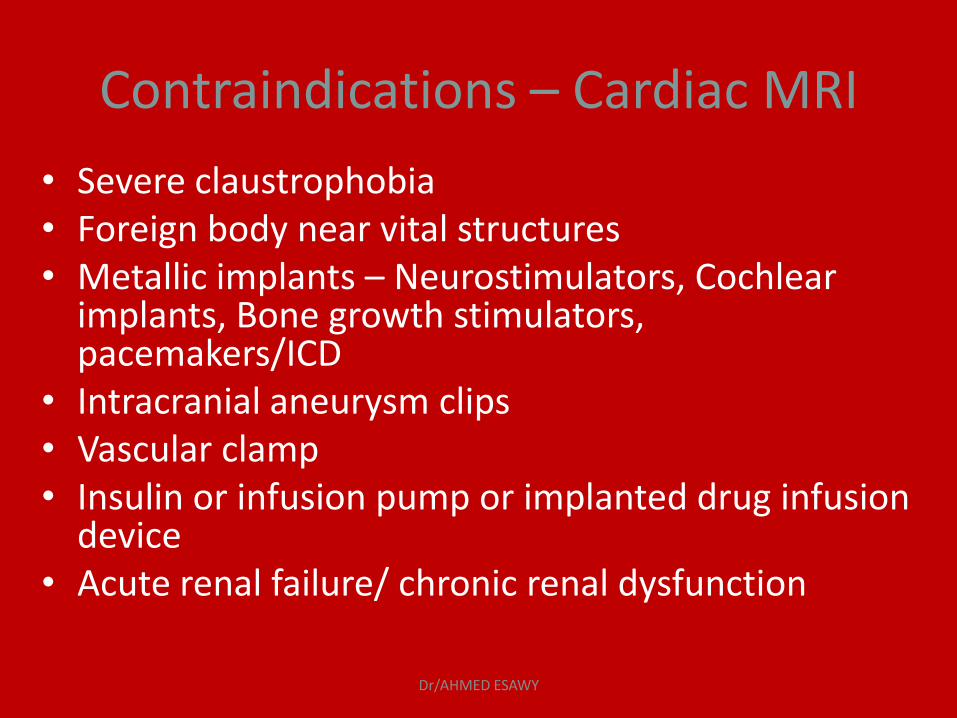
Contraindications – Cardiac MRI
• Severe claustrophobia • Foreign body near vital structures • Metallic implants – Neurostimulators, Cochlear
implants, Bone growth stimulators, pacemakers/ICD
• Intracranial aneurysm clips • Vascular clamp • Insulin or infusion pump or implanted drug infusion
device • Acute renal failure/ chronic renal dysfunction
Dr/AHMED ESAWY

Technique of
Multi-Slice CT /MRI
angiography of
coronary arteries
Dr/AHMED ESAWY

PREPERATION
Dr/AHMED ESAWY

• clinical history (symptoms such as chest pain and dyspnea);
• history of allergies (e.g., iodinated contrast material and medications);
• history of asthma or hyperthyroidism;
• history of renal disease or multiple myeloma (recent creatinine level);
• previous diagnostic examinations (stress test, electrocardiogram [ECG], and echocardiogram).
PRESCAN CONSIDERATIONS
Dr/AHMED ESAWY
Heart Rate Control Sinus rhythm
• Oral or intravenous b-blockers should be administered before the study, aiming for a resting regular heart rate less than 65 bpm
• Optimizing Heart Rate To obtain nearly motion free images.
Dr/AHMED ESAWY

Beta blocker
• contraindications:
» Severe asthma
» Severe obstructive lung disease
» Bradycardia
» Intolerance
Dr/AHMED ESAWY

Beta blocker complications
• » Hypotension
• » Bradycardia
• » Asthma
Slow injection
Have atropine on board
Dr/AHMED ESAWY

Go
• No beta blockers up to 70 bpm
• Beta blocker suggestions:
» Esmolol (Brevibloc), IV 20-30 mg/min
» Atenolol, oral, 50 mg 1 hour before scan if
heart rate above 65 b/m
• No caffeine
Beta blocker
Dr/AHMED ESAWY

Breath Holding 10 s breath hold (submaximum inspiration)
• During the test, a breath hold of 15–20 s will need to be performed.
• If the patient cannot hold still and follow breathing instructions, he or she should not be scanned.
• Breathing during the scan significantly compromises image quality and produces segments that cannot be evaluated.
• Before the scan, practicing breath holding helps to avoid such artifacts
Dr/AHMED ESAWY

Technique
• ECG-synchronized CT Scan Acquisition
Dr/AHMED ESAWY

Decreasing scan time,Course and duration
Dr/AHMED ESAWY

Nitroglycerin Always use it
• maximize coronary artery dilation during imaging
• Nitroglycerin suggestions:
» 0.8-1.2 mg glycerol trinitrate » 5 mg isosorbide dinitrate
• Nitroglycerin complications:
» Hypotension » Tachycardia
Dr/AHMED ESAWY

Nitrogylcerine Always use it
Nitroglycerin contraindications : » Inhibitors of phosphodiesterase » Severe aortic stenosis » Hypertrophic obstructive cardiomyopathy » Hypotension (<100 mm Hg) » Intolerance
Dr/AHMED ESAWY

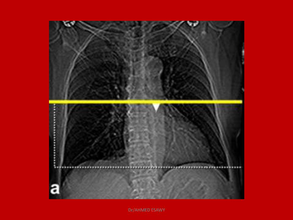
Scan Protocol Initial scout images were acquired to determine anatomic
coverage. The CT angiographic volume extended from 1 cm below the carina
to the base of the heart.
Then the entire heart volume from aortic root to diaphragm is scanned within a single breath hold (20 s). Total examination time (door to door) is 15 minutes
A preliminary scan without contrast injection may be performed to
determine the total calcium burden (calcium score) of the coronary tree .
Dr/AHMED ESAWY
Dr/AHMED ESAWY

Scan Protocol
120 ml iodinated contrast material is administered.
To achieve good timing either we use Bolus Pro-Ultra or time lapse method.
Dr/AHMED ESAWY

Scan Protocol
Bolus Pro-Ultra: automatic method in which after the density within the ROI crosses a predefined threshold value, the spiral scan starts after an adjustable offset delay.(The scan is triggered automatically when the concentration of contrast in the ascendinga aorta reaches 150 Hu (automatic bolus-triggering technique) .
Dr/AHMED ESAWY
Scan Protocol
In Time lapse method: a test bolus is given and the time/density curve within the ROI is plotted. The optimal delay to start the scan is determined manually as the peak of the time lapse curve.
Dr/AHMED ESAWY

Scan Protocol
bolus of 50 ml of saline is injected immediately after the iodinated contrast medium for prevention of streak artifacts.
Dr/AHMED ESAWY
Contraindication to contrast
• Absolute Contraindication: renal failure allergy to contrast • relative Contraindication : pregnancy thyrotoxicosis anaemia heart failure multiple myeloma
Dr/AHMED ESAWY

RECONSTRUCTION
Dr/AHMED ESAWY
Maximum intensity projection
Multiplanar reformations
Three-dimensional display:
Advanced visualization tools
Data Display
Dr/AHMED ESAWY
Image Display
Overlapped axial images are produced being able to produce smoother 3D renderings.
3D rendering techniques including Shaded surface display, maximum intensity projection, multi planar reformation, direct volume rendering and virtual endoscopy.
Dr/AHMED ESAWY

Shaded surface display
• Adjacent pixels above a predefined threshold are modeled by the computer in a single 3D structure. Those below the threshold are excluded.
• This results in 3D display of the surfaces of structures of interest.
• It provides excellent view of complex 3D relationships.
Dr/AHMED ESAWY

Maximum Intensity Projection
• It evaluates each voxel along a line from the viewer's eye and selects the maximum voxel value.
• So that the vessel of interest should have the highest number of HU.
Dr/AHMED ESAWY

Multi Planar Reformation
• First a plane is defined inside the 3D volume and only the data in this plane are displayed.
• It is easy and fast to use and provides images containing all available information.
• However it depends on the manual orientation of the planes which may result in false-positive or false-negative stenoses.
Dr/AHMED ESAWY

Multi Planar Reformation
• Oblique MPR of the right coronary artery performed with straight planes taken inside the 3D volume image.
Dr/AHMED ESAWY

3D Volume Rendering Technique
• It takes the entire volume of data, sums the contributions of each voxel along a line from the viewer's eye through the data set, and displays the resulting composite for each pixel of the display.
• Volume rendering provides good 3D depth and can use all the data available.
• However it is time-consuming requires the multitude of user definable parameters and settings. Also requires much more powerful computers.
Dr/AHMED ESAWY

3D Volume Rendering Technique
• CT coronary angiography. Colored volume rendering of right coronary artery (RCA) displayed in slightly cranial right anterior oblique view. This method provides better display of anatomy of tortuous coronary arteries
Dr/AHMED ESAWY

Virtual Endoscopy
• It is the visualization of the inner surface of a hollow structure.
• The very dense contrast medium inside the vessels is made transparent and the walls of the vessels are made opaque.
• This technique produce spectacular images but, its clinical use is limited.
Dr/AHMED ESAWY

Virtual Endoscopy
• Virtual endoscopic image of the ascending aorta shows extensive calcified plaque (white) within the vessel
Dr/AHMED ESAWY