Embed Size (px)
Citation preview
CELL MEMBRANE PHYSIOLOGY &PHARMACOLOGY
Dr Shahid SaacheDept. of Pharmacology
BJ GMC, PuneMentor- Dr Sujeet
Divhare
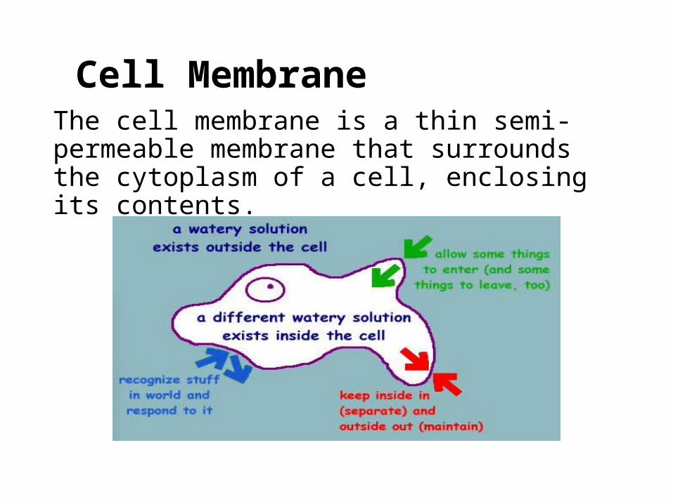
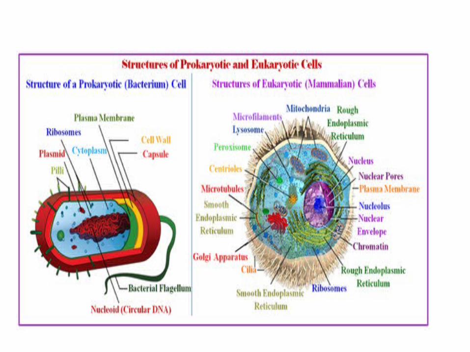
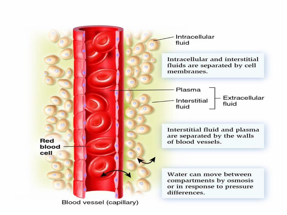
Cell MembraneThe cell membrane is a thin semi-permeable membrane that surrounds the cytoplasm of a cell, enclosing its contents.

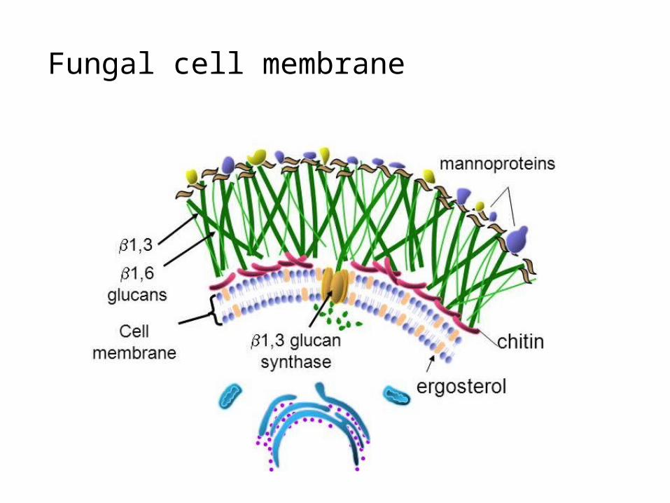
Fungal cell membrane
Why cell membrane is important to study??
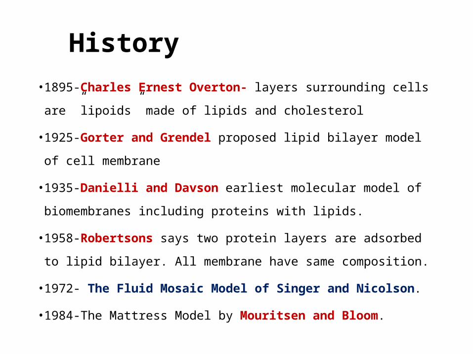
History• 1895-Charles Ernest Overton- layers surrounding cells are ”lipoids” made
of lipids and cholesterol
• 1925-Gorter and Grendel proposed lipid bilayer model of cell membrane
• 1935-Danielli and Davson earliest molecular model of biomembranes
including proteins with lipids.
• 1958-Robertsons says two protein layers are adsorbed to lipid bilayer. All
membrane have same composition.
• 1972- The Fluid Mosaic Model of Singer and Nicolson.
• 1984-The Mattress Model by Mouritsen and Bloom.
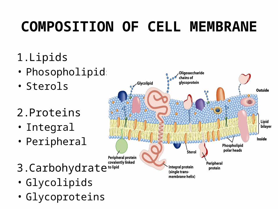
COMPOSITION OF CELL MEMBRANE
1. Lipids• Phosopholipids• Sterols
2. Proteins• Integral• Peripheral
3. Carbohydrates• Glycolipids• Glycoproteins
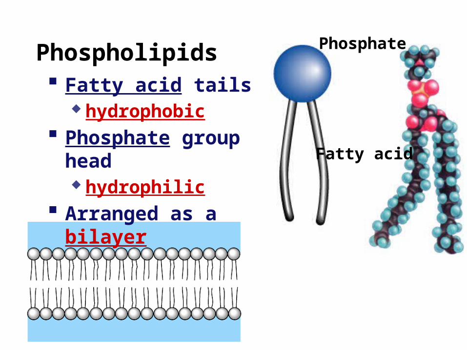
Phospholipids
Fatty acid
Phosphate
Fatty acid tails hydrophobic
Phosphate group head hydrophilic
Arranged as a bilayer
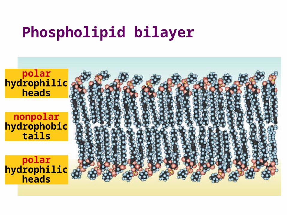
Phospholipid bilayer
polarhydrophilic
heads
nonpolarhydrophobic
tails
polarhydrophilic
heads
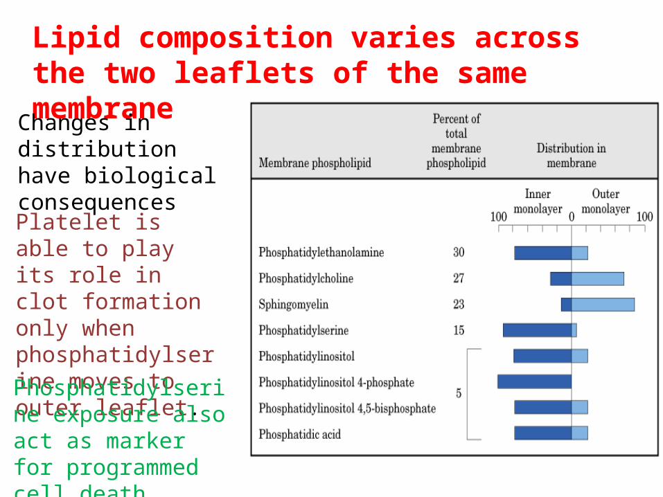
Lipid composition varies across the two leaflets of the same membrane
Changes in distribution have biological consequences
Platelet is able to play its role in clot formation only when phosphatidylserine moves to outer leaflet.
Phosphatidylserine exposure also act as marker for programmed cell death
14
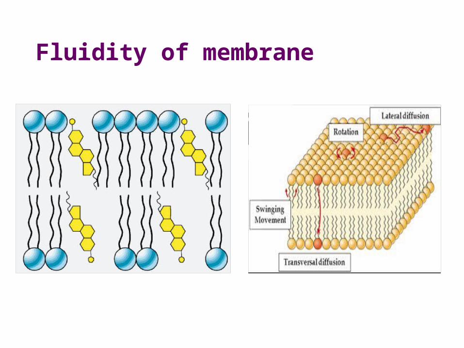
Fluidity of membrane
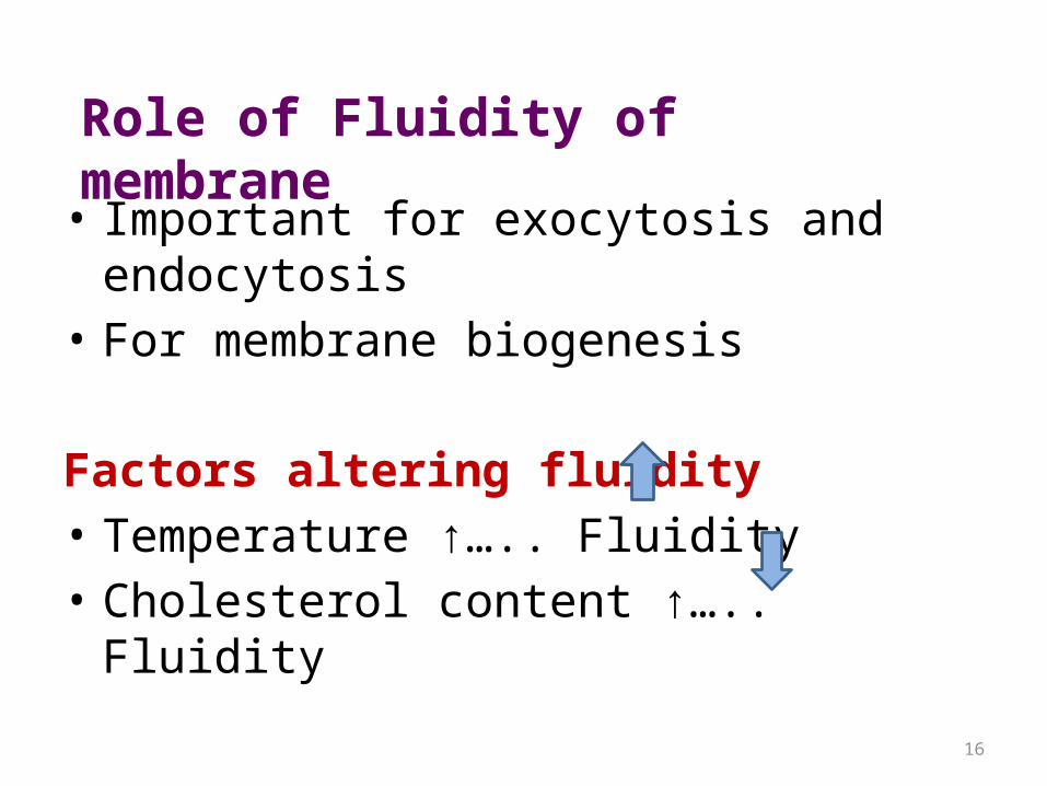
• Important for exocytosis and endocytosis• For membrane biogenesis
Factors altering fluidity• Temperature ↑….. Fluidity• Cholesterol content ↑….. Fluidity
16
Role of Fluidity of membrane
17
18
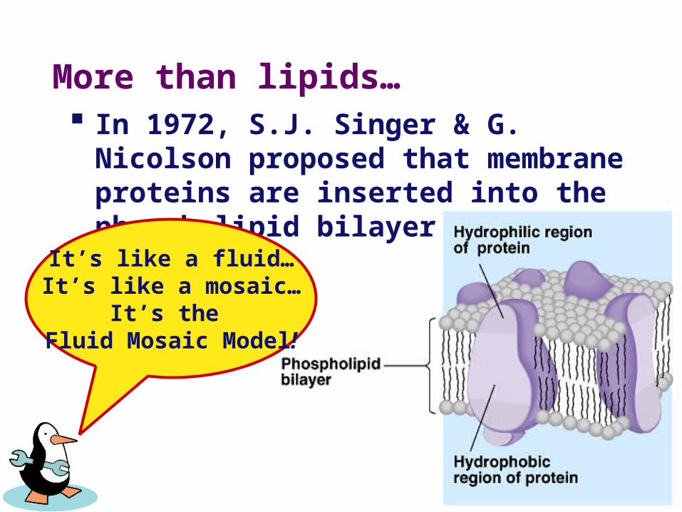
More than lipids… In 1972, S.J. Singer & G. Nicolson
proposed that membrane proteins are inserted into the phospholipid bilayer
It’s like a fluid…It’s like a mosaic…
It’s the Fluid Mosaic Model!
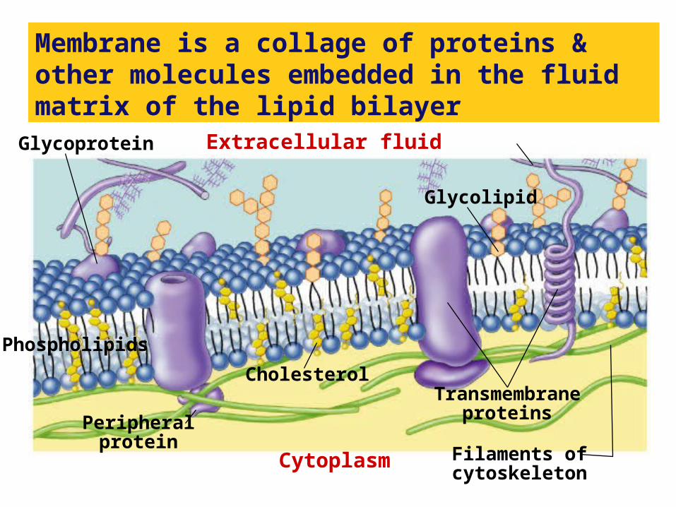
Membrane is a collage of proteins & other molecules embedded in the fluid matrix of the lipid bilayer
Extracellular fluid
Cholesterol
Cytoplasm
Glycolipid
Transmembraneproteins
Filaments ofcytoskeleton
Peripheralprotein
Glycoprotein
Phospholipids
Membrane proteins
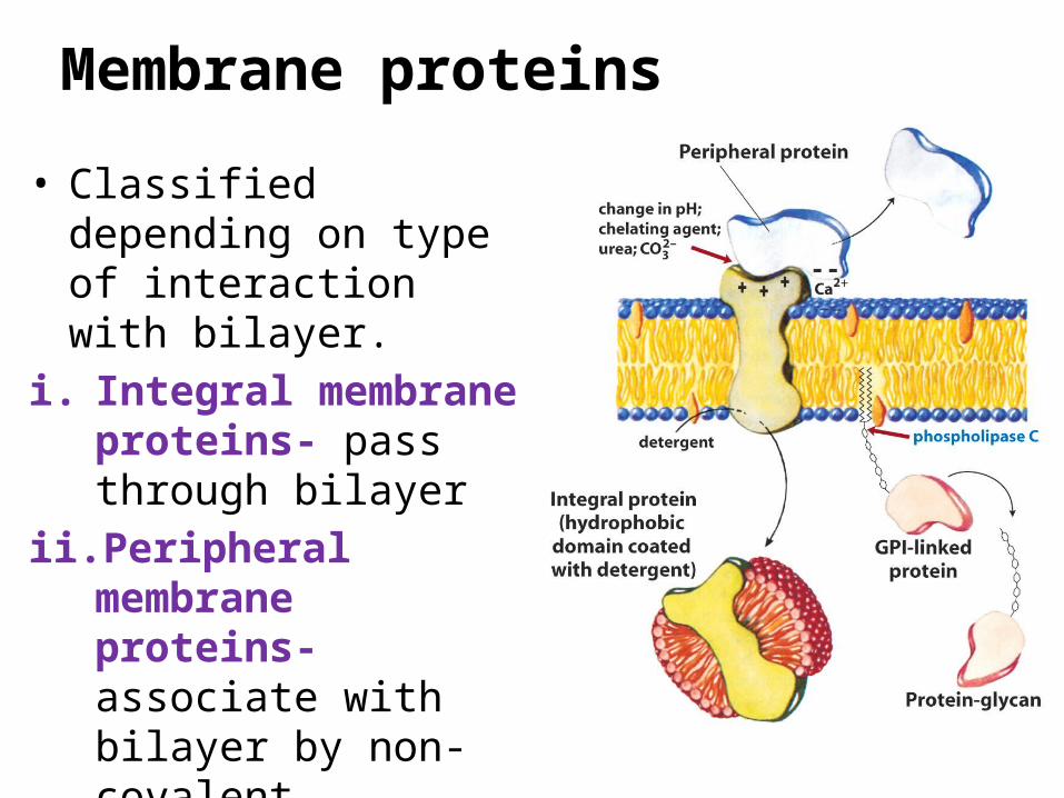
• Classified depending on type of interaction with bilayer.
i. Integral membrane proteins- pass through bilayer
ii. Peripheral membrane proteins- associate with bilayer by non-covalent interactions
iii. Lipid-anchored proteins.
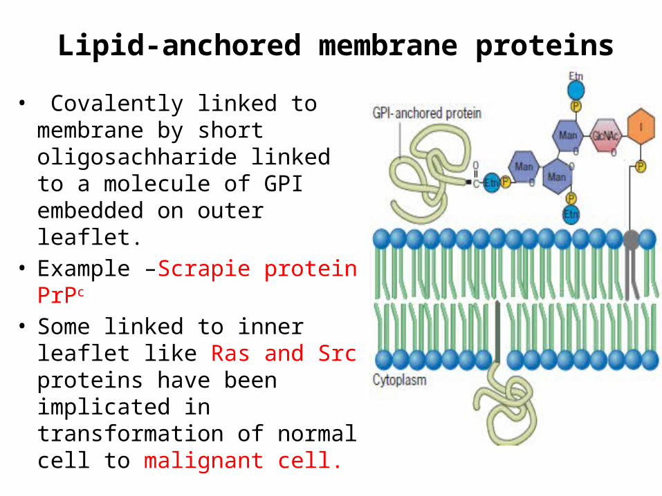
Lipid-anchored membrane proteins
• Covalently linked to membrane by short oligosachharide linked to a molecule of GPI embedded on outer leaflet.
• Example –Scrapie protein PrPc
• Some linked to inner leaflet like Ras and Src proteins have been implicated in transformation of normal cell to malignant cell.
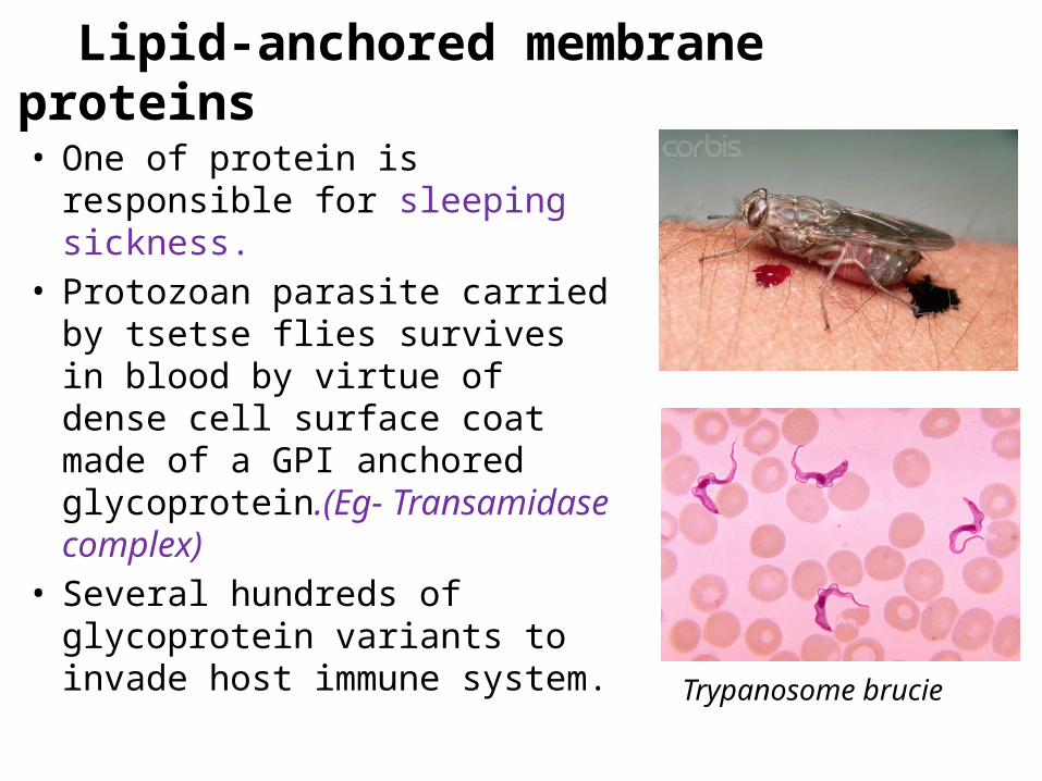
Lipid-anchored membrane proteins• One of protein is responsible for
sleeping sickness.• Protozoan parasite carried by
tsetse flies survives in blood by virtue of dense cell surface coat made of a GPI anchored glycoprotein.(Eg- Transamidase complex)
• Several hundreds of glycoprotein variants to invade host immune system.
Trypanosome brucie
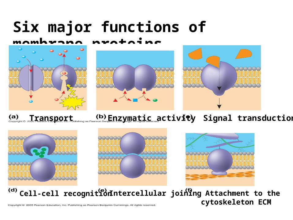
Six major functions of membrane proteins
Transport Enzymatic activity Signal transduction
Cell-cell recognition Intercellular joining Attachment to thecytoskeleton ECM
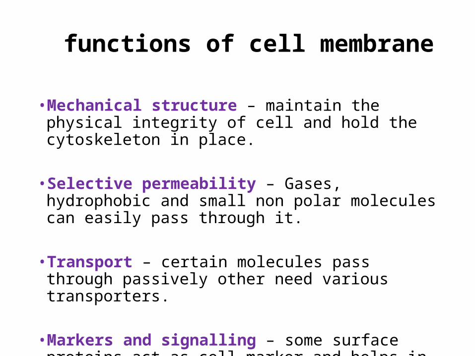
•Mechanical structure – maintain the physical integrity of cell and hold the cytoskeleton in place.
• Selective permeability – Gases, hydrophobic and small non polar molecules can easily pass through it.
• Transport – certain molecules pass through passively other need various transporters.
•Markers and signalling – some surface proteins act as cell marker and helps in cell signalling.
functions of cell membrane
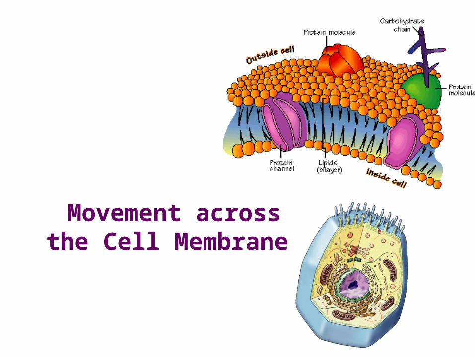
Movement across the Cell Membrane
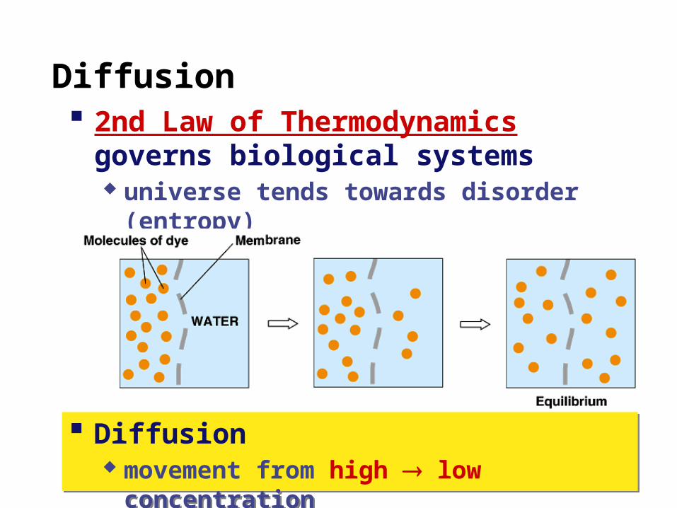
Diffusion 2nd Law of Thermodynamics
governs biological systems universe tends towards disorder (entropy)
Diffusion movement from high low concentration
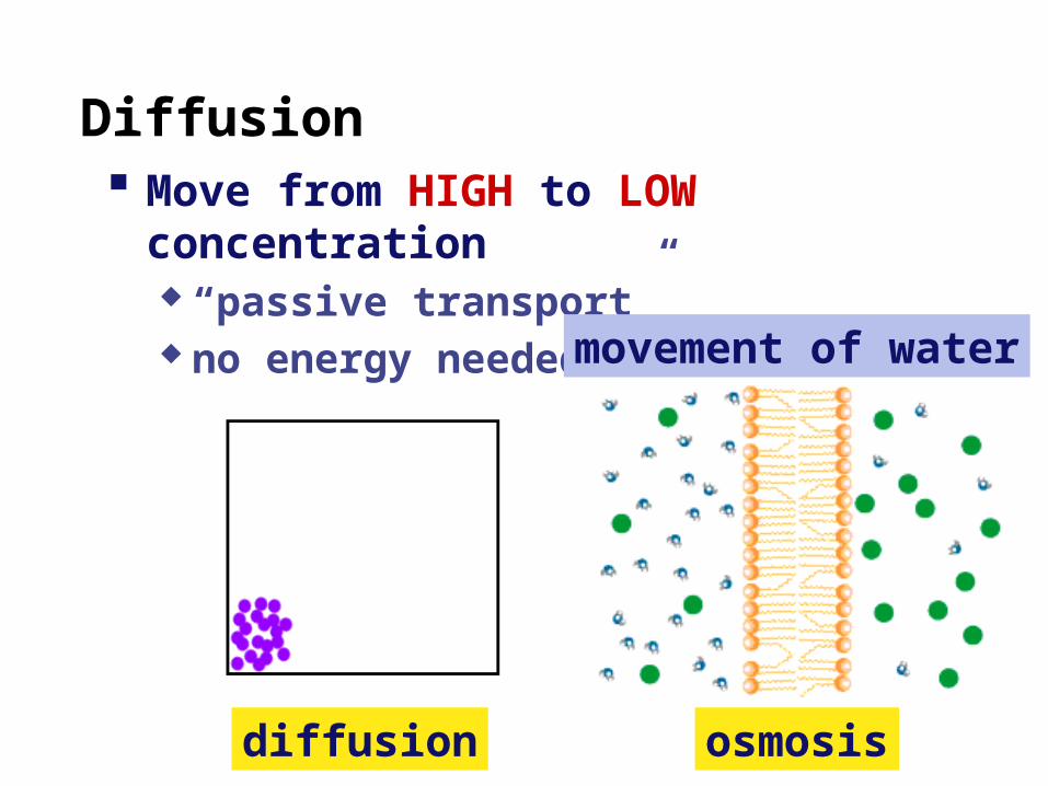
Diffusion Move from HIGH to LOW concentration
“passive transport” no energy needed
diffusion osmosis
movement of water
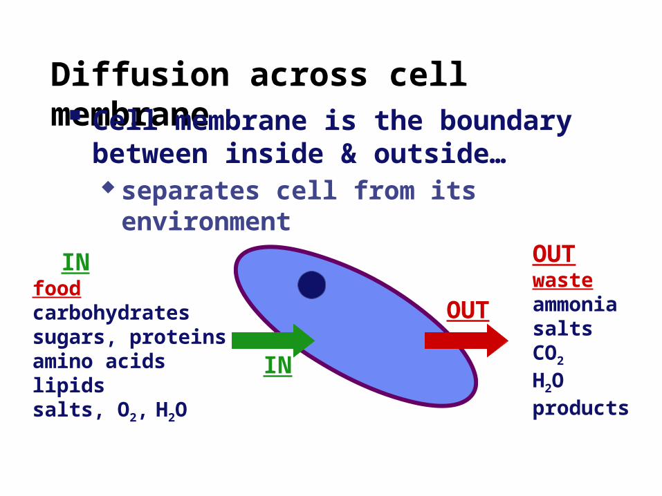
Diffusion across cell membrane Cell membrane is the boundary between
inside & outside… separates cell from its environment
INfoodcarbohydratessugars, proteinsamino acidslipidssalts, O2, H2O
OUTwasteammoniasaltsCO2
H2O products
IN
OUT
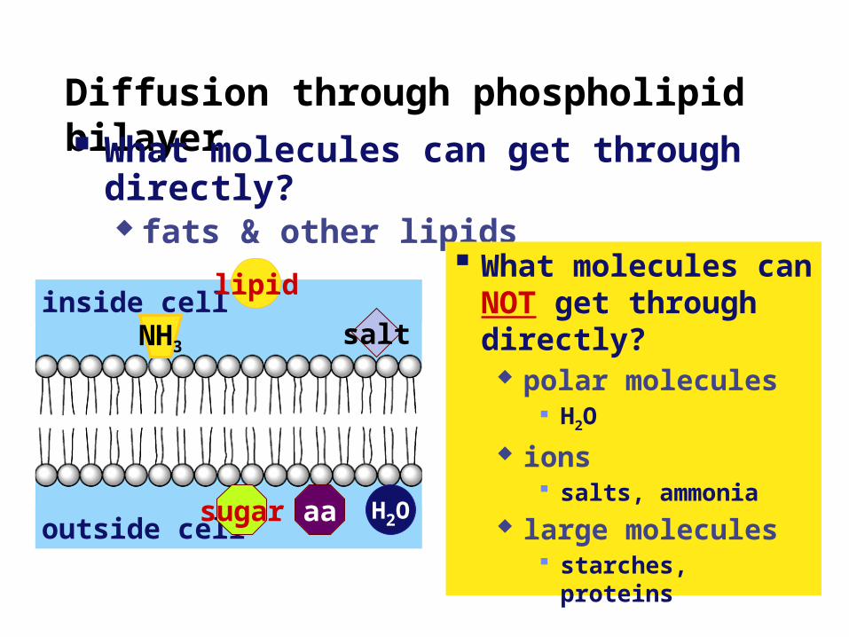
Diffusion through phospholipid bilayer What molecules can get through directly?
fats & other lipids
inside cell
outside cell
lipidsalt
aa H2Osugar
NH3
What molecules can NOT get through directly?
polar molecules H2O
ions salts, ammonia
large molecules starches, proteins
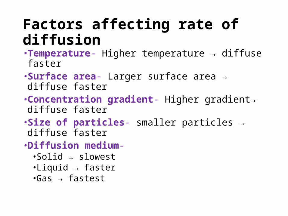
Factors affecting rate of diffusion•Temperature- Higher temperature → diffuse faster•Surface area- Larger surface area → diffuse faster•Concentration gradient- Higher gradient→ diffuse
faster•Size of particles- smaller particles → diffuse faster•Diffusion medium- • Solid → slowest• Liquid → faster•Gas → fastest
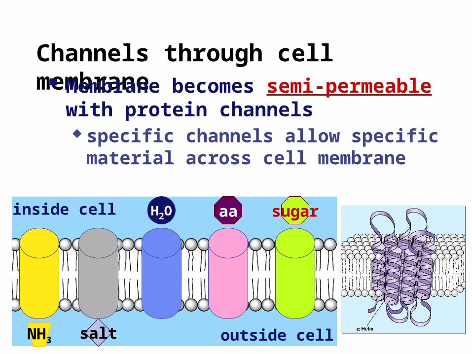
Channels through cell membrane Membrane becomes semi-permeable
with protein channels specific channels allow specific material
across cell membrane
inside cell
outside cell
sugaraaH2O
saltNH3
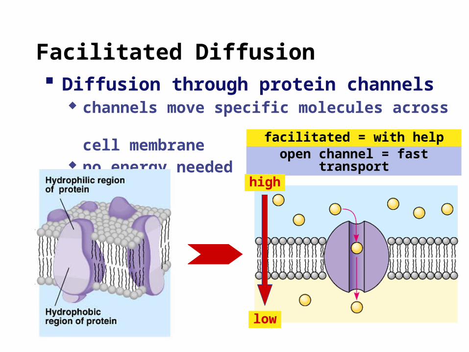
Facilitated Diffusion Diffusion through protein channels
channels move specific molecules across cell membrane
no energy neededopen channel = fast transport
facilitated = with help
high
low
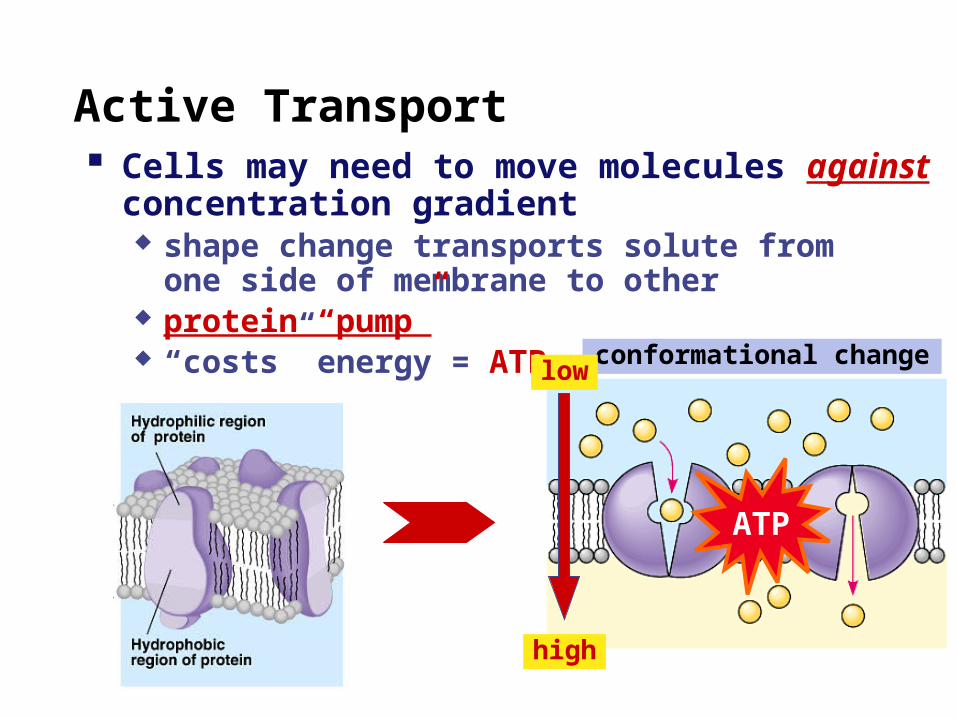
Active Transport
conformational change
Cells may need to move molecules against concentration gradient shape change transports solute from
one side of membrane to other protein “pump” “costs” energy = ATP
ATP
low
high
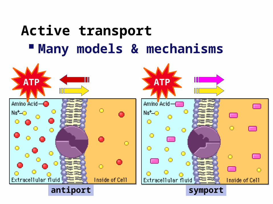
symportantiport
Active transport Many models & mechanisms
ATP ATP
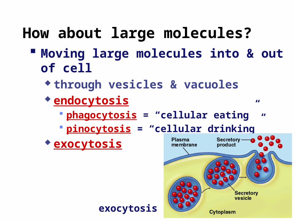
How about large molecules? Moving large molecules into & out of cell
through vesicles & vacuoles endocytosis
phagocytosis = “cellular eating” pinocytosis = “cellular drinking”
exocytosis
exocytosis
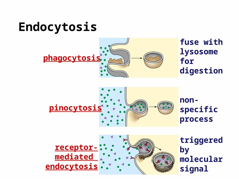
Endocytosis
phagocytosis
pinocytosis
receptor-mediated endocytosis
fuse with lysosome for digestion
non-specificprocess
triggered bymolecular signal
2007-2008
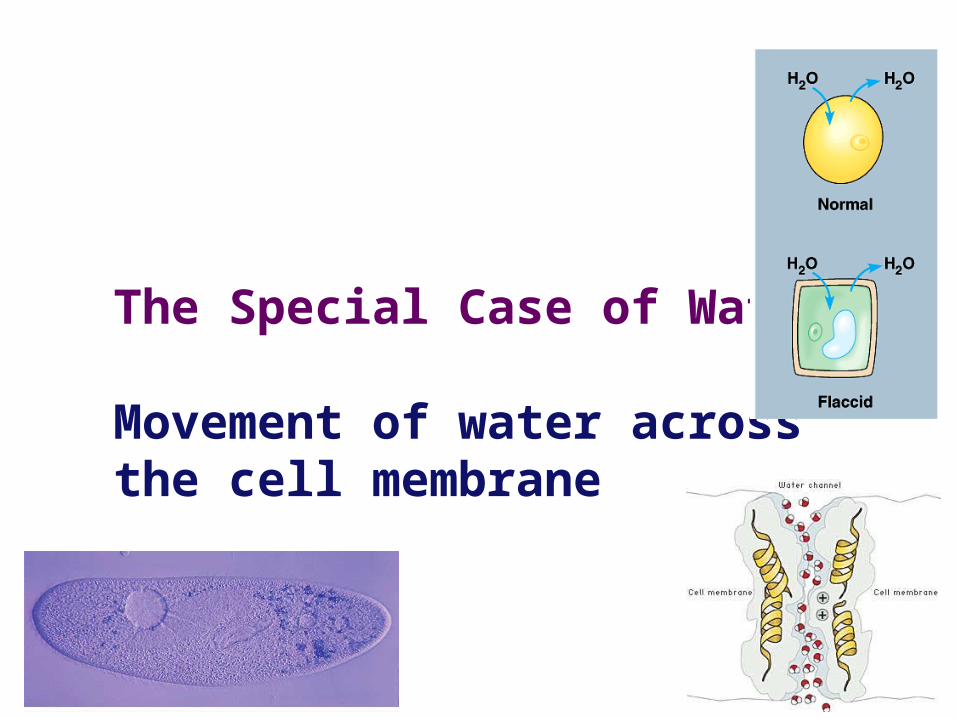
The Special Case of Water
Movement of water across the cell membrane
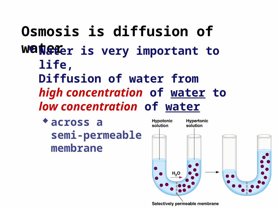
Osmosis is diffusion of water Water is very important to life,
Diffusion of water from high concentration of water to low concentration of water across a
semi-permeable membrane
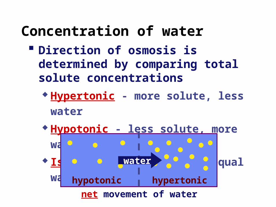
Concentration of water Direction of osmosis is determined by
comparing total solute concentrations Hypertonic - more solute, less water Hypotonic - less solute, more water Isotonic - equal solute, equal water
hypotonic hypertonic
water
net movement of water
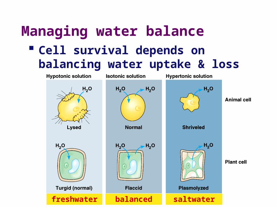
freshwater balanced saltwater
Managing water balance Cell survival depends on balancing
water uptake & loss
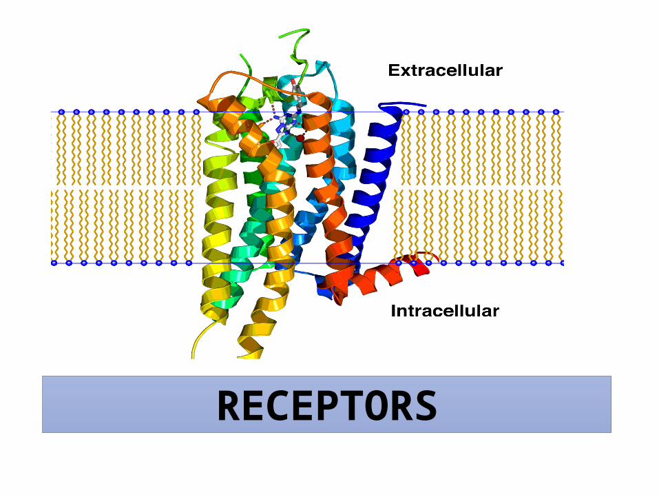
RECEPTORS
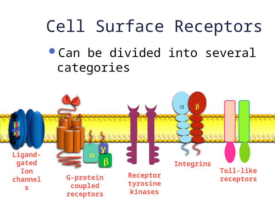
Cell Surface ReceptorsCan be divided into several
categories
gb
aLigand-gated
Ion channel
sG-protein coupled
receptors
Receptor tyrosine kinases
a b
IntegrinsToll-like
receptors
N
C
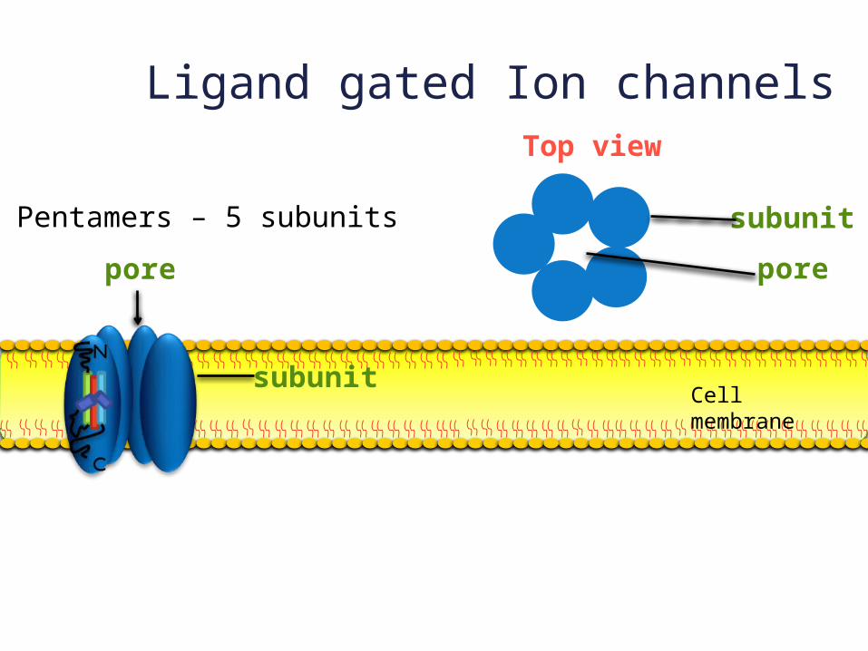
Ligand gated Ion channels
Pentamers – 5 subunits
Top view
subunitpore
subunit
pore
Cell membrane
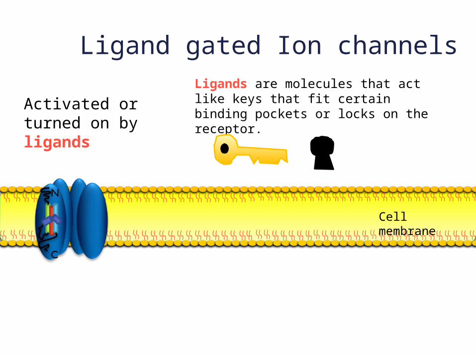
Ligand gated Ion channelsLigands are molecules that act like keys that fit certain binding pockets or locks on the receptor.
Activated or turned on by ligands
Cell membrane
GABAGlycineNicotinic
acetylcholineSerotoninGlutamate
Examples of these:
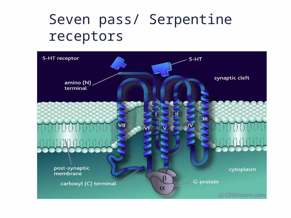
Seven pass/ Serpentine receptors
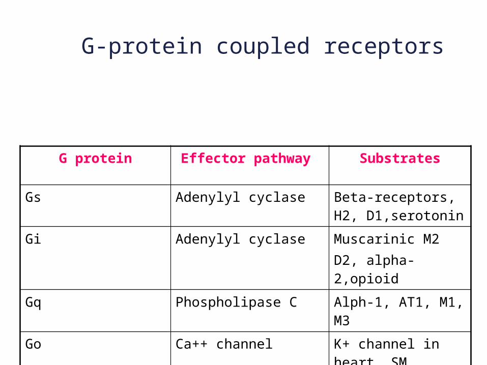
G protein Effector pathway Substrates
Gs Adenylyl cyclase Beta-receptors, H2, D1,serotonin
Gi Adenylyl cyclase Muscarinic M2D2, alpha-2,opioid
Gq Phospholipase C Alph-1, AT1, M1, M3
Go Ca++ channel K+ channel in heart, SM
G-protein coupled receptors
g
ba
G-protein coupled receptors
Cell membrane
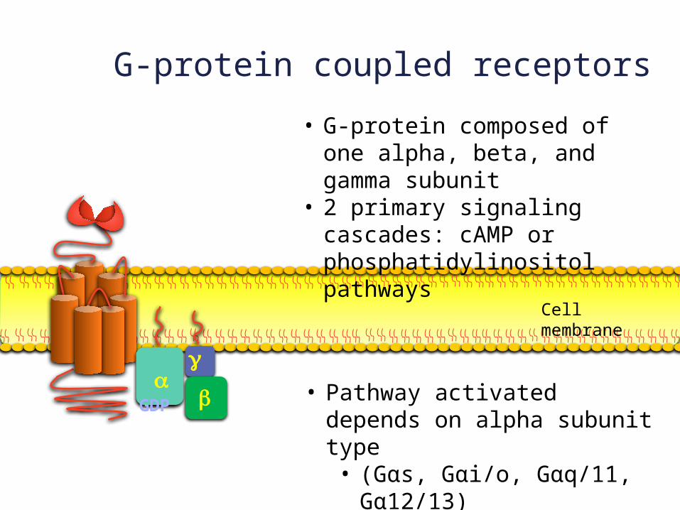
• G-protein composed of one alpha, beta, and gamma subunit
• 2 primary signaling cascades: cAMP or phosphatidylinositol pathways
• Pathway activated depends on alpha subunit type• (Gαs, Gαi/o, Gαq/11,
Gα12/13)• GDP bound to a when
inactive
GDP
g
ba
G-protein coupled receptors
Cell membrane
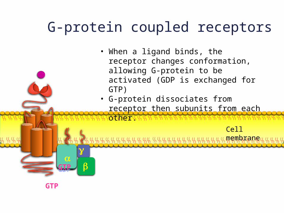
• When a ligand binds, the receptor changes conformation, allowing G-protein to be activated (GDP is exchanged for GTP)
• G-protein dissociates from receptor then subunits from each other.
GDP
GTP
aGTP
g
ba
cAMP pathway
Cell membrane
GDP
GTP
aGTP
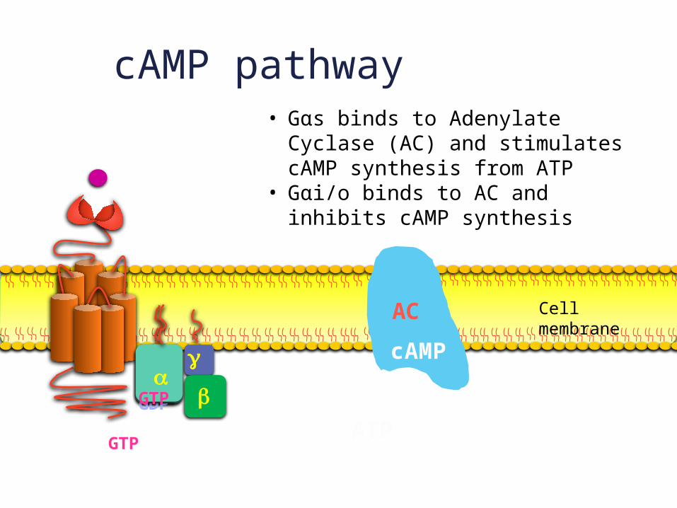
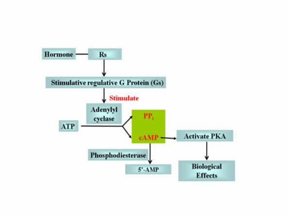
• Gαs binds to Adenylate Cyclase (AC) and stimulates cAMP synthesis from ATP
• Gαi/o binds to AC and inhibits cAMP synthesis
AC
ATP
cAMP
g
ba
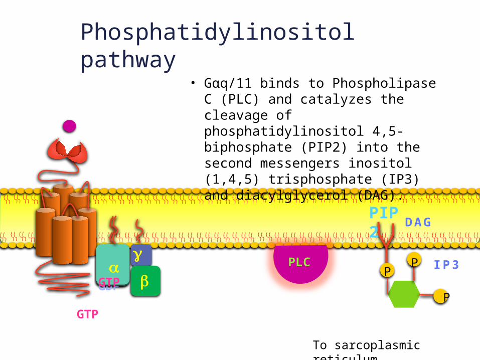
Phosphatidylinositol pathway
GDP
GTP
aGTP
• Gαq/11 binds to Phospholipase C (PLC) and catalyzes the cleavage of phosphatidylinositol 4,5-biphosphate (PIP2) into the second messengers inositol (1,4,5) trisphosphate (IP3) and diacylglycerol (DAG).
PLCPLC
DAG
IP3
PIP2
P P
P
To sarcoplasmic reticulum…
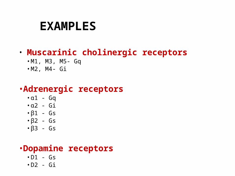
EXAMPLES
• Muscarinic cholinergic receptors• M1, M3, M5- Gq• M2, M4- Gi
•Adrenergic receptors• α1 - Gq• α2 - Gi• β1 - Gs• β2 - Gs• β3 - Gs
•Dopamine receptors• D1 - Gs• D2 - Gi
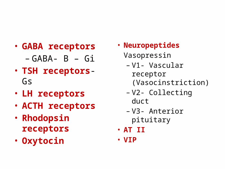
• GABA receptors– GABA- B – Gi
• TSH receptors- Gs• LH receptors• ACTH receptors• Rhodopsin receptors• Oxytocin
• NeuropeptidesVasopressin – V1- Vascular receptor
(Vasocinstriction)– V2- Collecting duct– V3- Anterior pituitary
• AT II• VIP
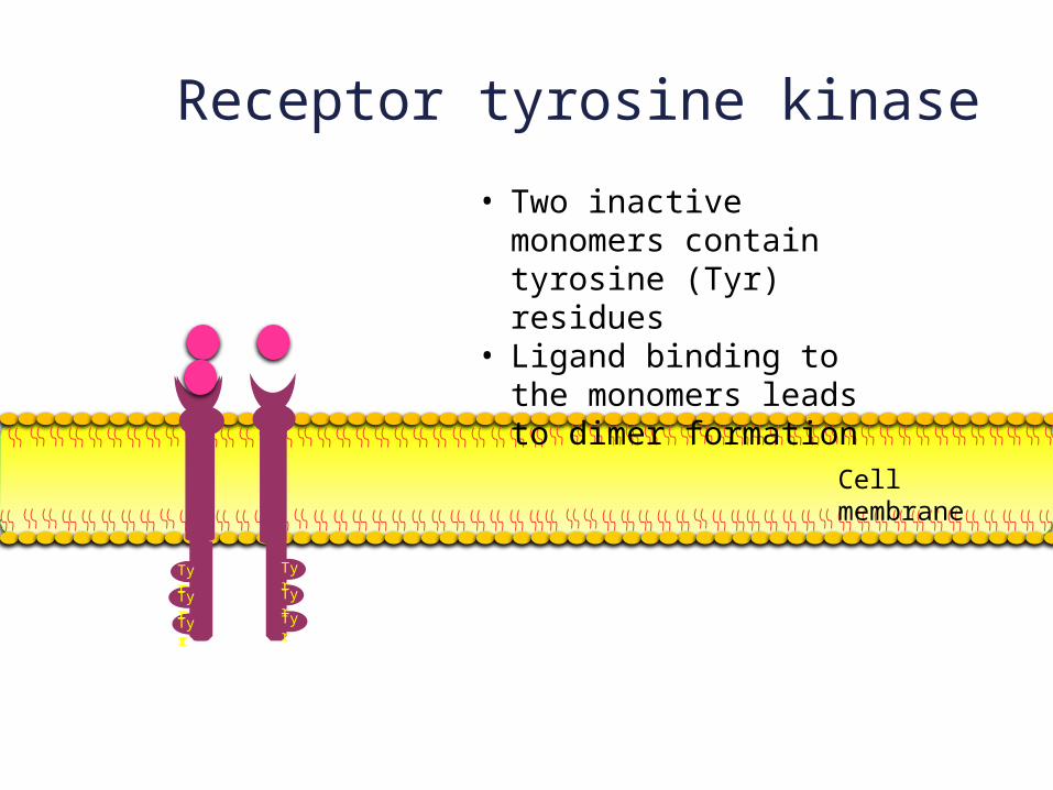
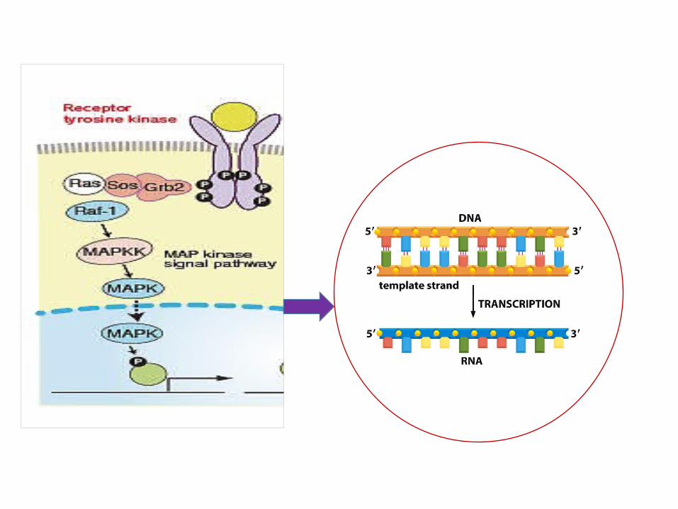
Receptor tyrosine kinase
TyrTyrTyr
TyrTyrTyr
• Two inactive monomers contain tyrosine (Tyr) residues
• Ligand binding to the monomers leads to dimer formation
TyrTyrTyr
Cell membrane
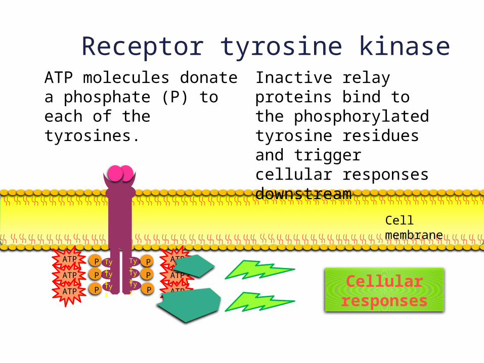
Receptor tyrosine kinase
TyrTyrTyr
TyrTyrTyr
ATPATPATP
ATPATPATP
PPP
PPP
ATP molecules donate a phosphate (P) to each of the tyrosines.
Inactive relay proteins bind to the phosphorylated tyrosine residues and trigger cellular responses downstream
Cellular responses
Cell membrane
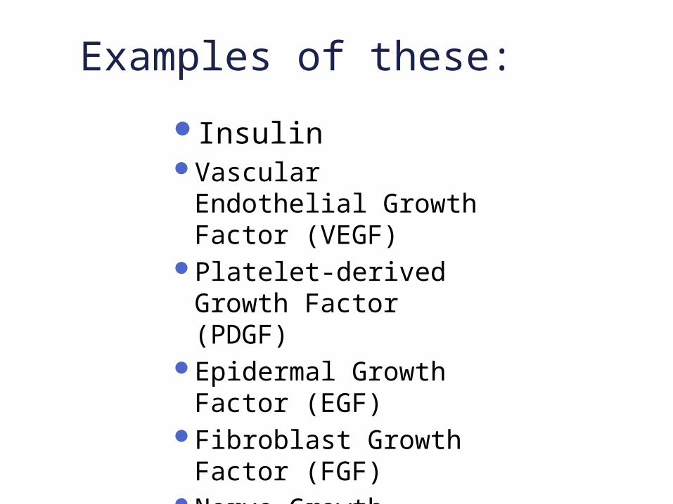
InsulinVascular Endothelial
Growth Factor (VEGF)Platelet-derived
Growth Factor (PDGF)Epidermal Growth
Factor (EGF)Fibroblast Growth
Factor (FGF)Nerve Growth Factor
(NGF)
Examples of these:
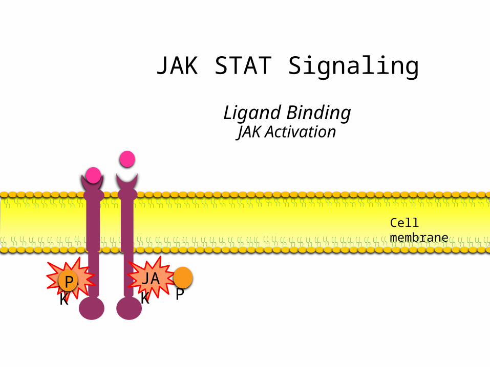
JAK STAT Signaling
Ligand BindingJAK Activation
Cell membrane
JAKJAK PP
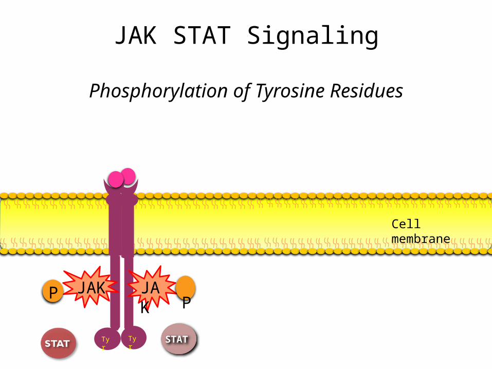
JAK STAT Signaling
Phosphorylation of Tyrosine Residues
Tyr Tyr
JAKJAK PP
Cell membrane
STAT
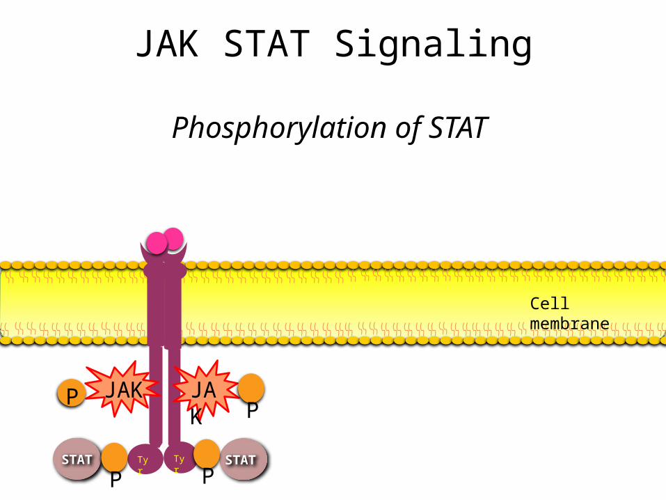
JAK STAT Signaling
Phosphorylation of STAT
Tyr Tyr
JAKJAK PP
Cell membrane
STATSTAT P P
STAT
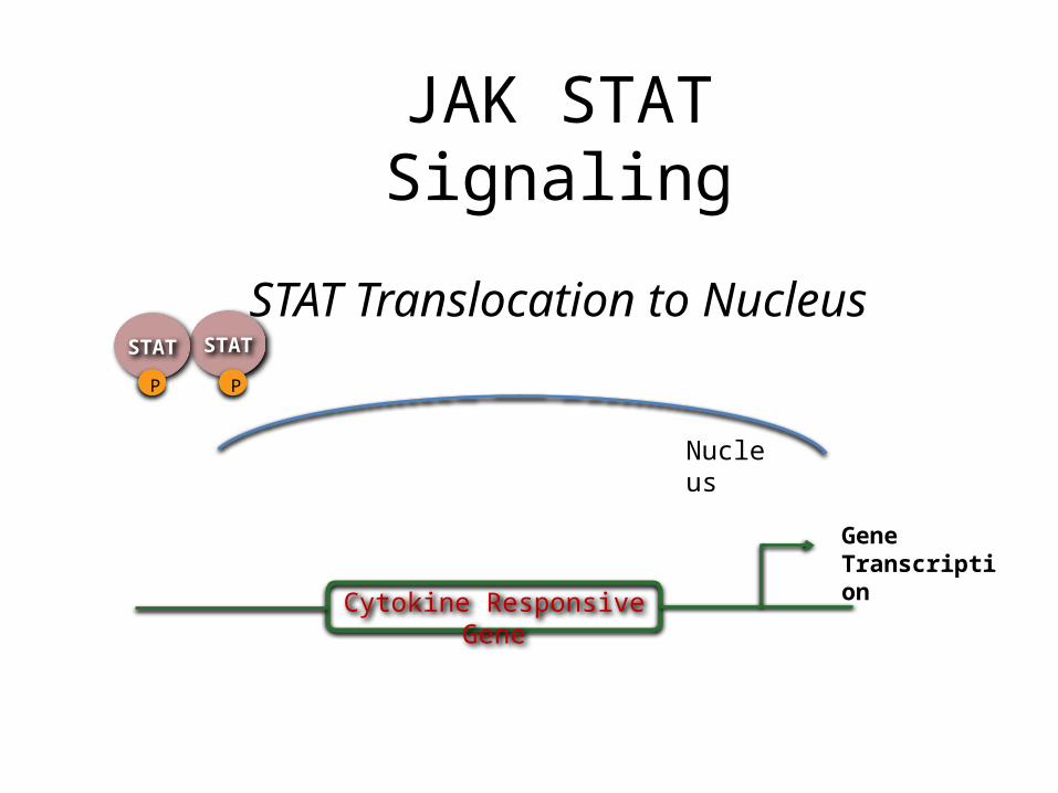
Cytokine Responsive Gene
Gene Transcription
Nucleus
STATP P
JAK STAT Signaling
STAT Translocation to Nucleus
Growth HormoneProlactinIL-2CytokinesT cell receptorsB cell receptors
Examples of these:
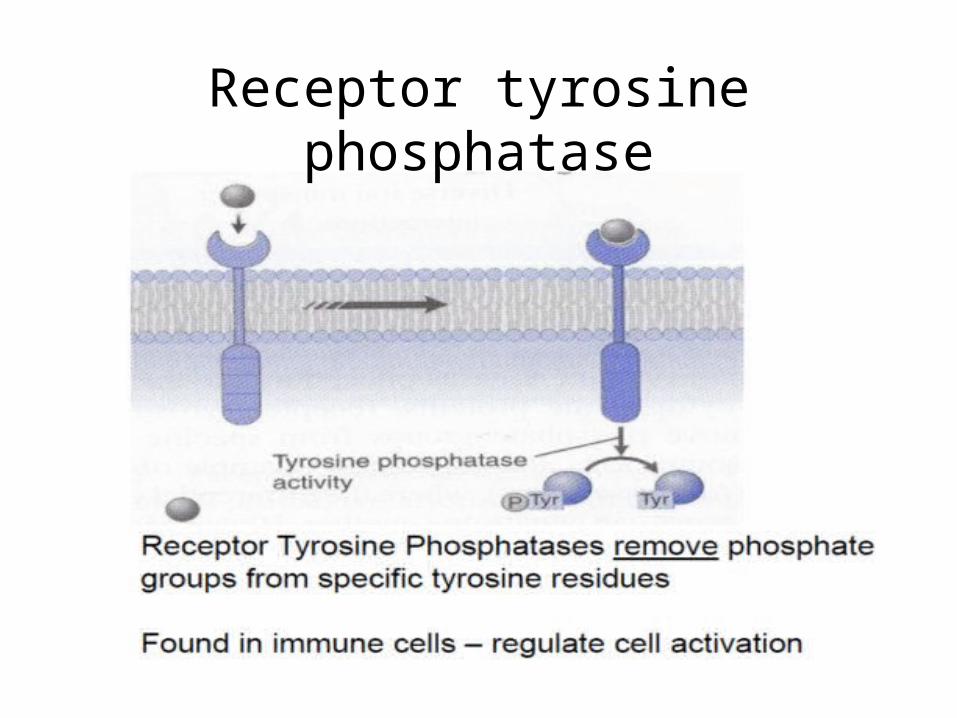
Receptor tyrosine phosphatase
Serine Threonine kinase pathway/ B Raf
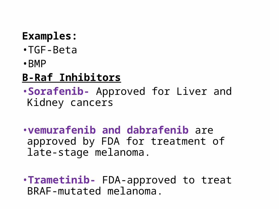
Examples:•TGF-Beta•BMPB-Raf Inhibitors•Sorafenib- Approved for Liver and Kidney cancers
•vemurafenib and dabrafenib are approved by FDA for treatment of late-stage melanoma.
•Trametinib- FDA-approved to treat BRAF-mutated melanoma.
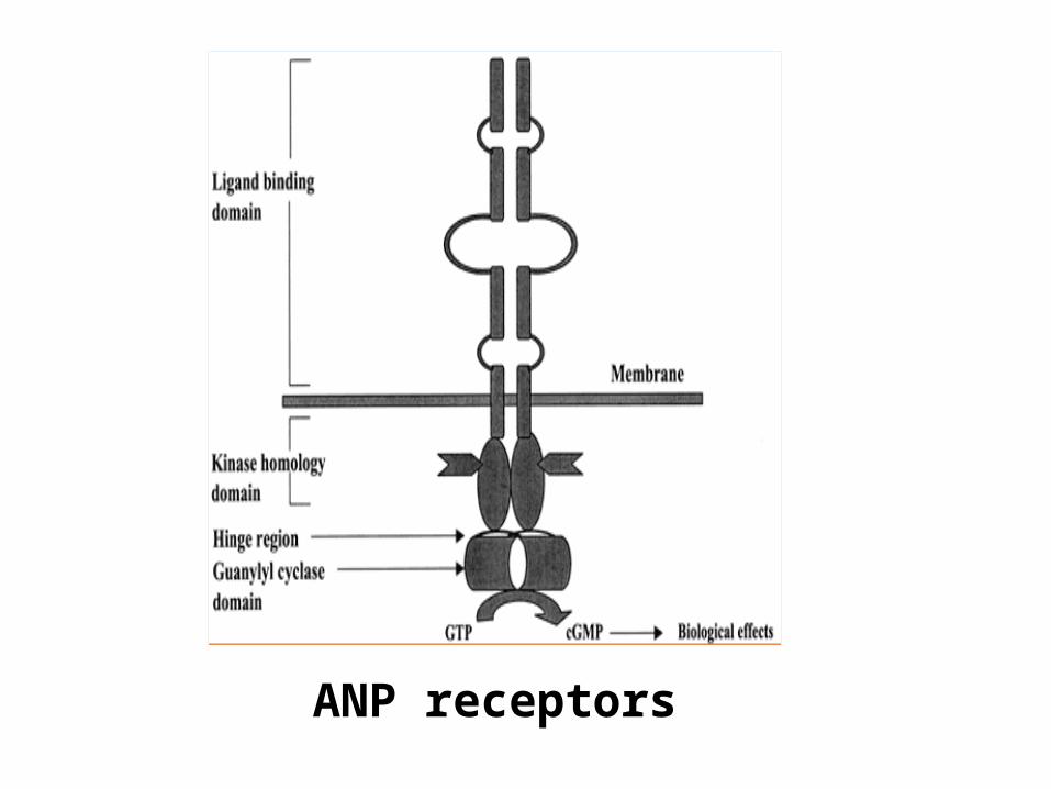
ANP receptors
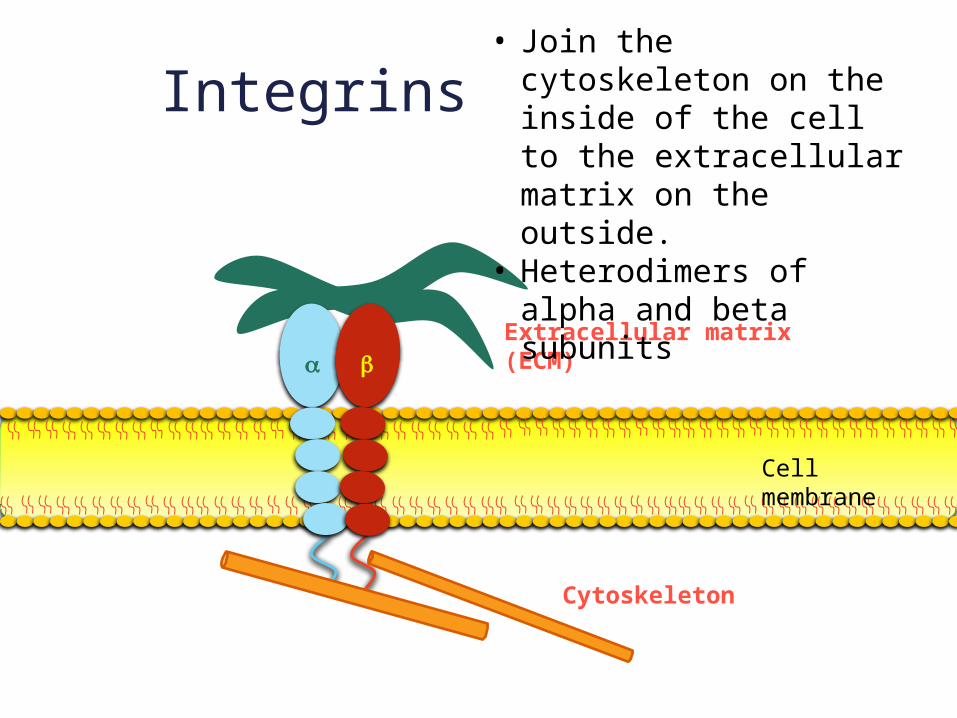
Integrins
Cytoskeleton
a bExtracellular matrix (ECM)
• Join the cytoskeleton on the inside of the cell to the extracellular matrix on the outside.
• Heterodimers of alpha and beta subunits
Cell membrane
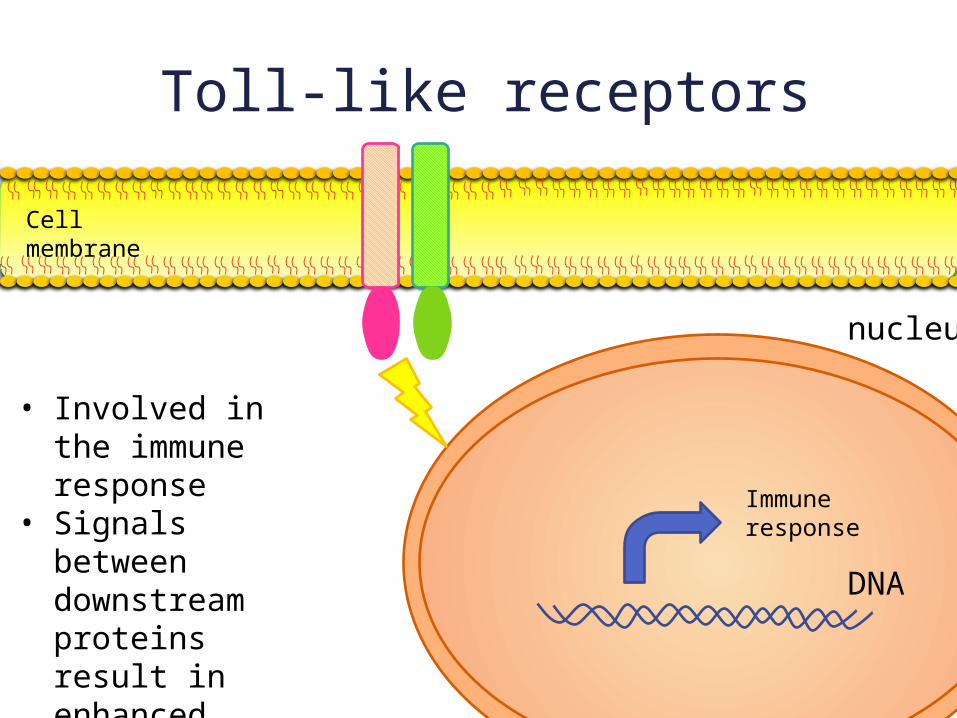
Toll-like receptors
nucleus
Cell membrane
DNA
• Involved in the immune response
• Signals between downstream proteins result in enhanced transcription of inflammatory genes
Immune response
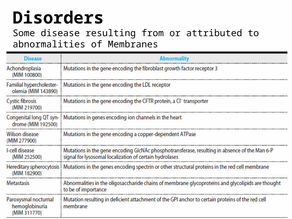
DisordersSome disease resulting from or attributed to abnormalities of Membranes
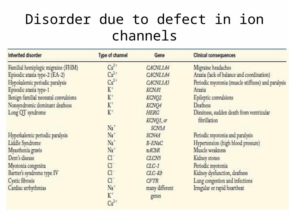
Disorder due to defect in ion channels
Some drugs acting on cell membrane
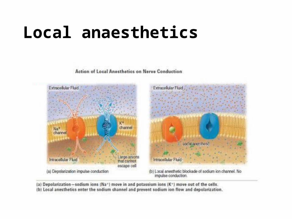
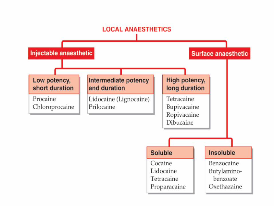
Local anaesthetics
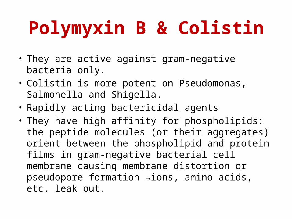
Polymyxin B & Colistin
• They are active against gram-negative bacteria only.• Colistin is more potent on Pseudomonas, Salmonella
and Shigella.• Rapidly acting bactericidal agents• They have high affinity for phospholipids: the peptide
molecules (or their aggregates) orient between the phospholipid and protein films in gram-negative bacterial cell membrane causing membrane distortion or pseudopore formation →ions, amino acids, etc. leak out.
• Given orally, side effects are limited to the g.i.t.• Systemic toxicity of these drugs (when injected) is
high: flushing and paresthesias (due to liberation of histamine from mast cells), marked kidney damage, neurological disturbances, neuromuscular blockade.
• Uses: skin infections, burns, otitis externa, conjunctivitis, corneal ulcer—caused by gram-negative bacteria including Pseudomonas.
• Gram-negative bacillary (E. coli, Salmonella, Shigella) diarrhoeas, especially in infants and children
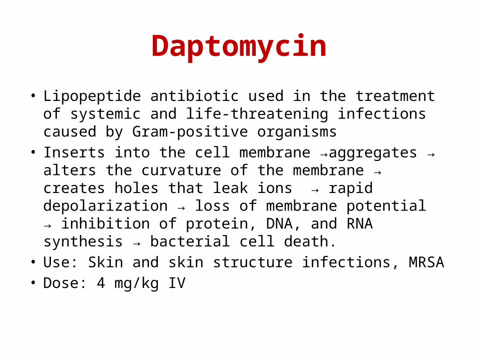
Daptomycin
• Lipopeptide antibiotic used in the treatment of systemic and life-threatening infections caused by Gram-positive organisms
• Inserts into the cell membrane →aggregates → alters the curvature of the membrane → creates holes that leak ions → rapid depolarization → loss of membrane potential → inhibition of protein, DNA, and RNA synthesis → bacterial cell death.
• Use: Skin and skin structure infections, MRSA• Dose: 4 mg/kg IV

Cell membrane active Antifungals