Embed Size (px)
Citation preview
1
British Journal of Pharmacology Special Issue: Themed Section: Relaxin Family Peptides and
Their Receptors: Biological and Therapeutic Advances.
Guest Editor: Roger Summers
REVIEW
Distribution, physiology and pharmacology of relaxin-3/RXFP3
systems in brain
Sherie Ma1,2, Craig M. Smith1-3, Anna Blasiak4, Andrew L. Gundlach1,2,5*
1The Florey Institute of Neuroscience and Mental Health, Parkville, Victoria, Australia 2Florey Department of Neuroscience and Mental Health, The University of Melbourne,
Victoria, Australia 3School of Medicine, Deakin University, Geelong, Victoria, Australia 4Department of Neurophysiology and Chronobiology, Institute of Zoology, Jagiellonian
University, Krakow, Poland 5Department of Anatomy and Neuroscience, The University of Melbourne, Victoria, Australia
Running title: Relaxin-3/RXFP3 systems in brain
*Corresponding Author:
Andrew L. Gundlach, PhD
The Florey Institute of Neuroscience and Mental Health
30 Royal Parade, Parkville, Victoria 3052, Australia
Phone: +61-3-9035-6507, Fax: +61-3-9035-3102
Email: [email protected]
This article is protected by copyright. All rights reserved.
This is the author manuscript accepted for publication and has undergone full peer review buthas not been through the copyediting, typesetting, pagination and proofreading process, whichmay lead to differences between this version and the Version of Record. Please cite this articleas doi: 10.1111/bph.13659
2
Abstract
Relaxin-3 is a member of a superfamily of structurally-related peptides that includes relaxin
and insulin-like peptide hormones. Soon after the discovery of the relaxin-3 gene, relaxin-3
was identified as an abundant neuropeptide in brain with a distinctive topographical
distribution within a small number of GABA neuron populations that is well conserved across
species. Relaxin-3 is thought to exert its biological actions through a single class-A GPCR –
relaxin-family peptide receptor 3 (RXFP3). Class-A comprises GPCRs for relaxin-3 and
insulin-like peptide-5 and other peptides such as orexin and the monoamine transmitters.
RXFP3 is selectively activated by relaxin-3, whereas insulin-like peptide-5 is the cognate
ligand for the related RXFP4 (see Ang SY et al. and Patil NA et al. this issue). Anatomical
and pharmacological evidence obtained over the last decade supports a function of relaxin-
3/RXFP3 systems in modulating responses to stress, anxiety-like and motivated behaviours,
circadian rhythms, and learning and memory. Electrophysiological studies have identified the
ability of RXFP3 agonists to directly hyperpolarise thalamic neurons in vitro, but there are no
reports of direct cell signalling effects in vivo. This article provides an overview of earlier
studies and highlights more recent research that implicates relaxin-3/RXFP3 neural network
signalling in the integration of arousal, motivation, emotion and related cognition, and has
begun to identify the associated neural substrates and mechanisms. Future research directions
to better elucidate the connectivity and function of different relaxin-3 neuron populations and
their RXFP3-positive target neurons in major experimental species and humans are also
identified.
Abbreviations: ACTH, adrenocorticotropic hormone; AgRP, agouti-related peptide; BNST,
bed nucleus of the stria terminalis; CRF, corticotropin-releasing factor; DREADD, designer
receptors exclusively activated by designer drugs; HCN, hyperpolarisation-activated cyclic
nucleotide-gated channels; icv, intracerebroventricular; IGL, intergeniculate leaflet; INSL3/5,
insulin-like peptide 3/5; MCH, melanin-concentrating hormone; NPY, neuropeptide-Y;
POMC, pro-opiomelanocortin; PV, parvalbumin; PVN, paraventricular nucleus of
hypothalamus; RXFP1-4, relaxin-family peptide receptor 1-4; SALPR, somatostatin- and
angiotensin-like peptide receptor.
This article is protected by copyright. All rights reserved.

3
This article is protected by copyright. All rights reserved.
4
Table of Links
TARGETS Enzymesb
GPCRsa ChAT
CRF1 receptor ERK1
CRF2 receptor ERK2
Dopamine D2 receptor GAD
Orexin OX2 receptor PKA
RXFP1-4 receptors tryptophan hydroxylase
Serotonin-1A receptor
These Tables of Links list key protein targets and ligands in this article that are hyperlinked to
corresponding entries in http://www.guidetopharmacology.org, the common portal for data
from the IUPHAR/BPS Guide to PHARMACOLOGY (Southan et al., 2016), and are
LIGANDS Melanin-concentrating hormone
Acetylcholine Neuropeptide-Y
Agouti-related peptide Orexin-A
Calcitonin gene-related peptide Oxytocin
cAMP Pro-opiomelanocortin
Chlorpromazine R3/I5
Clozapine Relaxin
Corticotropin-releasing factor Relaxin-3
Fluphenazine Serotonin
Dopamine Thyroid-stimulating hormone
GABA Vasoactive intestinal peptide
Insulin-like peptide 5 Vasopressin
This article is protected by copyright. All rights reserved.

5
permanently archived in The Concise Guide to PHARMACOLOGY 2015/16 (Alexander et
al., 2015a,b).
This article is protected by copyright. All rights reserved.
6
Introduction
Discovery of relaxin-3 and RXFP3
Relaxin-3 was discovered in 2001 by searching for homologues of the relaxin gene in the
Celera Discovery System and Celera Genomics databases (Bathgate et al., 2002) and due to
its predominant expression in brain, was subsequently classified as a neuropeptide. Relaxin-3,
like other relaxin and insulin-like peptide family members, is a 5 kDa peptide that consists of
two chains with three disulphide bonds (Bathgate et al., 2002; Liu et al., 2003b; Hossain et
al., 2013). All family members contain the characteristic sequence ‘RXXXRXX(I/V)’ within
their B-chain, which is essential for binding the different cognate receptors (Bullesbach and
Schwabe, 2000; Bathgate et al., 2002; 2006b).
The cognate receptor for relaxin-3 is relaxin-family peptide 3 receptor (RXFP3) (Bathgate et
al., 2006a). Also known as G-protein-coupled receptor 135 (GPCR135) (Liu et al., 2003b), it
was first discovered in 2000 and named somatostatin- and angiotensin-like peptide receptor
(SALPR), due to its high amino acid similarity with somatostatin receptor-5 and the
angiotensin AT1 receptor (Matsumoto et al., 2000). RXFP3 is a Gi/o-protein-coupled receptor,
and its activation produces inhibition of intracellular cAMP accumulation and activation of
extracellular-regulated kinase 1/2 (ERK1/2) (Liu et al., 2003b; van der Westhuizen et al.,
2007). Although in cell-based studies, relaxin-3 can bind and activate three related G-protein
coupled receptors – RXFP3, RXFP1 (originally named LGR7; Sudo et al., 2003), and RXFP4
(originally named GPCR142; Liu et al., 2003a), there is considerable evidence that RXFP3 is
the native receptor for relaxin-3. Firstly, RXFP3 displays the highest affinity for relaxin-3
(Bathgate et al., 2006b), and the genes encoding the peptide and protein have
phylogenetically co-evolved (Hsu et al., 2005; Wilkinson et al., 2005). Furthermore, there is a
strong overlap between the distribution of relaxin-3-positive nerve fibres and RXFP3
mRNA/binding sites throughout the rat (Sutton et al., 2004; Ma et al., 2007), mouse (Smith et
al., 2010) and macaque brain (Ma et al., 2009b, 2009c). Moreover, RXFP4 is primarily
expressed within the gastrointestinal tract and is largely absent from brain (Sutton et al.,
2006) and is, in fact, a pseudogene in rat (Chen et al., 2005). Also, although RXFP1 is
expressed widely throughout the rodent brain (Ma et al., 2006), its distribution pattern does
This article is protected by copyright. All rights reserved.
7
not correspond with that of the relaxin-3 innervation, and relaxin is expressed by specific
neuron populations (Ma et al., 2006). Finally, relaxin-3 is the only relaxin-peptide family
member that can activate RXFP3 (Liu et al., 2003b), whereas relaxin is the preferred ligand
for RXFP1 and also binds to RXFP2 (Sudo et al., 2003). The related insulin-like peptide 5
(INSL5) is uniquely expressed in enteroendocrine L-cells of the colon and it is the cognate
ligand for RXFP4 (Liu et al., 2005b; Sutton et al., 2006; Grosse et al., 2014).
Distribution of relaxin-3 and RXFP3 in the brain – a road map to function
Relaxin-3/RXFP3 systems conserved across various mammalian species
The brain is the main site of relaxin-3 mRNA synthesis, with high levels of expression
observed in various species including zebrafish (Donizetti et al., 2009), mouse (Bathgate et
al., 2002; Smith et al., 2010), rat (Burazin et al., 2002; Tanaka et al., 2005), macaque (Ma et
al., 2009b), and human (Liu et al., 2003b). The presence and anatomical distribution of
relaxin-3-producing neurons has been best studied in rat and mouse brain, with the largest
population observed in the brainstem nucleus incertus (Figure 1; Bathgate et al., 2002;
Burazin et al., 2002; Tanaka et al., 2005; Ma et al., 2007; Smith et al., 2010; Ryan et al.,
2011). Relaxin-3 neurons, which use GABA as their primary transmitter, are also present in
smaller populations in the pontine raphé nucleus (~350 neurons) medial and ventrolateral
periaqueductal grey (~550 neurons), and in an area dorsal to the substantia nigra (~350
neurons), relative to the ~2000 relaxin-3-positive neurons in the rat nucleus incertus (Tanaka
et al., 2005; Ma et al., 2007; Smith et al., 2010).
Major inputs to the nucleus incertus, which lies in the midline periventricular central grey,
arise from the prefrontal cortex, lateral habenula, interpeduncular nucleus, median raphe and
lateral hypothalamus (see Ma and Gundlach, 2015 for review), but only limited data is
available regarding the specific inputs to the relaxin-3 and non-relaxin-3 neurons in the area.
Additionally, the proximity of the nucleus incertus to the fourth ventricle in rodent, primate
and human (Ma and Gundlach, 2015) makes it a potential target for neurohumoral signals, as
described for similarly located structures like the dorsal raphe nucleus (Torterolo et al., 2008).
Nonetheless, the identified neural inputs point to a likely role for nucleus incertus/relaxin-
This article is protected by copyright. All rights reserved.
8
3/RXFP3 networks in the integration of multiple physiological functions, including energy
and endocrine homeostasis, circadian rhythmicity, reward, and emotional processing (Figure
2). Moreover, nucleus incertus neurons broadly innervate cortical and subcortical structures,
such as prefrontal and cingulate cortex, septum, hippocampus, thalamus, hypothalamus, and
innervate the brainstem (Goto et al., 2001; Olucha-Bordonau et al., 2003; Ma and Gundlach,
2015), suggesting that nucleus incertus relaxin-3 neurons integrate behavioural and
physiological responses to internal and external stimuli.
In the rat, the distribution of relaxin-3-containing fibres throughout the brain largely parallels
that of nucleus incertus efferent projections assessed by anterograde neural tract-tracing (Goto
et al., 2001; Olucha-Bordonau et al., 2003), suggesting that a substantial component of
axonally-transported relaxin-3 originates from nucleus incertus. However, there is evidence
for distinct relaxin-3 pathways arising from the smaller populations outside nucleus incertus.
For example, the thalamic intergeniculate leaflet (IGL) receives dense relaxin-3 projections
(Tanaka et al., 2005; Ma et al., 2007; Smith et al., 2010), that arise from neurons in the
periaqueductal grey, not nucleus incertus. Indeed, RXFP3 agonist peptides depolarise
neuropeptide-Y (NPY) neurons in the intergeniculate leaflet (Figure 3; Blasiak et al., 2013),
which are known to modulate suprachiasmatic nucleus function and associated circadian
rhythms. Therefore, further studies are required to establish the detailed projection patterns of
the different relaxin-3 neuron populations, a task that may eventually be facilitated by genetic
and/or viral based methods (see e.g. Schwarz et al., 2015).
The distribution of relaxin-3-containing nerve fibres is similar in rat, mouse and macaque
brain (Ma et al., 2009b), and ‘matches’ the distribution of RXFP3, as reflected by the
distribution of RXFP3 mRNA, and binding sites for a relaxin-3 agonist analogue, [125I]-R3/I5
(Sutton et al., 2004; Ma et al., 2007; Smith et al., 2010). The relaxin-3/RXFP3 system can be
generally viewed as being closely associated with functional circuits involving the septum
and hippocampus (septohippocampal system) and hippocampal-modulating regions, the
hypothalamus, limbic areas, and the thalamus/cortex. For further details, see Ma and
Gundlach (2007) and Smith et al. (2011).
This article is protected by copyright. All rights reserved.
9
Notably, however, the presence of a strong relaxin-3 innervation to the infralimbic, prelimbic
and anterior cingulate and posterior retrosplenial areas of the cortex in rat and mouse was not
observed in the macaque brain (Ma et al., 2009b). Otherwise, the distribution of the relaxin-3
innervation largely parallels that of nucleus incertus projections, which have been described
to be positioned to modulate various higher-cognitive brain circuits, related to behavioural
planning and state, motivation, emotion, and learning and memory (Goto et al., 2001; Olucha-
Bordonau et al., 2003). With respect to learning and memory, the dense relaxin-3 innervation
of septum (Olucha-Bordonau et al., 2012) and hippocampus further suggests the relaxin-
3/RXFP3 system modulates cognition via the septohippocampal system and associated effects
on hippocampal function (Ma et al., 2009a). To date, however, anatomical and functional
studies in human are limited, although in a preliminary study, relaxin-3-like immunoreactivity
was reported to be present in neurons in the dorsal raphe and pontine reticular nuclei, and
regions of the dorsal and ventral tegmental nucleus, with immunoreactive fibres in the
ventrolateral tegmental area, basis pontis, pontine nucleus, and pontocerebellar tracts
(Silvertown et al., 2010), and confirmatory studies are now required.
In other studies, human neocortex lysates from Alzheimer’s disease patients were reported to
contain a moderately higher level of RXFP3 protein detected by immunoblotting, which
correlated with longitudinal scores of depression (Lee et al., 2016), and the RXFP3 antiserum
was shown to recognise an appropriate sized protein, although tissues from Rxfp3 gene
knockout mice were not tested. Also, in a cohort of patients treated with antipsychotics, two
RXFP3 polymorphisms and a relaxin-3 gene polymorphism displayed significant associations
with hypercholesterolemia, suggesting a role for relaxin-3/RXFP3 signalling in metabolic
disturbances linked to antipsychotic treatment (Munro et al., 2012). In a cohort of female
patients, a moderate increase in serum relaxin-3 levels was correlated with component traits
of metabolic syndrome (Ghattas et al., 2013), although in this study, the specificity of the
assay for relaxin-3 detection was not fully demonstrated, so further confirmation of such links
is required. A further issue, given growing pre-clinical evidence for a role of relaxin-3/RXFP3
signalling in modulating central processes underlying cognition and behaviour, is a need for
This article is protected by copyright. All rights reserved.
10
more comprehensive studies of the system in human brain and its potential involvement in, or
therapeutic impact on, dementia, neurodegeneration, and neuropsychiatric disorders (see
Kumar et al. 2016).
Physiology of relaxin-3 and RXFP3 in the brain
Responsiveness to stress
Substantial anatomical and functional data (e.g. Potter et al., 1994; Bittencourt and
Sawchenko, 2000; Banerjee et al., 2010) suggest the nucleus incertus and its relaxin-3 neuron
population are highly ‘stress-reactive’ (see Ryan et al., 2011 for review). Notably, while
dorsal raphe serotonergic neurons express corticotrophin-releasing factor (CRF) receptor-1
and 2 (CRF1/2) (Kirby et al., 2008), the nucleus incertus expresses higher levels of CRF1 than
CRF2 (Bittencourt and Sawchenko, 2000; Van Pett et al., 2000; Justice et al., 2008).
Neurogenic stress in rats resulting from forced-swim, increased relaxin-3 heteronuclear RNA
and mRNA levels in the nucleus incertus, via a CRF1-dependent action (Banerjee et al.,
2010). A major nucleus incertus neuron population expressing CRF1 (including relaxin-3-
containing neurons) exhibited a long-lasting and non-desensitising depolarisation response to
CRF (Ma et al., 2013). These responses differ from those within the neighbouring dorsal
raphe nucleus, where serotonergic and non-serotonergic display differential, dose-dependent
responses to CRF that are rapidly desensitized (Kirby et al., 2008). Similarly, relaxin-3
neurons exhibited increased firing frequency following intracerebroventricular (icv) infusion
of CRF (1-3 µg), whereas decreased firing was only observed in relaxin-3 negative neurons
(Figure 4; Ma et al., 2013). These findings suggest distinct neural populations in the nucleus
incertus respond differentially to the stress hormone, but relaxin-3 neurons are robustly
stimulated by CRF. The stress reactivity of other relaxin-3 neuron populations has yet to be
investigated.
Furthermore, the activity of hypothalamic CRF neurons has been reported to be influenced by
central administration of relaxin-3, although the nature of these actions are currently unclear.
Intracerebroventricular infusion of relaxin-3 has been shown to increase c-fos (a marker of
neuronal activation) and CRF mRNA expression in CRF neurons in the rat PVN (Watanabe et
This article is protected by copyright. All rights reserved.
11
al., 2010), and to elevate plasma adrenocorticotropic hormone (ACTH) levels (Watanabe et
al., 2010; McGowan et al., 2014). Thus, there appears to exist a reciprocal interaction
between relaxin-3 and CRF systems, but further studies are required to determine the nature
of any direct or indirect effects of relaxin-3 inputs on the activity of CRF neurons and related
physiological/behavioural measures of hypothalamic CRF neural activity. Studies are also
required to catalogue the location and identity of the CRF neurons that innervate nucleus
incertus relaxin-3 neurons as there are many candidate extrahypothalamic CRF neuron
populations that may do so (Lenglos et al., 2013; Ma et al., 2013; Walker et al., 2016). More
generally, there is a need to identify and characterise other neurochemical/neural inputs to
relaxin-3 neurons that are altered by acute or chronic stressors, such as the hypothalamic
orexin neurons (Blasiak et al., 2015; Kastman et al., 2016).
Pharmacological effects of RXFP3 activation
Neurophysiological effects
Relaxin-3 activation of its cognate receptor, RXFP3, leads to the inhibition of intracellular
cAMP accumulation and activation of the ERK1/2 enzyme in cell-based assays (Liu et al.,
2003b; van der Westhuizen et al., 2007). Regulation of the cAMP pathway is a common
intracellular signalling cascade target for neuropeptides (e.g. CRF, vasoactive intestinal
peptide and calcitonin gene-related peptide (Haug and Storm, 2000) and other transmitters,
including catecholamines (Pedarzani and Storm, 1995). cAMP activates the protein kinase A
(PKA) enzyme, which phosphorylates target proteins, including ion channels that can mediate
suppression of membrane ion currents (e.g. the slow calcium-activated potassium current)
(Haug and Storm, 2000; Hu et al., 2011). Moreover, cAMP can exert direct effects on ion
channels independent of PKA, such as hyperpolarization-activated cyclic nucleotide–gated
(HCN) channels, whereby activation increases non-selective Ih cation currents that lead to
membrane depolarisation (Pedarzani and Storm, 1995; Sun et al., 2003). Currently, there are
few published reports of the direct impact of RXFP3 activation on the physiological or
neurochemical activity of target neurons, but these studies are underway and there are several
candidate target areas/neurons for investigation.
This article is protected by copyright. All rights reserved.

12
For example, the medial septum component of the septohippocampal system is a major
innervation target of relaxin-3 neurons and contains a high density of RXFP3 mRNA
expressing neurons (Sutton et al., 2004; Ma et al., 2007). Electrical stimulation of the nucleus
incertus in anesthetised rats evoked hippocampal theta oscillations and lesions of the nucleus
incertus abolished theta rhythm evoked by brainstem stimulation (Nunez et al., 2006).
Moreover, selective activation of RXFP3 in the medial septum promoted hippocampal theta
rhythm, as well as spatial memory and exploratory activity (Ma et al., 2009a). In this regard,
HCN h-currents exist in septal fast-spiking GABAergic and, to a lesser extent, fast-firing
glutamatergic neurons (Sotty et al., 2003); and rhythmic firing at theta frequency is
characteristic of all HCN-expressing neurons (Varga et al., 2008). Therefore, the role of
relaxin-3 in the regulation of septohippocampal activity may rely on RXFP3-dependent
modulation of cAMP in GABAergic septal neurons, which play a critical role in
synchronising the hippocampal neuron network at theta frequency (Toth et al., 1997).
Importantly, inhibition of cAMP accumulation reduces neuronal excitability and produces
membrane hyperpolarisation (Molosh et al., 2013) and RXFP3 activation inhibits a
population of IGL neurons in vitro (Figure 3B; Blasiak et al., 2013).
In addition to GABAergic neurons, hippocampal theta rhythm is also regulated by cholinergic
pacemaker neurons of the medial septum (Yoder and Pang, 2005). A recent study reported
that icv administration of the selective RXFP3 agonist, RXFP3-A2, increased ERK
phosphorylation in septal cholinergic neurons (20 and 60 min post-injection) and impaired
spatial working memory in a spontaneous alternation test assessed 5 min post-treatment
(Albert-Gasco et al., 2016). ERK1/2 activation is capable of increasing neuronal excitability
through inhibition of transient potassium (A-type) currents (Fu et al., 2008), but the recent
study did not assess the direct or indirect nature of the excitatory/inhibitory effect of RXFP3
activation on different septal neurons, as the site of peptide administration was outside the
septum (Albert-Gasco et al., 2016). Moreover, these recent behavioural findings contrast
earlier studies, which reported an increase in the power of hippocampal theta activity
following infusion of RXFP3 agonist, R3/I5, directly into the medial septum, and an
impairment in spatial memory performance in the spontaneous alternation task with intra-
This article is protected by copyright. All rights reserved.
13
septal infusion of an RXFP3 antagonist, R3(BΔ23-27)R/I5 (Ma et al., 2009a). Thus,
additional studies are required to investigate the precise nature of relaxin-3/RXFP3 signalling
within the medial septum, which may differ depending on the neural circuits and the neuronal
cell types involved when using different ‘pharmacological’ approaches. Notably however, a
key goal is to determine the physiological/behavioural effects of ‘global’ RXFP3 modulation
initiated via a peripheral route of administration, as this is vital in a therapeutic context.
Feeding and other motivated behaviours
The first reported pharmacological effect of relaxin-3 on behaviour in rats, was a potent
orexigenic action (McGowan et al., 2005; see also Calvez et al. 2016b). In satiated rats,
relaxin-3 injected into the lateral cerebral ventricle (180 pmol) or the paraventricular nucleus
of the hypothalamus (PVN, 18 pmol) during the early light phase, produced a marked
increase in food intake. This orexinergic response did not appear to involve classical
peptidergic feeding pathways, as no change in NPY, pro-opiomelanocortin (POMC) or
agouti-related peptide (AgRP) mRNA levels was produced by the peptide. Later studies
indicated that chronic intra-PVN relaxin-3 injections (180 pmol/twice a day for 7 days) also
promoted food intake, an effect associated with an increase in plasma leptin levels and
decreased thyroid-stimulating hormone levels (McGowan et al., 2006). Similar effects were
produced by chronic (14-day) relaxin-3 infusion into the cerebral ventricles via osmotic
minipumps (Hida et al., 2006), which in addition to the increase in food intake and body
weight, caused severe hyperleptinemia and hyperinsulinemia - symptoms that accompany
obesity in humans (Leon-Cabrera et al., 2013). A caveat of these early studies was the
possible activation of RXFP3 and RXFP1 by exogenously administrated relaxin-3, as both are
expressed in the hypothalamus and PVN (Sutton et al., 2004; Bathgate et al., 2006b; Ma et
al., 2006; Ganella et al., 2013b).
Studies using the first selective RXFP3 agonist, R3/I5 (Liu et al., 2005a; Sutton et al., 2009)
and the ‘next generation’ minimised agonist, RXFP3-A2 (Shabanpoor et al., 2012) confirmed
the involvement of RXFP3 in promoting feeding in rats. Furthermore, the likely involvement
of oxytocin and vasopressin signalling in the orexigenic action of relaxin-3 was revealed as
This article is protected by copyright. All rights reserved.
14
viral-mediated, chronic secretion of R3/I5 in the PVN region (Ganella et al., 2013a),
produced a robust reduction in whole hypothalamic oxytocin and vasopressin mRNA levels
(50% and 25% decrease relative to control, respectively). Importantly, chronic activation of
RXFP3 in this study led to a modest, but significant, increase in body weight and in daily
food intake, and so similar studies using an RXFP3 antagonist to determine its ability to
attenuate feeding in rats would be of interest. In this regard, RXFP3 antagonist peptides are
capable of blocking acute agonist-induced feeding (Kuei et al., 2007; Haugaard-Kedstrom et
al., 2011) and stress-induced increase in sucrose intake in binge-like eating prone, but not
binge-like eating resistant, female rats (Calvez et al., 2016a,b). Therefore, while these studies
suggest a lack of a strong direct influence of RXFP3 activation on hypothalamic NPY, AgRP
and POMC neurons, the mechanisms and hypothalamic neural circuits underlying relaxin-3-
induced feeding including effects via oxytocin and/or vasopressin, and other feeding-related
peptides such as orexins require further investigation. These studies should also examine
other experimental species such as mice and non-human primates and investigate the impact
of stress and different diet compositions on outcomes.
Other motivation and stress-sensitive behaviours are also influenced by relaxin-3/RXFP3
signalling, including alcohol seeking and self-administration, and stress-induced relapse to
alcohol seeking following abstinence in alcohol-preferring (iP) rats (Ryan et al., 2013b).
Infusion of the RXFP3-selective antagonist, R3(B1-22)R, into the lateral cerebral ventricle or
directly into the bed nucleus of the stria terminalis (BNST) of iP rats significantly attenuated
lever pressing for alcohol, and cue- and stress-induced reinstatement of lever pressing (Ryan
et al., 2013b). Importantly, these rats display increased stress/CRF responsiveness, and
decreased brain CRF levels (Ehlers et al., 1992); and relaxin-3 mRNA levels in the nucleus
incertus are positively correlated with their alcohol and sucrose intake (Ryan et al., 2014).
Together, these findings suggest relaxin-3/RXFP3 signalling in key hypothalamic and limbic
circuits is capable of integrating stress-related external and internal information, by regulating
networks responsible for orexigenic and goal-directed (motivated) behaviours.
This article is protected by copyright. All rights reserved.

15
Although most relaxin-3-related pharmacological research to date has been conducted in rats,
studies in mice have contributed to our knowledge of relaxin-3 biology. In agreement with a
role in motivated feeding, which is well-established in rats, icv infusion of the RXFP3
antagonist, R3(B1-22)R, in mice reduced the consumption of palatable food, and of regular
chow during the early dark phase and following mild food deprivation (Smith et al., 2014a).
Furthermore, icv infusion of this same RXFP3 antagonist reduced the consumption of NaCl
salt in sodium-depleted mice (Smith et al., 2015), and Rxfp3 gene knockout mice displayed
reduced motivation to consume sucrose compared to wildtype controls (Walker et al., 2015b).
Despite a clear ability of relaxin-3/RXFP3 signalling to modulate feeding in both rats and
mice, it is interesting that central infusion of RXFP3 agonists (or native relaxin-3 peptide)
potently increases food consumption in rats (e.g. Shabanpoor et al., 2012, but not mice (Smith
et al., 2013b; 2014a)). The reason for this species discrepancy is not obvious, as, for example,
both species display strong and roughly equivalent regional patterns of RXFP3 expression
within hypothalamic feeding centres (Ma et al., 2007; Smith et al., 2010). However, the
neurochemical identity of RXFP3-positive neurons within each of these regions, and their
efferent and afferent connectivity, remains to be determined in each species. For example,
differences exist between rat and mouse hypothalamic melanin-concentrating hormone
(MCH) neurons as reflected by their gene expression and projection patterns, birthdates and a
divergence in their developmental differentiation, which may underlie observed species-
specific effects of MCH signalling in the control of feeding behaviour and the sleep/wake
cycle (Croizier et al., 2010).
Another consummatory behaviour relaxin-3 signalling is able to modulate in both rats and
mice, is alcohol consumption. In line with rat studies, in which icv infusion of the RXFP3
antagonist R3(B1-22)R reduced alcohol seeking (Ryan et al., 2013b), Rxfp3 gene knockout
mice on a C57BL/6J background displayed reduced alcohol preference relative to wildtype
controls following chronic stress (Walker et al., 2015a). This study also demonstrated that
basal alcohol preferences were equivalent between genotypes; while a recent study reported
that male Rln3 gene knockout mice on a C57BL/6N background displayed increased baseline
alcohol intake compared to wildtype controls (Shirahase et al., 2016). These differences may
This article is protected by copyright. All rights reserved.
16
be attributable to genetic differences in the C57BL/6 mice used, as it has been established that
substrains of these mice display marked behavioural differences (Kiselycznyk and Holmes,
2011). Again, further studies will be required to explore these possibilities and clarify the true
nature and biological importance of the alcohol consumption differences observed.
Circadian rhythm and arousal
An ability of relaxin-3/RXFP3 signalling to promote a range of consummatory behaviours is
in line with its likely primary role in driving arousal and motivated behaviour more broadly
(Smith et al., 2011; Ma and Gundlach, 2015). For example, male and female relaxin-3 (Rln3)
(Smith et al., 2012) and Rxfp3 gene knockout mice (Hosken et al., 2015) display reduced
circadian dark phase running wheel activity compared to wildtype controls (Figure 5).
Furthermore, acute icv injection of the RXFP3 antagonist, R3(B1-22)R, reduced food
anticipatory activity displayed by pre-conditioned mice (Smith et al., 2014a), and viral vector-
mediated chronic secretion of an RXFP3 agonist within the mouse cerebral ventricular system
reduced locomotor habituation to a novel environment (Smith et al., 2013a). Central arousal
systems are also strongly involved in mediating the response to stress (Smith et al., 2014b),
and similar to rats (Ryan et al., 2013a), icv injection of an RXFP3 agonist reduced (elevated)
anxiety-like behaviour in mice (Zhang et al., 2015). Although subtle signs of altered anxiety-
like behaviour have been detected in Rln3 (Watanabe et al., 2011) and Rxfp3 knockout mice
(Hosken et al., 2015), life-long relaxin-3 or RXFP3 deletion did not alter depressive-like
behaviours relative to wildtype controls during methamphetamine withdrawal (Haidar et al.,
2016). Although the mechanisms underlying the ability of relaxin-3/RXFP3 signalling to
promote arousal and modulate stress responses in mice are unknown, based on the similar
distribution of ligand and receptor in both species (Ma et al., 2007; Smith et al., 2010),
mechanisms identified in rats (such as modulation of the septohippocampal system, amygdala
and PVN; see above) are likely to be involved.
Furthermore, in the context of arousal, recent studies have demonstrated that nucleus incertus
relaxin-3 neurons receive an excitatory orexinergic innervation from the lateral hypothalamus
and perifornical area, and that orexin-A produces depolarisation and action potential firing of
This article is protected by copyright. All rights reserved.
17
neurons in vitro via the OX2 receptor (Blasiak et al., 2015). Conversely, nucleus incertus
relaxin-3 neurons also express inhibitory D2 dopamine receptors which, when
pharmacologically activated, result in decreased locomotor activity in rats (Kumar et al.,
2015, 2016).
Among brain sites that might underlie the relaxin-3/RXFP3 signalling modulation of arousal
patterns, the IGL, which is a primary regulator of circadian rhythm, is a candidate. The
largely GABAergic and NPY-expressing IGL neurons, have strong projections to the
suprachiasmatic nucleus, which is considered to be the main circadian ‘pacemaker’ in the
circadian timing system (Morin and Blanchard, 2005; Moore, 2013). The IGL displays dense
RXFP3 mRNA levels and relaxin-3-immunoreactive nerve fibres (Tanaka et al., 2005; Ma et
al., 2007), but is not a target of nucleus incertus projections (Goto et al., 2001; Olucha-
Bordonau et al., 2003). Thus, retrograde neural tract-tracing studies identified that a large
population of relaxin-3 neurons in the periaqueductal grey innervate the IGL (Blasiak et al.,
2013). Furthermore, in vitro electrophysiological studies of these neurons revealed that
RXFP3 activation led to excitation or inhibition of neurons (Figure 3), depending on their
neurochemical nature; and suggesting that the actions of relaxin-3/RXFP3 signalling can be
bidirectional/opposing within different neural circuits (Blasiak et al., 2013).
Other findings that support a putative involvement of relaxin-3/RXFP3 in arousal arise from
studies of the nucleus incertus, which has been described as a ‘key GABAergic projection hub
for the regulation of cortical arousal’ (Brown and McKenna, 2015). Consistent with this
hypothesis, our laboratory has recently demonstrated that chemogenetic activation of the
nucleus incertus network in rats led to long-lasting wakefulness, and enhanced EEG measures
of cortical arousal/desynchronisation that was independent of movement; and enhanced
vigilance in response to impending threat (Ma et al., 2016). Similarly, unilateral electrical
stimulation of the nucleus incertus induced forward locomotion and rotation, accompanied by
an increase in movement velocity (Farooq et al., 2016). In both studies, it is suggested that the
promotion of arousal and movement may be via the septohippocampal system, as
glutamatergic neuron activation in the medial septum controls the initiation and velocity and
This article is protected by copyright. All rights reserved.
18
locomotion, and associated entrainment of hippocampal theta oscillations (Fuhrmann et al.,
2015; Robinson et al., 2016). Furthermore, the septohippocampal system also underlies
anxiety-related hippocampal theta rhythm (Wells et al., 2013). Thus, further studies
examining the impact of nucleus incertus (and relaxin-3) neurons in modulating stress-
associated arousal and related behaviours, will be of immense interest.
Learning, memory and hippocampal theta rhythm
Neural substrates underlying learning and memory chiefly reside in the hippocampus and
associated brain regions that regulate its activity, particularly a distinct activity known as
hippocampal theta rhythm, which are distinct oscillations at theta frequency (4-12 Hz) that
reflects mnemonic processing (Vertes, 2005). Theta rhythm is detectable in the
electroencephalogram (EEG) recording of brain activity in many mammals, and the temporal
aspects and behavioural correlations of these detected brain rhythms are highly conserved
(Buzsaki et al., 2013). In addition to memory, hippocampal theta rhythm has also been
associated with arousal states, exploratory behaviour and spatial navigation, rapid eye
movement (REM) sleep, and in anxiety-related behaviours (Vertes, 1984; McNaughton and
Gray, 2000; Vertes, 2005; Stujenske et al., 2014).
The ‘septohippocampal system’ is an important regulator of hippocampal theta rhythm,
whereby GABAergic and cholinergic neurons located in the medial septum function as
“pacemakers” for the genesis and pacing of hippocampal theta rhythm (Vertes and Kocsis,
1997; Simon et al., 2006; Hangya et al., 2009). Both septum and hippocampus receive a
dense relaxin-3 innervation, and relaxin-3-positive nerve fibres make close contacts (putative
synapses) with various types of pacemaker cells, including choline acetyltransferase (ChAT-),
and inhibitory GAD67-positive neurons, and those containing the calcium-binding proteins
parvalbumin, calbindin and calretinin (Olucha-Bordonau et al., 2012). In addition, medial
septum calretinin-positive neurons project to the nucleus incertus (Sanchez-Perez et al.,
2015), forming a closed-loop neural circuit, although the function of this bidirectional
feedback is still unknown. The effects of relaxin-3 on cognitive performance and EEG
markers of septohippocampal activity have been investigated in rats, whereby the RXFP3-
This article is protected by copyright. All rights reserved.
19
selective agonist, R3/I5, or antagonist, R3(BΔ23-27)R/I5, were locally infused into the medial
septum. Infusion of RXFP3 agonist significantly enhanced, whereas the antagonist attenuated
hippocampal theta power in freely-moving rats, and impaired spatial working memory
performance in a spontaneous alternation task (Ma et al., 2009a).
In electrophysiological studies in anesthetized rats, hippocampal theta oscillations were
induced by electrical stimulation of the nucleus incertus (Nunez et al., 2006). In contrast,
brainstem-induced hippocampal theta rhythm was blocked by electrolytic lesion of, or
muscimol injection into, the nucleus incertus (Nunez et al., 2006), suggesting it may act as a
key relay node between brainstem and forebrain theta-pacing regions (Brown and McKenna,
2015). Notably in this regard, nucleus incertus relaxin-3 neurons exhibit spontaneous firing
activity that is coherent with the early ascending phase of theta oscillations (while other
neurons do not), further supporting the proposed functional link (Ma et al., 2013).
Emotional and anxiety-like behaviour
Dysfunction in neural circuits controlling emotional behaviour underlies disorders such as
anxiety, depression and related psychiatric illnesses. In addition to broad modulatory effects
on cognition and arousal, which have interrelated importance for affective behaviour, RXFP3
is also densely expressed in regions critical for emotional control, such as the amygdala,
ventral hippocampus, bed nucleus of the stria terminalis (BNST) and prefrontal cortex (see
Smith et al., 2014b for review). A key transmitter that is an established regulator of anxiety
states and anxiety-related behaviour is serotonin, and the dorsal raphe nucleus is a major
source of this monoamine (Hale et al., 2012). Early studies in rats demonstrated that most
relaxin-3 neurons of the nucleus incertus co-express the inhibitory serotonin-1A receptor and
depletion of serotonin by pharmacological inhibition of tryptophan hydroxylase, resulted in
increased expression of relaxin-3, suggesting that serotonin normally suppresses relaxin-3
expression (Miyamoto et al., 2008). More recent studies revealed that treatment of rats with
the anxiogenic benzodiazepine, FG-7142, resulted in enhanced anxiety-like behaviour in the
elevated plus maze that was associated with activated populations of relaxin-3 neurons in the
nucleus incertus and serotonergic neurons in the dorsal raphe (Lawther et al., 2015). Such co-
This article is protected by copyright. All rights reserved.
20
activation of serotonergic and relaxin-3 systems suggests a functional association between
these signalling systems that warrants further investigation.
Indeed, previous studies demonstrated that icv administration of relaxin-3 (Nakazawa et al.,
2013) or the RXFP3-selective agonist, RXFP3-A2 (Ryan et al., 2013a), resulted in anxiolytic
and antidepressant-like behavioural effects in rats, although in studies in which relaxin-3
mRNA knockdown was achieved by viral driven expression of relaxin-3 microRNA in
nucleus incertus of rats, no overt changes in measures of anxiety-like behaviour were
observed in the light-dark box (Callander et al., 2012). However, because relaxin-3 neurons
are highly stress-responsive, such a behavioural change may have been better observed if pre-
stressed rats were studied. Administration of typical (chlorpromazine and fluphenazine) and
atypical (clozapine) antipsychotic drugs to rats activates nucleus incertus neurons, suggesting
that nucleus incertus relaxin-3 neurons are directly responsive to antipsychotic drugs of
various modes of action (Rajkumar et al., 2013).
Novel technologies to investigate relaxin-3/RXFP3 function in vivo
The recent boom in the use of viral vector technology for the dissection of complex neural
circuits underlying physiology and behaviour (Schaffer et al., 2008), has revolutionised our
understanding of how the brain works. Gene delivery technology, coupled with optogenetic
and chemogenetic methods, now allows researchers to investigate and dissect complex neural
circuit neuroanatomy and neurophysiology (Wulff and Wisden, 2005; Betley and Sternson,
2011; Deisseroth, 2015; Roth, 2016), and furthermore, gene therapy is currently being
assessed for clinical applications related to CNS treatments (Ojala et al., 2015). To date, there
have been limited studies using these technologies to investigate the relaxin-3/RXFP3 system,
but viral vectors have been used to determine physiological effects of relaxin-3 mRNA
knockdown in the nucleus incertus (Callander et al., 2012), and effects of chronic local
secretion of a selective RXFP3 agonist peptide in hypothalamus (Ganella et al., 2013a) on
feeding and body weight regulation. The effect of chemogenetic activation of the nucleus
incertus on cortical and behavioural arousal (as reflected by EEG and locomotor activity
changes) has also been explored (Ma et al., 2016).
This article is protected by copyright. All rights reserved.
21
Future applications of optogenetic and chemogenetics methods to study the role of relaxin-3
and RXFP3-regulated neurons should be greatly facilitated by the development of tools such
as viral vectors driven by a cell-specific promoter to regulate relaxin-3 neurons and/or a
relaxin-3-Cre or RXFP3-Cre transgenic mouse/rat, which would allow discrete functional
manipulations of relaxin-3 neurons and their specific target neurons (Madisen et al., 2015).
Furthermore, such technology could also address the importance of relaxin-3 and GABA co-
transmission in brain, in studies similar to those used to evaluate histaminergic and GABA
co-transmission in controlling wakefulness (Yu et al., 2015).
In light of growing evidence the nucleus incertus is a heterogeneous population of relaxin-3
positive and negative neurons that co-express a range of inhibitory neuron markers and other
neuropeptides (Ma et al., 2013), viral-based methods could be used to map the efferent and
afferent connections of relaxin-3 neurons, which would complement and advance current
mappings of the ‘whole’ nucleus incertus (Goto et al., 2001; Olucha-Bordonau et al., 2003).
The connectivity of the populations of relaxin-3 neurons in the pontine raphé nucleus,
periaqueductal grey, and dorsal substantia nigra could also be characterised.
Relaxin-3/RXFP3 related transgenic mouse strains
Although ‘whole-body/whole-of-life’ Rln3 and Rxfp3 gene knockout mouse strains have been
useful tools for exploring relaxin-3/RXFP3 biology (Watanabe et al., 2011; Smith et al.,
2012; Hosken et al., 2015), they potentially undergo developmental compensatory adaptations
in their behaviour and brain chemistry. For example, differences in the consumption of
palatable food (Smith et al., 2014a) and salt appetite (Smith et al., 2015) were detected in
wildtype mice following acute injection of the RXFP3 antagonist, R3(B1-22)R, compared to
vehicle, but there were no differences in these behaviours between Rxfp3 gene knockout and
wildtype mice. Therefore, anticipated future studies that utilise conditional Rxfp3 gene
knockout mice, which might combine the use of ‘floxed Rxfp3’ mice with viral vector-
induced expression of Cre recombinase to produce local receptor deletion, will be important,
not only to avoid developmental compensation (i.e. provide temporal control), but also to
This article is protected by copyright. All rights reserved.
22
allow chronic Rxfp3 gene depletion within one or more target region(s) of the brain (i.e.
spatial control). Transgenic mice that express a fluorophore within RXFP3-positive neurons
would be of benefit for histological and electrophysiological studies, as a fully-validated
RXFP3 antibody is not currently available. Indeed, studies using commercially-available
RXFP3 antibodies have been conducted (Meadows and Byrnes, 2015; Albert-Gasco et al.,
2016; Lee et al., 2016), although these antibodies have not yet been tested in Rxfp3 gene
knockout mice, which will be an important validation of specificity. Finally, transgenic mice
that express Cre recombinase within relaxin-3- or RXFP3-positive neurons would be
invaluable for facilitating viral-vector optogenetic or DREADD approaches to selectively
activate or inhibit target neuron populations within conscious, freely-behaving mice, as this
approach has been widely adopted to study neurons of a particular neurochemical phenotype
(see e.g. Krashes et al., 2014; Fuzesi et al., 2016).
Conclusions and Future Perspectives
In light of demonstrated anatomical and/or functional interactions between relaxin-3 and
multiple transmitter and neuropeptide systems (i.e. serotonin, dopamine, CRF and orexin);
evidence for a role for relaxin-3/RXFP3 signalling in arousal, motivation and cognition,
particularly in response to stress; and a range of additional putative such interactions and
functions, research on relaxin-3/RXFP3 neurobiology should flourish in the future, both basic
investigations and in relation to human neuropathology and the system’s plasticity in animal
models of psychiatric illness, metabolic/feeding disorders and neurodegenerative disease. For
example, growing evidence for the impact of stress and CRF in the aetiology of
neurodegenerative disorders such as Alzheimer’s disease (Campbell et al., 2015; Park et al.,
2015; Zhang et al., 2016), and the involvement of serotonin, orexin and other arousal
networks in normal and abnormal cognitive processing and in the expression of comorbid
symptoms of sleep dysregulation, anxiety and depression in multiple disorders (Chen et al.,
2015; Kohler et al., 2016), suggest there are exciting opportunities to examine the
importance/involvement and/or therapeutic potential of relaxin-3/RXFP3 signalling for the
treatment of cognitive, affective and mood deficits and/or neurological disease progression in
This article is protected by copyright. All rights reserved.

23
a range of clinical conditions or their validated experimental models (Smith et al., 2014b; see
Kumar et al. 2016).
Acknowledgements
Research by the authors is supported by research grants from the National Health and Medical
Research Council (Australia) (1024885, 1067522, 1106330 ALG), a NARSAD Independent
Investigator Award (ALG), the Polish Ministry of Science and Higher Education (N N303
569939, AB and ALG); The National Science Centre (Poland) (DEC-2012/05/D/NZ4/02984,
AB and ALG) and an EU-funded Exchange Program (FP7-PEOPLE-IRSES PIRSES-GA-
2012-318997 NEUREN project, AB and ALG).
Conflict of Interest
The authors have no conflict of interest to declare.
This article is protected by copyright. All rights reserved.
24
References
Albert-Gasco H, Garcia-Aviles A, Moustafa S, Sanchez-Sarasua S, Gundlach AL, Olucha-Bordonau FE, Sanchez-Perez AM (2016). Central relaxin-3 receptor (RXFP3) activation increases ERK phosphorylation in septal cholinergic neurons and impairs spatial working memory. Brain Struct Funct DOI: 10.1007/s00429-016-1227-8. Alexander SP, Davenport AP, Kelly E, Marrion N, Peters JA, Benson HE, Faccenda E, Pawson AJ, Sharman JL, Southan C, Davies JA (2015a). The Concise Guide to PHARMACOLOGY 2015/16: G protein-coupled receptors. Br J Pharmacol 172: 5744-5869. Alexander SP, Fabbro D, Kelly E, Marrion N, Peters JA, Benson HE, Faccenda E, Pawson AJ, Sharman JL, Southan C, Davies JA (2015b). The Concise Guide to PHARMACOLOGY 2015/16: Enzymes. Br J Pharmacol 172: 6024-6109. Banerjee A, Shen PJ, Ma S, Bathgate RAD, Gundlach AL (2010). Swim stress excitation of nucleus incertus and rapid induction of relaxin-3 expression via CRF1 activation. Neuropharmacology 58: 145-155. Bathgate RAD, Ivell R, Sanborn BM, Sherwood OD, Summers RJ (2006a). International union of pharmacology LVII: Recommendations for the nomenclature of receptors for relaxin family peptides. Pharmacol Rev 58: 7-31. Bathgate RAD, Lin F, Hanson NF, Otvos L, Jr., Guidolin A, Giannakis C, Bastiras S, Layfield SL, Ferraro T, Ma S, Zhao C, Gundlach AL, Samuel CS, Tregear GW, Wade JD (2006b). Relaxin-3: Improved synthesis strategy and demonstration of its high-affinity interaction with the relaxin receptor LGR7 both in vitro and in vivo. Biochemistry 45: 1043-1053. Bathgate RAD, Samuel CS, Burazin TCD, Layfield S, Claasz AA, Reytomas IG, Dawson NF, Zhao C, Bond C, Summers RJ, Parry LJ, Wade JD, Tregear GW (2002). Human relaxin gene 3 (H3) and the equivalent mouse relaxin (M3) gene. Novel members of the relaxin peptide family. J Biol Chem 277: 1148-1157. Betley JN, Sternson SM (2011). Adeno-associated viral vectors for mapping, monitoring, and manipulating neural circuits. Hum Gene Ther 22: 669-677. Bittencourt JC, Sawchenko PE (2000). Do centrally administered neuropeptides access cognate receptors? An analysis in the central corticotropin-releasing factor system. J Neurosci 20: 1142-1156. Blasiak A, Blasiak T, Lewandowski MH, Hossain MA, Wade JD, Gundlach AL (2013). Relaxin-3 innervation of the intergeniculate leaflet of the rat thalamus - neuronal tract-tracing and in vitro electrophysiological studies. Eur J Neurosci 37: 1284-1294.
This article is protected by copyright. All rights reserved.
25
Blasiak A, Siwiec M, Grabowiecka A, Blasiak T, Czerw A, Blasiak E, Kania A, Rajfur Z, Lewandowski MH, Gundlach AL (2015). Excitatory orexinergic innervation of rat nucleus incertus - Implications for ascending arousal, motivation and feeding control. Neuropharmacology 99: 432-447. Brown RE, McKenna JT (2015). Turning a negative into a positive: ascending GABAergic control of cortical activation and arousal. Front Neurol 6: 135. Bullesbach EE, Schwabe C (2000). The relaxin receptor-binding site geometry suggests a novel gripping mode of interaction. J Biol Chem 275: 35276-35280. Burazin TCD, Bathgate RAD, Macris M, Layfield S, Gundlach AL, Tregear GW (2002). Restricted, but abundant, expression of the novel rat gene-3 (R3) relaxin in the dorsal tegmental region of brain. J Neurochem 82: 1553-1557. Buzsaki G, Logothetis N, Singer W (2013). Scaling brain size, keeping timing: evolutionary preservation of brain rhythms. Neuron 80: 751-764. Callander GE, Ma S, Ganella DE, Wimmer VC, Gundlach AL, Thomas WG, Bathgate RAD (2012). Silencing relaxin-3 in nucleus incertus of adult rodents: a viral vector-based approach to investigate neuropeptide function. PLoS One 7: e42300. Calvez J, de Avila C, Matte LO, Guevremont G, Gundlach AL, Timofeeva E (2016a). Role of relaxin-3/RXFP3 system in stress-induced binge-like eating in female rats. Neuropharmacology 102: 207-215. Calvez J, de Avila C, Timofeeva E (2016b). Sex-specific effects of relaxin-3 on food intake and body weight gain. Br J Pharmacol DOI:10.1111/bph.13530. Campbell SN, Zhang C, Monte L, Roe AD, Rice KC, Tache Y, Masliah E, Rissman RA (2015). Increased tau phosphorylation and aggregation in the hippocampus of mice overexpressing corticotropin-releasing factor. J Alzheimers Dis 43: 967-976. Chen J, Kuei C, Sutton SW, Bonaventure P, Nepomuceno D, Eriste E, Sillard R, Lovenberg TW, Liu C (2005). Pharmacological characterization of relaxin-3/INSL7 receptors GPCR135 and GPCR142 from different mammalian species. J Pharmacol Exp Ther 312: 83-95. Chen Q, de Lecea L, Hu Z, Gao D (2015). The hypocretin/orexin system: an increasingly important role in neuropsychiatry. Med Res Rev 35: 152-197. Croizier S, Franchi-Bernard G, Colard C, Poncet F, La Roche A, Risold PY (2010). A comparative analysis shows morphofunctional differences between the rat and mouse melanin-concentrating hormone systems. PLoS One 5: e15471.
This article is protected by copyright. All rights reserved.
26
Deisseroth K (2015). Optogenetics: 10 years of microbial opsins in neuroscience. Nat Neurosci 18: 1213-1225. Donizetti A, Fiengo M, Minucci S, Aniello F (2009). Duplicated zebrafish relaxin-3 gene shows a different expression pattern from that of the co-orthologue gene. Dev Growth Differ 51: 715-722. Ehlers CL, Chaplin RI, Wall TL, Lumeng L, Li TK, Owens MJ, Nemeroff CB (1992). Corticotropin releasing factor (CRF): studies in alcohol preferring and non-preferring rats. Psychopharmacology (Berl) 106: 359-364. Farooq U, Kumar JR, Rajkumar R, Dawe GS (2016). Electrical microstimulation of the nucleus incertus induces forward locomotion and rotation in rats. Physiol Behav 160: 50-58. Fu Y, Han J, Ishola T, Scerbo M, Adwanikar H, Ramsey C, Neugebauer V (2008). PKA and ERK, but not PKC, in the amygdala contribute to pain-related synaptic plasticity and behavior. Mol Pain 4: 26. Fuhrmann F, Justus D, Sosulina L, Kaneko H, Beutel T, Friedrichs D, Schoch S, Schwarz MK, Fuhrmann M, Remy S (2015). Locomotion, theta oscillations, and the speed-correlated firing of hippocampal neurons are controlled by a medial septal glutamatergic circuit. Neuron 86: 1253-1264. Fuzesi T, Daviu N, Wamsteeker Cusulin JI, Bonin RP, Bains JS (2016). Hypothalamic CRH neurons orchestrate complex behaviours after stress. Nat Commun 7: 11937. Ganella DE, Callander GE, Ma S, Bye CR, Gundlach AL, Bathgate RAD (2013a). Modulation of feeding by chronic rAAV expression of a relaxin-3 peptide agonist in rat hypothalamus. Gene Ther 20: 703-716. Ganella DE, Ma S, Gundlach AL (2013b). Relaxin-3/RXFP3 signaling and neuroendocrine function - A perspective on extrinsic hypothalamic control. Front Endocrinol (Lausanne) 4: 128. Ghattas MH, Mehanna ET, Mesbah NM, Abo-Elmatty DM (2013). Relaxin-3 is associated with metabolic syndrome and its component traits in women. Clin Biochem 46: 45-48. Goto M, Swanson LW, Canteras NS (2001). Connections of the nucleus incertus. J Comp Neurol 438: 86-122. Grosse J, Heffron H, Burling K, Hossain MA, Habib AM, Rogers GJ, et al. (2014). Insulin-like peptide 5 is an orexigenic gastrointestinal hormone. Proc Natl Acad Sci USA 111: 11133-11138.
This article is protected by copyright. All rights reserved.

27
Haidar M, Lam M, Chua BE, Smith CM, Gundlach AL (2016). Sensitivity to chronic methamphetamine administration and withdrawal in mice with relaxin-3/RXFP3 deficiency. Neurochem Res 41: 481-491. Hale MW, Shekhar A, Lowry CA (2012). Stress-related serotonergic systems: implications for symptomatology of anxiety and affective disorders. Cell Mol Neurobiol 32: 695-708. Hangya B, Borhegyi Z, Szilagyi N, Freund TF, Varga V (2009). GABAergic neurons of the medial septum lead the hippocampal network during theta activity. J Neurosci 29: 8094-8102. Haug T, Storm JF (2000). Protein kinase A mediates the modulation of the slow Ca(2+)-dependent K(+) current, I(sAHP), by the neuropeptides CRF, VIP, and CGRP in hippocampal pyramidal neurons. J Neurophysiol 83: 2071-2079. Haugaard-Kedstrom LM, Shabanpoor F, Hossain MA, Clark RJ, Ryan PJ, Craik DJ, Gundlach AL, Wade JD, Bathgate RA, Rosengren KJ (2011). Design, synthesis, and characterization of a single-chain peptide antagonist for the relaxin-3 receptor RXFP3. J Am Chem Soc 133: 4965-4974. Hida T, Takahashi E, Shikata K, Hirohashi T, Sawai T, Seiki T, Tanaka H, Kawai T, Ito O, Arai T, Yokoi A, Hirakawa T, Ogura H, Nagasu T, Miyamoto N, Kuromitsu J (2006). Chronic intracerebroventricular administration of relaxin-3 increases body weight in rats. J Recept Signal Transduct Res 26: 147-158. Hosken IT, Sutton SW, Smith CM, Gundlach AL (2015). Relaxin-3 receptor (Rxfp3) gene knockout mice display reduced running wheel activity: implications for role of relaxin-3/RXFP3 signalling in sustained arousal. Behav Brain Res 278: 167-175. Hossain MA, Smith CM, Ryan PJ, Buchler E, Bathgate RAD, Gundlach AL, Wade JD (2013). Chemical synthesis and orexigenic activity of rat/mouse relaxin-3. Amino Acids 44: 1529-1536. Hsu SY, Semyonov J, Park JI, Chang CL (2005). Evolution of the signaling system in relaxin-family peptides. Ann N Y Acad Sci 1041: 520-529. Hu E, Demmou L, Cauli B, Gallopin T, Geoffroy H, Harris-Warrick RM, Paupardin-Tritsch D, Lambolez B, Vincent P, Hepp R (2011). VIP, CRF, and PACAP act at distinct receptors to elicit different cAMP/PKA dynamics in the neocortex. Cereb Cortex 21: 708-718. Justice NJ, Yuan ZF, Sawchenko PE, Vale W (2008). Type 1 corticotropin-releasing factor receptor expression reported in BAC transgenic mice: implications for reconciling ligand-receptor mismatch in the central corticotropin-releasing factor system. J Comp Neurol 511: 479-496.
This article is protected by copyright. All rights reserved.
28
Kastman HE, Blasiak A, Walker L, Siwiec M, Krstew EV, Gundlach AL, Lawrence AJ (2016). Nucleus incertus orexin-2 receptors mediate alcohol seeking in rats. Neuropharmacology DOI: 10.1016/j.neuropharm.2016.07.006. Kirby LG, Freeman-Daniels E, Lemos JC, Nunan JD, Lamy C, Akanwa A, Beck SG (2008). Corticotropin-releasing factor increases GABA synaptic activity and induces inward current in 5-hydroxytryptamine dorsal raphe neurons. J Neurosci 28: 12927-12937. Kiselycznyk C, Holmes A (2011). All (C57BL/6) mice are not created equal. Front Neurosci 5: 10. Kohler S, Cierpinsky K, Kronenberg G, Adli M (2016). The serotonergic system in the neurobiology of depression: Relevance for novel antidepressants. J Psychopharmacol 30: 13-22. Krashes MJ, Shah BP, Madara JC, Olson DP, Strochlic DE, Garfield AS, Vong L, Pei H, Watabe-Uchida M, Uchida N, Liberles SD, Lowell BB (2014). An excitatory paraventricular nucleus to AgRP neuron circuit that drives hunger. Nature 507: 238-242. Kuei C, Sutton S, Bonaventure P, Pudiak C, Shelton J, Zhu J, Nepomuceno D, Wu J, Chen J, Kamme F, Seierstad M, Hack MD, Bathgate RA, Hossain MA, Wade JD, Atack J, Lovenberg TW, Liu C (2007). R3(BΔ23-27)R/I5 chimeric peptide, a selective antagonist for GPCR135 and GPCR142 over relaxin receptor LGR7: in vitro and in vivo characterization. J Biol Chem 282: 25425-25435. Kumar JR, Rajkumar R, Farooq U, Lee LC, Tan FC, Dawe GS (2015). Evidence of D2 receptor expression in the nucleus incertus of the rat. Physiol Behav 151: 525-534. Kumar JR, Rajkumar R, Jayakody T, Marwari S, Hong JM, Ma S, Gundlach AL, Lai MKP, Dawe GS. (2016) Relaxin’ the brain: a case for targeting the nucleus incertus network and relaxin-3/RXFP3 system in neuropsychiatric disorders. Br J Pharmacol DOI: 10.1111/bph.13564. Lawther AJ, Clissold ML, Ma S, Kent S, Lowry CA, Gundlach AL, Hale MW (2015). Anxiogenic drug administration and elevated plus-maze exposure in rats activate populations of relaxin-3 neurons in the nucleus incertus and serotonergic neurons in the dorsal raphe nucleus. Neuroscience 303: 270-284. Lee JH, Koh SQ, Guadagna S, Francis PT, Esiri MM, Chen CP, Wong PT, Dawe GS, Lai MK (2016). Altered relaxin family receptors RXFP1 and RXFP3 in the neocortex of depressed Alzheimer's disease patients. Psychopharmacology (Berl) 233: 591-598.
This article is protected by copyright. All rights reserved.
29
Lenglos C, Mitra A, Guevremont G, Timofeeva E (2013). Sex differences in the effects of chronic stress and food restriction on body weight gain and brain expression of CRF and relaxin-3 in rats. Genes Brain Behav 12: 370-387. Leon-Cabrera S, Solis-Lozano L, Suarez-Alvarez K, Gonzalez-Chavez A, Bejar YL, Robles-Diaz G, Escobedo G (2013). Hyperleptinemia is associated with parameters of low-grade systemic inflammation and metabolic dysfunction in obese human beings. Front Integr Neurosci 7: 62. Liu C, Chen J, Kuei C, Sutton SW, Nepomuceno D, Bonaventure P, Lovenberg TW (2005a). Relaxin-3/insulin-like peptide 5 chimeric peptide, a selective ligand for G protein-coupled receptor (GPCR)135 and GPCR142 over leucine-rich repeat-containing G protein-coupled receptor 7. Mol Pharmacol 67: 231-240. Liu C, Chen J, Sutton SW, Roland B, Kuei C, Farmer N, Sillard R, Lovenberg TW (2003a). Identification of relaxin-3/INSL7 as a ligand for GPCR142. J Biol Chem 278: 50765-50770. Liu C, Eriste E, Sutton S, Chen J, Roland B, Kuei C, Farmer N, Jornvall H, Sillard R, Lovenberg TW (2003b). Identification of relaxin-3/INSL7 as an endogenous ligand for the orphan G-protein coupled receptor GPCR135. J Biol Chem 278: 50754-50764. Liu C, Kuei C, Sutton S, Chen J, Bonaventure P, Wu J, Nepomuceno D, Kamme F, Tran DT, Zhu J, Wilkinson T, Bathgate R, Eriste E, Sillard R, Lovenberg TW (2005b). INSL5 is a high affinity specific agonist for GPCR142 (GPR100). J Biol Chem 280: 292-300. Ma S, Allocca G, Ong-Palsson EK, Singleton CE, Hawkes D, McDougall SJ, Williams SJ, Bathgate RA, Gundlach AL (2016). Nucleus incertus promotes cortical desynchronization and behavioral arousal. Brain Struct Funct DOI: 10.1007/s00429-016-1230-0. Ma S, Blasiak A, Olucha-Bordonau FE, Verberne AJ, Gundlach AL (2013). Heterogeneous responses of nucleus incertus neurons to corticotrophin-releasing factor and coherent activity with hippocampal theta rhythm in the rat. J Physiol 591: 3981-4001. Ma S, Bonaventure P, Ferraro T, Shen PJ, Burazin TCD, Bathgate RAD, Liu C, Tregear GW, Sutton SW, Gundlach AL (2007). Relaxin-3 in GABA projection neurons of nucleus incertus suggests widespread influence on forebrain circuits via G-protein-coupled receptor-135 in the rat. Neuroscience 144: 165-190. Ma S, Gundlach AL (2015). Ascending control of arousal and motivation: role of nucleus incertus and its peptide neuromodulators in behavioural responses to stress. J Neuroendocrinol 27: 457-467. Ma S, Gundlach AL (2007). Relaxin-family peptide and receptor systems in brain: insights from recent anatomical and functional studies. Adv Exp Med Biol 612: 119-137.
This article is protected by copyright. All rights reserved.

30
Ma S, Olucha-Bordonau FE, Hossain MA, Lin F, Kuei C, Liu C, Wade JD, Sutton SW, Nunez A, Gundlach AL (2009a). Modulation of hippocampal theta oscillations and spatial memory by relaxin-3 neurons of the nucleus incertus. Learn Mem 16: 730-742. Ma S, Sang Q, Lanciego JL, Gundlach AL (2009b). Localization of relaxin-3 in brain of Macaca fascicularis: identification of a nucleus incertus in primate. J Comp Neurol 517: 856-872. Ma S, Shen P-J, Burazin TCD, Tregear GW, Gundlach AL (2006). Comparative localization of leucine-rich repeat-containing G-protein-coupled receptor-7 (RXFP1) mRNA and [(33)P]-relaxin binding sites in rat brain: Restricted somatic co-expression a clue to relaxin action? Neuroscience 141: 329-344. Ma S, Shen PJ, Sang Q, Lanciego JL, Gundlach AL (2009c). Distribution of relaxin-3 mRNA and immunoreactivity and RXFP3-binding sites in the brain of the macaque, Macaca fascicularis. Ann N Y Acad Sci 1160: 256-258. Madisen L, Garner AR, Shimaoka D, Chuong AS, Klapoetke NC, Li L, et al. (2015). Transgenic mice for intersectional targeting of neural sensors and effectors with high specificity and performance. Neuron 85: 942-958. Matsumoto M, Kamohara M, Sugimoto T, Hidaka K, Takasaki J, Saito T, Okada M, Yamaguchi T, Furuichi K (2000). The novel G-protein coupled receptor SALPR shares sequence similarity with somatostatin and angiotensin receptors. Gene 248: 183-189. McGowan BM, Minnion JS, Murphy KG, Roy D, Stanley SA, Dhillo WS, Gardiner JV, Ghatei MA, Bloom SR (2014). Relaxin-3 stimulates the neuro-endocrine stress axis via corticotrophin-releasing hormone. J Endocrinol 221: 337-346. McGowan BM, Stanley SA, Smith KL, Minnion JS, Donovan J, Thompson EL, Patterson M, Connolly MM, Abbott CR, Small CJ, Gardiner JV, Ghatei MA, Bloom SR (2006). Effects of acute and chronic relaxin-3 on food intake and energy expenditure in rats. Regul Pept 136: 72-77. McGowan BM, Stanley SA, Smith KL, White NE, Connolly MM, Thompson EL, Gardiner JV, Murphy KG, Ghatei MA, Bloom SR (2005). Central relaxin-3 administration causes hyperphagia in male Wistar rats. Endocrinology 146: 3295-3300. McNaughton N, Gray JA (2000). Anxiolytic action on the behavioural inhibition system implies multiple types of arousal contribute to anxiety. J Affect Disord 61: 161-176.
This article is protected by copyright. All rights reserved.
31
Meadows KL, Byrnes EM (2015). Sex- and age-specific differences in relaxin family peptide receptor expression within the hippocampus and amygdala in rats. Neuroscience 284: 337-348. Miyamoto Y, Watanabe Y, Tanaka M (2008). Developmental expression and serotonergic regulation of relaxin 3/INSL7 in the nucleus incertus of rat brain. Regul Pept 145: 54-59. Molosh AI, Sajdyk TJ, Truitt WA, Zhu W, Oxford GS, Shekhar A (2013). NPY Y1 receptors differentially modulate GABAA and NMDA receptors via divergent signal-transduction pathways to reduce excitability of amygdala neurons. Neuropsychopharmacology 38: 1352-1364. Moore RY (2013). The suprachiasmatic nucleus and the circadian timing system. Prog Mol Biol Transl Sci 119: 1-28. Morin LP, Blanchard JH (2005). Descending projections of the hamster intergeniculate leaflet: relationship to the sleep/arousal and visuomotor systems. J Comp Neurol 487: 204-216. Munro J, Skrobot O, Sanyoura M, Kay V, Susce MT, Glaser PE, de Leon J, Blakemore AI, Arranz MJ (2012). Relaxin polymorphisms associated with metabolic disturbance in patients treated with antipsychotics. J Psychopharmacol 26: 374-379. Nakazawa CM, Shikata K, Uesugi M, Katayama H, Aoshima K, Tahara K, Takahashi E, Hida T, Shibata H, Ogura H, Seiki T, Oda Y, Kuromitsu J, Miyamoto N (2013). Prediction of relaxin-3-induced downstream pathway resulting in anxiolytic-like behaviors in rats based on a microarray and peptidome analysis. J Recept Signal Transduct Res 33: 224-233. Nunez A, Cervera-Ferri A, Olucha-Bordonau F, Ruiz-Torner A, Teruel V (2006). Nucleus incertus contribution to hippocampal theta rhythm generation. Eur J Neurosci 23: 2731-2738. Ojala DS, Amara DP, Schaffer DV (2015). Adeno-associated virus vectors and neurological gene therapy. Neuroscientist 21: 84-98. Olucha-Bordonau FE, Otero-Garcia M, Sanchez-Perez AM, Nunez A, Ma S, Gundlach AL (2012). Distribution and targets of the relaxin-3 innervation of the septal area in the rat. J Comp Neurol 520: 1903-1939. Olucha-Bordonau FE, Teruel V, Barcia-Gonzalez J, Ruiz-Torner A, Valverde-Navarro AA, Martinez-Soriano F (2003). Cytoarchitecture and efferent projections of the nucleus incertus of the rat. J Comp Neurol 464: 62-97. Park HJ, Ran Y, Jung JI, Holmes O, Price AR, Smithson L, Ceballos-Diaz C, Han C, Wolfe MS, Daaka Y, Ryabinin AE, Kim SH, Hauger RL, Golde TE, Felsenstein KM (2015). The
This article is protected by copyright. All rights reserved.

32
stress response neuropeptide CRF increases amyloid-beta production by regulating gamma-secretase activity. EMBO J 34: 1674-1686. Pedarzani P, Storm JF (1995). Protein kinase A-independent modulation of ion channels in the brain by cyclic AMP. Proc Natl Acad Sci U S A 92: 11716-11720. Potter E, Sutton S, Donaldson C, Chen R, Perrin M, Lewis K, Sawchenko PE, Vale W (1994). Distribution of corticotropin-releasing factor receptor mRNA expression in the rat brain and pituitary. Proc Natl Acad Sci U S A 91: 8777-8781. Rajkumar R, See LK, Dawe GS (2013). Acute antipsychotic treatments induce distinct c-Fos expression patterns in appetite-related neuronal structures of the rat brain. Brain Res 1508: 34-43. Robinson J, Manseau F, Ducharme G, Amilhon B, Vigneault E, El Mestikawy S, Williams S (2016). Optogenetic activation of septal glutamatergic neurons drive hippocampal theta rhythms. J Neurosci 36: 3016-3023. Roth BL (2016). DREADDs for Neuroscientists. Neuron 89: 683-694. Ryan PJ, Buchler E, Shabanpoor F, Hossain MA, Wade JD, Lawrence AJ, Gundlach AL (2013a). Central relaxin-3 receptor (RXFP3) activation decreases anxiety- and depressive-like behaviours in the rat. Behav Brain Res 244: 142-151. Ryan PJ, Kastman HE, Krstew EV, Rosengren KJ, Hossain MA, Churilov L, Wade JD, Gundlach AL, Lawrence AJ (2013b). Relaxin-3/RXFP3 system regulates alcohol-seeking. Proc Natl Acad Sci U S A 110: 20789-20794. Ryan PJ, Krstew EV, Sarwar M, Gundlach AL, Lawrence AJ (2014). Relaxin-3 mRNA levels in nucleus incertus correlate with alcohol and sucrose intake in rats. Drug Alcohol Depend 140: 8-16. Ryan PJ, Ma S, Olucha-Bordonau FE, Gundlach AL (2011). Nucleus incertus-an emerging modulatory role in arousal, stress and memory. Neurosci Biobehav Rev 35: 1326-1341. Sanchez-Perez AM, Arnal-Vicente I, Santos FN, Pereira CW, ElMlili N, Sanjuan J, Ma S, Gundlach AL, Olucha-Bordonau FE (2015). Septal projections to nucleus incertus in the rat: Bidirectional pathways for modulation of hippocampal function. J Comp Neurol 523: 565-588. Schaffer DV, Koerber JT, Lim KI (2008). Molecular engineering of viral gene delivery vehicles. Annu Rev Biomed Eng 10: 169-194.
This article is protected by copyright. All rights reserved.
33
Schwarz LA, Miyamichi K, Gao XJ, Beier KT, Weissbourd B, DeLoach KE, Ren J, Ibanes S, Malenka RC, Kremer EJ, Luo L (2015). Viral-genetic tracing of the input-output organization of a central noradrenaline circuit. Nature 524: 88-92. Shabanpoor F, Akhter Hossain M, Ryan PJ, Belgi A, Layfield S, Kocan M, Zhang S, Samuel CS, Gundlach AL, Bathgate RAD, Separovic F, Wade JD (2012). Minimization of human relaxin-3 leading to high-affinity analogues with increased selectivity for relaxin-family peptide 3 receptor (RXFP3) over RXFP1. J Med Chem 55: 1671-1681. Shirahase T, Aoki M, Watanabe R, Watanabe Y, Tanaka M (2016). Increased alcohol consumption in relaxin-3 deficient male mice. Neurosci Lett 612: 155-160. Silvertown JD, Neschadim A, Liu HN, Shannon P, Walia JS, Kao JC, Robertson J, Summerlee AJ, Medin JA (2010). Relaxin-3 and receptors in the human and rhesus brain and reproductive tissues. Regul Pept 159: 44-53. Simon AP, Poindessous-Jazat F, Dutar P, Epelbaum J, Bassant MH (2006). Firing properties of anatomically identified neurons in the medial septum of anesthetized and unanesthetized restrained rats. J Neurosci 26: 9038-9046. Smith CM, Blasiak A, Ganella DE, Chua BE, Layfield SL, Bathgate RAD, Gundlach AL (2013a). Viral-mediated delivery of an RXFP3 agonist into brain promotes arousal in mice. Italian Journal of Anatomy and Embryology 118: 42. Smith CM, Chua BE, Zhang C, Walker AW, Haidar M, Hawkes D, Shabanpoor F, Hossain MA, Wade JD, Rosengren KJ, Gundlach AL (2014a). Central injection of relaxin-3 receptor (RXFP3) antagonist peptides reduces motivated food seeking and consumption in C57BL/6J mice. Behav Brain Res 268: 117-126. Smith CM, Hosken IT, Downer NL, Chua BE, Hossain MA, Wade JD, Gundlach AL (2013b). Pharmacological activation of RXFP3 is not orexigenic in C57BL/6J mice. Ital J Anat Embryol 118: 52-55. Smith CM, Hosken IT, Sutton SW, Lawrence AJ, Gundlach AL (2012). Relaxin-3 null mutation mice display a circadian hypoactivity phenotype. Genes Brain Behav 11: 94-104. Smith CM, Ryan PJ, Hosken IT, Ma S, Gundlach AL (2011). Relaxin-3 systems in the brain-the first 10 years. J Chem Neuroanat 42: 262-275. Smith CM, Shen PJ, Banerjee A, Bonaventure P, Ma S, Bathgate RAD, Sutton SW, Gundlach AL (2010). Distribution of relaxin-3 and RXFP3 within arousal, stress, affective, and cognitive circuits of mouse brain. J Comp Neurol 518: 4016-4045.
This article is protected by copyright. All rights reserved.
34
Smith CM, Walker AW, Hosken IT, Chua BE, Zhang C, Haidar M, Gundlach AL (2014b). Relaxin-3/RXFP3 networks: an emerging target for the treatment of depression and other neuropsychiatric diseases? Front Pharmacol 5: 46. Smith CM, Walker LL, Chua BE, McKinley MJ, Gundlach AL, Denton DA, Lawrence AJ (2015). Involvement of central relaxin-3 signalling in sodium (salt) appetite. Exp Physiol 100: 1064-1072. Sotty F, Danik M, Manseau F, Laplante F, Quirion R, Williams S (2003). Distinct electrophysiological properties of glutamatergic, cholinergic and GABAergic rat septohippocampal neurons: novel implications for hippocampal rhythmicity. J Physiol 551: 927-943. Southan C, Sharman JL, Benson HE, Faccenda E, Pawson AJ, Alexander SP, Buneman OP, Davenport AP, McGrath JC, Peters JA, Spedding M, Catterall WA, Fabbro D, Davies JA (2016). The IUPHAR/BPS Guide to PHARMACOLOGY in 2016: towards curated quantitative interactions between 1300 protein targets and 6000 ligands. Nucleic Acids Res 44: D1054-1068. Stujenske JM, Likhtik E, Topiwala MA, Gordon JA (2014). Fear and safety engage competing patterns of theta-gamma coupling in the basolateral amygdala. Neuron 83: 919-933. Sudo S, Kumagai J, Nishi S, Layfield S, Ferraro T, Bathgate RA, Hsueh AJ (2003). H3 relaxin is a specific ligand for LGR7 and activates the receptor by interacting with both the ectodomain and the exoloop 2. J Biol Chem 278: 7855-7862. Sun QQ, Prince DA, Huguenard JR (2003). Vasoactive intestinal polypeptide and pituitary adenylate cyclase-activating polypeptide activate hyperpolarization-activated cationic current and depolarize thalamocortical neurons in vitro. J Neurosci 23: 2751-2758. Sutton SW, Bonaventure P, Kuei C, Nepomuceno D, Wu J, Zhu J, Lovenberg TW, Liu C (2006). G-protein-coupled receptor (GPCR)-142 does not contribute to relaxin-3 binding in the mouse brain: Further support that relaxin-3 is the physiological ligand for GPCR135. Neuroendocrinology 82: 139-150. Sutton SW, Bonaventure P, Kuei C, Roland B, Chen J, Nepomuceno D, Lovenberg TW, Liu C (2004). Distribution of G-protein-coupled receptor (GPCR)135 binding sites and receptor mRNA in the rat brain suggests a role for relaxin-3 in neuroendocrine and sensory processing. Neuroendocrinology 80: 298-307. Sutton SW, Shelton J, Smith C, Williams J, Yun S, Motley T, Kuei C, Bonaventure P, Gundlach A, Liu C, Lovenberg T (2009). Metabolic and neuroendocrine responses to RXFP3 modulation in the central nervous system. Ann N Y Acad Sci 1160: 242-249.
This article is protected by copyright. All rights reserved.
35
Tanaka M, Iijima N, Miyamoto Y, Fukusumi S, Itoh Y, Ozawa H, Ibata Y (2005). Neurons expressing relaxin 3/INSL 7 in the nucleus incertus respond to stress. Eur J Neurosci 21: 1659-1670. Torterolo P, Lagos P, Sampogna S, Chase MH (2008). Melanin-concentrating hormone (MCH) immunoreactivity in non-neuronal cells within the raphe nuclei and subventricular region of the brainstem of the cat. Brain Res 1210: 163-178. Toth K, Freund TF, Miles R (1997). Disinhibition of rat hippocampal pyramidal cells by GABAergic afferents from the septum. J Physiol 500 ( Pt 2): 463-474. van der Westhuizen ET, Werry TD, Sexton PM, Summers RJ (2007). The relaxin family peptide receptor 3 activates extracellular signal-regulated kinase 1/2 through a protein kinase C-dependent mechanism. Mol Pharmacol 71: 1618-1629. Van Pett K, Viau V, Bittencourt JC, Chan RK, Li HY, Arias C, Prins GS, Perrin M, Vale W, Sawchenko PE (2000). Distribution of mRNAs encoding CRF receptors in brain and pituitary of rat and mouse. J Comp Neurol 428: 191-212. Varga V, Hangya B, Kranitz K, Ludanyi A, Zemankovics R, Katona I, Shigemoto R, Freund TF, Borhegyi Z (2008). The presence of pacemaker HCN channels identifies theta rhythmic GABAergic neurons in the medial septum. J Physiol 586: 3893-3915. Vertes RP (1984). Brainstem control of the events of REM sleep. Prog Neurobiol 22: 241-288. Vertes RP (2005). Hippocampal theta rhythm: a tag for short-term memory. Hippocampus 15: 923-935. Vertes RP, Kocsis B (1997). Brainstem-diencephalo-septohippocampal systems controlling the theta rhythm of the hippocampus. Neuroscience 81: 893-926. Walker AW, Smith CM, Chua BE, Krstew EV, Zhang C, Gundlach AL, Lawrence AJ (2015a). Relaxin-3 receptor (RXFP3) signalling mediates stress-related alcohol preference in mice. PLoS One 10: e0122504. Walker AW, Smith CM, Gundlach AL, Lawrence AJ (2015b). Relaxin-3 receptor (Rxfp3) gene deletion reduces operant sucrose- but not alcohol-responding in mice. Genes Brain Behav 14: 625-634. Walker LC, Kastman HE, Koeleman JA, Smith CM, Perry CJ, Krstew EV, Gundlach AL, Lawrence AJ (2016). Nucleus incertus corticotrophin-releasing factor 1 receptor signalling regulates alcohol seeking in rats. Addict Biol DOI: 10.1111/adb.12426.
This article is protected by copyright. All rights reserved.
36
Watanabe Y, Miyamoto Y, Matsuda T, Tanaka M (2010). Relaxin-3/INSL7 regulates the stress-response system in the rat hypothalamus. J Mol Neurosci 43: 169-174. Watanabe Y, Tsujimura A, Takao K, Nishi K, Ito Y, Yasuhara Y, Nakatomi Y, Yokoyama C, Fukui K, Miyakawa T, Tanaka M (2011). Relaxin-3-deficient mice showed slight alteration in anxiety-related behavior. Front Behav Neurosci 5: 50. Wells CE, Amos DP, Jeewajee A, Douchamps V, Rodgers J, O'Keefe J, Burgess N, Lever C (2013). Novelty and anxiolytic drugs dissociate two components of hippocampal theta in behaving rats. J Neurosci 33: 8650-8667. Wilkinson TN, Speed TP, Tregear GW, Bathgate RAD (2005). Coevolution of the relaxin-like peptides and their receptors. Ann N Y Acad Sci 1041: 534-539. Wulff P, Wisden W (2005). Dissecting neural circuitry by combining genetics and pharmacology. Trends Neurosci 28: 44-50. Yoder RM, Pang KC (2005). Involvement of GABAergic and cholinergic medial septal neurons in hippocampal theta rhythm. Hippocampus 15: 381-392. Yu X, Ye Z, Houston CM, Zecharia AY, Ma Y, Zhang Z, Uygun DS, Parker S, Vyssotski AL, Yustos R, Franks NP, Brickley SG, Wisden W (2015). Wakefulness is governed by GABA and histamine cotransmission. Neuron 87: 164-178. Zhang C, Chua BE, Yang A, Shabanpoor F, Hossain MA, Wade JD, Rosengren KJ, Smith CM, Gundlach AL (2015). Central relaxin-3 receptor (RXFP3) activation reduces elevated, but not basal, anxiety-like behaviour in C57BL/6J mice. Behav Brain Res 292: 125-132. Zhang C, Kuo CC, Moghadam SH, Monte L, Campbell SN, Rice KC, Sawchenko PE, Masliah E, Rissman RA (2016). Corticotropin-releasing factor receptor-1 antagonism mitigates beta amyloid pathology and cognitive and synaptic deficits in a mouse model of Alzheimer's disease. Alzheimers Dement 12: 527-537.
This article is protected by copyright. All rights reserved.
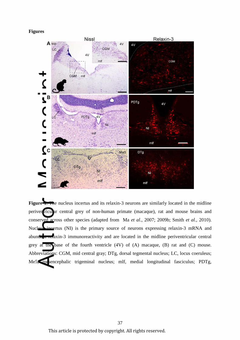
37
Figures
Figure 1. The nucleus incertus and its relaxin-3 neurons are similarly located in the midline
periventricular central grey of non-human primate (macaque), rat and mouse brains and
conserved across other species (adapted from Ma et al., 2007; 2009b; Smith et al., 2010).
Nucleus incertus (NI) is the primary source of neurons expressing relaxin-3 mRNA and
abundant relaxin-3 immunoreactivity and are located in the midline periventricular central
grey at the base of the fourth ventricle (4V) of (A) macaque, (B) rat and (C) mouse.
Abbreviations: CGM, mid central gray; DTg, dorsal tegmental nucleus; LC, locus coeruleus;
Me5, mesencephalic trigeminal nucleus; mlf, medial longitudinal fasciculus; PDTg,
This article is protected by copyright. All rights reserved.

38
posterodorsal tegmental nucleus; scp, superior cerebellar peduncle. Scale bars, (A) Nissl, 0.6
mm, inset, 80 µm, relaxin-3, 0.2 mm; (B) Nissl, 0.3 mm, relaxin-3, 0.1 mm; (C) 0.2 mm.
This article is protected by copyright. All rights reserved.
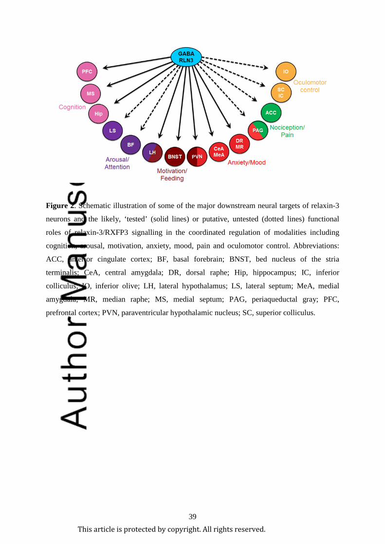
39
Figure 2. Schematic illustration of some of the major downstream neural targets of relaxin-3
neurons and the likely, ‘tested’ (solid lines) or putative, untested (dotted lines) functional
roles of relaxin-3/RXFP3 signalling in the coordinated regulation of modalities including
cognition, arousal, motivation, anxiety, mood, pain and oculomotor control. Abbreviations:
ACC, anterior cingulate cortex; BF, basal forebrain; BNST, bed nucleus of the stria
terminalis; CeA, central amygdala; DR, dorsal raphe; Hip, hippocampus; IC, inferior
colliculus; IO, inferior olive; LH, lateral hypothalamus; LS, lateral septum; MeA, medial
amygdala; MR, median raphe; MS, medial septum; PAG, periaqueductal gray; PFC,
prefrontal cortex; PVN, paraventricular hypothalamic nucleus; SC, superior colliculus.
This article is protected by copyright. All rights reserved.
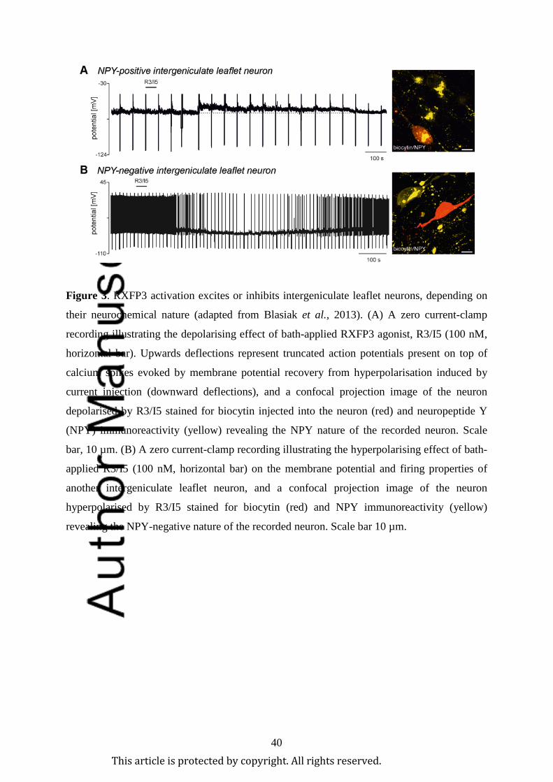
40
Figure 3. RXFP3 activation excites or inhibits intergeniculate leaflet neurons, depending on
their neurochemical nature (adapted from Blasiak et al., 2013). (A) A zero current-clamp
recording illustrating the depolarising effect of bath-applied RXFP3 agonist, R3/I5 (100 nM,
horizontal bar). Upwards deflections represent truncated action potentials present on top of
calcium spikes evoked by membrane potential recovery from hyperpolarisation induced by
current injection (downward deflections), and a confocal projection image of the neuron
depolarised by R3/I5 stained for biocytin injected into the neuron (red) and neuropeptide Y
(NPY) immunoreactivity (yellow) revealing the NPY nature of the recorded neuron. Scale
bar, 10 µm. (B) A zero current-clamp recording illustrating the hyperpolarising effect of bath-
applied R3/I5 (100 nM, horizontal bar) on the membrane potential and firing properties of
another intergeniculate leaflet neuron, and a confocal projection image of the neuron
hyperpolarised by R3/I5 stained for biocytin (red) and NPY immunoreactivity (yellow)
revealing the NPY-negative nature of the recorded neuron. Scale bar 10 µm.
This article is protected by copyright. All rights reserved.
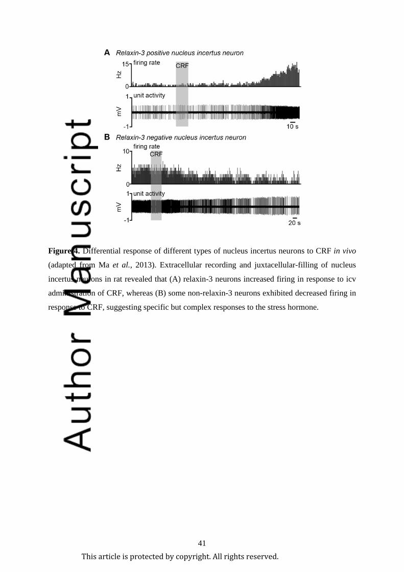
41
Figure 4. Differential response of different types of nucleus incertus neurons to CRF in vivo
(adapted from Ma et al., 2013). Extracellular recording and juxtacellular-filling of nucleus
incertus neurons in rat revealed that (A) relaxin-3 neurons increased firing in response to icv
administration of CRF, whereas (B) some non-relaxin-3 neurons exhibited decreased firing in
response to CRF, suggesting specific but complex responses to the stress hormone.
This article is protected by copyright. All rights reserved.
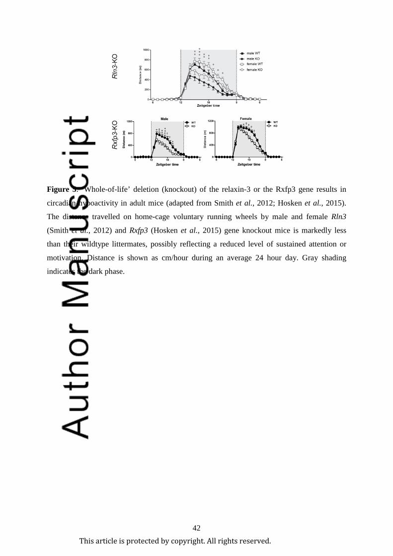
42
Figure 5. ‘Whole-of-life’ deletion (knockout) of the relaxin-3 or the Rxfp3 gene results in
circadian hypoactivity in adult mice (adapted from Smith et al., 2012; Hosken et al., 2015).
The distance travelled on home-cage voluntary running wheels by male and female Rln3
(Smith et al., 2012) and Rxfp3 (Hosken et al., 2015) gene knockout mice is markedly less
than their wildtype littermates, possibly reflecting a reduced level of sustained attention or
motivation. Distance is shown as cm/hour during an average 24 hour day. Gray shading
indicates the dark phase.
This article is protected by copyright. All rights reserved.
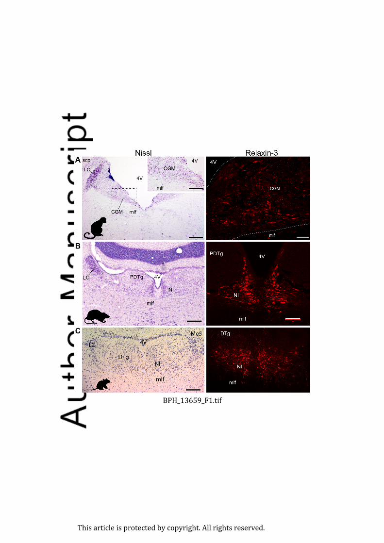
BPH_13659_F1.tif
This article is protected by copyright. All rights reserved.
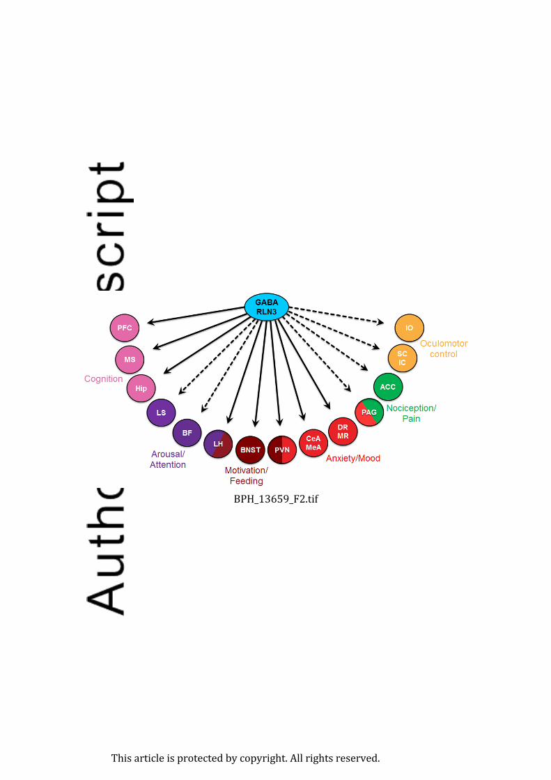
BPH_13659_F2.tif
This article is protected by copyright. All rights reserved.

BPH_13659_F3.tif
This article is protected by copyright. All rights reserved.

BPH_13659_F4.tif
This article is protected by copyright. All rights reserved.

BPH_13659_F5.tif
This article is protected by copyright. All rights reserved.
Minerva Access is the Institutional Repository of The University of Melbourne
Author/s:Ma, S;Smith, CM;Blasiak, A;Gundlach, AL
Title:Distribution, physiology and pharmacology of relaxin-3/RXFP3 systems in brain
Date:2017-05-01
Citation:Ma, S., Smith, C. M., Blasiak, A. & Gundlach, A. L. (2017). Distribution, physiologyand pharmacology of relaxin-3/RXFP3 systems in brain. BRITISH JOURNAL OFPHARMACOLOGY, 174 (10), pp.1034-1048. https://doi.org/10.1111/bph.13659.
Persistent Link:http://hdl.handle.net/11343/292206

![Amylin - Physiology and Pharmacology [Advances in Pharmacology, Vol 52] - A. Young (Elsevier, 2005) WW](https://img.pdfslide.us/doc/110x75/613caa889cc893456e1e9792/amylin-physiology-and-pharmacology-advances-in-pharmacology-vol-52-a-young.jpg)