Embed Size (px)
Citation preview
METABOLIC AND ENDOCRINE PHYSIOLOGY AND PHARMACOLOGY Annelise Kerr
1
METABOLIC AND ENDOCRINE PHYSIOLOGY AND PHARMACOLOGY
METABOLIC AND ENDOCRINE PHYSIOLOGY AND PHARMACOLOGY 1
METABOLIC AND ENDOCRINE PHYSIOLOGY 2Outline basic cellular physiology in particular 2
Structure of the cell membrane and transmembrane transport mechanisms 2Describe the structure of mitochrondria. Outline the metabolic processes that occur in the mitochrondria: PAST QUESTION 3Composition and regulation of intracellular fluid 4Generation of the trans-membrane potential 4Basic cellular physiology – other 5
Describe the structure and function of voltage sensitive ion channels: PAST QUESTION 5Describe role of intracellular tight junctions: PAST QUESTION 5What are membrane channels? How are they investigated? Describe one commonly interfered with in anaesthesia: PAST QUESTION 5Describe the mechanism of action of G proteins: PAST QUESTION 6Classify and describe the main intracellular and molecular mechanisms by which chemical neurotransmitters exert their effects. Use acetylcholine and adrenaline neurotransmitters as examples to illustrate: PAST QUESTION 6
Energy production by metabolic processes in cells 6Carbohydrate metabolism 7Fat metabolism 7Describe the role of insulin in fat metabolism: PAST QUESTION 8Protein metabolism 8Electron transport chain 9Anaerobic metabolism 10Exercise 11
Describe the physiological consequences of starvation 11Compare and contrast the physiological effects of a 6 hour fast of fluids and foods with a 20hr fast in a healthy adult: PAST QUESTION 33% 12Describe the fuel sources used during early and sustained fasting in man: PAST QUESTION: 51% 12Hormones involved in regulation of BSL: MAKEUP 12
Discuss the factors that influence metabolic rate 13Explain the control of blood glucose 13
Describe the physiological consequences of acute hypoglycaemia: PAST QUESTION 57% 14Describe the role of the hypothalamus in the integration of neuro-humoral responses 15Describe control of secretion and the functions of: 15
Pituitary hormones 15Thyroid hormones 16
Describe the physiological actions of thyroid homrones: PAST QUESTION 16Thyroid hormone synthesis: MAKEUP 16
Adrenocortical hormones 17Describe the physiological effects of the glucocorticoids: PAST QUESTION (high fail rate) 17Outline the physiological effects of bilateral adrenalectomy: PAST QUESTION 18
Adrenomedullary hormones 18Renin and angiotensin 19
Describe the secretion and function of renin and angiotensin: PAST QUESTION 19Atrial natriuretic peptide 19
Describe the regulation of plasma calcium including the actions and control of vitamin D, parathormone and calcitonin 20Outline the role of prostaglandins and other autocoids 20Metabolic and endocrine - other 21
Describe sepsis and describe the metabolic consequences of sepsis: PAST QUESTION 21Outline the components of parenteral nutrition, explaining the rationale for the use of each component: PAST QUESTION 42% 22Explain how an oxygen debt arises and how the body deals with it: PAST QUESTION 1996 22Describe the changes that occur with ageing that can affect O2 delivery to the tissues during moderate exercise 22
ENDOCRINE PHARMACOLOGY 24Describe the pharmacology of: 24
Insulin preparations 24Oral hypoglycaemics 25
List the main drug groups used in the treatment of diabetes mellitus. For each group explain the mechanism of action and give examples: PAST QUESTION 26
Corticosteroid drugs 27Describe the therapeutic and unwanted effects of dexamethasone: PAST QUESTION 28
Outline the pharmacology of: 30Thyroid hormone replacement and anti-thyroid drugs 30Glucagon, Vasopressin and analogues 31
METABOLIC AND ENDOCRINE PHYSIOLOGY AND PHARMACOLOGY Annelise Kerr
2
METABOLIC AND ENDOCRINE PHYSIOLOGYOutline basic cellular physiology in particular
Structure of the cell membrane and transmembrane transport mechanismsCell membrane
- Phospholipid bilayero hydrophilic (polar) ends outwardso hydrophobic (non-polar) fatty acid tail inwards o inserted protein channels (transmembrane proteins, peripheral proteins, glucoproteins, glycolipids)
- Function o Regulates passage of substances between ICF + ECF o prevents passage of water + hydrophilic substances o semi permeable: different ionic concentrations (and electrical charge) on either side of membrane o Functions of CM proteins
Structural Carriers for facilitated diffusion i.e. down EC gradient Pumps for ion active transport Ion channels: diffusion down electro or chemical gradient Receptors: for chemical messengers Enzymes Glycoproteins
Transmembrane transport mechanisms1. Diffusion
o continual random movement of molecules which relies only on normal kinetic motion of mattero Types of diffusion:
i. Simple diffusion – lipid soluble (O2, N2, CO2, EtOH) Small, highly soluble lipid molecules dissolve directly in lipid bilayer + diffusion through CM via conc gradient
ii. Simple diffusion – lipid insoluble (H2O) Water crosses CM through aquaporins
iii. Diffusion via gated protein channels Allows passage of charged molecules + exhibits selective permeability due to diameter, shape, electrical charge
o Voltage gated: e.g. Na, K, Cl; ∆shape in response to electrival stimulus (e.g. AP)o Chemical/ ligand: e.g. ACh, GABA, glycine, NMDA; binding of ligand open / close
iv. Facilitated diffusion E.g. glucose, amino acids Via carrier protein: binds substrate conformational change moves substrate across CM Concentration gradient dependent + limited by amount of carrier protein available
o Rate of diffusion across CM proportional to: Concentration difference across CM Electrical potential difference Osmotic pressure Carrier protein (for facilitated diffusion)
2. Active transporto Movement of substances across CM + carrier protein + against EC gradiento Relies on kinetic energy + ATPo Types of active transport
i. Primary active transport E.g. Na/K ATPase pump ATP hydrolysed by carrier protein as it moves substances across CM
ii. Secondary active transport Combination of primary active transport + facilitated diffusion Dependent on energy potential formed by EC gradient set up by primary active transport Classified as:
o Co transport / symport: both ions move in same direction e.g. SGLT-2o Counter transport: ions in opposite directions e.g. Na/K antiporter in CD
o Factors affecting active transport Concentration difference across CM Electrical portential difference Carrier proteins
3. Other methodso Endocytosiso Exocytosiso Transytosis
Relevant principles:1. Gibbs-donnan effect
o if a semi permeable membrane separates 2 solutions + at least 1 of those solutions contains a non-diffusible ion the distribution of other ions across the membrane will be altered
o The distribution of permeable charged ions will be influenced by both their valence + distribution of uncharged ionso Important for:
Stability of cell vol Plasma oncotic pressure RMP
2. Fick’s law of diffusion
METABOLIC AND ENDOCRINE PHYSIOLOGY AND PHARMACOLOGY Annelise Kerr
3
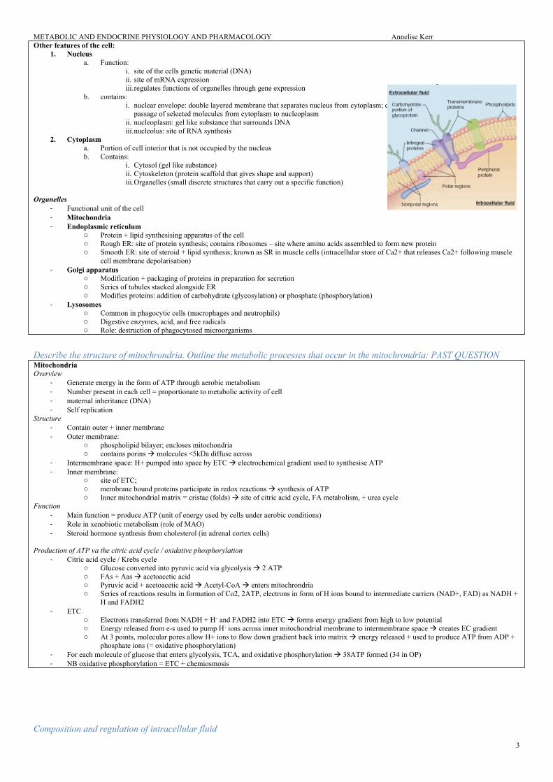
Other features of the cell: 1. Nucleus
a. Function:i. site of the cells genetic material (DNA)ii. site of mRNA expression iii.regulates functions of organelles through gene expression
b. contains:i. nuclear envelope: double layered membrane that separates nucleus from cytoplasm; contains nuclear pores that allow regulated
passage of selected molecules from cytoplasm to nucleoplasmii. nucleoplasm: gel like substance that surrounds DNAiii.nucleolus: site of RNA synthesis
2. Cytoplasma. Portion of cell interior that is not occupied by the nucleusb. Contains:
i. Cytosol (gel like substance)ii. Cytoskeleton (protein scaffold that gives shape and support)iii.Organelles (small discrete structures that carry out a specific function)
Organelles- Functional unit of the cell- Mitochondria- Endoplasmic reticulum
o Protein + lipid synthesising apparatus of the cello Rough ER: site of protein synthesis; contains ribosomes – site where amino acids assembled to form new proteino Smooth ER: site of steroid + lipid synthesis; known as SR in muscle cells (intracellular store of Ca2+ that releases Ca2+ following muscle
cell membrane depolarisation)- Golgi apparatus
o Modification + packaging of proteins in preparation for secretiono Series of tubules stacked alongside ERo Modifies proteins: addition of carbohydrate (glycosylation) or phosphate (phosphorylation)
- Lysosomeso Common in phagocytic cells (macrophages and neutrophils)o Digestive enzymes, acid, and free radicalso Role: destruction of phagocytosed microorganisms
Describe the structure of mitochrondria. Outline the metabolic processes that occur in the mitochrondria: PAST QUESTIONMitochondriaOverview
- Generate energy in the form of ATP through aerobic metabolism- Number present in each cell = proportionate to metabolic activity of cell - maternal inheritance (DNA)- Self replication
Structure- Contain outer + inner membrane- Outer membrane:
o phospholipid bilayer; encloses mitochondriao contains porins molecules <5kDa diffuse across
- Intermembrane space: H+ pumped into space by ETC electrochemical gradient used to synthesise ATP- Inner membrane:
o site of ETC; o membrane bound proteins participate in redox reactions synthesis of ATPo Inner mitochondrial matrix = cristae (folds) site of citric acid cycle, FA metabolism, + urea cycle
Function- Main function = produce ATP (unit of energy used by cells under aerobic conditions)- Role in xenobiotic metabolism (role of MAO)- Steroid hormone synthesis from cholesterol (in adrenal cortex cells)
Production of ATP va the citric acid cycle / oxidative phosphorylation - Citric acid cycle / Krebs cycle
o Glucose converted into pyruvic acid via glycolysis 2 ATPo FAs + Aas acetoacetic acido Pyruvic acid + acetoacetic acid Acetyl-CoA enters mitochrondriao Series of reactions results in formation of Co2, 2ATP, electrons in form of H ions bound to intermediate carriers (NAD+, FAD) as NADH +
H and FADH2- ETC
o Electrons transferred from NADH + H+ and FADH2 into ETC forms energy gradient from high to low potential o Energy released from e-s used to pump H+ ions across inner mitochondrial membrane to intermembrane space creates EC gradient o At 3 points, molecular pores allow H+ ions to flow down gradient back into matrix energy released + used to produce ATP from ADP +
phosphate ions (= oxidative phosphorylation)- For each molecule of glucose that enters glycolysis, TCA, and oxidative phosphorylation 38ATP formed (34 in OP)- NB oxidative phosphorylation = ETC + chemiosmosis
Composition and regulation of intracellular fluid
METABOLIC AND ENDOCRINE PHYSIOLOGY AND PHARMACOLOGY Annelise Kerr
4
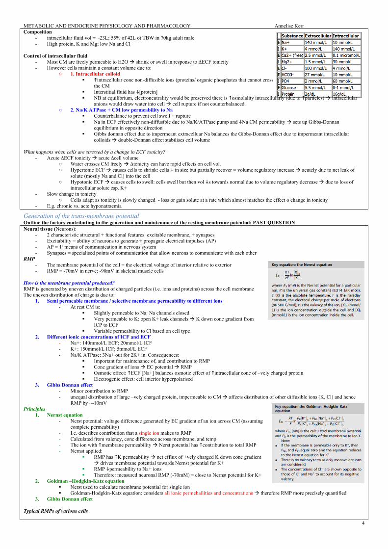
Composition- intracellular fluid vol = ~23L; 55% of 42L ot TBW in 70kg adult male- High protein, K and Mg; low Na and Cl
Control of intracellular fluid- Most CM are freely permeable to H2O shrink or swell in response to ∆ECF tonicity- However cells maintain a constant volume due to:
o 1. Intracellular colloid intracellular conc non-diffusible ions (proteins/ organic phosphates that cannot cross CM) Gibbs Donnan equilibrium across
the CM Interstitial fluid has [protein] NB at equilibrium, electroneutrality would be preserved there is osmolality intracellularly (due to particles) intracellular
anions would draw water into cell cell rupture if not counterbalanced. o 2. Na/K ATPase + CM low permeability to Na
Counterbalance to prevent cell swell + rupture Na in ECF effectively non-diffusible due to Na/K/ATPase pump and Na CM permeability sets up Gibbs-Donnan
equilibrium in opposite direction Gibbs donnan effect due to impermeant extracelluar Na balances the Gibbs-Donnan effect due to impermeant intracellular
colloids double-Donnan effect stabilises cell volume
What happens when cells are stressed by a change in ECF tonicity?- Acute ∆ECF tonicity acute ∆cell volume
o Water crosses CM freely ∆tonicity can have rapid effects on cell vol. o Hypertonic ECF causes cells to shrink: cells in size but partially recover = volume regulatory increase acutely due to net leak of
solute (mostly Na and Cl) into the cello Hypotonic ECF causes cells to swell: cells swell but then vol s towards normal due to volume regulatory decrease due to loss of
intracellular solute esp. K+- Slow change in tonicity
o Cells adapt as tonicity is slowly changed - loss or gain solute at a rate which almost matches the effect o change in tonicity- E.g. chronic vs. acte hyponatraemia
Generation of the trans-membrane potentialOutline the factors contributing to the generation and maintenance of the resting membrane potential: PAST QUESTIONNeural tissue (Neurons):
- 2 characteristic structural + functional features: excitable membrane, + synapses- Excitability = ability of neurons to generate + propagate electrical impulses (AP)- AP = 1o means of communication in nervous system- Synapses = specialised points of communication that allow neurons to communicate with each other
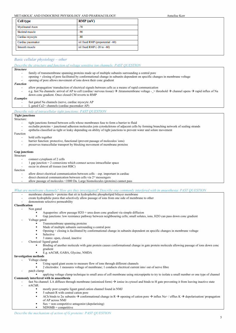
RMP- The membrane potential of the cell = the electrical voltage of interior relative to exterior- RMP = -70mV in nerve; -90mV in skeletal muscle cells
How is the membrane potential produced?RMP is generated by uneven distribution of charged particles (i.e. ions and proteins) across the cell membraneThe uneven distribution of charge is due to:
1. Semi permeable membrane / selective membrane permeability to different ions- At rest CM is:
Slightly permeable to Na: Na channels closed Very permeable to K: open K+ leak channels K down conc gradient from
ICP to ECF Variable permeability to Cl based on cell type
2. Different ionic concentrations of ICF and ECF- Na+: 140mmol/L ECF; 20mmol/L ICF- K+: 150mmol/L ICF; 5mmol/L ECF- Na/K ATPase: 3Na+ out for 2K+ in. Consequences:
Important for maintenance of, and contribution to RMP Conc gradient of ions EC potential RMP Osmotic effect: ECF [Na+] balances osmotic effect of intracellular conc of –vely charged protein Electrogenic effect: cell interior hyperpolarised
3. Gibbs Donnan effect- Minor contribution to RMP - unequal distribution of large –vely charged protein, impermeable to CM affects distribution of other diffusible ions (K, Cl) and hence
RMP by ~-10mVPrinciples
1. Nernst equation- Nerst potential: voltage difference generated by EC gradient of an ion across CM (assuming
complete permeability)- I.e. describes contribution that a single ion makes to RMP- Calculated from valency, conc difference across membrane, and temp- The ion with membrane permeability Nerst potential has contribution to total RMP- Nernst applied:
RMP has K permeability net efflux of +vely charged K down conc gradient drives membrane potential towards Nernst potential for K+
RMP permeability to Na+ ions Therefore: measured neuronal RMP (-70mM) = close to Nernst potential for K+
2. Goldman –Hodgkin-Katz equation Nerst used to calculate membrane potential for single ion Goldman-Hodgkin-Katz equation: considers all ionic permebailities and concentrations therefore RMP more precisely quantified
3. Gibbs Donnan effect
Typical RMPs of various cells
METABOLIC AND ENDOCRINE PHYSIOLOGY AND PHARMACOLOGY Annelise Kerr
5
Basic cellular physiology – otherDescribe the structure and function of voltage sensitive ion channels: PAST QUESTIONStructure
- family of transmembrane spanning proteins made up of multiple subunits surrounding a central pore- opening + closing of pore facilitated by conformational change in subunits dependent on specific changes in membrane voltage- opening of pore allows movement of ions down their conc gradient
Function- allow propagation/ transduction of electrical signals between cells as a means of rapid communication- e.g. fast Na channels: arrival of AP to cell (cardiac/ nervous tissue) ∆transmembrane voltage -_> threshold channel opens rapid influx of Na
down conc gradient. Once closed CM reverts to RMPExamples
- fast gated Na channels (nerve, cardiac myocyte AP- L gated Ca2+ channels (cardiac pacemaker AP)
Describe role of intracellular tight junctions: PAST QUESTIONTight junctionsStructure:
- tight junctions formed between cells whose membranes fuse to form a barrier to fluid- occludin proteins + junctional adhesion molecules join cytoskeletons of adjacent cells by forming branching network of sealing strands- epithelia classified as tight or leaky depending on ability of tight junctions to prevent water and solute movement
Function- hold cells together- barrier function: protective, functional (prevent passage of molecules/ ions)- preserves transcellular transport by blocking movement of membrane proteins
Gap junctionsStructure
- connect cytoplasm of 2 cells- 1 gap junction = 2 connexions which connect across intracellular space- occur in almost all tissues (not RBC)
function- allow direct electrical communication between cells – esp. important in cardiac- direct chemical communication between cells via 2nd messengers- allow passage of molecules <1000 Da. Large biomolecules (proteins) cannot pass
What are membrane channels? How are they investigated? Describe one commonly interfered with in anaesthesia: PAST QUESTION- membrane channels = proteins that sit in hydrophobic phospholipid bilayer membrane - create hydrophilic pores that selectively allow passage of ions from one side of membrane to other- demonstrate selective permeability
Classification- Non gated
Aquaporins: allow passage H2O + urea doen conc gradient via simple diffusion Gap junctions: low resistance pathway between neighbouring cells; small solutes, ions, H2O can pass down conc gradient
- Voltage gated Transmembrane spanning proteins Made of multiple subunits surrounding a central pore Opening + closing is facilitated by conformational change in subunits dependent on specific changes in membrane voltage Selective 3 states: open, closed, inactive
- Chemical/ ligand gated Binding of another molecule with gate protein causes conformational change in gate protein molecule allowing passage of ions down conc
gradient E.g. nAChR, GABA, Glycine, NMDA
Investigation methods- Voltage clamp
Using squid giant axons to measure flow of ions through different channels 2 electrodes: 1 measures voltage of membrane; 1 conducts electrical current into/ out of nerve fibre
- patch clamp applying voltage clamp technique to small area of cell membrane using micropipette to try to isolate a small number or one type of channel
Commonly interfered with in anaesthesia- fast Na channel: LA diffuses through membrane (unionised form) ionise in cytosol and binds to H gate preventing it from leaving inactive state- nAChR:
mostly post synaptic ligant gated cation channel found in NMJ 5 subunit R with central cation pore ACh binds to 2 subunits conformational change in R opening of cation pore influx Na+ / efflux K depolarisation/ propagation
of AP across NMJ Sux = non competitive antagonist (depolarising) NDNMB = competitive
Describe the mechanism of action of G proteins: PAST QUESTION
METABOLIC AND ENDOCRINE PHYSIOLOGY AND PHARMACOLOGY Annelise Kerr
6
G proteins and G protein coupled receptorsG proteins
- Multisubunit protein complex which exchange GDP for GTP in order to bring about an effect- GPCR: heterotrimetric G protein (subunits y) with GDP coupled to 7 transmembrane spanning receptor- MoA
o Activation: binding of ligand on ECF side of CM conformational change on cytosolic side exchange of GDP for GTPo Dissociation: -GTP complex dissociates from y and interacts with effector proteinso 2nd messenger effect: 2nd messengers then able to activate target proteins (e.g. cAMP) or ion channelso Inactivation: via intrinsic GTPase activity results in reformation of -GDP complex + reassociation with y complexo Amplification: each -GTP complex is capable of catalysing multiple reactions to form 2nd messengers amplification pathway
- Examples of GPCR/ -subunit variabilityo Gs (stimulatory): AC cAMP PKA; eg adrenaline, glucagon, PTH, ACTHo Gq: PLC 2nd messengers IP3 + DAG IP3 causes Ca2+ release from ER; DAG activates PKC eg vasopressin, TSH, angiotensino Gi (inhibitory): AC cAMP. E.g. M2 mediated cardiac AP conduction velocityo Gt: transducin: molecule responsible for generating signal in the rods of eye in response to light. Gai triggers breakdown of cGMP
- Targets: G protein can activate (2nd messengers)o Adenylate cyclase or guanylate cyclase cAMP or cGMP formationo Phospholipase C (PLC) on inner surface of CM catalyse hydrolysis of membrane lipid PIP2 to IP3 or DAG
IP3 diffuses to ER where it binds to IP3 receptor (ligand gated Ca channel) DAG stays in cell membrane where it activates protein kinase C
Classify and describe the main intracellular and molecular mechanisms by which chemical neurotransmitters exert their effects. Use acetylcholine and adrenaline neurotransmitters as examples to illustrate: PAST QUESTIONSee also: section on neurotransmittersGeneral:
- chemical neurotransmission = most common type of synaptic transmission - NT stored in presynaptic vesicles in nerve terminal- AP exocytosis release of NT into synaptic cleft diffuse across postsynaptic membrane- Rs on postsynaptic membrane. Classified as:
o Inotropic: direct action of ion channel membrane depolarisation / hyperpolarisationo GPCR (metabotropic): indirect action on ion channels changes in K+/Ca2+ conductance via 2nd messengers depolarisation/
hyperpolarisationACh
- nicotinic (nAChR): 5 subunit R with central cation pore- classic ionotropic receptor
o ACh binds 2 subunits conformational change in receptor opening of cation pore influx Na (small Ca influx) / efflux Ko Muscarinic (mAChR): 7 transmembrane spanning domains
5 subtypes M2 (conducting tissue of heart) AC K conductance membrane hyperpolarisation
Adrenaline- adrenergic receptors
o 1: PLC (2nd messenger) IP3/DAG Ca2+ depolarisation vasoconstriction GI smooth muscle relaxation Salivary secretion
o 2: AC cAMP Ca2+ platelet aggregation NA release (presynaptic inhibition)
- receptors: AC cAMP Ca2+o 1: positive inotrope/ chronotrope; relax gastric smooth muscle o 2: vasodilation; bronchodilationo 3: lipolysis of 1o brown fat
Energy production by metabolic processes in cellsMetabolism
- biochemical reactions that occur within living organisms- encompasses:
o Anabolism = building up of larger molecules from smaller oneso Catabolism = breaking down into smaller entities with extraction of energy
i. Cellular respiration 1. = series of catabolic processes by which carbohydrates, fats, and proteins are broken down to yield ATP through a
series of redox reactions – ultimately using O2 as the oxidising agent. 2. As O2 is too reactive to be used directly, this process employs a series of intermediate electron carriers, including
NAD+ and FADii. Catabolism involves a number of processes:
1. Glycolysis2. Lipolysis3. Protein catabolism4. The citric acid cycle5. The electron transport chain
METABOLIC AND ENDOCRINE PHYSIOLOGY AND PHARMACOLOGY Annelise Kerr
7
Carbohydrate metabolismCarbohydrate metabolism:
- glucose = basic unit of carbohydrates- Active form of glucose = glucose-6-phosphate
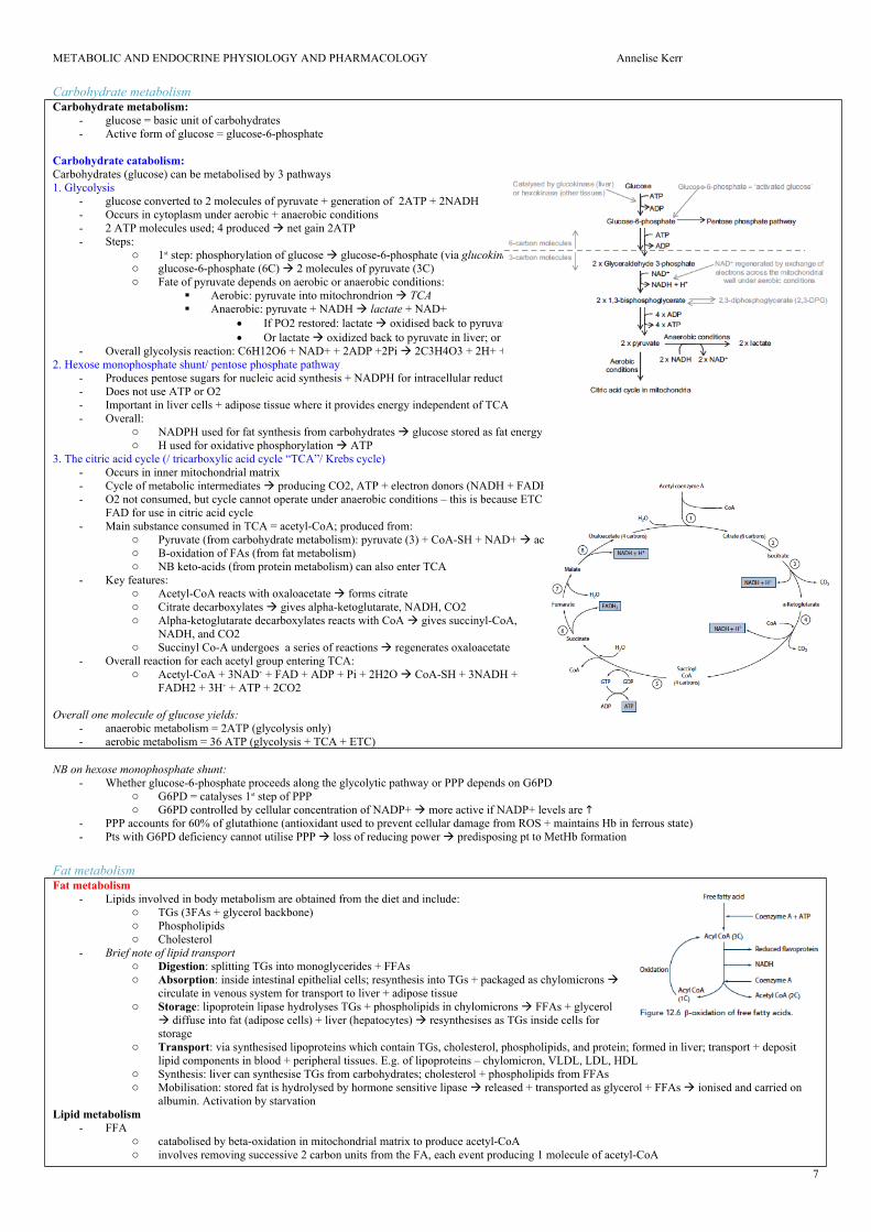
Carbohydrate catabolism:Carbohydrates (glucose) can be metabolised by 3 pathways1. Glycolysis
- glucose converted to 2 molecules of pyruvate + generation of 2ATP + 2NADH- Occurs in cytoplasm under aerobic + anaerobic conditions- 2 ATP molecules used; 4 produced net gain 2ATP- Steps:
o 1st step: phosphorylation of glucose glucose-6-phosphate (via glucokinase in liver; hexokinase in tissue cells)o glucose-6-phosphate (6C) 2 molecules of pyruvate (3C)o Fate of pyruvate depends on aerobic or anaerobic conditions:
Aerobic: pyruvate into mitochrondrion TCA Anaerobic: pyruvate + NADH lactate + NAD+
If PO2 restored: lactate oxidised back to pyruvate enter citric acid cycle Or lactate oxidized back to pyruvate in liver; or converted to glucose through gluconeogenesis (Cori cycle)
- Overall glycolysis reaction: C6H12O6 + NAD+ + 2ADP +2Pi 2C3H4O3 + 2H+ +2ATP + 2H2O + 2NADH2. Hexose monophosphate shunt/ pentose phosphate pathway
- Produces pentose sugars for nucleic acid synthesis + NADPH for intracellular reduction reactions- Does not use ATP or O2- Important in liver cells + adipose tissue where it provides energy independent of TCA- Overall:
o NADPH used for fat synthesis from carbohydrates glucose stored as fat energyo H used for oxidative phosphorylation ATP
3. The citric acid cycle (/ tricarboxylic acid cycle “TCA”/ Krebs cycle) - Occurs in inner mitochondrial matrix- Cycle of metabolic intermediates producing CO2, ATP + electron donors (NADH + FADH2) utilised in ETC to produce ATP- O2 not consumed, but cycle cannot operate under anaerobic conditions – this is because ETC (dependent upon O2) is needed to regenerate NAD+ and
FAD for use in citric acid cycle- Main substance consumed in TCA = acetyl-CoA; produced from:
o Pyruvate (from carbohydrate metabolism): pyruvate (3) + CoA-SH + NAD+ acetyl-CoA (2) + CO2 + NADHo B-oxidation of FAs (from fat metabolism) o NB keto-acids (from protein metabolism) can also enter TCA
- Key features:o Acetyl-CoA reacts with oxaloacetate forms citrateo Citrate decarboxylates gives alpha-ketoglutarate, NADH, CO2o Alpha-ketoglutarate decarboxylates reacts with CoA gives succinyl-CoA,
NADH, and CO2o Succinyl Co-A undergoes a series of reactions regenerates oxaloacetate
- Overall reaction for each acetyl group entering TCA:o Acetyl-CoA + 3NAD+ + FAD + ADP + Pi + 2H2O CoA-SH + 3NADH +
FADH2 + 3H+ + ATP + 2CO2
Overall one molecule of glucose yields:- anaerobic metabolism = 2ATP (glycolysis only)- aerobic metabolism = 36 ATP (glycolysis + TCA + ETC)
NB on hexose monophosphate shunt:- Whether glucose-6-phosphate proceeds along the glycolytic pathway or PPP depends on G6PD
o G6PD = catalyses 1st step of PPPo G6PD controlled by cellular concentration of NADP+ more active if NADP+ levels are
- PPP accounts for 60% of glutathione (antioxidant used to prevent cellular damage from ROS + maintains Hb in ferrous state) - Pts with G6PD deficiency cannot utilise PPP loss of reducing power predisposing pt to MetHb formation
Fat metabolismFat metabolism
- Lipids involved in body metabolism are obtained from the diet and include:o TGs (3FAs + glycerol backbone)o Phospholipidso Cholesterol
- Brief note of lipid transporto Digestion: splitting TGs into monoglycerides + FFAso Absorption: inside intestinal epithelial cells; resynthesis into TGs + packaged as chylomicrons
circulate in venous system for transport to liver + adipose tissueo Storage: lipoprotein lipase hydrolyses TGs + phospholipids in chylomicrons FFAs + glycerol
diffuse into fat (adipose cells) + liver (hepatocytes) resynthesises as TGs inside cells for storage
o Transport: via synthesised lipoproteins which contain TGs, cholesterol, phospholipids, and protein; formed in liver; transport + deposit lipid components in blood + peripheral tissues. E.g. of lipoproteins – chylomicron, VLDL, LDL, HDL
o Synthesis: liver can synthesise TGs from carbohydrates; cholesterol + phospholipids from FFAso Mobilisation: stored fat is hydrolysed by hormone sensitive lipase released + transported as glycerol + FFAs ionised and carried on
albumin. Activation by starvation Lipid metabolism
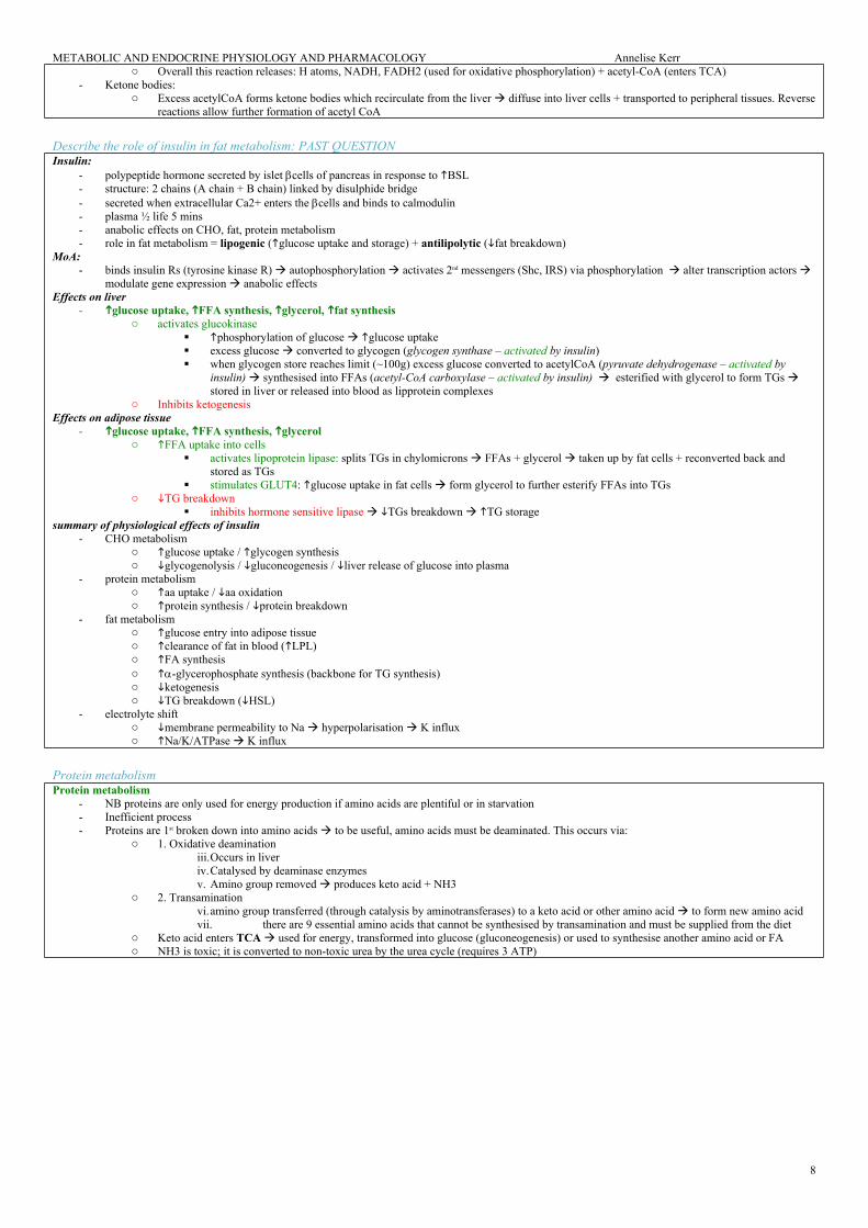
- FFA o catabolised by beta-oxidation in mitochondrial matrix to produce acetyl-CoAo involves removing successive 2 carbon units from the FA, each event producing 1 molecule of acetyl-CoA
METABOLIC AND ENDOCRINE PHYSIOLOGY AND PHARMACOLOGY Annelise Kerr
8
o Overall this reaction releases: H atoms, NADH, FADH2 (used for oxidative phosphorylation) + acetyl-CoA (enters TCA)- Ketone bodies:
o Excess acetylCoA forms ketone bodies which recirculate from the liver diffuse into liver cells + transported to peripheral tissues. Reverse reactions allow further formation of acetyl CoA
Describe the role of insulin in fat metabolism: PAST QUESTIONInsulin:
- polypeptide hormone secreted by islet cells of pancreas in response to BSL- structure: 2 chains (A chain + B chain) linked by disulphide bridge- secreted when extracellular Ca2+ enters the cells and binds to calmodulin- plasma ½ life 5 mins- anabolic effects on CHO, fat, protein metabolism - role in fat metabolism = lipogenic (glucose uptake and storage) + antilipolytic (fat breakdown)
MoA:- binds insulin Rs (tyrosine kinase R) autophosphorylation activates 2nd messengers (Shc, IRS) via phosphorylation alter transcription actors
modulate gene expression anabolic effects Effects on liver
- glucose uptake, FFA synthesis, glycerol, fat synthesis o activates glucokinase
phosphorylation of glucose glucose uptake excess glucose converted to glycogen (glycogen synthase – activated by insulin) when glycogen store reaches limit (~100g) excess glucose converted to acetylCoA (pyruvate dehydrogenase – activated by
insulin) synthesised into FFAs (acetyl-CoA carboxylase – activated by insulin) esterified with glycerol to form TGs stored in liver or released into blood as lipprotein complexes
o Inhibits ketogenesisEffects on adipose tissue
- glucose uptake, FFA synthesis, glycerol o FFA uptake into cells
activates lipoprotein lipase: splits TGs in chylomicrons FFAs + glycerol taken up by fat cells + reconverted back and stored as TGs
stimulates GLUT4: glucose uptake in fat cells form glycerol to further esterify FFAs into TGso TG breakdown
inhibits hormone sensitive lipase TGs breakdown TG storagesummary of physiological effects of insulin
- CHO metabolismo glucose uptake / glycogen synthesiso glycogenolysis / gluconeogenesis / liver release of glucose into plasma
- protein metabolismo aa uptake / aa oxidationo protein synthesis / protein breakdown
- fat metabolismo glucose entry into adipose tissueo clearance of fat in blood (LPL)o FA synthesiso -glycerophosphate synthesis (backbone for TG synthesis)o ketogenesiso TG breakdown (HSL)
- electrolyte shifto membrane permeability to Na hyperpolarisation K influxo Na/K/ATPase K influx
Protein metabolismProtein metabolism
- NB proteins are only used for energy production if amino acids are plentiful or in starvation- Inefficient process- Proteins are 1st broken down into amino acids to be useful, amino acids must be deaminated. This occurs via:
o 1. Oxidative deaminationiii.Occurs in liveriv.Catalysed by deaminase enzymesv. Amino group removed produces keto acid + NH3
o 2. Transaminationvi.amino group transferred (through catalysis by aminotransferases) to a keto acid or other amino acid to form new amino acidvii. there are 9 essential amino acids that cannot be synthesised by transamination and must be supplied from the diet
o Keto acid enters TCA used for energy, transformed into glucose (gluconeogenesis) or used to synthesise another amino acid or FAo NH3 is toxic; it is converted to non-toxic urea by the urea cycle (requires 3 ATP)
METABOLIC AND ENDOCRINE PHYSIOLOGY AND PHARMACOLOGY Annelise Kerr
9
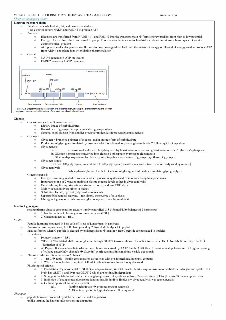
Electron transport chainElectron transport chain
- Final step of carbohydrate, fat, and protein catabolism- Uses electron donors NADH and FADH2 to produce ATP- Process:
o Electrons are transferred from NADH + H+ and FADH2 into the transport chain forms energy gradient from high to low potential o Energy released from electrons is used to pump H+ ions across the inner mitochondrial membrane to intermembrane space creates
electrochemical gradient o At 3 points, molecular pores allow H+ ions to flow down gradient back into the matrix energy is released energy used to produce ATP
from ADP + phosphate ions (= oxidative phosphorylation)- Overall:
o NADH generates 3 ATP moleculeso FADH2 generates 1 ATP molecule
Glucose - Glucose comes from 3 main sources:
o Dietary intake of carbohydrateso Breakdown of glycogen in a process called glycogenolysiso Generation of glucose from smaller precursor molecules in process gluconeogenesis
- Glycogeno Glycogen = branched polymer of glucose; major storage form of carbohydrateo Production of glycogen stimulated by insulin – which is released as plasma glucose levels following CHO ingestiono Glycogenesis:
viii. Glucose molecules are phosphorylated by hexokinase in tissue, and glucokinase in liver glucose-6-phosphateix.Glucose-6-phosphate converted into glucose-1-phosphate by phosphoglucomutasex. Glucose-1-phosphate molecules are joined together under action of glycogen synthase glycogen
o Glycogen stores: xi.Liver: 100g glycogen; skeletal muscle 200g glycogen (cannot be released into circulation; only used by muscle)
o Glycogenolysisxii. When plasma glucose levels release of glucagon + adrenaline stimulates glycogenolysis
- Gluconeogenesis o Energy consuming anabolic process in which glucose is synthesised from non-carbohydrate precursorso Importance: one of 2 ways to maintain plasma glucose levels (other is glycogenolysis)o Occurs during fasting, starvation, extreme exercise, and low CHO dietso Mainly occurs in liver; minor in kidneyo Substrates: lactate, pyruvate, glycerol, amino acidso Separate biochemical pathway – not simply the reverse of glycolysiso Glucagon + glucocorticoids promote gluconeogenesis; insulin inhibits it
Insulin + glucagon- resting plasma glucose concentration usually tightly controlled: 3.5-5.5mmol/L by balance of 2 hormones:
o 1. Insulin: acts to plasma glucose concentration (BSL)o 2. Glucagon: acts to BSL
Insulin:- Peptide hormone produced in beta cells of Islets of Langerhans in pancreas- Proinsulin: insulin precursor; A + B chain joined by 2 disulphide bridges + C peptide- Insulin: formed when C peptide is cleaved by endopeptidases insulin + free C peptide are packaged in vesicles - Exocytosis:
o Primary trigger = BSLo BSL facilitated diffusion of glucose through GLUT2 transmembrane channels into B-islet cells metabolic activity of cell
formation of ATPo ATP-gated K channels on beta islet cell membrane are closed by ATP levels K flux membrane depolarisation triggers opening
of voltage gated Ca2+ channels Ca2+ influx triggers insulin containing vesicle exocytosis - Plasma insulin secretion occurs in 2 phases:
o 1. BSL rapid insulin concentration as vesicles with pre-formed insulin empty contents o 2. When all vesicles have emptied B islet cells release insulin as it is synthesised
- Physiological effects:o 1. Facilitation of glucose uptake: GLUT4 in adipose tissue, skeletal muscle, heart – require insulin to facilitate cellular glucose uptake. NB
brain has GLUT-1 and liver has GLUT-2 which are not insulin dependento 2. Storage of metabolic substrates: hepatic glycogenesis, FA synthesis in liver, esterification of FAs (to make TGs) in adipose tissueo 3. Inhibition of endogenous glucose production: insulin inhibits lipolysis + glycogenolysis + gluconeogenesiso 4. Cellular uptake of amino acids and K
xiii. amino acid uptake promoes protein synthesisxiv. 2. K uptake: prevents hyperkalaemia following meal
Glucagon- peptide hormone produced by alpha cells of islets of Langerhans- unlike insulin, the have no glucose sensing apparatus
METABOLIC AND ENDOCRINE PHYSIOLOGY AND PHARMACOLOGY Annelise Kerr
10
- secretion:o stimulated by hypoglycaemia: hypoglycaemia-induced ANS activity (i.e. indirect) + adrenalineo inhibited by: insulin, somatostatin, freeFA and ketone body concentrations
- Actions:o plasma glucose concentration by:
xv. promoting gluconeogenesisxvi. promoting glycogenolysisxvii. inhibiting glycolysis in liver
o Esp. important during starvation
Anaerobic metabolismDescribe the formation, fate, and role of lactate in energy production: PAST QUESTION
- Aerobic conditions: pyruvate passes into mitochrondrion enters citric acid cycle- Anaerobic conditions: TCA cannot operate ETC is dependent upon O2 and is needed to regenerate NAD+ and FAD for use in TCA- lactate = 3C organic acid produced as a result of anaerobic metabolism of pyruvate catalysed by lactate dehydrogenase
Formation of lactate in energy production- Glycolysis: 9 step series of reactions occurring in cytoplasm: end result = 1 molecule of 6C glucose converted to 2x 3C pyruvate for net gain of 2ATP- Anaerobic glycolysis:
o Oxidative phosphorylation ceases: NADH not reoxidised accumulates: NAD+ NADHo TCA ceases acetyl CoA not utilised: o Lactic acid fermentation: pyruvate + NADH lactate + NAD+ via lactate dehydrogenase
Fate of lactate in energy production- Conversion back to pyruvate
o When O2 restored: lactate pyruvate + NADH able to be utilised by cell that formed the lactateo Pyruvate TCAo NAHD ETC
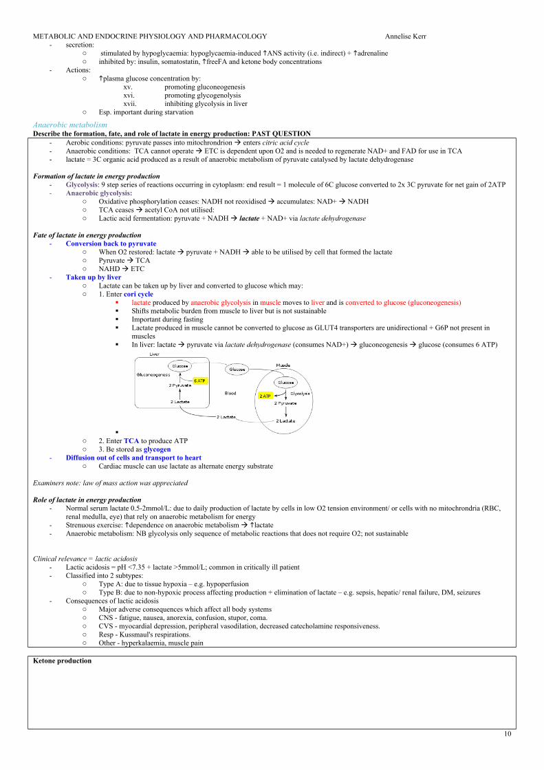
- Taken up by liver o Lactate can be taken up by liver and converted to glucose which may:o 1. Enter cori cycle
lactate produced by anaerobic glycolysis in muscle moves to liver and is converted to glucose (gluconeogenesis) Shifts metabolic burden from muscle to liver but is not sustainable Important during fasting Lactate produced in muscle cannot be converted to glucose as GLUT4 transporters are unidirectional + G6P not present in
muscles In liver: lactate pyruvate via lactate dehydrogenase (consumes NAD+) gluconeogenesis glucose (consumes 6 ATP)
o 2. Enter TCA to produce ATPo 3. Be stored as glycogen
- Diffusion out of cells and transport to hearto Cardiac muscle can use lactate as alternate energy substrate
Examiners note: law of mass action was appreciated
Role of lactate in energy production- Normal serum lactate 0.5-2mmol/L: due to daily production of lactate by cells in low O2 tension environment/ or cells with no mitochrondria (RBC,
renal medulla, eye) that rely on anaerobic metabolism for energy- Strenuous exercise: dependence on anaerobic metabolism lactate- Anaerobic metabolism: NB glycolysis only sequence of metabolic reactions that does not require O2; not sustainable
Clinical relevance = lactic acidosis- Lactic acidosis = pH <7.35 + lactate >5mmol/L; common in critically ill patient- Classified into 2 subtypes:
o Type A: due to tissue hypoxia – e.g. hypoperfusiono Type B: due to non-hypoxic process affecting production + elimination of lactate – e.g. sepsis, hepatic/ renal failure, DM, seizures
- Consequences of lactic acidosis o Major adverse consequences which affect all body systems o CNS - fatigue, nausea, anorexia, confusion, stupor, coma.o CVS - myocardial depression, peripheral vasodilation, decreased catecholamine responsiveness.o Resp - Kussmaul's respirations.o Other - hyperkalaemia, muscle pain
Ketone production
METABOLIC AND ENDOCRINE PHYSIOLOGY AND PHARMACOLOGY Annelise Kerr
11
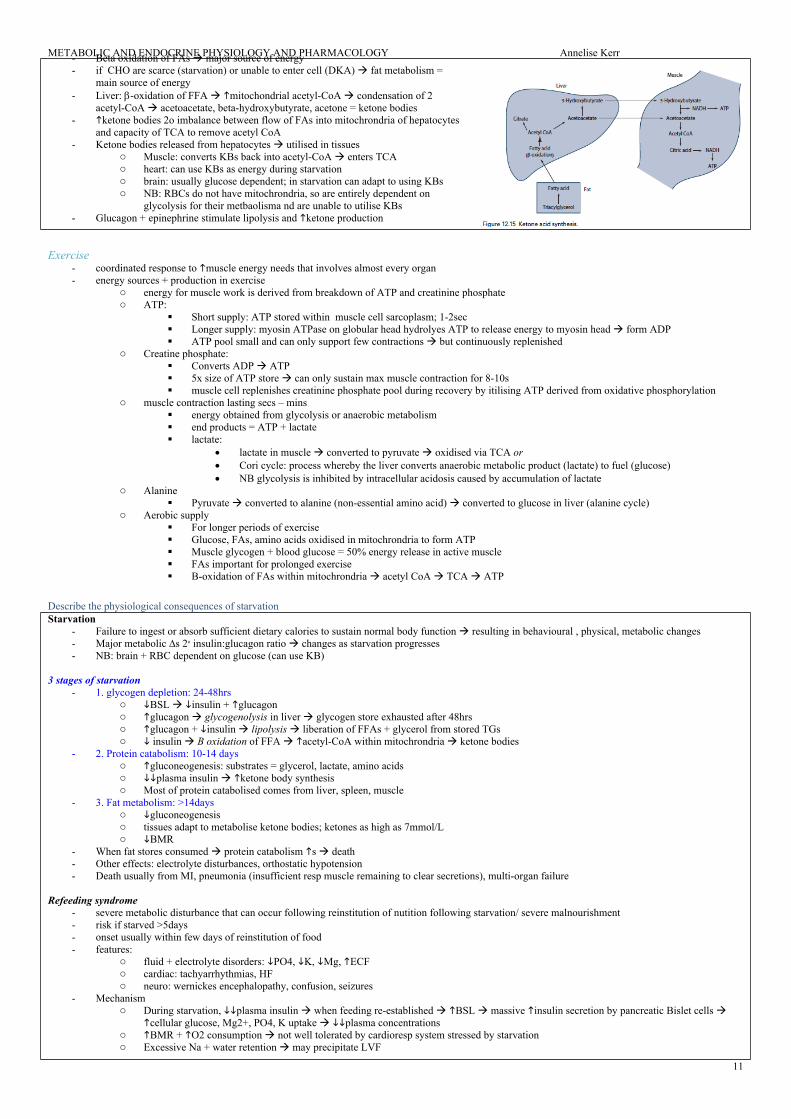
- Beta oxidation of FAs major source of energy - if CHO are scarce (starvation) or unable to enter cell (DKA) fat metabolism =
main source of energy- Liver: -oxidation of FFA mitochondrial acetyl-CoA condensation of 2
acetyl-CoA acetoacetate, beta-hydroxybutyrate, acetone = ketone bodies- ketone bodies 2o imbalance between flow of FAs into mitochrondria of hepatocytes
and capacity of TCA to remove acetyl CoA- Ketone bodies released from hepatocytes utilised in tissues
o Muscle: converts KBs back into acetyl-CoA enters TCAo heart: can use KBs as energy during starvationo brain: usually glucose dependent; in starvation can adapt to using KBso NB: RBCs do not have mitochrondria, so are entirely dependent on
glycolysis for their metbaolisma nd are unable to utilise KBs - Glucagon + epinephrine stimulate lipolysis and ketone production
Exercise- coordinated response to muscle energy needs that involves almost every organ- energy sources + production in exercise
o energy for muscle work is derived from breakdown of ATP and creatinine phosphateo ATP:
Short supply: ATP stored within muscle cell sarcoplasm; 1-2sec Longer supply: myosin ATPase on globular head hydrolyes ATP to release energy to myosin head form ADP ATP pool small and can only support few contractions but continuously replenished
o Creatine phosphate: Converts ADP ATP 5x size of ATP store can only sustain max muscle contraction for 8-10s muscle cell replenishes creatinine phosphate pool during recovery by itilising ATP derived from oxidative phosphorylation
o muscle contraction lasting secs – mins energy obtained from glycolysis or anaerobic metabolism end products = ATP + lactate lactate:
lactate in muscle converted to pyruvate oxidised via TCA or Cori cycle: process whereby the liver converts anaerobic metabolic product (lactate) to fuel (glucose) NB glycolysis is inhibited by intracellular acidosis caused by accumulation of lactate
o Alanine Pyruvate converted to alanine (non-essential amino acid) converted to glucose in liver (alanine cycle)
o Aerobic supply For longer periods of exercise Glucose, FAs, amino acids oxidised in mitochrondria to form ATP Muscle glycogen + blood glucose = 50% energy release in active muscle FAs important for prolonged exercise B-oxidation of FAs within mitochrondria acetyl CoA TCA ATP
Describe the physiological consequences of starvation Starvation
- Failure to ingest or absorb sufficient dietary calories to sustain normal body function resulting in behavioural , physical, metabolic changes- Major metabolic ∆s 2o insulin:glucagon ratio changes as starvation progresses- NB: brain + RBC dependent on glucose (can use KB)
3 stages of starvation- 1. glycogen depletion: 24-48hrs
o BSL insulin + glucagono glucagon glycogenolysis in liver glycogen store exhausted after 48hrso glucagon + insulin lipolysis liberation of FFAs + glycerol from stored TGso insulin B oxidation of FFA acetyl-CoA within mitochrondria ketone bodies
- 2. Protein catabolism: 10-14 dayso gluconeogenesis: substrates = glycerol, lactate, amino acidso plasma insulin ketone body synthesiso Most of protein catabolised comes from liver, spleen, muscle
- 3. Fat metabolism: >14dayso gluconeogenesis o tissues adapt to metabolise ketone bodies; ketones as high as 7mmol/Lo BMR
- When fat stores consumed protein catabolism s death - Other effects: electrolyte disturbances, orthostatic hypotension- Death usually from MI, pneumonia (insufficient resp muscle remaining to clear secretions), multi-organ failure
Refeeding syndrome- severe metabolic disturbance that can occur following reinstitution of nutition following starvation/ severe malnourishment - risk if starved >5days- onset usually within few days of reinstitution of food- features:
o fluid + electrolyte disorders: PO4, K, Mg, ECFo cardiac: tachyarrhythmias, HFo neuro: wernickes encephalopathy, confusion, seizures
- Mechanismo During starvation, plasma insulin when feeding re-established BSL massive insulin secretion by pancreatic Bislet cells
cellular glucose, Mg2+, PO4, K uptake plasma concentrationso BMR + O2 consumption not well tolerated by cardioresp system stressed by starvationo Excessive Na + water retention may precipitate LVF
METABOLIC AND ENDOCRINE PHYSIOLOGY AND PHARMACOLOGY Annelise Kerr
12
o Reintroduction of CHO resp quotient- Mx: slow institution of feeding + aggressive correction of electrolytes; vitamin supplements
Compare and contrast the physiological effects of a 6 hour fast of fluids and foods with a 20hr fast in a healthy adult: PAST QUESTION 33%Describe the fuel sources used during early and sustained fasting in man: PAST QUESTION: 51%
General:- Fluid requirements 30ml/kg/day; minimal energy requirement 30kcal/kg/day- Fasting = period of no food or fluid intake
o early fasting = <24hr; sustained fasting = >24hro 6hr fast = normal physiological event daily; well toleratedo 20hr fast = requires significant mobilisation of reserves o Sustained fasting many organs alter substrate utilisation pattern to preserve glucose for brain o Liver = central in adaptive process whereby BSL is maintained by the conversion of fat + amino acids to glucose
- Starvation = state of relative or absolute inadequate energy supply
Glucose- Primary energy source for brain + neural tissue (obligatory glucose utilisers), + main fuel for RBC, kidney- BSL must be tightly maintained (3.5-6mmol/L)- Glucose stored in circulation (5g) + ECF (10g) ~2hr supply- Glucose stored as glycogen in liver (~100g) + muscle (400g) 12-24hr supply
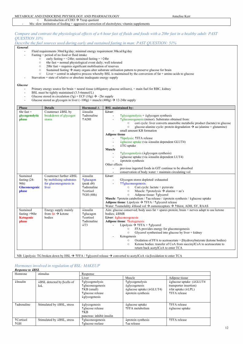
Phase Details Hormonal ∆ BSL maintained by:6hr fast = glycogenolytic phase
Counteract BSL by breakdown of glycogen stores
insulinadrenalineADH
Liver:- glycogenolysis + glycogen synthesis- gluconeogenesis (minor). Substrates obtained from:
o cori cycle: liver converts anaerobic metabolic product (lactate) to glucoseo glucose-alanine cycle: protein degradation aa (alanine + glutamine)
- small amount KB formation Adipose tissue
- lipolysis: FFA release- glucose uptake (via insulin dependent GLUT4)- TG uptake
Muscle- glycogenolysis (glycogen synthesis)- glucose uptake (via insulin dependent LUT4)- protein synthesis
Other effects- previous ingested foods in GIT continue to be absorbed- conservation of body water + maintain circulating vol
Sustained fasting (24-96h) Gluconeogenic phase
Counteract further BSL by mobilising substrates for gluconeogenesis in liver
insulinglucagon (peak d4)adrenalinecortisolGH (48h)
Liver: - Glycogen stores depleted/ exhausted- gluconeogenesis.
o Cori cycle: lactate + pyruvateo Muscle: protolysis alanine + aa’so Adipose tissue: glycerol
Muscle: protein catabolism / aa release / protein synthesis / glucose uptakeAdipose tissue: Lipolysis FFA / glycerol release Water: osmolality blood vol osmoreceptors thirst, ADH, SY, RAAS
Sustained fasting >96hrKetogenic phase
Energy supply mainly from fat ketone bodies
insulinglucagoncortisoladrenalineT3
Aim: glucose conserved; body uses fat + spares protein; brain + nerves adapt to use ketone bodies; BMRLiver: gluconeogenesis Adipose tissue: ketogenesis
- Lipolysis FFA + glycerol o FFA provides energy for gluconeogenesiso Glycerol synthesised into glucose by liver + kidney
- Ketogenesiso Oxidation of FFA to acetoacetate + hydroxybutyrate (ketone bodies)o Ketone bodies: transfer of CoA from succinylCoA to acetoacetate to
return back acetylCoA to enter TCA
NB: Lipolysis: TG broken down by HSL FFA / glycerol release converted to acetylCoA via oxidation to enter TCA
Hormones involved in regulation of BSL: MAKEUPResponse to BSLHomrone stimulus Response
Liver Muscle Adipose tissue Insulin BSL detected by cells of
IoLglycogenolysisgluconeogenesisKB (small)glucose releaseglycogenesis
glycogenolysisglycogenesisglucose uptake (GLUT4)protein synthesis
glucose uptake (GLUT4 transporter insertion)fat uptake (LPL)FFA release
adrenaline Stimulated by BSL, stress glycogenesisglucose releaseKBpancreas: inhibit insulin
glucose uptakeFFA metabolism
FFA releaseglucose uptake
Cortisol GH
Stimulated by BSL, stress gluconeogenesisglucose reelase
protein synthesisaa release
FFA release
METABOLIC AND ENDOCRINE PHYSIOLOGY AND PHARMACOLOGY Annelise Kerr
13
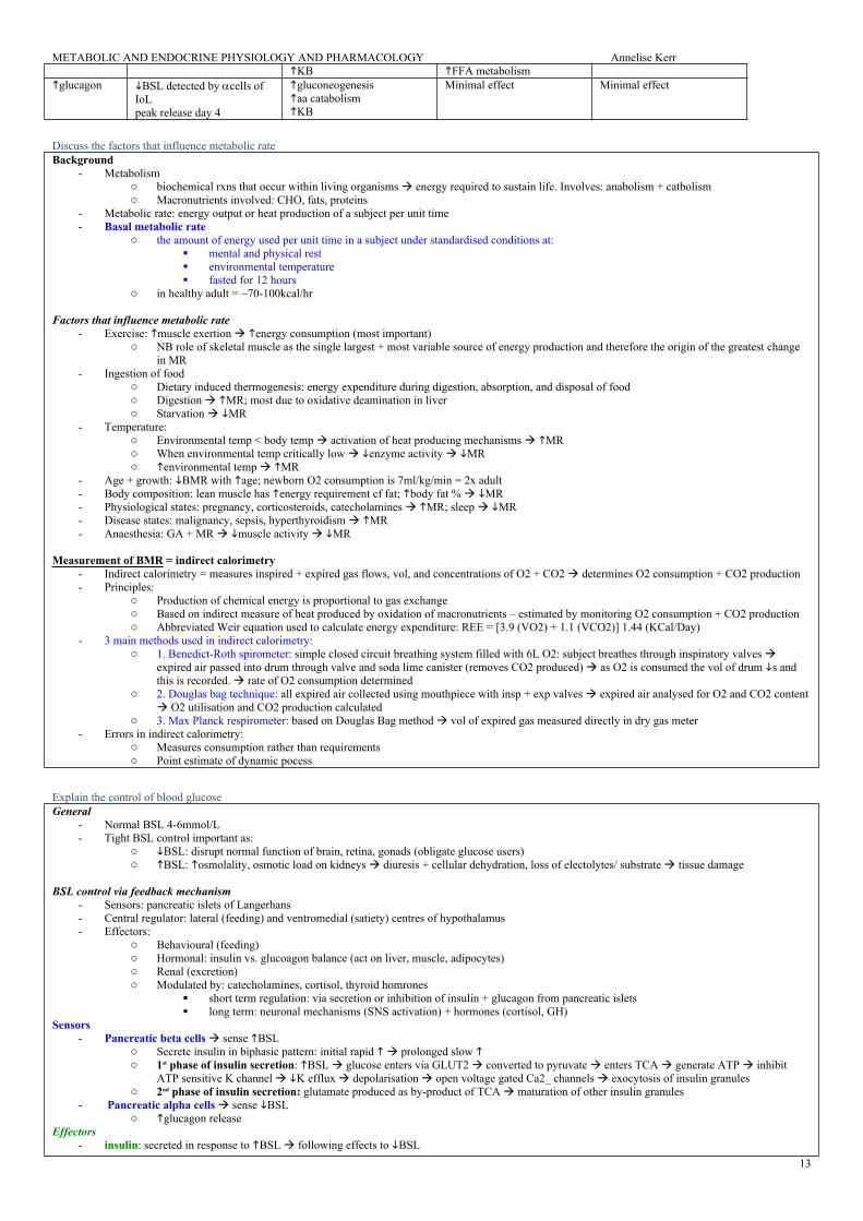
KB FFA metabolism glucagon BSL detected by cells of
IoLpeak release day 4
gluconeogenesisaa catabolism KB
Minimal effect Minimal effect
Discuss the factors that influence metabolic rate Background
- Metabolism o biochemical rxns that occur within living organisms energy required to sustain life. Involves: anabolism + catbolismo Macronutrients involved: CHO, fats, proteins
- Metabolic rate: energy output or heat production of a subject per unit time- Basal metabolic rate
o the amount of energy used per unit time in a subject under standardised conditions at: mental and physical rest environmental temperature fasted for 12 hours
o in healthy adult = ~70-100kcal/hr
Factors that influence metabolic rate- Exercise: muscle exertion energy consumption (most important)
o NB role of skeletal muscle as the single largest + most variable source of energy production and therefore the origin of the greatest change in MR
- Ingestion of foodo Dietary induced thermogenesis: energy expenditure during digestion, absorption, and disposal of food o Digestion MR; most due to oxidative deamination in liver o Starvation MR
- Temperature: o Environmental temp < body temp activation of heat producing mechanisms MRo When environmental temp critically low enzyme activity MRo environmental temp MR
- Age + growth: BMR with age; newborn O2 consumption is 7ml/kg/min = 2x adult- Body composition: lean muscle has energy requirement cf fat; body fat % MR- Physiological states: pregnancy, corticosteroids, catecholamines MR; sleep MR- Disease states: malignancy, sepsis, hyperthyroidism MR- Anaesthesia: GA + MR muscle activity MR
Measurement of BMR = indirect calorimetry- Indirect calorimetry = measures inspired + expired gas flows, vol, and concentrations of O2 + CO2 determines O2 consumption + CO2 production - Principles:
o Production of chemical energy is proportional to gas exchange o Based on indirect measure of heat produced by oxidation of macronutrients – estimated by monitoring O2 consumption + CO2 productiono Abbreviated Weir equation used to calculate energy expenditure: REE = [3.9 (VO2) + 1.1 (VCO2)] 1.44 (KCal/Day)
- 3 main methods used in indirect calorimetry:o 1. Benedict-Roth spirometer: simple closed circuit breathing system filled with 6L O2: subject breathes through inspiratory valves
expired air passed into drum through valve and soda lime canister (removes CO2 produced) as O2 is consumed the vol of drum s and this is recorded. rate of O2 consumption determined
o 2. Douglas bag technique: all expired air collected using mouthpiece with insp + exp valves expired air analysed for O2 and CO2 content O2 utilisation and CO2 production calculated
o 3. Max Planck respirometer: based on Douglas Bag method vol of expired gas measured directly in dry gas meter- Errors in indirect calorimetry:
o Measures consumption rather than requirementso Point estimate of dynamic pocess
Explain the control of blood glucose General
- Normal BSL 4-6mmol/L- Tight BSL control important as:
o BSL: disrupt normal function of brain, retina, gonads (obligate glucose users)o BSL: osmolality, osmotic load on kidneys diuresis + cellular dehydration, loss of electolytes/ substrate tissue damage
BSL control via feedback mechanism- Sensors: pancreatic islets of Langerhans- Central regulator: lateral (feeding) and ventromedial (satiety) centres of hypothalamus- Effectors:
o Behavioural (feeding)o Hormonal: insulin vs. glucoagon balance (act on liver, muscle, adipocytes)o Renal (excretion)o Modulated by: catecholamines, cortisol, thyroid homrones
short term regulation: via secretion or inhibition of insulin + glucagon from pancreatic islets long term: neuronal mechanisms (SNS activation) + hormones (cortisol, GH)
Sensors- Pancreatic beta cells sense BSL
o Secrete insulin in biphasic pattern: initial rapid prolonged slow o 1st phase of insulin secretion: BSL glucose enters via GLUT2 converted to pyruvate enters TCA generate ATP inhibit
ATP sensitive K channel K efflux depolarisation open voltage gated Ca2_ channels exocytosis of insulin granuleso 2nd phase of insulin secretion: glutamate produced as by-product of TCA maturation of other insulin granules
- Pancreatic alpha cells sense BSLo glucagon release
Effectors- insulin: secreted in response to BSL following effects to BSL
METABOLIC AND ENDOCRINE PHYSIOLOGY AND PHARMACOLOGY Annelise Kerr
14
o GLUT4 insertion into cell membrane glucose uptake into cells esp. muscle + fato glycogen synthesiso glucose utilisation + fat and protein synthesiso glycogenolysis / gluconeogenesis
- glucagon: secreted in reponse to BSL following effects to BSLo glycogenolysis / gluconeogenesiso minimal effect on adipose tissue and muscle
- Adrenaline: stimulated by BSL, stresso Inhibit insulino Liver: glycogenesis, glucose release, KBo Fat: FFA release, glucose uptakeo Muslce: glucose uptake, FFA metabolism
- Sustained BSL stimulates GH + cortisol release o glucose utilisation + fat utilisation limiting further BSLo protein synthesis / aa release / FFA metabolism
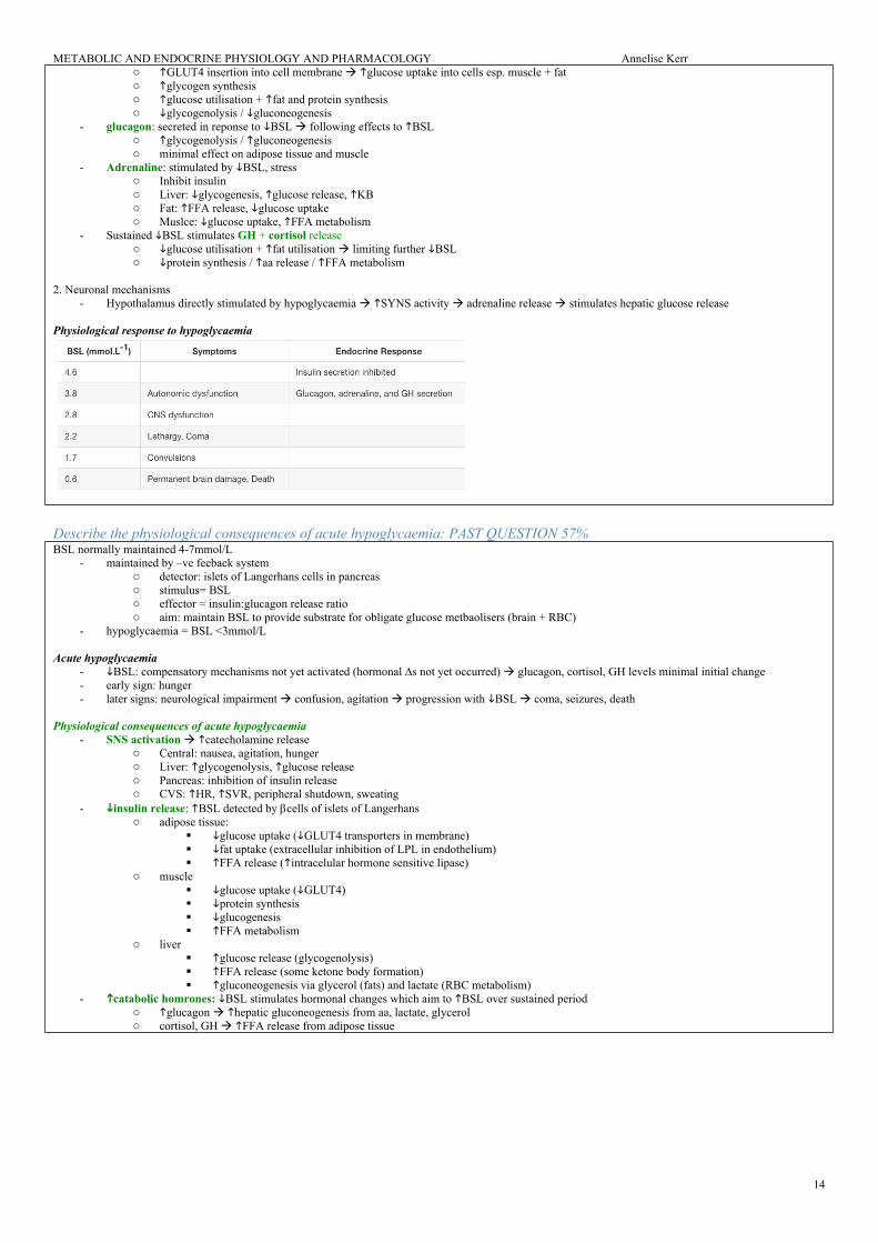
2. Neuronal mechanisms- Hypothalamus directly stimulated by hypoglycaemia SYNS activity adrenaline release stimulates hepatic glucose release
Physiological response to hypoglycaemia
Describe the physiological consequences of acute hypoglycaemia: PAST QUESTION 57%BSL normally maintained 4-7mmol/L
- maintained by –ve feeback systemo detector: islets of Langerhans cells in pancreaso stimulus= BSLo effector = insulin:glucagon release ratioo aim: maintain BSL to provide substrate for obligate glucose metbaolisers (brain + RBC)
- hypoglycaemia = BSL <3mmol/L
Acute hypoglycaemia- BSL: compensatory mechanisms not yet activated (hormonal ∆s not yet occurred) glucagon, cortisol, GH levels minimal initial change- early sign: hunger- later signs: neurological impairment confusion, agitation progression with BSL coma, seizures, death
Physiological consequences of acute hypoglycaemia- SNS activation catecholamine release
o Central: nausea, agitation, hungero Liver: glycogenolysis, glucose releaseo Pancreas: inhibition of insulin releaseo CVS: HR, SVR, peripheral shutdown, sweating
- insulin release: BSL detected by cells of islets of Langerhanso adipose tissue:
glucose uptake (GLUT4 transporters in membrane) fat uptake (extracellular inhibition of LPL in endothelium) FFA release (intracelular hormone sensitive lipase)
o muscle glucose uptake (GLUT4) protein synthesis glucogenesis FFA metabolism
o liver glucose release (glycogenolysis) FFA release (some ketone body formation) gluconeogenesis via glycerol (fats) and lactate (RBC metabolism)
- catabolic homrones: BSL stimulates hormonal changes which aim to BSL over sustained periodo glucagon hepatic gluconeogenesis from aa, lactate, glycerolo cortisol, GH FFA release from adipose tissue
METABOLIC AND ENDOCRINE PHYSIOLOGY AND PHARMACOLOGY Annelise Kerr
15
Describe the role of the hypothalamus in the integration of neuro-humoral responsesHypothalamus
- organ that regulates large number of autonomic/ endocrine processes- acts as control centre
Autonomic nervous system activity- CVS:
o Ant hypothalamic stimulation BP + HRo Post hypothalamic stimulation BP + HR
- Thermoregulatory: integrates thermoreceptor input + controls activity of heat loss + heat gain mechanisms- Satiety: hunger modulated by glucose, CCK, glucagon, and leptin- Water balance:
o Osmoreceptors: control ADH release from posterior pituitaryo ATII: stimulates thirst + ADH release via subfornical organ + organum vasculosum
- Circadian rhythm- Behaviour- Sexual function
Endocrine/ hormonal activity - Direct neural control of posterior pituitary gland - Pituitary neurosecreotry neurons
o Magnocellular neurons: consists of SON + PVN; synthesise and secrete ADH and oxytocino Parvocellular neurons: form tuberoinfundibular tract secrete hypophysiotropic hormones; release controlled by Nad, dopamine, 5-HT
- Hypothalamic hormones action in pituitary o Anterior pituitary by hormone secretion into long portal vein
GnRH stimulates FSH + LH release CRH stimulates ACTH release GHRH stimulates GH release TRH stimulates TSH release Somatostatin (GH inhibiting hormone) inhibits GH, TSH, ACTH, and PRL release PRL releasing hormone (PRH): stimulates PRL release Dopamine inhibits PRL release
o Posterior pituitary by neuronal innervation ACh stimulates release of ADH + oxytoxin NAd inhibits ADH + oxytocin secretion
Describe control of secretion and the functions of:
Pituitary hormonesPituitary
- HPA describes complex feedback loops between these endocrine organso Shortloop feedback: -ve feedback from pituitary on the hypothalamus e.g. thyroxin inhibiting TSH releaseo Long-loop feedback: -ve feedback from pituitary target gland (e.g. thyroid, adrenal, gonads) on the hypothalamus e.g. cortisol inhibiting
CRH (as well as ACTH) release- Pituitary hormones
o Anterior pituitary Secretes 6 hormones in response to hypothalamic endocrine stimulus Stimulating hormones:
Act at another gland Includes: ACTH, TSH, FSH, LH
Directly acting hormones Include: GH, prolactin
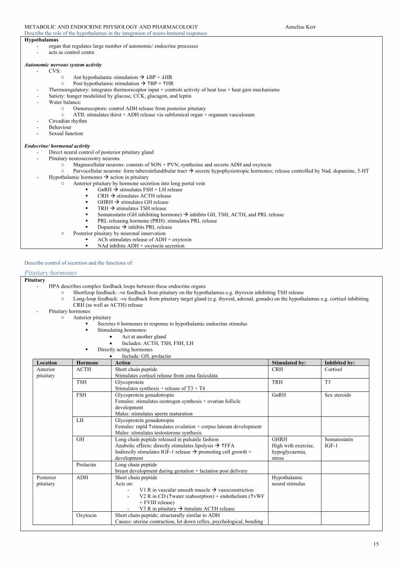
Location Hormone Action Stimulated by: Inhibited by:Anterior pituitary
ACTH Short chain peptide Stimulates cortisol release from zona fasiculata
CRH Cortisol
TSH GlycoproteinStimulates synthesis + release of T3 + T4
TRH T3
FSH Glycoprotein gonadotropinFemales: stimulates oestrogen synthesis + ovarian follicle developmentMales: stimulates sperm maturation
GnRH Sex steroids
LH Glycoprotein gonadotropinFemales: rapid stimulates ovulation + corpus luteum developmentMales: stimulates testosterone synthesis
GH Long chain peptide released in pulsatile fashionAnabolic effects: directly stimulates lipolysis FFAIndirectly stimulates IGF-1 release promoting cell growth + development
GHRHHigh with exercise, hypoglycaemia, stress
SomatostatinIGF-1
Prolactin Long chain peptide breast development during gestation + lactation post delivery
Posterior pituitary
ADH Short chain peptideActs on:
- V1 R in vascular smooth muscle vasoconstriction- V2 R in CD (water reabsorption) + endothelium (vWF
+ FVIII release)- V3 R in pituitary timulate ACTH release
Hypothalamic neural stimulus
Oxytocin Short chain peptide; structurally similar to ADHCauses: uterine contraction, let down reflex, psychological, bonding
METABOLIC AND ENDOCRINE PHYSIOLOGY AND PHARMACOLOGY Annelise Kerr
16
Thyroid hormonesDescribe the physiological actions of thyroid homrones: PAST QUESTIONGeneral
- 3 main thyroid hormoneso thyroxine (T4): 95%; ½ life 7 days; less activeo tri-iodothyronine (T3): 7%; ½ life 24hrs; 3-5x activity of T4o reverse T3 (rT3): inactive o T3 + T4 formed from iodination of aa tyrosineo iodine obtained from diet in form of iodide + actively taken up into thyroid follicular cells (req 120-150ug/day)
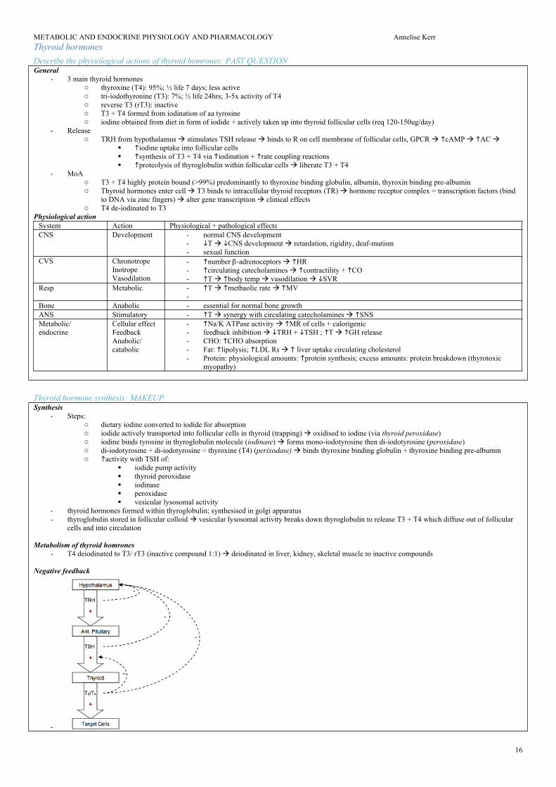
- Releaseo TRH from hypothalamus stimulates TSH release binds to R on cell membrane of follicular cells, GPCR cAMP AC
iodine uptake into follicular cells synthesis of T3 + T4 via iodination + rate coupling reactions proteolysis of thyroglobulin within follicular cells liberate T3 + T4
- MoAo T3 + T4 highly protein bound (>99%) predominantly to thyroxine binding globulin, albumin, thyroxin binding pre-albumino Thyroid hormones enter cell T3 binds to intracellular thyroid receptors (TR) hormone receptor complex = transcription factors (bind
to DNA via zinc fingers) alter gene transcription clinical effectso T4 de-iodinated to T3
Physiological actionSystem Action Physiological + pathological effectsCNS Development - normal CNS development
- T CNS development retardation, rigidity, deaf-mutism- sexual function
CVS ChronotropeInotropeVasodilation
- number -adrenoceptors HR- circulating catecholamines contractility + CO- T body temp vasodilation SVR
Resp Metabolic - T metbaolic rate MV-
Bone Anabolic - essential for normal bone growth ANS Stimulatory - T synergy with circulating catecholamines SNSMetabolic/ endocrine
Cellular effectFeedback Anabolic/ catabolic
- Na/K ATPase activity MR of cells + calorigenic - feedback inhibition TRH + TSH ; T GH release- CHO: CHO absorption- Fat: lipolysis; LDL Rs liver uptake circulating cholesterol - Protein: physiological amounts: protein synthesis; excess amounts: protein breakdown (thyrotoxic
myopathy)
Thyroid hormone synthesis: MAKEUPSynthesis
- Steps:o dietary iodine converted to iodide for absorptiono iodide actively transported into follicular cells in thyroid (trapping) oxidised to iodine (via thyroid peroxidase)o iodine binds tyrosine in thyroglobulin molecule (iodinase) forms mono-iodotyrosine then di-iodotyrosine (peroxidase) o di-iodotyrosine + di-iodotyrosine = thyroxine (T4) (perixodase) binds thyroxine binding globulin + thyroxine binding pre-albumino activity with TSH of:
iodide pump activity thyroid peroxidase iodinase peroxidase vesicular lysosomal activity
- thyroid hormones formed within thyroglobulin; synthesised in golgi apparatus- thyroglobulin stored in follicular colloid vesicular lysosomal activity breaks down thyroglobulin to release T3 + T4 which diffuse out of follicular
cells and into circulation
Metabolism of thyroid homrones- T4 deiodinated to T3/ rT3 (inactive compound 1:1) deiodinated in liver, kidney, skeletal muscle to inactive compounds
Negative feedback
-
METABOLIC AND ENDOCRINE PHYSIOLOGY AND PHARMACOLOGY Annelise Kerr
17
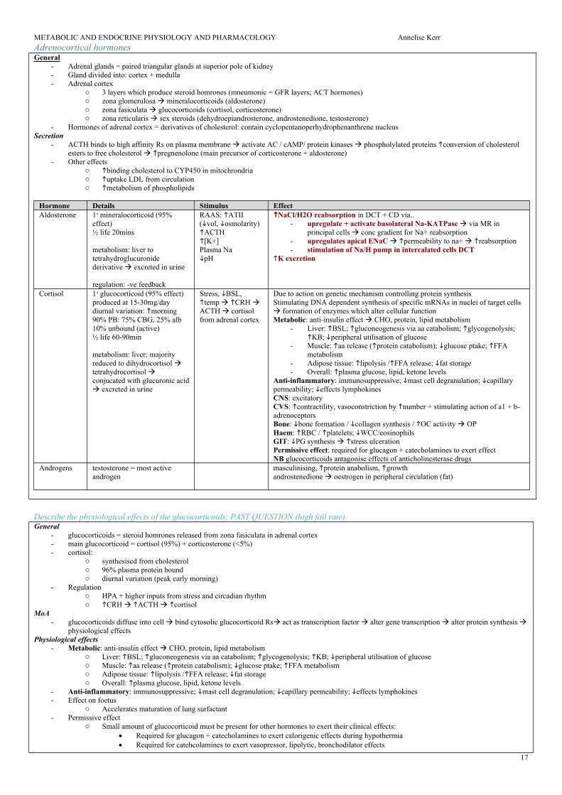
Adrenocortical hormonesGeneral
- Adrenal glands = paired triangular glands at superior pole of kidney- Gland divided into: cortex + medulla - Adrenal cortex
o 3 layers which produce steroid homrones (mneumonic = GFR layers; ACT hormones)o zona glomerulosa mineralocorticoids (aldosterone)o zona fasiculata glucocorticoids (cortisol, corticosterone)o zona reticularis sex steroids (dehydroepiandrosterone, androstenedione, testosterone)
- Hormones of adrenal cortex = derivatives of cholesterol: contain cyclopentanoperhydrophenanthrene nucleus Secretion
- ACTH binds to high affinity Rs on plasma membrane activate AC / cAMP/ protein kinases phospholylated proteins conversion of cholesterol esters to free cholesterol pregnenolone (main precursor of corticosterone + aldosterone)
- Other effectso binding cholesterol to CYP450 in mitochrondriao uptake LDL from circulationo metabolism of phospholipids
Hormone Details Stimulus EffectAldosterone 1o mineralocorticoid (95%
effect)½ life 20mins
metabolism: liver to tetrahydroglucuronide derivative excreted in urine
regulation: -ve feedback
RAAS: ATII (vol, osmolarity)ACTH[K+]Plasma NapH
NaCl/H2O reabsorption in DCT + CD via..- upregulate + activate basolateral Na-KATPase via MR in
principal cells conc gradient for Na+ reabsorption- upregulates apical ENaC permeability to na+ reabsorption - stimulation of Na/H pump in intercalated cells DCT
K excretion
Cortisol 1o glucocorticoid (95% effect)produced at 15-30mg/daydiurnal variation: morning90% PB: 75% CBG, 25% alb10% unbound (active)½ life 60-90min
metabolism: liver; majority reduced to dihydrocortisol tetrahydrocortisol conjucated with glucuronic acid excreted in urine
Stress, BSL, temp CRH ACTH cortisol from adrenal cortex
Due to action on genetic mechanism controlling protein synthesis Stimulating DNA dependent synthesis of specific mRNAs in nuclei of target cells formation of enzymes which alter cellular functionMetabolic: anti-insulin effect CHO, protein, lipid metabolism
- Liver: BSL; gluconeogenesis via aa catabolism; glycogenolysis; KB; peripheral utilisation of glucose
- Muscle: aa release (protein catabolism); glucose ptake; FFA metabolism
- Adipose tissue: lipolysis /FFA release; fat storage- Overall: plasma glucose, lipid, ketone levels
Anti-inflammatory: immunosuppressive; mast cell degranulation; capillary permeability; effects lymphokinesCNS: excitatory CVS: contractility, vasoconstriction by number + stimulating action of a1 + b-adrenoceptorsBone: bone formation / collagen synthesis / OC activity OPHaem: RBC / platelets; WCC/eosinophilsGIT: PG synthesis stress ulceration Permissive effect: required for glucagon + catecholamines to exert effectNB glucocorticoids antagonise effects of anticholinesterase drugs
Androgens testosterone = most active androgen
masculinising, protein anabolism, growthandrostenedione oestrogen in peripheral circulation (fat)
Describe the physiological effects of the glucocorticoids: PAST QUESTION (high fail rate)General
- glucocorticoids = steroid homrones released from zona fasiculata in adrenal cortex- main glucocorticoid = cortisol (95%) + corticosterone (<5%)- cortisol:
o synthesised from cholesterolo 96% plasma protein boundo diurnal variation (peak early morning)
- Regulationo HPA + higher inputs from stress and circadian rhythmo CRH ACTH cortisol
MoA- glucocorticoids diffuse into cell bind cytosolic glucocorticoid Rs act as transcription factor alter gene transcription alter protein synthesis
physiological effectsPhysiological effects
- Metabolic: anti-insulin effect CHO, protein, lipid metabolism o Liver: BSL; gluconeogenesis via aa catabolism; glycogenolysis; KB; peripheral utilisation of glucoseo Muscle: aa release (protein catabolism); glucose ptake; FFA metabolismo Adipose tissue: lipolysis /FFA release; fat storageo Overall: plasma glucose, lipid, ketone levels
- Anti-inflammatory: immunosuppressive; mast cell degranulation; capillary permeability; effects lymphokines- Effect on foetus
o Accelerates maturation of lung surfactant- Permissive effect
o Small amount of glucocorticoid must be present for other hormones to exert their clinical effects: Required for glucagon + catecholamines to exert calorigenic effects during hypothermia Required for catehcolamines to exert vasopressor, lipolytic, bronchodilator effects
METABOLIC AND ENDOCRINE PHYSIOLOGY AND PHARMACOLOGY Annelise Kerr
18
- Other systems o CNS: glucocorticoids irritability + poor concentrationo CVS: essential for normal CVS response to stress; response to catecholamines (permissive effect) +ve inotropeo GIT: PG synthesis + acid + pepsin production peptic ulcerso Renal: handling of body water: glucocorticoids unable to excrete free water loado Haem: RBC, platelet, neutrophil secretion; eosinophil, basophil, lymphocyte secretiono Endocrine: inhibits conversion of T4 to active T3; -ve feedback on HPA to inhibit release of CRH
Outline the physiological effects of bilateral adrenalectomy: PAST QUESTIONGeneral:
- Adrenal glando pair of triangular glands situated at superior pole of each kidneyo responsible for glucocorticoid, mineralocorticoid, and sex steroid synthesiso synthesise most adrenaline in body
- B/L adrenalectomy = acute form of “addisonian crisis”
Physiological consequences- absence of mineralocorticoids (aldosterone)
o Na reabsorption Na lost in urine water loss dehydration, hypovolaemia, shock, salt wasting crisiso K retained in plasma 2o K secretiono Mild acidosis due to lack of H secretiono Requires rapid resus + supplementation
- Absence of glucocorticoids (cortisol)o BSL 2o gluconeogenesis: impossible to maintain normal BSLo weaknesso mobilisation of protein + fats from tissueo highly susceptible to stress + unable to mount stress responseo effects of catecholamines
- adrenaline - ACTH
o ACTH secretion unopposed due to lack of –ve feedbacko Severe pigmentation (ACTH stimulates melanin formation)o Local effects: headache, visual disturbance
Adrenomedullary hormonesHormones produced in adrenal medulla = catecholamines:
- adrenaline + noradrenaline - dopamine
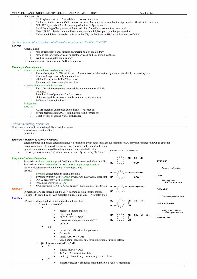
Structure + function of adrenal homrones - catecholamines all possess catechol nucleus = benzene ring with adjacent hydroxyl substitutions, O-dihydroxybenzene known as catechol- parent compound = -phenylethylamine: benzene ring + ethylamine side chain- optical isomerism conferred by substitution on either of ethyl C atoms- levorotary substitution at -C atoms produces naturally occurring NAd + Ad
Biosynthesis of catecholamines- Synthesis in adrenal medulla (modified SY ganglion composed of chromaffin cells)- Synthesis + release is dependent on ACh release by presynaptic neuron- NB catecholamine secretion is not a –ve feedback loop- Process
o Tyrosine concentrated in adrenal medullao Tyrosine hydroxylated to DOPA by tyrosine hydroxylase (rate limiting step)o DOPA decarboxylated to dopamineo Dopamine converted to NAdo NAd converted to Ad by PNMT (phenylethanolamine N-methyltansferase)
Stimulus:- In medulla: CAs are stored bound to ATP in granules with chromogranins- Release is triggered by an ACh mediated intracellular Ca2+ initiates exocytosis contents of granules released
Function- CAs act by direct binding to membrane bound receptors
o mobilisation of Ca2+ 1:
present in smooth muscle Gq coupled PLC IP3 Ca2+ vasoconstriction, relaxation of GIT
miscule 2:
present in CNS, arterioles, pancreas Gi coupled Inhibits AC cAMP vasodilation, sedation, analgesia, inhibition of insulin release
o 1 + 2 activation of AC + cAMP 1:
cardiac muscle + JGA cAMP intracellular Ca2+ inotropy, chronotromy, dromotropy, renin release
2: skeletal vascular + bronchial smooth muscle, liver, cell membrane
METABOLIC AND ENDOCRINE PHYSIOLOGY AND PHARMACOLOGY Annelise Kerr
19
vasodilation, bronchodilation, hepatic glycogenolysis, activity Na/K ATPase pump intracellular K
Renin and angiotensinDescribe the secretion and function of renin and angiotensin: PAST QUESTIONRAAS
- hormone system that regulates BP + water balance in bodyRenin
- acid protease; ½ life 80min- released by granular cells in JGA of kidney - rate limiting step in RAAS: splits angiotensinogen (produced in liver) to angiotensin I ATI ATII by pulmonary ACE- Release of renin controlled by:
o SNSo Intrarenal baroreceptorso Macula densa/ tubuloglomerular feedback o ATII
- Factors influencing rate of secretiono secretion
SNS activity: direct 1 effect pressure on intrarenal baroreceptors Na/K content at macula densa (tubuloglom feedback) PGI2, PGE2
o secretion ATII (-ve feedback) pressure intrarenal baroreceptors Na content at macula densa (tubuloglom feedback) vasopressin (ADH)
Angiotensin II- Glycoprotein; ½ life 1-2mins - Effector of RAAS- Actions via AT1R
o GFR mesangial cell constriction glom SA Kf GFR afferent > efferent constriction GFR
o Vasoconstriction peripheral : via AT1R, GPCR Gq: MAP constricts peritubular capillaries: capillary pressure fluid reabsorption
o Tubular absorption Direct effect: Na/H2O reabsorption CD Indirect effect: aldosterone K excretion from CD
o central effect Stimulate ADH release Thirst
o SNS stimulationo -ve feedback on renin production
Problems in anaesthetised pt taking ACEI/ ARBs- Hypotension: combination of long duration of action of ACEI/ ARB + anaesthetic agent BP + potential for CVS collapse- Angioedema with ACEI 2o bradykinin release- Renal failure esp. if NSAIDs- K risk arrhythmia
General- kidneys play role in regulation of body water + electrolytes- important component = JGA- JGA
o JG cells in wall of afferent arteriole Involved in pressure regulation through production of adenosine Pressure detection: afferent arteriolar baroreceptors Effector mechanism: production of renin via granular cells
- macula densao in walls of distal tubuleo primary role: tubuloglomerular feedback (autoregulation) through:
sensor for flow in DCT production of locally active vasoconstrictor
Atrial natriuretic peptideProduction Stimulus Site of action Effect
ANP RA Atrial stretch (CVP)
Efferent + afferent arterioleCD
- dilation afferent arteriole/ constriction efferent arteriole GFR H2O, solute filtered.
- Inhibit RAAS- ADH- Na absorption proportional to GFR, therefore reabsorption Na
glomerulotubular balance
METABOLIC AND ENDOCRINE PHYSIOLOGY AND PHARMACOLOGY Annelise Kerr
20
Describe the regulation of plasma calcium including the actions and control of vitamin D, parathormone and calcitoninFunctions of Ca2+
- Neuromuscular transmission + nerve function- Membrane excitation - Pacemaker potential - EC coupling + muscle contraction: binds to troponinC, displacing tropomyosin, and exposing binding site for myosin on actin- Release of hormones and NTs- Enzyme activation- Coagulation: activate FVII, VIII, V- Bone structure- Intracellular 2nd messenger
Storage- Total Ca2+ in body = 400mmol/kg: 99% bone / 1% ICF / 0.3% ECF- plasma:
o 40% protein bound (albumin)o 10% chelated to serum anions; o 50% free ionised
- ECF Ca2+: 2.45-2.55mmol/L / ionized Ca2+: 1-1.5mmol/LRegulation Absorption
- daily intake: 1000mg 10% absorption- GIT secretes up to 600mg/d reabsorbed
Short term regulation = renal reabsorption via PTH + calcitriol - large amount filtered by the kidneys; 98-99% reabsorbed - PT: 60% reabsorbed under control of PTH- Remainder reabsorbed in ascLoH and DT
Long term = osteoclast activity via PTH, calcitriol, calcitoninHormone Production Stimulus ActionsPTH In chief cells of parathyroid
Prehormone cleaved to prohormone hormone (ER +golgi)
[Ca2+] Ca2+ reabsorption DCT/CDPO4 reabsorption PCTactivation of vit D to calcitriolbone resorption/ bone formation Ca2+ release from bone
Vit D cholecalciferol (vit D3) produced in skin following exposure to UV lighthydroxylated in liver 25-hydroxycholecalciferol hydroxylated in prox nephron to 1,25-dihydroxycholecalciferol (calcitriol) (hydroxylase activity dependent on PTH)
[Ca2+] Ca2+ absorption in small intestinebone reabsorptionCa2+ + PO4 reabsorption from PCT
Calcitonin Secreted from parafollicular cells of thyroid [Ca2+] inhibit osteoblast activity (bone reabsorption)Ca/PO4 excretioncalcitriol synthesisjejunal absorption of dietary Ca2+
Clinical relevance- Hypercalcaemia:
o <3mmol/L: asymptomatic or non specific sx o 3-3.5mmol/L: polyuria, polydipsia, dehydration, anorexia, N+V, weaknesso CVS: short QT, HR, HTN
- Hypocalcaemiao Mild to seizures, heart failure, tetany
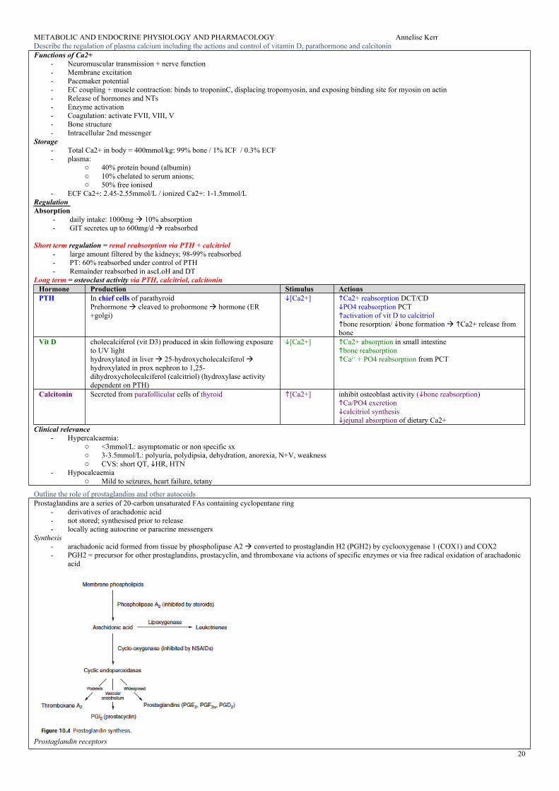
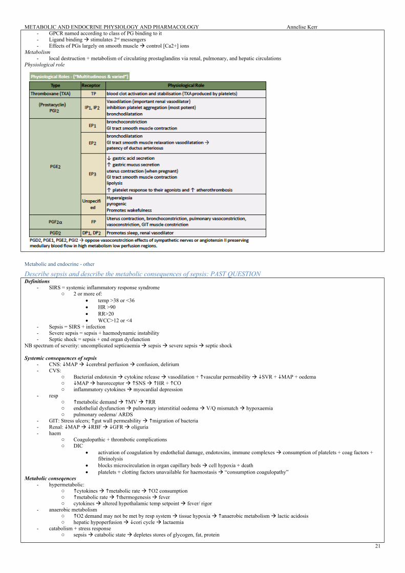
Outline the role of prostaglandins and other autocoidsProstaglandins are a series of 20-carbon unsaturated FAs containing cyclopentane ring
- derivatives of arachadonic acid- not stored; synthesised prior to release- locally acting autocrine or paracrine messengers
Synthesis- arachadonic acid formed from tissue by phospholipase A2 converted to prostaglandin H2 (PGH2) by cyclooxygenase 1 (COX1) and COX2- PGH2 = precursor for other prostaglandins, prostacyclin, and thromboxane via actions of specific enzymes or via free radical oxidation of arachadonic
acid
Prostaglandin receptors
METABOLIC AND ENDOCRINE PHYSIOLOGY AND PHARMACOLOGY Annelise Kerr
21
- GPCR named according to class of PG binding to it- Ligand binding stimulates 2nd messengers- Effects of PGs largely on smooth muscle control [Ca2+] ions
Metabolism- local destruction + metabolism of circulating prostaglandins via renal, pulmonary, and hepatic circulations
Physiological role
Metabolic and endocrine - other
Describe sepsis and describe the metabolic consequences of sepsis: PAST QUESTIONDefinitions
- SIRS = systemic inflammatory response syndromeo 2 or more of:
temp >38 or <36 HR >90 RR>20 WCC>12 or <4
- Sepsis = SIRS + infection- Severe sepsis = sepsis + haemodynamic instability- Septic shock = sepsis + end organ dysfunction
NB spectrum of severity: uncomplicated septicaemia sepsis severe sepsis septic shock
Systemic consequences of sepsis- CNS: MAP cerebral perfusion confusion, delirium- CVS:
o Bacterial endotoxin cytokine release vasodilation + vascular permeability SVR + MAP + oedemao MAP baroreceptor SNS HR + COo inflammatory cytokines myocardial depression
- respo metabolic demand MV RRo endothelial dysfunction pulmonary interstitial oedema V/Q mismatch hypoxaemiao pulmonary oedema/ ARDS
- GIT: Stress ulcers; gut wall permeability migration of bacteria- Renal: MAP RBF GFR oliguria- haem
o Coagulopathic + thrombotic complications o DIC
activation of coagulation by endothelial damage, endotoxins, immune complexes consumption of platelets + coag factors + fibrinolysis
blocks microcirculation in organ capillary beds cell hypoxia + death platelets + clotting factors unavailable for haemostasis “consumption coagulopathy”
Metabolic conseqences- hypermetabolic:
o cytokines metabolic rate O2 consumptiono metabolic rate thermogenesis fevero cytokines altered hypothalamic temp setpoint fever/ rigor
- anaerobic metabolismo O2 demand may not be met by resp system tissue hypoxia anaerobic metabolism lactic acidosiso hepatic hypoperfusion cori cycle lactaemia
- catabolism + stress response o sepsis catabolic state depletes stores of glycogen, fat, protein
METABOLIC AND ENDOCRINE PHYSIOLOGY AND PHARMACOLOGY Annelise Kerr
22
o glucocorticoid + catecholamines gluconeogenesis / glycogenolysis / peripheral insulin resistance / lipolysis / protein breakdown
- metabolic acidosiso compensation: buffered by HCO3, respiratory compensation, renal compensation
Outline the components of parenteral nutrition, explaining the rationale for the use of each component: PAST QUESTION 42%- TPN supples nutrients IV and is indicated when patients are unable to be fed via the GIT
Components- Water:
o Requirement: 30-35ml/kg/day- Energy in form of CHO + lipids
o Requirement: 125kg/kg/dayo Glucose (17kj/g): substrate required by brain + metabolised by all body tissues; prerequisite for protein metabolismo Lipids (37kj/g): in form of soybean emulsions; necessary for cell wall intefrity + PG synthesis
- Nitrogen in form of aao Requirement: 0.2g/kg/day nitrogen or 1.5g/kg/day amino acids for protein synthesis
- Electrolyteso Na+: 1-2mmol/kg/day; nerve condction + ECF tonicity o K+: 0.7-1mmol/kg/day: membrane potential + ICF tonicityo Ca2+: 0.1mmol/kg/day: bone metabolism, muscle contractiono PO4: 0.7mmol/kg/day: bone metabolism, tissue synthesis, phosphorylation of energy bondso Mg2+: 0.1mmol/kg/day: bone anabolism and enzyme systems
- Vitamins o Water soluble: C, B complex group + fat soluble (ADEK)o Catalyst or substrate in metabolic reactions
- Trace elementso Zinc: constituent of many enzymes e.g. carbonic anhydraseo Iron: Hb synthesiso Copper: RBC maturation + lipid metabolismo Iodine: thyroxine synthesiso Others: manganese, gluoride, chromium, selenium
Other considerations- extra H2O required to replace losses from vomiting, diarrhoea, sweating, fever- less water required in cardiac/ renal failure- energy intake supplied primarily as glucose > lipids in liver failure- energy intake supplied primarily as lipids > glucose in resp failure (CO2 production)
Explain how an oxygen debt arises and how the body deals with it: PAST QUESTION 1996General
- Active tissue utilises ATP for production of energy- 1st few seconds of vigorous exercise:
o ATP derived from cellular stores + creatine phosphate (limited stores) + depleted quicklt- 2nd stage
o body must switch to substrate metabolism to convert ADP to ATP for energyo oxidative phosphorylation within mitochrondria (uses CHO, glycogen, FFA)o anaerobic metabolism diversion of glycolytic pathway for production of lactate from CHO
O2 debt: - during initial stages of exercise, O2 uptake by exercising muscle < max that can be achieved during steady state exercise- O2 deficit = depletion of cellular ATP, myoglobin O2, and anaerobic metabolism- As exercise continues O2 offloading due to R shift OHDC venous pO2 maintains O2 requirements for exercise to continue- If exercise reaches aerobic threshold maximal O2 consumption body commences anaerobic metabolism lactate
How body deals with it:- at cessation of exercise: VO2 still this is the time that O2 stores are replenished
o myoglobin replenishedo creating phosphate/ ATP store replenished
Describe the changes that occur with ageing that can affect O2 delivery to the tissues during moderate exerciseGeneral
- O2 flux = amount of O2 delivered to the peripheral tissues per minuteo O2 flux = CO x arterial O2 content o In healthy young adult: tissue O2 dlivery = 1L O2/mino O2 flux/ or total body O2 delivery with age
- O2 flux equation = ([Hb] x SaO2 x 1.34) + (PaO2 x 0.0003)o 1.34 = Huffners constant: indicates amount of O2 which can combine with 1g of Hb when fully saturatedo 0.003 = proportionality constant; represents the amount fo O2 dissolved in blood (Henry’s law) i.e. 0.003ml O2 per mmHg pO2 per decilitre
of blood - During exercise, O2 delivery can be by:
o CO globally + locallyo O2 extraction from tissues
Effects of ageing- Cardiovascular
o CO max HR able to be achieved myocardial contractility SV afterload due to elasticity in large arteries preload due to VR and ventricular compliance
o cardiac work due to above +/- valve pathologyo atherosclerosis: blood flow + O2 supplyo overall: ability to CO to match exercising tissue demands
METABOLIC AND ENDOCRINE PHYSIOLOGY AND PHARMACOLOGY Annelise Kerr
23
- Respo PaO2: with age due to closing capacity; when CC>FRC airways closure during ventilation venous admixture PaO2o chest wall complianceo diffusion capacity 2o aveolar membrane thickness + functional surface areao WOBo anaemia O2 carrying capacity o overall: V/Q mismatch ability to oxygenate blood when tissue extraction s
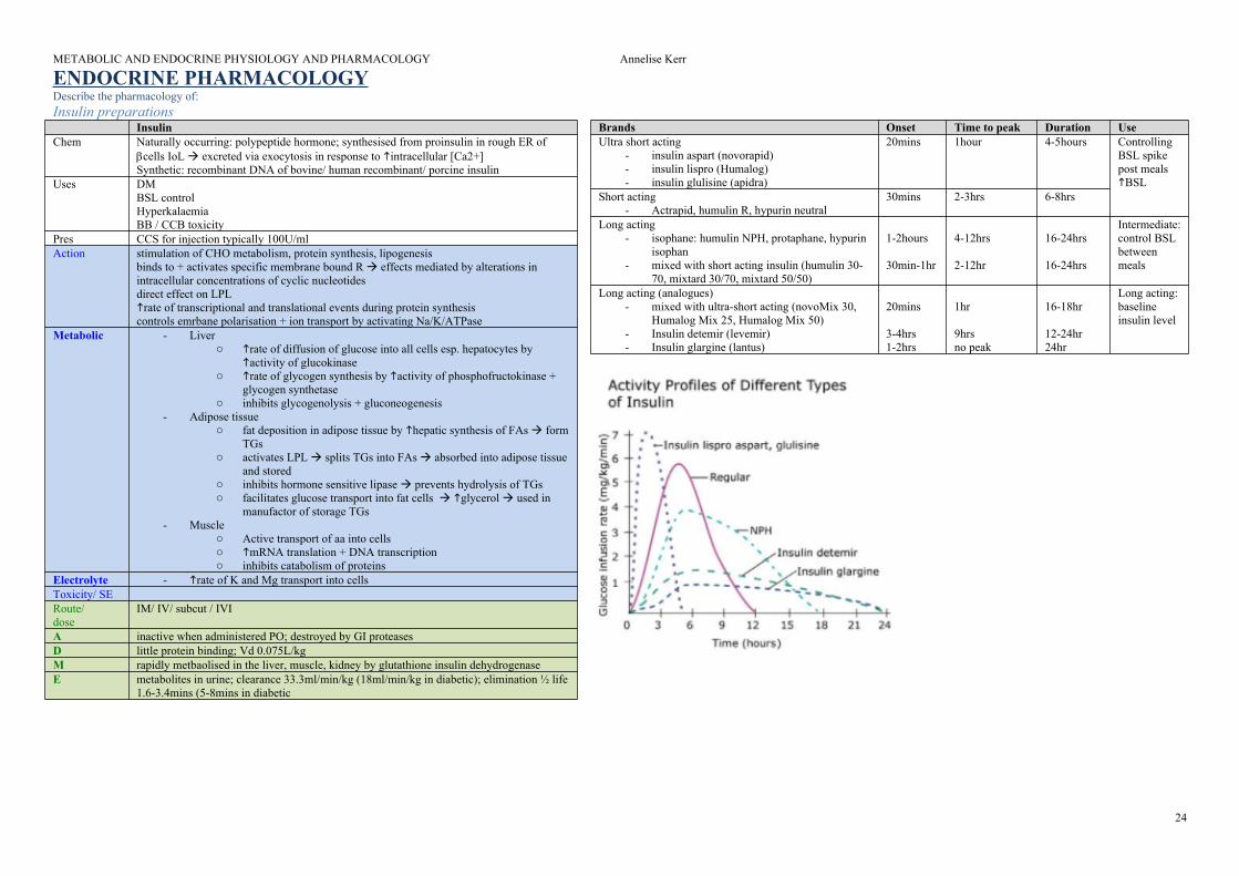
METABOLIC AND ENDOCRINE PHYSIOLOGY AND PHARMACOLOGY Annelise Kerr
24
ENDOCRINE PHARMACOLOGYDescribe the pharmacology of: Insulin preparations
Insulin Chem Naturally occurring: polypeptide hormone; synthesised from proinsulin in rough ER of
cells IoL excreted via exocytosis in response to intracellular [Ca2+]Synthetic: recombinant DNA of bovine/ human recombinant/ porcine insulin
Uses DMBSL controlHyperkalaemiaBB / CCB toxicity
Pres CCS for injection typically 100U/mlAction stimulation of CHO metabolism, protein synthesis, lipogenesis
binds to + activates specific membrane bound R effects mediated by alterations in intracellular concentrations of cyclic nucleotidesdirect effect on LPLrate of transcriptional and translational events during protein synthesiscontrols emrbane polarisation + ion transport by activating Na/K/ATPase
Metabolic - Livero rate of diffusion of glucose into all cells esp. hepatocytes by
activity of glucokinaseo rate of glycogen synthesis by activity of phosphofructokinase +
glycogen synthetaseo inhibits glycogenolysis + gluconeogenesis
- Adipose tissue o fat deposition in adipose tissue by hepatic synthesis of FAs form
TGso activates LPL splits TGs into FAs absorbed into adipose tissue
and stored o inhibits hormone sensitive lipase prevents hydrolysis of TGso facilitates glucose transport into fat cells glycerol used in
manufactor of storage TGs- Muscle
o Active transport of aa into cellso mRNA translation + DNA transcriptiono inhibits catabolism of proteins
Electrolyte - rate of K and Mg transport into cellsToxicity/ SERoute/ dose
IM/ IV/ subcut / IVI
A inactive when administered PO; destroyed by GI proteasesD little protein binding; Vd 0.075L/kgM rapidly metbaolised in the liver, muscle, kidney by glutathione insulin dehydrogenaseE metabolites in urine; clearance 33.3ml/min/kg (18ml/min/kg in diabetic); elimination ½ life
1.6-3.4mins (5-8mins in diabetic
Brands Onset Time to peak Duration UseUltra short acting
- insulin aspart (novorapid)- insulin lispro (Humalog)- insulin glulisine (apidra)
20mins 1hour 4-5hours Controlling BSL spike post mealsBSL
Short acting- Actrapid, humulin R, hypurin neutral
30mins 2-3hrs 6-8hrs
Long acting - isophane: humulin NPH, protaphane, hypurin
isophan- mixed with short acting insulin (humulin 30-
70, mixtard 30/70, mixtard 50/50)
1-2hours
30min-1hr
4-12hrs
2-12hr
16-24hrs
16-24hrs
Intermediate: control BSL between meals
Long acting (analogues)- mixed with ultra-short acting (novoMix 30,
Humalog Mix 25, Humalog Mix 50)- Insulin detemir (levemir)- Insulin glargine (lantus)
20mins
3-4hrs1-2hrs
1hr
9hrsno peak
16-18hr
12-24hr24hr
Long acting: baseline insulin level
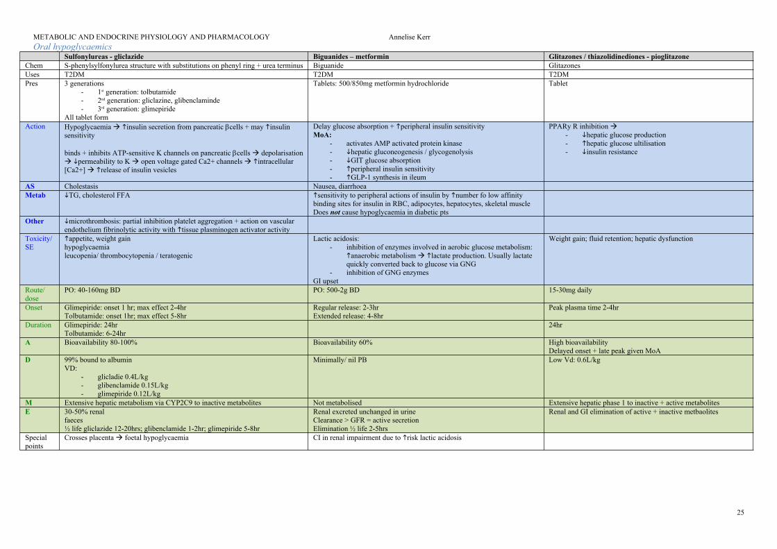
METABOLIC AND ENDOCRINE PHYSIOLOGY AND PHARMACOLOGY Annelise Kerr
25
Oral hypoglycaemicsSulfonylureas - gliclazide Biguanides – metformin Glitazones / thiazolidinediones - pioglitazone
Chem S-phenylsylfonylurea structure with substitutions on phenyl ring + urea terminus Biguanide Glitazones Uses T2DM T2DM T2DMPres 3 generations
- 1st generation: tolbutamide- 2nd generation: gliclazine, glibenclaminde- 3rd generation: glimepiride
All tablet form
Tablets: 500/850mg metformin hydrochloride Tablet
Action Hypoglycaemia insulin secretion from pancreatic cells + may insulin sensitivity
binds + inhibits ATP-sensitive K channels on pancreatic cells depolarisation permeability to K open voltage gated Ca2+ channels intracellular [Ca2+] release of insulin vesicles
Delay glucose absorption + peripheral insulin sensitivityMoA:
- activates AMP activated protein kinase - hepatic gluconeogenesis / glycogenolysis- GIT glucose absorption- peripheral insulin sensitivity- GLP-1 synthesis in ileum
PPARy R inhibition - hepatic glucose production- hepatic glucose ultilisation- insulin resistance
AS Cholestasis Nausea, diarrhoea Metab TG, cholesterol FFA sensitivity to peripheral actions of insulin by number fo low affinity
binding sites for insulin in RBC, adipocytes, hepatocytes, skeletal muscleDoes not cause hypoglycaemia in diabetic pts
Other microthrombosis: partial inhibition platelet aggregation + action on vascular endothelium fibrinolytic activity with tissue plasminogen activator activity
Toxicity/ SE
appetite, weight gainhypoglycaemia leucopenia/ thrombocytopenia / teratogenic
Lactic acidosis: - inhibition of enzymes involved in aerobic glucose metabolism:
anaerobic metabolism lactate production. Usually lactate quickly converted back to glucose via GNG
- inhibition of GNG enzymes GI upset
Weight gain; fluid retention; hepatic dysfunction
Route/ dose
PO: 40-160mg BD PO: 500-2g BD 15-30mg daily
Onset Glimepiride: onset 1 hr; max effect 2-4hrTolbutamide: onset 1hr; max effect 5-8hr
Regular release: 2-3hrExtended release: 4-8hr
Peak plasma time 2-4hr
Duration Glimepiride: 24hrTolbutamide: 6-24hr
24hr
A Bioavailability 80-100% Bioavailability 60% High bioavailabilityDelayed onset + late peak given MoA
D 99% bound to albuminVD:
- glicladie 0.4L/kg- glibenclamide 0.15L/kg- glimepiride 0.12L/kg
Minimally/ nil PB Low Vd: 0.6L/kg
M Extensive hepatic metabolism via CYP2C9 to inactive metabolites Not metabolised Extensive hepatic phase 1 to inactive + active metabolites E 30-50% renal
faeces½ life gliclazide 12-20hrs; glibenclamide 1-2hr; glimepiride 5-8hr
Renal excreted unchanged in urineClearance > GFR = active secretionElimination ½ life 2-5hrs
Renal and GI elimination of active + inactive metbaolites
Special points
Crosses placenta foetal hypoglycaemia CI in renal impairment due to risk lactic acidosis
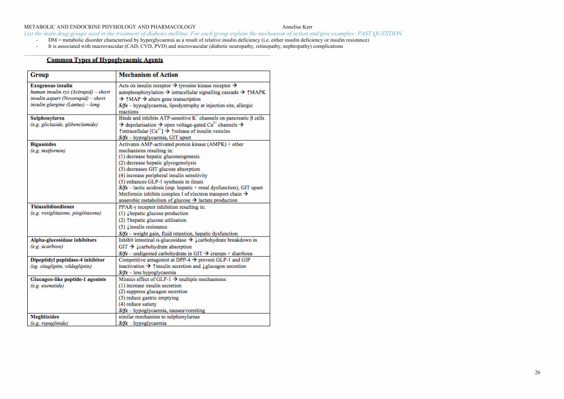
METABOLIC AND ENDOCRINE PHYSIOLOGY AND PHARMACOLOGY Annelise Kerr
26
List the main drug groups used in the treatment of diabetes mellitus. For each group explain the mechanism of action and give examples: PAST QUESTION- DM = metabolic disorder characterised by hyperglycaemia as a result of relative insulin deficiency (i.e. either insulin deficiency or insulin resistance)- It is associated with macrovascular (CAD, CVD, PVD) and microvascular (diabetic neuropathy, retinopathy, nephropathy) complications
METABOLIC AND ENDOCRINE PHYSIOLOGY AND PHARMACOLOGY Annelise Kerr
27
Corticosteroid drugs - Uses: adrenocortical deficiency / Allergy / anaphylaxis / Asthma / Autoimmune disorders / Eczema / chemo / immunosuppression post organ tx- MoA:
o Control rate of protein synthesis: react with cytoplasmic Rs form complex that directly influences rate of RNA transcription directs synthesis of lipcortinso Act on HPA axis at receptors on plasma membraneo Antiallergic, antitoxic, antishock, antipyretic, immunosuppressive properties
- Effectso Glucocorticoids
Metabolic: anti-insulin effect CHO, protein, lipid metabolism Liver: BSL; gluconeogenesis via aa catabolism; glycogenolysis; KB; peripheral utilisation of glucose Muscle: aa release (protein catabolism); glucose ptake; FFA metabolism Adipose tissue: lipolysis /FFA release; fat storage Overall: plasma glucose, lipid, ketone levels
Anti-inflammatory: immunosuppressive; mast cell degranulation; capillary permeability; effects lymphokines CNS: excitatory CVS: contractility, vasoconstriction by number + stimulating action of a1 + b-adrenoceptors Bone: bone formation / collagen synthesis / OC activity OP Haem: RBC / platelets; WCC/eosinophils GIT: PG synthesis stress ulceration Permissive effect: required for glucagon + catecholamines to exert effect NB glucocorticoids antagonise effects of anticholinesterase drugs
o mineralocorticoid: Na retention+++; K excretion; urinary Ca2+ excretin NB In large dose: inhibits endogenous adrenal cortical secretion, thymic activity, and ACTH excretion Aldosterone:
NaCl/H2O reabsorption in DCT + CD via..o upregulate + activate basolateral Na-KATPase via MR in principal cells conc gradient for Na+ reabsorptiono upregulates apical ENaC permeability to na+ reabsorption o stimulation of Na/H pump in intercalated cells DCT
K excretion Mineralocorticoid SE: Na+ + water retention / K / Oedema / muscle weakenss/ steroid myopathy / HTN / PUD, pancreatitis, oesophagitis
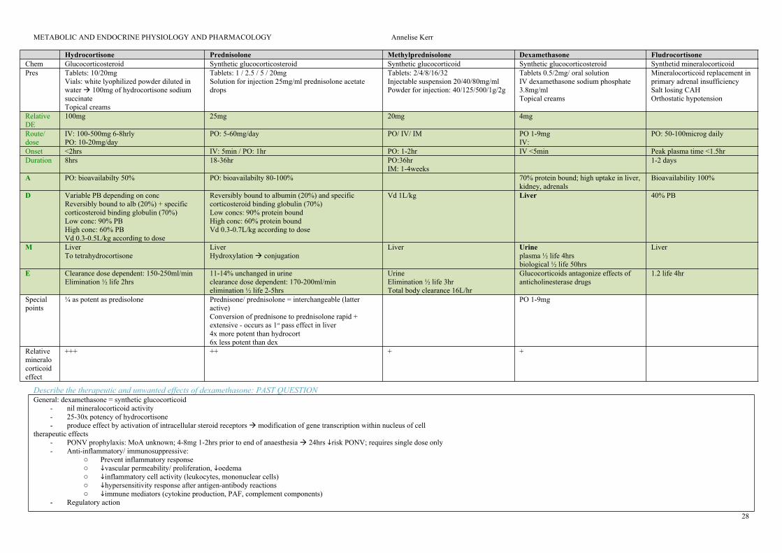
METABOLIC AND ENDOCRINE PHYSIOLOGY AND PHARMACOLOGY Annelise Kerr
28
Hydrocortisone Prednisolone Methylprednisolone Dexamethasone Fludrocortisone Chem Glucocorticosteroid Synthetic glucocorticosteroid Synthetic glucocorticoid Synthetic glucocorticosteroid Synthetid mineralocorticoidPres Tablets: 10/20mg
Vials: white lyophilized powder diluted in water 100mg of hydrocortisone sodium succinateTopical creams
Tablets: 1 / 2.5 / 5 / 20mgSolution for injection 25mg/ml prednisolone acetatedrops
Tablets: 2/4/8/16/32Injectable suspension 20/40/80mg/mlPowder for injection: 40/125/500/1g/2g
Tablets 0.5/2mg/ oral solutionIV dexamethasone sodium phosphate 3.8mg/mlTopical creams
Mineralocorticoid replacement in primary adrenal insufficiencySalt losing CAHOrthostatic hypotension
Relative DE
100mg 25mg 20mg 4mg
Route/ dose
IV: 100-500mg 6-8hrlyPO: 10-20mg/day
PO: 5-60mg/day PO/ IV/ IM PO 1-9mgIV:
PO: 50-100microg daily
Onset <2hrs IV: 5min / PO: 1hr PO: 1-2hr IV <5min Peak plasma time <1.5hrDuration 8hrs 18-36hr PO:36hr
IM: 1-4weeks1-2 days
A PO: bioavailabilty 50% PO: bioavailabilty 80-100% 70% protein bound; high uptake in liver, kidney, adrenals
Bioavailability 100%
D Variable PB depending on concReversibly bound to alb (20%) + specific corticosteroid binding globulin (70%)Low conc: 90% PBHigh conc: 60% PBVd 0.3-0.5L/kg according to dose
Reversibly bound to albumin (20%) and specific corticosteroid binding globulin (70%)Low concs: 90% protein boundHigh conc: 60% protein bound Vd 0.3-0.7L/kg according to dose
Vd 1L/kg Liver 40% PB
M LiverTo tetrahydrocortisone
LiverHydroxylation conjugation
Liver Urineplasma ½ life 4hrsbiological ½ life 50hrs
Liver
E Clearance dose dependent: 150-250ml/minElimination ½ life 2hrs
11-14% unchanged in urineclearance dose dependent: 170-200ml/minelimination ½ life 2-5hrs
Urine Elimination ½ life 3hrTotal body clearance 16L/hr
Glucocorticoids antagonize effects of anticholinesterase drugs
1.2 life 4hr
Special points
¼ as potent as predisolone Prednisone/ prednisolone = interchangeable (latter active)Conversion of prednisone to prednisolone rapid + extensive - occurs as 1st pass effect in liver4x more potent than hydrocort6x less potent than dex
PO 1-9mg
Relative mineralocorticoid effect
+++ ++ + +
Describe the therapeutic and unwanted effects of dexamethasone: PAST QUESTIONGeneral: dexamethasone = synthetic glucocorticoid
- nil mineralocorticoid activity- 25-30x potency of hydrocortisone- produce effect by activation of intracellular steroid receptors modification of gene transcription within nucleus of cell
therapeutic effects- PONV prophylaxis: MoA unknown; 4-8mg 1-2hrs prior to end of anaesthesia 24hrs risk PONV; requires single dose only- Anti-inflammatory/ immunosuppressive:
o Prevent inflammatory responseo vascular permeability/ proliferation, oedemao inflammatory cell activity (leukocytes, mononuclear cells)o hypersensitivity response after antigen-antibody reactionso immune mediators (cytokine production, PAF, complement components)
- Regulatory action
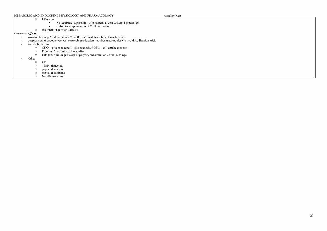
METABOLIC AND ENDOCRINE PHYSIOLOGY AND PHARMACOLOGY Annelise Kerr
29
o HPA axis -ve feedback suppression of endogenous corticosteroid production useful for suppression of ACTH production
o treatment in addisons diseaseUnwanted effects
- wound healing/ risk infection/ risk thrush/ breakdown bowel anastomoses- suppression of endogenous corticosteroid production: requires tapering dose to avoid Addisonian crisis- metabolic action
o CHO: gluconeogenesis, glycogenesis, BSL, cell uptake glucoseo Proteins: catabolism, anabolismo Fats (after prolonged use): lipolysis, redistribution of fat (cushings)
- Othero OPo IOP, glaucomao peptic ulcerationo mental disturbanceo Na/H2O retention
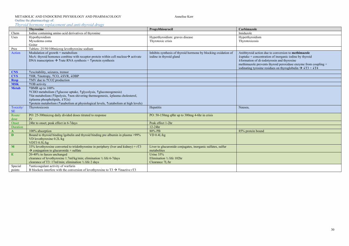
METABOLIC AND ENDOCRINE PHYSIOLOGY AND PHARMACOLOGY Annelise Kerr
30
Outline the pharmacology of:Thyroid hormone replacement and anti-thyroid drugs
Thyroxine Propylthiouracil Carbimazole Chem Iodine containing amino acid derivatives of thyronine Imidazole Uses Hypothyroidism
Myxedema comaGoiter
Hyperthyroidism: graves diseaseThyrotoxic crisis
HyperthyroidismThyrotoxicosis
Pres Tablets: 25/50/100microg levothyroxine sodiumAction Modulation of growth + metabolism
MoA: thyroid homones combine with receptor protein within cell nucleus activate DNA transcription rate RNA synthesis + protein synthesis
Inhibits synthesis of thyroid hormone by blocking oxidation of iodine in thyroid gland
Anithtyroid action due to conversion to methimazole uptake + concentration of inorganic iodine by thyroidformation of di-iodotyrosin and thyroxinemethimazole prevents thyroid peroxidase enzyme from coupling + iodinating tyrosine residues on thyroglobulin T3 + T4
CNS excitability, seizures, tremorCVS HR, inotropy, CO, SVR, DBPResp MV due to CO2 productionMSK OB activity Metab BMR up to 100%
CHO metabolism (glucose uptake, glycolysis, gluconeogenesis)fat metabolism (lipolysis, non shivering thermogenesis, plasma cholesterol, plasma phospholipids, TGs)protein metabolism (anabolism at physiological levels, catabolism at high levels)
Toxicity/ SE
Thyrotoxicosis Hepatitis Nausea,
Route/ dose
PO: 25-300microg daily divided doses titrated to responseIV
PO: 50-150mg q8hr up to 300mg 4-6hr in crisis
Onset 24hr to onset; peak effect in 6-7days Peak effect 1-2hrDuration 12-24hrA 100% absorption 80% PB 85% protein bound D Bound to thyroid binding lgobulin and thyroid binding pre albumin in plasma >99%
VD levothyroxine 0.2L/kgVDT3 0.5L/kg
VD 0.4L/kg
M 33% levothyroxine converted to triidothyronine in periphery (lver and kidney) + rT3 conjugation to glucuronide + sulfate
Liver to glucuronide conjugates, inorganic sulfates, sulfur metabolites
E 20-40% in faeces unchangedclearance of levothyroxine 1.7ml/kg/min; elimination ½ life 6-7daysclearance of T3: 17ml/min; elimination ½ life 2 days
Urine 35%Elimination ½ life 102hrClearance 7L/hr
Special points
anticoagulant activity of warfarinB blockers interfere with the conversion of levothyroxine to T3 inactive rT3
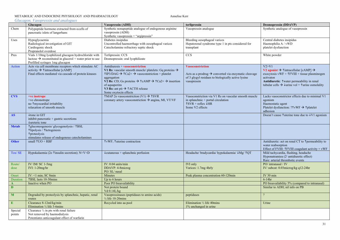
METABOLIC AND ENDOCRINE PHYSIOLOGY AND PHARMACOLOGY Annelise Kerr
31
Glucagon, Vasopressin and analoguesGlucagon Vasopressin (ADH) terlipressin Desmopressin (DDAVP)
Chem Polypeptide hormone extracted from cells of pancreatic islets of langerhans
Synthetic nonapeptide analogue of endogenous arginine vasopressin (ADH)Synthetic vasopressin = “argipressin”
Vasopressin analogue Synthetic analogue of vasopressin
Uses HypoglycaemiaRadiological investigation of GITCardiogenic shockPropranolol overdose
Diabetes insipidusUncontrolled haemorrhage with oesophageal varices Catecholamine refractory septic shock
Bleeding oesophageal varicesHepatorenal syndrome type 1 in pts considered for transplant
Central diabetes insipidusHaemophilia A / vWDplatelet dysfunction
Pres Vials 1/10mg lyophilized glucagon hydrochloride with lactose reconstituted in glucerol + water prior to usePrefilled syringes 1mg glucagon
Terlipressin: CCSDesmopressin: oral lyophilizate
CCS White powder
Action Acts via cell membrane receptors which stimulate AC activity intracellular [cAMP]Final effects mediated via cascade of protein kinases
Antidiuresis + vasoconstrictionV1 Rs: vascular smooth muscle/ platelets: Gq proteins IP3/DAG Ca2+ vasoconstriction + platelet aggregationV2 Rs: CD; Gs proteins cAMP Ca2+ insertion of aquaporinsV3 Rs: ant pit ACTH releaseSome oxytocin effects
Vasoconstriction
Acts as a prodrug converted via enzymatic cleavage of 3 glygyl residues to biologically active lysine vasopressin
V2>V1V2 agonist intracellular [cAMP] exocytosis vWF + FVIII + tissue plasminogen activatonAntidiuretic: water permeability in renal tubular cells urine vol + urine osmolality
CVS +ve inotrope+ve chronotropeno myocardial irritabilityrelaxation of smooth muscle
MAP 2o vasoconstriction (V1) SVRcoronary artery vasoconstriction angina, MI, VT/VF
Vasoconstriction via V1 Rs on vascular smooth muscle in splanchnic + portal circulationSVR + reflex HRSome V2 effects
Lacks vasoconstrictor effects due to minimal V1 agonism Haemostatic agentPlatelet dysfunction: ?vWF platelet adhesion
AS tone in GITinhibit pancreatic + gastric secretionsureteric tone
Doesn’t cause uterine tone due to V1 agonism
Metab gluconeogenesis/ glycogenolysis / BSLlipolysis / ketogenesisproteolysisstimulates release of endogenous catecholamines
Other small UO + RBF vWF; uterine contraction Antidiuretic: act on renal CT to permeability to water reabsorptionEffect of FVIII: FVIII coagulant activity + vWF
Tox/ SE Hypokalaemia (2o insulin secretion); N+V+D cutaneous + splanchnic perfusion Headache/ bradycardia/ hypokalaemia/ Mg/ QT Mild tachycardia, flushing, headache Hyponatramiea (2o antidiuretic effect)Rare: arterial thrombotic events
Route/ dose
IV/ IM/ SC 1-5mgIVI: 1-20mg/hr
IV: 0.04 units/minDDAVP: 4-8microgPO/ SL/ nasal
IVI onlyVarices: 1.7mg 4hrly
PO/ intranasal / IVIV/ subcut: 0.03microg/kg q12-24hr
Onset IV: <1 min; SC 8min Minutes Peak plasma concentration 60-120min IV 30 minDuration BSL lasts 10-30mins Up to 6 hours 6-14hr A Inactive when PO Poor PO bioavailability PO bioavailability 5% (compared to intranasal)D Not protein bound
Vd 0.14L/kgSimilar to ADH; nil info on PB
M Degraded by proteolysis by splanchnic, hepatic, renal routes
Vasopressinases (peptidases to amino acids)½ life 10-20mins
peptidases ?
E Clearance 8-12ml/kg/minElimination ½ life 3-6mins
Recycled into aa pool Elimination ½ life 40mins1% unchanged in urine
Urine
Special points
Clearance ½ in pts with renal failureNot removed by haemodialysisPotentiates anticoagulant effect of warfarin
METABOLIC AND ENDOCRINE PHYSIOLOGY AND PHARMACOLOGY Annelise Kerr
32