Copyright © 2010 Pearson Education, Inc.
THE HUMAN BODY: AN ORIENTATION CH. 1A
Copyright © 2010 Pearson Education, Inc.
Overview of Anatomy and Physiology
• Anatomy:
• Physiology:
Copyright © 2010 Pearson Education, Inc.
Principle of Complementarity
• Anatomy and physiology are inseparable.

Copyright © 2010 Pearson Education, Inc.
Levels of Structural Organization
• Chemical:
• Cellular:
• Tissue:
• Organ:
• Organ system:
• Organismal:

Copyright © 2010 Pearson Education, Inc.
Cardiovascularsystem
OrganelleMoleculeAtoms
Chemical level Cellular level
Tissue levelTissues consist of similartypes of cells.
Organ levelOrgans are made up of different typesof tissues.
Organ system levelOrgan systems consist of differentorgans that work together closely.
Organismal levelThe human organism is made upof many organ systems.
Smooth muscle cell
Smooth muscle tissue
Connective tissue
Blood vessel (organ)
HeartBloodvessels
Epithelialtissue
Smooth muscle tissue
12
3
4
56
Figure 1.1
Copyright © 2010 Pearson Education, Inc. Figure 1.3a
NailsSkin
Hair
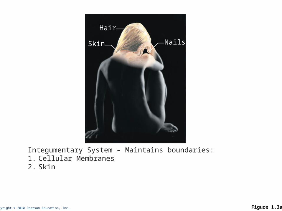
Integumentary System – Maintains boundaries:1. Cellular Membranes2. Skin
Copyright © 2010 Pearson Education, Inc. Figure 1.3b
Bones
Joint
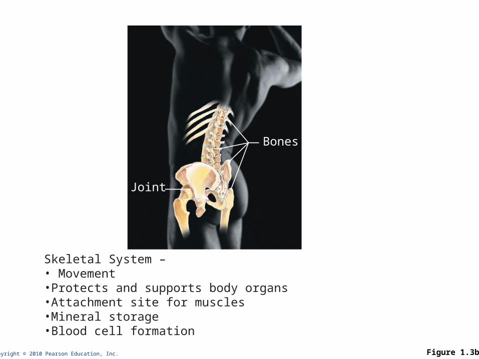
Skeletal System –• Movement •Protects and supports body organs•Attachment site for muscles•Mineral storage•Blood cell formation
Copyright © 2010 Pearson Education, Inc. Figure 1.3c
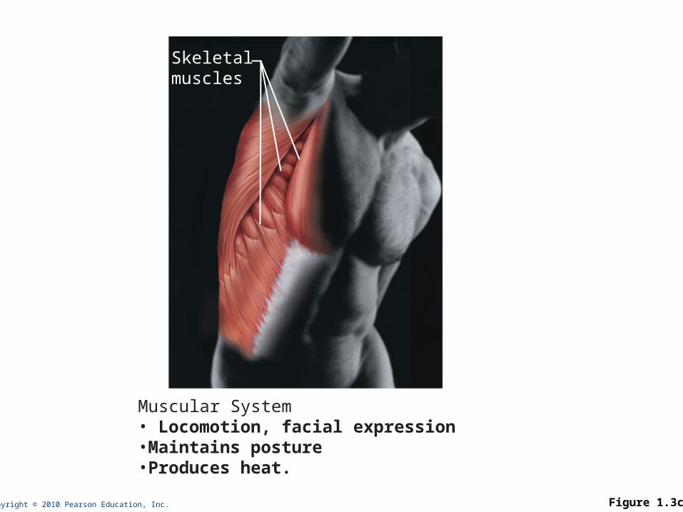
Skeletalmuscles
Muscular System• Locomotion, facial expression•Maintains posture•Produces heat.
Copyright © 2010 Pearson Education, Inc. Figure 1.3d
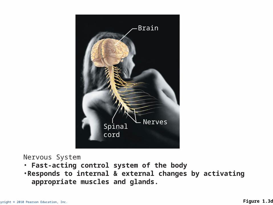
Brain
NervesSpinalcord
Nervous System• Fast-acting control system of the body•Responds to internal & external changes by activating appropriate muscles and glands.
Copyright © 2010 Pearson Education, Inc. Figure 1.3e
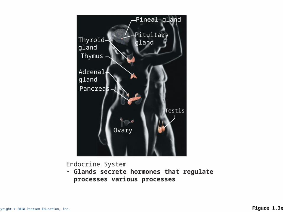
Pineal gland
PituitaryglandThyroid
glandThymus
AdrenalglandPancreas
Testis
Ovary
Endocrine System• Glands secrete hormones that regulate processes various processes

Copyright © 2010 Pearson Education, Inc. Figure 1.3f
Cardiovascular System• Blood vessels transport blood, which carries oxygen, carbon dioxide, nutrients, wastes, etc. The heart pumps blood.
Heart
Bloodvessels
Copyright © 2010 Pearson Education, Inc. Figure 1.3g
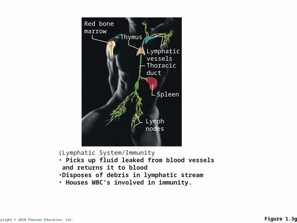
Lymphaticvessels
Red bonemarrow
Thoracicduct
Thymus
Spleen
Lymphnodes
(Lymphatic System/Immunity• Picks up fluid leaked from blood vessels and returns it to blood•Disposes of debris in lymphatic stream• Houses WBC’s involved in immunity.

Copyright © 2010 Pearson Education, Inc. Figure 1.3h
Nasalcavity
Bronchus
Pharynx
Larynx
Trachea
Lung
Respiratory System• Keeps blood constantly supplied with oxygen and removes carbon dioxide.
Copyright © 2010 Pearson Education, Inc. Figure 1.3i
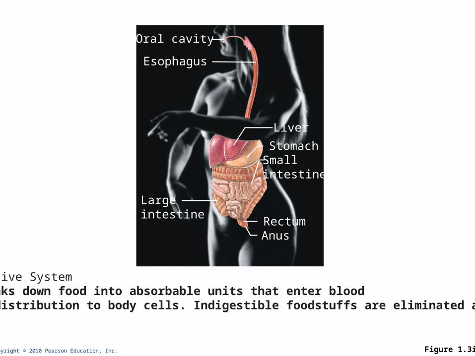
Liver
Oral cavity
Esophagus
Largeintestine
StomachSmallintestine
RectumAnus
Digestive System• Breaks down food into absorbable units that enter blood for distribution to body cells. Indigestible foodstuffs are eliminated as feces.
Copyright © 2010 Pearson Education, Inc. Figure 1.3j
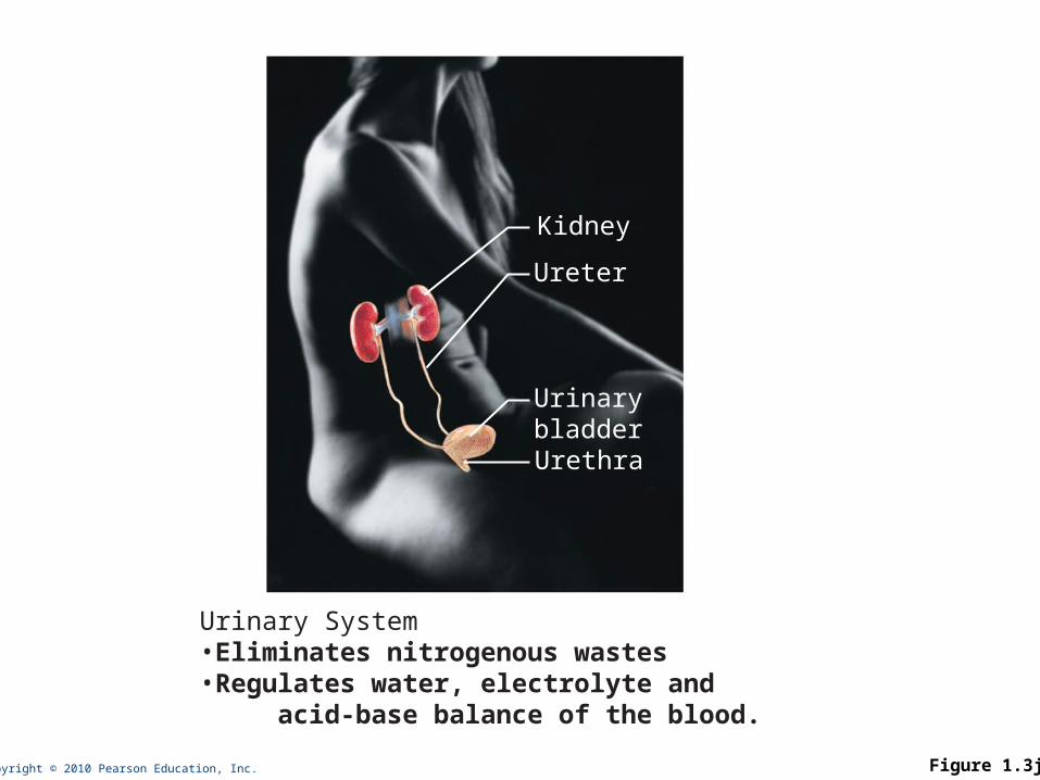
Kidney
Ureter
UrinarybladderUrethra
Urinary System•Eliminates nitrogenous wastes •Regulates water, electrolyte and acid-base balance of the blood.
Copyright © 2010 Pearson Education, Inc. Figure 1.3k-l
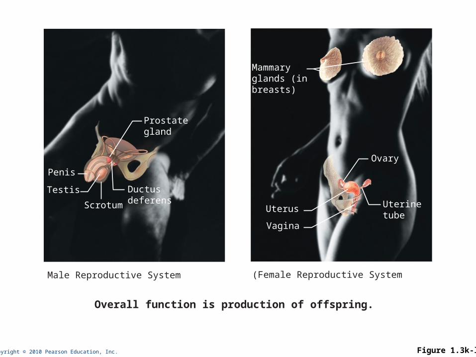
Prostategland
Ductusdeferens
Penis
Testis
Scrotum
Ovary
Uterinetube
Mammaryglands (inbreasts)
Uterus
Vagina
Overall function is production of offspring.
Male Reproductive System (Female Reproductive System
Copyright © 2010 Pearson Education, Inc. Figure 1.3
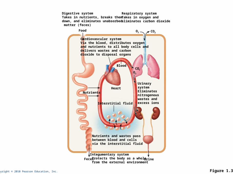
Digestive system Takes in nutrients, breaks them down, and eliminates unabsorbed matter (feces)
Respiratory systemTakes in oxygen and eliminates carbon dioxide
Food O2 CO2
Cardiovascular systemVia the blood, distributes oxygen and nutrients to all body cells and delivers wastes and carbon dioxide to disposal organs
Interstitial fluid
Nutrients
Urinary systemEliminates nitrogenouswastes andexcess ions
Nutrients and wastes pass between blood and cells via the interstitial fluid
Integumentary system Protects the body as a whole from the external environment
Blood
Heart
Feces Urine
CO2
O2
Copyright © 2010 Pearson Education, Inc.
Survival Needs
1. Nutrients
2. Oxygen
3. Water
4. Normal body temperature
5. Appropriate atmospheric pressure
Copyright © 2010 Pearson Education, Inc.
Homeostasis
• Definition:
• A dynamic state of equilibrium
Copyright © 2010 Pearson Education, Inc.
Components of a Homeostatic Control Mechanism
1. Receptor (sensor)
2. Control center
3. Effector
Copyright © 2010 Pearson Education, Inc.
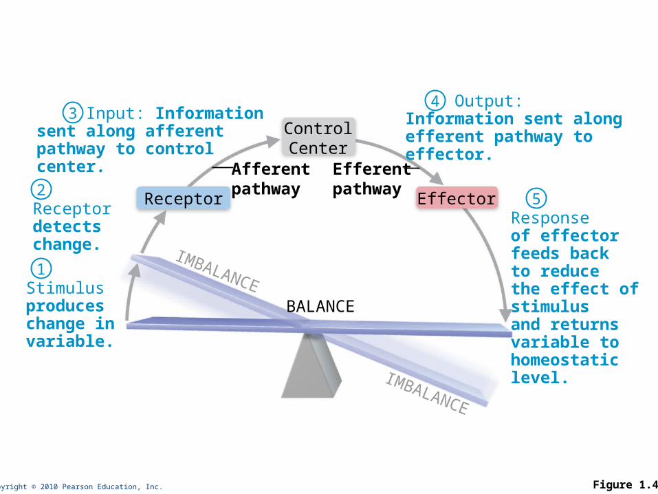
Stimulusproduceschange invariable.
Receptordetectschange.
Input: Informationsent along afferentpathway to controlcenter.
Output:Information sent alongefferent pathway toeffector.
Responseof effectorfeeds backto reducethe effect ofstimulusand returnsvariable tohomeostaticlevel.
Receptor Effector
ControlCenter
BALANCE
Afferentpathway
Efferentpathway
IMBALANCE
IMBALANCE
1
2
34
5
Figure 1.4
Copyright © 2010 Pearson Education, Inc.
Negative Feedback
• The response reduces or shuts off the original stimulus
• Example:
• Regulation of body temperature

Copyright © 2010 Pearson Education, Inc. Figure 1.5
Sweat glands activated
Shiveringbegins
StimulusBody temperaturerises BALANCE
Information sentalong the afferentpathway to controlcenter
Information sentalong the afferentpathway to controlcenter
Afferentpathway
Afferentpathway
Efferentpathway
Efferentpathway
Information sentalong the efferentpathway toeffectors
Information sentalong the efferentpathway to effectors
StimulusBody temperature falls
ReceptorsTemperature-sensitivecells in skin and brain
ReceptorsTemperature-sensitivecells in skin and brain
EffectorsSweat glands
EffectorsSkeletal muscles
Control Center(thermoregulatory
center in brain)
Control Center(thermoregulatory
center in brain)
ResponseEvaporation of sweatBody temperature falls;stimulus ends
ResponseBody temperature rises;stimulus ends
Copyright © 2010 Pearson Education, Inc.
Positive Feedback
• The response enhances or exaggerates the original stimulus
• Rare in biological systems
• Example:
• Enhancement of labor contractions by oxytocin
Copyright © 2010 Pearson Education, Inc.
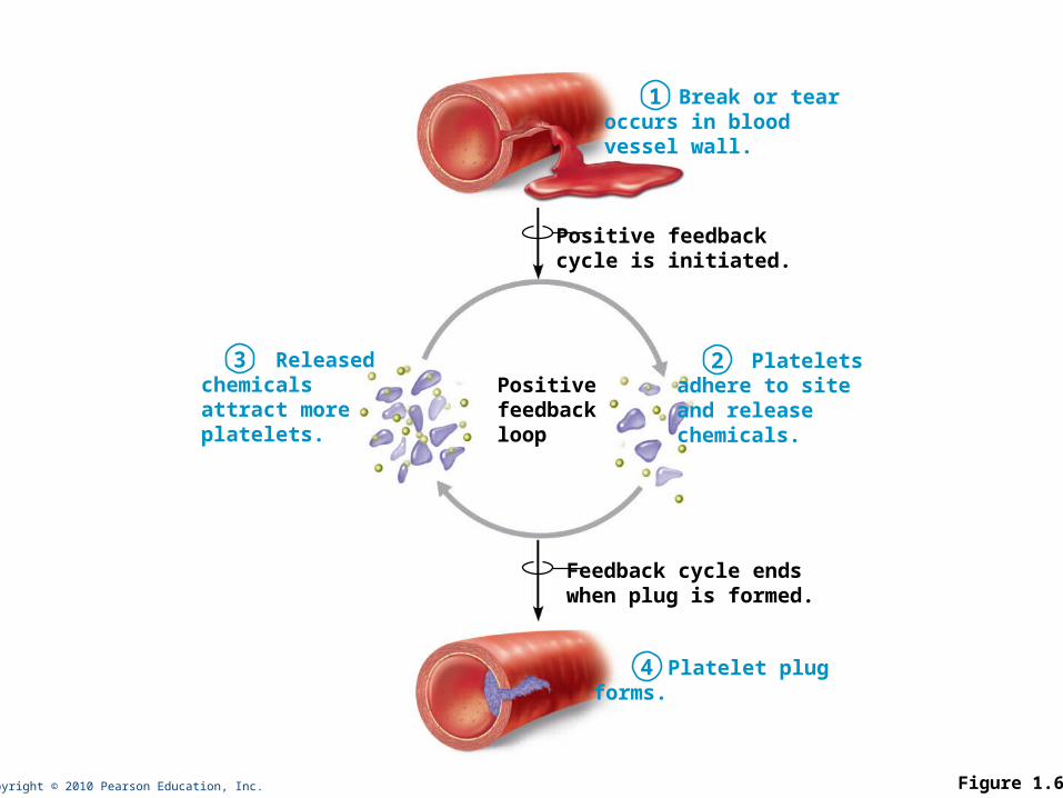
Feedback cycle endswhen plug is formed.
Positive feedbackcycle is initiated.
Positivefeedbackloop
Break or tearoccurs in bloodvessel wall.
Plateletsadhere to siteand releasechemicals.
Releasedchemicalsattract moreplatelets.
Platelet plugforms.
1
23
4
Figure 1.6

Copyright © 2010 Pearson Education, Inc.
CHAPTER 1B
Copyright © 2010 Pearson Education, Inc.
Anatomical Position
• Standard anatomical body position:
• Body erect
• Feet slightly apart
• Palms facing forward
Copyright © 2010 Pearson Education, Inc.
Regional Terms
• Regional Terms: designate specific areas/regions of the body.

Copyright © 2010 Pearson Education, Inc. Figure 1.5
Cervical
(a) Anterior/Ventral
Pubic
OrbitalNasalOral
ThoracicAxillary
SternalAbdominalUmbilicalPelvicInguinal
Upper limbAcromialBrachial (arm)AntecubitalAntebrachial (forearm)Carpal (wrist)
Digital
Lower limbCoxal (hip)Femoral (thigh)PatellarCrural (leg)Fibular
Tarsal (ankle)ThoraxAbdomenBack (Dorsum)
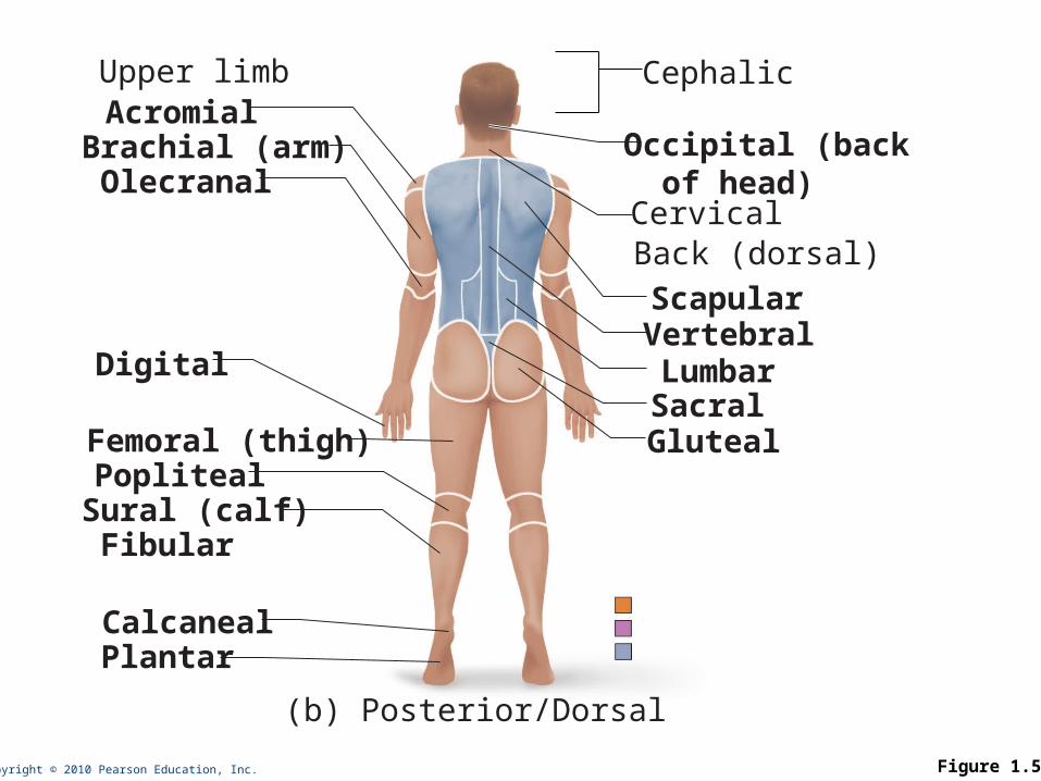
Copyright © 2010 Pearson Education, Inc. Figure 1.5
Cervical Back (dorsal)
(b) Posterior/Dorsal
Scapular Vertebral Lumbar Sacral Gluteal
Upper limb AcromialBrachial (arm) Olecranal
Digital
Femoral (thigh) Popliteal Sural (calf) Fibular
Calcaneal Plantar
Cephalic
Occipital (back of head)
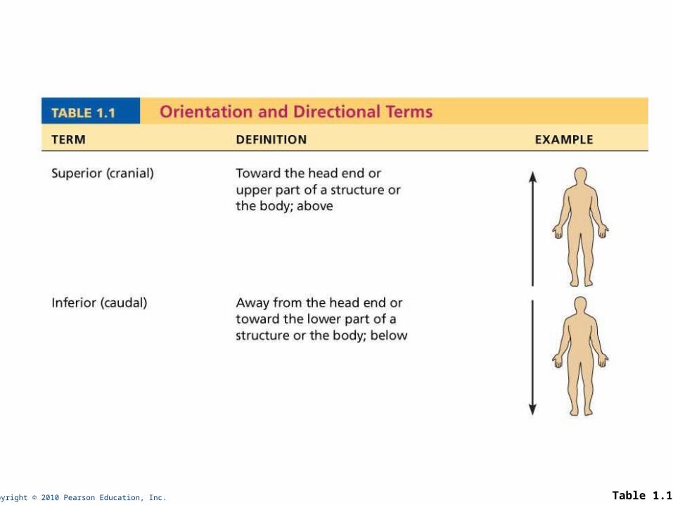
Copyright © 2010 Pearson Education, Inc. Table 1.1

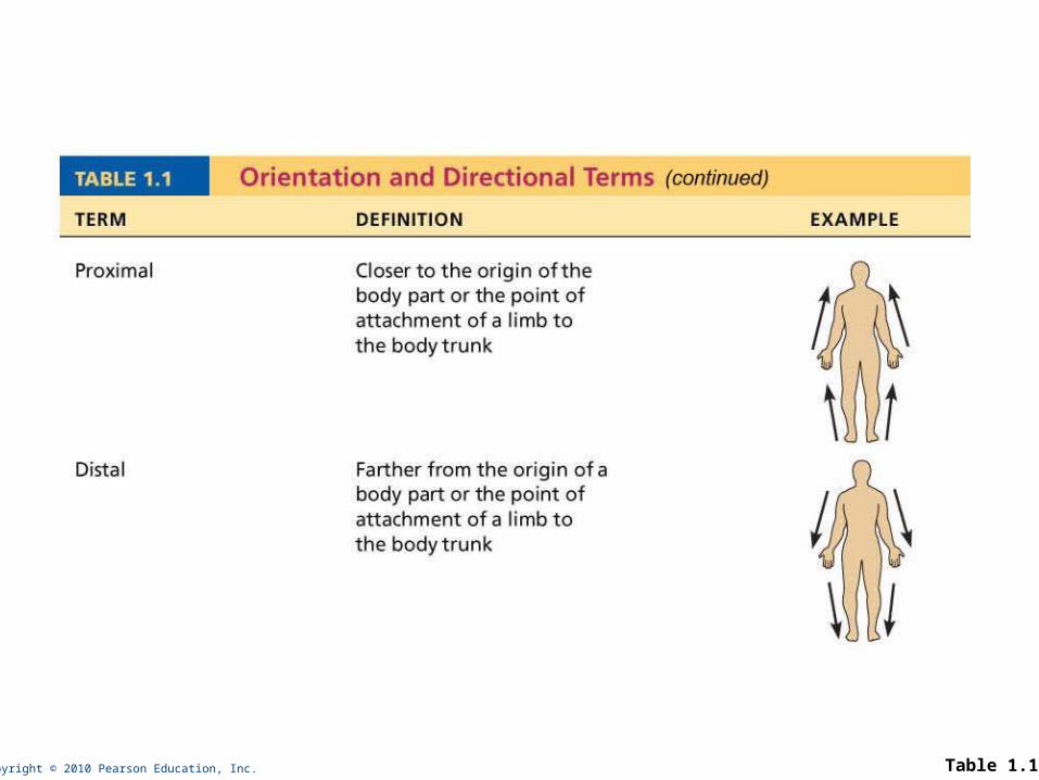
Copyright © 2010 Pearson Education, Inc. Table 1.1

Copyright © 2010 Pearson Education, Inc. Table 1.1
Copyright © 2010 Pearson Education, Inc. Table 1.1
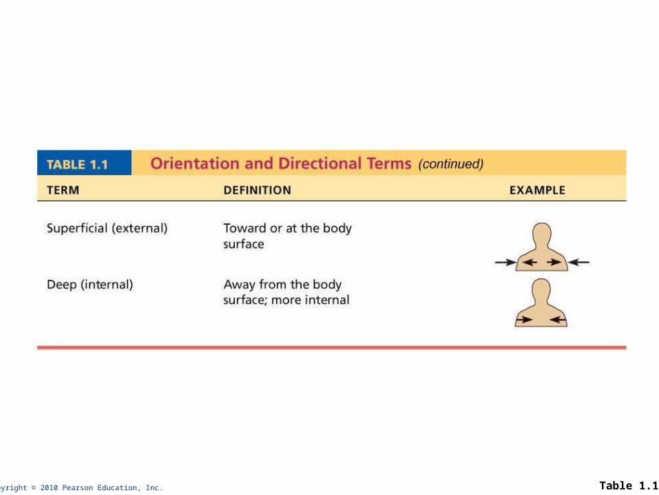
Copyright © 2010 Pearson Education, Inc. Table 1.1
Copyright © 2010 Pearson Education, Inc.
Body Planes and Sections
• Sagittal plane
• Midsagittal (median) plane
• Parasagittal plane
• Frontal (coronal) plane
• Transverse (horizontal) plane
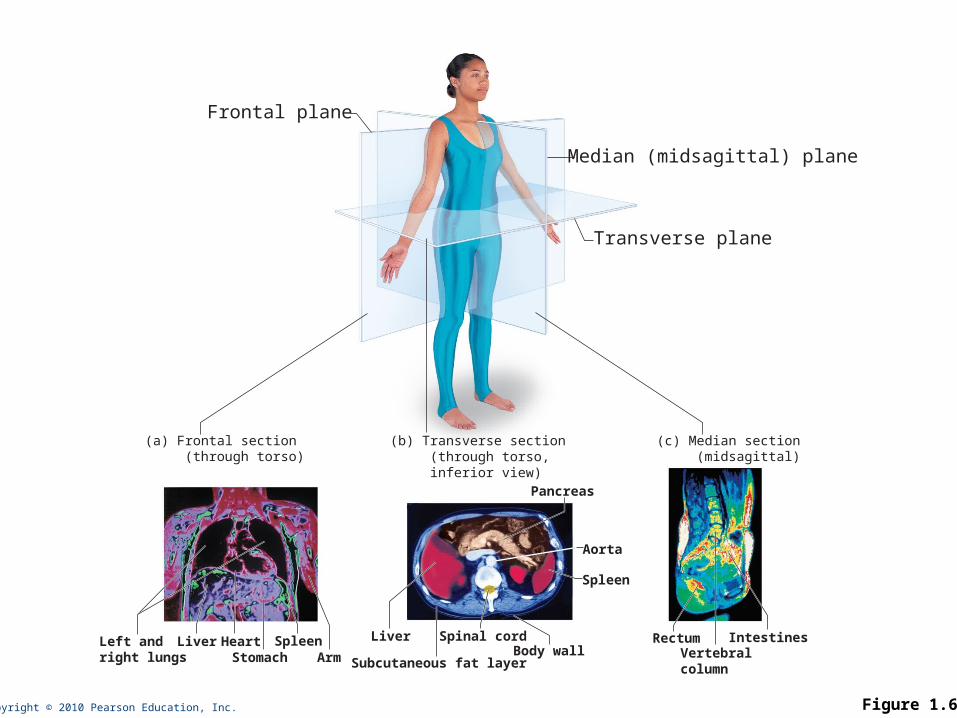
Copyright © 2010 Pearson Education, Inc. Figure 1.6
Transverse plane
Median (midsagittal) plane
Frontal plane
Liver
Spleen
Pancreas
Aorta
Vertebralcolumn
Spinal cord
Subcutaneous fat layerBody wall
Rectum IntestinesLeft andright lungs
Liver HeartStomach
SpleenArm
(a) Frontal section (through torso)
(b) Transverse section (through torso, inferior view)
(c) Median section (midsagittal)
Copyright © 2010 Pearson Education, Inc.
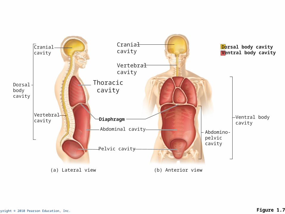
Body Cavities
• Two Large Cavities:
• Dorsal cavity
• Two subdivisions:
• Cranial cavity
• Vertebral cavity
Copyright © 2010 Pearson Education, Inc.
Body Cavities
• Ventral cavity
• Two subdivisions (separated by diaphragm):
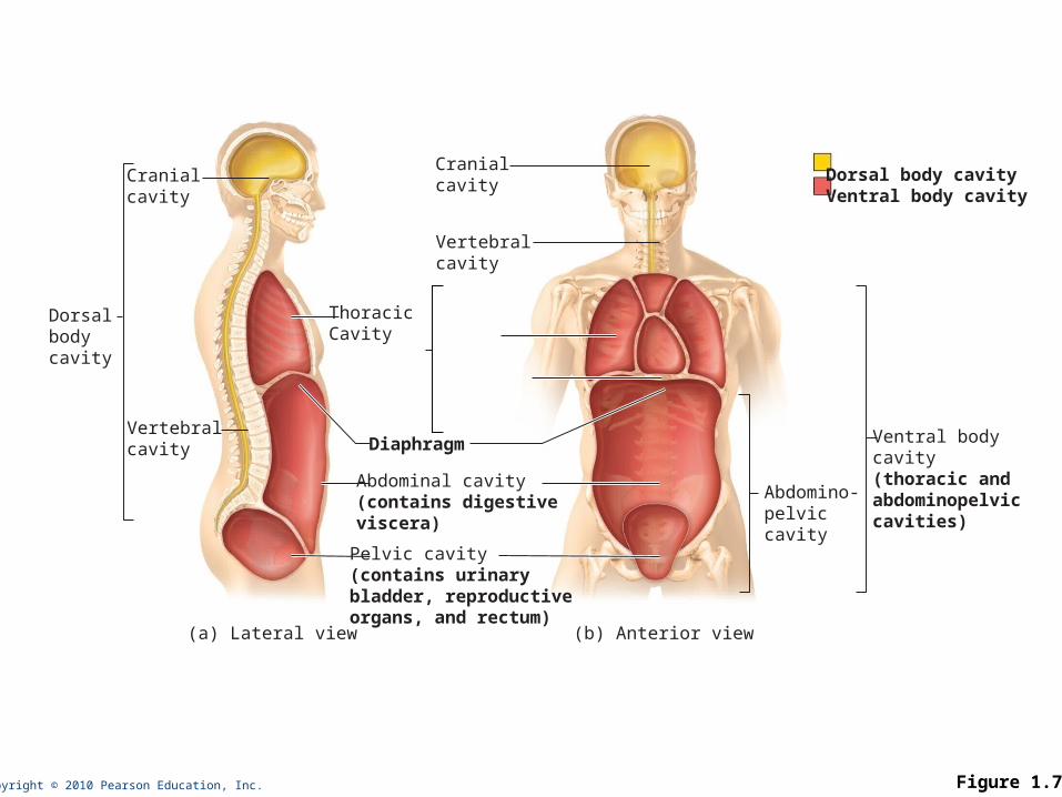
Copyright © 2010 Pearson Education, Inc. Figure 1.7
Cranialcavity
Dorsalbodycavity
Vertebralcavity
Cranialcavity
Vertebralcavity
Abdomino-pelviccavity
Ventral bodycavity(thoracic andabdominopelviccavities)
Abdominal cavity(contains digestiveviscera)
Diaphragm
Pelvic cavity(contains urinary bladder, reproductive organs, and rectum)
ThoracicCavity
(a) Lateral view (b) Anterior view
Dorsal body cavityVentral body cavity
Copyright © 2010 Pearson Education, Inc.
Ventral Body Cavities
• Thoracic cavity subdivisions:
• Two pleural cavities
• Mediastinum
• Pericardial cavity
Copyright © 2010 Pearson Education, Inc.
Ventral Body Cavities
• Abdominopelvic cavity subdivisions:
• Abdominal cavity
• Pelvic cavity
Copyright © 2010 Pearson Education, Inc. Figure 1.7
Cranialcavity
Dorsalbodycavity
Vertebralcavity
Cranialcavity
Vertebralcavity
Abdomino-pelviccavity
Ventral bodycavity
Abdominal cavity
Diaphragm
Pelvic cavity
Thoraciccavity
(a) Lateral view (b) Anterior view
Dorsal body cavityVentral body cavity
Copyright © 2010 Pearson Education, Inc.
Abdominopelvic Regions
• Nine divisions used primarily by anatomists
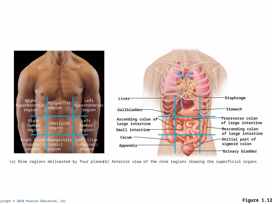
Copyright © 2010 Pearson Education, Inc. Figure 1.12
Epigastricregion
Umbilicalregion
Rightlumbarregion
Leftlumbarregion
Righthypochondriac
region
Lefthypochondriac
region
Hypogastric(pubic)region
Right iliac(inguinal)
region
Left iliac(inguinal)
region
Liver
Gallbladder
Ascending colon oflarge intestine
Small intestine
Appendix
Cecum
Diaphragm
Stomach
Descending colonof large intestine
Transverse colonof large intestine
Initial part ofsigmoid colon
Urinary bladder
(a) Nine regions delineated by four planes (b) Anterior view of the nine regions showing the superficial organs
Copyright © 2010 Pearson Education, Inc.
Abdominopelvic Quadrants
• Divisions used primarily by medical personnel
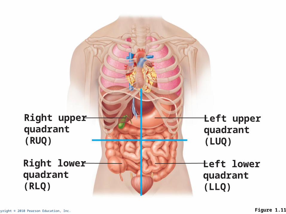
Copyright © 2010 Pearson Education, Inc. Figure 1.11
Right upperquadrant(RUQ)
Right lowerquadrant(RLQ)
Left upperquadrant(LUQ)
Left lowerquadrant(LLQ)
Recommended