Solution Precursor Plasma Spray Eu: Y2O3 PhosphorCoating
Dianying Chen*,† and Maurice Gell
Department of Chemical, Materials and Biomolecular Engineering, Institute of Materials Science,University of Connecticut, Storrs, Connecticut, 06269
Eric H. Jordan and Michael W. Renfro
Department of Mechanical Engineering, University of Connecticut, Storrs, Connecticut, 06269
Using the solution precursor plasma spray process Eu: Y2O3 phosphor coating was deposited. The phase composition,microstructure, and photoluminescent properties of the as-synthesized powders and as-deposited coatings were investigated.XRD analysis indicated that the coating is composed of cubic Y2O3. SEM micrographs reveal the as-sprayed coating is porous
with a thickness of ~150 lm. Photoluminescent property measurement indicated that the phosphor coating exhibits thestrongest emission at 612 nm, which is assigned to 5D0 ? 7F2 electric-dipole transition.
Introduction
The Eu3+-doped yttrium oxide (Y2O3) phosphorpowder, film and coating have gained wide investiga-tion recently. It has potential applications in cathoderay tube, field emission displays due to its extraordinarychemical stability, excellent luminescent efficiency, and
color purity.1 Eu: Y2O3 is also a thermographic phos-phor, whose luminescent properties are temperaturesensitive and which can be used to measure surfacetemperature by applying a thin coating of phosphors toa substrate.2,3 Eu: Y2O3 phosphor exhibits temperaturesensitivity at a relatively high temperature and maytherefore be suited for use in hostile high temperatureenvironments such as gas turbine combustors.2–4
Phosphor coatings have been deposited by a varietyof techniques such as R.F sputter,5 spray pyrolysis,6
sol–gel,7 and pulsed laser deposition.8 As is shown in
This work is supported by National Science Foundation under Grant No. CTS-0553623.†Present address: Sulzer Metco, Westbury, New York
© 2011 The American Ceramic Society
Int. J. Appl. Ceram. Technol., 9 [3] 636–641 (2012)DOI:10.1111/j.1744-7402.2011.02681.x
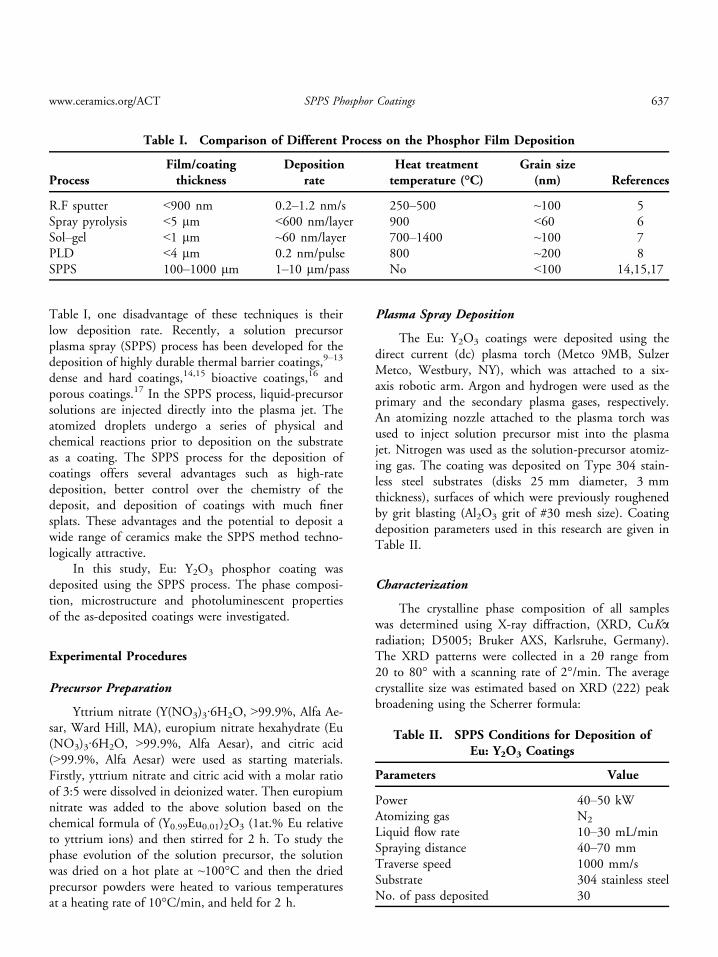
Table I, one disadvantage of these techniques is theirlow deposition rate. Recently, a solution precursorplasma spray (SPPS) process has been developed for thedeposition of highly durable thermal barrier coatings,9–13
dense and hard coatings,14,15 bioactive coatings,16 andporous coatings.17 In the SPPS process, liquid-precursorsolutions are injected directly into the plasma jet. Theatomized droplets undergo a series of physical andchemical reactions prior to deposition on the substrateas a coating. The SPPS process for the deposition ofcoatings offers several advantages such as high-ratedeposition, better control over the chemistry of thedeposit, and deposition of coatings with much finersplats. These advantages and the potential to deposit awide range of ceramics make the SPPS method techno-logically attractive.
In this study, Eu: Y2O3 phosphor coating wasdeposited using the SPPS process. The phase composi-tion, microstructure and photoluminescent propertiesof the as-deposited coatings were investigated.
Experimental Procedures
Precursor Preparation
Yttrium nitrate (Y(NO3)3·6H2O, >99.9%, Alfa Ae-sar, Ward Hill, MA), europium nitrate hexahydrate (Eu(NO3)3·6H2O, >99.9%, Alfa Aesar), and citric acid(>99.9%, Alfa Aesar) were used as starting materials.Firstly, yttrium nitrate and citric acid with a molar ratioof 3:5 were dissolved in deionized water. Then europiumnitrate was added to the above solution based on thechemical formula of (Y0.99Eu0.01)2O3 (1at.% Eu relativeto yttrium ions) and then stirred for 2 h. To study thephase evolution of the solution precursor, the solutionwas dried on a hot plate at ~100°C and then the driedprecursor powders were heated to various temperaturesat a heating rate of 10°C/min, and held for 2 h.
Plasma Spray Deposition
The Eu: Y2O3 coatings were deposited using thedirect current (dc) plasma torch (Metco 9MB, SulzerMetco, Westbury, NY), which was attached to a six-axis robotic arm. Argon and hydrogen were used as theprimary and the secondary plasma gases, respectively.An atomizing nozzle attached to the plasma torch wasused to inject solution precursor mist into the plasmajet. Nitrogen was used as the solution-precursor atomiz-ing gas. The coating was deposited on Type 304 stain-less steel substrates (disks 25 mm diameter, 3 mmthickness), surfaces of which were previously roughenedby grit blasting (Al2O3 grit of #30 mesh size). Coatingdeposition parameters used in this research are given inTable II.
Characterization
The crystalline phase composition of all sampleswas determined using X-ray diffraction, (XRD, CuKaradiation; D5005; Bruker AXS, Karlsruhe, Germany).The XRD patterns were collected in a 2h range from20 to 80° with a scanning rate of 2°/min. The averagecrystallite size was estimated based on XRD (222) peakbroadening using the Scherrer formula:
Table I. Comparison of Different Process on the Phosphor Film Deposition
ProcessFilm/coatingthickness
Depositionrate
Heat treatmenttemperature (°C)
Grain size(nm) References
R.F sputter <900 nm 0.2–1.2 nm/s 250–500 ~100 5Spray pyrolysis <5 lm <600 nm/layer 900 <60 6Sol–gel <1 lm ~60 nm/layer 700–1400 ~100 7PLD <4 lm 0.2 nm/pulse 800 ~200 8SPPS 100–1000 lm 1–10 lm/pass No <100 14,15,17
Table II. SPPS Conditions for Deposition ofEu: Y2O3 Coatings
Parameters Value
Power 40–50 kWAtomizing gas N2
Liquid flow rate 10–30 mL/minSpraying distance 40–70 mmTraverse speed 1000 mm/sSubstrate 304 stainless steelNo. of pass deposited 30
www.ceramics.org/ACT SPPS Phosphor Coatings 637

Dhkl ¼ 0:9kbhkl cos h
where Dhkl is the average dimensions of crystallites, k isthe wavelength of the X-ray radiation(k = 0.15405 nm), and h is the Bragg angle of reflec-tion of specific crystalline plane, bhkl is full width ofhalf maximum (FWHM) of the peak intensity
Photoluminescent properties of the Eu: Y2O3
powder and coating were investigated using a Perkin–Elmer LS50B Fluorometer (Perkin-Elmer Instruments,Shelton, CT). An environmental scanning electronmicroscope (ESEM 2020, Philips Electron Optics, Ein-dhoven, The Netherlands) and a JEOL JSM-6335F fieldemission scanning electron microscope (FESEM, JEOL,Tokyo, Japan) were used to characterize the coatingmicrostructure.
Results and Discussion
Crystallization Behavior and PhotoluminescentProperties of Eu: Y2O3Powders
When the precursor is heat treated at 100°C on ahot plate, it will automatically combust and producevoluminous, fluffy powders. The XRD patterns of thecombustion-synthesized and heat-treated powders atvarious temperatures for 2 h are shown in Fig. 1. Itcan be seen that the crystalline peaks of the combus-tion-synthesized powders are very weak, indicating poor
crystallinity. The precursor powders remained in poorcrystallinity up to 500°C. When the temperature isincreased from 600°C to 1000°C, cubic Y2O3 crystal-line phase begins to form and the crystalline peaksbecome sharper and stronger. The average grain size,D, of the heat-treated powder calculated using Scherrerformula is shown in Fig. 2. The calculated crystallinesize shows an increase from 15 to 48 nm as the calcina-tion temperature increases from 600°C to 1000°C.
Figure 3 shows the microstructure of Eu: Y2O3
powders calcined at 1000°C for 2 h. Well formed
20 30 40 50 60 70 80
(222
)
(e)
(d)(c)(b)(a)
2 θ (o)
Inte
nsity
(cps
)
Fig. 1. XRD patterns of Eu: Y2O3 powders calcined at varioustemperatures: (a) as-synthesized; (b) 500°C; (c) 600°C; (d) 800°C;(e) 1000°C.
600 700 800 900 10000
10
20
30
40
50
60
Gra
in S
ize
(nm
)
Temperature ( oC)
Fig. 2. Grain size of Eu: Y2O3 powders as a function of heattreatment temperatures.
Fig. 3. Microstructure of Eu: Y2O3 powder calcined at 1000°Cfor 2 h.
638 International Journal of Applied Ceramic Technology—Chen, et al. Vol. 9, No. 3, 2012
crystalline equiaxed grains with size of ~50 nm areclearly seen, which is consistent with the grain sizevalue calculated from the Scherrer equation.
The photoluminescent spectra of heat-treated Eu:Y2O3 powders excited by UV light at the wavelengthof 260 nm (kex = 260 nm) are shown in Fig. 4. Thespectra show the typical Eu: Y2O3 emission spectrum,which is described by the well known 5D0 ? 7Fj(j = 0, 1, 2, 3, 4) line emissions of the Eu3+ ions. Thestrongest emission at 612 nm is a hypersensitive forcedelectric-dipole emission from 5D0 ? 7F2 transition.The peaks at 580, 593, 653, and 710 correspond tothe 5D0 ? 7F0,
5D0 ? 7F1,5D0 ? 7F3, and
5D0 ? 7F4 transitions, respectively. It is also observedthat the photoluminescence intensity increases withincreasing heat treatment temperature from 600°C to1000°C. The increased photoluminescence withincreasing heat treatment temperature can be attributedto the improved crystallinity of powders, as is con-firmed by the XRD analysis.
SPPS Eu: Y2O3Coating
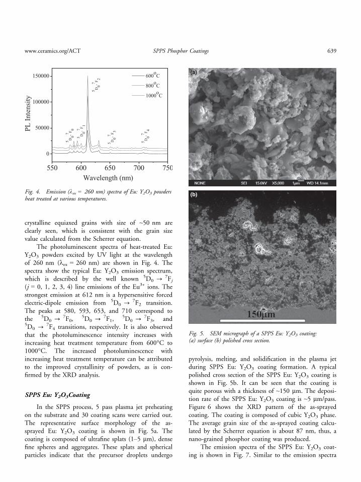
In the SPPS process, 5 pass plasma jet preheatingon the substrate and 30 coating scans were carried out.The representative surface morphology of the as-sprayed Eu: Y2O3 coating is shown in Fig. 5a. Thecoating is composed of ultrafine splats (1–5 lm), densefine spheres and aggregates. These splats and sphericalparticles indicate that the precursor droplets undergo
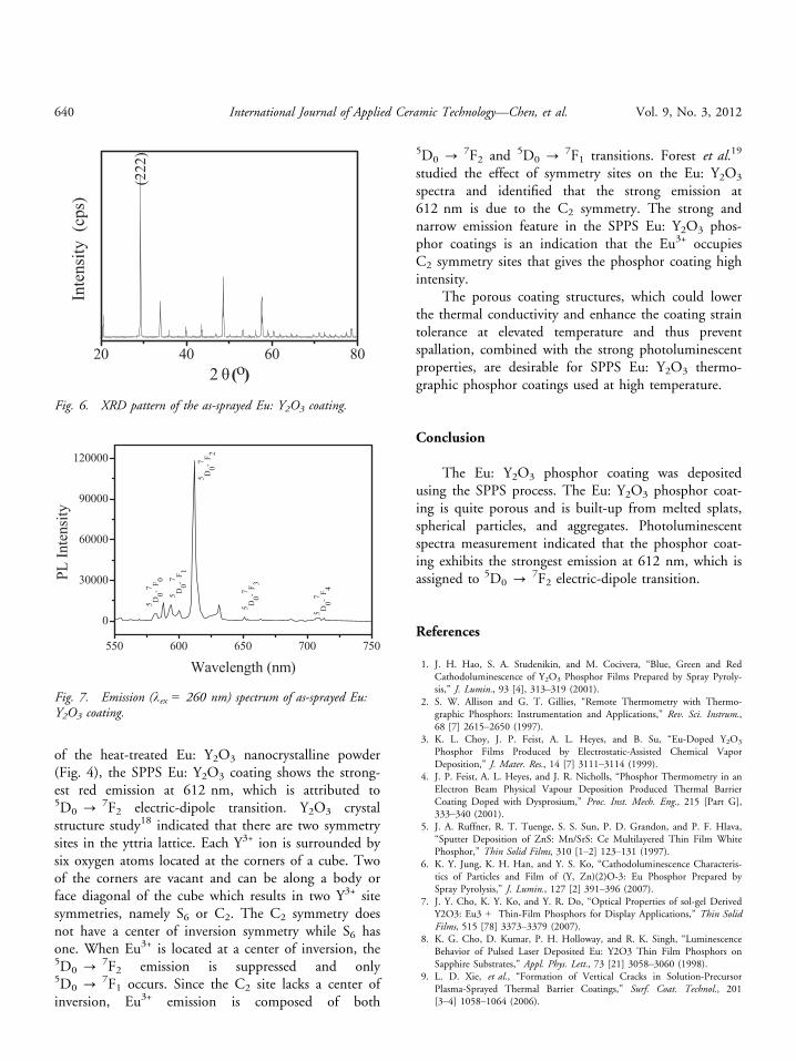
pyrolysis, melting, and solidification in the plasma jetduring SPPS Eu: Y2O3 coating formation. A typicalpolished cross section of the SPPS Eu: Y2O3 coating isshown in Fig. 5b. It can be seen that the coating isquite porous with a thickness of ~150 lm. The deposi-tion rate of the SPPS Eu: Y2O3 coating is ~5 lm/pass.Figure 6 shows the XRD pattern of the as-sprayedcoating. The coating is composed of cubic Y2O3 phase.The average grain size of the as-sprayed coating calcu-lated by the Scherrer equation is about 87 nm, thus, anano-grained phosphor coating was produced.
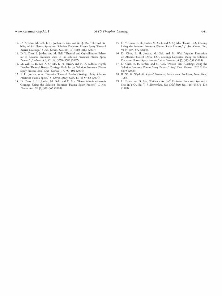
The emission spectra of the SPPS Eu: Y2O3 coat-ing is shown in Fig. 7. Similar to the emission spectra
550 600 650 700 750
0
50000
100000
150000
5 D0-7 F 05 D
0-7 F 1
5 D0-7 F 3
5 D0-7 F 4
5 D0-7 F 2
600oC
800oC
1000oC
Wavelength (nm)
PL In
tens
ity
Fig. 4. Emission (kex = 260 nm) spectra of Eu: Y2O3 powdersheat treated at various temperatures.
(a)
(b)
Fig. 5. SEM micrograph of a SPPS Eu: Y2O3 coating:(a) surface (b) polished cross section.
www.ceramics.org/ACT SPPS Phosphor Coatings 639
of the heat-treated Eu: Y2O3 nanocrystalline powder(Fig. 4), the SPPS Eu: Y2O3 coating shows the strong-est red emission at 612 nm, which is attributed to5D0 ? 7F2 electric-dipole transition. Y2O3 crystalstructure study18 indicated that there are two symmetrysites in the yttria lattice. Each Y3+ ion is surrounded bysix oxygen atoms located at the corners of a cube. Twoof the corners are vacant and can be along a body orface diagonal of the cube which results in two Y3+ sitesymmetries, namely S6 or C2. The C2 symmetry doesnot have a center of inversion symmetry while S6 hasone. When Eu3+ is located at a center of inversion, the5D0 ? 7F2 emission is suppressed and only5D0 ? 7F1 occurs. Since the C2 site lacks a center ofinversion, Eu3+ emission is composed of both
5D0 ? 7F2 and 5D0 ? 7F1 transitions. Forest et al.19
studied the effect of symmetry sites on the Eu: Y2O3
spectra and identified that the strong emission at612 nm is due to the C2 symmetry. The strong andnarrow emission feature in the SPPS Eu: Y2O3 phos-phor coatings is an indication that the Eu3+ occupiesC2 symmetry sites that gives the phosphor coating highintensity.
The porous coating structures, which could lowerthe thermal conductivity and enhance the coating straintolerance at elevated temperature and thus preventspallation, combined with the strong photoluminescentproperties, are desirable for SPPS Eu: Y2O3 thermo-graphic phosphor coatings used at high temperature.
Conclusion
The Eu: Y2O3 phosphor coating was depositedusing the SPPS process. The Eu: Y2O3 phosphor coat-ing is quite porous and is built-up from melted splats,spherical particles, and aggregates. Photoluminescentspectra measurement indicated that the phosphor coat-ing exhibits the strongest emission at 612 nm, which isassigned to 5D0 ? 7F2 electric-dipole transition.
References
1. J. H. Hao, S. A. Studenikin, and M. Cocivera, “Blue, Green and RedCathodoluminescence of Y2O3 Phosphor Films Prepared by Spray Pyroly-sis,” J. Lumin., 93 [4], 313–319 (2001).
2. S. W. Allison and G. T. Gillies, “Remote Thermometry with Thermo-graphic Phosphors: Instrumentation and Applications,” Rev. Sci. Instrum.,68 [7] 2615–2650 (1997).
3. K. L. Choy, J. P. Feist, A. L. Heyes, and B. Su, “Eu-Doped Y2O3
Phosphor Films Produced by Electrostatic-Assisted Chemical VaporDeposition,” J. Mater. Res., 14 [7] 3111–3114 (1999).
4. J. P. Feist, A. L. Heyes, and J. R. Nicholls, “Phosphor Thermometry in anElectron Beam Physical Vapour Deposition Produced Thermal BarrierCoating Doped with Dysprosium,” Proc. Inst. Mech. Eng., 215 [Part G],333–340 (2001).
5. J. A. Ruffner, R. T. Tuenge, S. S. Sun, P. D. Grandon, and P. F. Hlava,“Sputter Deposition of ZnS: Mn/SrS: Ce Multilayered Thin Film WhitePhosphor,” Thin Solid Films, 310 [1–2] 123–131 (1997).
6. K. Y. Jung, K. H. Han, and Y. S. Ko, “Cathodoluminescence Characteris-tics of Particles and Film of (Y, Zn)(2)O-3: Eu Phosphor Prepared bySpray Pyrolysis,” J. Lumin., 127 [2] 391–396 (2007).
7. J. Y. Cho, K. Y. Ko, and Y. R. Do, “Optical Properties of sol-gel DerivedY2O3: Eu3 + Thin-Film Phosphors for Display Applications,” Thin SolidFilms, 515 [78] 3373–3379 (2007).
8. K. G. Cho, D. Kumar, P. H. Holloway, and R. K. Singh, “LuminescenceBehavior of Pulsed Laser Deposited Eu: Y2O3 Thin Film Phosphors onSapphire Substrates,” Appl. Phys. Lett., 73 [21] 3058–3060 (1998).
9. L. D. Xie, et al., “Formation of Vertical Cracks in Solution-PrecursorPlasma-Sprayed Thermal Barrier Coatings,” Surf. Coat. Technol., 201[3–4] 1058–1064 (2006).
20 40 60 80
(222
)
Inte
nsity
(cp
s)
2 θ (o)
Fig. 6. XRD pattern of the as-sprayed Eu: Y2O3 coating.
550 600 650 700 750
0
30000
60000
90000
120000
PL In
tens
ity
Wavelength (nm)
5 D0-7 F 05 D
0-7 F 1
5 D0-7 F 3
5 D0-7 F 4
5 D0-7 F 2
Fig. 7. Emission (kex = 260 nm) spectrum of as-sprayed Eu:Y2O3 coating.
640 International Journal of Applied Ceramic Technology—Chen, et al. Vol. 9, No. 3, 2012
10. D. Y. Chen, M. Gell, E. H. Jordan, E. Cao, and X. Q. Ma, “Thermal Sta-bility of Air Plasma Spray and Solution Precursor Plasma Spray ThermalBarrier Coatings,” J. Am. Ceram. Soc., 90 [10] 3160–3166 (2007).
11. D. Y. Chen, E. Jordan, and M. Gell, “Thermal and Crystallization Behav-ior of Zirconia Precursor Used in the Solution Precursor Plasma SprayProcess,” J. Mater. Sci., 42 [14] 5576–5580 (2007).
12. M. Gell, L. D. Xie, X. Q. Ma, E. H. Jordan, and N. P. Padture, HighlyDurable Thermal Barrier Coatings Made by the Solution Precursor PlasmaSpray Process, Surf. Coat. Technol., 177 97–102 (2004).
13. E. H. Jordan, et al., “Superior Thermal Barrier Coatings Using SolutionPrecursor Plasma Spray,” J. Therm. Spray Tech., 13 [1] 57–65 (2004).
14. D. Chen, E. H. Jordan, M. Gell, and X. Ma, “Dense Alumina-ZirconiaCoatings Using the Solution Precursor Plasma Spray Process,” J. Am.Ceram. Soc., 91 [2] 359–365 (2008).
15. D. Y. Chen, E. H. Jordan, M. Gell, and X. Q. Ma, “Dense TiO2 CoatingUsing the Solution Precursor Plasma Spray Process,” J. Am. Ceram. Soc.,91 [3] 865–872 (2008).
16. D. Chen, E. H. Jordan, M. Gell, and M. Wei, “Apatite Formationon Alkaline-Treated Dense TiO2 Coatings Deposited Using the SolutionPrecursor Plasma Spray Process,” Acta Biomater., 4 [3] 553–559 (2008).
17. D. Chen, E. H. Jordan, and M. Gell, “Porous TiO2 Coatings Using theSolution Precursor Plasma Spray Process,” Surf. Coat. Technol., 202 6113–6119 (2008).
18. R. W. G. Wyckoff, Crystal Structures, Interscience Publisher, New York,1965.
19. H. Forest and G. Ban, “Evidence for Eu+3 Emission from two SymmetrySites in Y2O3: Eu
+3,” J. Electrochem. Soc: Solid State Sci., 116 [4] 474–478(1969).
www.ceramics.org/ACT SPPS Phosphor Coatings 641
Recommended