Embed Size (px)
Citation preview
CB30CH23-Giraldez ARI 11 September 2014 7:56
Zygotic Genome ActivationDuring the Maternal-to-Zygotic TransitionMiler T. Lee,1,∗ Ashley R. Bonneau,1,∗
and Antonio J. Giraldez1,2
1Department of Genetics, Yale University School of Medicine, New Haven, Connecticut 06520;email: [email protected], [email protected] Stem Cell Center, Yale University School of Medicine, New Haven, Connecticut 06520
Annu. Rev. Cell Dev. Biol. 2014. 30:581–613
First published online as a Review in Advance onAugust 11, 2014
The Annual Review of Cell and DevelopmentalBiology is online at cellbio.annualreviews.org
This article’s doi:10.1146/annurev-cellbio-100913-013027
Copyright c© 2014 by Annual Reviews.All rights reserved
∗Authors contributed equally to this review.
Keywords
embryogenesis, pluripotency, cellular reprogramming, pioneer factors,maternal clearance
Abstract
Embryogenesis depends on a highly coordinated cascade of genetically en-coded events. In animals, maternal factors contributed by the egg cytoplasminitially control development, whereas the zygotic nuclear genome is quies-cent. Subsequently, the genome is activated, embryonic gene products aremobilized, and maternal factors are cleared. This transfer of developmentalcontrol is called the maternal-to-zygotic transition (MZT). In this review,we discuss recent advances toward understanding the scope, timing, andmechanisms that underlie zygotic genome activation at the MZT in ani-mals. We describe high-throughput techniques to measure the embryonictranscriptome and explore how regulation of the cell cycle, chromatin, andtranscription factors together elicits specific patterns of embryonic gene ex-pression. Finally, we illustrate the interplay between zygotic transcriptionand maternal clearance and show how these two activities combine to repro-gram two terminally differentiated gametes into a totipotent embryo.
581
Ann
u. R
ev. C
ell D
ev. B
iol.
2014
.30:
581-
613.
Dow
nloa
ded
from
ww
w.a
nnua
lrev
iew
s.or
g A
cces
s pr
ovid
ed b
y Y
ale
Uni
vers
ity -
Med
ical
Lib
rary
on
08/2
0/17
. For
per
sona
l use
onl
y.
CB30CH23-Giraldez ARI 11 September 2014 7:56
Maternal-to-zygotictransition (MZT):the period duringembryogenesis whendevelopmental controltransitions frommaternally providedfactors to onesproduced by zygotic(embryonic)transcription
Totipotency/pluripotency: thecapacity of a cell togive rise to all(totipotent) or most(pluripotent)differentiated cells inan organism
Maternal clearance:the process ofregulated degradationof maternally providedRNAs and proteinsduring the MZT
Contents
INTRODUCTION . . . . . . . . . . . . . . . . . . . . . . . . . . . . . . . . . . . . . . . . . . . . . . . . . . . . . . . . . . . . . . . 582MEASURING ZYGOTIC GENE EXPRESSION . . . . . . . . . . . . . . . . . . . . . . . . . . . . . . . . . 583
The Developmental Context of Zygotic Genome Activation . . . . . . . . . . . . . . . . . . . . . . 583Distinguishing De Novo Zygotic Transcription from the Maternal Contribution . . 583Dynamics of Activation . . . . . . . . . . . . . . . . . . . . . . . . . . . . . . . . . . . . . . . . . . . . . . . . . . . . . . . . . 588
MECHANISMS OF GENOME ACTIVATION . . . . . . . . . . . . . . . . . . . . . . . . . . . . . . . . . . . 589General Models of Activation . . . . . . . . . . . . . . . . . . . . . . . . . . . . . . . . . . . . . . . . . . . . . . . . . . . 589Cell Cycle Regulation. . . . . . . . . . . . . . . . . . . . . . . . . . . . . . . . . . . . . . . . . . . . . . . . . . . . . . . . . . . 590Chromatin Competency . . . . . . . . . . . . . . . . . . . . . . . . . . . . . . . . . . . . . . . . . . . . . . . . . . . . . . . . 593Histone Modifications . . . . . . . . . . . . . . . . . . . . . . . . . . . . . . . . . . . . . . . . . . . . . . . . . . . . . . . . . . 594Epigenetic Prepatterning . . . . . . . . . . . . . . . . . . . . . . . . . . . . . . . . . . . . . . . . . . . . . . . . . . . . . . . 597Transcriptional Repressors and Activators . . . . . . . . . . . . . . . . . . . . . . . . . . . . . . . . . . . . . . . 598
DEVELOPMENTAL SIGNIFICANCE OF THE FIRST ZYGOTIC GENES . . . . . 600Functions of the First Zygotic Genes . . . . . . . . . . . . . . . . . . . . . . . . . . . . . . . . . . . . . . . . . . . . 600The Intimate Relationship between Zygotic Genome Activation
and Maternal Clearance . . . . . . . . . . . . . . . . . . . . . . . . . . . . . . . . . . . . . . . . . . . . . . . . . . . . . . 601CONCLUDING REMARKS . . . . . . . . . . . . . . . . . . . . . . . . . . . . . . . . . . . . . . . . . . . . . . . . . . . . . 603
Zygotic Gene Expression at the Maternal-to-Zygotic TransitionIs a Multifaceted Process . . . . . . . . . . . . . . . . . . . . . . . . . . . . . . . . . . . . . . . . . . . . . . . . . . . . . 603
Cellular Reprogramming During Embryogenesis and Beyond. . . . . . . . . . . . . . . . . . . . . 603
INTRODUCTION
Embryogenesis begins with a single cell composed of cytoplasm from the egg and nuclei fromboth parents that fuse to encode a single zygotic genome. How a fully formed organism arises,far removed in appearance and function from the zygote, has long been the subject of scientificinquiry. Efforts to understand the principles underlying organismal development were closelytied to experiments in the nineteenth century by Theodor Boveri and others, who used sea urchinembryos to investigate the relationship between cellular components and heredity (Laubichler& Davidson 2008). Cross-fertilization between gametes of different species was found to yieldlarvae with intermediate characteristics of both parents, suggesting that genetic determinantswere encoded in the nuclear material contributed by the sperm. However, a range of hybridcharacteristics was also observed in crosses using mechanically produced anucleate eggs, implyingthat some degree of genetic contribution was also conferred by the maternal cytoplasm (Laubichler& Davidson 2008).
These observations have laid the groundwork for our current understanding of embryonic de-velopment, a process subject to both cytoplasmic and nuclear control. Initially, the embryo is tran-scriptionally quiescent, and development is directed exclusively by maternally provided proteinsand RNAs from the egg cytoplasm. Subsequently, developmental control switches to the productsof an activated nuclear genome during a period called the maternal-to-zygotic transition (MZT).
The MZT encompasses two major molecular activities, which together reprogram the termi-nally differentiated oocyte and sperm to totipotency. One is maternal clearance, the deletion ofmaternal instructions—mRNA and proteins—that are necessary for oocyte maturation, homeo-stasis, and the first stages of embryogenesis but become unnecessary or possibly deleterious as theembryo develops. The other is the installation of new zygotic instructions through gene expression,
582 Lee · Bonneau · Giraldez
Ann
u. R
ev. C
ell D
ev. B
iol.
2014
.30:
581-
613.
Dow
nloa
ded
from
ww
w.a
nnua
lrev
iew
s.or
g A
cces
s pr
ovid
ed b
y Y
ale
Uni
vers
ity -
Med
ical
Lib
rary
on
08/2
0/17
. For
per
sona
l use
onl
y.
CB30CH23-Giraldez ARI 11 September 2014 7:56
Zygotic genomeactivation (ZGA):the period during theMZT when theembryonic genomefirst begins totranscribe RNA
Chromatin: thephysical structure of achromosome,consisting of DNAbound by nucleosomesand other proteins
Transcription factor:a protein that binds toDNA sequences toregulate geneexpression
Cellularization:the partitioning of amultinucleatedcytoplasm (syncytium)into individual cells, asobserved duringDrosophila melanogasterembryogenesis
a process that is activated by the maternal program and is called zygotic genome activation (ZGA).Together these two activities dramatically remodel the embryonic gene expression landscape andcellular identities, a process that will be revisited throughout development and adulthood as cellsdifferentiate and regenerate.
Recent reviews have provided extensive treatments of the mechanisms that regulate mater-nal clearance (Barckmann & Simonelig 2013, Walser & Lipshitz 2011). Here, we focus on theactivation of zygotic gene expression in animals, though similar processes also occur in plants(Baroux et al. 2008, Xin et al. 2012). In the first section, we highlight recent advances in mea-suring the onset of zygotic transcription and present them in the context of seminal discoveriesin the genetic control of embryogenesis. Next, we consider the mechanisms that drive ZGA anddescribe the interplay between the cell cycle, chromatin, and transcription factors in regulatingembryonic gene expression. Finally, we discuss the functional consequences of ZGA and showthat maternal clearance and zygotic transcription are intimately linked and combine to give riseto a reprogrammed embryo.
MEASURING ZYGOTIC GENE EXPRESSION
The Developmental Context of Zygotic Genome Activation
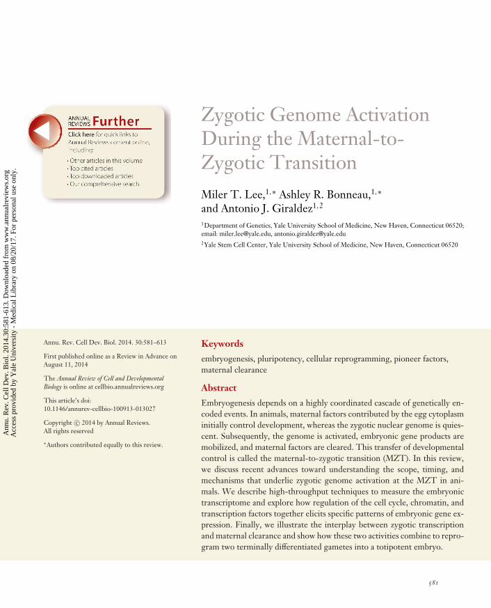
In most animals, the maternal contribution directs a series of synchronized mitoses while maintain-ing a relatively constant volume as it forms a blastula. Subsequently, coordinated cell movementsduring gastrulation form distinct germ layers that specify the various tissues in the mature or-ganism. Within this framework of embryogenesis, there is extensive variation in the timing andduration of these events among different species. The initial cell cycles of the amphibian Xenopus(35 min), zebrafish (15 min), and the fruit fly Drosophila melanogaster (8 min) are synchronizedand proceed in rapid succession, with gastrulation occurring within hours of fertilization (Foe& Alberts 1983, Gerhart 1980, Kane & Kimmel 1993). Development is also rapid in the nema-tode Caenorhabditis elegans, which forms a 28-cell gastrula after 100 min of asymmetric divisions(Figure 1) (Sulston et al. 1983). In contrast, mouse and other mammals have relatively long cell cy-cles, with the first cleavage occurring approximately one day after fertilization (Figure 1) followedby gastrulation five to six days later (Oron & Ivanova 2012).
The requirement for zygotic transcription for embryogenesis to proceed is universal acrossanimals. Upon transcriptional inhibition, zebrafish and Xenopus embryos will continue to dividebut fail to undergo gastrulation (Kane et al. 1996, Newport & Kirschner 1982a). Similarly, theC. elegans embryo experiences extreme morphological defects without zygotic transcription, de-spite reaching 100 cells before arresting (Edgar et al. 1994), and D. melanogaster, which does notcomplete cytokinesis for the first 13 cell cycles, requires zygotic transcripts for cellularization tooccur (Edgar et al. 1986, Merrill et al. 1988). In mouse, development proceeds no further thanthe second mitosis, commonly referred to as the 2-cell block (Goddard & Pratt 1983, Golbuset al. 1973, Warner & Versteegh 1974). In each of these organisms, ZGA occurs well before thesedefects arise, suggesting that zygotic transcription does not merely coincide with a requirementto replenish RNAs for homeostasis but also is essential to direct new developmental programs.Discovering the identity of these early zygotic RNAs is thus essential to understanding how de-velopment proceeds.
Distinguishing De Novo Zygotic Transcription from the Maternal Contribution
From the earliest studies of embryonic RNA content, it was clear that maternally deposited RNAsgreatly outnumber zygotic transcripts—representing between 40% and 75% of all protein-coding
www.annualreviews.org • Maternal-to-Zygotic Transition 583
Ann
u. R
ev. C
ell D
ev. B
iol.
2014
.30:
581-
613.
Dow
nloa
ded
from
ww
w.a
nnua
lrev
iew
s.or
g A
cces
s pr
ovid
ed b
y Y
ale
Uni
vers
ity -
Med
ical
Lib
rary
on
08/2
0/17
. For
per
sona
l use
onl
y.
CB30CH23-Giraldez ARI 11 September 2014 7:56
RN
A l
ev
els
Dros
ophi
la
Cell cycle
Xeno
pus
Zeb
rafis
hMo
use
48 min
4.5 h
2 h
Caen
orha
bditi
s el
egan
s
6 7 8 9 1021 11 12 13 14 1543 5
70 min10 h
4.3 h2 days 6 h90 min 2.5 h
Figure 1Timing of zygotic genome activation across various model organisms. Curves illustrate cumulative increasesin zygotic gene expression as development progresses. In the mouse, a minor wave of transcription duringthe first cell cycle is followed by a second wave during cycle 2. Caenorhabditis elegans divisions areasynchronous, with transcription detected by four cells. Zygotic transcription in Xenopus, zebrafish, andDrosophila melanogaster is detected several cell cycles later and increases rapidly. Approximate times postfertilization are indicated.
Transcriptome:the set of all RNAtranscripts in a cell,tissue, or organism
RNA-Seq: massivelyparallel RNAsequencing to measuregene expression levelsin a high-throughputmanner
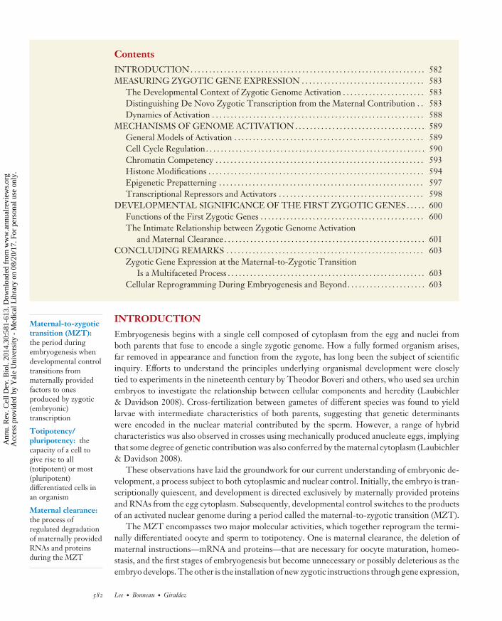
genes across various species (Tadros & Lipshitz 2009, Wang et al. 2004, Wei et al. 2006) and stillamounting to ∼60 to 70% of mRNA molecules at the peak of zygotic expression in zebrafish, forexample (Lee et al. 2013) (Figure 2a). This large maternal contribution presents a challenge fordetecting transcriptionally active genes, especially if maternal transcript copies greatly outnumberthe zygotic contribution or if degradation of maternal copies occurs concurrently with de novotranscription, effectively canceling out the signal (De Renzis et al. 2007). This, coupled withdynamic regulation of maternal RNAs, including changes in poly(A) tail length, highlights theneed to distinguish the maternal and zygotic contributions to the transcriptome pool.
Microarrays and high-throughput sequencing (RNA-Seq) using Illumina and SOLiDtechnologies (Table 1) have greatly enhanced our ability to dissect the maternal and zygoticRNA populations. Time-course experiments have revealed transcriptome-wide changes in geneexpression during the MZT in many different species (Aanes et al. 2011, Collart et al. 2014,Dobson et al. 2004, Hamatani et al. 2004, Harvey et al. 2013, Heyn et al. 2014, Lee et al. 2013,Paranjpe et al. 2013, Sirard et al. 2005, Tan et al. 2013, Vassena et al. 2011, Vesterlund et al. 2011,Wang et al. 2004, Wei et al. 2006, Xie et al. 2010, Xue et al. 2013, Zhang et al. 2009); however,attributing these changes to active zygotic transcription can still be a challenge. Explicit techniquesthat distinguish bona fide zygotic gene expression from post-transcriptional regulation of thematernal contribution are thus invaluable for understanding the dynamics and extent of ZGA.
Four general approaches have been used to more accurately assess the scope of ZGA by empha-sizing (Figure 2b) or removing (Figure 2c) the signal from zygotic transcripts. First, the maternalcontribution can be depleted using subtractive hybridization techniques (Figure 2d ). Zygoticsamples hybridized to biotinylated oocyte cDNA can yield libraries where maternally contributedtranscripts are underrepresented (Rothstein et al. 1992, Sive & St John 1988). This method hasbeen effective for detecting rare zygotic transcripts from limited amounts of RNA (Zeng & Schultz2003), but less so for accurate measurement of genome-wide transcript levels.
584 Lee · Bonneau · Giraldez
Ann
u. R
ev. C
ell D
ev. B
iol.
2014
.30:
581-
613.
Dow
nloa
ded
from
ww
w.a
nnua
lrev
iew
s.or
g A
cces
s pr
ovid
ed b
y Y
ale
Uni
vers
ity -
Med
ical
Lib
rary
on
08/2
0/17
. For
per
sona
l use
onl
y.
CB30CH23-Giraldez ARI 11 September 2014 7:56
Depleted zygotic signalEnriched zygotic signal
cb
h
–
i
–
ed f
–A
C
U
U+
g
Subtractive hybridization
4SU labeling Paternal allele Intron signal Transcriptioninhibition
Chromosomaldeletion
Early embryonic transcriptome
a
Zygotic contribution
Maternal contributionExon 1 Exon 2
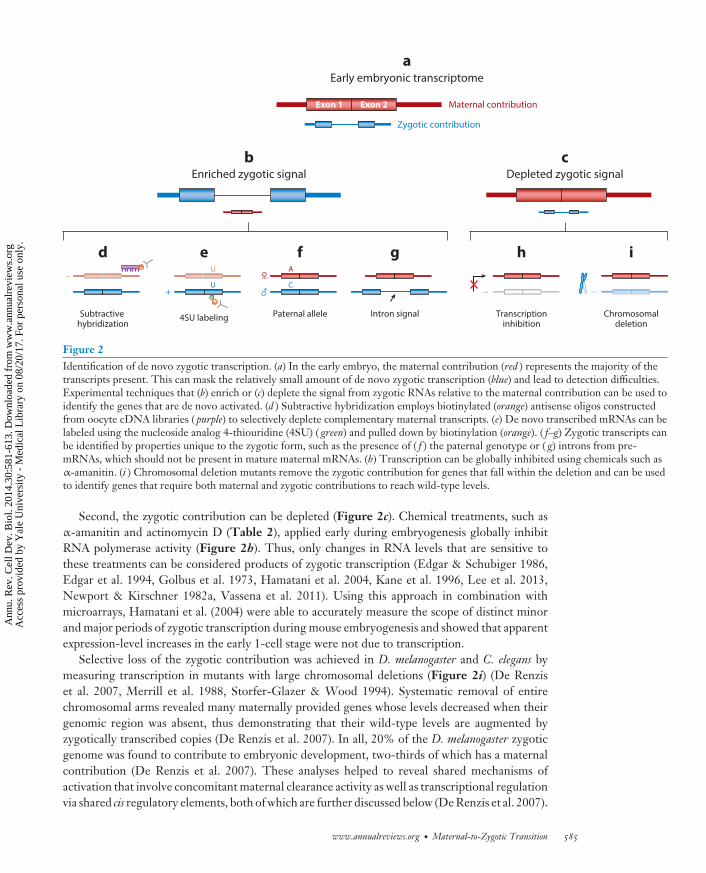
Figure 2Identification of de novo zygotic transcription. (a) In the early embryo, the maternal contribution (red ) represents the majority of thetranscripts present. This can mask the relatively small amount of de novo zygotic transcription (blue) and lead to detection difficulties.Experimental techniques that (b) enrich or (c) deplete the signal from zygotic RNAs relative to the maternal contribution can be used toidentify the genes that are de novo activated. (d ) Subtractive hybridization employs biotinylated (orange) antisense oligos constructedfrom oocyte cDNA libraries ( purple) to selectively deplete complementary maternal transcripts. (e) De novo transcribed mRNAs can belabeled using the nucleoside analog 4-thiouridine (4SU) ( green) and pulled down by biotinylation (orange). ( f–g) Zygotic transcripts canbe identified by properties unique to the zygotic form, such as the presence of ( f ) the paternal genotype or ( g) introns from pre-mRNAs, which should not be present in mature maternal mRNAs. (h) Transcription can be globally inhibited using chemicals such asα-amanitin. (i ) Chromosomal deletion mutants remove the zygotic contribution for genes that fall within the deletion and can be usedto identify genes that require both maternal and zygotic contributions to reach wild-type levels.
Second, the zygotic contribution can be depleted (Figure 2c). Chemical treatments, such asα-amanitin and actinomycin D (Table 2), applied early during embryogenesis globally inhibitRNA polymerase activity (Figure 2h). Thus, only changes in RNA levels that are sensitive tothese treatments can be considered products of zygotic transcription (Edgar & Schubiger 1986,Edgar et al. 1994, Golbus et al. 1973, Hamatani et al. 2004, Kane et al. 1996, Lee et al. 2013,Newport & Kirschner 1982a, Vassena et al. 2011). Using this approach in combination withmicroarrays, Hamatani et al. (2004) were able to accurately measure the scope of distinct minorand major periods of zygotic transcription during mouse embryogenesis and showed that apparentexpression-level increases in the early 1-cell stage were not due to transcription.
Selective loss of the zygotic contribution was achieved in D. melanogaster and C. elegans bymeasuring transcription in mutants with large chromosomal deletions (Figure 2i) (De Renziset al. 2007, Merrill et al. 1988, Storfer-Glazer & Wood 1994). Systematic removal of entirechromosomal arms revealed many maternally provided genes whose levels decreased when theirgenomic region was absent, thus demonstrating that their wild-type levels are augmented byzygotically transcribed copies (De Renzis et al. 2007). In all, 20% of the D. melanogaster zygoticgenome was found to contribute to embryonic development, two-thirds of which has a maternalcontribution (De Renzis et al. 2007). These analyses helped to reveal shared mechanisms ofactivation that involve concomitant maternal clearance activity as well as transcriptional regulationvia shared cis regulatory elements, both of which are further discussed below (De Renzis et al. 2007).
www.annualreviews.org • Maternal-to-Zygotic Transition 585
Ann
u. R
ev. C
ell D
ev. B
iol.
2014
.30:
581-
613.
Dow
nloa
ded
from
ww
w.a
nnua
lrev
iew
s.or
g A
cces
s pr
ovid
ed b
y Y
ale
Uni
vers
ity -
Med
ical
Lib
rary
on
08/2
0/17
. For
per
sona
l use
onl
y.
CB30CH23-Giraldez ARI 11 September 2014 7:56
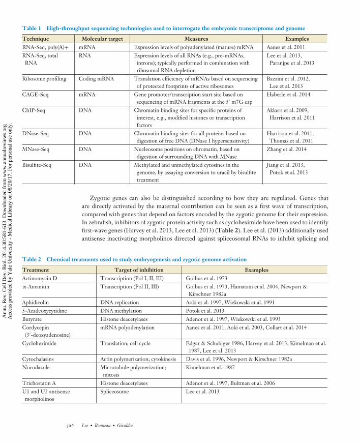
Table 1 High-throughput sequencing technologies used to interrogate the embryonic transcriptome and genome
Technique Molecular target Measures ExamplesRNA-Seq, poly(A)+ mRNA Expression levels of polyadenylated (mature) mRNA Aanes et al. 2011RNA-Seq, totalRNA
RNA Expression levels of all RNAs (e.g., pre-mRNAs,introns); typically performed in combination withribosomal RNA depletion
Lee et al. 2013,Paranjpe et al. 2013
Ribosome profiling Coding mRNA Translation efficiency of mRNAs based on sequencingof protected footprints of active ribosomes
Bazzini et al. 2012,Lee et al. 2013
CAGE-Seq mRNA Gene promoter/transcription start site based onsequencing of mRNA fragments at the 5′ m7G cap
Haberle et al. 2014
ChIP-Seq DNA Chromatin binding sites for specific proteins ofinterest, e.g., modified histones or transcriptionfactors
Akkers et al. 2009,Harrison et al. 2011
DNase-Seq DNA Chromatin binding sites for all proteins based ondigestion of free DNA (DNase I hypersensitivity)
Harrison et al. 2011,Thomas et al. 2011
MNase-Seq DNA Nucleosome positions on chromatin, based ondigestion of surrounding DNA with MNase
Zhang et al. 2014
Bisulfite-Seq DNA Methylated and unmethylated cytosines in thegenome, by assaying conversion to uracil by bisulfitetreatment
Jiang et al. 2013,Potok et al. 2013
Zygotic genes can also be distinguished according to how they are regulated. Genes thatare directly activated by the maternal contribution can be seen as a first wave of transcription,compared with genes that depend on factors encoded by the zygotic genome for their expression.In zebrafish, inhibitors of zygotic protein activity such as cycloheximide have been used to identifyfirst-wave genes (Harvey et al. 2013, Lee et al. 2013) (Table 2). Lee et al. (2013) additionally usedantisense inactivating morpholinos directed against spliceosomal RNAs to inhibit splicing and
Table 2 Chemical treatments used to study embryogenesis and zygotic genome activation
Treatment Target of inhibition ExamplesActinomycin D Transcription (Pol I, II, III) Golbus et al. 1973α-Amanitin Transcription (Pol II, III) Golbus et al. 1973, Hamatani et al. 2004, Newport &
Kirschner 1982aAphidicolin DNA replication Aoki et al. 1997, Wiekowski et al. 19915-Azadeoxycytidine DNA methylation Potok et al. 2013Butyrate Histone deacetylases Adenot et al. 1997, Wiekowski et al. 1993Cordycepin(3′-deoxyadenosine)
mRNA polyadenylation Aanes et al. 2011, Aoki et al. 2003, Collart et al. 2014
Cycloheximide Translation; cell cycle Edgar & Schubiger 1986, Harvey et al. 2013, Kimelman et al.1987, Lee et al. 2013
Cytochalasins Actin polymerization; cytokinesis Davis et al. 1996, Newport & Kirschner 1982aNocodazole Microtubule polymerization;
mitosisKimelman et al. 1987
Trichostatin A Histone deacetylases Adenot et al. 1997, Bultman et al. 2006U1 and U2 antisensemorpholinos
Spliceosome Lee et al. 2013
586 Lee · Bonneau · Giraldez
Ann
u. R
ev. C
ell D
ev. B
iol.
2014
.30:
581-
613.
Dow
nloa
ded
from
ww
w.a
nnua
lrev
iew
s.or
g A
cces
s pr
ovid
ed b
y Y
ale
Uni
vers
ity -
Med
ical
Lib
rary
on
08/2
0/17
. For
per
sona
l use
onl
y.
CB30CH23-Giraldez ARI 11 September 2014 7:56
Pronucleus: thehaploid nucleus of thesperm or egg followingfertilization, prior tofusion into a singlezygotic nucleus
Transcription startsite (TSS): the site oftranscription initiationproximal to thepromoter that definesthe 5′ end of a genetranscript
Single-nucleotidepolymorphism(SNP): variation of asingle base position inthe genome that differsbetween individuals ina population
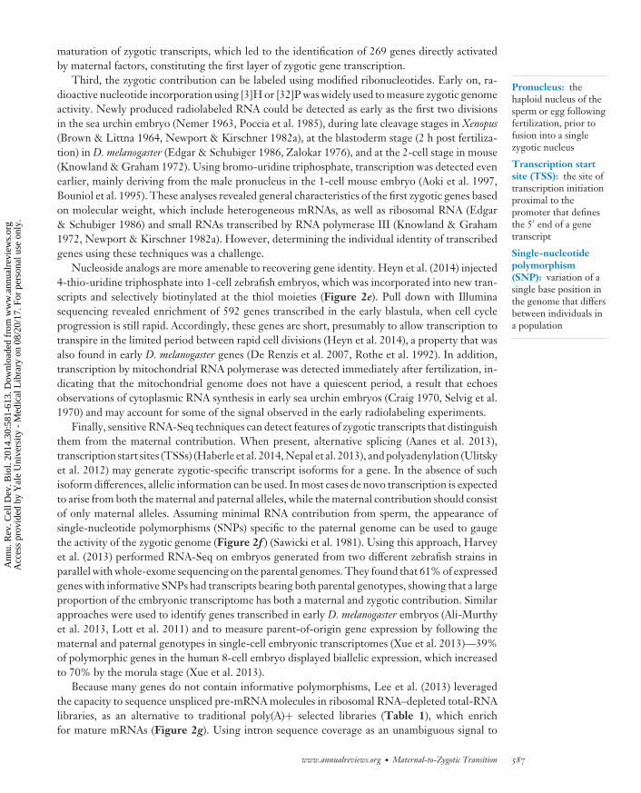
maturation of zygotic transcripts, which led to the identification of 269 genes directly activatedby maternal factors, constituting the first layer of zygotic gene transcription.
Third, the zygotic contribution can be labeled using modified ribonucleotides. Early on, ra-dioactive nucleotide incorporation using [3]H or [32]P was widely used to measure zygotic genomeactivity. Newly produced radiolabeled RNA could be detected as early as the first two divisionsin the sea urchin embryo (Nemer 1963, Poccia et al. 1985), during late cleavage stages in Xenopus(Brown & Littna 1964, Newport & Kirschner 1982a), at the blastoderm stage (2 h post fertiliza-tion) in D. melanogaster (Edgar & Schubiger 1986, Zalokar 1976), and at the 2-cell stage in mouse(Knowland & Graham 1972). Using bromo-uridine triphosphate, transcription was detected evenearlier, mainly deriving from the male pronucleus in the 1-cell mouse embryo (Aoki et al. 1997,Bouniol et al. 1995). These analyses revealed general characteristics of the first zygotic genes basedon molecular weight, which include heterogeneous mRNAs, as well as ribosomal RNA (Edgar& Schubiger 1986) and small RNAs transcribed by RNA polymerase III (Knowland & Graham1972, Newport & Kirschner 1982a). However, determining the individual identity of transcribedgenes using these techniques was a challenge.
Nucleoside analogs are more amenable to recovering gene identity. Heyn et al. (2014) injected4-thio-uridine triphosphate into 1-cell zebrafish embryos, which was incorporated into new tran-scripts and selectively biotinylated at the thiol moieties (Figure 2e). Pull down with Illuminasequencing revealed enrichment of 592 genes transcribed in the early blastula, when cell cycleprogression is still rapid. Accordingly, these genes are short, presumably to allow transcription totranspire in the limited period between rapid cell divisions (Heyn et al. 2014), a property that wasalso found in early D. melanogaster genes (De Renzis et al. 2007, Rothe et al. 1992). In addition,transcription by mitochondrial RNA polymerase was detected immediately after fertilization, in-dicating that the mitochondrial genome does not have a quiescent period, a result that echoesobservations of cytoplasmic RNA synthesis in early sea urchin embryos (Craig 1970, Selvig et al.1970) and may account for some of the signal observed in the early radiolabeling experiments.
Finally, sensitive RNA-Seq techniques can detect features of zygotic transcripts that distinguishthem from the maternal contribution. When present, alternative splicing (Aanes et al. 2013),transcription start sites (TSSs) (Haberle et al. 2014, Nepal et al. 2013), and polyadenylation (Ulitskyet al. 2012) may generate zygotic-specific transcript isoforms for a gene. In the absence of suchisoform differences, allelic information can be used. In most cases de novo transcription is expectedto arise from both the maternal and paternal alleles, while the maternal contribution should consistof only maternal alleles. Assuming minimal RNA contribution from sperm, the appearance ofsingle-nucleotide polymorphisms (SNPs) specific to the paternal genome can be used to gaugethe activity of the zygotic genome (Figure 2f ) (Sawicki et al. 1981). Using this approach, Harveyet al. (2013) performed RNA-Seq on embryos generated from two different zebrafish strains inparallel with whole-exome sequencing on the parental genomes. They found that 61% of expressedgenes with informative SNPs had transcripts bearing both parental genotypes, showing that a largeproportion of the embryonic transcriptome has both a maternal and zygotic contribution. Similarapproaches were used to identify genes transcribed in early D. melanogaster embryos (Ali-Murthyet al. 2013, Lott et al. 2011) and to measure parent-of-origin gene expression by following thematernal and paternal genotypes in single-cell embryonic transcriptomes (Xue et al. 2013)—39%of polymorphic genes in the human 8-cell embryo displayed biallelic expression, which increasedto 70% by the morula stage (Xue et al. 2013).
Because many genes do not contain informative polymorphisms, Lee et al. (2013) leveragedthe capacity to sequence unspliced pre-mRNA molecules in ribosomal RNA–depleted total-RNAlibraries, as an alternative to traditional poly(A)+ selected libraries (Table 1), which enrichfor mature mRNAs (Figure 2g). Using intron sequence coverage as an unambiguous signal to
www.annualreviews.org • Maternal-to-Zygotic Transition 587
Ann
u. R
ev. C
ell D
ev. B
iol.
2014
.30:
581-
613.
Dow
nloa
ded
from
ww
w.a
nnua
lrev
iew
s.or
g A
cces
s pr
ovid
ed b
y Y
ale
Uni
vers
ity -
Med
ical
Lib
rary
on
08/2
0/17
. For
per
sona
l use
onl
y.
CB30CH23-Giraldez ARI 11 September 2014 7:56
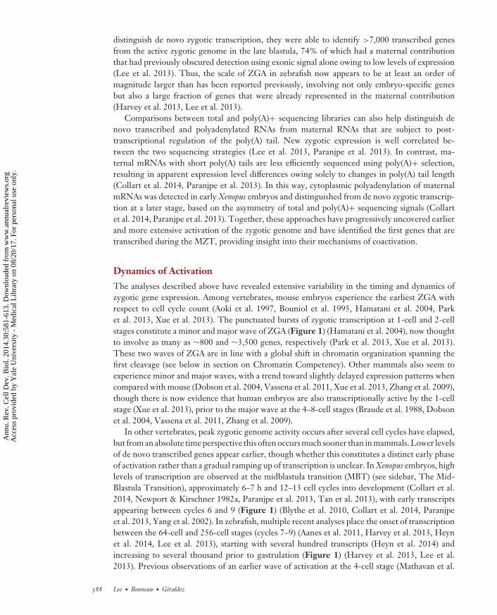
distinguish de novo zygotic transcription, they were able to identify >7,000 transcribed genesfrom the active zygotic genome in the late blastula, 74% of which had a maternal contributionthat had previously obscured detection using exonic signal alone owing to low levels of expression(Lee et al. 2013). Thus, the scale of ZGA in zebrafish now appears to be at least an order ofmagnitude larger than has been reported previously, involving not only embryo-specific genesbut also a large fraction of genes that were already represented in the maternal contribution(Harvey et al. 2013, Lee et al. 2013).
Comparisons between total and poly(A)+ sequencing libraries can also help distinguish denovo transcribed and polyadenylated RNAs from maternal RNAs that are subject to post-transcriptional regulation of the poly(A) tail. New zygotic expression is well correlated be-tween the two sequencing strategies (Lee et al. 2013, Paranjpe et al. 2013). In contrast, ma-ternal mRNAs with short poly(A) tails are less efficiently sequenced using poly(A)+ selection,resulting in apparent expression level differences owing solely to changes in poly(A) tail length(Collart et al. 2014, Paranjpe et al. 2013). In this way, cytoplasmic polyadenylation of maternalmRNAs was detected in early Xenopus embryos and distinguished from de novo zygotic transcrip-tion at a later stage, based on the asymmetry of total and poly(A)+ sequencing signals (Collartet al. 2014, Paranjpe et al. 2013). Together, these approaches have progressively uncovered earlierand more extensive activation of the zygotic genome and have identified the first genes that aretranscribed during the MZT, providing insight into their mechanisms of coactivation.
Dynamics of Activation
The analyses described above have revealed extensive variability in the timing and dynamics ofzygotic gene expression. Among vertebrates, mouse embryos experience the earliest ZGA withrespect to cell cycle count (Aoki et al. 1997, Bouniol et al. 1995, Hamatani et al. 2004, Parket al. 2013, Xue et al. 2013). The punctuated bursts of zygotic transcription at 1-cell and 2-cellstages constitute a minor and major wave of ZGA (Figure 1) (Hamatani et al. 2004), now thoughtto involve as many as ∼800 and ∼3,500 genes, respectively (Park et al. 2013, Xue et al. 2013).These two waves of ZGA are in line with a global shift in chromatin organization spanning thefirst cleavage (see below in section on Chromatin Competency). Other mammals also seem toexperience minor and major waves, with a trend toward slightly delayed expression patterns whencompared with mouse (Dobson et al. 2004, Vassena et al. 2011, Xue et al. 2013, Zhang et al. 2009),though there is now evidence that human embryos are also transcriptionally active by the 1-cellstage (Xue et al. 2013), prior to the major wave at the 4–8-cell stages (Braude et al. 1988, Dobsonet al. 2004, Vassena et al. 2011, Zhang et al. 2009).
In other vertebrates, peak zygotic genome activity occurs after several cell cycles have elapsed,but from an absolute time perspective this often occurs much sooner than in mammals. Lower levelsof de novo transcribed genes appear earlier, though whether this constitutes a distinct early phaseof activation rather than a gradual ramping up of transcription is unclear. In Xenopus embryos, highlevels of transcription are observed at the midblastula transition (MBT) (see sidebar, The Mid-Blastula Transition), approximately 6–7 h and 12–13 cell cycles into development (Collart et al.2014, Newport & Kirschner 1982a, Paranjpe et al. 2013, Tan et al. 2013), with early transcriptsappearing between cycles 6 and 9 (Figure 1) (Blythe et al. 2010, Collart et al. 2014, Paranjpeet al. 2013, Yang et al. 2002). In zebrafish, multiple recent analyses place the onset of transcriptionbetween the 64-cell and 256-cell stages (cycles 7–9) (Aanes et al. 2011, Harvey et al. 2013, Heynet al. 2014, Lee et al. 2013), starting with several hundred transcripts (Heyn et al. 2014) andincreasing to several thousand prior to gastrulation (Figure 1) (Harvey et al. 2013, Lee et al.2013). Previous observations of an earlier wave of activation at the 4-cell stage (Mathavan et al.
588 Lee · Bonneau · Giraldez
Ann
u. R
ev. C
ell D
ev. B
iol.
2014
.30:
581-
613.
Dow
nloa
ded
from
ww
w.a
nnua
lrev
iew
s.or
g A
cces
s pr
ovid
ed b
y Y
ale
Uni
vers
ity -
Med
ical
Lib
rary
on
08/2
0/17
. For
per
sona
l use
onl
y.
CB30CH23-Giraldez ARI 11 September 2014 7:56
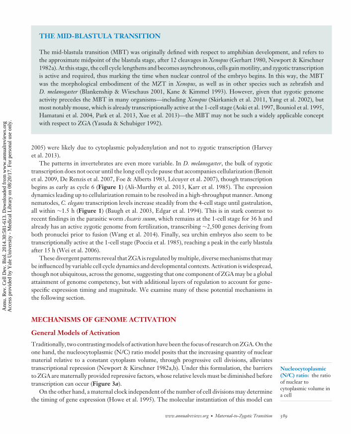
THE MID-BLASTULA TRANSITION
The mid-blastula transition (MBT) was originally defined with respect to amphibian development, and refers tothe approximate midpoint of the blastula stage, after 12 cleavages in Xenopus (Gerhart 1980, Newport & Kirschner1982a). At this stage, the cell cycle lengthens and becomes asynchronous, cells gain motility, and zygotic transcriptionis active and required, thus marking the time when nuclear control of the embryo begins. In this way, the MBTwas the morphological embodiment of the MZT in Xenopus, as well as in other species such as zebrafish andD. melanogaster (Blankenship & Wieschaus 2001, Kane & Kimmel 1993). However, given that zygotic genomeactivity precedes the MBT in many organisms—including Xenopus (Skirkanich et al. 2011, Yang et al. 2002), butmost notably mouse, which is already transcriptionally active at the 1-cell stage (Aoki et al. 1997, Bouniol et al. 1995,Hamatani et al. 2004, Park et al. 2013, Xue et al. 2013)—the MBT may not be such a widely applicable conceptwith respect to ZGA (Yasuda & Schubiger 1992).
Nucleocytoplasmic(N/C) ratio: the ratioof nuclear tocytoplasmic volume ina cell
2005) were likely due to cytoplasmic polyadenylation and not to zygotic transcription (Harveyet al. 2013).
The patterns in invertebrates are even more variable. In D. melanogaster, the bulk of zygotictranscription does not occur until the long cell cycle pause that accompanies cellularization (Benoitet al. 2009, De Renzis et al. 2007, Foe & Alberts 1983, Lecuyer et al. 2007), though transcriptionbegins as early as cycle 6 (Figure 1) (Ali-Murthy et al. 2013, Karr et al. 1985). The expressiondynamics leading up to cellularization remain to be resolved in a high-throughput manner. Amongnematodes, C. elegans transcription levels increase steadily from the 4-cell stage until gastrulation,all within ∼1.5 h (Figure 1) (Baugh et al. 2003, Edgar et al. 1994). This is in stark contrast torecent findings in the parasitic worm Ascaris suum, which remains at the 1-cell stage for 36 h andalready has an active zygotic genome from fertilization, transcribing ∼2,500 genes deriving fromboth pronuclei prior to fusion (Wang et al. 2014). Finally, sea urchin embryos also seem to betranscriptionally active at the 1-cell stage (Poccia et al. 1985), reaching a peak in the early blastulaafter 15 h (Wei et al. 2006).
These divergent patterns reveal that ZGA is regulated by multiple, diverse mechanisms that maybe influenced by variable cell cycle dynamics and developmental contexts. Activation is widespread,though not ubiquitous, across the genome, suggesting that one component of ZGA may be a globalattainment of genome competency, but with additional layers of regulation to account for gene-specific expression timing and magnitude. We examine many of these potential mechanisms inthe following section.
MECHANISMS OF GENOME ACTIVATION
General Models of Activation
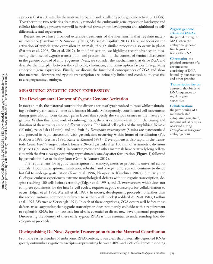
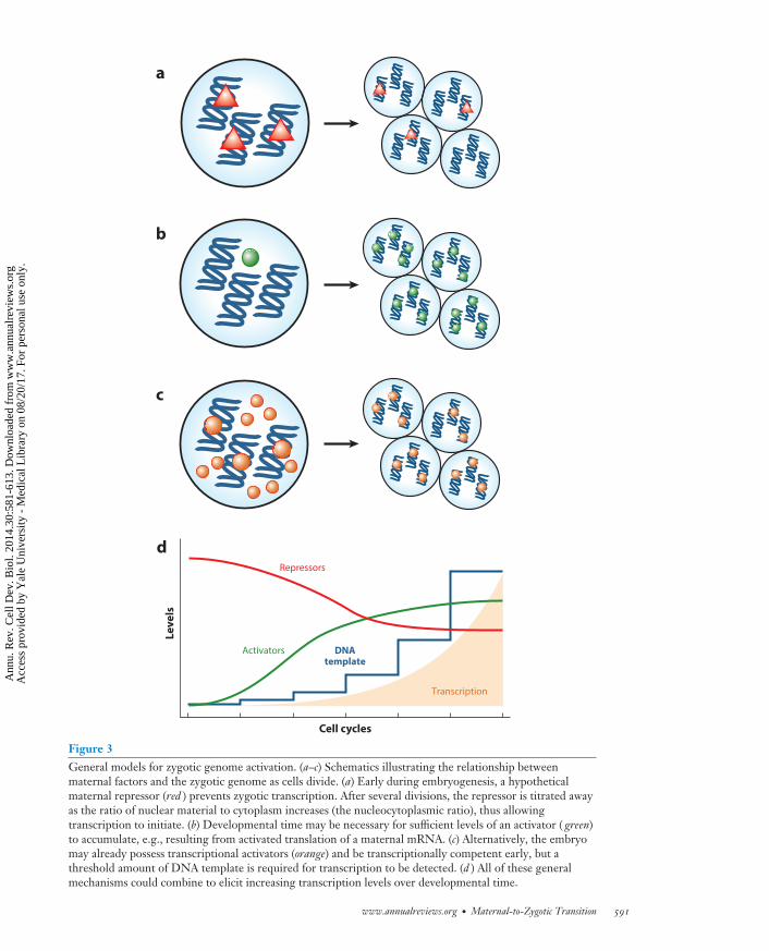
Traditionally, two contrasting models of activation have been the focus of research on ZGA. On theone hand, the nucleocytoplasmic (N/C) ratio model posits that the increasing quantity of nuclearmaterial relative to a constant cytoplasm volume, through progressive cell divisions, alleviatestranscriptional repression (Newport & Kirschner 1982a,b). Under this formulation, the barriersto ZGA are maternally provided repressive factors, whose relative levels must be diminished beforetranscription can occur (Figure 3a).
On the other hand, a maternal clock independent of the number of cell divisions may determinethe timing of gene expression (Howe et al. 1995). The molecular instantiation of this model can
www.annualreviews.org • Maternal-to-Zygotic Transition 589
Ann
u. R
ev. C
ell D
ev. B
iol.
2014
.30:
581-
613.
Dow
nloa
ded
from
ww
w.a
nnua
lrev
iew
s.or
g A
cces
s pr
ovid
ed b
y Y
ale
Uni
vers
ity -
Med
ical
Lib
rary
on
08/2
0/17
. For
per
sona
l use
onl
y.
CB30CH23-Giraldez ARI 11 September 2014 7:56
Epigenetic: refers toheritable changesaffecting geneexpression that are notencoded in the DNAsequence
RNA polymerase II(Pol II): the enzymeresponsible fortranscribing messengerRNAs in eukaryotes
be seen as an increase in quantity or activity of a maternal factor, which must reach a critical levelto trigger transcription (Figure 3b). This model is particularly appealing, given the prevalenceof cytoplasmic polyadenylation of maternally provided mRNAs (Aanes et al. 2011, Collart et al.2014, Harvey et al. 2013, Richter & Lasko 2011) and correlated increases in translation efficiency,which can be seen through polysome profiling (Qin et al. 2007) and high-throughput ribosomefootprinting (Table 1) (Lee et al. 2013). Thus, maternally provided mRNAs encoding criticalcomponents such as transcription factors and chromatin modifiers may be gradually mobilizedover time.
These models are not mutually incompatible, or necessarily independent, and there is evidencein D. melanogaster that both modes of regulation exist simultaneously for the activation of roughlyequal numbers of genes (Lu et al. 2009). However, it is also important to consider that all ofthese mechanisms also depend on the relative amount of DNA template available, which increasesexponentially after each cell cycle. Reaching a threshold quantity of DNA, as well as allowingsufficient time for transcript numbers to accumulate, is a factor in achieving detectable levelsof transcriptional output (Figure 3c,d). The mechanisms that regulate ZGA may in fact act atan earlier time than when the effects can be measured using current techniques. In the followingsubsections, rather than focusing on these models, we explore control of ZGA from the perspectiveof three aspects of the developing embryo: the cell cycle; changes in chromatin structure, histonemarks, and epigenetic prepatterning; and the activity of transcription factors.
Cell Cycle Regulation
In Xenopus, zygotic transcriptional activation accompanies the loss of cell cycle synchrony at theMBT (Gerhart 1980). In investigating the mechanisms that influence the timing of these events,Newport & Kirschner (1982a,b) discovered that an increasing N/C ratio controls the MBT andzygotic transcription, rather than the number of cleavages or rounds of DNA synthesis.
In a series of experiments that altered the N/C ratio using cleavage inhibitors (Table 2), me-chanical constriction of the cytoplasm, induced polyspermy, and injections of exogenous nonspe-cific DNA, they found that transcription could be prematurely activated when the DNA contentequaled that found in wild-type cycle 13 embryos, independent of cell cycle count (Newport &Kirschner 1982a,b). From these observations, Newport & Kirschner (1982a,b) proposed that titra-tion of some maternally provided repressive factor, relative to an exponentially increasing amountof DNA, ultimately determined the timing of ZGA. Such factors could include heterochromatin-promoting histones or transcription inhibitors, both of which are discussed in the followingsubsections.
This role of the N/C ratio was subsequently observed in the zebrafish mutant futile cycle ( fue), inwhich failure of chromosomal segregation leads to polyploid cells with locally higher N/C ratiosand premature RNA polymerase II (Pol II) initiation (Dekens et al. 2003). Zygotic transcription inD. melanogaster embryos also depends on an increasing N/C ratio; however, it seemed to stronglyaffect only a subset of genes, suggesting that other mechanisms are in place to regulate ZGA(Edgar et al. 1986, Lu et al. 2009, Yasuda et al. 1991).
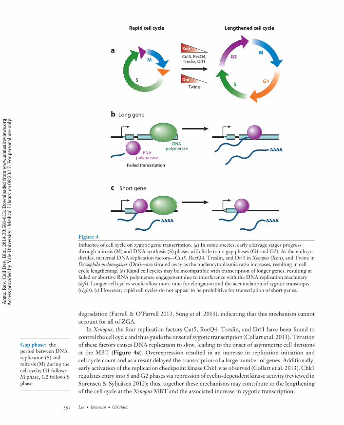
Other evidence suggests that the N/C ratio affects transcription activation indirectly, throughregulation of cell cycle rate. Edgar et al. (1986) found that D. melanogaster mitotic cycle lengthprior to cellularization slows according to the N/C ratio. Recently, the Cdc25 homolog Twinehas been implicated in effecting this cell cycle pause: Degradation of Twine, possibly in an N/Cratio–dependent manner, stabilizes Cdk1 phosphorylation, thus preventing entry into mitosis andallowing zygotic transcription to occur (Figure 4a) (Di Talia et al. 2013, Farrell & O’Farrell2013). However, early zygotic transcription prior to cellularization is in turn required for Twine
590 Lee · Bonneau · Giraldez
Ann
u. R
ev. C
ell D
ev. B
iol.
2014
.30:
581-
613.
Dow
nloa
ded
from
ww
w.a
nnua
lrev
iew
s.or
g A
cces
s pr
ovid
ed b
y Y
ale
Uni
vers
ity -
Med
ical
Lib
rary
on
08/2
0/17
. For
per
sona
l use
onl
y.
CB30CH23-Giraldez ARI 11 September 2014 7:56
d
Le
ve
ls
Cell cycles
Repressors
Transcription
a
b
c
Activators DNAtemplate
Figure 3General models for zygotic genome activation. (a–c) Schematics illustrating the relationship betweenmaternal factors and the zygotic genome as cells divide. (a) Early during embryogenesis, a hypotheticalmaternal repressor (red ) prevents zygotic transcription. After several divisions, the repressor is titrated awayas the ratio of nuclear material to cytoplasm increases (the nucleocytoplasmic ratio), thus allowingtranscription to initiate. (b) Developmental time may be necessary for sufficient levels of an activator ( green)to accumulate, e.g., resulting from activated translation of a maternal mRNA. (c) Alternatively, the embryomay already possess transcriptional activators (orange) and be transcriptionally competent early, but athreshold amount of DNA template is required for transcription to be detected. (d ) All of these generalmechanisms could combine to elicit increasing transcription levels over developmental time.
www.annualreviews.org • Maternal-to-Zygotic Transition 591
Ann
u. R
ev. C
ell D
ev. B
iol.
2014
.30:
581-
613.
Dow
nloa
ded
from
ww
w.a
nnua
lrev
iew
s.or
g A
cces
s pr
ovid
ed b
y Y
ale
Uni
vers
ity -
Med
ical
Lib
rary
on
08/2
0/17
. For
per
sona
l use
onl
y.
CB30CH23-Giraldez ARI 11 September 2014 7:56
b
a
c
Rapid cell cycle Lengthened cell cycle
Failed transcription
RNApolymerase
DNApolymerase
Long gene
Short gene
Twine
Cut5, RecQ4,Treslin, Drf1
Dm
Xen
G1
G2
S
M
S
M
Figure 4Influence of cell cycle on zygotic gene transcription. (a) In some species, early cleavage stages progressthrough mitosis (M) and DNA synthesis (S) phases with little to no gap phases (G1 and G2). As the embryodivides, maternal DNA replication factors—Cut5, RecQ4, Treslin, and Drf1 in Xenopus (Xen), and Twine inDrosophila melanogaster (Dm)—are titrated away as the nucleocytoplasmic ratio increases, resulting in cellcycle lengthening. (b) Rapid cell cycles may be incompatible with transcription of longer genes, resulting infailed or abortive RNA polymerase engagement due to interference with the DNA replication machinery(left). Longer cell cycles would allow more time for elongation and the accumulation of zygotic transcripts(right). (c) However, rapid cell cycles do not appear to be prohibitive for transcription of short genes.
Gap phase: theperiod between DNAreplication (S) andmitosis (M) during thecell cycle; G1 followsM phase, G2 follows Sphase
degradation (Farrell & O’Farrell 2013, Sung et al. 2013), indicating that this mechanism cannotaccount for all of ZGA.
In Xenopus, the four replication factors Cut5, RecQ4, Treslin, and Drf1 have been found tocontrol the cell cycle and thus guide the onset of zygotic transcription (Collart et al. 2013). Titrationof these factors causes DNA replication to slow, leading to the onset of asymmetric cell divisionsat the MBT (Figure 4a). Overexpression resulted in an increase in replication initiation andcell cycle count and as a result delayed the transcription of a large number of genes. Additionally,early activation of the replication checkpoint kinase Chk1 was observed (Collart et al. 2013). Chk1regulates entry into S and G2 phases via repression of cyclin-dependent kinase activity (reviewed inSørensen & Syljuasen 2012); thus, together these mechanisms may contribute to the lengtheningof the cell cycle at the Xenopus MBT and the associated increase in zygotic transcription.
592 Lee · Bonneau · Giraldez
Ann
u. R
ev. C
ell D
ev. B
iol.
2014
.30:
581-
613.
Dow
nloa
ded
from
ww
w.a
nnua
lrev
iew
s.or
g A
cces
s pr
ovid
ed b
y Y
ale
Uni
vers
ity -
Med
ical
Lib
rary
on
08/2
0/17
. For
per
sona
l use
onl
y.
CB30CH23-Giraldez ARI 11 September 2014 7:56
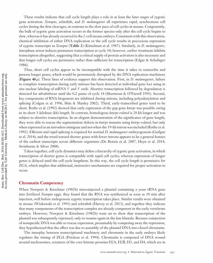
These results indicate that cell cycle length plays a role in at least the later stages of zygoticgene activation. Xenopus, zebrafish, and D. melanogaster all experience rapid, synchronous cellcycles during the first cleavages, in contrast to the slow pace of cell cycles in mouse. Congruently,the bulk of zygotic gene activation occurs in the former species only after the cell cycle begins toslow, whereas it has already occurred in the 2-cell mouse embryo. Consistent with this observation,chemical inhibition of either DNA replication or the cell cycle results in precocious expressionof zygotic transcripts in Xenopus (Table 2) (Kimelman et al. 1987). Similarly, in D. melanogaster,interphase arrest induces premature transcription at cycle 10; however, earlier treatment inhibitstranscription altogether, suggesting that a critical supply of protein activators is also necessary andthat longer cell cycles are permissive rather than sufficient for transcription (Edgar & Schubiger1986).
Thus, short cell cycles appear to be incompatible with the time it takes to transcribe andprocess longer genes, which would be prematurely disrupted by the DNA replication machinery(Figure 4b,c). Three lines of evidence support this observation. First, in D. melanogaster, failureto complete transcription during early mitoses has been detected at individual gene loci using insitu nuclear labeling of mRNA 5′ and 3′ ends. Abortive transcription followed by degradation isdetected for ultrabithorax until the G2 pause of cycle 14 (Shermoen & O’Farrell 1991). Second,key components of RNA biogenesis are inhibited during mitosis, including polyadenylation andsplicing (Colgan et al. 1996, Shin & Manley 2002). Third, early-transcribed genes tend to beshort. Rothe et al. (1992) showed that early expression of the gap gene knirps was possible owingto its short 3-kilobase (kb) length. In contrast, homologous knirps-related is 20 kb longer and wassubject to abortive transcription. In an elegant demonstration of the significance of gene length,they were able to rescue the segmentation defects in knirps mutants using knirps-related, but onlywhen expressed as an intronless minigene and not when the 19-kb intron was included (Rothe et al.1992). Efficient and rapid splicing is required for normal D. melanogaster embryogenesis (Guilguret al. 2014), and the trend toward shorter genes with fewer introns appears to be a general featureof the earliest transcripts across different organisms (De Renzis et al. 2007, Heyn et al. 2014,Swinburne & Silver 2008).
Taken together, cell cycle dynamics may define a hierarchy of zygotic gene activation, in whichtranscription of shorter genes is compatible with rapid cell cycles, whereas expression of longergenes is delayed until the cell cycle lengthens. In this way, the cell cycle length is permissive forZGA, which implies that additional, instructive mechanisms are required for proper activation tooccur.
Chromatin Competency
When Newport & Kirschner (1982b) microinjected a plasmid containing a yeast tRNA geneinto fertilized Xenopus eggs, they found that the RNA was synthesized as soon as 10 min afterinjection, well before endogenous zygotic transcription takes place. Similar results were obtainedin mouse (Wiekowski et al. 1993) and zebrafish (Harvey et al. 2013), and together they indicatethat many components of the transcription complex are already competent in the early vertebrateembryo. However, Newport & Kirschner (1982b) went on to show that transcription of theplasmid was subsequently repressed, only to resume again in the late blastula. Because coinjectionof nonspecific DNA was able to rescue expression, presumably by competing away the repression,they hypothesized that the effect was due to assembly of the plasmid DNA into closed chromatin.
The interplay between transcriptional machinery and chromatin in the early embryo likelyregulates the timing of ZGA (Prioleau et al. 1994). Chromatin is composed of DNA woundaround nucleosomes, octamers of the core histone proteins H2A, H2B, H3, and H4, which are in
www.annualreviews.org • Maternal-to-Zygotic Transition 593
Ann
u. R
ev. C
ell D
ev. B
iol.
2014
.30:
581-
613.
Dow
nloa
ded
from
ww
w.a
nnua
lrev
iew
s.or
g A
cces
s pr
ovid
ed b
y Y
ale
Uni
vers
ity -
Med
ical
Lib
rary
on
08/2
0/17
. For
per
sona
l use
onl
y.
CB30CH23-Giraldez ARI 11 September 2014 7:56
turn joined together by histone linker proteins (e.g., H1) to form compacted heterochromatin. Astranscription factors bind to regulatory regions in the DNA sequence, access to a particular genelocus can be occluded by this closed structure.
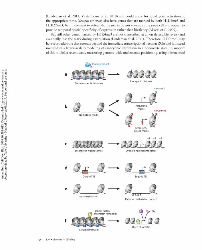
Chromatin accessibility is regulated through nucleosome position and configuration, which isinfluenced by histone variants as well as post-transcriptional modifications of histone N-terminaltails (marks), such as methylation and acetylation. In mouse, hyperaccessible chromatin seems tounderlie ZGA (Cho et al. 2002). Prior to fusion into the zygotic nucleus, the uncondensed malepronucleus is transcriptionally competent, and low levels of endogenous transcription during midto late S phase contribute to the minor wave of ZGA (Aoki et al. 1997, Bouniol et al. 1995, Parket al. 2013, Ram & Schultz 1993, Wiekowski et al. 1993, Xue et al. 2013). In contrast, the femalepronucleus remains transcriptionally silent (Wiekowski et al. 1993). This asymmetry is likely dueto a transient open chromatin state induced by the repackaging of the paternal genome. SpermDNA is bound by arginine-rich protamines, which are exchanged for maternal histones prior to Sphase (Nonchev & Tsanev 1990). These new histones are subject to a transcriptionally permissivepattern of modifications, including H4 hyperacetylation (Adenot et al. 1997, van der Heijden et al.2006) and H3K9 and H3K27 monomethylation (Santos et al. 2005). Protamines, interspersed withpaternal histones, are also found in human, Xenopus, and D. melanogaster sperm DNA (Hammoudet al. 2009, Jayaramaiah Raja & Renkawitz-Pohl 2005, Shechter et al. 2009), but surprisingly notin that of zebrafish (Wu et al. 2011).
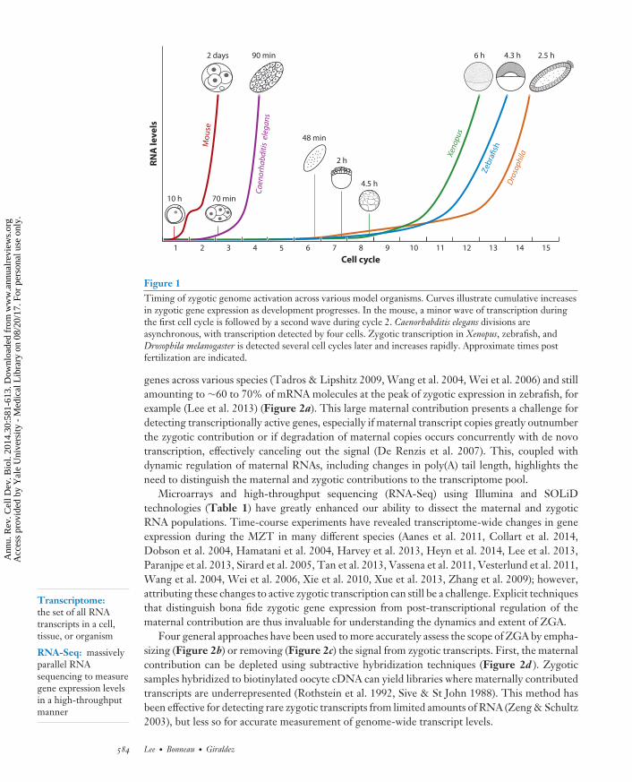
Histone exchange is a general mechanism during embryogenesis, occurring as gamete-specificvariants are replaced by somatic versions. This process could mediate gradual nucleosome un-packing prior to ZGA, as maternal histones are diluted in favor of permissive zygotic versions(Figure 5a). The repressive H2A variant macroH2A is found preferentially in the mouse femalepronucleus and appears to contribute to its transcriptional silence; macroH2A is progressivelylost as the embryo becomes transcriptionally active (Chang et al. 2005). In contrast, embryonicH2A.Z is required for development (Faast et al. 2001, Whittle et al. 2008). In C. elegans, H2A.Z(HTZ-1) was found to be translated from maternal mRNAs and incorporated proximal to devel-opmentally critical genes, suggesting that it influences expression specificity (Whittle et al. 2008).H3.3 incorporation is required for male pronuclear formation in D. melanogaster (Torres-Padillaet al. 2006), whereas H3.3 knockdown in mouse induces chromosomal overcondensation by the2-cell stage and impairs zygotic transcription in the morula (Lin et al. 2013). Finally, repressiveoocyte-specific H1 linker histone variants are replaced by somatic versions coincident with thealleviation of transcriptional quiescence (Fu et al. 2003, Perez-Montero et al. 2013, Smith et al.1988, Tanaka et al. 2001). In D. melanogaster, loss of embryonic H1 variant dBigH1 leads to pre-mature Pol II activity and gene expression (Perez-Montero et al. 2013), showing that one barrierto ZGA is the early prevalence of higher-order chromatin.
Thus, a globally permissive chromatin conformation is a prerequisite for ZGA, which is shapedin part by dynamic incorporation of embryonic histone variants. However, the specificity ofactivation—i.e., the genes that are eventually transcribed from the competent genome—likelyrequires local changes to chromatin accessibility. In the following two subsections, we examinethe roles that histone marks and epigenetic prepatterning may play in guiding these changes.
Histone Modifications
Two types of histone modifications have been implicated in shaping gene expression during theMZT: lysine acetylation and lysine (tri)methylation. In mouse, permissive H4 acetylation distin-guishes the transcriptionally active male pronucleus from the silent female pronucleus (Adenotet al. 1997, van der Heijden et al. 2006). Following the minor wave of ZGA, histone deacetylase
594 Lee · Bonneau · Giraldez
Ann
u. R
ev. C
ell D
ev. B
iol.
2014
.30:
581-
613.
Dow
nloa
ded
from
ww
w.a
nnua
lrev
iew
s.or
g A
cces
s pr
ovid
ed b
y Y
ale
Uni
vers
ity -
Med
ical
Lib
rary
on
08/2
0/17
. For
per
sona
l use
onl
y.
CB30CH23-Giraldez ARI 11 September 2014 7:56
H3K4me3 andH3K27me3:post-transcriptionalmodifications ofhistone H3 at lysine 4and 27, consisting oftrimethylation of theepsilon amino group
Embryonic stem (ES)cells: pluripotent cellsderived from theblastocyst capable ofdifferentiating into anyof the three germlayers
activity contributes to a transient period of transcriptional repression, such that injected plas-mids as well as endogenous genes that are normally expressed at the 1-cell stage are not readilytranscribed in the 2-cell embryo (Martınez-Salas et al. 1989, Wiekowski et al. 1991). Inhibitionof histone deacetylases (Davis et al. 1996) or DNA synthesis (Table 2) (Christians et al. 1995)relieves this repression, suggesting that replication-dependent deacetylation provides transcrip-tional specificity for the major wave of ZGA. Widespread H4 deacetylation leading up to theMBT has also been observed in Xenopus (Dimitrov et al. 1993), consistent with the creation of adefault off transcriptional state coupled with gene-specific regulation of chromatin accessibility.Chromatin remodelers that induce or respond to acetylation are likely involved in this process.Two maternally provided components of the ATP-dependent chromatin remodeling SWI/SNFcomplex, Brg1 and SRG3, are required for mouse embryogenesis (Bultman et al. 2006, Sun et al.2007), with loss of Brg1 resulting in reduced levels of 30% of zygotic genes and arrest at the 2-cellstage (Bultman et al. 2006).
H3 lysine methylation also influences the timing of gene activation during the MZT (Akkerset al. 2009, Chen et al. 2013, Lindeman et al. 2011, Schuettengruber et al. 2009, Vastenhouw et al.2010), though its effects vary widely across different species and contexts. The opposing marksH3K4 and H3K27 trimethylation (H3K4me3 and H3K27me3) at gene promoter regions havereceived considerable attention for their association with active and repressed gene expression,respectively (Figure 5b). In mouse, the transcriptionally inactive female pronucleus is associatedwith H3K27me3, with the transcriptionally competent male pronucleus acquiring H3K27me3only toward the end of the minor ZGA (Santos et al. 2005). However, H3K4me3 is also foundpreferentially in the female pronucleus despite its transcriptional quiescence, and this asymmetryrapidly diminishes as the male pronucleus incorporates maternal H3 histones (Lepikhov & Walter2004). Thus, H3K27me3, but not H3K4me3, seems to affect early mouse gene activity.
In other organisms where large numbers of embryos are more readily available, high-throughput chromatin immunoprecipitation assays (Table 1) show greater association ofH3K4me3 with transcription, but its role in guiding ZGA is not straightforward. In D. melanogaster,H3K4me3 becomes strongly enriched in zygotic-transcribed genes that have a maternal contri-bution (Chen et al. 2013, Schuettengruber et al. 2009), but only later in development. NeitherH3K4me3 nor H3K27me3 is present prior to cellularization, even among early-expressed genes(Chen et al. 2013).
For both zebrafish and Xenopus, H3K4me3 is found across embryonic gene promoters, witha preference for those with housekeeping roles expressed soon after ZGA (Akkers et al. 2009,Lindeman et al. 2011, Vastenhouw et al. 2010). In contrast, H3K27me3 preferentially associateswith genes encoding specific developmental functions (Lindeman et al. 2011, Vastenhouw et al.2010) and subject to differential spatial regulation (Akkers et al. 2009). Thus, in these species,H3K4me3 and H3K27me3 appear to distinguish earlier versus later zygotic transcription.
However, H3K4me3 is also found on inactive genes in zebrafish embryos, based on lack ofH3K36me3 signal (which marks Pol II elongation) or evidence of transcription in RNA-Seqexperiments (Lindeman et al. 2011, Vastenhouw et al. 2010). These genes do tend to be expressedat later stages, suggesting that H3K4 trimethylation also marks promoters that are poised forrapid mobilization in the appropriate developmental context (Lindeman et al. 2011, Vastenhouwet al. 2010). In addition, many of these promoters are also simultaneously occupied by inactivatingH3K27me3 (Figure 5b) (Vastenhouw et al. 2010), reminiscent of so-called bivalent domainsthat have been described in embryonic stem (ES) cells. In ES cells, bivalent marks are associatedwith genes with later roles in differentiated lineages, but whose expression is repressed duringpluripotency (Bernstein et al. 2006). Analogously, during zebrafish embryogenesis, bivalent marksare found in the promoters of lineage-specific genes that are not expressed immediately at ZGA
www.annualreviews.org • Maternal-to-Zygotic Transition 595
Ann
u. R
ev. C
ell D
ev. B
iol.
2014
.30:
581-
613.
Dow
nloa
ded
from
ww
w.a
nnua
lrev
iew
s.or
g A
cces
s pr
ovid
ed b
y Y
ale
Uni
vers
ity -
Med
ical
Lib
rary
on
08/2
0/17
. For
per
sona
l use
onl
y.
CB30CH23-Giraldez ARI 11 September 2014 7:56
(Lindeman et al. 2011, Vastenhouw et al. 2010) and could allow for rapid gene activation atthe appropriate time. Xenopus embryos also have genes that are marked by both H3K4me3 andH3K27me3, but in contrast to zebrafish, the marks do not coexist in the same cell and appear toprovide temporal-spatial specificity of expression rather than bivalency (Akkers et al. 2009).
But still other genes marked by H3K4me3 are not transcribed at all (at detectable levels) andeventually lose the mark during gastrulation (Lindeman et al. 2011). Therefore, H3K4me3 mayhave a broader role that extends beyond the immediate transcriptional needs at ZGA and is insteadinvolved in a larger-scale remodeling of embryonic chromatin to a nonoocyte state. In supportof this model, a recent study measuring genome-wide nucleosome positioning, using micrococcal
e
HypomethylatedPaternal methylation pattern
d
Oocyte TSS Zygotic TSS
No histone marks
Repressed/poised marks
Activatingmarks
H3K4me3
H3K27me3
b
Disordered nucleosomes Ordered nucleosome arrays
c
+1 +2 +3
a
Histone variant
Gamete-specific histonesEmbryonic histones
Closed chromatinOpen chromatin
TFsPioneer factor/chromatin remodeler
f
AT CG
596 Lee · Bonneau · Giraldez
Ann
u. R
ev. C
ell D
ev. B
iol.
2014
.30:
581-
613.
Dow
nloa
ded
from
ww
w.a
nnua
lrev
iew
s.or
g A
cces
s pr
ovid
ed b
y Y
ale
Uni
vers
ity -
Med
ical
Lib
rary
on
08/2
0/17
. For
per
sona
l use
onl
y.
CB30CH23-Giraldez ARI 11 September 2014 7:56
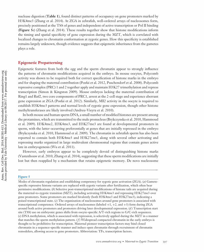
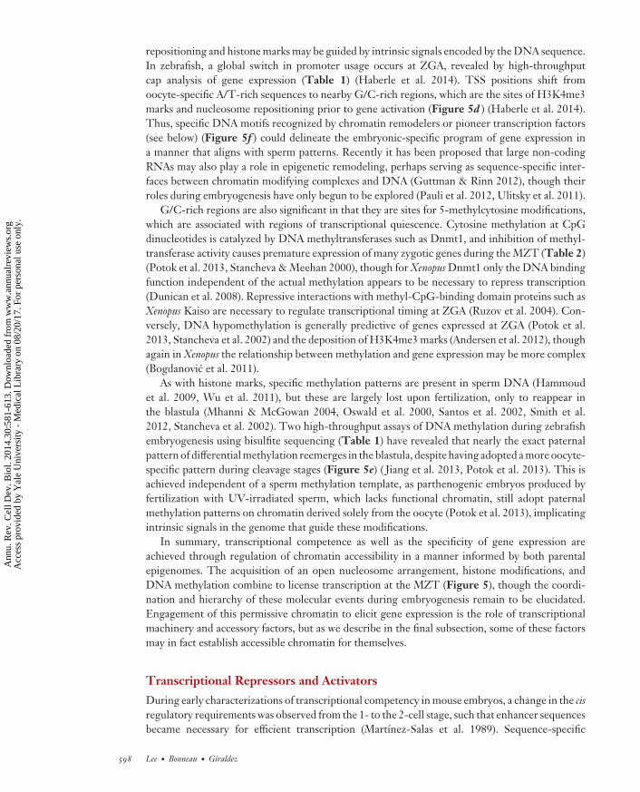
nuclease digestion (Table 1), found distinct patterns of occupancy on gene promoters marked byH3K4me3 (Zhang et al. 2014). At ZGA in zebrafish, well-ordered arrays of nucleosomes form,precisely positioned at the TSS of genes and independent of active transcription or Pol II binding(Figure 5c) (Zhang et al. 2014). These results together show that histone modifications informthe timing and spatial specificity of gene expression during the MZT, which is correlated withlocalized changes to chromatin conformation at zygotic genes. How this specificity is establishedremains largely unknown, though evidence suggests that epigenetic inheritance from the gametesplays a role.
Epigenetic Prepatterning
Epigenetic features from both the egg and the sperm chromatin appear to strongly influencethe patterns of chromatin modifications acquired in the embryo. In mouse oocytes, Polycombactivity was shown to be required both for correct specification of histone marks in the embryoand for shaping the maternal contribution (Posfai et al. 2012, Puschendorf et al. 2008). Polycombrepressive complex (PRC) 1 and 2 together apply and maintain H3K27 trimethylation and represstranscription (Simon & Kingston 2009). Mouse embryos lacking the maternal contribution ofRing1 and Rnf2, two core components of PRC1, arrest at the 2-cell stage and experience aberrantgene expression at ZGA (Posfai et al. 2012). Similarly, Mll2 activity in the oocyte is required toestablish H3K4me3 patterns and normal levels of zygotic gene expression, though other histonemethyltransferases are likely involved (Andreu-Vieyra et al. 2010).
In both mouse and human sperm DNA, a small number of modified histones are present amongthe protamines, which are transmitted to the male pronucleus (Brykczynska et al. 2010, Hammoudet al. 2009). H3K4me2, H3K4me3, and H3K27me3 are found at developmental promoters insperm, with the latter occurring preferentially at genes that are initially repressed in the embryo(Brykczynska et al. 2010, Hammoud et al. 2009). The chromatin in zebrafish sperm has also beenreported to contain both H3K4me3 and H3K27me3, along with several other activating andrepressing marks organized in large multivalent chromosomal regions that contain genes activelate in embryogenesis (Wu et al. 2011).
However, early embryos appear to be completely devoid of distinguishing histone marks(Vastenhouw et al. 2010, Zhang et al. 2014), suggesting that these sperm modifications are initiallylost but then reapplied by a mechanism that retains epigenetic memory. De novo nucleosome
←−−−−−−−−−−−−−−−−−−−−−−−−−−−−−−−−−−−−−−−−−−−−−−−−−−−−−−−−−−−−−−−−−−−−−−−−Figure 5Modes of chromatin regulation and establishing competency for zygotic gene activation (ZGA). (a) Gamete-specific repressive histone variants are replaced with zygotic variants after fertilization, which often bearpermissive modifications. (b) Selective post-transcriptional modifications of histone tails are acquired duringthe maternal-to-zygotic transition (MZT), including activating H3K4me3 and repressing H3K27me3 overgene promoters. Some promoters are marked bivalently (both H3K4me3 and H3K27me3), indicating apoised transcriptional state. (c) The organization of nucleosomes around gene promoters is associated withtranscriptional competence. Ordered arrays of nucleosomes (labeled +1, +2, and +3) form during ZGAaround both active promoters and promoters driving later developmental expression. (d ) Transcription startsite (TSS) use on embryonic genes shifts from oocyte-specific A/T-rich regions to G/C-rich sequences.(e) DNA methylation, which is associated with repression, is selectively applied during the MZT in a mannerthat matches the sperm methylation pattern. ( f ) Widespread compacted chromatin in the early embryo isthought to be prohibitive for transcription. Maternal pioneer transcription factors may bind closedchromatin in a sequence-specific manner and induce open chromatin through recruitment of chromatinremodelers, allowing access to gene promoters. Abbreviation: TFs, transcription factors.
www.annualreviews.org • Maternal-to-Zygotic Transition 597
Ann
u. R
ev. C
ell D
ev. B
iol.
2014
.30:
581-
613.
Dow
nloa
ded
from
ww
w.a
nnua
lrev
iew
s.or
g A
cces
s pr
ovid
ed b
y Y
ale
Uni
vers
ity -
Med
ical
Lib
rary
on
08/2
0/17
. For
per
sona
l use
onl
y.
CB30CH23-Giraldez ARI 11 September 2014 7:56
repositioning and histone marks may be guided by intrinsic signals encoded by the DNA sequence.In zebrafish, a global switch in promoter usage occurs at ZGA, revealed by high-throughputcap analysis of gene expression (Table 1) (Haberle et al. 2014). TSS positions shift fromoocyte-specific A/T-rich sequences to nearby G/C-rich regions, which are the sites of H3K4me3marks and nucleosome repositioning prior to gene activation (Figure 5d ) (Haberle et al. 2014).Thus, specific DNA motifs recognized by chromatin remodelers or pioneer transcription factors(see below) (Figure 5f ) could delineate the embryonic-specific program of gene expression ina manner that aligns with sperm patterns. Recently it has been proposed that large non-codingRNAs may also play a role in epigenetic remodeling, perhaps serving as sequence-specific inter-faces between chromatin modifying complexes and DNA (Guttman & Rinn 2012), though theirroles during embryogenesis have only begun to be explored (Pauli et al. 2012, Ulitsky et al. 2011).
G/C-rich regions are also significant in that they are sites for 5-methylcytosine modifications,which are associated with regions of transcriptional quiescence. Cytosine methylation at CpGdinucleotides is catalyzed by DNA methyltransferases such as Dnmt1, and inhibition of methyl-transferase activity causes premature expression of many zygotic genes during the MZT (Table 2)(Potok et al. 2013, Stancheva & Meehan 2000), though for Xenopus Dnmt1 only the DNA bindingfunction independent of the actual methylation appears to be necessary to repress transcription(Dunican et al. 2008). Repressive interactions with methyl-CpG-binding domain proteins such asXenopus Kaiso are necessary to regulate transcriptional timing at ZGA (Ruzov et al. 2004). Con-versely, DNA hypomethylation is generally predictive of genes expressed at ZGA (Potok et al.2013, Stancheva et al. 2002) and the deposition of H3K4me3 marks (Andersen et al. 2012), thoughagain in Xenopus the relationship between methylation and gene expression may be more complex(Bogdanovic et al. 2011).
As with histone marks, specific methylation patterns are present in sperm DNA (Hammoudet al. 2009, Wu et al. 2011), but these are largely lost upon fertilization, only to reappear inthe blastula (Mhanni & McGowan 2004, Oswald et al. 2000, Santos et al. 2002, Smith et al.2012, Stancheva et al. 2002). Two high-throughput assays of DNA methylation during zebrafishembryogenesis using bisulfite sequencing (Table 1) have revealed that nearly the exact paternalpattern of differential methylation reemerges in the blastula, despite having adopted a more oocyte-specific pattern during cleavage stages (Figure 5e) ( Jiang et al. 2013, Potok et al. 2013). This isachieved independent of a sperm methylation template, as parthenogenic embryos produced byfertilization with UV-irradiated sperm, which lacks functional chromatin, still adopt paternalmethylation patterns on chromatin derived solely from the oocyte (Potok et al. 2013), implicatingintrinsic signals in the genome that guide these modifications.
In summary, transcriptional competence as well as the specificity of gene expression areachieved through regulation of chromatin accessibility in a manner informed by both parentalepigenomes. The acquisition of an open nucleosome arrangement, histone modifications, andDNA methylation combine to license transcription at the MZT (Figure 5), though the coordi-nation and hierarchy of these molecular events during embryogenesis remain to be elucidated.Engagement of this permissive chromatin to elicit gene expression is the role of transcriptionalmachinery and accessory factors, but as we describe in the final subsection, some of these factorsmay in fact establish accessible chromatin for themselves.
Transcriptional Repressors and Activators
During early characterizations of transcriptional competency in mouse embryos, a change in the cisregulatory requirements was observed from the 1- to the 2-cell stage, such that enhancer sequencesbecame necessary for efficient transcription (Martınez-Salas et al. 1989). Sequence-specific
598 Lee · Bonneau · Giraldez
Ann
u. R
ev. C
ell D
ev. B
iol.
2014
.30:
581-
613.
Dow
nloa
ded
from
ww
w.a
nnua
lrev
iew
s.or
g A
cces
s pr
ovid
ed b
y Y
ale
Uni
vers
ity -
Med
ical
Lib
rary
on
08/2
0/17
. For
per
sona
l use
onl
y.
CB30CH23-Giraldez ARI 11 September 2014 7:56
transcription factors that bind gene loci to recruit the transcriptional machinery may thus help nav-igate a repressive chromatin landscape during the major ZGA (Majumder & DePamphilis 1995).
Across all species, the availability of both general and specific transcription factors is expected tohelp regulate gene expression at ZGA. Components of the TFIID complex, which binds the corepromoter to activate basal transcription, are one point of regulation. In C. elegans, TAF-4 is se-questered in the cytoplasm by phosphorylated OMA-1 and OMA-2, thus preventing TFIID fromassembling onto nuclear DNA during the first cleavages (Guven-Ozkan et al. 2008). Phosphor-ylation is mediated by fertilization through activation of the kinase MBK-2, which interestinglyis also responsible for phosphorylating other maternal proteins to mark them for degradation(Stitzel et al. 2006). Pol II inactivity is maintained until the 4-cell embryo, when OMA-1 and -2themselves are degraded and ZGA begins (Guven-Ozkan et al. 2008).
Another TFIID subunit, TATA-binding protein (TBP), along with its paralogs TBP2 andTBP-like, was found to be essential for embryonic development in Xenopus, C. elegans, and zebrafish(Bartfai et al. 2004; Dantonel et al. 2000; Ferg et al. 2007; Jallow et al. 2004; Kaltenbach et al. 2000;Martianov et al. 2002; Muller et al. 2001; Prioleau et al. 1994; Veenstra et al. 1999, 2000), withsemiredundant roles in transcription activation. In Xenopus, TBP is rate limiting for embryonictranscription (Prioleau et al. 1994, Veenstra et al. 1999). Although TBP mRNA is maternallyprovided, protein levels are undetectable until the early blastula (Veenstra et al. 1999). Thisis in contrast to other basal transcription factors, such as TGIIB and TFIIF RAP74 (Veenstraet al. 1999), suggesting that specific translational repression of TBP, likely mediated by delayedcytoplasmic polyadenylation, contributes to early transcriptional quiescence. In mouse, TBP lossresults in failed transcriptional activation in the blastocyst, though the effect was observed only forPol I and III transcription (Martianov et al. 2002). Additionally, in D. melanogaster, TBP is stronglyassociated with the earliest transcribed genes (Chen et al. 2013). Overall, differential TATA boxusage in gene promoters coupled with post-transcriptional regulation of maternal TATA-bindingfactors may be a mechanism to define temporally specific patterns of activation for different setsof genes during the MZT.
Establishing critical levels of other activating and repressive transcription factors likely playsa significant role during ZGA; however, to date few such factors have been identified. InD. melanogaster, Tramtrack (TTK) is a maternal inhibitor of fushi tarazu expression (Brown et al.1991, Pritchard & Schubiger 1996). Nuclear concentration of TTK is simultaneously reduced byan increasing N/C ratio and by regulated degradation of ttk mRNA by the maternal clearancefactor Smaug (Benoit et al. 2009, Tadros et al. 2007). Loss of Smaug has a widespread effect onzygotic gene expression, likely beyond the effect of sustained TTK activity, suggesting that Smaugis responsible for inactivating several other unknown repressive factors that are involved in ZGA(Benoit et al. 2009, Chen et al. 2014).
Once repression is overcome, early gene activation in D. melanogaster is driven by Zelda (alsoknown as Vielfaltig) (Liang et al. 2008), a maternally deposited transcription factor with ubiquitousexpression in the preblastoderm embryo and specific patterns of subcellular localization over thecourse of the cell cycle (Staudt et al. 2006). Zelda binds promoter regions of early zygotic genesvia a heptamer DNA motif called the TAGteam (De Renzis et al. 2007, ten Bosch et al. 2006)that is conserved in other insects (Biedler et al. 2012). Loss of Zelda causes mitotic defects (Staudtet al. 2006) and a failure to activate 120 early zygotic genes during cycles 8–13 (Liang et al. 2008).However, Zelda has also been shown to bind hundreds of additional gene promoters and enhancersprior to their eventual activation (Harrison et al. 2011, Nien et al. 2011), suggesting a broaderrole in licensing zygotic transcription.
In zebrafish, early zygotic genes are enriched in binding sites for the pluripotency-inducingfactors Nanog, Pou5f3 (formerly known as Pou5f1 and homologous to mammalian Oct4), and
www.annualreviews.org • Maternal-to-Zygotic Transition 599
Ann
u. R
ev. C
ell D
ev. B
iol.
2014
.30:
581-
613.
Dow
nloa
ded
from
ww
w.a
nnua
lrev
iew
s.or
g A
cces
s pr
ovid
ed b
y Y
ale
Uni
vers
ity -
Med
ical
Lib
rary
on
08/2
0/17
. For
per
sona
l use
onl
y.
CB30CH23-Giraldez ARI 11 September 2014 7:56
Induced pluripotentstem (iPS) cell: apluripotent cell that isderived from adifferentiated cell byreprogramming itsgene expression
Sox19b (a homolog of Sox2 in the SoxB1 family) (Lee et al. 2013, Leichsenring et al. 2013). Thesethree factors are maternally provided (Burgess et al. 2002, Okuda et al. 2010, Onichtchouk et al.2010, Schuff et al. 2012, Xu et al. 2012) and are the most highly translated sequence-specific tran-scription factors prior to ZGA, as determined by ribosome profiling (Lee et al. 2013). Combinedloss of these factors has a profound effect on transcriptional output and development, with 82%of genes deficient in the late blastula and a corresponding failure to initiate gastrulation, similarto an α-amanitin phenotype (Lee et al. 2013). Binding of the factors appears to be coordinatedand widespread (Bogdanovic et al. 2012, Leichsenring et al. 2013, Xu et al. 2012), reminiscent oftheir homologs’ behaviors in ES cells and induced pluripotent stem (iPS) cells, where they guidespecific gene expression patterns to induce or maintain pluripotency (reviewed in Young 2011).
Nanog, Sox2, and Oct4 are associated with chromatin remodeling activities in ES and iPS cells(Orkin & Hochedlinger 2011) and may have pioneering activity by binding to regions of repressedchromatin to induce nucleosome repositioning and allow other factors to bind in a cooperativemanner (Figure 5f ) (Zaret & Carroll 2011). In fact, they may play similar roles during ZGA (Leeet al. 2013, Leichsenring et al. 2013) and would thus provide a mechanism by which the silentembryonic genome is initially engaged. In Xenopus, β-catenin has been shown to induce epigeneticmodifications at genes prior to their eventual expression during early embryogenesis, implicatingit as another maternal factor involved in establishing chromatin competency (Blythe et al. 2010).
The widespread binding pattern of Zelda on gene regulatory regions suggests that it too mayhave pioneering activity. Zelda binding is correlated with accessible chromatin, as measured byDNase I hypersensitivity (Table 1) (Harrison et al. 2011, Thomas et al. 2011), and TAGteam se-quences are additionally bound by other factors (Satija & Bradley 2012), including Bicoid StabilityFactor (De Renzis et al. 2007), STAT92E (Tsurumi et al. 2011), and Grainyhead (Harrison et al.2010). In this way, gene activation may be the product of combinatorial binding of Zelda, whichprimes the chromatin, and other transcription factors, which would provide the temporal-spatialspecificity and magnitude of the transcriptional output (Figure 5f ). In sum, maternal transcriptionfactors have widespread roles in guiding ZGA at specific gene loci. However, specific factors withpioneering activity may ultimately determine ZGA by binding repressed chromatin and induc-ing remodeling, thus allowing the transcriptional machinery to access gene promoters and drivezygotic gene expression.
DEVELOPMENTAL SIGNIFICANCE OF THE FIRST ZYGOTIC GENES
Functions of the First Zygotic Genes
Zygotic gene expression during the MZT provides the embryo with what the maternal contribu-tion lacks. To some extent, these genes will encode transcription factors and signaling pathwaycomponents that prepare the embryo for gastrulation and cell specification and differentiation,but there will also be genes that carry out basic functions common to all cell types and tissues,the so-called housekeeping genes. Increased cell number, protein turnover rates, or the need toestablish specific spatial expression patterns may explain why it is necessary to supplement orreplenish large parts of the maternal contribution.
The relative proportion of these two classes of genes varies widely among different organisms,likely correlated with the different times at which ZGA occurs in relation to developmental mile-stones. In mouse, where both the minor and major waves of ZGA occur prior to two cleavages,spanning two days of development, activated genes predominantly encode basic cellular functions,including protein and RNA metabolism; transcription factors and patterning genes seem to acti-vate slightly later (Hamatani et al. 2004, Park et al. 2013, Xue et al. 2013). In contrast, zebrafish
600 Lee · Bonneau · Giraldez
Ann
u. R
ev. C
ell D
ev. B
iol.
2014
.30:
581-
613.
Dow
nloa
ded
from
ww
w.a
nnua
lrev
iew
s.or
g A
cces
s pr
ovid
ed b
y Y
ale
Uni
vers
ity -
Med
ical
Lib
rary
on
08/2
0/17
. For
per
sona
l use
onl
y.
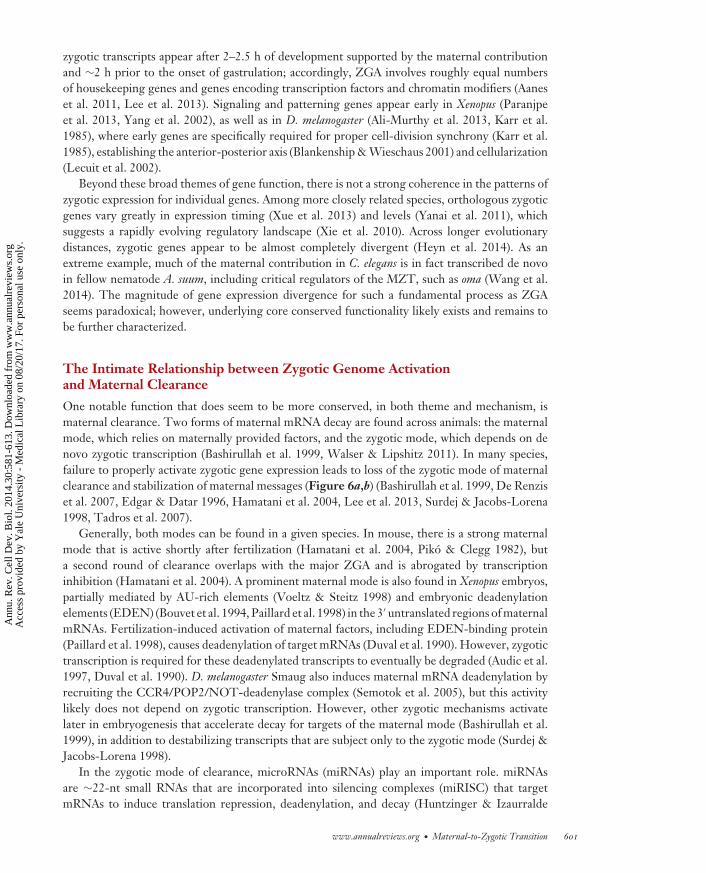
CB30CH23-Giraldez ARI 11 September 2014 7:56
zygotic transcripts appear after 2–2.5 h of development supported by the maternal contributionand ∼2 h prior to the onset of gastrulation; accordingly, ZGA involves roughly equal numbersof housekeeping genes and genes encoding transcription factors and chromatin modifiers (Aaneset al. 2011, Lee et al. 2013). Signaling and patterning genes appear early in Xenopus (Paranjpeet al. 2013, Yang et al. 2002), as well as in D. melanogaster (Ali-Murthy et al. 2013, Karr et al.1985), where early genes are specifically required for proper cell-division synchrony (Karr et al.1985), establishing the anterior-posterior axis (Blankenship & Wieschaus 2001) and cellularization(Lecuit et al. 2002).
Beyond these broad themes of gene function, there is not a strong coherence in the patterns ofzygotic expression for individual genes. Among more closely related species, orthologous zygoticgenes vary greatly in expression timing (Xue et al. 2013) and levels (Yanai et al. 2011), whichsuggests a rapidly evolving regulatory landscape (Xie et al. 2010). Across longer evolutionarydistances, zygotic genes appear to be almost completely divergent (Heyn et al. 2014). As anextreme example, much of the maternal contribution in C. elegans is in fact transcribed de novoin fellow nematode A. suum, including critical regulators of the MZT, such as oma (Wang et al.2014). The magnitude of gene expression divergence for such a fundamental process as ZGAseems paradoxical; however, underlying core conserved functionality likely exists and remains tobe further characterized.
The Intimate Relationship between Zygotic Genome Activationand Maternal Clearance
One notable function that does seem to be more conserved, in both theme and mechanism, ismaternal clearance. Two forms of maternal mRNA decay are found across animals: the maternalmode, which relies on maternally provided factors, and the zygotic mode, which depends on denovo zygotic transcription (Bashirullah et al. 1999, Walser & Lipshitz 2011). In many species,failure to properly activate zygotic gene expression leads to loss of the zygotic mode of maternalclearance and stabilization of maternal messages (Figure 6a,b) (Bashirullah et al. 1999, De Renziset al. 2007, Edgar & Datar 1996, Hamatani et al. 2004, Lee et al. 2013, Surdej & Jacobs-Lorena1998, Tadros et al. 2007).
Generally, both modes can be found in a given species. In mouse, there is a strong maternalmode that is active shortly after fertilization (Hamatani et al. 2004, Piko & Clegg 1982), buta second round of clearance overlaps with the major ZGA and is abrogated by transcriptioninhibition (Hamatani et al. 2004). A prominent maternal mode is also found in Xenopus embryos,partially mediated by AU-rich elements (Voeltz & Steitz 1998) and embryonic deadenylationelements (EDEN) (Bouvet et al. 1994, Paillard et al. 1998) in the 3′ untranslated regions of maternalmRNAs. Fertilization-induced activation of maternal factors, including EDEN-binding protein(Paillard et al. 1998), causes deadenylation of target mRNAs (Duval et al. 1990). However, zygotictranscription is required for these deadenylated transcripts to eventually be degraded (Audic et al.1997, Duval et al. 1990). D. melanogaster Smaug also induces maternal mRNA deadenylation byrecruiting the CCR4/POP2/NOT-deadenylase complex (Semotok et al. 2005), but this activitylikely does not depend on zygotic transcription. However, other zygotic mechanisms activatelater in embryogenesis that accelerate decay for targets of the maternal mode (Bashirullah et al.1999), in addition to destabilizing transcripts that are subject only to the zygotic mode (Surdej &Jacobs-Lorena 1998).
In the zygotic mode of clearance, microRNAs (miRNAs) play an important role. miRNAsare ∼22-nt small RNAs that are incorporated into silencing complexes (miRISC) that targetmRNAs to induce translation repression, deadenylation, and decay (Huntzinger & Izaurralde
www.annualreviews.org • Maternal-to-Zygotic Transition 601
Ann
u. R
ev. C
ell D
ev. B
iol.
2014
.30:
581-
613.
Dow
nloa
ded
from
ww
w.a
nnua
lrev
iew
s.or
g A
cces
s pr
ovid
ed b
y Y
ale
Uni
vers
ity -
Med
ical
Lib
rary
on
08/2
0/17
. For
per
sona
l use
onl
y.
CB30CH23-Giraldez ARI 11 September 2014 7:56
miRNAs
Zygotic genesMaternal genes
TFs
Time
Le
ve
ls
Zygotic genesMaternal genes
Time
Le
ve
ls
c Zebrafish d Drosophila melanogaster
a Reprogrammed
miRNAsTFs
b Persistent oocyte program
Zelda
?
Zygotic
Smaug
miR-309
Maternal
?
ZygoticmiR-430
MaternalNanog,
Pou5f3, Sox19b
Figure 6Activation of the zygotic genome and maternal clearance together reprogram the embryo during thematernal-to-zygotic transition. (a) Maternally provided factors (red ) activate transcription of zygotic genes(blue) through the action of transcription factors (TFs) that engage the early embryonic genome. In turn,zygotic genes, including microRNAs, direct clearance of maternal RNAs. (b) Failure to activate the zygoticgenome by inhibiting maternal TF activity results in stabilization of the maternal program and arresteddevelopment. (c) In zebrafish, the TFs Nanog, Pou5f3, and Sox19b are required for widespread zygotic geneexpression, which includes miR-430. miR-430, along with other unknown factors, induces clearance of alarge subset of maternal RNAs. (d ) In Drosophila melanogaster, Zelda plays a similar role and is responsible foractivating miR-309, which in turn mediates maternal clearance. Maternal Smaug activity is also required formaternal clearance, as well as for miR-309 expression, through an unknown intermediate.
2011). The role of miRNAs in maternal clearance was first identified in zebrafish with the aidof a maternal-zygotic mutant for dicer (MZdicer) (Giraldez et al. 2006), which encodes an RNaseIII enzyme required for canonical miRNA biogenesis. MZdicer embryos are deficient in maturemiR-430, which results in the stabilization of hundreds of maternal mRNAs that are normallytranslationally repressed and degraded in the wild-type embryo, comprising at least 20% ofmaternally contributed genes (Bazzini et al. 2012, Giraldez et al. 2006). The miR-430 gene clusteris one of the earliest and most highly transcribed genes from the zygotic genome (Chen et al.2005, Heyn et al. 2014, Lee et al. 2013) and is directly activated by ZGA transcription factorsNanog, Pou5f3, and SoxB1 (Figure 6c) (Lee et al. 2013). Loss of Nanog in particular results inseverely reduced miR-430 levels leading to stabilized maternal mRNAs (Lee et al. 2013), thusillustrating the tight coordination of ZGA and maternal clearance (Figure 6).
miR-430 has conserved zygotic expression in another teleost fish, medaka (Tani et al. 2010),and has sequence similarity to several other embryonic miRNAs, including miR-295 in mouse;miR-302, miR-327, and miR-516-520 in human (Chen et al. 2005, Giraldez 2010, Svoboda &Flemr 2010); members of the vertebrate miR-17-92 cluster (Tang et al. 2007); and miR-427 inXenopus (Lund et al. 2009, Watanabe et al. 2005). miR-427 has similar expression dynamics tomiR-430 in the early Xenopus embryo (Watanabe et al. 2005) and was likewise shown to mediatematernal clearance activity (Lund et al. 2009), whereas miR-302 was found to be activated by Oct4and Sox2 in ES cells (Card et al. 2008).
602 Lee · Bonneau · Giraldez
Ann
u. R
ev. C
ell D
ev. B
iol.
2014
.30:
581-
613.
Dow
nloa
ded
from
ww
w.a
nnua
lrev
iew
s.or
g A
cces
s pr
ovid
ed b
y Y
ale
Uni
vers
ity -
Med
ical
Lib
rary
on
08/2
0/17
. For
per
sona
l use
onl
y.
CB30CH23-Giraldez ARI 11 September 2014 7:56
Pioneer factor:a transcription factorthat can bind closedchromatin to inducechromatin remodelingand accessibility forother factors
Beyond vertebrates, the miR-309 cluster plays an analogous role in D. melanogaster embryoge-nesis, suggesting that flies have independently evolved a similar strategy to clear maternal mRNAs(Bushati et al. 2008). miR-309 is activated by Zelda along with other early zygotic protein-codingand miRNA genes (Fu et al. 2014, Liang et al. 2008), and loss of miR-309 function leads tostabilization of ∼13% of maternal transcripts cleared in wild-type embryos (Bushati et al. 2008).Interestingly, mRNA clearance activity mediated by maternally provided Smaug is also required fornormal miR-309 expression (Benoit et al. 2009) (Figure 6d ). Thus, distinct pathways of maternalclearance may be functionally linked through the regulation of zygotic transcription (Figure 6).
CONCLUDING REMARKS
Zygotic Gene Expression at the Maternal-to-Zygotic TransitionIs a Multifaceted Process
In conclusion, we have described the context, mechanisms, and function of ZGA and gene expres-sion during the MZT, a process that is fundamentally conserved across animals and yet in manyways is surprisingly variable among different species. High-throughput transcriptome analysesshow widespread, but specific, patterns of zygotic gene transcription, much of which overlapswith the maternal contribution of RNAs. Regulation of the cell cycle, chromatin, and transcrip-tional machinery by maternal factors together elicits transcriptional output with individual-genespecificity. Zygotic genes span the functional requirements of the developing embryo, includingthe clearance of maternal instructions, as the embryonic genome assumes developmental control.Despite the diversity of mechanisms found across different species to regulate gene expression,unifying themes are emerging, including the roles of key transcription factors and miRNAs indirecting the MZT (Figure 6). We anticipate that other common regulatory paradigms will soonemerge.
Although great advances have been made in understanding the function of each of these aspectsof the MZT and ZGA, we still lack a unified picture of how all of these components combine toprecisely activate transcription. Does ZGA arise from the cumulative effects of several independentmechanisms, or is there a cascade of molecular events that has a single point of control? Specifically,understanding how maternal transcription factors influence the embryonic chromatin will be keyto distinguishing these two scenarios, and it may be that the activity of pioneer factors is ultimatelywhat induces genome competency and initiates the zygotic program.
We also lack a complete understanding of how zygotic gene activation and maternal clearancecomplement each other and feed back on themselves. Zygotic transcription is already known tobe required for some maternal clearance, and vice versa, but how this is accomplished and whatfactors are involved are still largely unknown. It is tempting to speculate that there are additionalmechanisms that directly link these two RNA metabolic activities.
Cellular Reprogramming During Embryogenesis and Beyond
We close by revisiting the idea that the MZT is a cellular reprogramming event in vivo, bydeleting the old, maternal program and installing and maintaining the new zygotic program.Maternal clearance, zygotic gene expression, and the cross talk between them combine to inducea transformation in cellular identity, away from the terminally differentiated gametes and towardtransient stages of totipotency, pluripotency, and eventually redifferentiation (Giraldez 2010).Aspects of this process and the mechanisms regulating it are found beyond embryogenesis, mostnotably in stem cells, which have simultaneous requirements to give rise to differentiated cell typesand to self-renew to maintain an undifferentiated state.
www.annualreviews.org • Maternal-to-Zygotic Transition 603
Ann
u. R
ev. C
ell D
ev. B
iol.
2014
.30:
581-
613.
Dow
nloa
ded
from
ww
w.a
nnua
lrev
iew
s.or
g A
cces
s pr
ovid
ed b
y Y
ale
Uni
vers
ity -
Med
ical
Lib
rary
on
08/2
0/17
. For
per
sona
l use
onl
y.
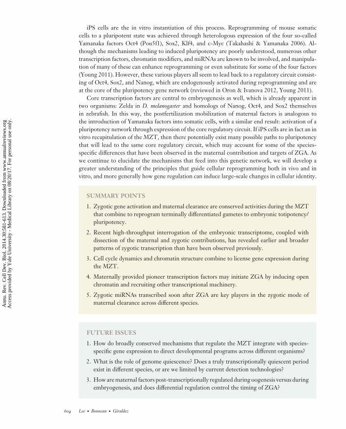
CB30CH23-Giraldez ARI 11 September 2014 7:56
iPS cells are the in vitro instantiation of this process. Reprogramming of mouse somaticcells to a pluripotent state was achieved through heterologous expression of the four so-calledYamanaka factors Oct4 (Pou5f1), Sox2, Klf4, and c-Myc (Takahashi & Yamanaka 2006). Al-though the mechanisms leading to induced pluripotency are poorly understood, numerous othertranscription factors, chromatin modifiers, and miRNAs are known to be involved, and manipula-tion of many of these can enhance reprogramming or even substitute for some of the four factors(Young 2011). However, these various players all seem to lead back to a regulatory circuit consist-ing of Oct4, Sox2, and Nanog, which are endogenously activated during reprogramming and areat the core of the pluripotency gene network (reviewed in Oron & Ivanova 2012, Young 2011).
Core transcription factors are central to embryogenesis as well, which is already apparent intwo organisms: Zelda in D. melanogaster and homologs of Nanog, Oct4, and Sox2 themselvesin zebrafish. In this way, the postfertilization mobilization of maternal factors is analogous tothe introduction of Yamanaka factors into somatic cells, with a similar end result: activation of apluripotency network through expression of the core regulatory circuit. If iPS cells are in fact an invitro recapitulation of the MZT, then there potentially exist many possible paths to pluripotencythat will lead to the same core regulatory circuit, which may account for some of the species-specific differences that have been observed in the maternal contribution and targets of ZGA. Aswe continue to elucidate the mechanisms that feed into this genetic network, we will develop agreater understanding of the principles that guide cellular reprogramming both in vivo and invitro, and more generally how gene regulation can induce large-scale changes in cellular identity.
SUMMARY POINTS
1. Zygotic gene activation and maternal clearance are conserved activities during the MZTthat combine to reprogram terminally differentiated gametes to embryonic totipotency/pluripotency.
2. Recent high-throughput interrogation of the embryonic transcriptome, coupled withdissection of the maternal and zygotic contributions, has revealed earlier and broaderpatterns of zygotic transcription than have been observed previously.
3. Cell cycle dynamics and chromatin structure combine to license gene expression duringthe MZT.
4. Maternally provided pioneer transcription factors may initiate ZGA by inducing openchromatin and recruiting other transcriptional machinery.
5. Zygotic miRNAs transcribed soon after ZGA are key players in the zygotic mode ofmaternal clearance across different species.
FUTURE ISSUES
1. How do broadly conserved mechanisms that regulate the MZT integrate with species-specific gene expression to direct developmental programs across different organisms?
2. What is the role of genome quiescence? Does a truly transcriptionally quiescent periodexist in different species, or are we limited by current detection technologies?
3. How are maternal factors post-transcriptionally regulated during oogenesis versus duringembryogenesis, and does differential regulation control the timing of ZGA?
604 Lee · Bonneau · Giraldez
Ann
u. R
ev. C
ell D
ev. B
iol.
2014
.30:
581-
613.
Dow
nloa
ded
from
ww
w.a
nnua
lrev
iew
s.or
g A
cces
s pr
ovid
ed b
y Y
ale
Uni
vers
ity -
Med
ical
Lib
rary
on
08/2
0/17
. For
per
sona
l use
onl
y.
CB30CH23-Giraldez ARI 11 September 2014 7:56
4. What are the biochemical determinants of maternal mRNA regulation (e.g., structuralelements, ribonucleotide modifications, and RNA-binding proteins)?
5. What changes to embryonic chromatin are directly required for ZGA versus instructivefor gene expression beyond the MZT?
6. What is the role of pioneer transcription factors in inducing open chromatin prior toZGA?
7. How is maternal clearance informative for ZGA, and what other mechanisms regulatematernal mRNA stability?
8. How do the mechanisms underlying the MZT induce cellular reprogramming in vitro?
DISCLOSURE STATEMENT
The authors are not aware of any affiliations, memberships, funding, or financial holdings thatmight be perceived as affecting the objectivity of this review.
ACKNOWLEDGMENTS
We thank K. Neugebauer, C. Rushlow, N. Vastenhouw, M. Stoeckius, C. Takacs, an anony-mous reviewer, and all the members of the Giraldez Lab for insightful discussions and com-ments on the manuscript. The Giraldez Lab is supported by National Institutes of Health grantsF32HD071697-03 (M.T.L.), T32GM007499 (A.R.B.), R01-GM081602-06A1, R01-GM101108-3, R01-GM102251-2, R01-GM103789-2, R01-HD074078-3, and R21-HD073768-2, the PewScholars Program in Biomedical Sciences, and the March of Dimes.
LITERATURE CITED
Aanes H, Ostrup O, Andersen IS, Moen LF, Mathavan S, et al. 2013. Differential transcript isoform usagepre- and post-zygotic genome activation in zebrafish. BMC Genomics 14:331
Aanes H, Winata CL, Lin CH, Chen JP, Srinivasan KG, et al. 2011. Zebrafish mRNA sequencing deciphersnovelties in transcriptome dynamics during maternal to zygotic transition. Genome Res. 21(8):1328–38
Adenot PG, Mercier Y, Renard JP, Thompson EM. 1997. Differential H4 acetylation of paternal and maternalchromatin precedes DNA replication and differential transcriptional activity in pronuclei of 1-cell mouseembryos. Development 124(22):4615–25
Akkers RC, van Heeringen SJ, Jacobi UG, Janssen-Megens EM, Francoijs KJ, et al. 2009. A hierarchy ofH3K4me3 and H3K27me3 acquisition in spatial gene regulation in Xenopus embryos. Dev. Cell 17(3):425–34
Ali-Murthy Z, Lott SE, Eisen MB, Kornberg TB. 2013. An essential role for zygotic expression in the pre-cellular Drosophila embryo. PLOS Genet. 9(4):e1003428
Andersen IS, Reiner AH, Aanes H, Alestrom P, Collas P. 2012. Developmental features of DNA methylationduring activation of the embryonic zebrafish genome. Genome Biol. 13(7):R65
Andreu-Vieyra CV, Chen R, Agno JE, Glaser S, Anastassiadis K, et al. 2010. MLL2 is required in oocytes forbulk histone 3 lysine 4 trimethylation and transcriptional silencing. PLOS Biol. 8(8):e1000453
Aoki F, Hara KT, Schultz RM. 2003. Acquisition of transcriptional competence in the 1-cell mouse embryo:requirement for recruitment of maternal mRNAs. Mol. Reprod. Dev. 64(3):270–74
Aoki F, Worrad DM, Schultz RM. 1997. Regulation of transcriptional activity during the first and second cellcycles in the preimplantation mouse embryo. Dev. Biol. 181(2):296–307
www.annualreviews.org • Maternal-to-Zygotic Transition 605
Ann
u. R
ev. C
ell D
ev. B
iol.
2014
.30:
581-
613.
Dow
nloa
ded
from
ww
w.a
nnua
lrev
iew
s.or
g A
cces
s pr
ovid
ed b
y Y
ale
Uni
vers
ity -
Med
ical
Lib
rary
on
08/2
0/17
. For
per
sona
l use
onl
y.
CB30CH23-Giraldez ARI 11 September 2014 7:56
Audic Y, Omilli F, Osborne HB. 1997. Postfertilization deadenylation of mRNAs in Xenopus laevis embryosis sufficient to cause their degradation at the blastula stage. Mol. Cell. Biol. 17(1):209–18
Barckmann B, Simonelig M. 2013. Control of maternal mRNA stability in germ cells and early embryos.Biochim. Biophys. Acta Gene Regul. Mech. 1829(6–7):714–24
Baroux C, Autran D, Gillmor CS, Grimanelli D, Grossniklaus U. 2008. The maternal to zygotic transition inanimals and plants. Cold Spring Harb. Symp. Quant. Biol. 73:89–100
Bashirullah A, Halsell SR, Cooperstock RL, Kloc M, Karaiskakis A, et al. 1999. Joint action of two RNAdegradation pathways controls the timing of maternal transcript elimination at the midblastula transitionin Drosophila melanogaster. EMBO J. 18(9):2610–20
Baugh LR, Hill AA, Slonim DK, Brown EL, Hunter CP. 2003. Composition and dynamics of the Caenorhabditiselegans early embryonic transcriptome. Development 130(5):889–900
Bartfai R, Balduf C, Hilton T, Rathmann Y, Hadzhiev Y, et al. 2004. TBP2, a vertebrate-specific member ofthe TBP family, is required in embryonic development of zebrafish. Curr. Biol. 14(7):593–98
Bazzini AA, Lee MT, Giraldez AJ. 2012. Ribosome profiling shows that miR-430 reduces translation beforecausing mRNA decay in zebrafish. Science 336(6078):233–37
Benoit B, He CH, Zhang F, Votruba SM, Tadros W, et al. 2009. An essential role for the RNA-bindingprotein Smaug during the Drosophila maternal-to-zygotic transition. Development 136(6):923–32
Bernstein BE, Mikkelsen TS, Xie X, Kamal M, Huebert DJ, et al. 2006. A bivalent chromatin structure markskey developmental genes in embryonic stem cells. Cell 125(2):315–26
Biedler JK, Hu W, Tae H, Tu Z. 2012. Identification of early zygotic genes in the yellow fever mosquito Aedesaegypti and discovery of a motif involved in early zygotic genome activation. PLOS ONE 7(3):e33933
Blankenship JT, Wieschaus E. 2001. Two new roles for the Drosophila AP patterning system in early morpho-genesis. Development 128(24):5129–38
Blythe SA, Cha SW, Tadjuidje E, Heasman J, Klein PS. 2010. β-Catenin primes organizer gene expressionby recruiting a histone H3 arginine 8 methyltransferase, Prmt2. Dev. Cell 19(2):220–31
Bogdanovic O, Fernandez-Minan A, Tena JJ, de la Calle-Mustienes E, Hidalgo C, et al. 2012. Dynamicsof enhancer chromatin signatures mark the transition from pluripotency to cell specification duringembryogenesis. Genome Res. 22(10):2043–53
Bogdanovic O, Long SW, van Heeringen SJ, Brinkman AB, Gomez-Skarmeta, et al. 2011. Temporal un-coupling of the DNA methylome and transcriptional repression during embryogenesis. Genome Res.21(8):1313–27
Bouniol C, Nguyen E, Debey P. 1995. Endogenous transcription occurs at the 1-cell stage in the mouseembryo. Exp. Cell Res. 218(1):57–62
Bouvet P, Omilli F, Arlot-Bonnemains Y, Legagneux V, Roghi C, et al. 1994. The deadenylation conferredby the 3′ untranslated region of a developmentally controlled mRNA in Xenopus embryos is switched topolyadenylation by deletion of a short sequence element. Mol. Cell. Biol. 14(3):1893–900
Braude P, Bolton V, Moore S. 1988. Human gene expression first occurs between the four- and eight-cellstages of preimplantation development. Nature 332(6163):459–61
Brown DD, Littna E. 1964. RNA synthesis during the development of Xenopus laevis, the South African clawedtoad. J. Mol. Biol. 8(5):669–87
Brown JL, Sonoda S, Ueda H, Scott MP, Wu C. 1991. Repression of the Drosophila fushi tarazu (ftz) segmen-tation gene. EMBO J. 10(3):665–74
Brykczynska U, Hisano M, Erkek S, Ramos L, Oakeley EJ, et al. 2010. Repressive and active histone methyl-ation mark distinct promoters in human and mouse spermatozoa. Nat. Struct. Mol. Biol. 17(6):679–87
Bultman SJ, Gebuhr TC, Pan H, Svoboda P, Schultz RM, Magnuson T. 2006. Maternal BRG1 regulateszygotic genome activation in the mouse. Genes Dev. 20(13):1744–54
Burgess S, Reim G, Chen W, Hopkins N, Brand M. 2002. The zebrafish spiel-ohne-grenzen (spg) gene encodesthe POU domain protein Pou2 related to mammalian Oct4 and is essential for formation of the midbrainand hindbrain, and for pre-gastrula morphogenesis. Development 129(4):905–16
Bushati N, Stark A, Brennecke J, Cohen SM. 2008. Temporal reciprocity of miRNAs and their targets duringthe maternal-to-zygotic transition in Drosophila. Curr. Biol. 18(7):501–6
Card DAG, Hebbar PB, Li L, Trotter KW, Komatsu Y, et al. 2008. Oct4/Sox2-regulated miR-302 targetscyclin D1 in human embryonic stem cells. Mol. Cell. Biol. 28(20):6426–38
606 Lee · Bonneau · Giraldez
Ann
u. R
ev. C
ell D
ev. B
iol.
2014
.30:
581-
613.
Dow
nloa
ded
from
ww
w.a
nnua
lrev
iew
s.or
g A
cces
s pr
ovid
ed b
y Y
ale
Uni
vers
ity -
Med
ical
Lib
rary
on
08/2
0/17
. For
per
sona
l use
onl
y.
CB30CH23-Giraldez ARI 11 September 2014 7:56
Chang C-C, Ma Y, Jacobs S, Tian XC, Yang X, Rasmussen TP. 2005. A maternal store of macroH2A isremoved from pronuclei prior to onset of somatic macroH2A expression in preimplantation embryos.Dev. Biol. 278(2):367–80
Chen K, Johnston J, Shao W, Meier S, Staber C, Zeitlinger J. 2013. A global change in RNA polymerase IIpausing during the Drosophila midblastula transition. eLife 2:e00861
Chen L, Dumelie JG, Li X, Cheng MHK, Yang Z, et al. 2014. Global regulation of mRNA translation andstability in the early Drosophila embryo by the Smaug RNA-binding protein. Genome Biol. 15(1):R4
Chen PY, Manninga H, Slanchev K, Chien M, Russo JJ, et al. 2005. The developmental miRNA profiles ofzebrafish as determined by small RNA cloning. Genes Dev. 19(11):1288–93
Cho T, Sakai S, Nagata M, Aoki F. 2002. Involvement of chromatin structure in the regulation of mousezygotic gene activation. Anim. Sci. J. 73(2):113–22
Christians E, Campion E, Thompson EM, Renard JP. 1995. Expression of the HSP 70.1 gene, a landmarkof early zygotic activity in the mouse embryo, is restricted to the first burst of transcription. Development121(1):113–22
Colgan DF, Murthy KGK, Prives C, Manley JL. 1996. Cell-cycle related regulation of poly(A) polymeraseby phosphorylation. Nature 384(6606):282–85
Collart C, Allen GE, Bradshaw CR, Smith JC, Zegerman P. 2013. Titration of four replication factors isessential for the Xenopus laevis midblastula transition. Science 341(6148):893–96
Collart C, Owens NDL, Bhaw-Rosun L, Cooper B, De Domenico E, et al. 2014. High-resolution analysis ofgene activity during the Xenopus mid-blastula transition. Development 141(9):1927–39
Craig SP. 1970. Synthesis of RNA in non-nucleate fragments of sea urchin eggs. J. Mol. Biol. 47(3):615–18Dantonel J-C, Quintin S, Lakatos L, Labouesse M, Tora L. 2000. TBP-like factor is required for embryonic
RNA polymerase II transcription in C. elegans. Mol. Cell 6(3):715–22Davis WJ, De Sousa PA, Schultz RM. 1996. Transient expression of translation initiation factor eIF-4C during
the 2-cell stage of the preimplantation mouse embryo: identification by mRNA differential display andthe role of DNA replication in zygotic gene activation. Dev. Biol. 174(2):190–201
De Renzis S, Elemento O, Tavazoie S, Wieschaus EF. 2007. Unmasking activation of the zygotic genomeusing chromosomal deletions in the Drosophila embryo. PLOS Biol. 5(5):e117
Dekens MPS, Pelegri FJ, Maischein HM, Nusslein-Volhard C. 2003. The maternal-effect gene futile cycle isessential for pronuclear congression and mitotic spindle assembly in the zebrafish zygote. Development130(17):3907–16
Di Talia S, She R, Blythe SA, Lu X, Zhang QF, Wieschaus EF. 2013. Posttranslational control of Cdc25degradation terminates Drosophila’s early cell-cycle program. Curr. Biol. 23(2):127–32
Dimitrov S, Almouzni G, Dasso M, Wolffe AP. 1993. Chromatin transitions during early Xenopus embryoge-nesis: changes in histone H4 acetylation and in linker histone type. Dev. Biol. 160(1):214–27
Dobson AT, Raja R, Abeyta MJ, Taylor T, Shen S, et al. 2004. The unique transcriptome through day 3 ofhuman preimplantation development. Hum. Mol. Genet. 13(14):1461–70
Dunican DS, Ruzov A, Hackett JA, Meehan RR. 2008. xDnmt1 regulates transcriptional silencing in pre-MBTXenopus embryos independently of its catalytic function. Development 135(7):1295–302
Duval C, Bouvet P, Omilli F, Roghi C, Dorel C, et al. 1990. Stability of maternal mRNA in Xenopus embryos:role of transcription and translation. Mol. Cell. Biol. 10(8):4123–29
Edgar BA, Datar SA. 1996. Zygotic degradation of two maternal Cdc25 mRNAs terminates Drosophila’s earlycell cycle program. Genes Dev. 10(15):1966–77
Edgar BA, Kiehle CP, Schubiger G. 1986. Cell cycle control by the nucleo-cytoplasmic ratio in early Drosophiladevelopment. Cell 44(2):365–72
Edgar BA, Schubiger G. 1986. Parameters controlling transcriptional activation during early Drosophila de-velopment. Cell 44(6):871–77
Edgar LG, Wolf N, Wood WB. 1994. Early transcription in Caenorhabditis elegans embryos. Development120(2):443–51
Faast R, Thonglairoam V, Schulz TC, Beall J, Wells JRE, et al. 2001. Histone variant H2A.Z is required forearly mammalian development. Curr. Biol. 11(15):1183–87
Farrell JA, O’Farrell PH. 2013. Mechanism and regulation of Cdc25/Twine protein destruction in embryoniccell-cycle remodeling. Curr. Biol. 23(2):118–26
www.annualreviews.org • Maternal-to-Zygotic Transition 607
Ann
u. R
ev. C
ell D
ev. B
iol.
2014
.30:
581-
613.
Dow
nloa
ded
from
ww
w.a
nnua
lrev
iew
s.or
g A
cces
s pr
ovid
ed b
y Y
ale
Uni
vers
ity -
Med
ical
Lib
rary
on
08/2
0/17
. For
per
sona
l use
onl
y.
CB30CH23-Giraldez ARI 11 September 2014 7:56
Ferg M, Sanges R, Gehrig J, Kiss J, Bauer M, et al. 2007. The TATA-binding protein regulates maternalmRNA degradation and differential zygotic transcription in zebrafish. EMBO J. 26(17):3945–56
Foe VE, Alberts BM. 1983. Studies of nuclear and cytoplasmic behaviour during the five mitotic cycles thatprecede gastrulation in Drosophila embryogenesis. J. Cell Sci. 61(1):31–70
Fu G, Ghadam P, Sirotkin A, Khochbin S, Skoultchi AI, Clarke HJ. 2003. Mouse oocytes and early embryosexpress multiple histone H1 subtypes. Biol. Reprod. 68(5):1569–76
Fu S, Nien C-Y, Liang H-L, Rushlow C. 2014. Co-activation of microRNAs by Zelda is essential for earlyDrosophila development. Development 141(10):2108–18
Gerhart JC. 1980. Mechanisms regulating pattern formation in the amphibian egg and early embryo. InBiological Regulation and Development, ed. R Goldberger, pp. 133–316. Boston, MA: Springer
Giraldez AJ. 2010. microRNAs, the cell’s Nepenthe: clearing the past during the maternal-to-zygotic transitionand cellular reprogramming. Curr. Opin. Genet. Dev. 20(4):369–75
Giraldez AJ, Mishima Y, Rihel J, Grocock RJ, Van Dongen S, et al. 2006. Zebrafish MiR-430 promotesdeadenylation and clearance of maternal mRNAs. Science 312(5770):75–79
Goddard MJ, Pratt HPM. 1983. Control of events during early cleavage of the mouse embryo: an analysis ofthe “2-cell block.” J. Embryol. Exp. Morphol. 73(1):111–33
Golbus MS, Calarco PG, Epstein CJ. 1973. The effects of inhibitors of RNA synthesis (α-amanitin andactinomycin D) on preimplantation mouse embryogenesis. J. Exp. Zool. 186(2):207–16
Guilgur LG, Prudencio P, Sobral D, Liszekova D, Rosa A, Martinho RG. 2014. Requirement for highlyefficient pre-mRNA splicing during Drosophila early embryonic development. eLife 3:e02181
Guttman M, Rinn JL. 2012. Modular regulatory principles of large non-coding RNAs. Nature 482(7385):339–46
Guven-Ozkan T, Nishi Y, Robertson SM, Lin R. 2008. Global transcriptional repression in C. elegans germlineprecursors by regulated sequestration of TAF-4. Cell 135(1):149–60
Haberle V, Li N, Hadzhiev Y, Plessy C, Previti C, et al. 2014. Two independent transcription initiation codesoverlap on vertebrate core promoters. Nature 507:381–85
Hamatani T, Carter MG, Sharov AA, Ko MSH. 2004. Dynamics of global gene expression changes duringmouse preimplantation development. Dev. Cell 6(1):117–31
Hammoud SS, Nix DA, Zhang H, Purwar J, Carrell DT, Cairns BR. 2009. Distinctive chromatin in humansperm packages genes for embryo development. Nature 460(7254):473–78
Harrison MM, Botchan MR, Cline TW. 2010. Grainyhead and Zelda compete for binding to the promotersof the earliest-expressed Drosophila genes. Dev. Biol. 345(2):248–55
Harrison MM, Li XY, Kaplan T, Botchan MR, Eisen MB. 2011. Zelda binding in the early Drosophilamelanogaster embryo marks regions subsequently activated at the maternal-to-zygotic transition. PLOSGenet. 7(10):e1002266
Harvey SA, Sealy I, Kettleborough R, Fenyes F, White R, et al. 2013. Identification of the zebrafish maternaland paternal transcriptomes. Development 140(13):2703–10
Heyn P, Kircher M, Dahl A, Kelso J, Tomancak P, et al. 2014. The earliest transcribed zygotic genes areshort, newly evolved, and different across species. Cell Rep. 6(2):285–92
Howe JA, Howell M, Hunt T, Newport JW. 1995. Identification of a developmental timer regulating thestability of embryonic cyclin A and a new somatic A-type cyclin at gastrulation. Genes Dev. 9(10):1164–76
Huntzinger E, Izaurralde E. 2011. Gene silencing by microRNAs: contributions of translational repressionand mRNA decay. Nat. Rev. Genet. 12(2):99–110
Jallow Z, Jacobi UG, Weeks DL, Dawid IB, Veenstra GJ. 2004. Specialized and redundant roles of TBP anda vertebrate-specific TBP paralog in embryonic gene regulation in Xenopus. Proc. Natl. Acad. Sci. USA101(37):13525–30
Jayaramaiah Raja S, Renkawitz-Pohl R. 2005. Replacement by Drosophila melanogaster protamines and Mst77fof histones during chromatin condensation in late spermatids and role of sesame in the removal of theseproteins from the male pronucleus. Mol. Cell. Biol. 25(14):6165–77
Jiang L, Zhang J, Wang JJ, Wang L, Zhang L, et al. 2013. Sperm, but not oocyte, DNA methylome isinherited by zebrafish early embryos. Cell 153(4):773–84
Kaltenbach L, Horner MA, Rothman JH, Mango SE. 2000. The TBP-like factor CeTLF is required to activateRNA polymerase II transcription during C. elegans embryogenesis. Mol. Cell 6(3):705–13
608 Lee · Bonneau · Giraldez
Ann
u. R
ev. C
ell D
ev. B
iol.
2014
.30:
581-
613.
Dow
nloa
ded
from
ww
w.a
nnua
lrev
iew
s.or
g A
cces
s pr
ovid
ed b
y Y
ale
Uni
vers
ity -
Med
ical
Lib
rary
on
08/2
0/17
. For
per
sona
l use
onl
y.
CB30CH23-Giraldez ARI 11 September 2014 7:56
Kane DA, Hammerschmidt M, Mullins MC, Maischein HM, Brand M, et al. 1996. The zebrafish epibolymutants. Development 123:47–55
Kane DA, Kimmel CB. 1993. The zebrafish midblastula transition. Development 119(2):447–56Karr TL, Ali Z, Drees B, Kornberg T. 1985. The engrailed locus of D. melanogaster provides an essential
zygotic function in precellular embryos. Cell 43(3 Pt.2):591–601Kimelman D, Kirschner M, Scherson T. 1987. The events of the midblastula transition in Xenopus are regulated
by changes in the cell cycle. Cell 48(3):399–407Knowland J, Graham C. 1972. RNA synthesis at the two-cell stage of mouse development. J. Embryol. Exp.
Morphol. 27(1):167–76Laubichler MD, Davidson EH. 2008. Boveri’s long experiment: sea urchin merogones and the establishment
of the role of nuclear chromosomes in development. Dev. Biol. 314(1):1–11Lecuit T, Samanta R, Wieschaus E. 2002. slam encodes a developmental regulator of polarized membrane
growth during cleavage of the Drosophila embryo. Dev. Cell 2(4):425–36Lecuyer E, Yoshida H, Parthasarathy N, Alm C, Babak T, et al. 2007. Global analysis of mRNA localization
reveals a prominent role in organizing cellular architecture and function. Cell 131(1):174–87Lee MT, Bonneau AR, Takacs CM, Bazzini AA, DiVito KR, et al. 2013. Nanog, Pou5f1 and SoxB1 activate
zygotic gene expression during the maternal-to-zygotic transition. Nature 503(7476):360–64Leichsenring M, Maes J, Mossner R, Driever W, Onichtchouk D. 2013. Pou5f1 transcription factor controls
zygotic gene activation in vertebrates. Science 341(6149):1005–9Lepikhov K, Walter J. 2004. Differential dynamics of histone H3 methylation at positions K4 and K9 in the
mouse zygote. BMC Dev. Biol. 4:12Liang HL, Nien CY, Liu HY, Metzstein MM, Kirov N, Rushlow C. 2008. The zinc-finger protein Zelda is a
key activator of the early zygotic genome in Drosophila. Nature 456(7220):400–3Lin C-J, Conti M, Ramalho-Santos M. 2013. Histone variant H3.3 maintains a decondensed chromatin state
essential for mouse preimplantation development. Development 140(17):3624–34Lindeman LC, Andersen IS, Reiner AH, Li N, Aanes H, et al. 2011. Prepatterning of developmental gene
expression by modified histones before zygotic genome activation. Dev. Cell 21(6):993–1004Lott SE, Villalta JE, Schroth GP, Luo S, Tonkin LA, Eisen MB. 2011. Noncanonical compensation of zygotic
X transcription in early Drosophila melanogaster development revealed through single-embryo RNA-seq.PLOS Biol. 9(2):e1000590
Lu X, Li JM, Elemento O, Tavazoie S, Wieschaus EF. 2009. Coupling of zygotic transcription to mitoticcontrol at the Drosophila mid-blastula transition. Development 136(12):2101–10
Lund E, Liu M, Hartley RS, Sheets MD, Dahlberg JE. 2009. Deadenylation of maternal mRNAs mediatedby miR-427 in Xenopus laevis embryos. RNA 15(12):2351–63
Majumder S, DePamphilis ML. 1995. A unique role for enhancers is revealed during early mouse development.BioEssays 17(10):879–89
Martianov I, Viville S, Davidson I. 2002. RNA polymerase II transcription in murine cells lacking the TATAbinding protein. Science 298(5595):1036–39
Martınez-Salas E, Linney E, Hassell J, DePamphilis ML. 1989. The need for enhancers in gene expressionfirst appears during mouse development with formation of the zygotic nucleus. Genes Dev. 3(10):1493–506
Mathavan S, Lee SGP, Mak A, Miller LD, Murthy KRK, et al. 2005. Transcriptome analysis of zebrafishembryogenesis using microarrays. PLOS Genet. 1(2):260–76
Merrill PT, Sweeton D, Wieschaus E. 1988. Requirements for autosomal gene activity during precellularstages of Drosophila melanogaster. Development 104(3):495–509
Mhanni AA, McGowan RA. 2004. Global changes in genomic methylation levels during early developmentof the zebrafish embryo. Dev. Genes Evol. 214(8):412–17
Muller F, Lakatos T, Dantonel J, Strahle U, Tora L. 2001. TBP is not universally required for zygotic RNApolymerase II transcription in zebrafish. Curr. Biol. 11(4):282–87
Nemer M. 1963. Old and new RNA in the embryogenesis of the purple sea urchin. Proc. Natl. Acad. Sci. USA50(2):230
Nepal C, Hadzhiev Y, Previti C, Haberle V, Li N, et al. 2013. Dynamic regulation of the transcription initiationlandscape at single nucleotide resolution during vertebrate embryogenesis. Genome Res. 23(11):1938–50
www.annualreviews.org • Maternal-to-Zygotic Transition 609
Ann
u. R
ev. C
ell D
ev. B
iol.
2014
.30:
581-
613.
Dow
nloa
ded
from
ww
w.a
nnua
lrev
iew
s.or
g A
cces
s pr
ovid
ed b
y Y
ale
Uni
vers
ity -
Med
ical
Lib
rary
on
08/2
0/17
. For
per
sona
l use
onl
y.
CB30CH23-Giraldez ARI 11 September 2014 7:56
Newport J, Kirschner M. 1982a. A major developmental transition in early Xenopus embryos: I. Characteriza-tion and timing of cellular changes at the midblastula stage. Cell 30(3):675–86
Newport J, Kirschner M. 1982b. A major developmental transition in early Xenopus embryos: II. Control ofthe onset of transcription. Cell 30(3):687–96
Nien C-Y, Liang H-L, Butcher S, Sun Y, Fu S, et al. 2011. Temporal coordination of gene networks by Zeldain the early Drosophila embryo. PLOS Genet. 7(10):e1002339
Nonchev S, Tsanev R. 1990. Protamine-histone replacement and DNA replication in the male mouse pronu-cleus. Mol. Reprod. Dev. 25(1):72–76
Okuda Y, Ogura E, Kondoh H, Kamachi Y. 2010. B1 SOX coordinate cell specification with patterning andmorphogenesis in the early zebrafish embryo. PLOS Genet. 6(5):e1000936
Onichtchouk D, Geier F, Polok B, Messerschmidt DM, Mossner R, et al. 2010. Zebrafish Pou5f1-dependenttranscriptional networks in temporal control of early development. Mol. Syst. Biol. 6:354
Orkin SH, Hochedlinger K. 2011. Chromatin connections to pluripotency and cellular reprogramming. Cell145(6):835–50
Oron E, Ivanova N. 2012. Cell fate regulation in early mammalian development. Phys. Biol. 9(4):045002Oswald J, Engemann S, Lane N, Mayer W, Olek A, et al. 2000. Active demethylation of the paternal genome
in the mouse zygote. Curr. Biol. 10(8):475–78Paillard L, Omilli F, Legagneux V, Bassez T, Maniey D, Osborne HB. 1998. EDEN and EDEN-BP, a cis
element and an associated factor that mediate sequence-specific mRNA deadenylation in Xenopus embryos.EMBO J. 17(1):278–87
Paranjpe SS, Jacobi UG, van Heeringen SJ, Veenstra GJC. 2013. A genome-wide survey of maternal andembryonic transcripts during Xenopus tropicalis development. BMC Genomics 14:762
Park S-J, Komata M, Inoue F, Yamada K, Nakai K, et al. 2013. Inferring the choreography of parental genomesduring fertilization from ultralarge-scale whole-transcriptome analysis. Genes Dev. 27(24):2736–48
Pauli A, Valen E, Lin MF, Garber M, Vastenhouw NL, et al. 2012. Systematic identification of long noncodingRNAs expressed during zebrafish embryogenesis. Genome Res. 22(3):577–91
Perez-Montero S, Carbonell A, Moran T, Vaquero A, Azorın F. 2013. The embryonic linker histone H1variant of Drosophila, dBigH1, regulates zygotic genome activation. Dev. Cell 26(6):578–90
Piko L, Clegg KB. 1982. Quantitative changes in total RNA, total poly(A), and ribosomes in early mouseembryos. Dev. Biol. 89(2):362–78
Poccia D, Wolff R, Kragh S, Williamson P. 1985. RNA synthesis in male pronuclei of the sea urchin. Biochim.Biophys. Acta 824(4):349–56
Posfai E, Kunzmann R, Brochard V, Salvaing J, Cabuy E, et al. 2012. Polycomb function during oogenesis isrequired for mouse embryonic development. Genes Dev. 26(9):920–32
Potok ME, Nix DA, Parnell TJ, Cairns BR. 2013. Reprogramming the maternal zebrafish genome afterfertilization to match the paternal methylation pattern. Cell 153(4):759–72
Prioleau MN, Huet J, Sentenac A, Mechali M. 1994. Competition between chromatin and transcriptioncomplex assembly regulates gene expression during early development. Cell 77(3):439–49
Pritchard DK, Schubiger G. 1996. Activation of transcription in Drosophila embryos is a gradual processmediated by the nucleocytoplasmic ratio. Genes Dev. 10(9):1131–42
Puschendorf M, Terranova R, Boutsma E, Mao X, Isono K, et al. 2008. PRC1 and Suv39h specify parentalasymmetry at constitutive heterochromatin in early mouse embryos. Nat. Genet. 40(4):411–20
Qin X, Ahn S, Speed TP, Rubin GM. 2007. Global analyses of mRNA translational control during earlyDrosophila embryogenesis. Genome Biol. 8(4):R63
Ram PT, Schultz RM. 1993. Reporter gene expression in G2 of the 1-cell mouse embryo. Dev. Biol. 156(2):552–56
Richter JD, Lasko P. 2011. Translational control in oocyte development. Cold Spring Harb. Perspect. Biol.3(9):a002758
Rothe M, Pehl M, Taubert H, Jackle H. 1992. Loss of gene function through rapid mitotic cycles in theDrosophila embryo. Nature 359(6391):156–59
Rothstein JL, Johnson D, DeLoia JA, Skowronski J, Solter D, Knowles B. 1992. Gene expression duringpreimplantation mouse development. Genes Dev. 6(7):1190–201
610 Lee · Bonneau · Giraldez
Ann
u. R
ev. C
ell D
ev. B
iol.
2014
.30:
581-
613.
Dow
nloa
ded
from
ww
w.a
nnua
lrev
iew
s.or
g A
cces
s pr
ovid
ed b
y Y
ale
Uni
vers
ity -
Med
ical
Lib
rary
on
08/2
0/17
. For
per
sona
l use
onl
y.
CB30CH23-Giraldez ARI 11 September 2014 7:56
Ruzov A, Dunican DS, Prokhorchouk A, Pennings S, Stancheva I, et al. 2004. Kaiso is a genome-wide repressorof transcription that is essential for amphibian development. Development 131(24):6185–94
Santos F, Hendrich B, Reik W, Dean W. 2002. Dynamic reprogramming of DNA methylation in the earlymouse embryo. Dev. Biol. 241(1):172–82
Santos F, Peters AH, Otte AP, Reik W, Dean W. 2005. Dynamic chromatin modifications characterise thefirst cell cycle in mouse embryos. Dev. Biol. 280(1):225–36
Satija R, Bradley RK. 2012. The TAGteam motif facilitates binding of 21 sequence-specific transcriptionfactors in the Drosophila embryo. Genome Res. 22(4):656–65
Sawicki JA, Magnuson T, Epstein CJ. 1981. Evidence for expression of the paternal genome in the two-cellmouse embryo. Nature 294(5840):450–51
Schuettengruber B, Ganapathi M, Leblanc B, Portoso M, Janschek R, et al. 2009. Functional anatomy ofpolycomb and trithorax chromatin landscapes in Drosophila embryos. PLOS Biol. 7(1):e13
Schuff M, Siegel D, Philipp M, Bundschu K, Heymann N, et al. 2012. Characterization of Danio rerio Nanogand functional comparison to Xenopus Vents. Stem Cells Dev. 21(8):1225–38
Selvig SE, Gross PR, Hunter AL. 1970. Cytoplasmic synthesis of RNA in the sea urchin embryo. Dev. Biol.22(2):343–65
Semotok JL, Cooperstock RL, Pinder BD, Vari HK, Lipshitz HD, Smibert CA. 2005. Smaug recruits theCCR4/POP2/NOT deadenylase complex to trigger maternal transcript localization in the early Drosophilaembryo. Curr. Biol. 15(4):284–94
Shechter D, Nicklay JJ, Chitta RK, Shabanowitz J, Hunt DF, Allis CD. 2009. Analysis of histones in Xenopuslaevis. I. A distinct index of enriched variants and modifications exists in each cell type and is remodeledduring developmental transitions. J. Biol. Chem. 284(2):1064–74
Shermoen AW, O’Farrell PH. 1991. Progression of the cell cycle through mitosis leads to abortion of nascenttranscripts. Cell 67(2):303–10
Shin C, Manley JL. 2002. The SR protein SRp38 represses splicing in M phase cells. Cell 111(3):407–17Simon JA, Kingston RE. 2009. Mechanisms of polycomb gene silencing: knowns and unknowns. Nat. Rev.
Mol. Cell Biol. 10(10):697–708Sirard MA, Dufort I, Vallee M, Massicotte L, Gravel C, et al. 2005. Potential and limitations of bovine-
specific arrays for the analysis of mRNA levels in early development: preliminary analysis using a bovineembryonic array. Reprod. Fertil. Dev. 17(1–2):47–57
Sive HL, St John T. 1988. A simple subtractive hybridization technique employing photoactivatable biotinand phenol extraction. Nucleic Acids Res. 16(22):10937
Skirkanich J, Luxardi G, Yang J, Kodjabachian L, Klein P. 2011. An essential role for transcription before theMBT in Xenopus laevis. Dev. Biol. 357(2):478–91
Smith RC, Dworkin-Rastl E, Dworkin MB. 1988. Expression of a histone H1-like protein is restricted to earlyXenopus development. Genes Dev. 2(10):1284–95
Smith ZD, Chan MM, Mikkelsen TS, Gu H, Gnirke A, et al. 2012. A unique regulatory phase of DNAmethylation in the early mammalian embryo. Nature 484(7394):339–44
Sørensen CS, Syljuasen RG. 2012. Safeguarding genome integrity: the checkpoint kinases ATR, CHK1 andWEE1 restrain CDK activity during normal DNA replication. Nucleic Acids Res. 40(2):477–86
Stancheva I, El-Maarri O, Walter J, Niveleau A, Meehan RR. 2002. DNA methylation at promoter regionsregulates the timing of gene activation in Xenopus laevis embryos. Dev. Biol. 243(1):155–65
Stancheva I, Meehan RR. 2000. Transient depletion of xDnmt1 leads to premature gene activation in Xenopusembryos. Genes Dev. 14(3):313–27
Staudt N, Fellert S, Chung HR, Jackle H, Vorbruggen G. 2006. Mutations of the Drosophila zinc finger-encoding gene vielfaltig impair mitotic cell divisions and cause improper chromosome segregation. Mol.Biol. Cell 17(5):2356–65
Stitzel ML, Pellettieri J, Seydoux G. 2006. The C. elegans DYRK kinase MBK-2 marks oocyte proteins fordegradation in response to meiotic maturation. Curr. Biol. 16(1):56–62
Storfer-Glazer FA, Wood WB. 1994. Effects of chromosomal deficiencies on early cleavage patterning andterminal phenotype in Caenorhabditis elegans embryos. Genetics 137(2):499–508
Sulston JE, Schierenberg E, White JG, Thomson JN. 1983. The embryonic cell lineage of the nematodeCaenorhabditis elegans. Dev. Biol. 100(1):64–119
www.annualreviews.org • Maternal-to-Zygotic Transition 611
Ann
u. R
ev. C
ell D
ev. B
iol.
2014
.30:
581-
613.
Dow
nloa
ded
from
ww
w.a
nnua
lrev
iew
s.or
g A
cces
s pr
ovid
ed b
y Y
ale
Uni
vers
ity -
Med
ical
Lib
rary
on
08/2
0/17
. For
per
sona
l use
onl
y.
CB30CH23-Giraldez ARI 11 September 2014 7:56
Sun F, Tang F, Yan AY, Fang HY, Sheng HZ. 2007. Expression of SRG3, a chromatin-remodelling factor,in the mouse oocyte and early preimplantation embryos. Zygote 15(2):129–38
Sung H-W, Spangenberg S, Vogt N, Großhans J. 2013. Number of nuclear divisions in the Drosophila blas-toderm controlled by onset of zygotic transcription. Curr. Biol. 23(2):133–38
Surdej P, Jacobs-Lorena M. 1998. Developmental regulation of bicoid mRNA stability is mediated by the first43 nucleotides of the 3′ untranslated region. Mol. Cell. Biol. 18(5):2892–900
Svoboda P, Flemr M. 2010. The role of miRNAs and endogenous siRNAs in maternal-to-zygotic reprogram-ming and the establishment of pluripotency. EMBO Rep. 11(8):590–97
Swinburne IA, Silver PA. 2008. Intron delays and transcriptional timing during development. Dev. Cell14(3):324–30
Tadros W, Goldman AL, Babak T, Menzies F, Vardy L, et al. 2007. SMAUG is a major regulator of maternalmRNA destabilization in Drosophila and its translation is activated by the PAN GU kinase. Dev. Cell12(1):143–55
Tadros W, Lipshitz HD. 2009. The maternal-to-zygotic transition: a play in two acts. Development136(18):3033–42
Takahashi K, Yamanaka S. 2006. Induction of pluripotent stem cells from mouse embryonic and adult fibroblastcultures by defined factors. Cell 126(4):663–76
Tan MH, Au KF, Yablonovitch AL, Wills AE, Chuang J, et al. 2013. RNA sequencing reveals a diverse anddynamic repertoire of the Xenopus tropicalis transcriptome over development. Genome Res. 23(1):201–16
Tanaka M, Hennebold JD, Macfarlane J, Adashi EY. 2001. A mammalian oocyte-specific linker histone geneH1oo: homology with the genes for the oocyte-specific cleavage stage histone (cs-H1) of sea urchin andthe B4/H1M histone of the frog. Development 128(5):655–64
Tang F, Kaneda M, O’Carroll D, Hajkova P, Barton SC, et al. 2007. Maternal microRNAs are essential formouse zygotic development. Genes Dev. 21(6):644–48
Tani S, Kusakabe R, Naruse K, Sakamoto H, Inoue K. 2010. Genomic organization and embryonic expressionof miR-430 in medaka (Oryzias latipes): insights into the post-transcriptional gene regulation in earlydevelopment. Gene 449(1–2):41–49
ten Bosch JR, Benavides JA, Cline TW. 2006. The TAGteam DNA motif controls the timing of Drosophilapre-blastoderm transcription. Development 133(10):1967–77
Thomas S, Li XY, Sabo PJ, Sandstrom R, Thurman RE, et al. 2011. Dynamic reprogramming of chromatinaccessibility during Drosophila embryo development. Genome Biol. 12(5):R43
Torres-Padilla M-E, Bannister AJ, Hurd PJ, Kouzarides T, Zernicka-Goetz M. 2006. Dynamic distributionof the replacement histone variant H3.3 in the mouse oocyte and preimplantation embryos. Int. J. Dev.Biol. 50(5):455–61
Tsurumi A, Xia F, Li J, Larson K, LaFrance R, Li WX. 2011. STAT is an essential activator of the zygoticgenome in the early Drosophila embryo. PLOS Genet. 7(5):e1002086
Ulitsky I, Shkumatava A, Jan CH, Sive H, Bartel DP. 2011. Conserved function of lincRNAs in vertebrateembryonic development despite rapid sequence evolution. Cell 147(7):1537–50
Ulitsky I, Shkumatava A, Jan CH, Subtelny AO, Koppstein D, et al. 2012. Extensive alternative polyadenylationduring zebrafish development. Genome Res. 22(10):2054–66
van der Heijden GW, Derijck AAHA, Ramos L, Giele M, van der Vlag J, de Boer P. 2006. Transmissionof modified nucleosomes from the mouse male germline to the zygote and subsequent remodeling ofpaternal chromatin. Dev. Biol. 298(2):458–69
Vassena R, Boue S, Gonzalez-Roca E, Aran B, Auer H, et al. 2011. Waves of early transcriptional activation andpluripotency program initiation during human preimplantation development. Development 138(17):3699–709
Vastenhouw NL, Zhang Y, Woods IG, Imam F, Regev A, et al. 2010. Chromatin signature of embryonicpluripotency is established during genome activation. Nature 464(7290):922–26
Veenstra GJ, Destree OH, Wolffe AP. 1999. Translation of maternal TATA-binding protein mRNA potenti-ates basal but not activated transcription in Xenopus embryos at the midblastula transition. Mol. Cell. Biol.19(12):7972–82
Veenstra GJC, Weeks DL, Wolffe AP. 2000. Distinct roles for TBP and TBP-like factor in early embryonicgene transcription in Xenopus. Science 290(5500):2312–15
612 Lee · Bonneau · Giraldez
Ann
u. R
ev. C
ell D
ev. B
iol.
2014
.30:
581-
613.
Dow
nloa
ded
from
ww
w.a
nnua
lrev
iew
s.or
g A
cces
s pr
ovid
ed b
y Y
ale
Uni
vers
ity -
Med
ical
Lib
rary
on
08/2
0/17
. For
per
sona
l use
onl
y.
CB30CH23-Giraldez ARI 11 September 2014 7:56
Vesterlund L, Jiao H, Unneberg P, Hovatta O, Kere J. 2011. The zebrafish transcriptome during earlydevelopment. BMC Dev. Biol. 11:30
Voeltz GK, Steitz JA. 1998. AUUUA sequences direct mRNA deadenylation uncoupled from decay duringXenopus early development. Mol. Cell. Biol. 18(12):7537–45
Walser CB, Lipshitz HD. 2011. Transcript clearance during the maternal-to-zygotic transition. Curr. Opin.Genet. Dev. 21(4):431–43
Wang J, Garrey J, Davis RE. 2014. Transcription in pronuclei and one- to four-cell embryos drives earlydevelopment in a nematode. Curr. Biol. 24(2):124–33
Wang QT, Piotrowska K, Ciemerych MA, Milenkovic L, Scott MP, et al. 2004. A genome-wide study ofgene activity reveals developmental signaling pathways in the preimplantation mouse embryo. Dev. Cell6(1):133–44
Warner CM, Versteegh LR. 1974. In vivo and in vitro effect of α-amanitin on preimplantation mouse embryoRNA polymerase. Nature 248(450):678–80
Watanabe T, Takeda A, Mise K, Okuno T, Suzuki T, et al. 2005. Stage-specific expression of microRNAsduring Xenopus development. FEBS Lett. 579(2):318–24
Wei Z, Angerer RC, Angerer LM. 2006. A database of mRNA expression patterns for the sea urchin embryo.Dev. Biol. 300(1):476–84
Whittle CM, McClinic KN, Ercan S, Zhang X, Green RD, et al. 2008. The genomic distribution and functionof histone variant HTZ-1 during C. elegans embryogenesis. PLOS Genet. 4(9):e1000187
Wiekowski M, Miranda M, DePamphilis ML. 1991. Regulation of gene expression in preimplantation mouseembryos: effects of the zygotic clock and the first mitosis on promoter and enhancer activities. Dev. Biol.147(2):403–14
Wiekowski M, Miranda M, DePamphilis ML. 1993. Requirements for promoter activity in mouse oocytesand embryos distinguish paternal pronuclei from maternal and zygotic nuclei. Dev. Biol. 159(1):366–78
Wu S-F, Zhang H, Cairns BR. 2011. Genes for embryo development are packaged in blocks of multivalentchromatin in zebrafish sperm. Genome Res. 21(4):578–89
Xie D, Chen CC, Ptaszek LM, Xiao S, Cao X, et al. 2010. Rewirable gene regulatory networks in the preim-plantation embryonic development of three mammalian species. Genome Res. 20(6):804–15
Xin H-P, Zhao J, Sun M-X. 2012. The maternal-to-zygotic transition in higher plants. J. Integr. Plant Biol.54(9):610–15
Xu C, Fan ZP, Muller P, Fogley R, DiBiase A, et al. 2012. Nanog-like regulates endoderm formation throughthe Mxtx2-Nodal pathway. Dev. Cell 22(3):625–38
Xue Z, Huang K, Cai C, Cai L, Jiang CY, et al. 2013. Genetic programs in human and mouse early embryosrevealed by single-cell RNA sequencing. Nature 500(7464):593–97
Yanai I, Peshkin L, Jorgensen P, Kirschner MW. 2011. Mapping gene expression in two Xenopus species:evolutionary constraints and developmental flexibility. Dev. Cell 20(4):483–96
Yang J, Tan C, Darken RS, Wilson PA, Klein PS. 2002. β-Catenin/Tcf-regulated transcription prior to themidblastula transition. Development 129(24):5743–52
Yasuda GK, Baker J, Schubiger G. 1991. Temporal regulation of gene expression in the blastoderm Drosophilaembryo. Genes Dev. 5(10):1800–12
Yasuda GK, Schubiger G. 1992. Temporal regulation in the early embryo: Is MBT too good to be true? TrendsGenet. 8(4):124–27
Young RA. 2011. Control of the embryonic stem cell state. Cell 144(6):940–54Zalokar M. 1976. Autoradiographic study of protein and RNA formation during early development of
Drosophila eggs. Dev. Biol. 49(2):425–37Zaret KS, Carroll JS. 2011. Pioneer transcription factors: establishing competence for gene expression. Genes
Dev. 25(21):2227–41Zeng F, Schultz RM. 2003. Gene expression in mouse oocytes and preimplantation embryos: use of suppression
subtractive hybridization to identify oocyte- and embryo-specific genes. Biol. Reprod. 68(1):31–39Zhang P, Zucchelli M, Bruce S, Hambiliki F, Savreus-Evers A, et al. 2009. Transcriptome profiling of human
pre-implantation development. PLOS ONE 4(11):e7844Zhang Y, Vastenhouw NL, Feng J, Fu K, Wang C, et al. 2014. Canonical nucleosome organization at promoters
forms during genome activation. Genome Res. 24(2):260–66
www.annualreviews.org • Maternal-to-Zygotic Transition 613
Ann
u. R
ev. C
ell D
ev. B
iol.
2014
.30:
581-
613.
Dow
nloa
ded
from
ww
w.a
nnua
lrev
iew
s.or
g A
cces
s pr
ovid
ed b
y Y
ale
Uni
vers
ity -
Med
ical
Lib
rary
on
08/2
0/17
. For
per
sona
l use
onl
y.
ANNUAL REVIEWSConnect With Our Experts
New From Annual Reviews:Annual Review of Cancer Biologycancerbio.annualreviews.org • Volume 1 • March 2017
Co-Editors: Tyler Jacks, Massachusetts Institute of Technology Charles L. Sawyers, Memorial Sloan Kettering Cancer Center
The Annual Review of Cancer Biology reviews a range of subjects representing important and emerging areas in the field of cancer research. The Annual Review of Cancer Biology includes three broad themes: Cancer Cell Biology, Tumorigenesis and Cancer Progression, and Translational Cancer Science.
TABLE OF CONTENTS FOR VOLUME 1:• How Tumor Virology Evolved into Cancer Biology and
Transformed Oncology, Harold Varmus• The Role of Autophagy in Cancer, Naiara Santana-Codina,
Joseph D. Mancias, Alec C. Kimmelman• Cell Cycle–Targeted Cancer Therapies, Charles J. Sherr,
Jiri Bartek• Ubiquitin in Cell-Cycle Regulation and Dysregulation
in Cancer, Natalie A. Borg, Vishva M. Dixit• The Two Faces of Reactive Oxygen Species in Cancer,
Colleen R. Reczek, Navdeep S. Chandel• Analyzing Tumor Metabolism In Vivo, Brandon Faubert,
Ralph J. DeBerardinis• Stress-Induced Mutagenesis: Implications in Cancer
and Drug Resistance, Devon M. Fitzgerald, P.J. Hastings, Susan M. Rosenberg
• Synthetic Lethality in Cancer Therapeutics, Roderick L. Beijersbergen, Lodewyk F.A. Wessels, René Bernards
• Noncoding RNAs in Cancer Development, Chao-Po Lin, Lin He
• p53: Multiple Facets of a Rubik’s Cube, Yun Zhang, Guillermina Lozano
• Resisting Resistance, Ivana Bozic, Martin A. Nowak• Deciphering Genetic Intratumor Heterogeneity
and Its Impact on Cancer Evolution, Rachel Rosenthal, Nicholas McGranahan, Javier Herrero, Charles Swanton
• Immune-Suppressing Cellular Elements of the Tumor Microenvironment, Douglas T. Fearon
• Overcoming On-Target Resistance to Tyrosine Kinase Inhibitors in Lung Cancer, Ibiayi Dagogo-Jack, Jeffrey A. Engelman, Alice T. Shaw
• Apoptosis and Cancer, Anthony Letai• Chemical Carcinogenesis Models of Cancer: Back
to the Future, Melissa Q. McCreery, Allan Balmain• Extracellular Matrix Remodeling and Stiffening Modulate
Tumor Phenotype and Treatment Response, Jennifer L. Leight, Allison P. Drain, Valerie M. Weaver
• Aneuploidy in Cancer: Seq-ing Answers to Old Questions, Kristin A. Knouse, Teresa Davoli, Stephen J. Elledge, Angelika Amon
• The Role of Chromatin-Associated Proteins in Cancer, Kristian Helin, Saverio Minucci
• Targeted Differentiation Therapy with Mutant IDH Inhibitors: Early Experiences and Parallels with Other Differentiation Agents, Eytan Stein, Katharine Yen
• Determinants of Organotropic Metastasis, Heath A. Smith, Yibin Kang
• Multiple Roles for the MLL/COMPASS Family in the Epigenetic Regulation of Gene Expression and in Cancer, Joshua J. Meeks, Ali Shilatifard
• Chimeric Antigen Receptors: A Paradigm Shift in Immunotherapy, Michel Sadelain
ANNUAL REVIEWS | CONNECT WITH OUR EXPERTS
650.493.4400/800.523.8635 (us/can)www.annualreviews.org | [email protected]
ONLINE NOW!
Ann
u. R
ev. C
ell D
ev. B
iol.
2014
.30:
581-
613.
Dow
nloa
ded
from
ww
w.a
nnua
lrev
iew
s.or
g A
cces
s pr
ovid
ed b
y Y
ale
Uni
vers
ity -
Med
ical
Lib
rary
on
08/2
0/17
. For
per
sona
l use
onl
y.
CB30-FrontMatter ARI 2 September 2014 12:28
Annual Reviewof Cell andDevelopmentalBiology
Volume 30, 2014Contents
Twists and Turns: A Scientific JourneyShirley M. Tilghman � � � � � � � � � � � � � � � � � � � � � � � � � � � � � � � � � � � � � � � � � � � � � � � � � � � � � � � � � � � � � � � � � � � � � � � � � � 1
Basic Statistics in Cell BiologyDavid L. Vaux � � � � � � � � � � � � � � � � � � � � � � � � � � � � � � � � � � � � � � � � � � � � � � � � � � � � � � � � � � � � � � � � � � � � � � � � � � � � � � � � �23
Liquid-Liquid Phase Separation in BiologyAnthony A. Hyman, Christoph A. Weber, and Frank Julicher � � � � � � � � � � � � � � � � � � � � � � � � � � � � �39
Physical Models of Plant DevelopmentOlivier Ali, Vincent Mirabet, Christophe Godin, and Jan Traas � � � � � � � � � � � � � � � � � � � � � � � � � � �59
Bacterial Pathogen Manipulation of Host Membrane TraffickingSeblewongel Asrat, Dennise A. de Jesus, Andrew D. Hempstead, Vinay Ramabhadran,
and Ralph R. Isberg � � � � � � � � � � � � � � � � � � � � � � � � � � � � � � � � � � � � � � � � � � � � � � � � � � � � � � � � � � � � � � � � � � � � � � � � �79
Virus and Cell Fusion MechanismsBenjamin Podbilewicz � � � � � � � � � � � � � � � � � � � � � � � � � � � � � � � � � � � � � � � � � � � � � � � � � � � � � � � � � � � � � � � � � � � � � � � 111
Spatiotemporal Basis of Innate and Adaptive Immunity in SecondaryLymphoid TissueHai Qi, Wolfgang Kastenmuller, and Ronald N. Germain � � � � � � � � � � � � � � � � � � � � � � � � � � � � � � � 141
Protein Sorting at the trans-Golgi NetworkYusong Guo, Daniel W. Sirkis, and Randy Schekman � � � � � � � � � � � � � � � � � � � � � � � � � � � � � � � � � � � � 169
Intercellular Protein Movement: Deciphering theLanguage of DevelopmentKimberly L. Gallagher, Rosangela Sozzani, and Chin-Mei Lee � � � � � � � � � � � � � � � � � � � � � � � � � � 207
The Rhomboid-Like Superfamily: Molecular Mechanismsand Biological RolesMatthew Freeman � � � � � � � � � � � � � � � � � � � � � � � � � � � � � � � � � � � � � � � � � � � � � � � � � � � � � � � � � � � � � � � � � � � � � � � � � � � 235
Biogenesis, Secretion, and Intercellular Interactions of Exosomesand Other Extracellular VesiclesMarina Colombo, Graca Raposo, and Clotilde Thery � � � � � � � � � � � � � � � � � � � � � � � � � � � � � � � � � � � � � � 255
vii
Ann
u. R
ev. C
ell D
ev. B
iol.
2014
.30:
581-
613.
Dow
nloa
ded
from
ww
w.a
nnua
lrev
iew
s.or
g A
cces
s pr
ovid
ed b
y Y
ale
Uni
vers
ity -
Med
ical
Lib
rary
on
08/2
0/17
. For
per
sona
l use
onl
y.
CB30-FrontMatter ARI 2 September 2014 12:28
Cadherin Adhesion and MechanotransductionD.E. Leckband and J. de Rooij � � � � � � � � � � � � � � � � � � � � � � � � � � � � � � � � � � � � � � � � � � � � � � � � � � � � � � � � � � � � � � 291
Electrochemical Control of Cell and Tissue PolarityFred Chang and Nicolas Minc � � � � � � � � � � � � � � � � � � � � � � � � � � � � � � � � � � � � � � � � � � � � � � � � � � � � � � � � � � � � � � 317
Regulated Cell Death: Signaling and MechanismsAvi Ashkenazi and Guy Salvesen � � � � � � � � � � � � � � � � � � � � � � � � � � � � � � � � � � � � � � � � � � � � � � � � � � � � � � � � � � � 337
Determinants and Functions of Mitochondrial BehaviorKatherine Labbe, Andrew Murley, and Jodi Nunnari � � � � � � � � � � � � � � � � � � � � � � � � � � � � � � � � � � � � 357
Cytoplasmic Polyadenylation Element Binding Proteins inDevelopment, Health, and DiseaseMaria Ivshina, Paul Lasko, and Joel D. Richter � � � � � � � � � � � � � � � � � � � � � � � � � � � � � � � � � � � � � � � � � � 393
Cellular and Molecular Mechanisms of Synaptic SpecificityShaul Yogev and Kang Shen � � � � � � � � � � � � � � � � � � � � � � � � � � � � � � � � � � � � � � � � � � � � � � � � � � � � � � � � � � � � � � � � 417
Astrocyte Regulation of Synaptic BehaviorNicola J. Allen � � � � � � � � � � � � � � � � � � � � � � � � � � � � � � � � � � � � � � � � � � � � � � � � � � � � � � � � � � � � � � � � � � � � � � � � � � � � � � � 439
The Cell Biology of Neurogenesis: Toward an Understanding of theDevelopment and Evolution of the NeocortexElena Taverna, Magdalena Gotz, and Wieland B. Huttner � � � � � � � � � � � � � � � � � � � � � � � � � � � � � 465
Myelination of the Nervous System: Mechanisms and FunctionsKlaus-Armin Nave and Hauke B. Werner � � � � � � � � � � � � � � � � � � � � � � � � � � � � � � � � � � � � � � � � � � � � � � � � 503
Insights into Morphology and Disease from the DogGenome ProjectJeffrey J. Schoenebeck and Elaine A. Ostrander � � � � � � � � � � � � � � � � � � � � � � � � � � � � � � � � � � � � � � � � � � � 535
Noncoding RNAs and Epigenetic Mechanisms DuringX-Chromosome InactivationAnne-Valerie Gendrel and Edith Heard � � � � � � � � � � � � � � � � � � � � � � � � � � � � � � � � � � � � � � � � � � � � � � � � � � � 561
Zygotic Genome Activation During the Maternal-to-ZygoticTransitionMiler T. Lee, Ashley R. Bonneau, and Antonio J. Giraldez � � � � � � � � � � � � � � � � � � � � � � � � � � � � � � 581
Histone H3 Variants and Their Chaperones During Development andDisease: Contributing to Epigenetic ControlDan Filipescu, Sebastian Muller, and Genevieve Almouzni � � � � � � � � � � � � � � � � � � � � � � � � � � � � � � 615
The Nature of Embryonic Stem CellsGraziano Martello and Austin Smith � � � � � � � � � � � � � � � � � � � � � � � � � � � � � � � � � � � � � � � � � � � � � � � � � � � � � � 647
“Mesenchymal” Stem CellsPaolo Bianco � � � � � � � � � � � � � � � � � � � � � � � � � � � � � � � � � � � � � � � � � � � � � � � � � � � � � � � � � � � � � � � � � � � � � � � � � � � � � � � � � � 677
viii Contents
Ann
u. R
ev. C
ell D
ev. B
iol.
2014
.30:
581-
613.
Dow
nloa
ded
from
ww
w.a
nnua
lrev
iew
s.or
g A
cces
s pr
ovid
ed b
y Y
ale
Uni
vers
ity -
Med
ical
Lib
rary
on
08/2
0/17
. For
per
sona
l use
onl
y.
CB30-FrontMatter ARI 2 September 2014 12:28
Haploid Mouse Embryonic Stem Cells: Rapid Genetic Screeningand Germline TransmissionAnton Wutz � � � � � � � � � � � � � � � � � � � � � � � � � � � � � � � � � � � � � � � � � � � � � � � � � � � � � � � � � � � � � � � � � � � � � � � � � � � � � � � � � 705
Indexes
Cumulative Index of Contributing Authors, Volumes 26–30 � � � � � � � � � � � � � � � � � � � � � � � � � � � 723
Cumulative Index of Article Titles, Volumes 26–30 � � � � � � � � � � � � � � � � � � � � � � � � � � � � � � � � � � � � � 726
Errata
An online log of corrections to Annual Review of Cell and Developmental Biology articlesmay be found at http://www.annualreviews.org/errata/cellbio
Contents ix
Ann
u. R
ev. C
ell D
ev. B
iol.
2014
.30:
581-
613.
Dow
nloa
ded
from
ww
w.a
nnua
lrev
iew
s.or
g A
cces
s pr
ovid
ed b
y Y
ale
Uni
vers
ity -
Med
ical
Lib
rary
on
08/2
0/17
. For
per
sona
l use
onl
y.
![PLoS BIOLOGY Unmasking Activation of the Zygotic Genome ...€¦ · PLoS BIOLOGY [11]. However, in none of these organisms have the molecular players actually regulating activation](https://img.pdfslide.us/doc/110x75/612544d019f005295903256f/plos-biology-unmasking-activation-of-the-zygotic-genome-plos-biology-11-however.jpg)