Embed Size (px)
Citation preview
1
GLP-1 RECEPTOR ACTIVATION PROTECTS AGAINST THE
DETRIMENTAL RENAL CONSEQUENCES OF MATERNAL OBESITY IN
OFFSPRING
Authors: *Sarah J. Glastras1,2, Hui Chen3, Rachel T. McGrath2, Amgad A. Zaky1,
Anthony J. Gill4, Carol A. Pollock1 & Sonia Saad1
1 Department of Medicine, Kolling Institute, University of Sydney, Sydney, Australia
2 Department of Diabetes, Endocrinology and Metabolism, Royal North Shore
Hospital, St Leonards, NSW 2065, Australia
3 School of Life Sciences, Faculty of Science, University of Technology Sydney,
Australia
4. Department of Anatomical Pathology, Royal North Shore Hospital, St Leonards,
NSW, Australia
Corresponding Author: Sarah Glastras, Renal Research Laboratory, Level 9,
Kolling Institute, Royal North Shore Hospital, St Leonards, Sydney, NSW 2065,
Australia.
Phone: +61 (2) 9926 4751 Fax: +61 (2) 9463 1045
Email: [email protected]
2
Abstract
Maternal obesity is associated with an increased risk of chronic disease in offspring,
including type 2 diabetes (T2D). Exendin-4 (Exd-4) activates the glucagon like
peptide-1 (GLP-1) receptor thereby decreasing serum glucose levels and body weight.
In addition, Exd-4 has been shown to reduce renal and cardiac complications in
experimental models of T2D. We hypothesized that treatment with Exd-4 would
ameliorate the detrimental effects of maternal and diet-induced obesity on renal
characteristics in offspring. Female Sprague-Dawley rats were fed either normal or
high-fat diet (HFD) for 6 weeks prior to pregnancy, during pregnancy and lactation,
and their offspring were weaned to normal or HFD. The offspring were randomized to
Exd-4 or placebo from weaning and their kidneys harvested at Week 9. We found that
the kidneys of offspring from obese mothers, regardless of postnatal diet, had
significantly increased markers of inflammation, oxidative stress and fibrosis. Exd-4
ameliorated the negative renal effects of maternal obesity and in particular, reduced
renal inflammation, oxidative stress and fibrosis. In conclusion, maternal obesity has
persisting effects on renal structure in the offspring. GLP-1 analogues are potentially
useful for protecting against the deleterious effects of maternal obesity on renal
physiology in offspring.

3
Introduction
The metabolic consequences of obesity have been well described and include type 2
diabetes, insulin resistance, non-alcoholic fatty liver disease, dyslipidaemia and
hypertension. Furthermore, obesity has been shown to be an independent risk factor
for both cardiovascular and cerebrovascular events 1,2. Chronic kidney disease (CKD)
is less often considered as part of the metabolic syndrome or an end-organ
complication of obesity. However, epidemiological evidence suggests that CKD is
increasing in parallel to the rising epidemic of obesity 3. A recent study showed that
the risk of CKD was over 2.5 times higher in obese individuals compared with
normal-weight individuals after adjusting for confounding factors including diabetes
and hypertension 4. Postulated pathophysiological mechanisms of obesity-related
CKD include renal lipid accumulation and increased expression of proinflammatory
factors, oxidative stress and profibrotic growth factors, which in turn lead to renal
hyperfiltration, podocyte damage and mesangial expansion 5-9.
Obesity affects all stages of the lifespan including women of childbearing age.
Maternal obesity in pregnancy is associated with lifelong metabolic consequences in
the offspring, including obesity, dyslipidemia, insulin resistance and type 2 diabetes
10,11. Maternal obesity and its effects on offspring have raised considerable public
interest, especially considering that adult progeny of obese mothers have a 22%
increased risk of cardiovascular disease, stroke, and all-cause mortality independent
of their own weight 12. This, along with similar other associations, gives precedence
to the theory of the developmental origins of health and disease, which states that
offspring’s health is integrally related to maternal health as a result of long-lasting

4
effects of in utero and early postnatal exposure 13. The effect of maternal obesity on
the offspring risk of renal disease is less well characterized.
Weight reduction by conservative management alone has been repeatedly shown to be
difficult to achieve and sustain 14. Therapeutic options to assist with weight loss and
maintenance are limited and would be a useful adjunct to lifestyle change. Glucagon
like peptide-1 (GLP-1) receptor agonists, such as Exendin-4 (Exd-4) are a class of
medications used in the management of type 2 diabetes as they have potent glucose-
lowering effects. GLP-1 is a naturally occurring incretin hormone secreted from the
L-cells in the lower gut; which stimulates endogenous insulin secretion in a
physiological and glucose-dependent manner. In addition, GLP-1 receptor agonists
promote weight loss through a reduction in appetite and increased satiety. Of note, it
is increasingly understood that GLP-1 receptor activation has renoprotective
properties beyond glucose lowering, as demonstrated in both animal models and
preliminary data from human studies 15.
We have previously shown that offspring of obese rat dams display increased fat
mass, elevated blood triglyceride levels and glucose intolerance compared with those
from lean rats, which is further exaggerated by consuming a high fat diet (HFD) after
weaning 16. The current study utilized a rodent model to investigate whether maternal
obesity has detrimental renal effects on the offspring; in particular promoting CKD by
altering glucose and lipid metabolism, oxidative stress, inflammation, and fibrosis
within the kidney. We hypothesized that the treatment of offspring with Exd-4 would
reduce renal inflammation, oxidative stress and fibrotic changes induced by maternal
obesity.
5
Results
Effect of maternal obesity, HFD consumption and Exd-4 on weight and glucose
tolerance in offspring
We have previously shown that maternal obesity and diet-induced obesity have
important consequences on offspring metabolic outcomes.16 In the present sub-study,
despite no difference in total body weight, offspring of obese mothers had increased
retroperitoneal and epididymal fat deposition (Figure 1A-C). Conversely, the body
weight of offspring fed a HFD was significantly increased at 9 weeks alongside
increased fat deposition (p < 0.0001, Figure 1A-C). Exendin-4 treatment significantly
reduced body weight in all groups, regardless of maternal or offspring diet (p <
0.0001, Figure 1A).
Glucose tolerance was not affected by maternal obesity alone. However, HFD
consumption in the offspring in conjunction with maternal obesity was associated
with glucose intolerance as shown by increased AUC (p < 0.0001, Figure 1D). At 15-
and 30-minute during IPGTT, there was a significant rise in blood glucose levels in
the HFD-fed offspring group (HH), compared with the control group (CC) (p < 0.05
at both time-points, Figure 1E). Insulin resistance, measured by HOMA-IR, was
increased in offspring of the obese mothers regardless of postnatal diet (HC vs. CC; p
< 0.05 and HH vs. CC; p < 0.0001), however, HOMA-IR was significantly higher in
the offspring of obese mothers fed HFD compared with those fed normal chow (HH
vs. HC; p < 0.0001, Figure 1F).
6
Effect of maternal obesity, HFD consumption and Exd-4 on tubular and glomerular
damage in the offspring
Using Masson’s trichome staining, proximal renal tubules of control offspring
demonstrated a normal appearance with back-to-back distribution and epithelial cells
were cuboidal in shape (Figure 2A). In the offspring of obese mothers, there was
flattening of the tubular epithelium, diminishment of the total thickness of the cortex
and medulla, thickened basement membrane, a widened tubulointerstitial space, and
increased cellular infiltrate compared with the control group, regardless of postnatal
diet (composite endpoint; HC vs. CC p < 0.001, HH vs. CC p < 0.0001, Figure 2A
and B). The kidneys of HFD-fed offspring of obese mothers had more extensive
tubular damage compared with their chow-fed littermates (HH vs. HC p < 0.01).
Furthermore, PAS staining demonstrated significant glomerular structural changes
including glomerulomegaly, mesangial expansion and tubular vacuolization in the
HFD-fed offspring of obese mothers (p < 0.05, Figure 2C and D). Exd-4 treatment
prevented the development of interstitial fibrosis (p < 0.001, Figures 2A and B) and
significantly ameliorated the detrimental effects of overnutrition on the glomerulus (p
< 0.05, Figure 2C and D). Effect of maternal obesity, HFD consumption and Exd-4
on renal fibrosis in the offspring
Immunohistochemistry staining for fibronectin confirmed that postnatal HFD
consumption increased renal cortical fibronectin levels (p <0.0001, Figure 3A and B).
mRNA expression of fibronectin was also increased in the offspring of obese mothers
regardless of postnatal diet (p < 0.05 and p < 0.001 vs. CC respectively, Figure 2C).
Treatment with Exd-4 significantly reduced fibronectin protein and mRNA expression
levels (p < 0.0001 and p < 0.05 respectively, Fig 3B and C). Similarly, levels of

7
collagen IV were elevated by both maternal obesity and postnatal HFD-consumption,
as measured by immunohistochemistry and mRNA expression (P < 0.05, Figures 2D,
E and F). However, there was no effect of Exd-4 treatment on collagen IV expression
in either the HCE or HHE groups (Figure 2E and F).
To further explore the above findings, renal function in the offspring was assessed by
measurement of serum cystatin C. 17 Interestingly, neither maternal obesity nor diet-
induced obesity impaired renal function in offspring (Supplementary Data Figure 1).
Of note, treatment with Exendin-4 did not affect renal function in any of the groups.
GLP-1 receptor mRNA expression was not altered by either maternal obesity, diet-
induced obesity or Exendin-4 therapy itself (Supplementary Data Figure 2).
Effect of maternal obesity, HFD consumption and Exd-4 on renal inflammatory
markers in the offspring
MCP-1 mRNA expression in the kidney was increased by maternal obesity in both
chow- and HFD-fed offspring (HC vs. CC, p < 0.05 and HH vs. CC p < 0.01
respectively, Figure 4A). Similarly, mRNA expression of TGF-β1 was greater in
offspring of obese mothers and those fed a HFD from weaning (Figure 4B). CD68
protein expression was increased by maternal obesity regardless of offspring diet (HC
vs. CC, p < 0.01 and HH vs. CC p < 0.0001 respectively, Figure 4C and D). Taken
together, these results demonstrate that maternal and diet-induced obesity give rise to
inflammation in the kidney.
Exd-4 ameliorated the inflammatory effects of HFD and maternal obesity in
offspring, as seen by a reduction in MCP-1 mRNA expression (p < 0.05, Figure 4A).
There was a trend towards lower MCP-1 expression following Exd-4 treatment in
8
chow-fed offspring of the obese mothers; however this difference was not statistically
significant. Interestingly, Exd-4 reduced TGF-β1 expression in the chow-fed but not
HFD-fed offspring of the obese mothers (p < 0.05, Figure 4B), suggesting that diet-
induced obesity in postnatal life outweighs the beneficial effect of Exd-4 therapy on
TGF-β1 levels. CD68 protein level was reduced by Exd-4 in chow- and HFD-fed
offspring of the obese mothers (p < 0.05, Figures 4C and D).
To determine whether maternal obesity leads to a systemic inflammatory state or
renal-specific inflammation, circulating inflammatory markers were quantified.
Similar to what was observed in the kidney, serum MCP-1 was increased in the
offspring of obese mothers by 1.5 fold compared to the control group (HC vs. CC p <
0.05, Figure 4E). In addition, serum MCP-1 levels were greater in the HFD-fed
offspring of obese mothers (p < 0.01) and treatment with Exd-4 ameliorated this rise
(HHE vs. HH p < 0.001). Likewise, circulating levels of TGF-β1 were higher in the
offspring of obese mothers compared to control, regardless of postnatal diet (p < 0.05,
Figure 4F), which again was reduced by Exd-4 (HCE vs. HC p < 0.05, HHE vs. HH p
< 0.05). HFD feeding in the offspring of obese mothers gave rise to increased serum
IL-6 levels, which was ameliorated by Exd-4 (p < 0.01, p < 0.05, respectively, Figure
3G). Furthermore, serum PAI-1 was increased by more than threefold when offspring
of obese mothers were fed HFD (HH vs. HC p < 0.0001, Figure 3H), which was
normalised by Exd-4 (p < 0.0001). Taken together, these results suggest that maternal
and diet-induced obesity give rise to a systemic inflammatory state in the offspring,
which is also evident in the kidney and is improved by treatment with Exd-4.

9
Effect of maternal obesity, HFD consumption and Exd-4 on renal oxidative stress in
the offspring
To further characterise the molecular mechanisms contributing to structural changes
in the kidneys of offspring exposed to maternal and diet-induced obesity, quantitative
analysis of oxidative stress markers was carried out. mRNA expression of iNOS was
increased by almost threefold in the kidneys of offspring of obese mothers
irrespective of postnatal diet, suggesting that maternal obesity gives rise to renal
oxidative stress in the subsequent generation (HC vs. CC p < 0.05, HH vs. CC p <
0.01, Figure 5A). Exd-4 reduced iNOS mRNA expression to similar levels in both
chow- and HFD-fed offspring of the obese mothers (HHE vs. HH p < 0.05, Figure
5A).
In addition, the concentration of 8-iso PGF2α in kidney lysate were increased by
almost threefold in both chow and HFD-fed offspring of the obese mothers (p < 0.05,
p < 0.01 respectively, Figure 5B), which was reduced by Exd-4 treatment (p < 0.05,
Figure 5B).
Furthermore, 8-OHdG expression was greater in the offspring of obese mothers with
or without HFD; with a fourfold and threefold increase observed, respectively (p <
0.01, p < 0.0001, Figure 5C and 5D). A reduction in 8-OHdG was seen in HFD-fed
offspring of obese mothers treated with Exd-4 but no effect was observed for chow-
fed controls (p < 0.001).

10
Effect of maternal obesity, HFD consumption and Exd-4 on lipid profile in the
offspring
As previously described, maternal obesity gave rise to hypertriglyceridemia
irrespective of offspring diet which was ameliorated by treatment with Exd-4 16.
Furthermore, serum free fatty acid levels were increased in HFD-fed offspring of the
obese mothers and again a reduction was observed following Exd-4 therapy (p < 0.05,
p < 0.001 respectively, Figure 6A).
Oil red O staining demonstrated significant lipid deposition in the kidneys of HFD-
fed offspring, which was ameliorated by Exd-4 (p < 0.01, Figure 6B and C). Renal
FAS mRNA expression was increased by two fold by maternal obesity (HC vs. CC p
< 0.01, Figure 6D), suggestive of upregulated fatty acid production. Furthermore,
HFD consumption in the offspring of obese mothers was associated with a 7-fold
increase in FAS mRNA expression. Similarly, FAS protein was increased by both
maternal obesity and exposure of offspring to postnatal HFD (Figure 6E and F). Exd-
4 reduced FAS protein levels in the offspring kidney (p < 0.001, Figure 6F), however
mRNA expression was not significantly lowered (Figure 6D).
11
Discussion
The results of the present study demonstrate that maternal obesity leads to detrimental
effects on kidney health in the offspring, including increased inflammation, oxidative
stress and fibrotic changes. Strikingly, these outcomes are observed even in the
absence of HFD-induced obesity in postnatal life. This suggests a powerful effect of
developmental programming on kidney health as a consequence of maternal obesity.
In addition, treatment with Exd-4 is associated with protection against the deleterious
effects of maternal obesity on the renal physiology of offspring. As such, this study is
the first to show a beneficial effect of a GLP-1 analogue on the modulation of kidney
health in adolescence in the setting of maternal obesity.
Obesity is a key player in the development of metabolic syndrome and is associated
with a multitude of chronic complications. Of note, several recent studies have
suggested that obesity, independent of diabetes, can cause renal damage 4,5. We found
that maternal obesity gives rise to inflammatory, oxidative stress and fibrotic changes
within the offspring kidney, which collectively, are likely to lead to the development
of CKD later in life. Interestingly, the deleterious effects of maternal obesity on the
kidneys of rat offspring were observed irrespective of postnatal diet. Furthermore,
these adverse effects are independent of the body weight or glucose tolerance, which
were not altered by maternal obesity at Week 9 in chow-fed offspring 16. Thus,
developmental programming makes an important contribution towards kidney health.
Adipose tissue secretes chemokines and cytokines which induce an overall pro-
inflammatory state in the body 18. Many of these chemokines and cytokines have
known associations with renal fibrosis 19-22. Our model of both maternal obesity and
diet-induced obesity shows increased fat accumulation in fat depots (retroperitoneal
12
and gonadal fat) as well as in the kidney. We found increased systemic inflammatory
markers including: (1) MCP-1, an important chemokine secreted by the macrophages
and key inflammatory mediator of renal fibrosis, (2) PAI-1, a known mediator of
renal fibrosis in obesity, and (3) TGF-β1 which is an important regulator of
inflammation and fibrosis in the kidney. The results of our study suggest that
increased inflammation is a significant mediator of maternal obesity-induced kidney
changes in the offspring. Given that the direct effect of the maternal milieu in utero
and in early postnatal life has well passed by Week 9 in the rat offspring, we postulate
that the effects of developmental programming on inflammatory pathways must be
sustained to adult life.
Recent studies suggest that oxidative stress in utero may be a key event in response to
abnormal maternal nutrition 23,24. Nutrient excess leads to mitochondrial dysfunction
that in turns leads to obesity related pathologies, in part due to the harmful effects of
oxidative stress 25. Oxidative stress has been reported early on during embryogenesis
in offspring of obese mice, with the oocytes and zygotes having increased
mitochondrial membrane potential, higher levels of oxidative phosphorylation and
increased ROS production 26. Oxidative stress plays a critical role in the
pathophysiology of several kidney diseases 27. Our study is the first to report the
influence of maternal obesity on renal markers of oxidative stress suggesting that
maternal obesity induces sustained activation of deleterious oxidative stress pathways
in the offspring, rendering them vulnerable to develop kidney diseases.
GLP-1 receptors (GLP-1Rs) are predominantly expressed in the β-cells of the
pancreas, but are also expressed in many other tissues including the kidney, where
they have been localized to the glomeruli and endothelial cells within the kidney 28. A

13
recent study used autoradiographic labeling to demonstrate that the GLP-1R is
predominantly located in the renal microvasculature in rats, namely afferent arterioles
29. Thus, GLP-1R modulation may have beneficial effects in the kidney. We found
that GLP-1R was expressed in the rat kidney and importantly, its expression was not
altered by maternal obesity, diet-induced obesity or Exendin-4 treatment. It was
previously shown that treatment with the GLP-1R agonist liraglutide suppresses the
progression of diabetic nephropathy in mice, as demonstrated by reduced markers of
renal fibrosis and decreased glomerular oxidative stress 28. In the present study, Exd-4
therapy significantly reduced many of the adverse effects of maternal obesity and
HFD consumption on inflammation, oxidative stress and fibrotic changes in the
kidney. We did not see overt nephropathy, most likely due to the animals’ young age
at the time of harvest, which is equivalent to late adolescence. Further studies are
required to investigate the long-term outcome of maternal obesity on the renal
outcomes in such offspring.
A limitation of this study is that we were unable to fully determine whether the
detrimental changes in the offspring’s kidneys induced by maternal obesity correlates
with an alteration in kidney function and whether Exd-4 minimized this potential
effect. Renal function as measured by circulating cystatin C was not altered by
maternal obesity or Exd-4 due to the young age of the offspring. In this study, the
kidneys of offspring were studied at postnatal Week 9, which is young adulthood of
the rat. It is possible that with aging and longer exposure time of HFD, renal
functional changes may have been seen. This would be an interesting focus for future
studies. However, increased inflammation, oxidative stress and fibrosis are all known
precursors to the development of CKD and are integrally part of renal disease
pathogenesis 30. In addition, recent evidence has suggested that structural kidney
14
damage may preface frank impairment in kidney function, as evidenced by increased
early interstitial fibrotic changes and podocyte damage in the absence of
microalbuminuria 31. We found evidence of early renal fibrosis, increased oxidative
stress and inflammation within the kidneys of offspring exposed to maternal obesity.
This detrimental effect on renal parameters was further exacerbated by diet-induced
obesity in the offspring. Thus, our results strongly suggest that maternal obesity
induces pathophysiologic changes in the kidneys which may predispose offspring to
renal disease in later life.
In the current model, there are two possible explanations for the protective renal
effects of Exd-4 treatment: (1) direct pleiotropic effects of GLP-1 receptor activation
on the kidney by reduction in pro-inflammatory and antioxidant effects ameliorating
fibrotic changes induced by HFD; or (2) alterations in whole body metabolic profile
including weight loss, reduced blood lipids and improved glucose metabolism. To
further explore the above possibilities, a systemic or kidney-specific GLP-1 receptor
knockout animal could be utilized to determine whether the effect observed was due
to a systemic or kidney specific mechanism. Regardless of whether the effect is
directly or indirectly mediated, our study has clinical relevance given it shows the
beneficial renal outcomes of treatment with Exendin-4. An overview of the potential
mechanisms of Exd-4 therapy on kidney health in offspring exposed to maternal
obesity with or without diet-induced obesity is provided in Figure 7.
To conclude, obesity-related nephropathy is an important complication of
overnutrition. Maternal obesity has a detrimental impact on kidney health. As the
prevalence of obesity increases worldwide, it is of clinical significance to better
understand the effect of maternal obesity on kidney health and find therapies that may
15
ameliorate such detrimental effects. Our findings point towards GLP-1 receptor
agonists as a potential novel therapy for the reduction of kidney damage associated
with maternal obesity.

16
Methods
Animal experiments
Virgin outbred female Sprague Dawley (SD) rats (aged 9 weeks) were sourced from
Animal Resource Centre (WA, Australia). The study protocol has been previously
described 16. Briefly, dams were fed standard rodent chow (11kJ/g, 14% fat, Gordon’s
Specialty Stockfeeds, NSW, Australia) or pellet HFD (20kJ/g, 43% fat; SF03-020,
Speciality Feeds, WA, Australia) for 6 weeks prior to mating, throughout gestation
and lactation. Only male offspring were studied given that previous studies have
shown male gender to be more susceptible to renal damage 32. All procedures were
approved by the Animal Care and Ethics Committee (ACEC) of University of New
South Wales (ACEC#09/3A) and complied with the Australian Code of Practice for
the Care and Use of Animals for Scientific Purposes. At postnatal day 20 (normal
weaning age), pups from lean dams were fed standard rodent chow diet (CC). Half of
the pups from the obese dams within the same litter were fed chow (HC), while the
other half was fed HFD (HH). Within each dietary cohort, half the rats were treated
with daily injections of Exd-4 (15µg/kg/day i.p. Auspep, VIC, Australia) and the
other half were treated with saline. This resulted in 6 experimental groups, CC, CCE,
HC, HCE, HH, and HHE. At 9 weeks of age, the rats were fasted for 5 hours before
being deeply anesthetized with Pentothal® (0.1mg/g, i.p. Abbott Australasia PTY.
LTD, NSW, Australia) and weighed. Blood was collected via cardiac puncture and
kidneys were harvested at the time of death.
17
Quantification of inflammatory and oxidative stress markers
Expression levels of inflammatory markers, including monocyte chemotactic protein-
1 (MCP-1) and interleukin-6 (IL-6), were measured in serum using a rat specific
ELISA kit (Thermo, Rockford, IL, USA). Serum transforming growth factor-beta 1
(TGF-β1) was measured using a multispecies ELISA kit (Invitrogen, California,
USA). Plasminogen Activator Inhibitor-1 (PAI-1) was quantified as part of a Rat
Diabetes 5-Plex Kit (Bio-Rad Laboratories, Hercules, CA, USA). 8-iso-Prostaglandin
F2 was measured by ELISA in kidney lysate as a marker of oxidative stress according
to the manufacturer’s protocol (Cell Biolabs, San Diego, USA).
Quantitative real-time PCR
RNA was extracted from kidney tissue using RNeasy mini kit (Qiagen, Valencia, CA,
USA). cDNA was generated using Transcriptor First Strand cDNA Synthesis Kits
(Roche Diagnostics, Mannheim, Germany). RT-PCR was performed using SYBR
Green primers (Table 1) with GreenER qPCR Supermix (Life Technology, Foster
City, CA, USA) in an ABI Prism 7900 HT Sequence Detection System (Life
Technology). Results are presented as fold change against the control after
normalization to the housekeeping gene 18s.
Analysis of renal structural changes
Formalin-fixed hemisected kidneys were embedded in paraffin and stained with
Masson’s trichrome or Periodic Acid Schiff (PAS). They were examined using a
light microscope (Leica photomicroscope linked to a DFC 480 digital camera) and
five non-overlapping fields were captured for each kidney. Tubulointerstitial fibrosis
18
was graded on a scale of 0 to 4 in a blinded manner by two independent pathologists
(0 - normal; 1 - involvement of < 10% of the cortex; 2 - involvement of 10 –25% of
the cortex; 3 - involvement of 25–75% of the cortex; and 4 - extensive damage
involving > 75% of the cortex). In a similar manner, PAS staining was used to grade
glomerulosclerosis (0 - normal, 1 - < 25% involvement, 2 < 50% involvement, 3 - <
75%, and 4 - > 75% sclerosis) and then the glomerulosclerosis index (GSI) calculated
as GSI = [(1 x N1) + (2 x N2) + (3 x N3) + (4 x N4)]/(N0 + N1 + N2 + N3 + N4),
where Nx is the number of glomeruli with each given score for a given section.
Immunohistochemistry staining was performed as previously described 33. Briefly,
primary antibodies against fibronectin (dilution 1:1,000, Abcam, Cambridge, UK),
collagen IV (dilution 1:1,000, Abcam, Cambridge, UK), fatty acid synthase (FAS,
dilution 1:50, Cell Signaling, Danvers, USA), 8-Oxo-2'-deoxyguanosine (8-OHdG,
1:200, Cell Signaling) and nitrotyrosine (1:200, Abcam) were applied followed by
biotinylated secondary anti-rabbit IgG antibodies and finally horseradish peroxidase
(HRP)-conjugated streptavidin (Dako). The tissue specimens were examined by
brightfield microscopy (Leica photomicroscope). For fibronectin, collagen IV, FAS,
8-OHdG and nitrotyrosine, five consecutive nonoverlapping fields from each section
of renal cortex were photographed under high magnification. Areas of brown staining
were quantified using a computer-aided manipulator, Image J (National Institutes of
Health, US). The percentage of the stained area relative to the whole area in each
vision field was determined.
Oil Red O staining was performed on frozen sections to determine renal accumulation
of neutral lipids. Frozen sections were place in Oil Red O-saturated solution (0.5:100

19
isopropanol) for one hour and then observed under phase-contrast microscopy. Oil
Red O-stained lipid was examined by light microscopy and quantified with Image J.
In order to examine effects on renal function. Cystatin C was measured by
immunoassay as per manufacturer’s instructions (R&D Systems, Minneapolis, USA).
Statistical methods
All results are expressed as mean ± SEM. Data were analyzed using analysis of
variance (ANOVA), followed by post hoc Bonferroni tests (GraphPad Prism 6.0,
GraphPad Software, San Diego, CA, USA). A p value of < 0.05 was considered
statistically significant.
20
References
1 Strazzullo, P. et al. Excess body weight and incidence of stroke: meta-analysis of prospective studies with 2 million participants. Stroke 41, e418-426, doi:10.1161/STROKEAHA.109.576967 (2010).
2 Bogers, R. P. et al. Association of overweight with increased risk of coronary heart disease partly independent of blood pressure and cholesterol levels: a meta-analysis of 21 cohort studies including more than 300 000 persons. Arch Intern Med 167, 1720-1728, doi:10.1001/archinte.167.16.1720 (2007).
3 McClellan, W. M. & Plantinga, L. C. A public health perspective on CKD and obesity. Nephrology, dialysis, transplantation : official publication of the European Dialysis and Transplant Association - European Renal Association 28 Suppl 4, iv37-42, doi:10.1093/ndt/gft030 (2013).
4 MacLaughlin, H. L., Hall, W. L., Sanders, T. A. & Macdougall, I. C. Risk for chronic kidney disease increases with obesity: Health Survey for England 2010. Public health nutrition, 1-6, doi:10.1017/s1368980015000488 (2015).
5 Kanasaki, K., Kitada, M., Kanasaki, M. & Koya, D. The biological consequence of obesity on the kidney. Nephrol Dial Transplant 28 Suppl 4, iv1-7, doi:10.1093/ndt/gft098 (2013).
6 Jiang, T. et al. Diet-induced obesity in C57BL/6J mice causes increased renal lipid accumulation and glomerulosclerosis via a sterol regulatory element-binding protein-1c-dependent pathway. J Biol Chem 280, 32317-32325, doi:10.1074/jbc.M500801200 (2005).
7 Hall, M. E. et al. Obesity, hypertension, and chronic kidney disease. International journal of nephrology and renovascular disease 7, 75-88, doi:10.2147/IJNRD.S39739 (2014).
8 Stemmer, K. et al. High-fat-diet-induced obesity causes an inflammatory and tumor-promoting microenvironment in the rat kidney. Disease models & mechanisms 5, 627-635, doi:10.1242/dmm.009407 (2012).
9 Mathew, A. V., Okada, S. & Sharma, K. Obesity related kidney disease. Curr Diabetes Rev 7, 41-49 (2011).
10 Catalano, P. M. Obesity, insulin resistance, and pregnancy outcome. Reproduction 140, 365-371, doi:10.1530/rep-10-0088 (2010).
11 Dabelea, D. & Crume, T. Maternal environment and the transgenerational cycle of obesity and diabetes. Diabetes 60, 1849-1855, doi:10.2337/db11-0400 (2011).
12 Reynolds, R. M. et al. Maternal obesity during pregnancy and premature mortality from cardiovascular event in adult offspring: follow-up of 1 323 275 person years. Bmj 347, f4539, doi:10.1136/bmj.f4539 (2013).
13 Li, M., Sloboda, D. M. & Vickers, M. H. Maternal obesity and developmental programming of metabolic disorders in offspring: evidence from animal models. Exp Diabetes Res 2011, 592408, doi:10.1155/2011/592408 (2011).
14 Purcell, K. et al. The effect of rate of weight loss on long-term weight management: a randomised controlled trial. Lancet Diabetes Endocrinol 2, 954-962, doi:10.1016/S2213-8587(14)70200-1 (2014).

21
15 Skov, J. Effects of GLP-1 in the kidney. Rev Endocr Metab Disord 15, 197-207, doi:10.1007/s11154-014-9287-7 (2014).
16 Chen, H. et al. Exendin-4 is effective against metabolic disorders induced by intrauterine and postnatal overnutrition in rodents. Diabetologia 57, 614-622, doi:10.1007/s00125-013-3132-5 (2014).
17 Song, S. et al. Serum cystatin C in mouse models: a reliable and precise marker for renal function and superior to serum creatinine. Nephrology, dialysis, transplantation : official publication of the European Dialysis and Transplant Association - European Renal Association 24, 1157-1161, doi:10.1093/ndt/gfn626 (2009).
18 Jung, U. J. & Choi, M. S. Obesity and its metabolic complications: the role of adipokines and the relationship between obesity, inflammation, insulin resistance, dyslipidemia and nonalcoholic fatty liver disease. International journal of molecular sciences 15, 6184-6223, doi:10.3390/ijms15046184 (2014).
19 Duffield, J. S. Macrophages and immunologic inflammation of the kidney. Semin Nephrol 30, 234-254, doi:10.1016/j.semnephrol.2010.03.003 (2010).
20 Lee, S. B. & Kalluri, R. Mechanistic connection between inflammation and fibrosis. Kidney Int Suppl, S22-26, doi:10.1038/ki.2010.418 (2010).
21 Eddy, A. A. Molecular insights into renal interstitial fibrosis. J Am Soc Nephrol 7, 2495-2508 (1996).
22 Meng, X. M., Nikolic-Paterson, D. J. & Lan, H. Y. Inflammatory processes in renal fibrosis. Nat Rev Nephrol 10, 493-503, doi:10.1038/nrneph.2014.114 (2014).
23 Alfaradhi, M. Z. & Ozanne, S. E. Developmental programming in response to maternal overnutrition. Frontiers in genetics 2, 27, doi:10.3389/fgene.2011.00027 (2011).
24 Matsuzawa-Nagata, N. et al. Increased oxidative stress precedes the onset of high-fat diet-induced insulin resistance and obesity. Metabolism 57, 1071-1077, doi:10.1016/j.metabol.2008.03.010 (2008).
25 Bournat, J. C. & Brown, C. W. Mitochondrial dysfunction in obesity. Current opinion in endocrinology, diabetes, and obesity 17, 446-452, doi:10.1097/MED.0b013e32833c3026 (2010).
26 Igosheva, N. et al. Maternal diet-induced obesity alters mitochondrial activity and redox status in mouse oocytes and zygotes. PLoS One 5, e10074, doi:10.1371/journal.pone.0010074 (2010).
27 Ozbek, E. Induction of oxidative stress in kidney. International journal of nephrology 2012, 465897, doi:10.1155/2012/465897 (2012).
28 Fujita, H. et al. The protective roles of GLP-1R signaling in diabetic nephropathy: possible mechanism and therapeutic potential. Kidney Int 85, 579-589, doi:10.1038/ki.2013.427 (2014).
29 Jensen, E. P. et al. Activation of GLP-1 receptors on vascular smooth muscle cells reduces the autoregulatory response in afferent arterioles and increases renal blood flow. Am J Physiol Renal Physiol 308, F867-877, doi:10.1152/ajprenal.00527.2014 (2015).
30 Elmarakby, A. A. & Sullivan, J. C. Relationship between oxidative stress and inflammatory cytokines in diabetic nephropathy. Cardiovasc Ther 30, 49-59, doi:10.1111/j.1755-5922.2010.00218.x (2012).
22
31 Weil, E. J. et al. Podocyte detachment and reduced glomerular capillary endothelial fenestration promote kidney disease in type 2 diabetic nephropathy. Kidney Int 82, 1010-1017, doi:10.1038/ki.2012.234 (2012).
32 Silbiger, S. R. & Neugarten, J. The impact of gender on the progression of chronic renal disease. Am J Kidney Dis 25, 515-533 (1995).
33 Zhang, J. et al. A cationic-independent mannose 6-phosphate receptor inhibitor (PXS64) ameliorates kidney fibrosis by inhibiting activation of transforming growth factor-beta1. PLoS ONE 10, e0116888, doi:10.1371/journal.pone.0116888 (2015).

23
Acknowledgments
We would like to thank Professor Margaret J. Morris of the School of Medical
Sciences, University of New South Wales for the provision of facilities for the
collection of samples analyzed in this study.
Author Contributions
S.J.G. wrote the manuscript; H.C. performed the animal experiments; S.J.G., H.C.,
R.T.M., and A.A. analyzed data; S.J.G. prepared figures; A.J.G. contributed to
histological preparation of the slides; H.C., C.A.P. and S.S. provided conception and
design of research. S.J.G., H.C., R.M.G., C.A.P. and S.S. edited and reviewed the
manuscript. All authors accepted the final version of the manuscript.
Disclosure/Conflict of Interest
All authors have no conflicts to declare.
Figure legends
Figure 1. Schematic diagram depicting rodent model of maternal obesity, diet
exposure and Exd-4 treatment in offspring.
Figure 2. Structural changes in the kidney of offspring from obese vs. control dams at
9 weeks. (A) Representative images of interstitial fibrotic changes; (B) Interstitial
fibrosis scores; (C) Representative images of glomerular damage; (D)

24
Glomerulosclerosis scores (n = 4-8 per group). * p < 0.05, *** p < 0.001, **** p <
0.0001.
Figure 3. Fibronectin expression in the kidney of offspring from obese vs. control
dams at 9 weeks. (A) Representative images of immunohistochemistry; (B) Protein
expression of fibronectin; (C) mRNA expression of collagen IV; (D) Representative
images of Collagen IV protein by immunohistochemistry; (E) Quantification of
Collagen IV protein; (F) mRNA expression of fibronectin (n = 4-6 per group for
protein analysis, n = 6-9 per group for RT-PCR). * p < 0.05, *** p < 0.001, **** p <
0.0001.
Figure 4. Renal and serum inflammation markers in the offspring from obese vs.
control dams at 9 weeks. (A) MCP-1 mRNA expression (n = 6-9), (B), TGF-β1
mRNA expression (n = 6-9), (C and D) immunohistochemistry staining of CD68 (n =
4-6), serum level of (E) MCP-1, (F) PAI, (G) TGF-β1, (H) IL-6 (n = 5-8 per group).
(* p < 0.05, ** p < 0.01, *** p < 0.001, **** p < 0.0001.
Figure 5. Renal oxidative stress markers in the offspring from obese vs. control dams
at 9 weeks. (A) iNOS mRNA expression, (B) 8-iso PGF2a protein expression; (C and
D) Immunohistochemistry staining for 8-OHDG (n = 4-7 per group). *p < 0.05, **p<
0.01, *** - < 0.001, **** p < 0.0001.
Figure 6. Lipid and free fatty acid accumulation in the offspring from obese vs.
control dams at 9 weeks: (A) Serum free fatty acid concentrations (n = 9-13 per
group); (B and C) renal Oil red O staining (n = 4-6 per group); (D) mRNA expression
of fatty acid synthase (n = 6-9 per group) and (E and F) Fatty acid synthase protein by

25
immunohistochemistry (n = 4-6 per group). * p < 0.05, ** p < 0.01, *** p < 0.001,
**** p < 0.0001.
Figure 7. Potential mechanism of Exendin-4 activity in the reduction of deleterious
structural changes in kidneys of offspring of obese mothers.
26
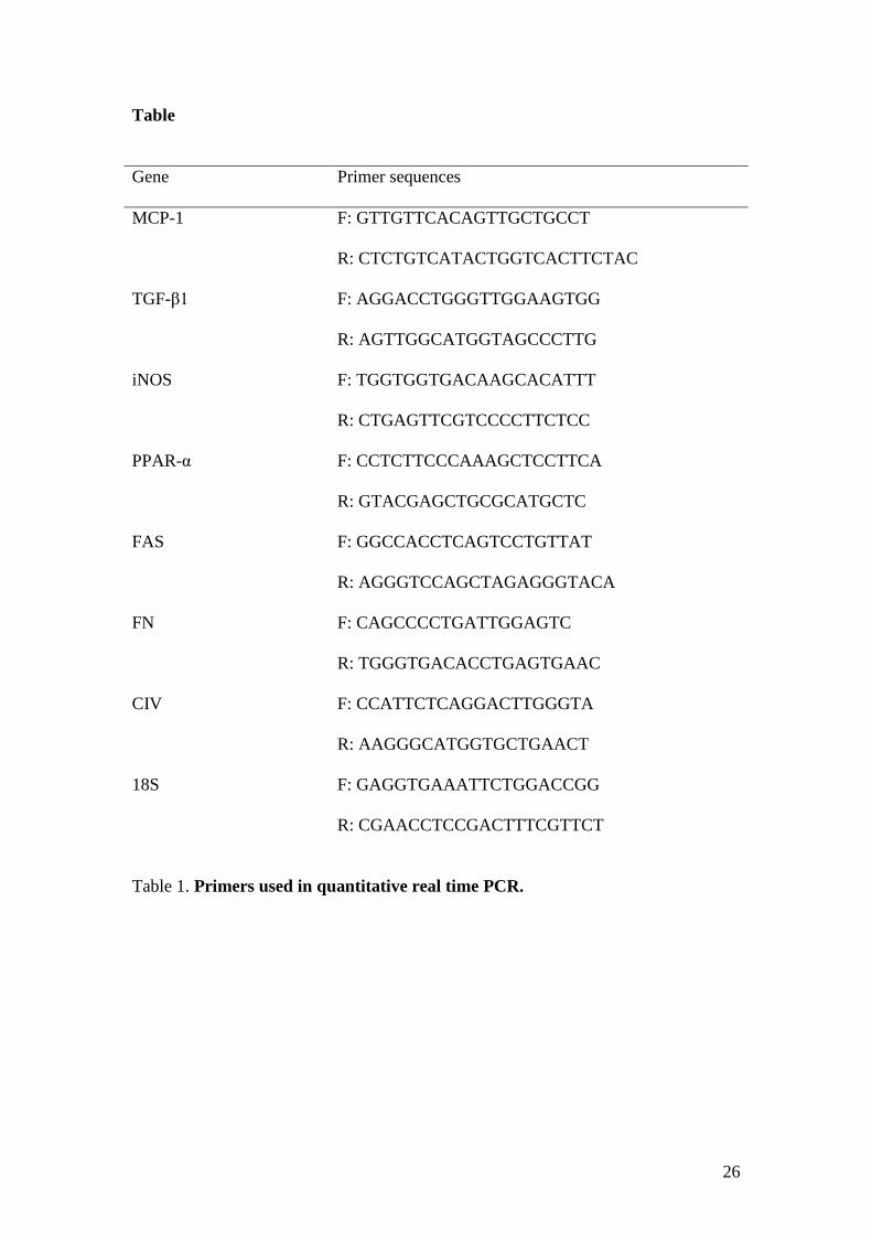
Table
Gene Primer sequences
MCP-1 F: GTTGTTCACAGTTGCTGCCT
R: CTCTGTCATACTGGTCACTTCTAC
TGF-β1 F: AGGACCTGGGTTGGAAGTGG
R: AGTTGGCATGGTAGCCCTTG
iNOS F: TGGTGGTGACAAGCACATTT
R: CTGAGTTCGTCCCCTTCTCC
PPAR-α F: CCTCTTCCCAAAGCTCCTTCA
R: GTACGAGCTGCGCATGCTC
FAS F: GGCCACCTCAGTCCTGTTAT
R: AGGGTCCAGCTAGAGGGTACA
FN F: CAGCCCCTGATTGGAGTC
R: TGGGTGACACCTGAGTGAAC
CIV F: CCATTCTCAGGACTTGGGTA
R: AAGGGCATGGTGCTGAACT
18S F: GAGGTGAAATTCTGGACCGG
R: CGAACCTCCGACTTTCGTTCT
Table 1. Primers used in quantitative real time PCR.


![[GLP]Health printer1](https://img.pdfslide.us/doc/110x75/554e7c60b4c90545698b5058/glphealth-printer1.jpg)