Embed Size (px)
Citation preview
441© Springer International Publishing Switzerland 2017 F. Pelegri et al. (eds.), Vertebrate Development, Advances in Experimental Medicine and Biology 953, DOI 10.1007/978-3-319-46095-6_9
Chapter 9Cell Cycle Remodeling and Zygotic Gene Activation at the Midblastula Transition
Maomao Zhang*, Jennifer Skirkanich*, Michael A. Lampson, and Peter S. Klein
Abstract Following fertilization, vertebrate embryos delay large-scale activation of the zygotic genome from several hours in fish and amphibians to several days in mam-mals. Externally developing embryos also undergo synchronous and extraordinarily rapid cell divisions that are accelerated by promiscuous licensing of DNA replication origins, absence of gap phases and cell cycle checkpoints, and preloading of the egg with maternal RNAs and proteins needed to drive early development. After a species-specific number of cell divisions, the cell cycle slows and becomes asynchronous, gap phases appear, checkpoint functions are acquired, and large- scale zygotic gene
M. Zhang Department of Biology, University of Pennsylvania, Philadelphia, PA, USA
Cell and Molecular Biology Graduate Group, Perelman School of Medicine at the University of Pennsylvania, Philadelphia, PA, USA
Department of Cancer Biology and Genetics, Memorial Sloan Kettering Cancer Center, New York, NY, USA
J. Skirkanich Cell and Molecular Biology Graduate Group, Perelman School of Medicine at the University of Pennsylvania, Philadelphia, PA, USA
Department of Biology, Bryn Mawr College, Bryn Mawr, PA, USA
M.A. Lampson (*) Department of Biology, University of Pennsylvania, Philadelphia, PA, USA
Cell and Molecular Biology Graduate Group, Perelman School of Medicine at the University of Pennsylvania, Philadelphia, PA, USAe-mail: [email protected]
P.S. Klein (*) Cell and Molecular Biology Graduate Group, Perelman School of Medicine at the University of Pennsylvania, Philadelphia, PA, USA
Department of Medicine (Hematology-Oncology), Perelman School of Medicine at the University of Pennsylvania, Philadelphia, PA, USAe-mail: [email protected]
*Author contributed equally with all other contributors.
442
activation begins. These events, along with clearance of maternal RNAs and proteins, define the maternal to zygotic transition and are coordinated at a developmental mile-stone termed the midblastula transition (MBT). Despite the relative quiescence of the zygotic genome in vertebrate embryos, genes required for clearance of maternal RNAs and for the initial steps in mesoderm induction are robustly transcribed before MBT. The coordination and timing of the MBT depends on a mechanism that senses the ratio of nuclear to cytoplasmic content as well as mechanisms that are independent of the nuclear–cytoplasm ratio. Changes in chromatin architecture anticipate zygotic gene activation, and maternal transcription factors identified as regulators of pluri-potency play critical roles in kick-starting the transition from the proliferative, plu-ripotent state of the early embryo to the more lineage-committed phase of development after the MBT. This chapter describes the regulation of the cell cycle and the activa-tion of zygotic gene expression before and after the MBT in vertebrate embryos.
Keywords Midblastula transition • Maternal zygotic transition • Zygotic transcrip-tion • Cell cycle checkpoint • Embryo • DNA damage • Nuclear cytoplasmic ratio • Cell cycle • Pluripotency
9.1 Introduction
Many metazoan embryos initiate development with a series of rapid, synchronous divisions that lack G1 and G2 phases, leading to reduction in cell size with each subsequent division (Bachvarova et al. 1966; Graham and Morgan 1966; Newport and Kirschner 1982a; Tadros and Lipshitz 2009; Gerhart 1980; Davidson 1986; Baroux et al. 2008). Rapid cleavage divisions are typically observed in embryos of vertebrates with larger eggs, such as amphibians, fish, birds, and reptiles. Although mammalian embryos undergo slower, asynchronous cell cycles from the outset, both mammalian and nonmammalian vertebrate embryos delay the onset of zygotic transcription and depend initially on maternal mRNAs and proteins to direct the earliest stages of development.
Rapid cell divisions and global suppression of zygotic transcription during cleav-age stages require unique adaptations for the control of DNA replication, mitosis, and early gene expression. During the transition from maternal to zygotic control of development, maternal mRNAs are degraded, the cell cycle lengthens, cell divisions become asynchronous, cells become motile, and large-scale zygotic transcription begins. In amphibians and fish, as well as invertebrates such as Drosophila, the appearance of gap phases (G1 and G2) in the cell cycle and the onset of large-scale zygotic transcription occur predictably after a species-specific number of cell/nuclear divisions, termed the midblastula transition (MBT). While not all organisms coordi-nate the cell cycle changes with the onset of zygotic transcription (mammalian cell cycles are initially slow, whereas sea urchins undergo rapid cleavages but initiate zygotic transcription at fertilization), there are nevertheless conserved features in the regulation of cell cycle and transcription that are reviewed in this chapter.
M. Zhang et al.
443
The terms MBT and MZT (maternal to zygotic transition) have been used vari-ously. We follow the clear definitions in Tadros and Lipshitz (2009) wherein MZT refers to the gradual transition from maternal to zygotic control and can span a broad developmental window in the pregastrula embryo. The MBT, in contrast, refers to a discrete milestone occurring in many species after a species-specific number of cell divisions and is marked by the coordinated acquisition of cell cycle checkpoints, cell cycle asynchrony, and the onset of large-scale zygotic transcrip-tion (Tadros and Lipshitz 2009; Gerhart 1980; Signoret and Lefresne 1971; Newport and Kirschner 1982a, b; Audic et al. 1997; Langley et al. 2014). We use the qualified term “large-scale zygotic transcription” because a subset of zygotic genes is tran-scribed before the MBT in Xenopus, zebrafish, and Drosophila (Tadros and Lipshitz 2009; Baroux et al. 2008; Nakakura et al. 1987; Yang et al. 2002; Skirkanich et al. 2011; Lindeman et al. 2011; Liang et al. 2008; Harrison et al. 2010; Edgar and Schubiger 1986; Yasuda et al. 1991; Heyn et al. 2014; Collart et al. 2014; Tan et al. 2013; Blythe et al. 2010; Lee et al. 2014), and this early wave of transcription is essential for development at the late blastula stage in Xenopus and Drosophila (Skirkanich et al. 2011; Liang et al. 2008; Harrison et al. 2010). In this chapter, we focus on the regulation of the cell cycle and zygotic transcription in vertebrate embryos and briefly touch on information from invertebrate species where it directly informs our understanding of vertebrate mechanisms. The MBT in nonvertebrate species has been discussed in more detail in several informative reviews (Yasuda and Schubiger 1992; Tadros and Lipshitz 2009; Baroux et al. 2008; Blythe and Wieschaus 2015a; Lee et al. 2014; Farrell and O'Farrell 2014). Degradation of maternal mRNAs is addressed in detail elsewhere in this volume (see Chapter 10).
9.2 Embryonic Cleavage Divisions
9.2.1 Unique Features of Embryonic Cleavage Cycles
Most actively proliferating cells progress through four distinct phases: the first gap phase (G1), DNA replication (S-phase), a second gap phase (G2), and mitosis (M). Newly fertilized embryos from many vertebrate and invertebrate species, including nonmammalian vertebrates such as fish and frogs, have highly specialized cell cycles that differ significantly from cells of later development and adult organisms (Oppenheimer 1936; Graham and Morgan 1966). These embryonic cell cycles, known as cleavage divisions, are extremely short and lack the gap phases typical of growing cells. Without cell growth, these cleavage cycles are reductive divisions that progressively subdivide a constant volume of cytoplasm from a single large cell into many smaller cells, increasing the nuclear to cytoplasmic (N:C) ratio with each round of DNA replication. Rapid cleavage cycles, which establish a large cell popu-lation necessary for gastrulation, continue until the embryo undergoes the mid- blastula transition (MBT).
9 Cell Cycle Remodeling and Zygotic Gene Activation at the Midblastula Transition
444
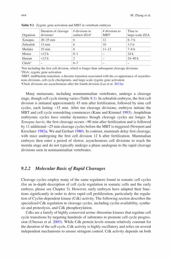
Many metazoans, including nonmammalian vertebrates, undergo a cleavage stage, though cell cycle timing varies (Table 9.1). In zebrafish embryos, the first cell division is initiated approximately 45 min after fertilization, followed by nine cell cycles, each lasting ~15 min. After ten cleavage divisions, embryos initiate the MBT and cell cycle remodeling commences (Kane and Kimmel 1993). Amphibian embryonic cycles have similar dynamics though cleavage cycles are longer. In Xenopus laevis, the first cleavage occurs ~90 min after fertilization and is followed by 11 additional ~25 min cleavage cycles before the MBT is triggered (Newport and Kirschner 1982a; Wu and Gerhart 1980). In contrast, mammals delay first cleavage, with mice undergoing the first cell division 12 h after fertilization. Mammalian embryos then enter a period of slower, asynchronous cell divisions to reach the morula stage and do not typically undergo a phase analogous to the rapid cleavage divisions seen in nonmammalian vertebrates.
9.2.2 Molecular Basis of Rapid Cleavages
Cleavage cycles employ many of the same regulators found in somatic cell cycles (for an in-depth description of cell cycle regulation in somatic cells and the early embryo, please see Chapter 3). However, early embryos have adapted their func-tions significantly in order to drive rapid cell proliferation, particularly the regula-tion of Cyclin-dependent kinase (Cdk) activity. The following section describes the specialized Cdk regulation in cleavage cycles, including cyclin availability, synthe-sis and proteolysis, and Cdk phosphorylation.
Cdks are a family of highly conserved serine–threonine kinases that regulate cell cycle transitions by targeting hundreds of substrates to promote cell cycle progres-sion (Ubersax et al. 2003). While Cdk protein levels remain relatively constant for the duration of the cell cycle, Cdk activity is highly oscillatory and relies on several independent mechanisms to ensure stringent control. Cdk activity depends on both
Table 9.1 Zygotic gene activation and MBT in vertebrate embryos
OrganismDuration of cleavage divisionsa
# divisions to earliest ZGAb
# divisions to MBTc
Time to large-scale ZGA
Xenopus 20–25 min 6 12 6–7 h
Zebrafish 15 min 6 10 3.5 h
Medaka 35 min 6 11–12 7–8 h
Mouse <12 h 0–1 – 24 h
Human <12 h 2 – 24–48 h
Chickd – 6–7 – –aNot including the first cell division, which is longer than subsequent cleavage divisionsbZGA: zygotic gene activationcMBT: midblastula transition, a discrete transition associated with the co-appearance of asynchro-nous divisions, cell cycle checkpoints, and large-scale zygotic gene activationdChick divisions are asynchronous after the fourth division (Lee et al. 2013a)
M. Zhang et al.
445
association with cyclin proteins and phosphorylation state. Furthermore, subcellular localization and degradation of cyclin and cyclin/Cdk complexes adds an additional level of regulation.
Many cyclins and Cdks have been identified in somatic cells. Cyclin A/Cdk2, cyclin D/Cdk4, cyclin D/Cdk6, and cyclin E/Cdk2 regulate cell cycle progression and replication during G1 and S phases. Cyclin B/Cdk1, on the other hand, is impor-tant for progression from G2 to M (Evans et al. 1983; Murray 2004). However, embryonic cleavage cycles are regulated by only three cyclins, A, B, and E, and two Cdks, Cdk1 and Cdk2 (Hartley et al. 1996). Cdk2 binds to cyclins A and E to medi-ate DNA replication and centrosome duplication while Cdk1 binds to cyclins A and B to drive mitotic progression (Murray and Kirschner 1989; Rempel et al. 1995; Strausfeld et al. 1996).
9.2.2.1 Phospho-regulation of Cdk1
During somatic cell cycles, Cdk1 activity is regulated by phosphorylation on key residues. Cyclin binding requires phosphorylation of a threonine adjacent to the active site (Ducommun et al. 1991). Additionally, the Wee1 and Myt1 kinases inhibit Cdk1 and Cdk2 by phosphorylating threonine 14 and tyrosine 15. Cyclin- bound Cdk remains in an inactive state until mitosis, when these inhibitory phos-phorylations are removed by Cdc25 phosphatases, which triggers mitotic entry (Atherton-Fessler et al. 1994; Krek and Nigg 1991).
In Xenopus embryos, Wee1 kinase phosphorylates Tyr15 on Cdk1 and Cdk2 in each pre-MBT cell cycle (Kim et al. 1999; Murakami and Vande Woude 1998). However, this inhibitory phosphorylation occurs at relatively low levels (Kim et al. 1999). Furthermore, although Cdc25A protein is not detected in Xenopus oocytes, maternally deposited cdc25 mRNA is translated upon fertil-ization, and Cdc25 protein steadily increases during the cleavage stages (Kim et al. 1999; Pomerening et al. 2003; Sha et al. 2003; Bouldin and Kimelman 2014; Yang and Ferrell 2013). The low level of Cdk1 inhibitory phosphorylation in cleavage-stage embryos keeps Cdk1 in a “primed” state, ready for activation upon cyclin binding.
9.2.2.2 Regulation of Mitotic Cyclin Protein Levels
Unlike somatic cell cycles, phospho-regulation of Cdk1 activity plays a minor role during early embryogenesis. Instead, Cdk in cleavage-stage vertebrate embryos is predominately regulated by cyclin protein synthesis and degradation. In Xenopus, protein levels of cyclin A and B oscillate once per cell cycle, with a nearly identical pattern of expression. Cdk1 activity closely parallels cyclin expression and also oscillates with each cell cycle (Hartley et al. 1996). In somatic cell cycles, cyclin protein expression is regulated by cell cycle phase-specific transcription (Pines 2011). In contrast, cyclin transcripts are preloaded maternally in embryos during
9 Cell Cycle Remodeling and Zygotic Gene Activation at the Midblastula Transition
446
oogenesis, and cyclin protein accumulation is posttranscriptionally regulated. Cell cycle phase-specific translation of cyclins relies mainly on the polyadenylation of mRNAs, which changes in a cell cycle-dependent manner (Groisman et al. 2002).
As in somatic cells, mitotic exit during the cleavage divisions is regulated by cyclin B degradation, mediated by the highly conserved E3 ubiquitin ligase Anaphase Promoting Complex/Cyclosome (APC/C) (Skaar and Pagano 2009). In Xenopus embryos, XErp1/Emi2, a homolog of early mitotic inhibitor 1 (Emi1) (Tung et al. 2005), inhibits APC/C activity during mitosis. However, when Cdk1 becomes maximally activated, it antagonizes XErp1/Emi2 function leading to APC/C activation (Tischer et al. 2012). Subsequently, activated APC/C polyubiqui-nates cyclin B, tagging it for proteasomal degradation. As cyclin B levels fall, Cdk1 activity diminishes and cells exit mitosis (King et al. 1996).
Cdk1 also regulates cyclin B protein expression: inhibiting Cdk1 leads to increased cyclin B protein levels while prematurely activating Cdk1 decreases cyclin B accumulation. These results demonstrate that Cdk1 participates in a negative- feedback loop that attenuates the production of cyclins before mitosis. Limiting cyclin production increases the efficiency and sensitivity of the Cdk1- APC/C negative feedback loop by decreasing the burden of cyclin B degradation at anaphase (Kang and Pomerening 2012). These negative feedback loops between Cdk1, APC/C activation, and cyclin degradation support the rapid cyclin oscilla-tions observed in cleavage-stage embryos.
9.2.2.3 Regulation of Cyclin E/Cdk2
In somatic cells, cyclin E/Cdk2 activity mediates the transition from G1 to S (Elledge et al. 1992). Similarly, cyclin E/Cdk2 regulates the progression of S-phase in cleavage-stage embryos. Inhibition of CyclinE/Cdk2 activity moderately increases cell cycle lengths in pre-MBT Xenopus embryos, showing that the activity of Cdk2 also contributes to rapid cell cycle progression (Hartley et al. 1996, 1997). Though cyclins A and B protein levels oscillate during the cleavage cycles, cyclin E protein levels steadily increase following fertilization (Hartley et al. 1996). Despite this, cyclin E/Cdk2 activity oscillates twice per cleavage cycle independently of protein synthesis. While more studies are required to elucidate the regulation of Cdk2 oscillations in cleavage-stage embryos, pre-MBT Cdk2 activity is likely regu-lated by phosphorylation state (Ciliberto et al. 2003). Indeed, Cdk2 activity is regu-lated by inhibitory phosphorylation by the Wee1 kinase in Xenopus egg extracts and embryos (D'Angiolella et al. 2001; Wroble et al. 2007).
9.2.2.4 Influence of Replication on Cleavage Cycles
Although accumulation and degradation of cyclins is certainly important for mitotic entry, RNAi knockdown of two Drosophila mitotic cyclins and reduction of gene dosage for the third cyclin did not prolong interphase but rather led to a
M. Zhang et al.
447
partial activation of mitotic events. These data suggest that cyclin accumulation alone is not enough to mediate all aspects of rapid cell cycle progression (McCleland et al. 2009a).
A second mechanism for maintaining short cell cycles could rely on DNA replication itself. Replication occupies the majority of interphase during cleavage divisions and proceeds quickly due to the close proximity of origins of replication (Harland and Laskey 1980; Spradling 1999). Inhibiting replication in syncytial embryos by injecting Geminin, which blocks the licensing of origins (McGarry and Kirschner 1998; Quinn et al. 2001), abolished S-phase and led to premature mitotic entry, demonstrating that replication defines interphase length in cleavage-stage embryos (McCleland et al. 2009b). This idea was corroborated in Xenopus, where replication factors were recently identified that can directly modify cleavage cycle lengths. Highly expressed during the cleavage stages, Cut5, Treslin, Drf5, and RecQ4 become limiting at the MBT, coinciding with cell cycle elongation. Importantly, overexpression of these factors abolished cell cycle lengthening at the MBT (Collart et al. 2013).
In conclusion, pre-MBT cells are preloaded with many of the same cell cycle regulators as seen in most somatic cells. However, it is their specialized regulation that leads to rapid cell proliferation.
9.2.3 Cell Cycle Checkpoints in Early Embryogenesis
Cell cycle checkpoints are present in almost all nonpathologic somatic cells to maintain genome integrity. The DNA damage checkpoint induces cell cycle arrest during interphase in response to DNA damage or stalled replication. The spindle assembly checkpoint (SAC) causes metaphase arrest in response to kinetochores that are not attached to microtubules during mitosis. However, cleavage-stage embryos forgo checkpoint function in their commitment to rapid cell proliferation. Little is known about how checkpoints are suppressed during cleavage stages or how they are acquired at the MBT. The following section reviews our current knowl-edge of SAC and DNA damage checkpoint signaling prior to the MBT.
9.2.3.1 The DNA Damage Checkpoint in Cleavage Cycles
In somatic cells, DNA damage activates two phosphoinositide 3-kinase-related pro-tein kinases (PIKKs): ataxia-telangiectasia mutated (ATM) and ATM and RAD3- related (ATR). ATM and ATR are similar in structure and share many of the same substrates, but are activated by distinct triggers. ATM is activated by DNA double- strand breaks (DSBs) and ATR is activated by single-strand DNA (ssDNA) or ssDNA–dsDNA junctions. One of the earliest consequences of DNA damage is phosphorylation of Serine 139 on the histone variant H2AX (γH2AX) by ATM and ATR. The forma-tion of γH2AX foci surrounding the damage site creates a docking site that recruits
9 Cell Cycle Remodeling and Zygotic Gene Activation at the Midblastula Transition
448
DNA damage response proteins, promoting DNA repair and checkpoint signal ampli-fication (Sirbu and Cortez 2013). γH2AX is an important read-out for DNA damage checkpoint initiation and the successful sensing of DNA damage (Dickey et al. 2009). ATM and ATR also activate the serine–threonine kinases Chk1 and Chk2, which play a central role in facilitating cell cycle arrest by phosphorylating multiple substrates to ultimately inhibit the activity of Cdks (Bartek and Lukas 2003). For example, Chk1 and Chk2 phosphorylate Cdc25 phosphatases, targeting them for degradation. Chk1 can also activate the Wee1 kinase via phosphorylation (Patil et al. 2013).
Post-MBT embryos have a robust DNA damage response and induce cell cycle arrest efficiently after DNA damage (Hensey and Gautier 1997; Maller et al. 2001). However, when zebrafish or Xenopus pre-MBT embryos are treated with ionizing radi-ation, cleavage cycles continue without arrest or cell cycle delay (Hensey and Gautier 1997; Zhang et al. 2014). Furthermore, DNA polymerase inhibitors or mutations that disrupt replication cause replication stalling and trigger S-phase arrest in somatic cells and post-MBT embryos, but not in pre-MBT Xenopus, zebrafish, or Drosophila embryos (Dasso and Newport 1990; Freeman and Glover 1987; Freeman et al. 1986; Kimelman et al. 1987; Shamanski and Orr-Weaver 1991; Ikegami et al. 1997).
Rapid DNA repair to explain the lack of cell cycle arrest after damage seems implausible, as irradiated embryos have high levels of DNA fragmentation (Anderson et al. 1997; Finkielstein et al. 2001; Hensey and Gautier 1997). Instead, checkpoint signaling is defective. In zebrafish, the DNA damage checkpoint is properly initiated by ionizing radiation, as irradiated pre-MBT embryos can phos-phorylate histone H2AX and activate the effector kinase Chk2. Chk1 is not acti-vated, however, leaving cleavage-stage embryos unable to arrest the cell cycle after DNA damage (Zhang et al. 2014).
DNA damage sustained during the cleavage stages results in embryonic lethality. Without checkpoints to resolve DNA lesions prior to the MBT, damaged DNA accu-mulates to irreparable levels by the MBT. When the checkpoint program finally becomes functional at the MBT, apoptosis is the only course of action; pre-MBT Xenopus embryos treated with ionizing radiation accumulate dense, small nuclei that are typical of apoptosis beginning at the onset of the MBT (Anderson et al. 1997). Further, TdT-mediated dUTP digoxigenin nick end labeling (TUNEL) detected apop-tosis after the MBT, but not before (Anderson et al. 1997; Hensey and Gautier 1997; Stack and Newport 1997; Sible et al. 1997). Hensey and Gautier, for example, reported that apoptosis is first detectable at the gastrula stage (Hensey and Gautier 1997).
9.2.3.2 The SAC in Cleavage Cycles
In most cells, the SAC delays anaphase onset and mitotic exit until all kineto-chores are attached to microtubules, in order to prevent chromosome missegrega-tion. Each unattached kinetochore recruits SAC proteins to form the mitotic checkpoint complex (MCC), which prevents APC/C activation by targeting its co-activator, Cdc20. SAC proteins are removed from each kinetochore as it binds microtubules. After all kinetochores are attached, MCC disassembly frees Cdc20 to activate the APC/C, which polyubiquinates cyclin B and securin, leading to
M. Zhang et al.
449
their destruction, which is required for anaphase onset and mitotic exit (Amon et al. 1994; Lara-Gonzalez et al. 2012).
Despite robust function after the cleavage stage, the SAC is not active in pre- MBT embryos. Nuclei in Xenopus cleavage-stage embryos or egg extracts treated with microtubule poisons have a dramatically different morphology than their post- MBT counterparts, forming irregularly shaped, fragmented micronuclei thought to arise from inappropriate anaphase onset (Clute and Masui 1992; Newport and Kirschner 1984). Furthermore, time spent in mitosis does not change in pre-MBT embryos after microtubule depolymerization (Clute and Masui 1992; Ikegami et al. 1997; Zhang et al. 2015).
9.2.4 Cell Cycle Remodeling at the MBT
Cell cycle remodeling, in which cells elongate their cell cycles, add gap phases, and gain functional cell cycle checkpoints, is a hallmark of the MBT. This section reviews our current understanding of cell cycle elongation and checkpoint acquisi-tion at the MBT.
9.2.4.1 Timing the Onset of Cell Cycle Lengthening
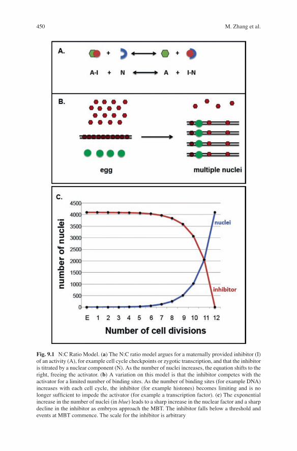
Cell cycle remodeling occurs after a fixed number of cleavages that is characteristic of the species. This observation led to the hypothesis that cell cycle elongation at the MBT is triggered by a mechanism that can measure the number of cell divisions or elapsed time after fertilization. However, a series of elegant experiments (Newport and Kirschner 1982a, b) demonstrated that the onset of cell cycle lengthening and asynchrony associated with the MBT is determined by a threshold N:C ratio (Fig. 9.1). Using a method adapted from Spemann (Sander and Faessler 2001), a strand of hair tied around the embryo partially constricted it at the single-cell stage to trap the nucleus on one side of the embryo. This manipulation effectively halved the cytoplas-mic volume carrying the nucleus. The section with the nucleus cleaved 11 times before cell cycles became asynchronous. However, after two divisions, a daughter nucleus migrated through the narrow channel of the constriction to the side that origi-nally had no nucleus. This side of the embryo now underwent 11 more cleavage cycles before becoming asynchronous, even though the nucleus had undergone two mitoses prior to migration (Newport and Kirschner 1982a). Similarly, a fourfold reduction in the cytoplasm of eggs of the Japanese newt accelerated the onset of mitotic asynchrony by two divisions (Kobayakawa and Kubota 1981).
Further experiments showed that the end of the cleavage stage is not determined by a mechanism that counts cell divisions. When cell division was blocked with cytochalasin B or by gently centrifuging fertilized eggs, the embryos continued to synthesize DNA at an exponential rate for 6 h and then abruptly slowed DNA syn-thesis, similar to control embryos at the MBT. Furthermore, increasing DNA con-tent in cleavage-stage embryos, for example in polyspermic eggs or eggs injected
9 Cell Cycle Remodeling and Zygotic Gene Activation at the Midblastula Transition
450
Fig. 9.1 N:C Ratio Model. (a) The N:C ratio model argues for a maternally provided inhibitor (I) of an activity (A), for example cell cycle checkpoints or zygotic transcription, and that the inhibitor is titrated by a nuclear component (N). As the number of nuclei increases, the equation shifts to the right, freeing the activator. (b) A variation on this model is that the inhibitor competes with the activator for a limited number of binding sites. As the number of binding sites (for example DNA) increases with each cell cycle, the inhibitor (for example histones) becomes limiting and is no longer sufficient to impede the activator (for example a transcription factor). (c) The exponential increase in the number of nuclei (in blue) leads to a sharp increase in the nuclear factor and a sharp decline in the inhibitor as embryos approach the MBT. The inhibitor falls below a threshold and events at MBT commence. The scale for the inhibitor is arbitrary
M. Zhang et al.
451
with exogenous DNA, advances the timing of the MBT (Dasso and Newport 1990; Newport and Kirschner 1982a, b; Mita and Obata 1984). Similarly in zebrafish, tetraploid embryos begin cell cycle lengthening about a cycle early and haploid embryos begin cell cycle lengthening about a cycle later than diploid embryos (Kane and Kimmel 1993). Partial enucleation of multicellular embryos, an approach analogous to the ligature experiment adapted from Spemann, also has a similar effect. At the early stages zebrafish blastomeres maintain intercellular bridges, allowing migration of a single nucleus into an enucleated cell. As the nucleus now occupies a larger cytoplasm, the daughter cells undergo additional cell divisions before cell cycles become asynchronous (Kane and Kimmel 1993). Further, haploid Drosophila embryos undergo one extra syncytial division, presumably because the N:C ratio associated with the MBT is achieved one cell cycle later than in diploid embryos (Di Talia et al. 2013; Edgar et al. 1986).
Taken together, work in Xenopus and other amphibians, zebrafish, and Drosophila indicate that cell cycle elongation and loss of cell cycle synchrony at the MBT is not determined by the chronological time after fertilization, the number of divisions, or a progressive change in chromatin state with each cell cycle. Rather, these MBT events are initiated when embryos reach a threshold N:C ratio resulting from rounds of nuclear replication without cell growth. A proposed mechanism of regulation is that a cytoplasmic factor that inhibits the MBT during the cleavage cycles is titrated out by binding DNA (Fig. 9.1) (Newport and Kirschner 1982b).
In addition to the ratio of DNA content to cytoplasm, as discussed above, the ratio of nuclear volume to cytoplasmic volume may contribute to the onset of the MBT. Injection of nuclear scaling factors, including import proteins, lamins, and reticulons, into cleavage-stage Xenopus embryos showed that increasing nuclear volume causes premature cell cycle remodeling, whereas a reduced N:C volume ratio increases the number of rapid cell divisions and delays cell cycle remodeling (Jevtic and Levy 2015). These experiments are discussed in more detail in the sec-tion on transcription.
9.2.4.2 Molecular Mechanisms of Cell Cycle Elongation
Cell cycle elongation at the MBT is achieved by the restraint and modification of cyclin/Cdk activities found in the cleavage stage. Embryos use several mechanisms to downregulate Cdc25 phosphatase, which allows Cdk1 to accumulate inhibitory phosphorylation and become inactivated after the last cleavage-stage mitosis. At the first asynchronous cycle after the MBT, lower Cdk1 activity results in an extended replication phase (Farrell et al. 2012). After replication is complete, cells must wait until zygotic Cdc25 is synthesized to restore Cdk1 activity for mitotic entry. In effect, these delays represent cell cycle elongation via extension of S-phase and the acquisition of G2 phase.
Regulation of Cdc25 at the MBT has been studied extensively in flies. Drosophila embryos express two maternally supplied Cdc25 homologs, String and Twine (Edgar et al. 1994). Altering the number of maternal copies of these Cdc25 homologs
9 Cell Cycle Remodeling and Zygotic Gene Activation at the Midblastula Transition
452
showed that mutant embryos with increased maternal supplies of Cdc25 have one extra rapid, synchronous mitotic cycle. Conversely, mutant embryos with reduced maternal Cdc25 elongate cell cycles prematurely. These findings provided strong evidence that maternally loaded Cdc25 phosphatases are dosage-sensitive regula-tors that determine when cell cycles elongate (Edgar and Datar 1996). Furthermore, cell cycle elongation depends on rapid degradation of Twine protein at the MBT (Di Talia et al. 2013; Farrell and O'Farrell 2013).
9.2.4.3 Cell Cycle Elongation via Zygotic Transcription
Cdc25 stability at the MBT is sensitive to the N:C ratio, as Twine protein in Drosophila haploid embryos is degraded one cell cycle later in haploids com-pared to diploids (Farrell and O'Farrell 2013). This finding may be a result of N:C ratio- regulated transcription of specific genes that control Cdc25 degradation. Embryos injected with α-amanitin, an RNA Polymerase II inhibitor, undergo an extra round of rapid division and exhibit extended Twine stabilization (Farrell and O'Farrell 2013). Furthermore, flies with the RNA polymerase II mutation RPII215, which prematurely activates zygotic transcription, have a reduced number of nuclear divisions before cellularization. These data independently corroborate that zygotic transcription affects the timing of cell cycle remodeling at the MBT (Sung et al. 2013).
The genes transcribed at the MBT that regulate Cdc25 destruction are just beginning to be elucidated. One candidate in Drosophila is tribbles, which medi-ates Cdc25 destruction via proteolysis (Mata et al. 2000). Precocious tribbles expression via mRNA injection can arrest embryos in cycle 13 due to significant reduction in Twine levels (Farrell and O'Farrell 2013; Grosshans and Wieschaus 2000). Additionally, RNA sequencing of staged embryos revealed that tribbles expression increases dramatically at the MBT. Importantly, gene expression pro-filing of haploid embryos demonstrated that tribbles expression is sensitive to the N:C ratio (Lott et al. 2011; Lu et al. 2009). The Cdk1 inhibitor (CKI) fruhstart is another Drosophila zygotic gene important for cell cycle elongation at the MBT. Also sensitive to the N:C ratio, fruhstart appears immediately after the last cleavage division at the beginning of the 14th cell cycle (Lu et al. 2009). Moreover, when precociously expressed via mRNA injection, fruhstart can arrest the cell cycle during cleavage divisions (Grosshans et al. 2003). To inhibit Cdk1, Fruhstart binds tightly to mitotic cyclins, sequestering them from Cdk1 (Gawlinski et al. 2007).
Work in zebrafish has provided additional insight into the influence of zygotic genome activation on cell cycle remodeling in vertebrate systems. Similar to the results in Drosophila, inhibiting zygotic transcription in zebrafish embryos hinders the acquisition of G1 phase at the MBT (Zamir et al. 1997). However, the relationship between cell cycle lengthening and zygotic transcription seems more complex, since acquisition of G2 is independent of zygotic transcription (Dalle Nogare et al. 2009).
M. Zhang et al.
453
9.2.4.4 Developmental Use of the Replication Checkpoint for Cell Cycle Elongation
Proteins involved in the cellular response to DNA damage or replication stress also contribute to cell cycle remodeling at the MBT. Most checkpoint proteins are dis-pensable in somatic cells; knocking out individual components leads to mutations and aneuploidy but rarely inviability. However, several checkpoint proteins are essential for viability in a variety of model systems, have non-checkpoint related cell cycle functions, and have important roles in early development (Brown and Baltimore 2000; Liu et al. 2000).
The Chk1 serine–threonine kinase is an important checkpoint component that mediates cell cycle arrest in response to DNA single-strand breaks caused by stalled replication forks and fork collapse. Cleavage stage embryos are sensitive to Chk1 activity, and exogenous expression of wild type Chk1 or constitutively active Chk1 in Xenopus and zebrafish embryos induces a dose-dependent delay of cleavage cycles (Kappas et al. 2000; Zhang et al. 2014, 2015). Chk1 is also tran-siently activated at the MBT and required for cell cycle lengthening, suggesting that embryos co-opt this checkpoint protein to remodel the cell cycle (Shimuta et al. 2002).
Developmental activation of Chk1 causes cell cycle lengthening by promoting inhibitory phosphorylation of Cdk1, through regulation of the Cdc25 phosphatases and Wee1 kinase. In Xenopus, Chk1 phosphorylates Cdc25A, targeting it for protea-somal degradation. Dominant-negative forms of Chk1 injected into Xenopus embryos stabilize Cdc25A protein, while wild-type Chk1 overexpression leads to precocious Cdc25A destruction (Shimuta et al. 2002). Chk1 can also phosphorylate Cdc25 to inhibit its interaction with cyclin/Cdk complexes (Petrus et al. 2004; Uto et al. 2004). Furthermore, Chk1 enhances Wee1 kinase activity, thereby using a two- pronged approach for maintaining inhibitory phosphorylation of Cdk1 (Lukas and Bartek 2009).
Chk1 is also essential for cell cycle remodeling at the MBT in Drosophila: the Chk1 mutant, grapes, undergoes extra syncytial divisions and dies at gastrulation (Sibon et al. 1997, 1999). Chk1 enhances Wee1 kinase activity, as in Xenopus (Campbell et al. 1995), but does not play a significant role in Cdc25 destruction at MBT. Although String destruction is Chk1 dependent, Chk1 does not affect the stability of Twine, the Cdc25 homolog that is primarily associated with MBT cell cycle elongation (Farrell and O'Farrell 2013; Di Talia et al. 2013). As an additional mechanism for Cdk1 inhibition, Chk1 can inhibit nuclear accumulation of cyclin B, which prevents its interaction with Cdk1 in the nucleus (Royou et al. 2008). Together, these findings in Xenopus and Drosophila demonstrate that Chk1 is a potent regulator of cell cycle length during early embryogenesis and is necessary for early embryonic development.
While the molecular mechanisms of Chk1 activation at the MBT have yet to be fully elucidated, several hypotheses have been proposed that implicate a role for the N:C ratio (Edgar et al. 1994; Edgar and O'Farrell 1989; Sibon et al. 1999). In one model, a maternally loaded replication factor is titrated by increasing chromatin concentrations
9 Cell Cycle Remodeling and Zygotic Gene Activation at the Midblastula Transition
454
in cleavage-stage embryos. Eventually, a limiting amount of this replication factor leads to a delay in replication. While this is not DNA damage per se, it may be effec-tively recognized as replication stress: ssDNA and ssDNA-dsDNA- binding proteins like RPA, Rad17-RPC and 9-1-1 may have an opportunity to bind, leading to Chk1 activation. Supporting this possibility, a recent study identified replication factors that could regulate cell cycle length during Xenopus cleavage divisions (Collart et al. 2013). When these replication factors are abundant in the cleavage-stage environment, the rapid origin firing may not allow binding of ATR and Chk1 activating factors to chro-matin. However, the replication factors may become limiting at the MBT as the N:C ratio increases, slowing replication and permitting ATR-Chk1 activation.
A second model is that the Chk1 adaptor protein Claspin is regulated by the N:C ratio. Chk1 activation in somatic cells and Xenopus egg extracts depends on Claspin, which recruits Chk1 to ATR for phosphorylation (Kumagai and Dunphy 2000, 2003; Chini and Chen 2003). Claspin is typically phosphorylated by ATR after replication stress, but Xenopus embryos phosphorylate Claspin at low levels even in the absence of ATR activity. However, Claspin phosphorylation is responsive to the N:C ratio: addi-tion of sperm nuclei to Xenopus egg extracts to increase N:C ratios to MBT levels resulted in Claspin phosphorylation in an ATR-independent manner. These data suggest that a threshold N:C ratio may either activate a novel kinase or titrate a kinase inhibitor to allow Claspin phosphorylation (Gotoh et al. 2011), in addition to the canonical phos-phorylation of Claspin by ATR after MBT activation of the replication checkpoint.
Finally, a third hypothesis suggests that transcriptional activity itself can lead to replication checkpoint activation, regardless of the products of transcription. In Drosophila, binding of the replication protein RPA70 to DNA is tightly correlated to DNA-binding of RNA polymerase II, which increases gradually throughout the cleavage stages in preparation for zygotic transcription at the MBT (Blythe and Wieschaus 2015b). These data support the hypothesis that sites of transcriptionally engaged DNA can be sources of replication stress that activate the replication checkpoint and confer cell cycle remodeling.
9.2.5 Checkpoint Acquisition at the MBT
The molecular mechanisms that underlie checkpoint acquisition at the MBT are poorly understood. The following section reviews what is known about DNA damage check-point and SAC acquisition at the MBT, with an emphasis on the role of the N:C ratio.
9.2.5.1 DNA Damage Checkpoint Acquisition
The first hints towards elucidating checkpoint regulation came from studies using Xenopus egg extracts. As mentioned above, addition of DNA replication inhibitors, DNA damaging agents, or microtubule poisons had no effect on cell cycle progression in control extracts (Dasso and Newport 1990; Kumagai et al. 1998).
M. Zhang et al.
455
However, when additional DNA was supplied via the addition of sperm chromatin, the extracts became sensitive to replication stress and DNA damage and arrested their cell cycles, indicative of restored checkpoint function.
Several models have been proposed to explain the influence of the N:C ratio on DNA damage checkpoint acquisition. One hypothesis suggests that pre-MBT embryos cannot efficiently amplify the DNA damage signaling response in the unusu-ally large cytoplasm associated with low N:C ratios. Injecting embryos with varying amounts of dsDNA to mimic DNA double-strand breaks (DSBs), together with plas-mid DNA to increase the DNA content (and thus N:C ratio), induced a precocious DNA damage response that activated Chk1, increased phosphorylation of Cdk1, and caused subsequent cell cycle delay (Conn et al. 2004). This activation of the check-point only occurs at a critical DNA to cytoplasm ratio, demonstrating the importance of N:C ratio in checkpoint acquisition (Conn et al. 2004; Peng et al. 2008).
Checkpoint acquisition at the MBT likely centers around Chk1 gain of function. In zebrafish pre-MBT embryos, checkpoint proteins are maternally supplied, and the ATM-Chk2 axis responds robustly to DNA damage, but Chk1 is not activated (Zhang et al. 2014). These results suggest that lack of Chk1 activity limits checkpoint func-tion prior to the MBT. Given these findings and the transient Chk1 activation at the MBT observed in Xenopus (Shimuta et al. 2002), we propose that Chk1 activation is sensitive to the N:C ratio and that Chk1 is a master regulator of cell cycle remodeling at the MBT, contributing to both cell cycle elongation and checkpoint acquisition.
9.2.5.2 SAC Acquisition
To examine SAC acquisition, “mini-embryos” with reduced cytoplasmic volume were created using a modified version of the Spemann method. A loop of baby’s hair was placed around the animal pole of a newly fertilized Xenopus embryo to constrict a portion of the nucleus-containing cytoplasm, effectively increasing the N:C ratio. These mini-embryos had a cytoplasmic volume of about 1/8–1/12 the size of a normal embryo and continued to cycle like their unperturbed counterparts, but with a much higher N:C ratio (Clute and Masui 1995). The cell cycles of these mini-zygotes became asynchronous two cycles before the usual time of the MBT (cleavage 10). At this point, the N:C ratio of the mini-embryos corresponds to the N:C ratio of unperturbed embryos at MBT (cleavage 12). This finding suggests that cell cycle remodeling is controlled by the N:C ratio. Surprisingly, however, mitotic delay after microtubule depolymerization occurred at the same time as in control embryos, despite the disparity in N:C ratio and precocious cell cycle elongation in the mini-zygotes. Similar findings were demonstrated with zebrafish embryos: when pre-MBT cell cycles were artificially lengthened with the addition of acti-vated Chk1, thereby slowing the increase of the N:C ratio, embryos still acquired a functional SAC at 3 h post fertilization (hpf), the normal time of the MBT, despite not having reached the usual MBT N:C ratio (Zhang et al. 2015).
These studies suggest that while the onset of cell cycle asynchrony depends on the N:C ratio, the SAC is acquired at an absolute time that is independent of N:C ratio. However, they differ from earlier findings in egg extracts, which can activate
9 Cell Cycle Remodeling and Zygotic Gene Activation at the Midblastula Transition
456
the SAC and arrest in metaphase if enough sperm chromatin is added (Minshull et al. 1994), implying that SAC acquisition is coupled to the N:C ratio. One explana-tion for this result is that a threshold N:C ratio could trigger zygotic transcription, leading to SAC acquisition indirectly by transcription of checkpoint components that are not maternally supplied. However, work in zebrafish and Xenopus shows that blocking transcription with α-amanitin in pre-MBT embryos does not abrogate SAC acquisition at the MBT (Zhang et al. 2015; Newport and Kirschner 1982b). Instead, the seemingly contradictory results may indicate that both a developmental timer and increases in the N:C ratio contribute to SAC acquisition. Full activation of the SAC relies on the generation of MCCs at kinetochores and diffusion throughout the cytoplasm to inhibit the APC/C. Cleavage stage embryos may express as-yet unidentified SAC inhibitors, or may not generate enough MCCs to overcome the large cytoplasmic volume. A threshold concentration of DNA (and also kineto-chores) may be required to generate enough active MCCs or to titrate a SAC inhibi-tor. During normal development, when the number of kinetochores is fixed, a set time may be required for the synthesis and accumulation of SAC proteins that amplify signaling downstream from initial SAC activation at kinetochores. However, the need for accumulation of these proteins could be bypassed if exogenous DNA is added, providing large numbers of kinetochores to amplify SAC signaling.
9.2.6 Summary
A principal goal of the early embryo is to accumulate a large cell population in preparation for gastrulation and the later stages of development. This feature is especially important in oviparous organisms where embryonic development occurs outside the mother and rapid embryonic development is a survival advantage. To do so, cleavage-stage embryos have modified several facets of cell cycle regulation to encourage rapid proliferation: cyclin/Cdk activities oscillate to achieve efficient cell cycling and checkpoint function is sacrificed. It is not until the MBT that cell cycles are remodeled to behave more like their somatic counterparts: cell cycles lengthen dramatically, acquire checkpoints, and become asynchronous. Though these fea-tures occur simultaneously, they are driven by various triggers, be it the N:C ratio, absolute time post fertilization, or zygotic transcription.
9.3 Regulation of Zygotic Transcription in the Early Embryo
9.3.1 Changes in Transcription from Oocyte Maturation Until Zygotic Gene Activation
During oogenesis, robust transcriptional activity generates maternal transcripts essential for post-fertilization development. However, the maternal genome becomes quiescent upon meiotic maturation and remains inactive after fertilization until
M. Zhang et al.
457
zygotic genome activation (Table 9.1) (Wormington and Brown 1983; De La Fuente and Eppig 2001; Bachvarova and Davidson 1966; Woodland and Gurdon 1969; Newport and Kirschner 1982a). In Xenopus, large-scale zygotic transcription is not activated until the 12th embryonic cleavage, approximately 6–7 h after fertilization (Newport and Kirschner 1982a). In zebrafish, large-scale zygotic gene activation is not observed until the tenth cleavage division (Kane and Kimmel 1993). In medaka, large-scale zygotic transcription was reported to begin around division 11–12 (Aizawa et al. 2003). The major wave of zygotic transcription in mammals is delayed from one to several days, depending on the species, and occurs after fewer cell divisions than observed in nonmammalian vertebrates. In mice and rabbits, the major wave of zygotic gene activation is established by the 2-cell stage, whereas in cows, sheep, pigs, and humans, major zygotic transcription begins at the 4- to 16-cell stage (Telford et al. 1990; Li et al. 2013; Hamatani et al. 2004; Wang et al. 2004; Zeng and Schultz 2005; Flach et al. 1982; Tesarik et al. 1988; Sawicki et al. 1981; Park et al. 2013; Xue et al. 2013; Aoki et al. 1997).
While the time until zygotic gene activation varies between organisms, it is highly reproducible in a given species (Table 9.1). Furthermore, most species that delay large-scale ZGA nevertheless demonstrate an earlier, minor wave of zygotic transcription (reviewed by Tadros and Lipshitz 2009; Baroux et al. 2008; Lee et al. 2014), consistent with mechanisms that both suppress early transcription and acti-vate zygotic genes at the appropriate stage. The following sections review the evi-dence that early embryos have the capacity for transcription despite global repression of most zygotic genes and discuss the regulation of zygotic gene activation, includ-ing potential mechanisms to regulate the timing of zygotic gene activation.
9.3.2 Transcriptional Repression in Pre-MBT Embryos
Despite the limited zygotic transcription after fertilization in vertebrates, the basal transcriptional machinery is present in oocytes and early embryos of Xenopus and other amphibians, zebrafish, mouse, and other mammals (Brown 2004; Veenstra 2002; Wiekowski et al. 1993; Li et al. 2013; Lee et al. 2014), as are multiple gene specific transcription factors. These findings suggest that the low level of transcrip-tion is due to a repressive mechanism that functions until the MBT/ZGA, as dis-cussed further in Sect. 9.3.5.
For example, RNA polymerases (RNAP) I, II, and III are present in Xenopus oocytes and eggs, which have served as an abundant source for purification of poly-merases and preparation of extracts competent for transcription of exogenous tem-plates (Roeder 1974a, b). Importantly, RNAPII is present in an active form in early embryos. RNAPII is phosphorylated in a repeat sequence within the C-terminal domain in manner that correlates with its initiating (serine-5 phosphorylated) and elongating (serine-2 phosphorylated) state. In vivo, RNAPII phosphorylated at serine- 2 is detectable in cleavage-stage embryos, consistent with a low level of transcription before the MBT. Although several studies reported that serine-2 phosphorylation
9 Cell Cycle Remodeling and Zygotic Gene Activation at the Midblastula Transition
458
decreases after fertilization and increases after the MBT, each of these studies nevertheless identify elongating RNAPII with a serine-2 phosphorylation specific antibody (H5) throughout pre-MBT stages (Palancade et al. 2001; Collart et al. 2009; Blythe et al. 2010). Phosphorylated RNAPII is also detectable before the MBT in zebrafish and increases steadily through the MBT (Zhang et al. 2014). This level of serine-2 phosphorylated RNAPII is actually high relative to the amount of DNA tem-plate in cleavage-stage embryos; while genome wide ChIP has not detected elongat-ing RNAPII associated with chromatin before the MBT, the sensitivity of ChIP has so far been limited by the low N:C ratio in early embryos (Lindeman et al. 2011; Akkers et al. 2009; Vastenhouw et al. 2010).
Furthermore, oocyte and egg extracts are transcriptionally competent. Xenopus egg extracts arrested in interphase will transcribe exogenous type III genes (Hartl et al. 1993; Wolffe and Brown 1987; Almouzni et al. 1990, 1991; Toyoda and Wolffe 1992; Wolffe 1989; Amodeo et al. 2015). In Xenopus oocytes and eggs, injected plasmids with type III promoters are transcribed transiently (Mertz and Gurdon 1977; Brown and Gurdon 1977; Newport and Kirschner 1982a; Prioleau et al. 1994; Almouzni and Wolffe 1995). Furthermore, a type II promoter-reporter is active throughout pre-MBT stages if an appropriate activator (GAL4-VP16) is present at sufficient levels (Almouzni and Wolffe 1995). A plasmid reporter with an EF1α promoter injected into zebrafish is also transcribed before the MBT (Harvey et al. 2013). The regulation of type II and type III promoters before and after the MBT is described in more detail below.
Mouse embryos are able to initiate transcription of exogenous templates—either injected plasmids or paternal transgenes—during S phase of the first cell cycle in the male pronucleus (Martinez-Salas et al. 1989; Ram and Schultz 1993; Wiekowski et al. 1993). Endogenous RNAPII dependent transcription has also been detected at the 1-cell stage (Bouniol et al. 1995; Aoki et al. 1997; Matsumoto et al. 1994), including transcription of specific mRNAs Hsp70.1 and MuERV-L (Christians et al. 1995; Kigami et al. 2003; Latham et al. 1992). The presence of ß2-microglobulin protein expressed from the paternal allele by the 2-cell stage in mice also indicates earlier transcription of this zygotic gene (Sawicki et al. 1981) (also see Hamatani et al. 2004; Xue et al. 2013; Park et al. 2013).
9.3.3 Detection of Pre-MBT Transcription
One of the strongest arguments that pre-MBT embryos are competent for transcrip-tion is the observation of RNAPII dependent transcription before the MBT. Although large-scale zygotic transcription is delayed in embryos of most commonly studied model organisms, a minor wave of pre-MBT zygotic transcription has been docu-mented in several model organisms (Edgar and Schubiger 1986; Kimelman et al. 1987; Nakakura et al. 1987; Shiokawa et al. 1989; Yasuda and Schubiger 1992; Yang et al. 2002; Heyn et al. 2014; Aoki et al. 1997; Liang et al. 2008; ten Bosch et al. 2006; De Renzis et al. 2007; Harrison et al. 2011; Tani et al. 2010; Kraeussling et al.
M. Zhang et al.
459
2011). The finding of pre-MBT transcription was for many years only a footnote to the study of ZGA, but over the past decade has been reinforced by functional studies, advances in gene profiling methods, and the discovery of mechanisms that regulate pre-MBT transcription in Drosophila, Xenopus, and zebrafish.
9.3.3.1 Metabolic Labeling
The initial work describing pre-MBT transcription of endogenous genes in Xenopus used metabolic labeling of anonymous transcripts in dissociated blastomeres (Nakakura et al. 1987) or in cleavage-arrested (coenocytic) embryos (Kimelman et al. 1987), perturbations that may disrupt the normal regulation of transcription. Indeed, Kimelman et al. noted “a generalized inhibition of pol II transcription in coenocytic embryos” (Kimelman et al. 1987). Furthermore, Lund et al. showed that perturbations that cause cleavage arrest reduce endogenous DNA content and impair RNAP II dependent transcription of 4-8S RNAs (Lund and Dahlberg 1992). Thus, measurements of RNAPII-dependent transcription in pre-MBT embryos may be confounded by cleavage arrest. However, injection of [32P]-UTP into otherwise unperturbed, cleaving embryos demonstrated readily detectable new transcription of heterogeneous, polyadenylated RNAs as early as the 128-cell stage, six divisions before the canonical MBT (Yang et al. 2002; Nakakura et al. 1987). While some of these early transcripts may have been mitochondrial RNAs, as observed in Lund et al., they also included specific RNAP II dependent transcripts as described in the next section.
Similarly, metabolic labeling in zebrafish embryos detects transcripts as early as the 64-cell stage (see Fig. 9.2 for selected pre-MBT genes) (Heyn et al. 2014), which is in contrast to the findings of Kane and Kimmel, who reported incorpora-tion of labeled nucleotides at the tenth division (Kane and Kimmel 1993); these differences may simply reflect the sensitivity of the respective detection methods. Metabolic labeling in mouse was used to identify a minor wave of zygotic transcrip-tion within the first 2 h of S-phase in 1-cell embryos (Aoki et al. 1997). Considerably less is known about the onset of zygotic transcription or the presence of an MBT in avians and reptiles, but recent staining for phosphorylated RNAPII in the chick sug-gested that transcription begins during cleavage divisions at the seventh to eighth division (64–128 cell stage) (Nagai et al. 2015). Similarly, zygotic mRNA synthesis begins in quail during cleavage stages, while new rRNA transcription is delayed until blastula stage (5000 cells) (Olszanska et al. 1984).
9.3.3.2 Identification of Specific Pre-MBT mRNAs
The first specific RNAPII dependent pre-MBT transcripts identified in vertebrate embryos were the Xenopus nodal related genes Xnr5 and Xnr6, which were detected as early as the 256-cell stage (Yang et al. 2002). Xnr5 and Xnr6 are multicopy genes regulated by Wnt signaling and by the maternal T box transcription factor VegT
9 Cell Cycle Remodeling and Zygotic Gene Activation at the Midblastula Transition
460
(Takahashi et al. 2000; Hilton et al. 2003). The pre-MBT expression of Xnr5/Xnr6 also requires Wnt signaling and VegT, is localized to early dorsal-vegetal blasto-meres, and is inhibited by α-amanitin (Yang et al. 2002; Skirkanich et al. 2011). In situ hybridization confirmed zygotic expression of Xnr5 in dorsal-vegetal blasto-meres before the MBT (Takahashi et al. 2006). Furthermore, Yanai et al. using microarrays reported a parallel increase in Xnr5 and Xnr6 expression between the 2-cell stage and the early to mid-blastula stage (stage 8) in X. laevis and X. tropicalis (Yanai et al. 2011), and Collart et al. also identified Xnr5 and Xnr6 as pre-MBT transcripts in X. tropicalis (Collart et al. 2014). Thus, Xnr5 and Xnr6 are transcribed in a highly regulated manner before the MBT.
As discussed above, type II transcription can be sustained before the MBT if a strong gene-specific activator is present. Thus, the zygotic expression of Xnr5/6 before the MBT may depend on maternal transcription factors that may in turn acti-vate additional zygotic genes before the MBT. Xnr5/6 are direct targets of VegT (Takahashi et al. 2000; Hilton et al. 2003), and other direct targets of VegT (Xanthos et al. 2001), including mixer, bix4, derriere, and sox17α are also newly transcribed between the seventh and eighth cleavage divisions (Skirkanich et al. 2011). Each of these zygotic transcripts was detected at the 256-cell stage and rose exponentially, with an increase of approximately 2 orders of magnitude by the onset of the MBT. The pre-MBT expression of these mesendodermal genes required RNAPII and maternal
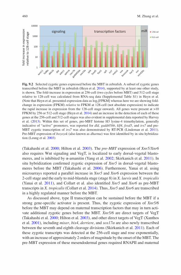
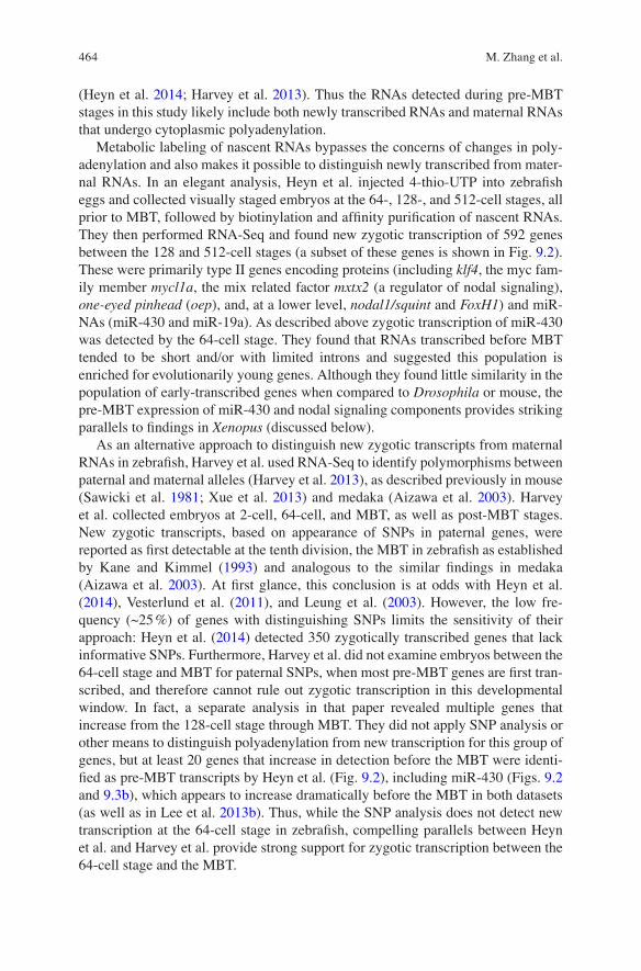
Fig. 9.2 Selected zygotic genes expressed before the MBT in zebrafish. A subset of zygotic genes transcribed before the MBT in zebrafish (Heyn et al. 2014), supported by at least one other study, is shown. The fold-increase in expression at 256-cell (two cycles before MBT) and 512-cell stage relative to 128-cell was calculated from RNA-seq data (Supplemental Table S1) in Heyn et al. (Note that Heyn et al. presented expression data as log2[FPKM] whereas here we are showing fold- change in expression (FPKM) relative to FPKM at 128-cell (not absolute expression) to indicate the rapid increase in expression from the 128-cell stage onward). All genes were present at >10 FPKM by 256 or 512-cell stage (Heyn et al. 2014) and an increase in the detection of each of these genes at the 256-cell and 512-cell stages was also evident in supplemental data reported by Harvey et al. (2013). Within this set of genes, pre-MBT histone H3 lysine-4 trimethylation, generally indicative of “active” promoters, was reported for dld, gadd45bb, klf4, foxd5, and irx7 and pre- MBT zygotic transcription of irx7 was also demonstrated by RT-PCR (Lindeman et al. 2011). Pre-MBT expression of bozozok (also known as dharma) was first identified by in situ hybridiza-tion (Leung et al. 2003)
M. Zhang et al.
461
VegT (Skirkanich et al. 2011). Hence, multiple direct targets of VegT are initially transcribed before the MBT in Xenopus laevis, consistent with gene- specific tran-scription factor dependent expression before the MBT, as seen with exogenous Gal4-VP16 (Almouzni and Wolffe 1995) in Xenopus and for endogenous genes in Drosophila (e.g., Liang et al. 2008; ten Bosch et al. 2006; Harrison et al. 2011).
The microRNA miR-427 (Watanabe et al. 2005) is a homologue of zebrafish miR-430 (Giraldez et al. 2006), and both of these miRNAs regulate maternal mRNA clear-ance and nodal signaling (Choi et al. 2007; Rosa et al. 2009). The miR-427 cluster is highly expressed before the MBT in X. laevis (Lund et al. 2009). The precursor form, pre-miR-427, and the primary transcript pri-miR-427 are detectable by Northern blot several hours before MBT (Lund et al. 2009) and an exponential increase in expres-sion begins at the 64-cell stage based on qRT-PCT (Fig. 9.3a, Jing Yang, personal communication). Pre-MBT transcription of miR-427 is RNAPII dependent, based on α-amanitin sensitivity. Interestingly, mIR-427 targets Xnr5 and Xnr6b and regulates mesendoderm development (Rosa et al. 2009). Inhibition of miR-427 with an antimir reduces expression of nodal inducible genes and mesoderm development, mimicking other inhibitors of nodal signaling. Although these effects on mesoderm development were associated with miR-427 regulation of lefty, another TGF-ß family member (Rosa et al. 2009), the coincident pre-MBT expression of both miR-427, Xnr5/6, and other regulators of mesendoderm induction is intriguing.
In zebrafish, miR-430 is also transcribed at a high level before the MBT, as early as the 64-cell stage (Fig. 9.3b) (Heyn et al. 2014), strongly supporting a conserved requirement for pre-MBT expression of this microRNA family. miR-430 transcrip-tion is activated by the maternal transcription factors Nanog, Pou5f3 (closely related to mammalian Oct4/Pou5f1 (Frankenberg et al. 2014)) and SoxB1, which also con-tribute to zygotic genome activation at the MBT (Lee et al. 2013b; Leichsenring et al. 2013). The regulation of miR-430 by these factors is analogous to miR-309 in Drosophila, which also regulates maternal mRNA clearance and is activated (by Zelda) before the MBT (Blythe and Wieschaus 2015b; Biemar et al. 2005). Similar to miR-427 in Xenopus, miR-430 interacts with nodal1/squint and lefty to regulate nodal signaling (Choi et al. 2007). miR-430 is also detectable by Northern blot before the proposed MBT in medaka (Tani et al. 2010). In addition, expression of the dorsal Wnt target gene bozozok (dharma) is detected in zebrafish before the MBT by in situ hybridization (Leung et al. 2003) and by RNA-Seq (Heyn et al. 2014; Harvey et al. 2013; Lee et al. 2013b).
9.3.3.3 Pre-MBT Transcription Identified by Gene Profiling
Multiple gene profiling studies have been reported on early developmental stages in Xenopus tropicalis, zebrafish, mouse, and other mammals. Most of the profiling studies in Xenopus and zebrafish have confirmed pre-MBT transcription of multiple protein coding genes and microRNAs. Interesting biological patterns emerge from interspecies comparisons, including the robust early expression of microRNAs that regulate degradation of maternal RNAs and early expression of nodal signaling components.
9 Cell Cycle Remodeling and Zygotic Gene Activation at the Midblastula Transition

462
Xenopus
Yanai et al. used custom microarrays to compare the temporal profiles for gene expression in X. laevis and X. tropicalis (Yanai et al. 2011). While they were not focused on gene expression changes during cleavage stages, they found multiple genes that increase in abundance from the 2-cell stage to stage 8 (MBT is at stage 8.5), which likely includes pre-MBT transcripts. Although a caveat of this analysis is that differences in signal could reflect changes in polyadenylation rather than zygotic transcription (see below), they observe in both X. tropicalis and X. laevis an increase at stage 8 in the VegT-regulated pre-MBT genes identified by Skirkanich et al., consistent with a conserved mode of regulation for these pre-MBT genes.
Subsequent RNA-Seq analyses using more detailed temporal analysis of multi-ple stages throughout cleavage stage development revealed additional type II zygotic genes transcribed before the MBT in X. tropicalis. Collart et al. used both oligo-dT primed and ribosome depleted RNA for libraries, and collected samples at 30 min intervals. They identified 960 RNAs that increased in apparent abundance
Egg 16-cell 64c 256c 1024c MBT St9 St10 St12
876543210m
iR42
7 ex
pres
sion
(Log
10)
miR-427 expression in Xenopus
miR-430 expression in zebrafish
32-cell 64-cell 128-cell 256-cell 512-cell + - + - + - + - + - RT
pri-mir430
pri-mir19a
a
b
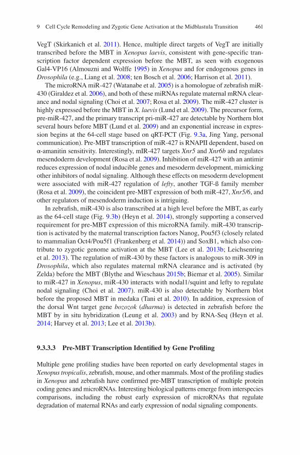
Fig. 9.3 Zygotic expression of miR-427/430 during cleavage stage in Xenopus and zebrafish. (a) Real-time RT-PCR analysis of pri-mir-427 at the indicated stages in Xenopus laevis. Data courtesy of Dr. Jing Yang, University of Illinois, Urbana-Champagne. Pre-MBT expression of mir-427 was first detected by Northern blot as shown by Lund et al. (2009). (b) RT-PCR analysis of pri-mir-430 and pri-mir-19a at the indicated stages in zebrafish, from Heyn et al. (2014) Supplemental data (Fig. S4C), reprinted with permission from the authors and from Cell Press. “+” and “−” indicate reaction products with and without reverse transcriptase (RT), respectively
M. Zhang et al.
463
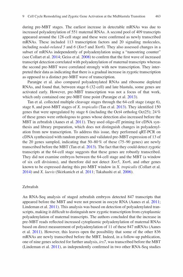
during pre-MBT stages. The earliest increase in detectable mRNAs was due to increased polyadenylation of 551 maternal RNAs. A second pool of 409 transcripts appeared around the 128-cell stage and these were confirmed as newly transcribed mRNAs. These included 113 transcription factors and 20 signaling molecules, including nodal-related 5 and 6 (Xnr5 and Xnr6). They also assessed changes in a subset of mRNAs independently of polyadenylation using a “nanostring counter” (see Collart et al. 2014; Geiss et al. 2008) to confirm that the first wave of increased transcript detection correlated with polyadenylation of maternal transcripts whereas the second pre-MBT wave correlated strongly with new transcription. They inter-preted their data as indicating that there is a gradual increase in zygotic transcription as opposed to a distinct pre-MBT wave of transcription.
Paranjpe et al. also compared polyadenylated RNAs and ribosome depleted RNAs, and found that, between stage 6 (32-cell) and late blastula, some genes are activated early. However, pre-MBT transcription was not a focus of that work, which only contained one pre-MBT time point (Paranjpe et al. 2013).
Tan et al. collected multiple cleavage stages through the 64-cell stage (stage 6), stage 8, and post-MBT stages of X. tropicalis (Tan et al. 2013). They identified 150 genes that were upregulated by stage 6 (including the Oct4 ortholog Oct25); 24 % of these genes were orthologous to genes whose detection also increased before the MBT in zebrafish (Aanes et al. 2011). They used oligo-dT priming for cDNA syn-thesis and library preparation, which does not distinguish changes in polyadenyl-ation from new transcription. To address this issue, they performed qRT-PCR on cDNA synthesized with random primers and validated pre-MBT expression of 13 of the 20 genes sampled, indicating that 50–80 % of these (75–90 genes) are newly transcribed before the MBT (Tan et al. 2013). The fact that they could detect zygotic transcripts at the 64-cell stage suggests that these genes are robustly transcribed. They did not examine embryos between the 64-cell stage and the MBT (a window of six cell divisions), and therefore did not detect Xnr5, Xnr6, and other genes known to be expressed during this pre-MBT window in X. tropicalis (Collart et al. 2014) and X. laevis (Skirkanich et al. 2011; Takahashi et al. 2006).
Zebrafish
An RNA-Seq analysis of staged zebrafish embryos detected 847 transcripts that appeared before the MBT and were not present in oocyte RNA (Aanes et al. 2011; Lindeman et al. 2011). This analysis was based on detection of polyadenylated tran-scripts, making it difficult to distinguish new zygotic transcription from cytoplasmic polyadenylation of maternal transcripts. The authors concluded that the increase in pre-MBT reads reflected increased cytoplasmic polyadenylation of maternal RNAs based on direct measurement of polyadenylation of 11 of these 847 mRNAs (Aanes et al. 2011). However, this leaves open the possibility that some of the other 836 mRNAs are newly transcribed before the MBT. Indeed, in a follow-up publication, one of nine genes selected for further analysis, irx7, was transcribed before the MBT (Lindeman et al. 2011), as independently confirmed in two other RNA-Seq studies
9 Cell Cycle Remodeling and Zygotic Gene Activation at the Midblastula Transition
464
(Heyn et al. 2014; Harvey et al. 2013). Thus the RNAs detected during pre- MBT stages in this study likely include both newly transcribed RNAs and maternal RNAs that undergo cytoplasmic polyadenylation.
Metabolic labeling of nascent RNAs bypasses the concerns of changes in poly-adenylation and also makes it possible to distinguish newly transcribed from mater-nal RNAs. In an elegant analysis, Heyn et al. injected 4-thio-UTP into zebrafish eggs and collected visually staged embryos at the 64-, 128-, and 512-cell stages, all prior to MBT, followed by biotinylation and affinity purification of nascent RNAs. They then performed RNA-Seq and found new zygotic transcription of 592 genes between the 128 and 512-cell stages (a subset of these genes is shown in Fig. 9.2). These were primarily type II genes encoding proteins (including klf4, the myc fam-ily member mycl1a, the mix related factor mxtx2 (a regulator of nodal signaling), one-eyed pinhead (oep), and, at a lower level, nodal1/squint and FoxH1) and miR-NAs (miR-430 and miR-19a). As described above zygotic transcription of miR-430 was detected by the 64-cell stage. They found that RNAs transcribed before MBT tended to be short and/or with limited introns and suggested this population is enriched for evolutionarily young genes. Although they found little similarity in the population of early-transcribed genes when compared to Drosophila or mouse, the pre-MBT expression of miR-430 and nodal signaling components provides striking parallels to findings in Xenopus (discussed below).
As an alternative approach to distinguish new zygotic transcripts from maternal RNAs in zebrafish, Harvey et al. used RNA-Seq to identify polymorphisms between paternal and maternal alleles (Harvey et al. 2013), as described previously in mouse (Sawicki et al. 1981; Xue et al. 2013) and medaka (Aizawa et al. 2003). Harvey et al. collected embryos at 2-cell, 64-cell, and MBT, as well as post-MBT stages. New zygotic transcripts, based on appearance of SNPs in paternal genes, were reported as first detectable at the tenth division, the MBT in zebrafish as established by Kane and Kimmel (1993) and analogous to the similar findings in medaka (Aizawa et al. 2003). At first glance, this conclusion is at odds with Heyn et al. (2014), Vesterlund et al. (2011), and Leung et al. (2003). However, the low fre-quency (~25 %) of genes with distinguishing SNPs limits the sensitivity of their approach: Heyn et al. (2014) detected 350 zygotically transcribed genes that lack informative SNPs. Furthermore, Harvey et al. did not examine embryos between the 64-cell stage and MBT for paternal SNPs, when most pre-MBT genes are first tran-scribed, and therefore cannot rule out zygotic transcription in this developmental window. In fact, a separate analysis in that paper revealed multiple genes that increase from the 128-cell stage through MBT. They did not apply SNP analysis or other means to distinguish polyadenylation from new transcription for this group of genes, but at least 20 genes that increase in detection before the MBT were identi-fied as pre-MBT transcripts by Heyn et al. (Fig. 9.2), including miR-430 (Figs. 9.2 and 9.3b), which appears to increase dramatically before the MBT in both datasets (as well as in Lee et al. 2013b). Thus, while the SNP analysis does not detect new transcription at the 64-cell stage in zebrafish, compelling parallels between Heyn et al. and Harvey et al. provide strong support for zygotic transcription between the 64-cell stage and the MBT.
M. Zhang et al.
465
As another approach to identify zygotic transcripts in zebrafish, Lee et al. performed RNA-Seq for intronic sequences, which also distinguishes new tran-scripts from maternal RNAs. That comprehensive study focused on gene expression after the canonical MBT (4 hpf) and did not examine stages between the 64-cell and MBT (Lee et al. 2013b); their approach nevertheless identified a large number of zygotic transcripts. A subset designated as “first wave” zygotic transcripts (still transcribed when zygotic gene function is blocked), including miR-430, klf4b, nnr, oep, blf, vent, her3, foxi1, mxtx2, were also identified as pre-MBT transcripts by Heyn et al. and some of these were also found to increase in detection before MBT by Harvey et al. Lee et al. further showed that loss of Nanog, SoxB1, and/or Pou5f3 (which has also been referred to as Pouf5/1 or Oct4, see Frankenberg et al. 2014) reduced the expression of “first-wave” genes, including multiple genes identified before the MBT in the Heyn et al. study (e.g., miR-430, klf4, blf, vent, her3, foxi1, mxtx2, vox, foxa3, foxd3, and sox32) (Lee et al. 2013b; Leichsenring et al. 2013). Consistent with this, Heyn et al. (2014) observed that 50 % of the pre-MBT genes that they identified contained Pou-Sox binding sites. Thus the transcription of a subset of “first zygotic wave” genes identified by Lee et al. may well begin between the 64-cell stage and the MBT.
Mammals
Gene profiling in mouse has revealed multiple mRNAs that increase in abundance in 1-cell mouse embryos (Xue et al. 2013; Park et al. 2013). Park et al. (2013) used ribosome depleted RNA in their studies and identified ~600 genes that increased in apparent expression in the 1-cell embryo. However, Abe have also found marked promiscuous transcription of RNAs in the 1-cell mouse embryo, with transcription of intergenic regions lacking clear promoters; these RNA species are low abundance and of as yet uncertain significance (Abe et al. 2015). Gene profiling studies of the maternal to zygotic transition have also been reported for various mammalian spe-cies (as cited in Li et al. 2013: Hamatani et al. 2004; Zeng and Schultz 2005; Misirlioglu et al. 2006; Sirard et al. 2005; Vallee et al. 2008; Wang et al. 2004; Whitworth et al. 2005; Zeng et al. 2004).
9.3.3.4 Limitations in Detecting New Transcription Before the MBT
These studies provide informative resources on early gene expression and also illus-trate some of the challenges inherent in analyzing new transcription in the early embryo. Most importantly the sensitivity of detection methods is a critical issue, especially when looking for new transcription over the background of a maternal store of RNAs that is orders of magnitude higher than the RNA in a typical somatic cell. This is especially true and underappreciated in RNA profiling approaches. Two recent studies on the sensitivity and reproducibility of next generation sequencing across multiple platforms (Li et al. 2014; SEQC/MAQC-III 2014) demonstrated that
9 Cell Cycle Remodeling and Zygotic Gene Activation at the Midblastula Transition
466
a substantial number of transcripts will be missed in somatic cell RNA at less than 1 billion mapped reads. Because of cost limitations, most RNA-Seq studies on staged early embryos are based on 10–100 times fewer mapped reads per stage; these stud-ies are informative about large scale changes in gene expression but not sufficiently powered to rule out the expression of low abundance or undetected transcripts. As a practical example, increasing sequencing depth to 1.5 billion reads identified numer-ous new lncRNAs in early zebrafish not detected in earlier studies (Pauli et al. 2012).
Detecting new gene expression in early embryos is also complicated by highly dynamic cytoplasmic polyadenylation and deadenylation of up to ~25 % of mRNAs during early development (Dworkin et al. 1985; McGrew et al. 1989; Graindorge et al. 2006; Paranjpe et al. 2013). Detection methods that depend on oligo-dT prim-ing alone cannot distinguish between changes in polyadenylation of maternal RNAs and changes in RNA abundance. This concern can be addressed using ribosome depleted RNAs, metabolic labeling of nascent RNAs, sequencing of introns, identi-fication of paternal polymorphisms, and/or validating by qRT-PCR using random- primed cDNA, as in the above studies. However, in general, distinct transcriptomes are represented in libraries prepared by polyA enrichment versus ribo-depletion, with more low abundance genes detected using ribosome depletion compared to oligo-dT priming (Li et al. 2014; SEQC/MAQC-III 2014).
Accurate staging of multicellular embryos is a potential concern, especially when comparing stages close to the MBT and when comparing different clutches, different rearing temperatures, or related but genetically distinct species (see Yanai et al. 2011; Harvey et al. 2013). For example, the first cell cycle in zebrafish is 45 min, compared with 15 min cleavage cycles subsequently; therefore the use of natural matings can introduce significant variation if staged samples are collected based on time alone (Langley et al. 2014; Steven Harvey, personal communication). This con-cern can be readily alleviated by using small numbers of embryos and visually stag-ing embryos during cleavage stages, as clearly described (Heyn et al. 2014; Karla Neugebauer, personal communication). In addition, several of the studies cited above did not examine pre-MBT stages later than the 64-cell stage of zebrafish or X. tropicalis, and therefore likely missed pre-MBT transcription detected by others between early cleavage stages and the MBT.
9.3.4 Function of Pre-MBT Gene Expression
The above expression analyses demonstrate that new transcription before the MBT is a conserved phenomenon in vertebrates (as in invertebrates) and suggest specific groups of zygotic genes that regulate early developmental events may be similarly regulated. However, many of these pre-MBT genes are initially present at low levels of expression, and the concern that this represents transcriptional noise has been raised. The evidence that early transcription is specifically regulated includes: (1) A limited set of genes is reproducibly transcribed before the MBT; (2) pre-MBT transcription is restricted to specific blastomeres for some genes (e.g., bozozok, Xnr5, Xnr6); (3)
M. Zhang et al.
467
maternal transcription factors are required for transcription of type II genes before the MBT; and (4) Pre-MBT genes regulate at least two fundamental biological processes that occur near the MBT—turnover of maternal RNAs and mesendoderm induction.
The Nodal genes derriere, Xnr5, and Xnr6 are expressed well before the MBT in Xenopus laevis (Yang et al. 2002; Blythe et al. 2010; Skirkanich et al. 2011; Takahashi et al. 2006; Collart et al. 2014). Nodal signaling is essential for meso-derm formation and, in coordination with dorsally localized Wnt/ß-catenin activity, establishes the dorsal-ventral axis of the embryo (Luxardi et al. 2010; Agius et al. 2000; Schier 2001; Xu et al. 2012; Schier and Talbot 2005). Nodal signaling is active at or before the MBT, as determined by the presence of phosphorylated Smad2, and pre-MBT expression of Xnr5 and Xnr6 are required for this activation (Skirkanich et al. 2011; Schohl and Fagotto 2002; Faure et al. 2000; Lee et al. 2001). Inhibition of Nodal signaling before the MBT abrogates mesoderm induction, whereas inhibition after the MBT no longer blocks induction of mesodermal mark-ers. Furthermore, activation of Nodal/Smad signaling before the MBT induces post- MBT expression of mesodermal markers, whereas activation of Smad2 after MBT no longer induces mesoderm markers. Taken together, these data demonstrate that pre-MBT Nodal signaling, initiated by pre-MBT transcription of Xnr5 and Xnr6, is required for patterning of the Xenopus embryo (Skirkanich et al. 2011).
MiR-430 in zebrafish and medaka and miR-427 in Xenopus are robustly expressed before the MBT and are essential for the degradation of maternal mRNAs during the maternal to zygotic transition. In Drosophila, the miR-309 cluster, which is activated by Zelda and is also essential for clearance of maternal mRNAs, strongly recruits RNAPII and is abundantly expressed at least two cycles before the MBT (Blythe and Wieschaus 2015b; Biemar et al. 2005). Whether the expression of these microRNAs specifically before the MBT is essential has not yet been tested, but their conserved pre-MBT expression in widely divergent species is consistent with an important role for their pre-MBT transcription in the maternal to zygotic transition. Furthermore, miR-430 and miR-427 regulate mesendoderm development in zebrafish and Xenopus (Choi et al. 2007; Rosa et al. 2009) and their early transcription is therefore consis-tent with the early transcription of other mesendoderm regulators, such as Xnr5/6 in Xenopus and nodal1/squint, mxtx2, and other nodal pathway genes in zebrafish.
These findings reinforce the developmental significance of pre-MBT transcription in Xenopus and zebrafish, as also reported in Drosophila (Liang et al. 2008; Harrison et al. 2010, 2011; Karr et al. 1985; Ali-Murthy et al. 2013; Biemar et al. 2005), and specifi-cally underline the critical importance of early transcription of microRNAs that regulate maternal mRNA clearance and of early transcriptional activation of nodal signaling.
9.3.5 Large Scale Genome Activation at the MBT
The large-scale increase in zygotic gene expression associated with the MBT is firmly established in Xenopus and zebrafish. This has been documented by metabolic labeling (Newport and Kirschner 1982a; Kane and Kimmel 1993; Kimelman et al.
9 Cell Cycle Remodeling and Zygotic Gene Activation at the Midblastula Transition
468
1987; Heyn et al. 2014; Yang et al. 2002) and gene profiling (Lee et al. 2014; Heyn et al. 2014; Harvey et al. 2013; Aanes et al. 2011; Collart et al. 2014; Paranjpe et al. 2013; Tan et al. 2013). Lee identified >7000 genes that are transcribed at or near the MBT in zebrafish, a majority of which were maternal genes not previously identi-fied as transcribed again at the onset of large-scale zygotic transcription. A major wave of transcription in the first few days of development has also been docu-mented in diverse mammalian species (reviewed in Li et al. 2013; Latham and Schultz 2001). The repression of transcription before MBT and activation at the MBT is also illustrated by the activity of injected RNAPIII dependent reporters.
Tadros and Lipschitz have nicely summarized the onset of zygotic transcription in diverse model organisms, and describe a minor wave of transcription that pre-cedes the major wave, even in organisms that begin zygotic transcription early in the developmental program, suggesting a requirement for limited transcription in early development followed by large-scale changes required for the maternal to zygotic transition (Tadros and Lipshitz 2005). While it has been suggested that the minor wave might simply be a shoulder to, and part of, the major wave of transcrip-tion at the MBT, the fact remains that genes dedicated to specific functions, e.g., microRNAs involved in maternal RNA degradation and signaling molecules involved in mesendoderm development, are consistently transcribed 4–6 cell cycles before the major wave of zygotic transcription in multiple organisms. This regula-tion suggests that the minor and major waves of transcription are distinct in both their regulation and their functions, as also seen in Drosophila (Liang et al. 2008; ten Bosch et al. 2006; De Renzis et al. 2007).
How then is the major wave of transcription regulated? Specifically, how is the onset of zygotic transcription coordinated with other events of the MBT and what are the mechanisms of repression before pre-MBT or activation of large-scale expression at the MBT? The timing of zygotic gene activation and the MBT in general has been proposed to be coupled to the nucleus–cytoplasm (N:C) ratio or alternatively depen-dent on a timer that is uncoupled from the N:C ratio. There is evidence for both mechanisms in vertebrates as in Drosophila. The mechanisms for transcriptional regulation before and after the MBT may reflect the absence of an activator that accu-mulates or the presence of an inhibitor that is disengaged at the MBT. Again, there is evidence for both mechanisms (Tadros and Lipshitz 2005; Blythe and Wieschaus 2015a; Lee et al. 2014; Langley et al. 2014; Farrell and O'Farrell 2014; Lu et al. 2009).
9.3.5.1 Mechanisms of Repression and the N:C Ratio
The evidence for a repressive factor that must be titrated out by the exponential increase in zygotic DNA was elegantly and persuasively presented in the classical work of Newport and Kirschner and supported by subsequent work in zebrafish (Kane and Kimmel 1993) and Drosophila (Di Talia et al. 2013; Blythe and Wieschaus 2015a; Lu et al. 2009; Edgar and Schubiger 1986). Those experiments are summarized in Sect. 9.2, and here we only address the titration model with respect to the control of zygotic transcription.
M. Zhang et al.
469
At the time of Newport and Kirschner’s experiments, it was clear that Xenopus embryos contain a large store of RNA polymerase that is active in extracts, suggest-ing that the lack of transcription before the MBT is due to repression. To test this hypothesis, Newport and Kirschner injected a yeast tRNA plasmid into cleavage- arrested Xenopus eggs. The tRNA was initially transcribed but was silenced within 2–3 h and remained silent until the MBT. This suppression correlated with assembly of the injected DNA into chromatin. As described for the onset of cell cycle asyn-chrony, injection of nonspecific DNA at approximately the amount found in the embryo at MBT caused premature activation of the yeast tRNA plasmid, as well as some endogenous, small RNAs, consistent with a repressive mechanism, at least for type III genes (Newport and Kirschner 1982b). In addition, Newport and Kirschner showed that increasing DNA content by generating polyspermic embryos led to transcription activation (of small RNAs) two cycles earlier than MBT in controls, also consistent with the timing of transcription depending on the N:C or more pre-cisely the DNA–cytoplasm ratio.
Prioleau found that the c-myc promoter could also be transiently activated if pre-loaded with TATA binding protein (TBP) before injection (Prioleau et al. 1994). Similar to type III genes described by Newport and Kirschner, this plasmid was also assembled into chromatin and repressed before MBT, and coinjecting nonspecific DNA blocked repression. TBP had to be preloaded on the plasmid; overexpression by injection of TBP mRNA was insufficient to allow early expression of the reporter. These authors proposed that assembly of exogenous DNA into chromatin prevents binding of activat-ing factors and thereby restrains transcription until the MBT (Almouzni et al. 1990; Newport and Kirschner 1982b; Prioleau et al. 1994; Amodeo et al. 2015).
Amodeo et al. (2015) further showed that histone H3 and H4 are titratable inhibitors of transcription in the pre-MBT embryo. They used Xenopus egg extracts to identify a threshold level of sperm chromatin needed to overcome an inhibitor present in extracts and in embryos. As nonspecific DNA will titrate the inhibitor, they assayed for removal of the inhibitor by DNA coated beads, then performed biochemical fractionation to isolate inhibitory factors identified by mass spec as histones H3 and H4. Purified H3/H4 inhibited transcription of sperm chromatin in vitro; furthermore, overexpression of H3 suppressed and knockdown of H3 accelerated the onset of RNAPIII dependent transcription in vivo. While this biochemical tour-de-force provides strong support for the findings of Newport and Kirschner, Prioleau et al., and Almouzni et al., who showed that addition of H2A/H2B dimers also inhibited transcription in Xenopus extracts (Almouzni et al. 1990, 1991), it also raises new questions. It remains unclear how the reduced ratio of core histones to DNA at the MBT results in gene specific activation; additional regulation, such as nucleosome remodeling, changes in chromatin architecture or attachment to the nuclear matrix, and posttranslational modification of core his-tones, likely contribute to the control of zygotic transcription (comprehensively reviewed in Bogdanovic et al. 2012; Veenstra 2002), as discussed further below.
The above work is consistent with the Newport and Kirschner hypothesis that zygotic transcription at the MBT is determined by the N:C ratio, similar also to findings in Drosophila. However, much of the work on transcription in early
9 Cell Cycle Remodeling and Zygotic Gene Activation at the Midblastula Transition
470
Xenopus and Drosophila has focused on type III transcription (Newport and Kirschner 1982b; Almouzni et al. 1990; Amodeo et al. 2015; Brown 2004; Brown and Gurdon 1977, 1978), and although Prioleau showed that the c-myc promoter is also repressed before MBT, a different story emerged for the regulation of other type II genes in cleavage stage Xenopus embryos. Increasing nonspecific DNA content did not cause premature activation of exogenous U1 genes, which are RNAPII dependent (Lund and Dahlberg 1992). Similarly, Almouzni and Wolffe found that injection of nonspecific DNA does not activate a CMV promoter or other type II genes (Almouzni and Wolffe 1995). Furthermore, an exogenous type II promoter can be activated if the transactivator is also present: Expression of GAL4-VP16 at sufficient levels drives a GAL4 dependent reporter throughout pre-MBT stages, showing that the type II transcription apparatus is functional (Almouzni and Wolffe 1995). Almouzni and Wolffe proposed that the suppression of type II transcription is through a mechanism distinct from type III regulation, and, in addition to regulation by nucleosome assembly, depends on the availability of gene-specific transcription factors (as discussed in detail above for endogenous genes in Xenopus and zebrafish).
Transcription During Short Cell Cycles
The notion that transcription is not compatible with the rapid cell cycles of cleavage divisions has been proposed. According to this model, the high density of replica-tion origins in cleavage stage embryos and the progress of the replication apparatus disrupt transcription complexes and furthermore the short duration of S-phase limits transcription to short RNAs. In this model, slowing of the cell cycle at the MBT allows transcription.
In support of this model, treatment of Xenopus embryos with cycloheximide two cycles before the MBT blocks progression into mitosis and causes premature acti-vation of type III zygotic gene expression, despite the DNA content being fourfold lower than MBT. Similar observations were made in Drosophila (Edgar and Schubiger 1986). However, cycloheximide is a blunt instrument, and treatment at earlier stages in either Xenopus or Drosophila has the opposite effect to block zygotic transcription at MBT (Edgar and Schubiger 1986; Lund and Dahlberg 1992; Blythe and Wieschaus 2015a; Lee et al. 2014; Sible et al. 1997).
Using a more targeted approach, Collart et al. showed that the replication factors Cut5, RecQ4, Treslin, and Drf1 become limiting for DNA replication as the N:C ratio increases in Xenopus embryos (Collart et al. 2013), as also discussed above. They pro-posed that the limited availability of these replication factors slows the cell cycle and permits transcription at the MBT. Overexpression of the four factors delayed expression for a subset of zygotic genes, consistent with these factors acting as repressors that are titrated out as the N:C ratio approaches a critical value. While it remains possible that overexpression of these factors impairs transcription of this subset of genes indepen-dently of the effects on replication, these data nevertheless show a compelling correla-tion between availability of the four replication factors and multiple events at the MBT.
M. Zhang et al.
471
However, a simple model involving constraint from rapid DNA replication does not take into account the following observations: (1) Sea urchins initiate large scale zygotic transcription during early cleavage stages despite cleavage divisions as rapid as Xenopus or zebrafish (Davidson 1986), and C. elegans similarly initiates zygotic transcription during a period of rapid cell division, arguing that rapid DNA replication alone is not sufficient to delay zygotic transcription. (2) Mammalian embryos delay zygotic transcription for up to several days, despite their slow cell cycles, implying the need for a distinct mechanism to delay zygotic gene activation in mammals. (3) Transcription of endogenous genes is readily detectable during early cleavage stages in Xenopus, zebrafish, medaka (Tani et al. 2010; Kraeussling et al. 2011), and Drosophila and is essential for post-MBT development in Xenopus and Drosophila. (4) Prolonging the cell cycle by activation of Chk1 before the MBT in zebrafish embryos does not cause premature zygotic transcription (Zhang et al. 2014). Similarly, prolonging the cell cycle before the MBT does not enhance tran-scription of exogenous type II genes in Xenopus (Almouzni and Wolffe 1995), 1-cell mouse embryos (Aoki et al. 1997), or Drosophila (inferred from normal tim-ing of cellularization; McCleland and O'Farrell 2008). (5) Xenopus embryos defi-cient for Wee-1 continue to undergo rapid cell cycles after the MBT, yet activate zygotic transcription at the 12th division, similar to control embryos, demonstrating (as in sea urchins) that slowing of the cell cycle is not required to activate transcrip-tion (Murakami et al. 2004), and (6) Overexpression of the four DNA replication factors described above extends rapid cell cycles beyond the 12th division (MBT), and while the onset of transcription of some genes is delayed, a “large number” of zygotic genes is still activated at the canonical MBT (Collart et al. 2013). These observations argue that the restriction of zygotic transcription is not explained solely by the constraints of rapid DNA replication. In fact, the converse appears to be true in Drosophila, where recruitment of RNAPII drives DNA replication stall-ing in early Drosophila embryos (Blythe and Wieschaus 2015b).
In contrast, transcription of most loci is suppressed during mitosis, although some transcription factors remain associated with chromatin throughout mitosis (Kadauke and Blobel 2013). If RNA polymerase moves at a rate of ~50–100 nt/s, then a 10 min S-phase may limit synthesis of unprocessed RNAs to <60 kilobases. Empirically, a bias toward short transcripts has been reported for pre-MBT genes in zebrafish (Heyn et al. 2014) and Drosophila (Rothe et al. 1992; Shermoen and O'Farrell 1991; De Renzis et al. 2007; Swinburne and Silver 2008).
DNA Methylation
Differential DNA methylation was at one time proposed as a mechanism to control zygotic gene activation at the MBT (Stancheva and Meehan 2000). Knockdown of the maintenance DNA methyltransferase, Dnmt1, reduces global 5-methylcytosine levels and leads to precocious activation of a subset of zygotic genes in Xenopus (Ruzov et al. 2004, 2009; Dunican et al. 2008). However, knockdown does not alter methylation patterns at the promoters of precociously expressed genes and a catalytically inactive
9 Cell Cycle Remodeling and Zygotic Gene Activation at the Midblastula Transition
472
form of Dnmt1 restores normal timing of expression, indicating that transcriptional repression by Dnmt1 is not mediated by DNA methylation (Dunican et al. 2008). These observations are consistent with the finding that global DNA methylation does not change from pre-MBT to post-MBT stages (Veenstra 2002).
Zebrafish and mammalian embryos undergo global demethylation and remethyl-ation after fertilization (Andersen et al. 2012, 2013; Potok et al. 2013). The inter-play of DNA methylation and regulation of early gene expression in mouse embryos has been thoroughly reviewed elsewhere (Plasschaert and Bartolomei 2014; Weaver et al. 2009; Paranjpe and Veenstra 2015; Rivera and Ross 2013; Li et al. 2010, 2013). In zebrafish, DNA methylation reaches a nadir at the 64-cell stage and then increases through the MBT. DNA methylation status mostly correlates with the time of gene expression, with genes expressed at MBT typically being hypomethylated and genes involved in later development being hypermethylated. Most promoters maintain their respective states of methylation from pre-MBT to post-MBT stages, although a minority undergoes dynamic changes in methylation (Andersen et al. 2012, 2013; Potok et al. 2013). Remarkably, the DNA methylation pattern in sperm is highly similar to embryonic DNA at MBT, whereas the maternal DNA methyla-tion pattern is reprogrammed during embryogenesis, suggesting that the methyla-tion pattern in sperm could inform later patterns of gene expression in the embryo (Potok et al. 2013).
N:C Volume Ratio
As discussed above, the N:C volume ratio also contributes to the timing of MBT, including the onset of zygotic transcription (Jevtic and Levy 2015). Jevtic and Levy increased nuclear volume by overexpressing importin-α and lamin B3 in one half of Xenopus embryos and examined the onset of expression of GS17, an established MBT marker (Krieg and Melton 1985; Harvey et al. 1986). In the injected halves, GS17 was expressed well before the MBT in ~80 % of embryos, but not until the MBT on the control side. Conversely, reducing nuclear volume by overexpression of the reticulon family protein Rtn4a delayed the onset of GS17 expression. They also observed increased pre-MBT expression of Xnr5, a known pre-MBT zygotic transcript (Yang et al. 2002), when they increased nuclear volume, suggesting that pre-MBT transcription may also be sensitive to the N:C volume ratio (Jevtic and Levy 2015).
9.3.5.2 Activating Factors and an N:C Independent Timer
In addition to taking a foot off the brakes, the embryo may need to step on the accel-erator. Accumulation of activating factors, for example by translation of maternal mRNAs, may contribute to a developmental timer that functions independently of the N:C ratio. Howe et al. described a developmental timer that coordinates cyclin E1 degradation with the MBT (Howe and Newport 1996). Evidence for a translation
M. Zhang et al.
473
coupled timer for the MBT is further supported by the observations in Xenopus and zebrafish that cycloheximide exposure during early cleavage stages (as opposed to treatment one or two cycles before MBT) blocks zygotic transcription, even when nonspecific DNA is injected to match the expected concentration of DNA at the MBT (Lund et al. 2009; Lee et al. 2014).
As discussed above, Nanog, Pou5f3, and SoxB1 are required for zygotic gene activation in zebrafish, and are in this sense analogous to Zelda in Drosophila. Knockdown of all three genes causes lethality similar to treatment with α-amanitin (Lee et al. 2013b). As these three factors are present as maternal mRNAs and are translated prior to the MBT, their progressive accumulation could function as an N:C independent timer. Notably, these factors are required for very early zygotic expression of miR-430, and therefore for the regulation of maternal mRNA stability as well as global gene activation at the MBT. Oct4 (Pou5f1) and Nanog have also been implicated in the activation of zygotic gene expression at the two-cell stage in mouse (Foygel et al. 2008; Tan et al. 2013). A similar role for pluripotency factors has not yet been reported in the regulation of zygotic transcription in Xenopus.
TBP
As described above, TBP can facilitate the expression of exogenous reporter plas-mids in cleavage stage Xenopus embryos, especially type III promoters. More gen-erally, TBP is required for basal and active transcription and is considered an essential component of the transcription complex (Hernandez 1993). TBP and closely related factors are essential for embryonic development in Xenopus, zebraf-ish, and C. elegans (reviewed in Lee et al. 2014; Bogdanovic et al. 2012; Veenstra 2002). TBP mRNA is present maternally but the protein is barely detectable in the egg. Translation during cleavage stages leads to accumulation of TBP protein that correlates with large-scale zygotic gene activation in frogs and mice, suggesting new translation of TBP contributes to transcriptional activation at the MBT (Veenstra et al. 1999; Bell and Scheer 1999; Bogdanovic et al. 2012; Veenstra 2002). Regulated translation of maternal TBP mRNA during early development could therefore be a component of an N:C independent timer for zygotic transcription at the MBT.
Smicl and RNAPII
Collart et al. reported an increase in C-terminal phosphorylation of RNAPII (at sites associated with elongating RNAPII) at the MBT that was mediated by the Smad- interacting CPSF 30-like factor (Smicl), which translocates from the cytoplasm to the nucleus at the MBT in Xenopus. Knockdown of Smicl expression reduced the expression of multiple genes at the MBT and delayed the degradation of several maternal mRNAs (Collart et al. 2009), supporting the hypothesis that enhanced nuclear translocation of Smicl and increased CTD phosphorylation of RNAPII facilitates zygotic transcription at the MBT.
9 Cell Cycle Remodeling and Zygotic Gene Activation at the Midblastula Transition
474
Geminin
Geminin is an unstable protein that inhibits the replication factor Cdt1 to prevent a second round of DNA replication during S-phase. Knockdown of Geminin or over-expression of Cdt1 in Xenopus embryos causes G2 arrest one cycle after the MBT and markedly reduces expression of multiple genes normally expressed at the MBT, but not expression of the pre-MBT zygotic transcripts for Xnr5 (Kerns et al. 2012). These observations suggest that Geminin is essential specifically for zygotic tran-scription at the MBT, but not before. However, Lim et al. (2011) found that Geminin overexpression represses expression of lineage specific markers after the MBT and knockdown (with a different morpholino than that used by Kerns et al.) enhanced their expression. These two studies appear incompatible, and the reasons for these differences are not clear.
9.3.6 Histone Modifications During Early Development
In addition to assembly of nucleosomes and DNA methylation, discussed above, posttranslational modifications of histones that correlate with both activation and repression are dynamic across development, from the gametes through zygotic gene activation. The chromatin landscape through development has been compre-hensively covered in authoritative reviews (Bogdanovic et al. 2012; Lee et al. 2014; Blythe and Wieschaus 2015a; Andersen et al. 2013), and is only touched on briefly here.
Surveys of histone modifications during development in Xenopus and zebrafish have revealed some shared general features and interesting differences. In general, chromatin modifications precede gene expression in many cases, consistent with a chromatin prepattern or poised chromatin state (Lindeman et al. 2011; Blythe et al. 2010; Potok et al. 2013). In general, the detection of both activating and repressive marks increases during the blastula stage, although the time that these marks can first be detected relative to the MBT depends on the sensitivity of the ChIP methods, a critical issue in an early embryo that contains a limiting number of nuclei/chromo-somal DNA within a large cytoplasmic volume, filled with abundant yolk proteins, maternal RNAs, and free histones that may also carry posttranslational modifica-tions (Shechter et al. 2009; Toyama et al. 2008).
Akkers et al. assessed genome-wide H3K4me3, H3K27me3, and RNAPII occu-pancy in gastrula stage Xenopus tropicalis by ChIP-Seq and showed that presence of both H3K4me3 and H3K27me was associated with genes that are transcribed in a localized manner (Akkers et al. 2009); these marks were present on distinct nucleosome populations, distinguishing them from bivalent marks described for example in mouse embryonic stem cells. Using ChIP-PCR for a selected set of developmental genes, they first detected H3K4me3 in post-MBT blastulae either coincident with or preceding expression. H3K27me appeared later, at the gastrula stage, and was predictive of localized expression. Examining a distinct set of
M. Zhang et al.
475
genes, Blythe et al. detected H3K4me3, RNAPII recruitment, H3R8me2a, and specific transcription factor binding in X. laevis two cell cycles before the MBT at both transcribed genes and nontranscribed, poised promoters (Blythe et al. 2010). The establishment of poised chromatin architecture at dorsal specifying genes before the MBT was required for localized expression and dorsal development after the MBT, demonstrating an essential developmental function for pre-MBT chromatin modifications.
An initial study in zebrafish used a microarray-based approach (ChIP-chip) to survey chromatin marks in 31 Mb of the zebrafish genome; in contrast to findings in X. tropicalis, they found that H3K4me3 and H3K27me3 were present within the same population of nucleosomes, consistent with bivalent chromatin marks (Vastenhouw et al. 2010). They were first able to detect H3K4me3 and H3K27me3 after the MBT. However, more sensitive methods in zebrafish subsequently detected >1000 promoters with H3K4me3 before the MBT (256-cell), and fewer genes marked with H3K27me3 (200) and H3K9me3 (500) (Lindeman et al. 2011). Lindeman et al. detected chromatin marks as early as three cell cycles before the MBT by western blot and immunofluorescence. Many of the genes containing H3K4me3 lacked evidence of elongating RNAPII and were presumed to be poised, non-expressed genes, although direct measurements demonstrated that a subset of genes with pre-MBT H3K4me3 marks are in fact expressed before the MBT (see above). Nevertheless, the prevalence of modified histones prior to the MBT at genes that are mostly inactive suggested a chromatin prepattern that anticipates later developmental gene expression.
The pattern of histone modifications at the MBT in zebrafish embryos was very similar to the pattern in sperm, as also observed for DNA methylation (Potok et al. 2013), consistent with inheritance of a chromatin prepattern that may lay the tran-scriptional groundwork for early development.
The regulation of zygotic gene activation by changes in chromatin architecture has also been investigated in mammals, principally mouse, and is more thoroughly reviewed elsewhere (Li et al. 2010, 2013). Maternal loss of Brg1, a core compo-nent of Swi/Snf chromatin remodeling complexes, impairs activation of ~30 % of zygotic genes, reduces H3K4 methylation, and causes peri-implantation lethality in the mouse (Bultman et al. 2006). Similarly, overexpression in mouse oocytes of a mutant form of histone H3.3 that cannot be methylated at lysine-4 (or knockdown of the H3K4 methyltransferase Mll3/4) causes developmental arrest after fertiliza-tion and impairs the minor wave of zygotic gene expression in the paternal pronu-cleus (Aoshima et al. 2015). Loss of TIF1a (transcription intermediary factor 1a), which is recruited to sites occupied by Brg1, also causes embryonic lethality at the 2–4 cell stage and disrupts activation of zygotic gene expression (Torres-Padilla and Zernicka-Goetz 2006). Activation of zygotic genes depends on additional fac-tors involved in chromatin or nuclear structure such as the PRC1 components Ring1 and Rnf2 (Posfai et al. 2012), CTCF (Wan et al. 2008), and nucleoplasmin 2 (Npm2) (Burns et al. 2003), the pluripotency factors Oct4 (Pou5f1) (Foygel et al. 2008) and Sox2 (Pan and Schultz 2011), as well as other factors, as cited by Li et al. (2010, 2013).
9 Cell Cycle Remodeling and Zygotic Gene Activation at the Midblastula Transition
476
9.3.7 Summary
Vertebrate embryos delay the first major wave of zygotic transcription from hours to days after fertilization, although the number of cell divisions until zygotic gene activation varies. Large-scale changes in chromatin architecture appear to anticipate and/or accompany these changes, although the timing and strategy for regulation of gene expression may vary between organisms. The capacity for transcription exists already in the egg, indicating that repressive mechanisms are programmed to delay zygotic transcription. In oviparous species a repressive activity linked to chromatin assembly is titrated out as the DNA content and/or nuclear volume increases relative to the cytoplasm, but there is also evidence for N:C independent timing mechanisms that may regulate zygotic gene activation, maternal RNA clearance, and cell cycle remodeling. Recent work has identified specific mRNAs and microRNAs robustly expressed before the MBT that are essential for critical developmental processes that define the maternal to zygotic transition, including clearance of maternal RNAs and activation of the earliest steps in germ layer formation. Whether expression of these early RNAs depends on the N:C ratio or instead is regulated by an N:C inde-pendent timer remains to be investigated.
Acknowledgements We thank Shelby Blythe, Jing Yang, Daniel Kessler, Mary Mullins, Karla Neugebauer, Patricia Heyn, Steven Harvey, and Derek Stemple for helpful discussions. We thank Jing Yang for sharing unpublished data and Karla Neugebauer and Patricia Heyn for permission to reprint their published data. We are indebted to authoritative reviews from many authors, as cited above, and thank Shelby Blythe and Eric Wieschaus for sharing their review prior to publication. M.Z. was supported in part by the Developmental Biology Training Grant at Penn (T32HD007516). J.S. was supported in part by the Cell and Molecular Biology Training Grant at Penn (T32-GM07229). M.A.L. was supported by the NIH (R01GM083988) and a Searle Scholar award. P.S.K. was supported by the NIH (R01HL110806 and R01MH100923) and the Institute for Regenerative Medicine at the University of Pennsylvania.
References
Aanes H, Winata CL, Lin CH, Chen JP, Srinivasan KG, Lee SG, Lim AY, Hajan HS, Collas P, Bourque G, Gong Z, Korzh V, Alestrom P, Mathavan S (2011) Zebrafish mRNA sequencing deciphers novelties in transcriptome dynamics during maternal to zygotic transition. Genome Res 21(8):1328–1338
Abe K, Yamamoto R, Franke V, Cao M, Suzuki Y, Suzuki MG, Vlahovicek K, Svoboda P, Schultz RM, Aoki F (2015) The first murine zygotic transcription is promiscuous and uncoupled from splicing and 3′ processing. EMBO J 34(11):1523–1537
Agius E, Oelgeschlager M, Wessely O, Kemp C, De Robertis EM (2000) Endodermal Nodal- related signals and mesoderm induction in Xenopus. Development 127(6):1173–1183
Aizawa K, Shimada A, Naruse K, Mitani H, Shima A (2003) The medaka midblastula transition as revealed by the expression of the paternal genome. Gene Expr Patterns 3(1):43–47
Akkers RC, van Heeringen SJ, Jacobi UG, Janssen-Megens EM, Francoijs KJ, Stunnenberg HG, Veenstra GJ (2009) A hierarchy of H3K4me3 and H3K27me3 acquisition in spatial gene regulation in Xenopus embryos. Dev Cell 17(3):425–434
M. Zhang et al.
477
Ali-Murthy Z, Lott SE, Eisen MB, Kornberg TB (2013) An essential role for zygotic expression in the pre-cellular Drosophila embryo. PLoS Genet 9(4):e1003428
Almouzni G, Mechali M, Wolffe AP (1990) Competition between transcription complex assembly and chromatin assembly on replicating DNA. EMBO J 9(2):573–582
Almouzni G, Mechali M, Wolffe AP (1991) Transcription complex disruption caused by a transi-tion in chromatin structure. Mol Cell Biol 11(2):655–665
Almouzni G, Wolffe AP (1995) Constraints on transcriptional activator function contribute to tran-scriptional quiescence during early Xenopus embryogenesis. EMBO J 14(8):1752–1765
Amodeo AA, Jukam D, Straight AF, Skotheim JM (2015) Histone titration against the genome sets the DNA-to-cytoplasm threshold for the Xenopus midblastula transition. Proc Natl Acad Sci U S A 112(10):E1086–E1095
Amon A, Irniger S, Nasmyth K (1994) Closing the cell cycle circle in yeast: G2 cyclin proteolysis initi-ated at mitosis persists until the activation of G1 cyclins in the next cycle. Cell 77(7):1037–1050
Andersen IS, Lindeman LC, Reiner AH, Ostrup O, Aanes H, Alestrom P, Collas P (2013) Epigenetic marking of the zebrafish developmental program. Curr Top Dev Biol 104:85–112
Andersen IS, Reiner AH, Aanes H, Alestrom P, Collas P (2012) Developmental features of DNA methylation during activation of the embryonic zebrafish genome. Genome Biol 13(7):R65
Anderson JA, Lewellyn AL, Maller JL (1997) Ionizing radiation induces apoptosis and elevates cyclin A1-Cdk2 activity before but not after the midblastula transition in Xenopus. Mol Biol Cell 8(7):1195–1206
Aoki F, Worrad DM, Schultz RM (1997) Regulation of transcriptional activity during the first and second cell cycles in the preimplantation mouse embryo. Dev Biol 181(2):296–307
Aoshima K, Inoue E, Sawa H, Okada Y (2015) Paternal H3K4 methylation is required for minor zygotic gene activation and early mouse embryonic development. EMBO Rep 16(7):803–812
Atherton-Fessler S, Liu F, Gabrielli B, Lee MS, Peng CY, Piwnica-Worms H (1994) Cell cycle regulation of the p34cdc2 inhibitory kinases. Mol Biol Cell 5(9):989–1001
Audic Y, Omilli F, Osborne HB (1997) Postfertilization deadenylation of mRNAs in Xenopus laevis embryos is sufficient to cause their degradation at the blastula stage. Mol Cell Biol 17(1):209–218
Bachvarova R, Davidson EH (1966) Nuclear activation at the onset of amphibian gastrulation. J Exp Zool 163(3):285–296
Bachvarova R, Davidson EH, Allfrey VG, Mirsky AE (1966) Activation of RNA synthesis associ-ated with gastrulation. Proc Natl Acad Sci U S A 55(2):358–365
Baroux C, Autran D, Gillmor CS, Grimanelli D, Grossniklaus U (2008) The maternal to zygotic transition in animals and plants. Cold Spring Harb Symp Quant Biol 73:89–100
Bartek J, Lukas J (2003) Chk1 and Chk2 kinases in checkpoint control and cancer. Cancer Cell 3(5):421–429
Bell P, Scheer U (1999) Developmental changes in RNA polymerase I and TATA box-binding protein during early Xenopus embryogenesis. Exp Cell Res 248(1):122–135
Biemar F, Zinzen R, Ronshaugen M, Sementchenko V, Manak JR, Levine MS (2005) Spatial regu-lation of microRNA gene expression in the Drosophila embryo. Proc Natl Acad Sci U S A 102(44):15907–15911
Blythe SA, Cha SW, Tadjuidje E, Heasman J, Klein PS (2010) Beta-catenin primes organizer gene expression by recruiting a histone H3 arginine 8 methyltransferase, Prmt2. Dev Cell 19(2) :220–231
Blythe SA, Wieschaus EF (2015a) Coordinating cell cycle remodeling with transcriptional activa-tion at the Drosophila MBT. Curr Top Dev Biol 113:113–148
Blythe SA, Wieschaus EF (2015b) Zygotic genome activation triggers the DNA replication check-point at the midblastula transition. Cell 160(6):1169–1181
Bogdanovic O, van Heeringen SJ, Veenstra GJ (2012) The epigenome in early vertebrate develop-ment. Genesis 50(3):192–206
Bouldin CM, Kimelman D (2014) Cdc25 and the importance of G2 control: insights from developmental biology. Cell Cycle 13(14):2165–2171
9 Cell Cycle Remodeling and Zygotic Gene Activation at the Midblastula Transition
478
Bouniol C, Nguyen E, Debey P (1995) Endogenous transcription occurs at the 1-cell stage in the mouse embryo. Exp Cell Res 218(1):57–62
Brown DD (2004) A tribute to the Xenopus laevis oocyte and egg. J Biol Chem 279(44) :45291–45299
Brown DD, Gurdon JB (1977) High-fidelity transcription of 5S DNA injected into Xenopus oocytes. Proc Natl Acad Sci U S A 74(5):2064–2068
Brown DD, Gurdon JB (1978) Cloned single repeating units of 5S DNA direct accurate transcrip-tion of 5S RNA when injected into Xenopus oocytes. Proc Natl Acad Sci U S A 75(6):2849–2853
Brown EJ, Baltimore D (2000) ATR disruption leads to chromosomal fragmentation and early embryonic lethality. Genes Dev 14(4):397–402
Bultman SJ, Gebuhr TC, Pan H, Svoboda P, Schultz RM, Magnuson T (2006) Maternal BRG1 regulates zygotic genome activation in the mouse. Genes Dev 20(13):1744–1754
Burns KH, Viveiros MM, Ren Y, Wang P, DeMayo FJ, Frail DE, Eppig JJ, Matzuk MM (2003) Roles of NPM2 in chromatin and nucleolar organization in oocytes and embryos. Science 300(5619):633–636
Campbell SD, Sprenger F, Edgar BA, O'Farrell PH (1995) Drosophila Wee1 kinase rescues fission yeast from mitotic catastrophe and phosphorylates Drosophila Cdc2 in vitro. Mol Biol Cell 6(10):1333–1347
Chini CC, Chen J (2003) Human claspin is required for replication checkpoint control. J Biol Chem 278(32):30057–30062
Choi WY, Giraldez AJ, Schier AF (2007) Target protectors reveal dampening and balancing of Nodal agonist and antagonist by miR-430. Science 318(5848):271–274
Christians E, Campion E, Thompson EM, Renard JP (1995) Expression of the HSP 70.1 gene, a landmark of early zygotic activity in the mouse embryo, is restricted to the first burst of tran-scription. Development 121(1):113–122
Ciliberto A, Petrus MJ, Tyson JJ, Sible JC (2003) A kinetic model of the cyclin E/Cdk2 develop-mental timer in Xenopus laevis embryos. Biophys Chem 104(3):573–589
Clute P, Masui Y (1992) Development of microtubule-dependence of the chromosome cycle at the midblastula transition in Xenopus laevis embryos. Dev Growth Differ 34:27–36
Clute P, Masui Y (1995) Regulation of the appearance of division asynchrony and microtubule- dependent chromosome cycles in Xenopus laevis embryos. Dev Biol 171(2):273–285
Collart C, Allen GE, Bradshaw CR, Smith JC, Zegerman P (2013) Titration of four replication factors is essential for the Xenopus laevis midblastula transition. Science 341(6148):893–896
Collart C, Owens ND, Bhaw-Rosun L, Cooper B, De Domenico E, Patrushev I, Sesay AK, Smith JN, Smith JC, Gilchrist MJ (2014) High-resolution analysis of gene activity during the Xenopus mid-blastula transition. Development 141(9):1927–1939
Collart C, Ramis JM, Down TA, Smith JC (2009) Smicl is required for phosphorylation of RNA polymerase II and affects 3′-end processing of RNA at the midblastula transition in Xenopus. Development 136(20):3451–3461
Conn CW, Lewellyn AL, Maller JL (2004) The DNA damage checkpoint in embryonic cell cycles is dependent on the DNA-to-cytoplasmic ratio. Dev Cell 7(2):275–281
D'Angiolella V, Costanzo V, Gottesman ME, Avvedimento EV, Gautier J, Grieco D (2001) Role for cyclin-dependent kinase 2 in mitosis exit. Curr Biol 11:1221–1226
Dalle Nogare DE, Pauerstein PT, Lane ME (2009) G2 acquisition by transcription-independent mechanism at the zebrafish midblastula transition. Dev Biol 326(1):131–142
Dasso M, Newport JW (1990) Completion of DNA replication is monitored by a feedback system that controls the initiation of mitosis in vitro: studies in Xenopus. Cell 61(5):811–823
Davidson EH (1986) Gene activity in early development, 3rd edn. Academic, Boston, MADe La Fuente R, Eppig JJ (2001) Transcriptional activity of the mouse oocyte genome: companion
granulosa cells modulate transcription and chromatin remodeling. Dev Biol 229(1):224–236De Renzis S, Elemento O, Tavazoie S, Wieschaus EF (2007) Unmasking activation of the zygotic
genome using chromosomal deletions in the Drosophila embryo. PLoS Biol 5(5):e117
M. Zhang et al.
479
Di Talia S, She R, Blythe SA, Lu X, Zhang QF, Wieschaus EF (2013) Posttranslational control of Cdc25 degradation terminates Drosophila’s early cell-cycle program. Curr Biol 23(2):127–132
Dickey JS, Redon CE, Nakamura AJ, Baird BJ, Sedelnikova OA, Bonner WM (2009) H2AX: functional roles and potential applications. Chromosoma 118(6):683–692
Ducommun B, Brambilla P, Félix M, Franza BR, Karsenti E, Draetta G (1991) Cdc2 phosphoryla-tion is required for its interaction with cyclin. EMBO J 10(11):3311–3319
Dunican DS, Ruzov A, Hackett JA, Meehan RR (2008) xDnmt1 regulates transcriptional silencing in pre-MBT Xenopus embryos independently of its catalytic function. Development 135(7):1295–1302
Dworkin MB, Shrutkowski A, Dworkin-Rastl E (1985) Mobilization of specific maternal RNA species into polysomes after fertilization in Xenopus laevis. Proc Natl Acad Sci U S A 82(22):7636–7640
Edgar BA, Datar SA (1996) Zygotic degradation of two maternal Cdc25 mRNAs terminates Drosophila’s early cell cycle program. Genes Dev 10(15):1966–1977
Edgar BA, Kiehle CP, Schubiger G (1986) Cell cycle control by the nucleo-cytoplasmic ratio in early Drosophila development. Cell 44(2):365–372
Edgar BA, O'Farrell PH (1989) Genetic control of cell division patterns in the Drosophila embryo. Cell 57(1):177–187
Edgar BA, Schubiger G (1986) Parameters controlling transcriptional activation during early Drosophila development. Cell 44(6):871–877
Edgar BA, Sprenger F, Duronio RJ, Leopold P, O'Farrell PH (1994) Distinct molecular mechanism regulate cell cycle timing at successive stages of Drosophila embryogenesis. Genes Dev 8(4):440–452
Elledge SJ, Richman R, Hall FL, Williams RT, Lodgson N, Harper JW (1992) CDK2 encodes a 33-kDa cyclin A-associated protein kinase and is expressed before CDC2 in the cell cycle. Proc Natl Acad Sci U S A 89:2907–2911
Evans T, Rosenthal ET, Youngblom J, Distel D, Hunt T (1983) Cyclin: a protein specified by mater-nal mRNA in sea urchin eggs that is destroyed at each cleavage division. Cell 33:389–396
Farrell JA, O'Farrell PH (2013) Mechanism and regulation of Cdc25/Twine protein destruction in embryonic cell-cycle remodeling. Curr Biol 23(2):118–126
Farrell JA, O'Farrell PH (2014) From egg to gastrula: how the cell cycle is remodeled during the Drosophila mid-blastula transition. Annu Rev Genet 48:269–294
Farrell JA, Shermoen AW, Yuan K, O'Farrell PH (2012) Embryonic onset of late replication requires Cdc25 down-regulation. Genes Dev 26(7):714–725
Faure S, Lee MA, Keller T, ten Dijke P, Whitman M (2000) Endogenous patterns of TGFbeta superfamily signaling during early Xenopus development. Development 127(13):2917–2931
Finkielstein CV, Lewellyn AL, Maller JL (2001) The midblastula transition in Xenopus embryos activates multiple pathways to prevent apoptosis in response to DNA damage. Proc Natl Acad Sci U S A 98:1006–1011
Flach G, Johnson MH, Braude PR, Taylor RA, Bolton VN (1982) The transition from maternal to embryonic control in the 2-cell mouse embryo. EMBO J 1(6):681–686
Foygel K, Choi B, Jun S, Leong DE, Lee A, Wong CC, Zuo E, Eckart M, Reijo Pera RA, Wong WH, Yao MW (2008) A novel and critical role for Oct4 as a regulator of the maternal- embryonic transition. PLoS One 3(12):e4109
Frankenberg SR, Frank D, Harland R, Johnson AD, Nichols J, Niwa H, Scholer HR, Tanaka E, Wylie C, Brickman JM (2014) The POU-er of gene nomenclature. Development 141(15):2921–2923
Freeman M, Glover DM (1987) The gnu mutation of Drosophila causes inappropriate DNA syn-thesis in unfertilized and fertilized eggs. Genes Dev 1:924–930
Freeman M, Nusslein-Volhard C, Glover DM (1986) The dissociation of nuclear and centrosomal division in gnu, a mutation causing giant nuclei in Drosophila. Cell 46(3):457–468
Gawlinski P, Nikolay R, Goursot C, Lawo S, Chaurasia B, Herz HM, Kussler-Schneider Y, Ruppert T, Mayer M, Grosshans J (2007) The Drosophila mitotic inhibitor Fruhstart specifically binds to the hydrophobic patch of cyclins. EMBO Rep 8(5):490–496
9 Cell Cycle Remodeling and Zygotic Gene Activation at the Midblastula Transition
480
Geiss GK, Bumgarner RE, Birditt B, Dahl T, Dowidar N, Dunaway DL, Fell HP, Ferree S, George RD, Grogan T, James JJ, Maysuria M, Mitton JD, Oliveri P, Osborn JL, Peng T, Ratcliffe AL, Webster PJ, Davidson EH, Hood L, Dimitrov K (2008) Direct multiplexed measurement of gene expression with color-coded probe pairs. Nat Biotechnol 26(3):317–325
Gerhart J (1980) Mechanisms regulating pattern formation in the amphibian egg and early embryo. In: Goldburger R (ed) Biological regulation and development, vol 2. Plenum Press, New York, pp 133–316
Giraldez AJ, Mishima Y, Rihel J, Grocock RJ, Van Dongen S, Inoue K, Enright AJ, Schier AF (2006) Zebrafish MiR-430 promotes deadenylation and clearance of maternal mRNAs. Science 312(5770):75–79
Gotoh T, Kishimoto T, Sible JC (2011) Phosphorylation of Claspin is triggered by the nucleocyto-plasmic ratio at the Xenopus laevis midblastula transition. Dev Biol 353(2):302–308
Graham CF, Morgan RW (1966) Changes in the cell cycle during early amphibian development. Dev Biol 14(3):439–460
Graindorge A, Thuret R, Pollet N, Osborne HB, Audic Y (2006) Identification of post- transcriptionally regulated Xenopus tropicalis maternal mRNAs by microarray. Nucleic Acids Res 34(3):986–995
Groisman I, Jung MY, Sarkissian M, Cao Q, Richter JD (2002) Translational control of the embry-onic cell cycle. Cell 109(4):473–483
Grosshans J, Muller HA, Wieschaus E (2003) Control of cleavage cycles in Drosophila embryos by fruhstart. Dev Cell 5(2):285–294
Grosshans J, Wieschaus E (2000) A genetic link between morphogenesis and cell division during formation of the ventral furrow in Drosophila. Cell 101:523–531
Hamatani T, Carter MG, Sharov AA, Ko MS (2004) Dynamics of global gene expression changes during mouse preimplantation development. Dev Cell 6(1):117–131
Harland RM, Laskey RA (1980) Regulated replication of DNA micro-injected into eggs of Xenopus laevis. Cell 21:761–771
Harrison MM, Botchan MR, Cline TW (2010) Grainyhead and Zelda compete for binding to the promoters of the earliest-expressed Drosophila genes. Dev Biol 345(2):248–255
Harrison MM, Li XY, Kaplan T, Botchan MR, Eisen MB (2011) Zelda binding in the early Drosophila melanogaster embryo marks regions subsequently activated at the maternal-to- zygotic transition. PLoS Genet 7(10):e1002266
Hartl P, Gottesfeld J, Forbes DJ (1993) Mitotic repression of transcription in vitro. J Cell Biol 120(3):613–624
Hartley RS, Rempel RE, Maller JL, Rebecca SH, Rem E (1996) In vivo regulation of the early embryonic cell cycle in Xenopus. Dev Biol 419:408–419
Hartley RS, Sible JC, Lewellyn AL, Maller JL (1997) A role for cyclin E/Cdk2 in the timing of the midblastula transition in Xenopus embryos. Dev Biol 188(2):312–321
Harvey RP, Tabin CJ, Melton DA (1986) Embryonic expression and nuclear localization of Xenopus homeobox (Xhox) gene products. EMBO J 5:1237–1244
Harvey SA, Sealy I, Kettleborough R, Fenyes F, White R, Stemple D, Smith JC (2013) Identification of the zebrafish maternal and paternal transcriptomes. Development 140(13):2703–2710
Hensey C, Gautier J (1997) A developmental timer that regulates apoptosis at the onset of gastrula-tion. Mech Dev 69(1–2):183–195
Hernandez N (1993) TBP, a universal eukaryotic transcription factor? Genes Dev 7(7B):1291–1308Heyn P, Kircher M, Dahl A, Kelso J, Tomancak P, Kalinka AT, Neugebauer KM (2014) The earli-
est transcribed zygotic genes are short, newly evolved, and different across species. Cell Rep 6(2):285–292
Hilton E, Rex M, Old R (2003) VegT activation of the early zygotic gene Xnr5 requires lifting of Tcf-mediated repression in the Xenopus blastula. Mech Dev 120(10):1127–1138
Howe JA, Newport JW (1996) A developmental timer regulates degradation of cyclin E1 at the mid-blastula transition during Xenopus embryogenesis. Proc Natl Acad Sci U S A 93(5):2060–2064
Ikegami R, Zhang J, Rivera-Bennetts AK, Yager TD (1997) Activation of the metaphase checkpoint and an apoptosis programme in the early zebrafish embryo, by treatment with the spindle- destabilising agent nocodazole. Zygote 5(4):329–350
M. Zhang et al.
481
Jevtic P, Levy DL (2015) Nuclear size scaling during Xenopus early development contributes to midblastula transition timing. Curr Biol 25(1):45–52
Kadauke S, Blobel GA (2013) Mitotic bookmarking by transcription factors. Epigenetics Chromatin 6(1):6
Kane DA, Kimmel CB (1993) The zebrafish midblastula transition. Development 119(2):447–456Kang Q, Pomerening JR (2012) Punctuated cyclin synthesis drives early embryonic cell cycle
oscillations. Mol Biol Cell 23(2):284–296Kappas NC, Savage P, Chen KC, Walls AT, Sible JC (2000) Dissection of the XChk1 signaling
pathway in Xenopus laevis embryos. Mol Biol Cell 11(9):3101–3108Karr TL, Ali Z, Drees B, Kornberg T (1985) The engrailed locus of D. melanogaster provides an
essential zygotic function in precellular embryos. Cell 43(3 Pt 2):591–601Kerns SL, Schultz KM, Barry KA, Thorne TM, McGarry TJ (2012) Geminin is required for
zygotic gene expression at the Xenopus mid-blastula transition. PLoS One 7(5):e38009Kigami D, Minami N, Takayama H, Imai H (2003) MuERV-L is one of the earliest transcribed
genes in mouse one-cell embryos. Biol Reprod 68(2):651–654Kim SH, Li C, Maller JL (1999) A maternal form of the phosphatase Cdc25A regulates early
embryonic cell cycles in Xenopus laevis. Dev Biol 212(2):381–391Kimelman D, Kirschner M, Scherson T (1987) The events of the midblastula transition in Xenopus
are regulated by changes in the cell cycle. Cell 48(3):399–407King RW, Glotzer M, Kirschner MW (1996) Mutagenic analysis of the destruction signal of
mitotic cyclins and structural characterization of ubiquitinated intermediates. Mol Biol Cell 7:1343–1357
Kobayakawa Y, Kubota HY (1981) Temporal pattern of cleavage and the onset of gastrulation in amphibian embryos developed from eggs with the reduced cytoplasm. J Embryol Exp Morphol 62:83–94
Kraeussling M, Wagner TU, Schartl M (2011) Highly asynchronous and asymmetric cleavage divisions accompany early transcriptional activity in pre-blastula medaka embryos. PLoS One 6(7):e21741
Krek W, Nigg E (1991) Differential phosphorylation of vertebrate p34cdc2 kinase at the G1/S and G2/M transitions of the cell cycle: identification of major phosphorylation sites. EMBO J 10(2):305–316
Krieg PA, Melton DA (1985) Developmental regulation of a gastrula-specific gene injected into fertilized Xenopus eggs. EMBO J 4:3463–3471
Kumagai A, Dunphy WG (2000) Claspin, a novel protein required for the activation of Chk1 dur-ing a DNA replication checkpoint response in Xenopus egg extracts. Mol Cell 6:839–849
Kumagai A, Dunphy WG (2003) Repeated phosphopeptide motifs in Claspin mediate the regu-lated binding of Chk1. Nat Cell Biol 5(2):161–165
Kumagai A, Guo Z, Emami KH, Wang SX, Dunphy WG (1998) The Xenopus Chk1 protein kinase mediates a caffeine-sensitive pathway of checkpoint control in cell-free extracts. J Cell Biol 142(6):1559–1569
Langley AR, Smith JC, Stemple DL, Harvey SA (2014) New insights into the maternal to zygotic transition. Development 141(20):3834–3841
Lara-Gonzalez P, Westhorpe FG, Taylor SS (2012) The spindle assembly checkpoint. Curr Biol 22(22):R966–R980
Latham KE, Schultz RM (2001) Embryonic genome activation. Front Biosci 6:D748–D759Latham KE, Solter D, Schultz RM (1992) Acquisition of a transcriptionally permissive state dur-
ing the 1-cell stage of mouse embryogenesis. Dev Biol 149(2):457–462Lee HC, Choi HJ, Park TS, Lee SI, Kim YM, Rengaraj D, Nagai H, Sheng G, Lim JM, Han JY (2013a)
Cleavage events and sperm dynamics in chick intrauterine embryos. PLoS One 8(11):e80631Lee MA, Heasman J, Whitman M (2001) Timing of endogenous activin-like signals and regional
specification of the Xenopus embryo. Development 128(15):2939–2952Lee MT, Bonneau AR, Giraldez AJ (2014) Zygotic genome activation during the maternal-to-
zygotic transition. Annu Rev Cell Dev Biol 30:581–613
9 Cell Cycle Remodeling and Zygotic Gene Activation at the Midblastula Transition
482
Lee MT, Bonneau AR, Takacs CM, Bazzini AA, DiVito KR, Fleming ES, Giraldez AJ (2013b) Nanog, Pou5f1 and SoxB1 activate zygotic gene expression during the maternal-to-zygotic transition. Nature 503(7476):360–364
Leichsenring M, Maes J, Mossner R, Driever W, Onichtchouk D (2013) Pou5f1 transcription fac-tor controls zygotic gene activation in vertebrates. Science 341(6149):1005–1009
Leung T, Bischof J, Soll I, Niessing D, Zhang D, Ma J, Jackle H, Driever W (2003) bozozok directly represses bmp2b transcription and mediates the earliest dorsoventral asymmetry of bmp2b expression in zebrafish. Development 130(16):3639–3649
Li L, Lu X, Dean J (2013) The maternal to zygotic transition in mammals. Mol Aspects Med 34(5):919–938
Li L, Zheng P, Dean J (2010) Maternal control of early mouse development. Development 137(6):859–870
Li S, Tighe SW, Nicolet CM, Grove D, Levy S, Farmerie W, Viale A, Wright C, Schweitzer PA, Gao Y, Kim D, Boland J, Hicks B, Kim R, Chhangawala S, Jafari N, Raghavachari N, Gandara J, Garcia-Reyero N, Hendrickson C, Roberson D, Rosenfeld J, Smith T, Underwood JG, Wang M, Zumbo P, Baldwin DA, Grills GS, Mason CE (2014) Multi-platform assessment of tran-scriptome profiling using RNA-seq in the ABRF next-generation sequencing study. Nat Biotechnol 32(9):915–925
Liang HL, Nien CY, Liu HY, Metzstein MM, Kirov N, Rushlow C (2008) The zinc-finger protein Zelda is a key activator of the early zygotic genome in Drosophila. Nature 456(7220):400–403
Lim JW, Hummert P, Mills JC, Kroll KL (2011) Geminin cooperates with Polycomb to restrain multi-lineage commitment in the early embryo. Development 138(1):33–44
Lindeman LC, Andersen IS, Reiner AH, Li N, Aanes H, Ostrup O, Winata C, Mathavan S, Muller F, Alestrom P, Collas P (2011) Prepatterning of developmental gene expression by modified histones before zygotic genome activation. Dev Cell 21(6):993–1004
Liu Q, Guntuku S, Cui XS, Matsuoka S, Cortez D, Tamai K, Luo G, Carattini-Rivera S, DeMayo F, Bradley A, Donehower LA, Elledge SJ (2000) Chk1 is an essential kinase that is regulated by Atr and required for the G(2)/M DNA damage checkpoint. Genes Dev 14(12):1448–1459
Lott SE, Villalta JE, Schroth GP, Luo S, Tonkin LA, Eisen MB (2011) Noncanonical compensation of zygotic X transcription in early Drosophila melanogaster development revealed through single-embryo RNA-seq. PLoS Biol 9(2):e1000590
Lu X, Li JM, Elemento O, Tavazoie S, Wieschaus EF (2009) Coupling of zygotic transcription to mitotic control at the Drosophila mid-blastula transition. Development 136(12):2101–2110
Lukas J, Bartek J (2009) DNA repair: new tales of an old tail. Nature 458(7238):581–583Lund E, Dahlberg JE (1992) Control of 4-8S RNA transcription at the midblastula transition in
Xenopus laevis embryos. Genes Dev 6(6):1097–1106Lund E, Liu M, Hartley RS, Sheets MD, Dahlberg JE (2009) Deadenylation of maternal mRNAs
mediated by miR-427 in Xenopus laevis embryos. RNA 15(12):2351–2363Luxardi G, Marchal L, Thome V, Kodjabachian L (2010) Distinct Xenopus Nodal ligands sequen-
tially induce mesendoderm and control gastrulation movements in parallel to the Wnt/PCP pathway. Development 137(3):417–426
Maller JL, Gross SD, Schwab MS, Finkielstein CV, Taieb FE, Qian YW (2001) Cell cycle transi-tions in early Xenopus development. Novartis Found Symp 237:58–73
Martinez-Salas E, Linney E, Hassell J, DePamphilis ML (1989) The need for enhancers in gene expression first appears during mouse development with formation of the zygotic nucleus. Genes Dev 3(10):1493–1506
Mata J, Curado S, Ephrussi A, Rørth P (2000) Tribbles coordinates mitosis and morphogenesis in Drosophila by regulating string/CDC25 proteolysis. Cell 101:511–522
Matsumoto K, Anzai M, Nakagata N, Takahashi A, Takahashi Y, Miyata K (1994) Onset of pater-nal gene activation in early mouse embryos fertilized with transgenic mouse sperm. Mol Reprod Dev 39(2):136–140
McCleland ML, Farrell JA, O'Farrell PH (2009a) Influence of cyclin type and dose on mitotic entry and progression in the early Drosophila embryo. J Cell Biol 184(5):639–646
McCleland ML, O'Farrell PH (2008) RNAi of mitotic cyclins in Drosophila uncouples the nuclear and centrosome cycle. Curr Biol 18(4):245–254
M. Zhang et al.
483
McCleland ML, Shermoen AW, O'Farrell PH (2009b) DNA replication times the cell cycle and contributes to the mid-blastula transition in Drosophila embryos. J Cell Biol 187(1):7–14
McGarry TJ, Kirschner MW (1998) Geminin, an inhibitor of DNA replication, is degraded during mitosis. Cell 93(6):1043–1053
McGrew LL, Dworkin-Rastl E, Dworkin MB, Richter JD (1989) Poly(A) elongation during Xenopus oocyte maturation is required for translational recruitment and is mediated by a short sequence element. Genes Dev 3(6):803–815
Mertz JE, Gurdon JB (1977) Purified DNAs are transcribed after microinjection into Xenopus oocytes. Proc Natl Acad Sci U S A 74(4):1502–1506
Minshull J, Sun H, Tonks NK, Murray A (1994) A MAP kinase-dependent spindle assembly checkpoint in Xenopus egg extracts. Cell 79(3):475–486
Misirlioglu M, Page GP, Sagirkaya H, Kaya A, Parrish JJ, First NL, Memili E (2006) Dynamics of global transcriptome in bovine matured oocytes and preimplantation embryos. Proc Natl Acad Sci U S A 103(50):18905–18910
Mita I, Obata C (1984) Timing of early morphogenetic events in tetraploid starfish embryos. J Exp Zool 229:215–222
Murakami MS, Moody SA, Daar IO, Morrison DK (2004) Morphogenesis during Xenopus gastru-lation requires Wee1-mediated inhibition of cell proliferation. Development 131(3):571–580
Murakami MS, Vande Woude GF (1998) Analysis of the early embryonic cell cycles of Xenopus; regulation of cell cycle length by Xe-wee1 and Mos. Development 125(2):237–248
Murray AW (2004) Recycling the cell cycle: cyclins revisited. Cell 116(2):221–234Murray AW, Kirschner MW (1989) Cyclin synthesis drives the early embryonic cell cycle. Nature
339:275–280Nagai H, Sezaki M, Kakiguchi K, Nakaya Y, Lee HC, Ladher R, Sasanami T, Han JY, Yonemura
S, Sheng G (2015) Cellular analysis of cleavage-stage chick embryos reveals hidden conserva-tion in vertebrate early development. Development 142(7):1279–1286
Nakakura N, Miura T, Yamana K, Ito A, Shiokawa K (1987) Synthesis of heterogeneous mRNA- like RNA and low-molecular-weight RNA before the midblastula transition in embryos of Xenopus laevis. Dev Biol 123(2):421–429
Newport J, Kirschner M (1982a) A major developmental transition in early Xenopus embryos: 1. Characterization and timing of cellular changes at the midblastula transition. Cell 30:675–686
Newport J, Kirschner M (1982b) A major developmental transition in early Xenopus embryos: II. Control of the onset of transcription. Cell 30(3):687–696
Newport JW, Kirschner MW (1984) Regulation of the cell cycle during early Xenopus develop-ment. Cell 37(3):731–742
Olszanska B, Kludkiewicz B, Lassota Z (1984) Transcription and polyadenylation processes dur-ing early development of quail embryo. J Embryol Exp Morphol 79:11–24
Oppenheimer JM (1936) Transplantation experiments on developing teleosts (Fundulus and Perca). J Exp Zool 72:409–437
Palancade B, Bellier S, Almouzni G, Bensaude O (2001) Incomplete RNA polymerase II phos-phorylation in Xenopus laevis early embryos. J Cell Sci 114(Pt 13):2483–2489
Pan H, Schultz RM (2011) Sox2 modulates reprogramming of gene expression in two-cell mouse embryos. Biol Reprod 85(2):409–416
Paranjpe SS, Jacobi UG, van Heeringen SJ, Veenstra GJ (2013) A genome-wide survey of mater-nal and embryonic transcripts during Xenopus tropicalis development. BMC Genomics 14:762
Paranjpe SS, Veenstra GJ (2015) Establishing pluripotency in early development. Biochim Biophys Acta 1849(6):626–636
Park SJ, Komata M, Inoue F, Yamada K, Nakai K, Ohsugi M, Shirahige K (2013) Inferring the choreography of parental genomes during fertilization from ultralarge-scale whole- transcriptome analysis. Genes Dev 27(24):2736–2748
Patil M, Pabla N, Dong Z (2013) Checkpoint kinase 1 in DNA damage response and cell cycle regulation. Cell Mol Life Sci 70(21):4009–4021
Pauli A, Valen E, Lin MF, Garber M, Vastenhouw NL, Levin JZ, Fan L, Sandelin A, Rinn JL, Regev A, Schier AF (2012) Systematic identification of long noncoding RNAs expressed dur-ing zebrafish embryogenesis. Genome Res 22(3):577–591
9 Cell Cycle Remodeling and Zygotic Gene Activation at the Midblastula Transition
484
Peng A, Lewellyn AL, Maller JL (2008) DNA damage signaling in early Xenopus embryos. Cell Cycle 7(1):3–6
Petrus MJ, Wilhelm DE, Murakami M, Kappas NC, Carter AD, Wroble BN, Sible JC (2004) Altered expression of Chk1 disrupts cell cycle remodeling at the midblastula transition in Xenopus laevis embryos. Cell Cycle 3(2):212–217
Pines J (2011) Cubism and the cell cycle: the many faces of the APC/C. Nat Rev Mol Cell Biol 12(7):427–438
Plasschaert RN, Bartolomei MS (2014) Genomic imprinting in development, growth, behavior and stem cells. Development 141(9):1805–1813
Pomerening JR, Sontag ED, Ferrell JE (2003) Building a cell cycle oscillator: hysteresis and bista-bility in the activation of Cdc2. Nat Cell Biol 5:346–351
Posfai E, Kunzmann R, Brochard V, Salvaing J, Cabuy E, Roloff TC, Liu Z, Tardat M, van Lohuizen M, Vidal M, Beaujean N, Peters AH (2012) Polycomb function during oogenesis is required for mouse embryonic development. Genes Dev 26(9):920–932
Potok ME, Nix DA, Parnell TJ, Cairns BR (2013) Reprogramming the maternal zebrafish genome after fertilization to match the paternal methylation pattern. Cell 153(4):759–772
Prioleau MN, Huet J, Sentenac A, Mechali M (1994) Competition between chromatin and transcrip-tion complex assembly regulates gene expression during early development. Cell 77(3):439–449
Quinn LM, Herr A, McGarry TJ, Richardson H (2001) The Drosophila Geminin homolog: roles for Geminin in limiting DNA replication, in anaphase and in neurogenesis. Genes Dev 15(20):2741–2754
Ram PT, Schultz RM (1993) Reporter gene expression in G2 of the 1-cell mouse embryo. Dev Biol 156(2):552–556
Rempel RE, Sleight SB, Maller JL (1995) Maternal Xenopus Cdk2-cyclin E complexes function dur-ing meiotic and early embryonic cell cycles that lack a G1 phase. J Biol Chem 270:6843–6855
Rivera RM, Ross JW (2013) Epigenetics in fertilization and preimplantation embryo development. Prog Biophys Mol Biol 113(3):423–432
Roeder RG (1974a) Multiple forms of deoxyribonucleic acid-dependent ribonucleic acid poly-merase in Xenopus laevis. Isolation and partial characterization. J Biol Chem 249(1):241–248
Roeder RG (1974b) Multiple forms of deoxyribonucleic acid-dependent ribonucleic acid poly-merase in Xenopus laevis. Levels of activity during oocyte and embryonic development. J Biol Chem 249(1):249–256
Rosa A, Spagnoli FM, Brivanlou AH (2009) The miR-430/427/302 family controls mesendoder-mal fate specification via species-specific target selection. Dev Cell 16(4):517–527
Rothe M, Pehl M, Taubert H, Jackle H (1992) Loss of gene function through rapid mitotic cycles in the Drosophila embryo. Nature 359(6391):156–159
Royou A, McCusker D, Kellogg DR, Sullivan W (2008) Grapes(Chk1) prevents nuclear CDK1 activation by delaying cyclin B nuclear accumulation. J Cell Biol 183(1):63–75
Ruzov A, Dunican DS, Prokhortchouk A, Pennings S, Stancheva I, Prokhortchouk E, Meehan RR (2004) Kaiso is a genome-wide repressor of transcription that is essential for amphibian devel-opment. Development 131:6185–6194
Ruzov A, Savitskaya E, Hackett JA, Reddington JP, Prokhortchouk A, Madej MJ, Chekanov N, Li M, Dunican DS, Prokhortchouk E, Pennings S, Meehan RR (2009) The non-methylated DNA- binding function of Kaiso is not required in early Xenopus laevis development. Development 136(5):729–738
Sander K, Faessler PE (2001) Introducing the Spemann-Mangold organizer: experiments and insights that generated a key concept in developmental biology. Int J Dev Biol 45(1):1–11
Sawicki JA, Magnuson T, Epstein CJ (1981) Evidence for expression of the paternal genome in the two-cell mouse embryo. Nature 294(5840):450–451
Schier AF (2001) Axis formation and patterning in zebrafish. Curr Opin Genet Dev 11(4):393–404Schier AF, Talbot WS (2005) Molecular genetics of axis formation in zebrafish. Annu Rev Genet
39:561–613Schohl A, Fagotto F (2002) Beta-catenin, MAPK and Smad signaling during early Xenopus devel-
opment. Development 129(1):37–52
M. Zhang et al.
485
SEQC/MAQC-III C (2014) A comprehensive assessment of RNA-seq accuracy, reproducibility and information content by the Sequencing Quality Control Consortium. Nat Biotechnol 32(9):903–914
Sha W, Moore J, Chen K, Lassaletta AD, Yi C-S, Tyson JJ, Sible JC (2003) Hysteresis drives cell- cycle transitions in Xenopus laevis egg extracts. Proc Natl Acad Sci U S A 100:975–980
Shamanski FL, Orr-Weaver TL (1991) The Drosophila plutonium and pan gu genes regulate entry into S phase at fertilization. Cell 66:1289–1300
Shechter D, Nicklay JJ, Chitta RK, Shabanowitz J, Hunt DF, Allis CD (2009) Analysis of histones in Xenopus laevis: I. A distinct index of enriched variants and modifications exists in each cell type and is remodeled during developmental transitions. J Biol Chem 284(2):1064–1074
Shermoen AW, O'Farrell PH (1991) Progression of the cell cycle through mitosis leads to abortion of nascent transcripts. Cell 67(2):303–310
Shimuta K, Nakajo N, Uto K, Hayano Y, Okazaki K, Sagata N (2002) Chk1 is activated transiently and targets Cdc25A for degradation at the Xenopus midblastula transition. EMBO J 21(14) :3694–3703
Shiokawa K, Misumi Y, Tashiro K, Nakakura N, Yamana K, Oh-uchida M (1989) Changes in the pat-terns of RNA synthesis in early embryogenesis of Xenopus laevis. Cell Differ Dev 28(1):17–25
Sible JC, Anderson JA, Lewellyn AL, Maller JL (1997) Zygotic transcription is required to block a maternal program of apoptosis in Xenopus embryos. Dev Biol 189(2):335–346
Sibon OC, Laurencon A, Hawley R, Theurkauf WE (1999) The Drosophila ATM homologue Mei- 41 has an essential checkpoint function at the midblastula transition. Curr Biol 9(6):302–312
Sibon OC, Stevenson VA, Theurkauf WE (1997) DNA-replication checkpoint control at the Drosophila midblastula transition. Nature 388(6637):93–97
Signoret J, Lefresne J (1971) Contribution a l'etude de la segmentation de l'oef d'axolotl: I. Definition de la transition blastuleenne. Ann Embryol Morphogen 4:113–123
Sirard MA, Dufort I, Vallee M, Massicotte L, Gravel C, Reghenas H, Watson AJ, King WA, Robert C (2005) Potential and limitations of bovine-specific arrays for the analysis of mRNA levels in early development: preliminary analysis using a bovine embryonic array. Reprod Fertil Dev 17(1–2):47–57
Sirbu BM, Cortez D (2013) DNA damage response: three levels of DNA repair regulation. Cold Spring Harb Perspect Biol 5(8):a012724
Skaar JR, Pagano M (2009) Control of cell growth by the SCF and APC/C ubiquitin ligases. Curr Opin Cell Biol 21(6):816–824
Skirkanich J, Luxardi G, Yang J, Kodjabachian L, Klein PS (2011) An essential role for transcrip-tion before the MBT in Xenopus laevis. Dev Biol 357(2):478–491
Spradling AC (1999) ORC binding, gene amplification, and the nature of metazoan replication origins. Genes Dev 13(20):2619–2623
Stack JH, Newport JW (1997) Developmentally regulated activation of apoptosis early in Xenopus gastrulation results in cyclin A degradation during interphase of the cell cycle. Development 124(16):3185–3195
Stancheva I, Meehan RR (2000) Transient depletion of xDnmt1 leads to premature gene activation in Xenopus embryos. Genes Dev 14(3):313–327
Strausfeld UP, Howell M, Descombes P, Chevalier S, Rempel RE, Adamczewski J, Maller JL, Hunt T, Blow JJ (1996) Both cyclin A and cyclin E have S-phase promoting (SPF) activity in Xenopus egg extracts. J Cell Sci 109(Pt 6):1555–1563
Sung HW, Spangenberg S, Vogt N, Grosshans J (2013) Number of nuclear divisions in the Drosophila blastoderm controlled by onset of zygotic transcription. Curr Biol 23(2):133–138
Swinburne IA, Silver PA (2008) Intron delays and transcriptional timing during development. Dev Cell 14(3):324–330
Tadros W, Lipshitz HD (2005) Setting the stage for development: mRNA translation and stability during oocyte maturation and egg activation in Drosophila. Dev Dyn 232(3):593–608
Tadros W, Lipshitz HD (2009) The maternal-to-zygotic transition: a play in two acts. Development 136(18):3033–3042
9 Cell Cycle Remodeling and Zygotic Gene Activation at the Midblastula Transition
486
Takahashi S, Onuma Y, Yokota C, Westmoreland JJ, Asashima M, Wright CV (2006) Nodal- related gene Xnr5 is amplified in the Xenopus genome. Genesis 44(7):309–321
Takahashi S, Yokota C, Takano K, Tanegashima K, Onuma Y, Goto J, Asashima M (2000) Two novel nodal-related genes initiate early inductive events in Xenopus Nieuwkoop center. Development 127(24):5319–5329
Tan MH, Au KF, Yablonovitch AL, Wills AE, Chuang J, Baker JC, Wong WH, Li JB (2013) RNA sequencing reveals a diverse and dynamic repertoire of the Xenopus tropicalis transcriptome over development. Genome Res 23(1):201–216
Tani S, Kusakabe R, Naruse K, Sakamoto H, Inoue K (2010) Genomic organization and embryonic expression of miR-430 in medaka (Oryzias latipes): insights into the post-transcriptional gene regulation in early development. Gene 449(1–2):41–49
Telford NA, Watson AJ, Schultz GA (1990) Transition from maternal to embryonic control in early mammalian development: a comparison of several species. Mol Reprod Dev 26(1):90–100
ten Bosch JR, Benavides JA, Cline TW (2006) The TAGteam DNA motif controls the timing of Drosophila pre-blastoderm transcription. Development 133(10):1967–1977
Tesarik J, Kopecny V, Plachot M, Mandelbaum J (1988) Early morphological signs of embryonic genome expression in human preimplantation development as revealed by quantitative electron microscopy. Dev Biol 128(1):15–20
Tischer T, Hormanseder E, Mayer TU (2012) The APC/C inhibitor XErp1/Emi2 is essential for Xenopus early embryonic divisions. Science 338(6106):520–524
Torres-Padilla ME, Zernicka-Goetz M (2006) Role of TIF1alpha as a modulator of embryonic transcription in the mouse zygote. J Cell Biol 174(3):329–338
Toyama R, Rebbert ML, Dey A, Ozato K, Dawid IB (2008) Brd4 associates with mitotic chromo-somes throughout early zebrafish embryogenesis. Dev Dyn 237(6):1636–1644
Toyoda T, Wolffe AP (1992) In vitro transcription by RNA polymerase II in extracts of Xenopus oocytes, eggs, and somatic cells. Anal Biochem 203(2):340–347
Tung JJ, Hansen DV, Ban KH, Loktev AV, Summers MK, Adler JR 3rd, Jackson PK (2005) A role for the anaphase-promoting complex inhibitor Emi2/XErp1, a homolog of early mitotic inhibitor 1, in cytostatic factor arrest of Xenopus eggs. Proc Natl Acad Sci U S A 102(12):4318–4323
Ubersax JA, Woodbury EL, Quang PN, Paraz M, Blethrow JD, Shah K, Shokat KM, Morgan DO (2003) Targets of the cyclin-dependent kinase Cdk1. Nature 425(6960):859–864
Uto K, Inoue D, Shimuta K, Nakajo N, Sagata N (2004) Chk1, but not Chk2, inhibits Cdc25 phos-phatases by a novel common mechanism. EMBO J 23(16):3386–3396
Vallee M, Aiba K, Piao Y, Palin MF, Ko MS, Sirard MA (2008) Comparative analysis of oocyte tran-script profiles reveals a high degree of conservation among species. Reproduction 135(4):439–448
Vastenhouw NL, Zhang Y, Woods IG, Imam F, Regev A, Liu XS, Rinn J, Schier AF (2010) Chromatin signature of embryonic pluripotency is established during genome activation. Nature 464(7290):922–926
Veenstra GJ (2002) Early embryonic gene transcription in Xenopus. Adv Dev Biol Biochem 12:85–105
Veenstra GJ, Destree OH, Wolffe AP (1999) Translation of maternal TATA-binding protein mRNA potentiates basal but not activated transcription in Xenopus embryos at the midblastula transi-tion. Mol Cell Biol 19(12):7972–7982
Vesterlund L, Jiao H, Unneberg P, Hovatta O, Kere J (2011) The zebrafish transcriptome during early development. BMC Dev Biol 11:30
Wan LB, Pan H, Hannenhalli S, Cheng Y, Ma J, Fedoriw A, Lobanenkov V, Latham KE, Schultz RM, Bartolomei MS (2008) Maternal depletion of CTCF reveals multiple functions during oocyte and preimplantation embryo development. Development 135(16):2729–2738
Wang QT, Piotrowska K, Ciemerych MA, Milenkovic L, Scott MP, Davis RW, Zernicka-Goetz M (2004) A genome-wide study of gene activity reveals developmental signaling pathways in the preimplantation mouse embryo. Dev Cell 6(1):133–144
Watanabe T, Takeda A, Mise K, Okuno T, Suzuki T, Minami N, Imai H (2005) Stage-specific expression of microRNAs during Xenopus development. FEBS Lett 579(2):318–324
Weaver JR, Susiarjo M, Bartolomei MS (2009) Imprinting and epigenetic changes in the early embryo. Mamm Genome 20(9–10):532–543
M. Zhang et al.
487
Whitworth KM, Agca C, Kim JG, Patel RV, Springer GK, Bivens NJ, Forrester LJ, Mathialagan N, Green JA, Prather RS (2005) Transcriptional profiling of pig embryogenesis by using a 15-K member unigene set specific for pig reproductive tissues and embryos. Biol Reprod 72(6): 1437–1451
Wiekowski M, Miranda M, DePamphilis ML (1993) Requirements for promoter activity in mouse oocytes and embryos distinguish paternal pronuclei from maternal and zygotic nuclei. Dev Biol 159(1):366–378
Wolffe AP (1989) Transcriptional activation of Xenopus class III genes in chromatin isolated from sperm and somatic nuclei. Nucleic Acids Res 17(2):767–780
Wolffe AP, Brown DD (1987) Differential 5S RNA gene expression in vitro. Cell 51(5):733–740Woodland HR, Gurdon JB (1969) RNA synthesis in an amphibian nuclear-transplant hybrid. Dev
Biol 20(2):89–104Wormington WM, Brown DD (1983) Onset of 5S RNA gene regulation during Xenopus embryo-
genesis. Dev Biol 99(1):248–257Wroble BN, Finkielstein CV, Sible JC (2007) Wee1 kinase alters cyclin E/Cdk2 and promotes
apoptosis during the early embryonic development of Xenopus laevis. BMC Dev Biol 7:119Wu M, Gerhart JC (1980) Partial purification and characterization of the maturation-promoting
factor from eggs of Xenopus laevis. Dev Biol 79(2):465–477Xanthos JB, Kofron M, Wylie C, Heasman J (2001) Maternal VegT is the initiator of a molecular
network specifying endoderm in Xenopus laevis. Development 128(2):167–180Xu C, Fan ZP, Muller P, Fogley R, DiBiase A, Trompouki E, Unternaehrer J, Xiong F, Torregroza
I, Evans T, Megason SG, Daley GQ, Schier AF, Young RA, Zon LI (2012) Nanog-like regulates endoderm formation through the Mxtx2-Nodal pathway. Dev Cell 22(3):625–638
Xue Z, Huang K, Cai C, Cai L, Jiang CY, Feng Y, Liu Z, Zeng Q, Cheng L, Sun YE, Liu JY, Horvath S, Fan G (2013) Genetic programs in human and mouse early embryos revealed by single-cell RNA sequencing. Nature 500(7464):593–597
Yanai I, Peshkin L, Jorgensen P, Kirschner MW (2011) Mapping gene expression in two Xenopus species: evolutionary constraints and developmental flexibility. Dev Cell 20(4):483–496
Yang J, Tan C, Darken RS, Wilson PA, Klein PS (2002) Beta-catenin/Tcf-regulated transcription prior to the midblastula transition. Development 129(24):5743–5752
Yang Q, Ferrell JE Jr (2013) The Cdk1-APC/C cell cycle oscillator circuit functions as a time- delayed, ultrasensitive switch. Nat Cell Biol 15(5):519–525
Yasuda GK, Baker J, Schubiger G (1991) Temporal regulation of gene expression in the blasto-derm Drosophila embryo. Genes Dev 5(10):1800–1812
Yasuda GK, Schubiger G (1992) Temporal regulation in the early embryo: is MBT too good to be true? Trends Genet 8(4):124–127
Zamir E, Kam Z, Yarden A (1997) Transcription-dependent induction of G1 phase during the zebrafish midblastula transition. Mol Cell Biol 17(2):529–536
Zeng F, Baldwin DA, Schultz RM (2004) Transcript profiling during preimplantation mouse devel-opment. Dev Biol 272(2):483–496
Zeng F, Schultz RM (2005) RNA transcript profiling during zygotic gene activation in the preim-plantation mouse embryo. Dev Biol 283(1):40–57
Zhang M, Kothari P, Lampson MA (2015) Spindle assembly checkpoint acquisition at the mid- blastula transition. PLoS One 10(3):e0119285
Zhang M, Kothari P, Mullins M, Lampson MA (2014) Regulation of zygotic genome activation and DNA damage checkpoint acquisition at the mid-blastula transition. Cell Cycle 13(24):3828–3838
9 Cell Cycle Remodeling and Zygotic Gene Activation at the Midblastula Transition