Embed Size (px)
Citation preview

WOUND CARE PROTOCOL MEDICAL DEPT – OCB / 06.2018
‘YOU NEED TO TREAT THE WHOLE PATIENT AND NOT JUST THE HOLE IN THE PATIENT’. (DOWSETT & NEWTON, 2005)
MEDICAL PROTOCOLPR
© M
SF /
Fred
eric
NO
Y / S
outh
Sud
an

2WOUND CARE PROTOCOL / 06.2018
FOREWORD
PR
FOREWORDWound care is a regular component of the package of care we offer in the majority of our health care facilities and represents a high volume of activities. The current practices in MSF projects are often based on the habits of each individual supervisor, the wound care material we offer is partly outdated and does not allow optimal wound care. There is a need for standardization of wound care and it needs to be evidence based as much as possible, taking into account the realities of the field.
The scope of this document is to guide the caregiver in the wound care process. It does not intend to provide in depth information on wound healing or physiology. There is a wide range of literature and background information available for this purpose in the references and in the list of extra reading.
WOUND CARE PROTOCOLMEDICAL DEPT - OCB / 06.2018
AUTHORS Andrea Marelli, An Calwuaerts, Erika Wagner, Karolien D’hollander, Sofia Goudmaeker
ACKNOWLEDGMENTSThis WOUND CARE PROTOCOL is the product of the collaboration of many different contributors who all worked substantially in the definition of the technical content, the writing and the revision /proofreading. The full list of contributors can be found on the back cover.
Special thanks toHubert Vuagnat (Hôpitaux Universitaires de Genève), Kris Bernaerts (UZ Leuven), Steven Smet (UZ Gent)
GRAPHIC & LAYOUT: Philippe Maillot Bibop Code : L029NURM02E-P
MEDICAL PROTOCOLPR

3WOUND CARE PROTOCOL / 06.2018
CONTENT
PR
CONTENTFOREWORD 2CONTENT 3SUMMARY OF THE WOUND CARE PROTOCOL 6Step 1 – ASSESS 6Step 2 – OBSERVE & ACT 6Step 3 – DRESSING CHOICE 7INTRODUCTION TO THE PROTOCOL 9Main points 10Methods used for the development of the protocol 10Challenges and limitations 10Rationale behind the selection of wound care material 11CHAPTER 1 - GENERAL PRINCIPLES OF MANAGEMENT OF PATIENTS WITH WOUNDS 15First aid/emergency treatment 16Tetanus prophylaxis 17Simple burns 19Holistic approach 20Factors influencing wound healing 21Wound bed preparation 25CHAPTER 2 - PATIENT WITH WOUNDS: ASSESSMENT AND PREPARATION OF THE PATIENT 29Pain management in wound care 30Nutrition and hydration 33Influencing factors 37CHAPTER 3 - WOUND: ASSESSMENT AND CARE 41Tissue viability - T of TIME-D 43Infection prevention and management - I of TIME-D 48Moisture balance - M of TIME-D 65Edges - E of TIME-D 68Diseases - D of TIME-D 73Fixation / cover 74 ANNEX 3.1 - HYPERGRANULATION 75ANNEX 3.2 - EXTERNAL FIXATOR 78

4WOUND CARE PROTOCOL / 06.2018
CONTENT
PR
CHAPTER 4 - DRESSING MATERIAL 81When to use what 82List of items and specifications 83ANNEX 4.1 – EXAMPLES OF “WHEN TO USE WHAT” 99CHAPTER 5 - GENERAL WOUND CARE TECHNIQUE 101Before the procedure 103During the procedure 105After the procedure 111ANNEX 5.1 – PRESCRIPTIONS AND SAFE MEDICATION PRACTICES 113ANNEX 5.2 – DOCUMENTATION 115CHAPTER 6 – SUPERVISION AND MANAGEMENT OF WOUND CARE ACTIVITIES 117Supervision of a wound care activity 118Supply and management of medical material 121ANNEX 6.1 – SKILL ASSESSMENT CHECK-LISTS 123ANNEX 6.2 – ORDER TOOL 125TECHNICAL SHEETS 127Neonatal Facial Coding System 128Behavioural Pain Assessment Scale (FLACC) 129Simple Verbal Scale - Visual Analogue Scale 130Pain Management for Wound Care 131Autolytic debridement with hydrogel 133Sharp or Surgical debridement 134Wound infection with P. aeruginosa and the use of acetic acid 136External fixator wound care procedure 138Wound follow up sheet 141Wound care procedure check-list 143Illustrations for T 144Illustrations for I 148Illustrations for M 149Illustrations for E - Wound edges 150Illustrations for E - Periwound skin 152REFERENCES 155LIST OF EXTRA READINGS 161

5WOUND CARE PROTOCOL / 06.2018
LIST OF ABBREVIATIONS
PR
LIST OF ABBREVIATIONS AIDS Acquired Immune Deficiency SyndromeANTT Aseptic Non-Touch TechniqueBMI Body Mass IndexCHX ChlorhexidineCOPD Chronic Obstructive Pulmonary DiseaseHDU High Dependency UnitHIV Human Immunodeficiency VirusHR Human ResourcesICU Intensive Care UnitIV IntravenousIPC Infection Prevention and ControlIPD In-Patient DepartmentMUAC Mid-Upper Arm CircumferenceNB Nota BeneNSAIDS Non-Steroidal Anti-Inflammatory DrugsOPD Out-Patient DepartmentOT Operating TheatrePO Per OsPPE Personal Protective EquipmentPSI Pin Site Infection PVI Povidone IodineRUSF Ready-to Use Supplementary FoodRUTF Ready-to-Use Therapeutic FoodS.U. Single UseSC SubcutaneousSOP Standard Operational ProcedureTCV Tetanus-toxoid Containing VaccineWBP Wound bed preparation
LIST OF ICONS
Chapter index Observation
Attention point
Action Summary

6WOUND CARE PROTOCOL / 06.2018
SUMMARY OF THE WOUND CARE PROTOCOL
PR
SUMMARY OF THE WOUND CARE PROTOCOLThe following 3 steps are the same for all types of wounds, regardless the aetiology of the wound, location of the wound, chronic or acute wounds etc.
Step 1 – ASSESS: factors influencing wound healing and pain management
The wound should not be treated in isolation but in the context of the patient’s overall wellbeing. Before deciding on any wound action, products and materials, the clinician must undertake and document a holistic assessment of the patient. To obtain optimal wound healing conditions comorbidities and underlying diseases must be treated together with the wound.This step includes also pain assessment and the administration of pain medication before wound care is performed. Correct pain management can improve the patients condition and facilitates and accelerates the wound healing process.
Step 2 – OBSERVE & ACT: TIME assessment, wound cleansing and disinfection (if necessary)
TIME
T Tissue viability
I Infection prevention and management
M Moisture balance
E Edges
Cleansing can be done mechanically, or by irrigation; whether with NaCl 0,9% only or in combination with povidone iodine (PVI) 7,5% soap. Indications for each product are described in the protocol.Disinfection is indicated only for non-healing wounds, wounds with signs of infection or for cases with specific influencing factors and increased risk of infection.

7WOUND CARE PROTOCOL / 06.2018
SUMMARY OF THE WOUND CARE PROTOCOL
PR
ACTION
T The type of tissue will define if we need to debride or to protect
I The observations gathered in the ‘I’ will define if we need to use an antiseptic or not
M The moisture balance will define if we need to hydrate, maintain or absorb the exudate
E Always provide wound edges and periwound skin protection.
Step 3 – DRESSING CHOICE: hydrate/absorb and protect the wound
The dressing should offer mechanical protection of the wound, be impermeable to micro-organisms and avoid pain and trauma during its removal. Moreover, it should respect the principle of moist wound healing by adding moisture when the wound is too dry, maintaining a good moisture balance in moderately moist wounds and absorbing exudate when the wound is too wet.

8WOUND CARE PROTOCOL / 06.2018PR

9WOUND CARE PROTOCOL / 06.2018
INTRODUCTION TO THE PROTOCOL
PR
INTRODUCTION TO THE PROTOCOL
Main pointsMethods used for the development of the protocolChallenges and limitationsRationale behind the selection of wound care material
This protocol aims to guide the treatment of the majority of wounds encountered in the field, following the same structured approach for all different types. This will make it easier for paramedical staff, including nurses and nurse-aid, as well as doctors, to perform wound care in any context as long as the materials are available.
First aid/emergency treatment of wounds in the field, in triage situations and in emergency rooms is not included in this protocol.
As one of the main objectives of a proper wound care is to prevent and treat wound infections, tetanus prophylaxis needs always to be taken into consideration during the first treatment of all non-surgical wounds.
The treatment of severe burns and skin graft is not included in this protocol as they will be treated by a specific document. Nevertheless simple burns can be treated using the same protocol without additional material.
In case of children, neonates and kwashiorkor patients the document with specificities for wound care in these populations need to be consulted.
On top of the treatment proposed in this protocol, some specific situations such as diabetic foot wounds, arterial wounds, etc…need supplementary specific treatment, beside the local basic wound care.
For all these cases more information can be found through the links mentioned at the end of the protocol, contacting the more appropriate HQ Referents or using the telemedicine service.

10WOUND CARE PROTOCOL / 06.2018
INTRODUCTION TO THE PROTOCOL
PR
1. Main points
This protocol:
• puts the focus on the cleansing of the wound • recommends restricted indications for using antiseptics during wound care • recommends the respect of keeping a moisture balance in the wound • emphasizes on the documentation of the wound observation and evolution.
We use the TIME-D concept to guide the process of wound care.
2. Methods used for the development of the protocol
The process started with an extensive literature search, followed by a proposal of a protocol. This first draft was presented to a panel that consisted of wound care experts not working with MSF and medical MSF-field experts. The members of the panel proposed changes and reached a consensus on this final version.
We have tested the protocol in 2 projects in 2016 and 2017 to check effectiveness and feasibility of implementation.
3. Challenges and limitations
Evidence related to wound care is very heterogeneous and almost non-existent in low resource settings.
It is necessary to balance the ideal protocol and field realities and challenges such as:
• Human resources: ➢ Variation in level of training of health care workers performing wound care ➢ In some contexts, restricted supervision capacities
• Infrastructure: not always adapted to the level of care• Material: some wound care items are produced in only one country; to avoid supply
chain issues, materials have been chosen that are manufactured by big companies or by different companies.
• Patient related factors: ➢ Comorbidities, nutritional status ➢ Living conditions, personal hygiene ➢ Socio-economic characteristics
• Climate: often very hot and humid

11WOUND CARE PROTOCOL / 06.2018
INTRODUCTION TO THE PROTOCOL
PR
4. Rationale behind the selection of wound care material
The selection of wound care materials and products was guided by information from the literature, recommendations from a panel of experts and input from those responsible for these products at MSF Supply.
The guiding principle was to keep the protocol as simple as possible. Some wound care products are not retained in this protocol because of the risk of doing more harm than benefit in case of misuse or because not suitable for use in difficult conditions.
Field realities and challenges, as mentioned before, have also been taken into account .
For example: we did not include an alginate dressing because if misused this dressing can damage wound healing; the use of hydrocolloid dressings was rejected because of the potential to melt in hot climates.
Material discussed:
Tap water versus NaCl 0,9% for cleansing wounds and the periwound area
In projects where we can’t guarantee the quality of the tap water in terms of bacteriology, NaCl 0,9% should be used to avoid supplementary contamination of the wounds.
Polyvidone iodine (PVI) surgical scrub instead of neutral liquid soap to clean wounds
The panel of experts recommended to use a normal, neutral liquid soap (for dirty skin) or NaCl 0,9% (for not visibly dirty skin) to clean surrounding, healthy skin. PVI soap for daily cleansing of the surrounding, healthy skin is discouraged because there will be an increased risk of excessively drying out the skin with consequent risk of infections. Inside the dirty or infected wound it is acceptable to use PVI soap. Due to the potential risk of confusing the two types of PVI (i.e. solution and soap), the panel suggested to use the neutral, liquid soap also for cleansing dirty, non-healing, infected wounds. However MSF Supply and soap suppliers emphasized that normal liquid soaps are only indicated to be used on intact skin, thus we have decided to use the PVI soap to clean wounds.
Non-woven compresses instead of gauze compresses to cover wounds
Non-woven compresses have less risk of sticking on the wound compared to gauze compresses.An additional unintended advantage is that non-woven compresses are cheaper than gauze compresses. Gauze compresses can still be used for cleaning wounds.

12WOUND CARE PROTOCOL / 06.2018
INTRODUCTION TO THE PROTOCOL
PR
Non-adherent compresses
The panel of experts recommended to use non-woven compresses rather than non-adherent compresses: “Once an osmotic hydrogel/PVI gel is used there is a permanent attraction of fluid, implying a reduced risk of sticking into the wound.” By covering the hydrogel/PVI gel with paraffin gauze the panel of experts suggested that there is a reduced need for non-adherent compresses.
Furthermore, based on their personal experiences the wound care experts mentioned that the different layers of some non-adherent compresses easily slide away from each other. Besides this – according to some of them – non-adherent compresses might facilitate maceration.
All-in-one postop dressings
According to the panel experts an all-in-one postop dressing with a non-adherent compress as wound contact layer is the best option to avoid sticking of the dressing onto a wound that is sutured or stapled.
There was a discussion regarding two types of outside layers for these dressings, i.e. polyurethane film and non-woven. The advantage of film is that the patient can shower with the dressing, but during the field test it was observed that in hot and humid climates the dressing can release from the skin. The recommendation is to use the all-in one postop non-woven adhesive dressing.
Hydrogel
According to the panel of experts the selected hydrogel is basically water made up in a gel by the adjunction of carboxymethyl cellulose (= CMC). The main role will thus be to bring fluids to a dry wound and to maintain the moisture balance in wounds that are moderately moist.
Additionally, they advised that the selected product should only be partially hydrated: the closer to the 100% saturation with water, the less absorption capacity and the higher the risk of evaporation. After consulting the literature, different wound care experts and MSF Supply, it turned out to be impossible to link exact percentages to the term “partially hydrated”, as manufactures rarely disclose details of the composition of their products.

13WOUND CARE PROTOCOL / 06.2018
INTRODUCTION TO THE PROTOCOL
PR
PVI gel
Following the principle of moist wound healing and parallel with the introduction of hydrogel for healing wounds without signs of infection, the panel of experts advised to include PVI gel for non-healing wounds with or without overt signs of infection with mild to moderate amount of exudate. Using PVI solution as an antiseptic in these wounds could make them too dry.
Next, the choice had to be made between PVI gel and PVI ointment. The choice for gel was based on the following points:
– Gel is hydrophilic (↔ ointment = hydrophobic: sticks to everything except to the wound bed): the gel contains fluid absorbing macrogols and prevents maceration;
– Gel is easier to spread into a wound and easier to clean out of the wound; – Ointments have an occlusive effect.
Absorbent compresses instead of super absorbent compresses
The panel of experts came to the consensus that absorbent compresses will be sufficient (and cheaper) as they will be used in infected and/or non-healing wounds, involving a dressing change at least once a day.In case of wet non-infected, healing wounds the exudate will rapidly decrease once the underlying oedema is treated.
Zinc oxide ointment
This product was already available in MSF missions. According to the panel of experts it is sufficient for periwound protection against maceration (no need to include more sophisticated products).
Baby oil
The zinc oxide ointment needs to be removed with an oily product. Together with the panel we searched for such a product that can be found locally in most of our contexts: baby oil.
Sugar
Sugar in wound care might have a range of valid indications, but we did not include it in the protocol because of the lack of quality evidence, and the fact that it might contain impurities that can cause allergic reactions. When there is more evidence available we can reconsider including sugar in the protocol.
14WOUND CARE PROTOCOL / 06.2018
INTRODUCTION TO THE PROTOCOL
PR
Medicalized honey
A Cochrane review of 2015 states that “It is difficult to draw overall conclusions regarding the effects of honey as a topical treatment for wounds due to the heterogeneous nature of the patient populations and comparators studied and the mostly low quality of the evidence”.
Next to this, some countries may be reluctant to import medicalized honey.
Never use pure natural honey for wound care due to: – lack of standardization – possibility of contamination with pesticides, antibiotics or viable spores, including
clostridium. – risk of botulism.

15WOUND CARE PROTOCOL / 06.2018
CHAPTER 1 - General principles of management of patients with wounds
PR
CHAPTER 1 - GENERAL PRINCIPLES OF MANAGEMENT OF PATIENTS WITH WOUNDS
First aid/emergency treatmentTetanus prophylaxisSimple burnsHolistic approachFactors influencing wound healingWound bed preparation
16WOUND CARE PROTOCOL / 06.2018
CHAPTER 1 - General principles of management of patients with wounds
PR
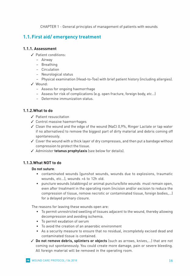
1.1. First aid/ emergency treatment
1.1.1. Assessment 俵 Patient conditions:
– Airway – Breathing – Circulation – Neurological status – Physical examination (Head-to-Toe) with brief patient history (including allergies).
俵 Wound: – Assess for ongoing haemorrhage – Assess for risk of complications (e.g. open fracture, foreign body, etc…) – Determine immunization status.
1.1.2.What to do 俵 Patient resuscitation 俵 Control massive haemorrhages 俵 Clean the wound and the edge of the wound (NaCl 0,9%, Ringer Lactate or tap water if no alternatives) to remove the biggest part of dirty material and debris coming off spontaneously.
俵 Cover the wound with a thick layer of dry compresses, and then put a bandage without compression to protect the tissue.
俵 Administer tetanus prophylaxis (see below for details).
1.1.3.What NOT to doDo not suture:
• contaminated wounds (gunshot wounds, wounds due to explosions, traumatic wounds, etc…), wounds >6 to 12h old.
• puncture wounds (stabbings) or animal puncture/bite wounds must remain open, even after treatment in the operating room (incision and/or excision to reduce the compression of tissue, remove necrotic or contaminated tissue, foreign bodies,...) for a delayed primary closure.
The reasons for leaving these wounds open are:• To permit unrestricted swelling of tissues adjacent to the wound, thereby allowing
decompression and avoiding ischemia.• To permit exudation of serum• To avoid the creation of an anaerobic environment• As a security measure to ensure that no residual, incompletely excised dead and
contaminated tissue is contained. 俵 Do not remove debris, splinters or objects (such as arrows, knives,...) that are not coming out spontaneously. You could create more damage, pain or severe bleeding. All foreign material will be removed in the operating room.

17WOUND CARE PROTOCOL / 06.2018
CHAPTER 1 - General principles of management of patients with wounds
PR
1.2. Tetanus prophylaxis
Risk of tetanus disease depends on the type and condition of the wound and on the immune status of the patient.
The following steps should be taken to prevent tetanus:
1. Assess the type of wound and provide appropriate wound care.
Wounds may be clean or contaminated and dirty, superficial or deep and penetrating. Dirty wounds pose an increased risk for tetanus.
All wounds should be cleaned, dirt or foreign material removed, and necrotic tissues removed or debrided.
2. Evaluate the origin of the wound(s) and risk of contamination using a careful anamnesis.
3. Evaluate the immunization status of the patient: this will determine the choice of the post-exposure prophylaxis.
4. Administer the most appropriated post-exposure prophylaxis.
WOUND CLASSIFICATION
Clinical features Tetanus Prone Non-Tetanus Prone
Age of wound > 6 hours ≤ 6 hours
Configuration Stellate, avulsion Linear
Depth > 1 cm ≤ 1 cm
Mechanism of injury Missile, crush, burn, frostbite
Sharp surface (glass, knife)
Devitalized tissue Present Absent
Contaminants (dirt, saliva, etc.)
Present Absent

18WOUND CARE PROTOCOL / 06.2018
CHAPTER 1 - General principles of management of patients with wounds
PR
IMMUNIZATION SCHEDULE
History of Tetanus Immunization
Dirty, Tetanus-Prone Wound
Clean, non-Tetanus-Prone Wound
TCV Anti-tetanus immunoglobulins TCV Anti-tetanus
immunoglobulinsUnknown or < 3 doses Yes Yes Yes No
3 or more doses No No No NoRef: MMWR 60:13, 2011; MMWR 61:468, 2012; MMWR 62:131, 2013 (pregnancy)
In case of wounds at minor risk of tetanus with record of vaccination status and the person has been fully vaccinated in the past, a booster dose of toxoid is required only if this was more than 10 years ago.
Unvaccinated persons should start and complete a primary series with an age-appropriate TCV (tetanus toxoid-containing vaccine as DTaP, TdaP, or Td) depending on the formulation available in each project.
Persons with unknown or uncertain history of previous prior doses tetanus toxoid-containing vaccines should be considered to have had no previous tetanus toxoid-containing vaccine and a primary series should be initiated. This is because earlier doses of toxoid may not induce adequate immunity, but only prime the immune system.
Only in case of major risk of tetanus with no record of tetanus vaccination or doubtful protection: give the first dose of tetanus toxoid, plus tetanus immunoglobulins.
Dosage: – Human anti-tetanus immunoglobulins:
Children and adults: 250 IU as a single dose or 500 IU for wounds more than 24 hours old. To be injected IM only.Inject the vaccine and the immunoglobulins in two different sites, using a separate syringe for each. In case only equine immunoglobulins are available in the field administration must follow leaflet recommendations (as they might vary between manufacturers).
– TCV (tetanus toxoid-containing vaccines):
One dose=0,5ml per injection - To be injected IM or SC into the anterolateral part of the thigh or the deltoid muscle.

19WOUND CARE PROTOCOL / 06.2018
CHAPTER 1 - General principles of management of patients with wounds
PR
Each person should receive a vaccination card and must be instructed to return at 4 weeks and then 6 months afterwards to receive respectively the 2nd and 3rd dose of TCV . For more details about degree and duration of protection following tetanus vaccination: MSF clinical guidelines.
1.3. Simple burns
Burn patients have the same priorities as other trauma patients.
1.3.1.Assess: 俵 Airway 俵 Breathing: beware of inhalation and rapid airway compromise (check of soot)
俵 Circulation: fluid replacement 俵 Disability: compartment syndrome 俵 Exposure: percentage burn surface
1.3.2.Essential management points: 俵 Stop the burning 俵 ABCDE 俵 Determine the percentage of burned surface (Rule of 9) 俵 Good IV access and early fluid replacement
The severity of the burn is determined by:
Simple Severe
Burned surface < 9% ≥ 9%
Depth of burn 1st or 2nd degree -
Location -Special regions: face, hands, feet,
perineum, genitals
Patient age -Any burn in the very young, the elderly or in case of pre-burns comorbidities
Other considerations -Circumferential burns
Inhalation injury

20WOUND CARE PROTOCOL / 06.2018
CHAPTER 1 - General principles of management of patients with wounds
PR
First aid and preventive treatment
俵 If the patient arrives at the health facility without having been given first aid, drench the burn thoroughly with cool water to prevent further damage and remove all burned clothing when not excessively adhered into the wound.
俵 If the burned area is limited, immerse the site in cold water for 30 minutes to reduce pain and oedema and to minimize tissue damage. Elevation of the burned limb can also relief the pain.
俵 If the area of the burn is large, after it has been showered with cold water, apply clean wraps around the burned area (or the whole patient) to prevent systemic heat loss and hypothermia.
俵 Hypothermia is a particular risk in young children 俵 First 6 hours following injury are critical: transport the patient with severe burns to a hospital as soon as possible.
俵 In all cases, administer tetanus prophylaxis 俵 Assess and treat pain as by pain management protocol 俵 Patient with simple burns but presence of influencing factors (see Chapter 2) such as comorbidities, specific medications, psychological/social specific conditions, in need of intensive care or with increased risk of infection should be seen by a clinician.
俵 If specific protocols and material for treating burns are available, the health care worker should opt for the most appropriate care.
1.4. Holistic approach
‘You need to treat the whole patient and not just the hole in the patient’.
(Dowsett & Newton, 2005)
The healing process is the result of a complex interaction between the patient and wound-related factors, the treatment used, and the skills and knowledge of healthcare professionals. Thus, wound management requires a holistic approach.
This wound care protocol mainly focuses on aspects related to the wound. Nevertheless, the other factors that can influence wound healing should also be taken into account to ensure optimal wound healing.

21WOUND CARE PROTOCOL / 06.2018
CHAPTER 1 - General principles of management of patients with wounds
PR
1.5. Factors influencing wound healing
Patient relatedPathology, comorbidity, malnutrition, allergy, medication, psychosocial aspects, pain, coping
Wound related
Type, size (surface and depth), wound bed condition, ischemia, oedema, infection, anatomical site, treatment response
Health care professional related
Skills, knowledge and multidisciplinary care (nurse, doctor, physiotherapist,…), supervision
Resources/treatment related
Availability of material, suitability, effectiveness
Environmental related Hygiene, cold / hot weather, humidity
22WOUND CARE PROTOCOL / 06.2018
CHAPTER 1 - General principles of management of patients with wounds
PR
1.5.1.Patient related factorsAny factor that weakens the patient, impairs the immune resistance or reduces tissue perfusion, e.g.:
– Comorbidities• Malnutrition/cachexia• Immunodeficiency status• Autoimmune disorders (e.g. rheumatoid arthritis)• Diabetes mellitus• Hypoxia/poor tissue oxygenation (e.g. due to anaemia, arterial/cardiac/
respiratory disease, peripheral vascular disease, ageing, diabetes, ischemia)
• Malignancy• Medical problem causing oedema.
– Pain – Nutrition and hydration – Medication: e.g. corticosteroids, cytotoxic agents, immunosuppressant drugs – Psychosocial factors: e.g. hospitalisation/institutionalisation, poor personal
hygiene, unhealthy lifestyle choices; (e.g. excess alcohol consumption, tobacco smoking, lack of exercise, …).
– Patient environment: patient hospitalized in critical care ward (intensive care unit).
1.5.2.Wound related factorsWounds at increased risk of infection:
– Any wound with a traumatic origin (involving contaminated materials) – Trauma with delayed treatment – Contaminated surgery (cfr. Altemeier score; see table 1) – Long operative procedure (cfr. Length of intervention; see table 2) – Diabetic foot wound – Anatomically situated near a site of potential contamination, e.g. anal area, groins,
deep skinfolds. – Presence of β-haemolytic streptococci – Age (neonates and age above 60 years).

23WOUND CARE PROTOCOL / 06.2018
CHAPTER 1 - General principles of management of patients with wounds
PR
Table 1 - Altemeier ClassificationContamination class of the surgical intervention:
Type of Surgery Selection Criteria Examples
Class IClean surgery
– Without opening the gastroin-testinal tract
– No evidence of injury or probable inflammation
– Simple hernia – Scheduled caesarean not in labour
Class IIClean-contami-nated surgery
– Opening of gastrointestinal tract with minor contamination
– Minor breach of asepsis
– Appendectomy – Scheduled caesarean in labour
– Urgent caesarean
Class IIIContaminated surgery
– Significant contamination by intestinal contents
– Major breach of asepsis – Recent traumatic wound less than 4 hours old
– Genitourinary or biliary tract open with infected bile or urine
– Strangulated hernia with intestinal resection
Class IVDirty surgery
– Traumatic wound more than 4 hours old and/or with devitalised tissues
– Faecal contamination – Foreign body – Perforated viscera – Acute bacterial inflammation without pus.
– Presence of pus
– Peritonitis
⇒ At enhanced risk = class III and IV

24WOUND CARE PROTOCOL / 06.2018
CHAPTER 1 - General principles of management of patients with wounds
PR
Table 2 - Length of surgical procedure
Percentile 75 as a function of the type of intervention (examples)
More than one hour – appendectomy – amputation – caesarean
More than two hours
– cholecystectomy – abdominal or vaginal hysterectomy – laparotomy – hernia – breast surgery
More than three hours
– colon, gastric, iliac surgery – nephrectomy – joint prosthesis – vascular surgery
More than four hours – prostate – neurosurgery – surgery of the biliary tract, liver, pancreas
More than five hours – cardiac surgery – coronary bypass
– Low risk: length of intervention equal to or less than percentile 75 of the distribution of the length of this intervention in the general population.
– Enhanced risk: length of intervention greater than percentile 75 of this distribution.Should be based on the specific standards of the project!

25WOUND CARE PROTOCOL / 06.2018
CHAPTER 1 - General principles of management of patients with wounds
PR
1.5.3.Health care professional related factorsTraining is needed for all relevant health professionals so that they have the basic knowledge and skills to evaluate, initiate and perform wound care in a standardized and systematic way.
1.5.4.Resources/treatment related factorsSpecific material is needed to perform wound care activities and health care workers must know how to use it. Supply chain, stock management and end-user-pharmacy supervision are capital to avoid out-of-stock of items or unsuitable storage conditions.
1.5.5.Environmental related factorsHygiene is a big challenge in many of the countries where MSF works. Health structures are often not answering to minimum standards and compromises are too easily made . Moreover, climate conditions (e.g. temperature and humidity) are sometimes also affecting the wound healing process and it could happen that the effectiveness of products is heavily reduced (like the capacity of a dressing to stick on the skin).
1.6. Wound bed preparation
The overall goal of the wound bed preparation (WBP) is to create an optimal wound healing environment with a balance of moisture to produce a well-vascularized, stable wound bed and wound edges.
1.6.1.Wound healing in moist environmentProduction of exudate is part of the body’s response to tissue damage. Creating a moisture balance at the wound interface is essential for wound healing.
Wound healing must be seen as a biological process and it must be remembered that in humans all biological processes take place in a moist environment as our body consists of nearly 70% water.
A moist environment reduces the risk of infection, and stimulates granulation and epithelialisation. Acute wound exudate contains both proteolytic enzymes (to clean up the wound) and growth factors (to stimulate the cleaning and de proliferation of the necessary cells of the wound bed (granulation tissue).
26WOUND CARE PROTOCOL / 06.2018
CHAPTER 1 - General principles of management of patients with wounds
PR
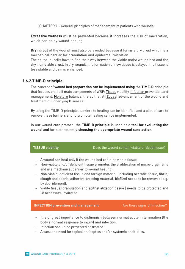
Excessive wetness must be prevented because it increases the risk of maceration, which can delay wound healing.
Drying out of the wound must also be avoided because it forms a dry crust which is a mechanical barrier for granulation and epidermal migration. The epithelial cells have to find their way between the viable moist wound bed and the dry, non-viable crust. In dry wounds, the formation of new tissue is delayed, the tissue is less stable and pain is enhanced.
1.6.2.TIME-D principleThe concept of wound bed preparation can be implemented using the TIME-D principle that focuses on the 5 main components of WBP: Tissue viability, Infection prevention and management, Moisture balance, the epithelial (Edges) advancement of the wound and treatment of underlying Diseases.
By using the TIME-D principle, barriers to healing can be identified and a plan of care to remove these barriers and to promote healing can be implemented.
In our wound care protocol the TIME-D principle is used as a tool for evaluating the wound and for subsequently choosing the appropriate wound care action.
TISSUE viability Does the wound contain viable or dead tissue?
– A wound can heal only if the wound bed contains viable tissue – Non-viable and/or deficient tissue promotes the proliferation of micro-organisms
and is a mechanical barrier to wound healing. – Non-viable, deficient tissue and foreign material (including necrotic tissue, fibrin,
slough and debris, adherent dressing material, biofilm) needs to be removed (e.g. by debridement).
– Viable tissue (granulation and epithelialization tissue ) needs to be protected and -if necessary- hydrated.
INFECTION prevention and management Are there signs of infection?
– It is of great importance to distinguish between normal acute inflammation (the body’s normal response to injury) and infection.
– Infection should be prevented or treated – Assess the need for topical antiseptics and/or systemic antibiotics.

27WOUND CARE PROTOCOL / 06.2018
CHAPTER 1 - General principles of management of patients with wounds
PR
MOISTURE balance Does the wound produce too little or too much exudate?
– Moisture balance should be achieved in order to encourage healing. – Evaluate the amount, type and odour of the exudate. – Appropriate choice of dressing should add or absorb moisture to preserve the
moisture balance. – The cause of excessive exudate should be investigated: inspect for “I” (inflammation
/ infection) and/or oedema.
EDGES Is the epidermis able to cover the granulation tissue?
– Assessment of the wound edges and the condition of the periwound skin ( = the skin within 4 cm of the wound edge as well as any skin under the dressing): is the wound contracting and is epithelialization progressing?
– As the epithelialization progresses the wound edges should be healthy, free of maceration, necrotic tissue and crusts.
– Properly evaluate and execute all actions associated with T, I and M. – Ensure contact between the dressing and the wound bed, prevent/treat maceration,
debride wound edges. – Be careful when removing dressing materials to avoid additional damage. – If necessary, surgical techniques may be used to close the wound.
Disease Is there any underlying disease that needs treatment?
– To emphasise the importance of managing the comorbidities (diseases) of the patient during treatment of the wound, the acronym “TIME” is extended to “TIME-D”.

28WOUND CARE PROTOCOL / 06.2018
CHAPTER 1 - General principles of management of patients with wounds
PR
Table 3- Illustration wound bed preparation according to the TIME principles
Observation Consequence Aim
T = Tissue non-viable or deficient
Necrosis, fibrin, debris, foreign material
Barrier for wound healing process, place for infection, exacerbates inflammatory response
Viable wound base
I = Infection
Prolonged inflammation phase, oedema, redness and pain at edges, ↑ exudate, ↑odour, discoloration of surface, purulent drainage, …
Barrier for wound healing process
Bacterial balance and reduced inflammation
M = Moisture imbalance
Dehydration: no or too little exudate with dry wound bedToo much exudate with maceration of wound edges
Delayed wound contraction and epithelialization
Moisture balance
E = Edge of the wound, non-advancing or undermining
Prolonged inflammation phase, wound size not decreasing over time, irritation of wound edges
Failure of migration of the epidermal cells across the wound bed
Advancing of wound edges

29WOUND CARE PROTOCOL / 06.2018
CHAPTER 2 - Patient with wound: assessment and preparation of the patient
PR
CHAPTER 2 - PATIENT WITH WOUNDS: ASSESSMENT AND PREPARATION OF THE PATIENT
Pain management in wound careNutrition and hydrationInfluencing factors
– Comorbidities and/or medical condition of the patient – Medications – Psychological condition, body image and psychosocial
factors – Patient admitted in critical care ward – Increased risk of infection

30WOUND CARE PROTOCOL / 06.2018
CHAPTER 2 - Patient with wound: assessment and preparation of the patient
PR
2.1. Pain management in wound care
Wounds can be painful, especially when they are new, infected or granulating. Wounds located in areas exposed to pressure, friction or frequent movement may also be more painful.
Dressing changes can be associated with significant pain. Frequent dressing changes may increase wound sensitivity and levels of background pain, especially when debridement or scrubbing is necessary. Wound pain is also affected by choice of dressing materials and cleansing products.
Unrelieved pain affects the wound healing process. Inhibition of deep breathing may lead to impaired tissue oxygenation and generalised vasoconstriction associated with severe pain leads to impaired tissue perfusion. Both factors impair healing and predispose to infection. Untreated wound pain also increases the likelihood of a patient developing a chronic pain condition.
Effective management of wound pain includes attention to wound care, positioning of the affected body part, rest and immobilisation or controlled mobilisation, avoidance of environmental stresses and the use of analgesic medication.
Before applying a dressing on a wound it is important to assess the background pain due to the wound and anticipate the pain generated during the wound care procedure.
Assessment of pain
It is helpful to understand the location, timing and intensity of a patient’s pain, as well as aggravating and relieving factors. Wound pain can be classified as background pain that may be intermittent or continuous, incident pain that is often associated with mobilisation, coughing etc. and procedural pain associated with dressing changes or debridement. Procedural pain may persist for several hours after a dressing change. Each type of pain requires a different approach to treatment.
Systematic use of a pain scoring tool to quantify and record pain severity allows to evaluate the success of analgesic and wound care choices. The choice of the tool depends on patient age and individual circumstances, but it is important that both patient and clinician/care giver understand how it is used and interpreted.

31WOUND CARE PROTOCOL / 06.2018
CHAPTER 2 - Patient with wound: assessment and preparation of the patient
PR
Examples of pain scoring tools (see technical sheets 1,2,3)
Infants 2 - 12 months
Neonatal Facial Coding System (NFCS Scale)
Children 1-4 years (and patients unable to communicate their pain)
EVENDOL scale
Children ≥ 5 years and adults Self-assessment method: - Simple Verbal Scale 1-5 (SVS)- Visual Analogue Scale (VAS)
A pain scoring tool should be used to assess background and incident pain, as well as pain before, during and after a dressing change. It is recommended to continue with the same scale once used to ensure consistency in pain management strategy and documentation.
Assessment should also consider the characteristics of the wound and the patient’s indi-vidual circumstances, medical history and behaviour.
Management of pain
Non-pharmacological approaches to wound pain should always be considered.
– Elevation or splinting of a wounded extremity – rest and stress less environment – Careful mobilization – Physiotherapy (may assist mobilisation) – Explain the procedure to reduce anxiety and fear; presence of parent – Simple relaxation techniques – “Pauses” during the procedure – Shift from a dry to a moist environment – Hydrating the surrounding skin – If dressing sticks to the tissue ⇒ take time to remove, avoid tearing fragile tissues.
Use lots of NaCl 0.9% to moisten the dressing!
Drug treatment of wound pain should follow the same step-wise approach described by the WHO Pain Ladder.

32WOUND CARE PROTOCOL / 06.2018
CHAPTER 2 - Patient with wound: assessment and preparation of the patient
PR
Significant background pain should be treated with, oral analgesia that is given at regularly scheduled intervals. Background pain is usually mild or moderate in intensity and can often be managed with non-opioid analgesics. For example, regular paracetamol, alone or combined with a regular non-steroidal anti-inflammatory drug (NSAID) is a very effective combination.
Incident or breakthrough pain can be treated with intermittent doses of a rapidly-acting analgesic, as required. This may be paracetamol or a NSAID, if they are not already prescribed regularly, or a weak opioid e.g. codeine or tramadol. If incident pain is associated with specific activities, a dose of breakthrough analgesia can be given pre-emptively.
Procedural pain may be severe and is very severe in some patients, requiring a weak or strong opioid in addition to non-opioid analgesia. It should be anticipated and managed pre-emptively. It is important to allow adequate time for analgesia to take effect before starting the procedure and to ensure the procedure is completed during the period of peak analgesic effect. Some patients experience increased pain for several hours after a dressing change, which should be considered. A NSAID such as ibuprofen or diclofenac often provides effective, post-procedural pain relief.
Some patients, particularly those with longstanding wounds and significant pain may suffer from a combination of nociceptive and neuropathic, or chronic, pain. In this situation, adjuvant drugs such as tricyclic antidepressants (amitriptylline) or anticonvulsants (carbamazepine, gabapentin) may improve symptoms and quality of life if prescribed regularly.
For the general principles of pain management refer to the MSF Clinical Guidelines Diag-nosis and Treatment Manual and the MSF Neonatal Care guideline and to technical sheet 4 that summarizes the pain management and the action time of analgesics.
2.1.1.Monitor and record keepingIt is helpful to maintain a record of the patient’s symptoms and pain scores, alongside a record of the pain treatment used. Patients with problematic pain may require a variety of approaches and analgesic regimens to be tried, which can be compared using a pain scoring tool. Moderate or severe pain recorded during or after a procedure should prompt a review of the treatment used.

33WOUND CARE PROTOCOL / 06.2018
CHAPTER 2 - Patient with wound: assessment and preparation of the patient
PR
2.2. Nutrition and hydration
Good nutrition and hydration have an essential role in wound healing. During the healing process, the body needs increased amounts of calories, proteins and vitamins. Proper hydration is important for wound care as it assists in every stage of wound healing.
The wound healing process needs proteins, sugars, fats, vitamins (especially A, B, C, E and K), minerals and trace elements (especially iron, copper, zinc and manganese). Malnourished patients or patients with dietary imbalances have a higher risk of wound infection and often experience chronic non-healing wounds with decreased tensile strength.
On the other hand, big and/or infected wounds need higher nutritional intake to regenerate lost tissues or to face infection processes with a consequent increase of energy and particular nutrient consumption, especially protein and calories. If nutrients intake is not consistent to the needs, potential risks are delayed wound healing and prolonged catabolic phase with consequent protein-energy malnutrition status.
Dehydration has also a negative impact in wound healing. Dehydrated skin becomes inelastic, fragile and more susceptible to breakdown (Thomas, 2001). Dehydration can reduce tissue perfusion at the wound site by reducing the blood volume, limiting the supply of oxygen and nutrients. Drainage from a wound (exudate) can be a major source of fluid loss.
Patients with dietary imbalances need nutrition therapy and dehydrated patients need to be rehydrated in order to enable the wound(s) to heal.

34WOUND CARE PROTOCOL / 06.2018
CHAPTER 2 - Patient with wound: assessment and preparation of the patient
PR
Assessing a patient’s nutritional status
It is essential to know whether the patient is well nourished or suffers from some degree of acute malnutrition as well as to plan the appropriate nutritional support.
This assessment is made up of:
– anthropometric measurements – assessment for oedema – dietary history plus food security assessment.
For detailed guidance on how to do this nutritional assessment, please refer to the following documents:
For adults: – Nutritional Support & Enteral Feeding for Adult in Intensive Care Unit or Surgery
Ward MSFOCB. – Protocol for Malnutrition in Teenagers and Adults MSFOCB – Protocol for Nutrition support and Malnutrition treatment in Pregnant and lactating
Women MSFOCB.
For children: – Nutritional and Medical Protocol for Treatment of Severe Malnutrition – Inpatient
Children from 6 months to 10 years MSFOCB. – Nutritional and Medical Protocol for Treatment of Severe Malnutrition – outpatient
Children from 6 to 10 years MSFOCB . – MSF HIV/TB Clinical Guide.
All patients with big or complicated wounds (extensive gap of tissue, not healing and/or signs of infection and presence of comorbidities) need to have this nutritional status assessment. If a patient is found to suffer from moderate or acute malnutrition, he should be referred to a nutrition service and started on treatment as per protocol in the wound care providing health facility providing the wound care.
However, in keeping in mind the holistic wound care approach, patients with a normal nutritional status must not be forgotten. They still need nutrition counselling and regular follow-up to ensure they do not deteriorate from a nutritional perspective.
Furthermore, it is likely there is a group of patients “at risk of acute malnutrition”. They may be close to a BMI/MUAC cut-off for acute malnutrition or have severe food insecurity at home.

35WOUND CARE PROTOCOL / 06.2018
CHAPTER 2 - Patient with wound: assessment and preparation of the patient
PR
These patients need close follow-up and although there is currently no evidence about the best form of treatment, it is wise to be proactive and consider supplementation either with RUSF, RUTF or fortified flour such as supercereal, on a case-by-case basis.Be aware that overweight patients can also be undernourished even if they have a high BMI.
Nutrition counselling
There are a number of tools available for nutritional counselling. The list below covers the essential topics:
– Importance of nutrition in aiding wound recovery (diagram above) – Identification of locally available food sources and more importantly, what can the
patient actually access (money, transport, time, etc.) ? – Identification of specific conditions impacting intake (e.g. painful mouth from
ulcers, nausea, gastro-oesophageal reflux, etc.) – Nutritional needs according to comorbidities (e.g. hypertension, diabetes, renal
disease, etc.) – Meal planning (guided by daily energy needs) – Hygiene in food preparation – Linkage to community support and opportunities for economic strengthening

36WOUND CARE PROTOCOL / 06.2018
CHAPTER 2 - Patient with wound: assessment and preparation of the patient
PR
The table below gives some guidance on treatment/care options for the different groups. This can be adapted to the context and in discussion with the medical team to the most feasible for the project.
Nutritional Status
Hospitalised(for wound care or wound care is a major reason for
hospitalisation)
Outpatient wound care follow-up
Normal
Nutrition counsellingRegular anthropometric assessment (weekly)Meal plans (see protocols above)
Nutrition counsellingRegular anthropometric assessment (weekly)
At risk of acute malnutrition
Nutrition counsellingRegular anthropometric assessment (weekly)Monitored meal times (to assess intake/feeding difficulties)Meal plans (see protocols above)Consider supplementation if there is difficulty with oral intake
Nutrition counsellingRegular anthropometric assessment (weekly)Consider supplementation if there is difficulty with oral intake or significant food insecurity in the household
Moderate acute malnutrition
Treat as SAM – inpatient malnutrition protocol
Treat as SAM – outpatient malnutrition protocol
Severe acute malnutrition Inpatient malnutrition protocol Outpatient malnutrition protocol

37WOUND CARE PROTOCOL / 06.2018
CHAPTER 2 - Patient with wound: assessment and preparation of the patient
PR
2.3. Influencing factors
The wound should not be treated in isolation but in the context of the patient’s overall wellbeing.
Before deciding on any wound action, products and materials, the clinician must undertake and document a holistic assessment of the patient. This should include an assessment of his/her comorbidities, any medications the patient is receiving and psychological and psychosocial factors.
Comorbidities and/or medical condition of the patient
Wound healing needs a good functioning of the blood circulation, metabolism, respiratory system and immune system. Any disease or condition that hinders partially or completely these physiological processes will affect the healing of the wound.
Examples
– Vascular insufficiency and other circulatory disorders leading to reduced blood flow and/or to poor tissue oxygenation (oxygen is essential for cell metabolism and critical to all wound-healing processes; reduced blood flow hinders cell, nutrient and oxygen transport to the wound bed): e.g. atherosclerosis, chronic venous insufficiency, peripheral vascular disease, hypovolemia.
– Metabolic diseases: e.g. renal insufficiency, (poorly controlled) diabetes mellitus (⇒ peripheral vascular disease, neuropathy, impaired transport of vitamin C leading to impaired collagen synthesis and inferior connective tissue, impaired functioning of immune and inflammatory cells,… ⇒ increased risk of wound infection, decreased potential for wound healing masking indicators of wound infection such as inflammation, pain and discomfort).
– Lung diseases (oxygen is essential for cell metabolism and critical to all wound-healing processes): e.g. COPD, cystic fibrosis.
– Disorders associated with a reduced activity, immobility, impaired sensory perception, neurological and motoric deficits: e.g. paralyzed patients, cerebrovascular accident, multiple sclerosis.
– Impaired immune responses due to age (neonates and the elderly are at particular risk of delayed wound healing and wound infection).
– Connective tissue diseases: e.g. rheumatoid arthritis, scleroderma.
– Diseases in which the immune system is suppressed: e.g. HIV-AIDS.
– Oncologic diseases: can lead to a debilitated physical condition.

38WOUND CARE PROTOCOL / 06.2018
CHAPTER 2 - Patient with wound: assessment and preparation of the patient
PR
⇒ Associated comorbidities need to be addressed in order to enable the wound(s) to heal
Medications
Certain drugs negatively affect the wound-healing process. In all cases, liaise with the prescriber to analyse risks and benefits before stopping or modifying prescriptions.
Examples
– Anti-inflammatory medication: corticosteroids (local and systemic) and NSAIDs (in case of long term use): counteract inflammation and thus interfere with the first stage of wound healing. Next to it corticosteroids have also a direct impact on fibroblasts leading to an impaired collagen formation.
– Antibiotics: might reduce the wound’s tensile strength, impeding final wound closure.
– Antiplatelet drugs: some of them can lead to prolonged bleeding and deficient coagulation and thus interfere with first stage of wound healing.
– Immunosuppressant drugs: immunosuppression, consequent weakening of the patient and increased risk of superinfection.
– Chemotherapeutic drugs: are used to stop the growth of rapidly dividing cancer cells, but most of them also delay the cell division in other rapidly dividing tissues, such as the skin. In addition, they weaken the patient’s immune functions, thereby impeding the inflammatory phase of wound healing and increasing the risk of wound infection.
⇒ For each patient, it should be checked whether the benefits of these drugs outweigh the negative impact on wound healing. If cessation is not advisable it is important that both the patient and the wound(s) are carefully monitored and reassessed in a timely manner.

39WOUND CARE PROTOCOL / 06.2018
CHAPTER 2 - Patient with wound: assessment and preparation of the patient
PR
Psychological condition, body image and psychosocial factors
– Factors such as stress and anxiety may adversely affect the wound healing. – Adequate sleep and rest are important for an optimal cellular metabolism and a
good wound healing. – Good personal hygiene is necessary for an optimal wound healing.
⇒ Psychological condition, body image and psychosocial factors need to be taken into account/optimized in order to promote wound healing.
⇒ Anthropological considerations should be assessed in particular contexts where witchcraft is part of the local culture. Sometimes wounds (especially tropical chronic ulcers) are seen by the community as a punishment or malediction.
Patient admitted in critical care ward
– Patient admitted in ICU level 2 and 3, are more at risk of having a longer or more complicate wound healing process, due to their critical conditions and a consequent immunity weakness.
⇒ ICU patients need to be monitored and treated with particular attention and precaution in order to early identify local or systemic signs of infection

40WOUND CARE PROTOCOL / 06.2018
CHAPTER 2 - Patient with wound: assessment and preparation of the patient
PR
Increased risk of infection
– Any wound with a traumatic origin (involving contaminated materials) – Contaminated surgery (cfr. Altemeier score; see table 1) – Long operative procedure (cfr. Length of intervention; see table 2) – Trauma with delayed treatment – Diabetic foot wound – Anatomically situated near a site of potential contamination, e.g. anal area, groins,
deep skinfolds. – Presence of β-haemolytic streptococci – Age (neonates and age above 60 years).
⇒ All wounds that are at increased risk of infection should be treated as a wound with signs of infection during the first treatment

41WOUND CARE PROTOCOL / 06.2018
CHAPTER 3 - Wound: assessment and care
PR
CHAPTER 3 - WOUND: ASSESSMENT AND CARE
Tissue viabilityInfection prevention and managementMoisture balanceEdgesDiseasesFixation / cover
Annex 3.1 - HypergranulationAnnex 3.2 - External fixator

42WOUND CARE PROTOCOL / 06.2018
CHAPTER 3 - Wound: assessment and care
PR
In order to perform a correct wound cleansing and care, the wound must be carefully observed and evaluated. Based on this evaluation we will decide which action(s) should be taken and which dressing material we have to apply. This process is guided by the TIME principle that focuses on the 5 main components of WOUND BED PREPARATION:
TIME
T Tissue viability
I Infection prevention and management
M Moisture balance
E Edges
D Diseases
Before starting any action, the whole TIME assessment of the wound must be completed.
Each step of the assessment has an impact on the decision of each action.
The removal of the previous dressing covering a wound needs to be done carefully (see Chapter 5 for the specific technique). When fragile new granulation and/or epithelialization tissue starts to develop, a too strong detachment of the dressing can nullify all the improvements already achieved.

43WOUND CARE PROTOCOL / 06.2018
CHAPTER 3 - Wound: assessment and care
PR
TIS
SU
E
3.1. Tissue viability
T of TIME-D
The observation of the type of tissue present in a wound is one of the factors to be taken in consideration for choosing the technique for the cleaning and the type of product in the dressing phase.
Some tissues (necrosis and fibrin) need a more “aggressive” approach with a direct mechanical action (from mechanical cleansing with simple gauze compresses to more invasive procedures like surgical debridement) because of their thickness or resistance and negative effects on the underlying cells. On the contrary, other tissues (granulation and epithelialization) are signs of a good healing process. Due to their fragility they need to be protected as much as possible from any direct mechanical or chemical action during the phases of cleaning, covering and dressing removal.
Another very important information to be collected and registered is the size of the wound. The positive or negative evolution of the healing process is also defined by the reduction or not of the wound dimensions. This information can influence the decision to be taken on the treatment.
Type of tissue
Sutured woundSuturing is used to promote primary healing. It realigns tissue layers and holds the skin edges together until sufficient healing occurs to withstand stress without mechanical support. When wounds are completely sutured, internal tissues are not visible on the wound surface and are rapidly physically protected from external injuring mechanisms and germs.
Picture 1 – Sutured wound

44WOUND CARE PROTOCOL / 06.2018
CHAPTER 3 - Wound: assessment and care
PR
TIS
SU
E
GranulationDuring the proliferation phase of the wound healing process, a new connective tissue with microscopic blood vessels starts to grow from the base of the wound. This new tissue will fill the whole wound and will be the base for the migration of epithelial cells resulting in wound closure.
Healthy granulation tissue is light red or dark pink in colour because of its abundant vascularization. It is soft and moist, granular in appearance and a good indicator of wound healing.
Picture 2 – Granulated wound
EpithelializationNew epithelial cells start migrating across the granulation tissue in order to form a barrier between the wound and the environment. These cells usually begin their proliferation at wound edges and from the adnexa (such as hair follicles, sweat glands, and sebaceous glands) and the covering will move from there to the wound centre. At the beginning, epithelial cells are very fragile and need to be protected and preserved. They have a pink pearly appearance and form first an almost invisible film. If rubbed, they will detach from the granulation tissue.
Picture 3 – Epithelialization tissue
Fibrinous tissueAccumulation of fibrin can generate a yellowish fibrinous tissue. Fibrin is a protein formed by the action of the protease thrombin on fibrinogen. With platelets it forms a
haemostatic plug or clot over a wound site. Fibrin can usually be removed with high pressure rinsing or with a simple mechanical cleansing with sterile gauze compresses or by scraping with a metallic instrument, especially when the thickness is limited and the wound is humid or wet. However, dry and thicker fibrinous tissues could be more complicated to manage, needing more advanced procedures. As for necrotic tissue, a fibrinous layer over the wound bed impedes the normal healing process.
Picture 4 – Fibrinous tissue

45WOUND CARE PROTOCOL / 06.2018
CHAPTER 3 - Wound: assessment and care
PR
TIS
SU
E
Necrosis
Necrosisis the consequence of devitalisation and death of tissue cells due to different reasons and can easily delay wound healing and put the patient’s life at risk. Necrotic tissues can be dry, thick and strongly attached to the underlying layer or more moist, loose and stringy in appearance.
The presence of necrotic tissue in a wound prevents the normal healing process and can hide or cause infection.
Picture 5 – Necrotic tissue
Visible supporting structures
While assessing the wound bed tissue, it is important to recognize supporting structures that are visible in the wound. Tendons, bones, fascia, joint capsules, etc…are examples of structures not to be confused with other types of tissues and should be correctly protected and treated.
Picture 6 – Wound with exposed tendonds
HypergranulationHypergranulation is the excess of granulation tissue, beyond the amount required to replace the tissue deficit. The production of granulation tissue continues beyond the
height of the epithelium surface / periwound skin resulting in a raised mass (or peduncle) in excess of the wound itself. Because epithelial cells are unable to grow over this raised tissue, epithelialization will stop.
Annex 3.1 gives more background information about causes and treatment of hypergranulation.
Picture 7 – External fixator with hyper-granulation around pin sites

46WOUND CARE PROTOCOL / 06.2018
CHAPTER 3 - Wound: assessment and care
PR
TIS
SU
E
Size of the wound
Accurate and objective wound measurement is a vital component of wound management and it should be part of routine practices.
Aside from the type of tissue (necrosis, fibrin, granulation, epithelialization, hypergranulation, visible structures), the depth, length and width (together with the shape) of the wound should be evaluated and described.
Measuring a wound at the start of treatment is seen as best practice to enable accurate assessment of the impact of the intervention. Subsequent measuring can identify whether or not a wound is failing to heal or deteriorating.
A wound that decreases with 30-40% in 2 to 3 weeks is considered as healing.
The size of a wound can be measured using different methods, with different levels of accuracy and complexity.
With a simple disposable paper ruler (many examples are available on internet) it is possible to assess the three main dimensions: length, width and depth.
Clock method
The “clock method” is the most common and easiest way for linear measurement of a wound.
Imagine the head of the patient is at 12:00 on the clock and the feet at 6:00:
– length is measured by placing the ruler at the point of greatest length or from 12:00 to 6:00 (vertical axis);
– width is measured by placing the ruler at the point of greatest width or from 9:00 to 3:00 (horizontal axis).
The wound depth is the difference between the deepest part of the wound and the skin level. It can be measured using one of the sterile instruments such as forceps or peans already present in the dressing set.
Undermining and tunnelling parts of wound should be measured too and documented in the patient file for a complete follow-up of the healing process.

47WOUND CARE PROTOCOL / 06.2018
CHAPTER 3 - Wound: assessment and care
PR
TIS
SU
E
Acetate tracing
A specific technique for monitoring the evolution of the wound for both size and shape is the “acetate tracing”. It requires a transparent acetate sheet and a permanent marker. The shape of the wound is simply retraced on the sheet and then it will be possible to easily measure length and width with a ruler and the area using a graph paper. Each tracing in a sequence is easy to compare with the others and tracing is relatively unobtrusive for the patient. Tracings can be immediately stored in the patient’s records.
A huge attention must be put on the side of the acetate paper that is in contact with the wound: as it will get contaminated by the wound it will be necessary to disinfect it using an appropriate technique.
An alternate solution would be to use the sterile blister of the dressing on the wound and draw on its non-sterile side.
Photography
This is an easy way of charting wound progression but it requires many conditions to be in place:
– the patient has to give his verbal consent: patient’s willingness and sensitivity need always to be respected, especially when wounds are located in private body areas;
– all pictures have to be taken with the same technique: from the same distance, possibly the same camera, patient in the same position, perpendicular to the middle of the lesion, etc…;
– pictures have to include the patient identification number, a ruler (for proportion) and the date when they were taken;
– the use of the camera has to respect of hygiene precautions to avoid contaminations and cross infections.
Some specific software or smartphone/tablet applications could include a measuring tool.

48WOUND CARE PROTOCOL / 06.2018
CHAPTER 3 - Wound: assessment and care
PR
INFE
CTIO
N
3.2. Infection prevention and management
I of TIME-D
The microbial bioburden in a wound can range from contamination, colonization and critical colonization to ultimately local and systemic infection if not appropriately controlled.
Contamination All wounds contain micro-organisms. If suitable nutritive and physical conditions are not available for each microbial species, or they are not able to successfully evade host defences, they will not multiply or persist. Their presence is only transient and wound healing is not delayed: they do not cause clinical problems and there will be no signs of infection.
ColonizationMicro-organisms multiply but they do not cause damage to the host, wound healing is not delayed and there will be no signs of infection.
Critical colonization (covert infection)Micro-organisms multiply to the extent that healing is impaired. It may also mean that biofilm communities are present in the wound bed. As this stage is rather difficult to visualize, many authors tend to rule it out.
Infection Micro-organisms multiply, healing is disrupted and wound tissues are damaged (local infection). Micro-organisms may spread from the wound, causing problems in the nearby healthy tissue (spreading infection, e.g. cellulitis and erythema).Micro-organisms may also cause infection throughout the body (systemic infection, with systemic inflammatory response, sepsis and organ dysfunction).
As first step, wounds (or patients with wounds) should be classified in one of the following categories:
– Healing wound and no signs of infection
– Non-healing wound and/or signs of infection
– Surgical foreign object in the wound (e.g. drain, external fixator pin site)
– Patient hospitalized in ICU
– Wound at increased risk of infection
• Any wound with a traumatic origin (involving contaminated materials)

49WOUND CARE PROTOCOL / 06.2018
CHAPTER 3 - Wound: assessment and care
PR
INFECTION
• Contaminated surgery (cfr. Altemeier score)
• Long operative procedure
• Trauma with delayed treatment
• Diabetic foot wound
• Anatomically situated near a site of potential contamination, e.g. anal area, groins, deep skinfolds
• Presence of β-haemolytic streptococci
• Age (neonates and age above 60 years)
– Patient with comorbidity
• Malnutrition/cachexia
• Immunodeficiency status
• Autoimmune disorders; rheumatoid arthritis
• Diabetes mellitus
• Hypoxia/poor tissue oxygenation (e.g. due to anaemia, arterial/cardiac/respiratory disease, peripheral vascular disease, ageing, diabetes, ischemia)
• Malignancy
• Medical problem causing oedema
The decision to apply an antiseptic or not or to start a systemic antibiotic or not will be taken based on this classification.
Healing wound and no signs of infection
It is of great importance to distinguish between normal acute inflammation (the body’s normal response to injury) and infection.
Because wound infection hampers the wound healing process it has to be prevented or treated as soon as possible.

50WOUND CARE PROTOCOL / 06.2018
CHAPTER 3 - Wound: assessment and care
PR
INFE
CTIO
N
Prevention can be done by:
– using aseptic dressing technique; – appropriate cleansing of the wound and – if necessary – debridement; – protecting the wound against contamination.
The effectivity of any actions to improve T, M and/or E will be nullified if (increased risk of) infection is not managed.
For a healing wound without signs of infection there is no need to use an antiseptic.
Most of the time cleansing with normal saline is enough.
Non-healing wound and/or signs of infection
Wound infection: case definitionThe following case definition can be used as guidance during diagnosis, reporting and analysis of data. However, the symptoms mentioned further in this chapter should also be kept in mind while observing a wound.
The wound is infected if one or more of the following criteria are present:
– Symptoms of infection: pain or tenderness, localized swelling, redness or heat/fever(> 38°C), modification of the granulation tissue from nice red and firm to pale and friable, spontaneous dehiscence (bursting open) of the surgical wound and diagnosis of wound infection is made by the surgeon or another physician;
– Purulent discharge the wound;
– Abscess formation;
– Stagnated wound healing and diagnosis of wound infection is made by the surgeon or another physician;
– Positive culture of tissue or fluid from the wound.

51WOUND CARE PROTOCOL / 06.2018
CHAPTER 3 - Wound: assessment and care
PR
INFECTION
Clinical assessment and investigationsAs stated before the clinician has to distinguish signs and symptoms of inflammation related to normal physiological healing from those related to excessive inflammation caused by underlying aetiologies and infection.
Some important definitions: – Inflammation = defensive reaction to tissue injury. It involves increased blood flow
and capillary permeability and facilitates physiologic clean-up of the wound. It is accompanied by increased heat, redness, swelling and pain in the affected area.
– Acute wounds = follow the orderly process of healing. – Chronic wounds = the usual orderly process of healing is disrupted at one or more
points in the process, resulting in delayed healing or failure to heal (more than 6 weeks). A wound becomes chronic because of an underlying pathology (e.g. arterial/venous insufficiency, diabetes, etc.) or an external factor (e.g. infection) or an improper treatment (e.g. lack of compression in venous leg ulcers).
In acute wounds in otherwise healthy patients, infection is usually obvious (classical signs and symptoms of infection).
In chronic wounds and debilitated patients, obvious indicators of infection are not always present and diagnosis may rely on recognition of more subtle local signs or non-specific general signs.
Microbiology investigation of wound samplesCurrent clinical practice assumes swab cultures from wounds are unreliable and therefore not a relevant base for wound management nor for decision to introduce antibiotic treatment.
Sampling of wounds (whether by biopsy, needle aspiration or superficial swabbing as very last choice) should only be done according to strict criteria and following prescription from a clinician.

52WOUND CARE PROTOCOL / 06.2018
CHAPTER 3 - Wound: assessment and care
PR
INFE
CTIO
N
The following criteria will need to be met:
1. Context criteria: – Reliable and accessible microbiology laboratory (validated by HQ) – Adequate material for sampling and for storage/transport available – Medical expertise for interpretation of results accessible and available (incl.
via Telemedicine). – Relevant antibiotics and their SOP (administration/monitoring/follow up)
available and understood.
2. Wound criteria: – Non-healing wound or clinical signs of wound infection not improving after
10 -14 days with adequate wound care/disinfection, or – Deteriorating wound for several days although adequate wound care, or – Non–healing wounds although first line empirical antibiotic treatment has
been given for the wound infection, or – Chronic wounds, before deciding to start antibiotic treatment.
Exclusion criteria are:
– Superficial wound only involving epidermis, without infiltration of underlying tissue (not reliable, difficult interpretation of result because of presence of commensal micro flora).
– Abscesses – Wounds penetrating to bone or joint should ONLY be done in the operating theatre.
Other investigationsDepending on the possibilities in the field, blood sampling and imaging can also be done for example to detect complications such as osteomyelitis.

53WOUND CARE PROTOCOL / 06.2018
CHAPTER 3 - Wound: assessment and care
PR
INFECTION
Table 4- Evolution of acute infected wounds and chronic woundsACUTE WOUNDS
Localized infection Spreading infection – Classic signs and symptoms:
• New or increasing pain• Redness, erythema• Local warmth• Swelling, oedema • Purulent discharge• Loss of function
– Fever (in surgical wound, typically 5 to 7 days post-surgery)
– Delayed (or stalled) healing – Abscess – Malodour
As for localized infection PLUS one or more criteria:
– Further extension of erythema
– Lymphangitis
– Crepitus in soft tissues
– Wound breakdown/dehiscence
SYST
EMIC
INFE
CTIO
N
Sepsis = documented infection with pyrexia or hypothermia, tachycardia, tachypnea, raised or depressed white blood cell count
↓Severe sepsis = sepsis and multiple organ dysfunction
↓Septic shock = sepsis and hypotension despite adequate volume resuscitation
↓Death
NB: other sites of infection should be excluded before assuming that systemic infection is related to wound infection
CHRONIC WOUNDSLocalized infection Spreading infection – Delayed (or stalled) healing – New, increased or altered pain or tenderness – Periwound oedema – Bleeding or friable granulation tissue – Distinctive malodour or change in odour – Wound bed discoloration – Discharge: increased or altered/purulent exudate – Pocketing at the base of the wound – Epithelial bridging – Often biofilm (not easy to see) – New areas of necrosis – Increased size or not progressing – Undermining – Abscess formation
– Wound breakdown – Erythema extending from wound edge
(> 1-2 cm) – Cellulitis – Crepitus, warmth, induration or discoloration
spreading into periwound area – Lymphangitis – Malaise, loss of appetite or other non-specific
deterioration in patient’s general condition
Notes – In patients who are immunocompromised and/or who have motor or sensory neuropathies, symptoms may be
modified and less obvious (e.g. in a diabetic patient with an infected foot ulcer and peripheral neuropathy, pain may not be a prominent feature).
– Arterial ulcers: previously dry ulcers may become wet when infected. – In the diabetic foot, inflammation is not necessarily indicative of infection.
��

54WOUND CARE PROTOCOL / 06.2018
CHAPTER 3 - Wound: assessment and care
PR
INFE
CTIO
N
Foreign object in the wound (e.g. drain, external fixator)Drains and external fixators create an excellent medium for bacterial growth, thus impaired wound healing and postoperative infections are inherent risks.
Infections in external fixator pins or wound drains are often the result of bacterial adhesion as a consequence of the development of a biofilm.
Technical sheet 8 describes the care for wounds with external fixators and annex 3.2 gives more background on external fixators.
Patient requiring Intensive Care Unit (ICU) level 2 and 3Patients requiring critical care have by definition an impaired immune resistance which will hamper the wound healing.
Patients in ICU level 1 can be considered as patients at risk when they need higher level of ICU not available in that specific hospital/context.
On the contrary, if a higher level of ICU is available and ICU level 1 is used as a step-down unit, these patients aren’t considered as ICU patients.
Wound at increased risk of infections: the first treatmentAt their first treatment these wounds will always be considered as potentially infected, contaminated or with a negative prognosis.
Patient with comorbiditiesMedical advice is needed to define whether there is a specific need for using an antiseptic on the wound even in absence of signs of infection.
Wound cleansing
Cleansing of the wound is one of the most important acts in wound care. It aims to remove from the wound all the dirty external material, dead tissues, useless inflammatory proteins, proteases and part of the micro-organisms that could potentially lead to a wound infection.
Picture 8 – External fixator

55WOUND CARE PROTOCOL / 06.2018
CHAPTER 3 - Wound: assessment and care
PR
INFECTION
Optimal healing of a wound is only possible if inflammation inducing material and foreign bodies are removed.
Depending on the type of tissue, the cleansing of the wound is done mechanically (by rubbing gently with sterile gauze compresses over the wound surface: in case of fibrin and necrosis) or by irrigation (using a syringe with or without a fine catheter or a perfusion tube: in case of granulation and epithelialization tissue). Cleansing by irrigation should use sufficient pressure to effectively remove debris and micro-organisms without damaging the wound or driving micro-organisms into wound tissues.
The cleansing of healing wounds is done with a large quantity of NaCl 0,9% and sterile gauze compresses.
For cleansing dirty, non-healing, or infected wounds and wounds at risk during the first treatment, Povidone Iodine 7,5% soap and sterile gauze compresses are used together with NaCl 0,9% for rinsing.This is to maximize the potential impact of antiseptics on the bacterial load. Moreover, the surfactant is necessary to help breaking down a potential biofilm.
Not only the wound but also the intact skin surrounding the wound has to be cleansed widely to avoid colonization of the wound with micro-organisms of the skin. Don’t forget the parts of the skin that have been in contact with irrigation fluid/wound exudate. The surrounding, healthy skin should be cleansed with NaCl 0,9% and sterile gauze compresses or – if visibly soiled – with a neutral liquid soap, NaCl 0,9% and sterile gauze compresses. Neutral liquid soap shouldn’t be used systematically to avoid drying out of the skin. The PVI soap should not be used for routinely cleansing the surrounding healthy skin. By doing this the skin risks to dry out involving an increased risk of wound infection because the normal skin barrier is disturbed.
Wound cleansing: product
Surrounding skin/limb cleansing: – cleanse with NaCl 0,9% (if necessary with liquid neutral soap, but not
routinely because of the risk of drying out the skin)Sutured wound and open healing wound:
– cleanse with NaCl 0,9%Dirty wound/non-healing wound/infected wound/first treatment of wound at risk:
– cleanse with PVI 7,5% soap + rinse with NaCl 0,9% afterwards

56WOUND CARE PROTOCOL / 06.2018
CHAPTER 3 - Wound: assessment and care
PR
INFE
CTIO
N
Debridement
DefinitionThe word debridement is derived from the French word “débridement” which means “remove a constraint”. It is the act of removing necrotic material, fibrin, eschar, devitalised tissue, serocrusts, infected tissue, hyperkeratosis, slough, pus, haematomas, foreign bodies, debris, bone fragments or any other type of bioburden from a wound with the objective to promote wound healing. This bioburden is a barrier for the wound healing process, provides a focus for infection, exacerbates the inflammatory response, and impedes the optimal progression of wound granulation, contraction and epithelialization.
Debridement includes not only the removal of bioburden from the wound bed, but also the liberation of wound edges as well as of periwound skin.
Goals – Promotion and acceleration of wound healing – Removal of the supportive environment for infections – Decrease of odour – Decrease of excess of exudate – Assessment of the depth of the wound – Preparation of the wound for other techniques/therapies.
Types of debridement Over the last years many new debridement techniques appeared on the wound care market and they are continuously developing. We describe only the techniques that are applicable within most of MSF projects.
1. Autolytic debridementThis is selective debridement by the activation of phagocytes and release of the patients’ endogenous proteolytic enzymes. These enzymes will soften, break down and dissolve necrotic or sloughy tissue, enabling it to be digested by macrophages.
Another aspect of autolytic debridement is mediated by the high water content and the moisturising effect, which leads to swelling of necrotic tissue and fibrin coatings, facilitating their de-attachment.
For an autolytic debridement, wound conditions must be created that are optimised for leucocytes and macrophages activity. This is achieved by creating a moist wound environment.

57WOUND CARE PROTOCOL / 06.2018
CHAPTER 3 - Wound: assessment and care
PR
INFECTION
Products to promote autolytic debridement can be found in many different varieties (hydrogel or hydrogel-based dressings, hydrocolloids, hydrofibers, multi-component dressings). In this protocol hydrogel is used for this purpose.
See technical sheet 5 for indications, contra-indications, etc.
2. Sharp and surgical debridementSharp debridement is a minor surgical bedside procedure, involving cutting away non-viable tissue using a scalpel, scissors, forceps, and/or curette.
Surgical debridement is a procedure performed under general or local anaesthesia, using various surgical instruments (various sizes of scissors, scalpels, curettes, saws, drills, osteotomes, forceps, needle holders etc.), in a facility dedicated to surgical interventions (OT or advanced dressing room).
As with any treatment, it is important to explain the process to the patient. In case of surgical debridement informed consent is necessary.
See technical sheet 6 for indications, contra indications, etc.
Other types of debridement (not recommend/practiced in MSF settings)3. Mechanical debridement
This is the use of dry gauze dressings (wet-to-dry gauze dressings) to remove non-viable tissue from the wound bed. As the devitalised tissue dries, it re-hardens and becomes attached to the gauze. When the dressing is removed, the adhered material is pulled free. This technique is discouraged and not recommended in our protocol because it may result in increased risk of infection (lack of procedural concordance and gauze remnants can potentially act as foreign bodies within the wound bed), risk of damage to healthy tissue (not selective) and pain.
A debriding pad (monofilament fibre pad) to mechanically remove slough and devitalized cells from the wound bed has recently been on the market. It shows the potential to advance mechanical debridement as a viable technique, by providing a rapid, safe and easy-to-use method with limited pain for the patient. However, further research, including clinical use on a variety of acute and chronic wound types, is needed. Consequently this product is not yet available within MSF.
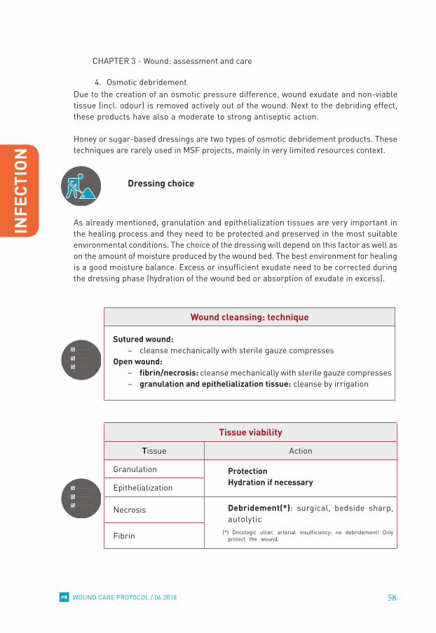
58WOUND CARE PROTOCOL / 06.2018
CHAPTER 3 - Wound: assessment and care
PR
INFE
CTIO
N
4. Osmotic debridementDue to the creation of an osmotic pressure difference, wound exudate and non-viable tissue (incl. odour) is removed actively out of the wound. Next to the debriding effect, these products have also a moderate to strong antiseptic action.
Honey or sugar-based dressings are two types of osmotic debridement products. These techniques are rarely used in MSF projects, mainly in very limited resources context.
Dressing choice
As already mentioned, granulation and epithelialization tissues are very important in the healing process and they need to be protected and preserved in the most suitable environmental conditions. The choice of the dressing will depend on this factor as well as on the amount of moisture produced by the wound bed. The best environment for healing is a good moisture balance. Excess or insufficient exudate need to be corrected during the dressing phase (hydration of the wound bed or absorption of exudate in excess).
Wound cleansing: technique
Sutured wound: – cleanse mechanically with sterile gauze compresses
Open wound: – fibrin/necrosis: cleanse mechanically with sterile gauze compresses – granulation and epithelialization tissue: cleanse by irrigation
Tissue viability
Tissue Action
Granulation Protection Hydration if necessaryEpithelialization
Necrosis Debridement(*): surgical, bedside sharp, autolytic
(*) Oncologic ulcer, arterial insufficiency: no debridement! Only protect the wound.Fibrin

59WOUND CARE PROTOCOL / 06.2018
CHAPTER 3 - Wound: assessment and care
PR
INFECTION
Management of infected - non healing wounds
Effective management of wound infection often requires a multidisciplinary approach and may involve referral to a specialist. We aim to readjust the interaction between the patient and the infecting micro-organism(s) in favour of the patient by:
– optimizing the host response – reducing the number of micro-organisms
The treatment is based on:
– removal of infected foci by cleansing and debridement (see “Tissue viability”) – antiseptics – systemic antibiotics if necessary
AntisepticsAgents used to inhibit or kill micro-organisms within a wound or on intact skin. They are applied topically and are non-selective. They may also have toxic effects on human cells. Development of resistance to antiseptics is less likely as they work at all levels of cell biology but already documented in literature (e.g. against CHX). The use should be rationalized!.
Indications for the use of antiseptics
- Prevention of wound infection or recurrence of infection in patient with increased risk of wound infection with:
• Foreign object in the wound (e.g. drain, external fixator, deep sutures)• Patient in ICU• An increased risk of infection at first dressing: during the next dressing change it
has to be evaluated whether the use of an antiseptic is still indicated.• Patient with comorbidity (as described in the list) upon advice of medical doctor/
trained health care worker: if the wound is healing and has no signs of infection the wound should be disinfected if the clinical condition of the patient is not supportive for wound healing. This means that the risk of impaired wound healing and/or wound infection is bigger than a potentially negative impact of antiseptic on wound healing (e.g. well-regulated diabetic patient might not need systematic use of antiseptic while a patient with poorly controlled diabetes mellitus needs systematic use of antiseptic in order to prevent wound infection).
- Treatment of critical colonization/covert infection: when progression towards overt infection is suspected or when interrupted healing is observed (non-healing wounds).
- Treatment of infection; consider combination with systemic antibiotics.

60WOUND CARE PROTOCOL / 06.2018
CHAPTER 3 - Wound: assessment and care
PR
INFE
CTIO
N
Review antiseptics regimen: STOP and/or OTHER TREATMENTS:
– If the wound deteriorates or the patient experiences symptoms suggestive of spreading or systemic infection.
– If a chronic wound with localized infection shows no improvement after 10–14 days of antiseptic therapy alone → re-evaluate the patient and the wound; send samples for microbiological analysis; consider whether there are any indications for systemic antibiotic treatment (and/or for further debridement).
Discontinue antiseptics – When the signs of infection resolve – When the wound starts to heal – If the patient experiences an antiseptic-related adverse event.
Some important considerations for the use of antiseptics – Do not mix different antiseptics. Never use two different
antiseptics after each other.
– Avoid alcoholic based antiseptics because of the pain generated.
– Always use the prescribed concentration.
– Check possible intolerances and allergies before using an antiseptic.
– Respect contact time
– Organic materials (e.g. debris, pus, dirt) can adversely affect the action of antiseptics
⇒ Mechanical cleansing of dirty and infected wounds should be done before disinfection.
– Respect the shelf life after opening
⇒ Write date of opening on the packaging.
– Open the bottle as instructed by the manufacturer and close the bottle after each use.
– Do not soil the bottles when handling them: don’t touch the bottle opening with hands, soiled gauze, instruments, the wound itself, etc.
– Always keep the bottle closed to avoid contamination.
– Clinicians need to consider whether, for a particular wound in a particular patient, the clinical benefit of the use of an antiseptic outweighs any possible negative effect on wound healing.

61WOUND CARE PROTOCOL / 06.2018
CHAPTER 3 - Wound: assessment and care
PR
INFECTION
Antibiotics (if needed)Antibiotics are substances that act selectively against bacteria and can be administered topically (not recommended) or systemically. As antibiotics work more specifically, they give bacteria an opportunity to mutate and to form resistance. Development of resistance to antibiotics is an increasing problem.
Topical antibioticsThe use of topical antibiotics in the management of infected wounds should be avoided to minimize the risk of allergy and the emergence of bacterial resistance.They should only be used in infected wounds under very specific circumstances, prescribed by experienced clinicians (e.g. topical metronidazole might be used for the treatment of malodour in fungating wounds or oncologic ulcers).Additionally, most topical antibiotics have a lower antimicrobial activity than most antiseptics.
Systemic antibiotics
1. INDICATIONS FOR THE USE OF SYSTEMIC ANTIBIOTICS
– Prophylaxis where risk of wound infection is high, e. g. contaminated colon surgery or ‘dirty’ traumatic wounds.
– Spreading or systemic wound infection. – When culture results reveal β-haemolytic streptococci. – If a chronic wound (with localized infection) shows no improvement after 10–14
days of antiseptic therapy alone → re-evaluate the patient and the wound; send samples for microbiological analysis (based on physician order); consider whether there are indications for systemic antibiotic treatment or re-debridement of the wound.
2. DISCONTINUE/REVIEW SYSTEMIC ANTIBIOTICS
– At the end of the prescribed course (according to type of infection, wound type, patient comorbidities and local prescribing policy).
– If there is no improvement of systemic or local signs and symptoms → re-evaluate the patient and the wound, consider microbiological analysis and changing antibiotic regimen.
– If the patient has an antibiotic-related adverse event → discontinue the causative antibiotic.

62WOUND CARE PROTOCOL / 06.2018
CHAPTER 3 - Wound: assessment and care
PR
INFE
CTIO
N
3. CHOICE OF SYSTEMIC ANTIBIOTICS
– If empirical treatment is necessary, start with an appropriate broad-spectrum antibiotic. When antibiotic susceptibilities become available, follow local microbiological/infectious disease advice, possibly switching to a narrower spectrum agent.
– Empirical antibiotic treatment must take into account the local antimicrobial susceptibility patterns of the possible pathogens.
– In chronic wounds, unless the patient is systemically unwell or a limb is in danger, microbiological results should usually be awaited before commencing systemic antibiotics.
– Administration of a combination of antibiotics may be necessary. Intravenous antibiotics are usually reserved for serious or life-threatening infections.
The decision to start or discontinue a systemic antibiotic and the choice of specific treatment has to be taken by a clinician.
For more information about medication prescription, see annex 5.1 about “Prescriptions and safe medication practices”.

63WOUND CARE PROTOCOL / 06.2018
CHAPTER 3 - Wound: assessment and care
PR
INFECTION
In summary: – Never use an antiseptic for a healing wound without signs of infection. – Always use an antiseptic for a non-healing wound and/or signs of infection,
foreign object in the wound (e.g. drain, external fixator), and patient requiring Intensive Care Unit (ICU).
– Use an antiseptic at the first treatment for a wound at risk of infection and re-evaluate at the next dressing if there is still a need for an antiseptic.
– Use an antiseptic upon advice of a medical doctor or a trained health care worker for a healing wound without signs of infection in a patient with comorbidity.
Cleansing and Disinfection
Infection Action Product, antiseptic, material
Healing wound and no signs of infection
Cleansing NaCl 0,9%
Disinfection No disinfection
Non-healing wound and/or signs of infection
Cleansing PVI 7,5% soap + rinse with NaCl 0,9%
DisinfectionPVI 10% aqueous solutionException: Pseudomonas Aeruginosa: acetic acid 1% if no improvement with PVI.
Foreign object in the wound (e.g. drain, external fixator)
Cleansing-Healing wound: NaCl 0,9%
-Dirty/non-healing/infected wound/first treatment of wound at risk: PVI 7,5% soap + rinse with NaCl 0,9%
Disinfection PVI 10% aqueous solution + see specific procedure!
Patient requiring Intensive Care Unit (ICU) level 2 and 3
Cleansing-Healing wound: NaCl 0,9%
-Dirty/non-healing/infected wound/first treatment of wound at risk: PVI 7,5% soap + rinse with NaCl 0,9%
Disinfection PVI 10% aqueous solution
Wound at increased risk of infection, at first treatment
CleansingPVI 7,5% soap + rinse with NaCl 0,9%
From the second treatment, always assess the wound and disinfect only if needed.
Disinfection PVI 10% aqueous solution
Patient with comorbidity
Cleansing-Healing wound: NaCl 0,9%-Dirty/non-healing/infected wound/first treatment of wound at risk: PVI 7,5% soap + rinse with NaCl 0,9%
Disinfection
-PVI 10% aqueous solution. After medical advice in case of healing wound. -Dirty/non-healing/infected wound/first treatment of wound at risk: PVI 10%
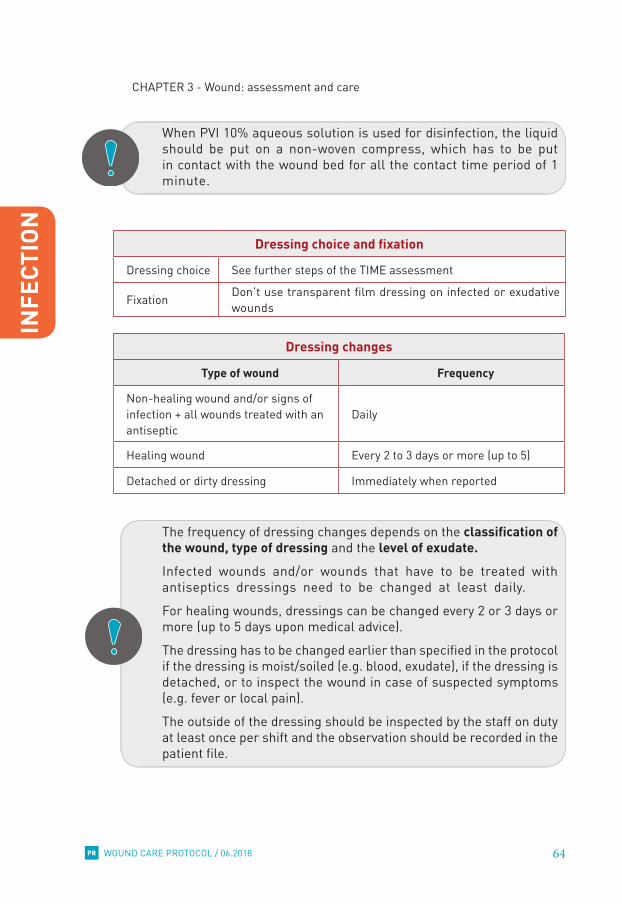
64WOUND CARE PROTOCOL / 06.2018
CHAPTER 3 - Wound: assessment and care
PR
INFE
CTIO
N
When PVI 10% aqueous solution is used for disinfection, the liquid should be put on a non-woven compress, which has to be put in contact with the wound bed for all the contact time period of 1 minute.
Dressing choice and fixation
Dressing choice See further steps of the TIME assessment
Fixation Don’t use transparent film dressing on infected or exudative wounds
Dressing changes
Type of wound Frequency
Non-healing wound and/or signs of infection + all wounds treated with an antiseptic
Daily
Healing wound Every 2 to 3 days or more (up to 5)
Detached or dirty dressing Immediately when reported
The frequency of dressing changes depends on the classification of the wound, type of dressing and the level of exudate.Infected wounds and/or wounds that have to be treated with antiseptics dressings need to be changed at least daily.
For healing wounds, dressings can be changed every 2 or 3 days or more (up to 5 days upon medical advice).
The dressing has to be changed earlier than specified in the protocol if the dressing is moist/soiled (e.g. blood, exudate), if the dressing is detached, or to inspect the wound in case of suspected symptoms (e.g. fever or local pain).
The outside of the dressing should be inspected by the staff on duty at least once per shift and the observation should be recorded in the patient file.

65WOUND CARE PROTOCOL / 06.2018
CHAPTER 3 - Wound: assessment and care
PR
MOISTURE
3.3. Moisture balance
M of TIME-D
Creating a moisture balance in the wound is essential to achieve wound healing. Exudate is produced as part of the body’s response to tissue damage. A wound which progresses through the normal wound healing cycle produces enough moisture to promote cell proliferation and supports the removal of devitalised tissue through autolysis.
Exudate
Evaluation of the exudate is an important part of wound management. The amount, type and viscosity of the exudate should be recorded and dressing material should be selected based on the exudate’s characteristics. If a wound is too dry, rehydration should be the principle of management; if the wound is producing an excess of exudate we need to absorb.
Table 5 – Exudate observation
Dry wound: no exudate or +
Moderately moist wound: exudate ++
Wet wound: exudate +++
Besides the amount of exudate, the type and odour should be observed: the colour, brightness and odour of the wound exudate can also give an indication of the degree of contamination of the wound.

66WOUND CARE PROTOCOL / 06.2018
CHAPTER 3 - Wound: assessment and care
PR
MOI
STUR
E
Table 6 - Exudate types and significanceType Colour Consistency Significance
SerousClear, straw- coloured
Thin, watery
Normal. Possibly a sign of infection if increasing. Some bacteria produce fibrinolysis, which degrade fibrin clots or coagulated plasma.
Fibrinous Cloudy ThinContains fibrin protein strands. Typical during inflammation phase.
Serosanguinous Clear, pink Thin, watery Normal.
Sanguinolent Red Thin, watery Trauma to blood vessels.
Seropurulent
Murky, yellow, cream-coffee
Thicker, creamy Infection.
Purulent Yellow, grey, green Thick
Infection. Contains pyogenic organisms and other inflammatory cells.
HaemopurulentDark, blood-stained
Viscous,sticky
Contains neutrophils, dead/dying bacteria and inflammatory cells. This means an established infection is present. Consequent damage to dermal capillaries leads to blood leakage.
Haemorrhagic Red Thick
Infection. Trauma. Capillaries are so friable they readily break down and spontaneous bleeding occurs. Not to be confused with bloody exudate produced by overenthusiastic debridement.
http://www.worldwidewounds.com/2006/september/White/Modern-Exudate-Mgt.html

67WOUND CARE PROTOCOL / 06.2018
CHAPTER 3 - Wound: assessment and care
PR
MOISTURE
Assure a correct moisture balance
Depending on the amount of exudate the wound will need hydration or fluids absorption in order to obtain the correct balance.
Different products are available for each specific situation and the choice will be not only based on the moisture but also on the presence of signs/risks of infections and the wound bed tissue type.
The hydration of the wound bed is obtained by the use of specific gels (hydrogel or polyvidone iodine gel) and paraffin gauzes while absorbent gauzes can efficiently absorb the excess of exudate.
The fixation (extensible adhesive tape or bandages versus transparent polyurethane film dressing) can also have an effect in promoting a correct moisture balance and avoiding dry or too moist tissues.
Moisture balance
Assessment Action
Dry wound: no exudate or + Hydration
Moderately moist wound: exudate ++ Preserve moist balance
Wet wound: exudate +++ – Absorption – Check the cause of the excess of exudate: check the “I” and/or oedema

68WOUND CARE PROTOCOL / 06.2018
CHAPTER 3 - Wound: assessment and care
PR
EDGE
S
3.4. Edges
E of TIME-D
During the last stage of wound closure, epithelial cells migrate across the wound bed to cover the surface of a wound (epithelialization). To allow this migration, wound edges need to be moist, intact (e.g. free of maceration, necrotic tissue and crusts) and attached to the base of the wound.
Wound edges
Evaluation of wound edges can indicate whether wound contraction and epithelialization are progressing, and confirm either the effectiveness of the wound treatment or the need for re-evaluation.There are many reasons why the epidermal margins of a wound fail to migrate across the wound bed or the wound edges fail to contract and reduce in size, e.g. hypoxia, infection, desiccation, dressing trauma, hyperkeratosis and callus at the wound margins.
When this is the case it should be checked whether all aspects (= T, I and M) of wound bed preparation have been considered. Furthermore, the underlying cause should be detected and corrected (ask advice of a specialist). If, despite proper wound bed preparation and treatment of the underlying cause, the edges fail to close alternative therapies to stimulate wound healing (e.g. surgical reconstruction/ skin graft) should be considered.
Normal edges
Normal wound edges are usually firmly attached to the wound bed with a healthy moist balanced periwound skin and in more advanced phase of healing also epithelial cells growing and migrating on the wound bed.

69WOUND CARE PROTOCOL / 06.2018
CHAPTER 3 - Wound: assessment and care
PR
EDGES
Hard-to-stimulate epithelialization, not closing, macerated wound edges
Problems with wound edges include:
Maceration
Dehydration
Undermining
Keratinized
Rolled (epibole)
Preserve or improve wound edges
To prevent damaging the wound edges and periwound area dressings should be removed carefully.
Normal progressing epithelialization and healthy wound edges
– Promotion of closure of the wound edges: see T,I and M
– Fill cavities (dressing has to have contact with wound bed)
– Prevention of maceration in wounds with exudate +++: see M + protection of wound environment
– Be careful while removing dressing materials because at first, the newly grown epithelial cells do not adhere firmly on the wound bed.
Hard-to-stimulate epitheliali-zation, not-closing, macerated wound edges
Evaluate cause and consider alternative therapies to stimulate wound healing (e.g. skin graft/surgical reconstruction/flaps, …)

70WOUND CARE PROTOCOL / 06.2018
CHAPTER 3 - Wound: assessment and care
PR
EDGE
S
Problem Action (in consultation with the doctor/surgeon)
Maceration
Establish and correct cause and minimize contact with moisture (absorbent dressings, periwound protection with zinc oxide ointment).
In order to prevent maceration the periwound area of wet wounds (exudate +++) should be protected by applying zinc oxide ointment.
There is no need to remove all the paste, just complete where the cream is missing after cleansing. If removal is necessary this should be done with an oily substance such as baby oil.
DehydrationEstablish and correct cause and rehydrate periwound skin (emollients, e.g. vaseline, shea butter, palm oil or olive oil).
Undermining
Establish and correct cause (e.g. can be indicative of a chronic wound and in particular those that are also critically colonized with bacteria or infected).
Undermining wounds and sinuses with narrow necks that are difficult to dress may be amenable to be laid open to facilitate drainage and dressing. Wounds associated with multiple sinuses or fistulas should be referred for surgical intervention.
Undermining should be treated by filling gently (to avoid shearing) cavities with compresses (the dressing has to have contact with wound bed) without creating tension.
Rolled (epibole)
Establish and correct cause (e.g. may be a sign of infection, can be present in wounds that have an inflammatory origin such as pyoderma gangrenosum or in malignancy), debridement.
Keratinized
Establish and correct cause. Remove hyperkeratotic skin using a scalpel or a curette and rehydrate (emollients, e.g. vaseline or shea butter).
Remember, callous means that bellow there is epithelium, so do not scrape too much not to avoid injuring it.

71WOUND CARE PROTOCOL / 06.2018
CHAPTER 3 - Wound: assessment and care
PR
EDGES
Periwound skin
In addition to the wound edges, the clinician should also consider the condition of the periwound skin (i.e. the skin within 4 cm of the wound edge as well as any skin under the dressing). Problems of the periwound skin may delay healing, provoke pain and discomfort, enlarge the wound, and adversely affect the patient’s quality of life.
The amount of exudate is a key factor for increasing the risk of periwound skin damage. Greater moisture exposure reduces skin barrier function and increases the risk of skin breakdown and maceration. This may make patients more susceptible to develop inflammation and contact dermatitis. Erythema and swelling may also indicate infection, which should be treated. In addition to the periwound skin, patients with wounds should also be assessed for problems that may be affecting their skin more widely.
Moreover, in these cases the underlying cause should be detected and corrected (ask advice of a specialist).
Periwound problems include:
Maceration Excoriation Dry skin
Hyperkeratosis Callus Eczema

72WOUND CARE PROTOCOL / 06.2018
CHAPTER 3 - Wound: assessment and care
PR
EDGE
S
Preserve or improve periwound skin
Problem Action (in consultation with the doctor/surgeon)
Maceration
Establish and correct cause and minimize contact with moisture (absorbent dressings, periwound protection with zinc oxide ointment).
Excoriation Establish and correct cause and rehydrate periwound skin (emollients, e.g. vaseline or shea butter).
Dry skin Establish and correct cause and rehydrate periwound skin (emollients, e.g. vaseline or shea butter).
Hyperkeratosis and callusEstablish and correct cause, remove hyperkeratotic skin plaques and rehydrate (emollients, e.g. vaseline or shea butter).
Callus Establish and correct cause, remove callus and offload to prevent recurrence.
Eczema Establish and correct cause, relieve symptoms and avoid allergens.
Edges
Assessment Action
Wound edges
– Protect healthy wound edges from becoming dry or macerated
– Investigate problematic/non-healing wound edges and treat accordingly
Periwound skin
– Protect healthy periwound skin against becoming dry, macerated or irritated
– Investigate problematic/non-healing wound edges and treat accordingly

73WOUND CARE PROTOCOL / 06.2018
CHAPTER 3 - Wound: assessment and care
PR
DISEASES
3.5. Diseases
D of TIME-D
The wound should not be treated in isolation but in the context of the patient’s overall wellbeing.
Comorbidities must be treated in order to optimize the general condition of the patient and facilitate the wound healing process. If comorbidities are not taken in consideration in the patient care plan, there are high risks to prolong the healing time or even to never achieve a complete healing of the wound.
A multidisciplinary approach is a key strategy for success in treating patients with wounds and this is not limited to nurses and medical doctors only, but also includes specific profiles like specialists, microbiologists, pharmacists, physiotherapists, occupational therapists, social workers, psychologists, health educators, etc…
Unfortunately all of these resources are not always available in all projects. This is why it is important to identify as soon as possible the existence and the quality of external resources where patients can be referred if necessary.

74WOUND CARE PROTOCOL / 06.2018
CHAPTER 3 - Wound: assessment and care
PR
3.6. Fixation / cover
Protection and hydration of wounds have been mentioned several times during the description of the TIME-D process and their importance has been highlighted.
After the choice of the cleaning method, the choice of dressing material is a second key element of the wound care process and it will play a relevant role in the protection and hydration/control of moisture of wounds.
Dressing materials are described with more details in chapter 4.
1. Protection
The dressing should offer mechanical protection of the wound, should be impermeable to micro-organisms, and avoid pain or trauma during removal of the dressing.
Wounds will be covered with non-woven compresses instead of gauze compresses, an all-in-one postop dressing with non-adherent wound contact layer, or an absorbent compress with non-adherent wound contact layer. These dressing materials will reduce the risk of sticking into the wound bed affecting the healing process.By covering ointments (i.e. hydrogel and PVI gel) with paraffin gauze (before non-woven gauzes) the risk of a dressing sticking into the wound is also reduced.
2. Hydration
The dressing(s) should respect the principle of moist wound healing by adding moisture when the wound is too dry, maintaining a good moisture balance in moderate moist wounds and absorbing exudate when the wound is too wet.
This has been explained in details in the section “Moisture balance”.
A good dressing will have:
– A layer that will maintain the moisture balance. – A layer which is non-adherent to the wound. For example paraffin gauze. – The third layer will be the fixation itself. Tapes are often too aggressive for the
skin as their adhesive is very strong. A good alternative is to use a bandage. This bandage should not be put just over the wound dressing but cover a much larger area. For example, for a wound on the lower leg the bandage will begin from the base of the toes and end below the knee. When wrapping, the pressure at the foot can be rather tight and released little by little going to the knee. Before applying the bandage, do not forget to “hydrate” the limb with a lipidic substance. In order to avoid a “tourniquet” effect, do not finish the bandage by many turns over each other (every turn adds its own pressure). Do not use metallic pins or a knot to close it, this could harm the tissues.
The aim is to leave this dressing in place for at least 24h, without seeing secretions going through it.

75WOUND CARE PROTOCOL / 06.2018
Chap 3. Annex 1 - Hypergranulation
PR
Annex 3.1 - Hypergranulation
Definition
Hypergranulation is the excess of granulation tissue, beyond the amount required to replace the tissue deficit. The production of granulation tissue continues beyond the height of the epithelium surface / periwound skin resulting in a raised mass (or peduncle) in excess of the wound itself.Hypergranulation creates a humping of the tissue, impeding the epidermal cell to cover and resurface the wound.
The cause of hypergranulation is not really known. It may be linked to:
– Prolonged inflammation (Type1) which may be caused by:• Infection• Foreign body irritant (e.g. dressing fibers)• External friction and traction (e.g. gastrostomy tubes, supra-pubic catheter
sites )• Allergic reaction (e.g. to the dressing or adhesive backing)• Tube material: higher incidence of overgranulation in areas surrounding
latex tubes then with other materials such as silicone.
– Use of occlusive dressings (Type 2):Growth factors may be extra stimulated under an occlusive dressing; fluids remain stuck under the dressing resulting in oedema and the possibility of cytotoxic effects due to occlusion.
– Cellular imbalance (Type 3)Cellular imbalance: an imbalance between collagen synthesis and lysis which could result in the unchecked proliferation of collagen leading to hypergranulation formation.
To date, there is no consensus on the best way to manage hypergranulating wounds, but in the protocol we propose certain action points.
Characteristics
– Usually similar composition as the normal granulation tissue. – Commonly it is seen as a sponge, friable, exuberant mass of tissue; sometimes
beefy red, sometimes almost purple in colour. – Highly vascularized with a dense network of blood vessels and capillaries. – Generally it is not painful as it contains little nerve tissue, however, if left untreated,
innervation can occur which will increase sensation.

76WOUND CARE PROTOCOL / 06.2018
Chap 3. Annex 1 - Hypergranulation
PR
– Healthy hypergranulation tissue: red-pink, moist tissue; may bleed but not readily; no other symptoms.
– Unhealthy hypergranulation tissue: dark red or pale bluish-purple uneven mass; rising above the level of the surrounding skin; may bleed readily; can be associated with wound infection; high exudate levels with associated maceration can be present.
Consequences
➢ Granulation tissue is highly vascularized but lacks a protective epithelial layer ⇒ it is unable to withstand even minor trauma (very fragile tissue).
– Prone to damage from contact (e.g. rubbing) with dressings and clothing. – Leakage of haemoserous exudate can lead to painful periwound maceration and
soiling and can require prolonged use of protective and absorbent dressings. ➢ Hypergranulation tissue can impede wound healing in several ways:
• Hypergranulation tissue prevents the migration of epithelial cells across the wound surface (these cells do not travel vertically) and delay wound closure.
• May increase the risk of bacterial contamination, biofilm formation and infection
• May increase the risk of scar formation (by forcing the wound edges further apart).
➢ Hypergranulation in wounds around or near devices (e.g. stoma flanges, gastrostomy tubes and tracheostomy tubes):• Can cause a physical barrier to device placement.• Can prevent close fitting. • Exudate and discharge is able to be in contact with the peristomal skin
causing a possible breakdown
Treatment
As mention before, to date, there is no consensus on the best way to manage these wounds.
BEFORE PROCEEDING IT IS IMPORTANT TO RULE OUT THE POSSIBILITY OF MALIGNANCY!
The removal of malignant tissue could lead to significant blood loss and would have a negative rather than a positive impact on the wound and the patient!
TYPE 1: INFLAMMATION RELATED
➢ Remove the irritant or inflammatory factors. ➢ Secure external medical devices (e.g. gastrostomy tubes and central lines) in
such a way to minimize friction around the wound site. ➢ Identify wound infection

77WOUND CARE PROTOCOL / 06.2018
Chap 3. Annex 1 - Hypergranulation
PR
TYPE 2: DRESSING TYPE RELATED
➢ Change the dressing type.• An occlusive dressing is changed to a dressing with more permeability. • The dressing choice will depend on exudate level.
➢ Secure dressings in such way to allow management of exudate and vapour loss properly. Avoid layers of adhesive film.
➢ If possible apply moderate direct prolonged pressure to the wound directly with fingers (sterile gloves) by using dressing pads, crepe or a tubular bandage. Avoid constriction of the blood supply!
➢ Alternatively, corticoid cream can be applied for a few days on the hypergranulation tissues. As this can also stop normal wound healing and favor infection, the wound will have to be kept on tighter surveillance.
Where bandaging is not recommended, local pressure may be applied by alternative means, e.g. positioning the patient on the wound for short periods. Take care not to cause any harm as a consequence of applying pressure.
TYPE 3: CELLULAR IMBALANCE
WHEN ABOVE MENTIONED TREATMENT OPTIONS HAVE FAILED, APPLICATIONS WITH SILVER NITRATE PENCIL OR SURGICAL EXCISION ARE POSSIBLE TREATMENT OPTIONS.
A SURGICAL EXCISION IS A VERY LAST TREATMENT OPTION. BY CUTTING AWAY THE HYPERGRANULATION A NEW WOUND IS CREATED, WHICH MAY AGAIN LEAD TO FORMATION OF HYPERGRANULATION TISSUE.

78WOUND CARE PROTOCOL / 06.2018
Chap 3. Annex 2 - External fixator
PR
Annex 3.2 - External fixator
Introduction
Up till now there is not enough evidence for any particular strategy of pin site care to reduce the incidence of pin site infections. The lack of undisputed research to determine best practice has resulted in many variations of pin site care.
We have composed a SOP for the care of external fixators, based on the consensus of the panel of wound care experts and with input from the surgical referents from MSF.
We refer to technical sheet n°8 for dressing procedure, attention points in care, when to inform a doctor, etc.
Definitions
External fixator
External fixation is a process by which pins are inserted into bone fragments through small incisions in the skin, and then held together with an external clamp or framework. This orthopaedic procedure is used to treat fractures or for correction of bone deformities associated with malunion, reconstructive surgery or limb-lengthening procedures.To carry out external fixation, pins and wires are surgically inserted and penetrate through the skin and soft tissue into the bone. Then, additional rods and bars are attached to the pins or wires to create an external fixator device which stabilizes the segment(s) of bone.
Pin sites These are the skin entry points of the skin-metal interface.
Possible complication: pin site infection
One of the main complications is pin site infection (PSI), which can lead to the development of osteomyelitis and systemic infection (sepsis).
Definition, classification
There is no uniformly accepted definition nor widely accepted criteria for the diagnosis of pin site infection. Consensus opinions from experts on pin site management have attempted to differentiate between reaction, colonization and infection:
– Reaction = the normal changes that occur at the pin site after pin insertion (i.e. changes in normal skin colour, skin warmth and drainage at the pin site). These are expected to subside after 72 hours.
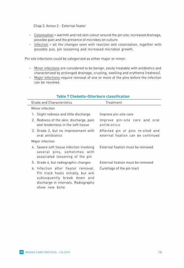
79WOUND CARE PROTOCOL / 06.2018
Chap 3. Annex 2 - External fixator
PR
– Colonization = warmth and red skin colour around the pin site, increased drainage, possible pain and the presence of microbes on culture.
– Infection = all the changes seen with reaction and colonization, together with possible pus, pin loosening and increased microbial growth.
Pin site infections could be categorized as either major or minor:
– Minor infections are considered to be benign, easily treatable with antibiotics and characterized by prolonged drainage, crusting, swelling and erythema (redness).
– Major infections require removal of one or more of the pins before the infection can be resolved.
Table 7 Cheketts-Otterburn classificationGrade and Characteristics TreatmentMinor infection1. Slight redness and little discharge Improve pin–site care2. Redness of the skin, discharge, pain
and tenderness in the soft tissueImprove pin–site care and oral ant ib io t ics
3. Grade 2, but no improvement with oral antibiotics
Affected pin or pins re-sited and external fixation can be continued
Major infection4. Severe soft tissue infection involving
several pins, sometimes with associated loosening of the pin
External fixation must be removed
5. Grade 4, but radiographic changes External fixation must be removed6. Infection after fixator removal.
Pin track heals initially, but will subsequently break down and discharge in intervals. Radiographs show new bone
Curettage of the pin tract
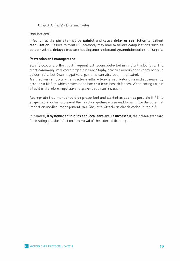
80WOUND CARE PROTOCOL / 06.2018
Chap 3. Annex 2 - External fixator
PR
Implications
Infection at the pin site may be painful and cause delay or restriction to patient mobilization. Failure to treat PSI promptly may lead to severe complications such as osteomyelitis, delayed fracture healing, non-union and systemic infection and sepsis.
Prevention and management
Staphylococci are the most frequent pathogens detected in implant infections. The most commonly implicated organisms are Staphylococcus aureus and Staphylococcus epidermidis, but Gram negative organisms can also been implicated.An infection can occur when bacteria adhere to external fixator pins and subsequently produce a biofilm which protects the bacteria from host defences. When caring for pin sites it is therefore imperative to prevent such an ‘invasion’.
Appropriate treatment should be prescribed and started as soon as possible if PSI is suspected in order to prevent the infection getting worse and to minimize the potential impact on medical management: see Cheketts-Otterburn classification in table 7.
In general, if systemic antibiotics and local care are unsuccessful, the golden standard for treating pin site infection is removal of the external fixator pin.

81WOUND CARE PROTOCOL / 06.2018
CHAPTER 4 - Dressing material
PR
CHAPTER 4 - DRESSING MATERIAL
When to use whatList of items and specifications
– Antiseptics and drugs for external use – Dressing material – Fixation material Annex 4.1 - Examples of “when to use what”

82WOUND CARE PROTOCOL / 06.2018
CHAPTER 4 - Dressing material
PR
4.1. When to use what
Based on the TIME observation, we will define which material and product we need to use. The main influencing factors are:
– Tissue viability: need for autolytic debridement or protection? – Infection prevention and management: need for antiseptic or not? – Moisture balance: need to add or absorb moisture? – Edges: need to protect or correct?
3 . D R E S S I N G C H O I C EPROTECTION OF WOUND + CONTROL OF MOISTURE
1st and 2nd layer: maintain the moisture balance & non-adherent to the wound3rd layer: fixation of the dressing
SUTURED WOUNDSCover with all-in-one dressing
Hea
ling
wou
ndN
o si
gns
of
infe
ctio
n
• Hydrogel• Paraffin gauzes• Non-woven compress• Fixation
• (Hydrogel)• (Paraffin gauzes)• Non-woven compress• Fixation
• Non-woven compress• Absorbent dressing• Fixation (NO FILM)
Non
-hea
ling
wou
ndSi
gns
of in
fect
ion • PVI 10% gel
• Paraffin gauzes• Non-woven compress• Fixation (NO FILM)
• PVI 10% gel• Paraffin gauzes• Non-woven compress• Fixation (NO FILM)
• PVI 10% aqueous sol• Non-woven compress• Absorbent dressing• Fixation (NO FILM)
REMEMBER: Re-evaluate the patient after 10-14 days of disinfection
HYDRATATION PRESERVEMOIST BALANCE
ABSORB
Rehydratation of periwound skin
Zinc Oxide 10% on surrounding skin if needed
Evaluate cause & consider alternative therapies to stimulate wound healing (e.g. surgical reconstruction/ skin graft)
Practical examples about dressing choice can be found in Annex 4.1.
Fi l l c a v i t i e s - D r e s s i n g i n c o n t a c t w i t h w o u n d b e d
DRY HUMID WET

83WOUND CARE PROTOCOL / 06.2018
CHAPTER 4 - Dressing material
PR
4.2. List of items and specifications
俵 Antiseptics and drugs for external use
HYDROGEL, amorphous, 25g, tube, sterile SDREWHYGA25T
POLYVIDONE IODINE, 10%, gel, 100g, tube DEXTIODP1G1
POLYVIDONE IODINE, 10%, solution, 200 ml, dropper bot. DEXTIODP1S2
POLYVIDONE IODINE, surgical scrub, 7.5 %, 500 ml, bot. DEXTIODPS75
SILVER NITRATE, 40%, pencil DEXTSILN1U-
ZINC OXIDE, 10%, ointment, 100 g, tube DEXTYINO1O1
Liquid soap, pH neutral DEXTSOAP1L1
俵 Dressing material
NON-WOVEN ADHESIVE DRESSSING WITH PAD, sterile, L SDRETAPAP1L
NON-WOVEN ADHESIVE DRESSSING WITH PAD, sterile, M SDRETAPAP1M
NON-WOVEN ADHESIVE DRESSSING WITH PAD, sterile, S SDRETAPAP1S
ABSORBENT DRESSING, small, sterile, s.u.NON-WOVEN ADHESIVE SDREABSD1S
ABSORBENT DRESSING, medium, sterile, s.u.NON-WOVEN ADHESIVE SDREABSD1M
ABSORBENT DRESSING, large, sterile, s.u.NON-WOVEN ADHESIVE SDREABSD1L
COMPRESS, NON WOVEN, 4 plies, 10 cm, sterile SDRECOMN10S
COMPRESS, GAUZE, paraffin, 10 cm x 10 cm, sterile SDRECOMP1P-
COMPRESS, GAUZE, 10 cm, 12 plies, 17 threads, sterile SDRECOMP1S-
俵 Fixation material
FILM DRESSING, semi-permeable, adhesive, 10cmx10m, roll SDREFIDSR1010
FILM DRESSING, semi-permeable, adhesive, 15cmx10m, roll SDREFIDSR1510
TAPE, ADHESIVE, roll, 2 cm SDRETAPA025
TAPE, ADHESIVE, roll, extensible, nonwoven, 10 cm x 10 m SDRETAPA100
BANDAGE, EXTENSIBLE, non adhesive, 6 to 7 cm x 4 m SDREBANE06N
BANDAGE, CREPE (Velpeau), 10 cm x 4 m SDREBANE104
BANDAGE, ELASTIC TUBULAR NET, … , roll 25 m SDREBTNE00++

84WOUND CARE PROTOCOL / 06.2018
CHAPTER 4 - Dressing material
PR
Antiseptics and drugs for external use
HYDROGEL, amorphous, 25g, tube, sterile SDREWHYGA25T
Indications
Healing wounds without signs of infection, which are dry (no exudate or +) or moderately moist (exudate ++), with necrosis and/or fibrin, granulation tissue or epithelialization tissue.
Therapeutic action
– Adds moisture to wounds that are too dry (rehydration of dry tissue). – Facilitates autolytic debridement (necrosis and fibrin). – Limited absorption capacity: absorbs exudate and slough in wounds that are
minimally exudative. – In moderately moist wounds with granulation/epithelialization tissue it is advised
to add a layer of hydrogel to prevent the wounds become too dry when exudate is absorbed by the non-woven compress.
Contra-indications
– Wounds with heavy exudate (exudate +++) (risk of maceration). – Infection (the available moisture may increase the risk of bacterial proliferation). – High potential for anaerobic infections; evidence of gangrenous tissue (keep dry!). – Allergy to ingredients or known hypersensitivity to the gel or any of its ingredients – Necrotic feet/digits because of ischaemia and/or neuropathy.
Attention points
– Should be used with care in the vicinity of the eyes and in deep wounds with narrow openings (e.g. fistulas) where removal of the gel may be difficult.
– May overhydrate: excessive use, or use in a highly exudative wound, may lead to maceration of the periwound skin.
– The wound may initially appear to increase in size in the early stages of treatment. This is normal because wound debris is removed.

85WOUND CARE PROTOCOL / 06.2018
CHAPTER 4 - Dressing material
PR
Instructions for use
– Cleanse the wound properly – Apply a layer of +/- 5mm of hydrogel into the wound in an aseptic way (without
touching the wound surface with the tip) or apply the hydrogel on a sterile compress/sterile paraffin gauze to be put on the wound, or impregnate packing rope with the gel.
– Avoid overspill to the surrounding skin. – Cover the hydrogel with paraffin gauze to avoid fast evaporation – Secondary dressing + fixation is necessary – As hydrogel creates a moist environment, it may be necessary to protect the
wound edges against moistening (zinc oxide ointment). – Hydrogel can be removed from the wound by rinsing with NaCl 0,9%. If the
hydrogel becomes difficult to remove soak the area with sterile water or NaCl 0,9% to facilitate removal.
– Frequency of dressing changes: every 2 or 3 days (depending on the clinical condition of the wound and the amount of exudate produced).
– In a necrotic eschar: scaring the surface of the necrotic eschar can help the hydrogel to rehydrate it and thus accelerate the debridement.
POLYVIDONE IODINE, 10%, gel, 100g, tube DEXTIODP1G1
Indications
Non-healing wounds with or without overt signs of infection, which are dry (no exudate or +) or moderately moist (exudate ++), with necrosis and/or fibrin, granulation or epithelialization tissue.
In addition, according to our protocol: all dry wounds (no exudate or +) or moderately moist (exudate ++) in:
– patients in ICU – wounds at increased risk of infection at first treatment – patients with comorbidities after medical advice.
Therapeutic action
Broad spectrum antimicrobial.

86WOUND CARE PROTOCOL / 06.2018
CHAPTER 4 - Dressing material
PR
Contra-indications
– Healing wounds without signs of infection except for patients in ICU, wounds at increased risk of infection at first treatment, patients with comorbidities after medical advice.
– Wet (exudate +++) non-healing wounds with or without overt signs of infections. – Patient with a known hypersensitivity to the gel or any of its ingredients. History of
iodine intolerance. – Thyroid dysfunction – Together with antiseptics of other groups (incompatibility) or with mercury,
lithium, alkali or sodium thiosulfate compounds. – Preterm neonates, neonates < 1,5 kg and children younger than 30 months – Without medical advice in children between 30 months and 5 years – Concomitant use of topical products containing mercury derivatives – Vicinity of the eyes.
According to the advice of external wound care experts the risk of negative effects in pediatric patients is very limited. Povidone iodine is contraindicated in infants aged < 1 month.
Attention points
– During treatment of extensive skin surfaces, on mucous membranes, or during long-term administration, it is necessary to take into account the possible resorption of iodine through the skin.
– A regular or prolonged use should be avoided in patients with burns involving more than 20% of the skin surface, with large open wounds, in patients treated with lithium, in pregnant and lactating women and neonates. During pregnancy and lactation only to be prescribed with a defined indication and in limited doses (due to the risk for iodine absorption).
– Do not use with other antiseptics such as chlorhexidine-cetrimide (incompatibility), silver, hydrogen peroxide or mercury components.
Instructions for use
– Cleanse the wound properly – Apply a layer of +/- 5mm into the wound in an aseptic way (without touching the
wound surface with the tip of the tube) /apply the gel on a sterile compress to spread out on the wound/on the sterile paraffin gauze to be put on the wound/impregnate packing rope with the gel.
– Contact with the skin around the wound edges/intact skin should be minimised. – Secondary dressing + fixation is necessary. Do NOT use polyurethane film for
fixation of the secondary dressing on infected wounds. – Tightly close the tube after each use to prevent contamination – Frequency: at least once a day – The gel changes colour as iodine is released: if brown colour has disappeared, the
product has to be applied again.

87WOUND CARE PROTOCOL / 06.2018
CHAPTER 4 - Dressing material
PR
POLYVIDONE IODINE, 10%, solution, 200 ml, dropper bot. DEXTIODP1G1
N.B Aqueous solution
Indications
For disinfection: – of non-healing and/or infected wounds. – In addition, according to our protocol in:
• patients in ICU• wounds at increased risk of infection at first treatment• patients with comorbidities after medical advice• Wounds with foreign bodies (exudate 0, +, ++ and +++).
To cover: – non-healing and/or infected wounds with exudate +++
Therapeutic actions
Broad spectrum antimicrobial.
Contra-indications
– Healing wounds without signs of infection, except for patients in ICU, wounds at increased risk of infection at first treatment, patients with comorbidities after medical advice, wounds with foreign objects.
– Patients with a known sensitivity to any of its ingredients – History of iodine intolerance – Thyroid dysfunction – Together with antiseptics of other groups (incompatibility) or with mercury,
lithium, alkali or sodium thiosulfate compounds. – Preterm neonates, neonates < 1,5kg and children younger than 30 month. – Without medical advice in children between 30 months and 5 years – Repeatedly or on very large areas, especially in pregnant and lactating women and
neonates. – Contact with eyes.
According to the advice of external wound care experts the risk of negative effects in pediatric patients is very limited. Povidone iodine is contraindicated in infants aged < 1 month.
Alternative if PVI is contra-indicated = chlorhexidine 0,5% in aqua.

88WOUND CARE PROTOCOL / 06.2018
CHAPTER 4 - Dressing material
PR
Attention points
– Regular or prolonged use and treatment of extensive skin surfaces: only under medical supervision.
– During pregnancy and lactation: only upon medical advice.
Instructions for use
– The antiseptic effect of PVI 10% solution starts after 30 seconds, but a minimum contact time of 1 minute is recommended.
– Frequency of dressing: at least daily. If brown colour has disappeared, the product has to be applied again.
– When should PVI 10% solution be diluted?
Type of care provided Dilution
Wound rinsing 10ml PVI 10% solution + 90ml NaCl 0,9%
Intra-abdominal washing 10ml PVI 10% solution + 90ml NaCl 0,9%
Bladder rinsing 50ml PVI 10% solution + 200ml NaCl 0,9%
Constant irrigation 2 ml PVI 10% solution + 98 ml NaCl 0,9%
During contact with the exudate the iodine is little by little released. In case of dilution: all the iodine is released immediately with no more long-term effect.
POLYVIDONE IODINE, surgical scrub, 7.5 %, 500 ml, bot. DEXTIODPS75
N.B Soap solution
Indications
Cleansing of dirty wounds, non-healing wounds, infected wounds and wounds at risk at first treatment.
Therapeutic action
Broad spectrum antimicrobial soap.

89WOUND CARE PROTOCOL / 06.2018
CHAPTER 4 - Dressing material
PR
Contra-indications
– Patients with a known sensitivity to any of its ingredients – History of iodine intolerance – Thyroid dysfunction – Together with antiseptics of other groups (incompatibility) or with mercury,
lithium, alkali or sodium thiosulfate compounds. – Preterm neonates, neonates < 1,5kg and children younger than 30 months – Without medical advice in children between 30 months and 5 years – Repeatedly nor on very large areas, especially in pregnant and lactating women
and neonates. Pregnancy and breastfeeding are no contra-indication for brief application.
– Contact with eyes.
According to the advice of external wound care experts the risk of negative effects in pediatric patients is very limited. Povidone iodine is contraindicated in infants aged < 1 month.
Instructions for use
The solution needs to be used undiluted, in the same way as a normal soap.
The scrub solution is first brown. While using, the soap becomes less brown. When the foam is light coloured, it can be rinsed with NaCl 0,9%.
Polyvidone iodine 4% is a possible alternative in case of stock rupture.
SILVER NITRATE, 40%, pencil DEXTSILN1U-
Indications
– Use only with great care in more stubborn areas of hypergranulation.
Therapeutic action
– Astringent cauterization of wounds – Germicidal removal of granulation tissue, corns, and warts – To “burn back” hypergranulation tissue.
Contra-indications
– Hypersensitivity to silver nitrate or any component of the formulation – Broken skin, cuts, or wounds outside of the hypergranulation affected area.

90WOUND CARE PROTOCOL / 06.2018
CHAPTER 4 - Dressing material
PR
Attention points
– Not recommended for prolonged or excessive use – Only for small surfaces – For individual use.
Side effects
– May cause pain – Risk for damaging the surrounding skin – Can promote tissue necrosis, which represents a further risk of infection – Provokes a further inflammatory response – Reduces fibroblast proliferation – Risk for chemical burns, which are more likely to occur with prolonged application – May cause systemic effects if used over a large area (e.g. hyponatraemia, …).
Instruction for use
Apply once daily, 3 to 6 days: – Cleanse and dry the wound according to the protocol – Moisten the silver nitrate pencil with water for injection (do not use
NaCl0,9%solution or water containing salt or chlorides). – Protect the healthy skin around the area to be treated (e.g. vaseline ointment). – Do not allow the water or tip to touch healthy skin or tissue. – Touch the area to be treated with the wet tip. Use light pressure, do not press or
rub. Respect the contact time (cfr. manufacturer) to avoid too deep damage of the tissue.
– Dry the tip of the pencil with a sterile compress. – Apply a secondary dressing as chosen for the wound stage and fixate.
ZINC OXIDE, 10%, ointment, 100 g, tube DEXTYINO1O1
Indications
– Skin protector for periwound in case of wounds with exudate +++: it protects the skin against the influence of body fluids.
– Moderate drying effect.
Contra-indications
Pregnancy and breastfeeding are no contra-indications, but do not apply on breasts.

91WOUND CARE PROTOCOL / 06.2018
CHAPTER 4 - Dressing material
PR
Attention points
Can interfere with dressing adherence.
Instructions for use
– Can be used up to 1 to 3 applications/day; depending on dressing changes and clinical response.
– Apply only on the periwound area, not in the wound! – Removal with something oily, e.g. baby oil. Note that you don’t have to remove all
the zinc oxide paste, completing the layer already present is enough.
LIQUID NEUTRAL SOAP, 1000 ml DEXTSOAP1L1
Indications
Cleansing of dirty, intact surrounding skin/limb.
Contra-indications
Non-intact skin.
Attention points
If used routinely, the skin may dry out.
Instructions for use
Rinse after cleansing with soap with an abundant amount of NaCl 0,9 %.

92WOUND CARE PROTOCOL / 06.2018
CHAPTER 4 - Dressing material
PR
Dressing material
NON-WOVEN ADHESIVE DRESSSING WITH PAD, sterile, LNON-WOVEN ADHESIVE DRESSSING WITH PAD, sterile, MNON-WOVEN ADHESIVE DRESSSING WITH PAD, sterile, S
SDRETAPAP1LSDRETAPAP1MSDRETAPAP1S
Indications
All-in-one dressing for covering sutured/stapled wounds
SizesSmall - Medium - Large.
Contra-indications
- Deep cavity wounds- Wet wounds (exudate +++).
Attention points
- Not waterproof: patient cannot shower with the dressing- Once the dressing is saturated or has become wet from the outside the barrier function is broken.
Instructions for use
Open the dressing and apply to the wound respecting aseptic careWhen removing, be gentle as not to harm the skin.

93WOUND CARE PROTOCOL / 06.2018
CHAPTER 4 - Dressing material
PR
ABSORBENT DRESSING, small, sterile, s.u.NON-WOVEN ADHESIVE ABSORBENT DRESSING, medium, sterile, s.u.NON-WOVEN ADHESIVE ABSORBENT DRESSING, large, sterile, s.u.NON-WOVEN ADHESIVE
SDREABSD1SSDREABSD1MSDREABSD1L
Exact dimensions may vary according to manufacturer.Examples of sizes:
– Small: 10-10 cm, 15-15 cm – Medium: 10-20 cm, 15-20 cm, 15-25cm – Large: 20-20 cm, 20-25 cm.
Indications
Wet wounds (exudate +++).
Contra-indications
Wounds which are dry (no exudate or +) or moderately moist (exudate ++).
Instructions for use
– White side = wound side; marked side = outside – Secondary dressing with fixation is necessary – Cannot be cut.

94WOUND CARE PROTOCOL / 06.2018
CHAPTER 4 - Dressing material
PR
COMPRESS, NON WOVEN, 4 plies, 10 cm, sterile DRECOMN10S
Indications
To cover wounds.
Instructions for use
Unless in a very wet wound, it will stick and should be used over a non-adherent layer.Fixation is necessary.
COMPRESS, GAUZE, paraffin, 10 cm x 10 cm, sterile SDRECOMP1P-
Indications
– To cover hydrogel and PVI gel (to avoid rapid evaporation of these products) – To reduce adherence of the compresses to the wound bed.
Contra-indications
– Wounds with heavy exudate.
Attention points
Adheres to the wound if it remains too long in place. Removal can be obtained by pouring NaCl 0,9 % on the wound area until the dressing is sufficiently soft to remove.
Instructions for use
- Can be cut to size (use sterile scissors)- Maximum 2 layers above each other (to ensure the permeability of exudate)- Secondary dressing and fixation necessary.

95WOUND CARE PROTOCOL / 06.2018
CHAPTER 4 - Dressing material
PR
COMPRESS, GAUZE, 10 cm, 12 plies, 17 threads, sterile SDRECOMP1S-
Indications
Cleansing, drying and disinfecting of wounds and the periwound area.
Attention points
As these woven compresses can prove to be very adherent to the wound bed, caregivers should prefer using non-woven compresses to be in direct contact.
Fixation material
FILM DRESSING, semi-permeable, adhesive, 10cmx10m, roll FILM DRESSING, semi-permeable, adhesive, 15cmx10m, roll
SDREFIDSR1010SDREFIDSR1510
Indications
Fixating of primary dressings in healing wounds without signs of infectionWaterproof: patient can shower when the primary dressing is covered with this transparent film. Once over a dressing, it will help create moist environment.
Contra-indications
– Infected wounds – Heavy levels of exudate (risk of maceration).
In these cases the dressing can be covered with a film dressing to give the patient the possibility to take a shower. Afterwards the dressing should be renewed.

96WOUND CARE PROTOCOL / 06.2018
CHAPTER 4 - Dressing material
PR
Instructions for use
– Do not use as a primary dressing on the wound: the film on the roll is not sterile. – Do not stretch while applying, because the film will gradually shrink back to the
normal shape and may pull the skin, causing discomfort and blistering. – Apply a margin of overlap with the skin of approximately 4 to 6 cm to provide an
adequate area of contact for adherence. – To ensure adequate dressing adhesion, the periwound skin should be clean, dry
and free of moisturizers or grease. – Presence of hair on the skin may affect the adhesiveness of the dressing and
complicate the technique for the removal. If hair removal is not possible (e.g. no consent of patient, risk of infection associated with shaving …) a non-adhesive method of dressing fixing should be considered (e.g. tubular gauze or light retention bandage).
– Caution is required when removing the dressing to avoid that the adhesive damages the skin. The corner of the dressing should be lifted gently from the skin and then stretched horizontally (tangentially) away from the wound (not vertically).
TAPE, ADHESIVE, roll, 2 cm SDRETAPA025
Adhesive tape used to secure certain medical devices on the skin and to maintain dressings. Caution is required when removing the dressing to avoid that the adhesive damages the skin. The corner of the dressing should be lifted gently from the skin.

97WOUND CARE PROTOCOL / 06.2018
CHAPTER 4 - Dressing material
PR
TAPE, ADHESIVE, roll, extensible, nonwoven, 10 cm x 10 m SDRETAPA100
– Self-adhesive, extensible, nonwoven tape used to secure dressings. – Can be easily cut to the desired shape and size. – Conforms well to body contours and allows body movements when in place.
Suitable for flexing areas such as neck, elbows, knees. – Do not apply under tension to prevent shearing forces causing damage to skin.
This is particularly important when applied over joints. – Caution is required when removing the dressing to avoid that the adhesive
damages the skin. The corner of the dressing should be lifted gently from the skin.
BANDAGE, EXTENSIBLE, non adhesive, 6 to 7 cm x 4 m SDREBANE06N
– Stretch bandage used to secure dressings.
Note that it should be fixated by tape, not with metallic pins or knots. When finishing the bandage DO NOT circle more than once (it would add pressure and act as a “tourniquet”).

98WOUND CARE PROTOCOL / 06.2018
CHAPTER 4 - Dressing material
PR
BANDAGE, CREPE (Velpeau), 10 cm x 4 m SDREBANE104
– Stretch bandage used to secure dressings by exerting a certain pressure.
Note that it should be fixated by tape, not metallic pins or knots. When finishing the bandage DO NOT circle more than once (it would add pressure and act as a “tourniquet”.
BANDAGE, ELASTIC TUBULAR NET, … , roll 25 m SDREBTNE00++
Finger SDREBTNE001Wrist/hand/foot SDREBTNE002Arm/leg SDREBTNE003Head/small chest SDREBTNE004Chest/hip SDREBTNE005
Tubular shaped elastic net used to hold dressings securely in place without compression or immobilization, even for moving areas or areas which are difficult to access (head, thorax, limbs, and joints).

99WOUND CARE PROTOCOL / 06.2018
Chap 4. Annex 1 – Examples of “when to use what”
PR
Annex 4.1 – Examples of “when to use what”
Healing wound without signs of infection, exudate (0 or +), no foreign object in the wound, not in ICU, no increased risk of infection, no comorbidities
Moisture is added by application of hydrogel. The hydrogel is covered with 1 (max. 2) layer(s) of vaseline gauze, to avoid rapid evaporation of the gel and sticking into the wound.The hydrogel and paraffin gauze are covered with one or more non-woven compresses. The whole is fixed with film dressing (roll), tape or a bandage.
Exception to the application of hydrogel: dry sutured/stapled wound → has to be covered with an all-in-one postop dressing [or with non-woven compresses and film dressing (roll) or nonwoven adhesive tape (roll) if postop dressings are not available or if more absorption capacity is necessary].
Healing wound without signs of infection, exudate: ++, no foreign object in the wound, not in ICU, no increased risk of infection, no comorbidities
– Necrosis and/or fibrin: apply hydrogel, cover with 1 (max. 2) layer(s) of paraffin gauze, non-woven compress(es) and fix with film dressing (roll), tape or a bandage. Next to maintaining a good moisture balance, the hydrogel aims autolytic debridement of the fibrin and/or necrosis. For this purpose the gel has to stay for a sufficiently long period of time on the wound. To avoid rapid evaporation of the gel, paraffin gauze is applied.
– Granulation and/or epithelialization tissue: apply hydrogel, cover with non-woven compress(es) and fix with film dressing (roll), tape or a bandage.Hydrogel is applied to avoid that the all wound exudate is absorbed by the compresses, resulting in a wound that is too dry and a dressing sticking into the wound.
Healing wound without signs of infection, with a lot of exudate: +++, no foreign object in the wound, not in ICU, no increased risk of infection, no comorbidities (= very rare condition)
In order to absorb the excess of exudate, an absorbent compress or extra non-woven compresses + absorbent compress are applied and fixed with non-woven adhesive roll, tape or a bandage (NO film!).
Non-healing wound with or without overt signs of infection, exudate 0, or + or ++, no foreign body
Moisture is added by application of PVI gel, which has a prolonged antiseptic action. The PVI gel is covered with 1 (max. 2) layer(s) of paraffin gauze, to avoid rapid evaporation of the gel and sticking into the wound.

100WOUND CARE PROTOCOL / 06.2018
Chap 4. Annex 1 – Examples of “when to use what”
PR
The PVI gel and paraffin gauze are covered with one or more non-woven compresses. The whole is fixed with tape or a bandage. Film dressing (roll) should not be used in wound with overt/covert infection, because it can create an environment that is too moist which further sustains the infection.
Non-healing wound with or without overt signs of infection, exudate: +++
Instead of PVI gel the wound bed is covered with non-woven compress(es) moistened with PVI aqueous solution. PVI solution has a drying effect; application of PVI gel in this type of wounds increases the risk of maceration. Next, an absorbent compress is applied. The whole is fixed with tape or a bandage. Film dressing (roll) should not be used in wound with overt/covert infection, because it can create an environment that is too moist which further sustains the infection. Deep wounds
In deep wounds the same materials and products are used as in the superficial ones with the difference that the wound is packed with non-woven compresses in order to maintain contact between the wound bed and the dressing (the wound has to grow upwards from the bottom). The absorbent compress, depending on the moisture level of the wound, should be applied on top of the other compresses (as final layer before fixation).

101WOUND CARE PROTOCOL / 06.2018
CHAPTER 5 - General wound care technique
PR
CHAPTER 5 - GENERAL WOUND CARE TECHNIQUE
Before the procedure – Patient – Environment – Material
During the procedure – Preparation – Removal of dressing – Observe and act
After the procedure – Patient – Infection prevention and control – DocumentationAnnex 5.1 - Prescription and safe medication practiceAnnex 5.2 - Documentation
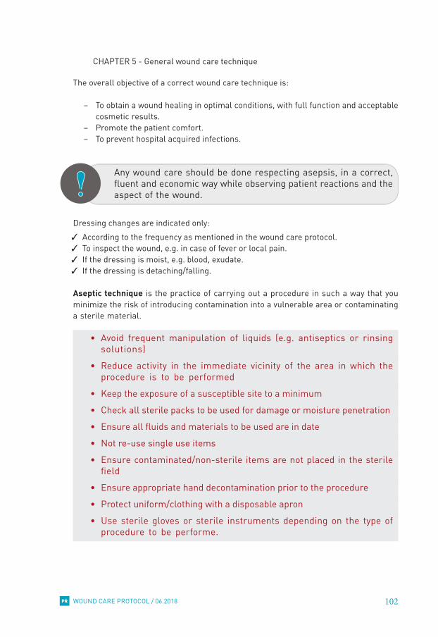
102WOUND CARE PROTOCOL / 06.2018
CHAPTER 5 - General wound care technique
PR
The overall objective of a correct wound care technique is:
– To obtain a wound healing in optimal conditions, with full function and acceptable cosmetic results.
– Promote the patient comfort. – To prevent hospital acquired infections.
Any wound care should be done respecting asepsis, in a correct, fluent and economic way while observing patient reactions and the aspect of the wound.
Dressing changes are indicated only: 俵 According to the frequency as mentioned in the wound care protocol. 俵 To inspect the wound, e.g. in case of fever or local pain. 俵 If the dressing is moist, e.g. blood, exudate. 俵 If the dressing is detaching/falling.
Aseptic technique is the practice of carrying out a procedure in such a way that you minimize the risk of introducing contamination into a vulnerable area or contaminating a sterile material.
• Avoid frequent manipulation of liquids (e.g. antiseptics or rinsing solutions)
• Reduce activity in the immediate vicinity of the area in which the procedure is to be performed
• Keep the exposure of a susceptible site to a minimum
• Check all sterile packs to be used for damage or moisture penetration
• Ensure all fluids and materials to be used are in date
• Not re-use single use items
• Ensure contaminated/non-sterile items are not placed in the sterile field
• Ensure appropriate hand decontamination prior to the procedure
• Protect uniform/clothing with a disposable apron
• Use sterile gloves or sterile instruments depending on the type of procedure to be performe.

103WOUND CARE PROTOCOL / 06.2018
CHAPTER 5 - General wound care technique
PR
5.1. Before the procedure
BEFORE- VERIFY
IDENTITY & MEDICAL ORDERS
- INFORM PATIENT
- PROPER IPC
- COMFORT & PRIVACY
- FORESEE & PREPARE
PATIENT ENVIRONMENT MATERIAL
Patient
– Verify the patient identity and the correspondence with the patient file. – Check the information on the follow up sheet for wound care and prepare
accordingly. – Greet and inform the patient in a reassuring and understandable way about
the aim of the procedure, the cooperation needed, the procedure itself and the expected sensations.
– Evaluate pain, provide prescribed analgesic (see Chapter 2 – Pain management and Technical sheet 4) if required and wait the time needed for the drug to act.
– Position the patient and yourself comfortably.
EnvironmentIn all health structures, rigorous organization and preparation in the dressing of wounds can reduce the risk of accidental wound contamination and transmission of microorganisms from one patient to another.
In general:
– Having a dedicated dressing room is a plus. – It must have a clear clean to dirty pathway, clean wounds should be seen first. – Prepare everything in advance in order to avoid interrupting the care/leaving the
room. Having an aid can be most useful. – During dressing changes visits are not allowed, beds are not being made and the
room is not being cleaned. Switch off any fan and close all windows. In this way circulation of micro-organisms in the air is avoided.
– Create enough space to work comfortably at any moment. – Respect patient privacy: use a screen if needed. – The room needs adequate lighting. – Clean and disinfect all working surfaces with Surfanios® diluted solution and let
it work for 15 minutes.
104WOUND CARE PROTOCOL / 06.2018
CHAPTER 5 - General wound care technique
PR
– Use a clean, disinfected trolley, with sterile and/or clean materials (dressing set, compresses, etc.) on the upper tray, and septic materials (sharps container, trash bag, etc.) on the lower tray.
– Clean and disinfect the trolley in between patients, before and after the procedure.
For IPD patients:
– Preferably perform dressings at the bedside. – For specific reasons (e.g. not possible to assure privacy) dressings can be done in
a specific designated dressing area/room. – Advanced dressings which require more invasive procedure or basic anaesthesia
can be done in specific procedure rooms or can be planned for the operating room. – In case of a dressing care in isolation rooms: bring only the needed material.
Any unused material that has been brought inside the isolation area should remain there or should be disposed even if has not been used. Respect local recommendations based on the type of isolation adopted.
For OPD patients (or in case of use of dressing room in IPD):
– Organized circuit from clean to contaminated. – Program “clean” patients first. – Always clean and disinfect the examination table at the end of each procedure,
before installing the next patient. – Clean and disinfect the room (floor and all the other surfaces) on daily basis or
immediately in case of body fluids spills. – Disposal of waste must be done daily, at each time the container is full or whenever
there is the necessity (e.g. bad smell due to biological fluids/material).
Material
Select your material based on the wound care protocol.
Foresee:
– Cleaning material: • Surfanios® diluted solution +wipes wipes (e.g. cleaning cloth, paper roll)
– Alcohol based hand rub. – Non-sterile gloves. – Clean scissors to remove the dressing (one per patient). – Clean scissor for preparing the fixation material (only for cutting non sterile clean
material; to be disinfected after each patient). – Dressing sets and/or sterile gloves.

105WOUND CARE PROTOCOL / 06.2018
CHAPTER 5 - General wound care technique
PR
– Material for:• Cleaning of the wound• Disinfection of the wound• Dressing and covering of the wound
– Additional dressing material and products, depending on the type of wound care, e.g.:
• Removal of sutures/staples: stitch cutter/staple remover• Wound sampling/drain sampling: sterile swab/ sterile recipient• Drain: clamps, sterile safety pins, ...
– Protection for the bed (e.g. clean drapes or sterile drape in case of deep wound, plastic sheets could be used for very exudative wounds).
– Recipients for used:• Linen• Disposable material/waste disposal• Instruments
➢ Where applicable check expiration date and the integrity of materials to be used.
➢ Be aware of the characteristics of each specific material (indications, contra-indications, attention points and instructions for use).
REMEMBER
5 MOMENTS of HAND HYGIENE
BE CAREFUL WHILE REMOVING DRESSING MATERIALS
CLEAN PERIWOUNDSKIN
TECHNIQUECLEANEST TO DIRTIESTASEPTIC TECHNIQUE
5.2. During the procedure
Preparation
俵 Clean and disinfect all the surfaces: wipe with Surfanios® diluted solution and leave it for 15 minutes.
俵 Perform hand hygiene. 俵 If risk of contact with body liquids: use Personal Protective Equipment (PPE) and non-sterile gloves (except if the procedure needs to be carried out with sterile gloves).
俵 Protect the bed / the stretcher to avoid body fluids and liquids to contaminate the surface, the bed linen or patient dress.
Picture 9 – Cleaning and disinfection of surfaces

106WOUND CARE PROTOCOL / 06.2018
CHAPTER 5 - General wound care technique
PR
Removal of dressing
俵 Wear non-sterile gloves. 俵 Carefully remove the old dressing:
– If dressing is fibbed with bandages:• Use a clean scissor for removing bandages (one scissor per patient).
– If dressing is fixed with adhesive fixation:• Loosen the edges of the dressing.• Start at the end of the adhesive tape and stretch it out horizontally parallel
to skin while supporting the skin with your fingers. If loosening the adhesive tape is too painful: moisten with NaCl 0,9%.
俵 Remove remaining compresses with a non-sterile glove (instrumental removal in case of a wick).
俵 If the dressing adheres to the wound: moisten generously with NaCl 0,9% to prevent tissue damage and wait enough time for it to act.
俵 Avoid damage to newly formed tissue by roughly rubbing, clumsy or rough handling of instruments.
俵 Observe the dressing to evaluate the amount, the colour and the odour of the exudate (as described in the wound care protocol).
Picture 10 – Removal of dressing

107WOUND CARE PROTOCOL / 06.2018
CHAPTER 5 - General wound care technique
PR
俵 Dispose of the dressing immediately in the correct waste container (follow the waste management procedure locally in use).
俵 Remove non-sterile gloves and perform hand hygiene.
Observe and act
俵 Observe pain. 俵 Observe wound as by TIME assessment: Tissue ⇒ Infection ⇒ Moisture ⇒ Edges. 俵 Actions: always work from cleanest to dirtiest: if a patient has several wounds, start dressing at the cleanest and after pass to the dirtiest or infected ones.
俵 Perform hand hygiene. 俵 The dressing can be carried out with sterile instruments or sterile gloves.
Using sterile instruments
– Open the dressing set – Open the sterile single use drape or use the inner layer of the crepe paper as
sterile field. – Open the sachets of sterile compresses and lay them on the sterile field without
touching them. – Open the NaCl 0,9% bags and antiseptics if required – Pick up one of the sterile forceps without touching anything else with your fingers. – Pick up the second forceps, using the first forceps. – Grasp a sterile compress with one of the two forceps and make a sponge by folding
it in quarters with the other forceps. – Pour the liquids on the sponge, holding the bottle above it so that it does not touch.
Using sterile gloves
– Prepare all materials before beginning the procedure, or work with an assistant:• Open the NaCl 0,9% bags and the antiseptics if required.• Open the packet containing the sterile gloves.• Open the sterile single use drape or use the paper from the gloves as a
sterile field for laying out the materials.• Open the sachets of sterile compresses and lay them on the sterile field
without touching them.• Pour the liquids on the compresses, holding the bottle above them so that it
does not touch. – Put on the sterile gloves. – Grasp a compress and make a sponge by folding it in quarters.

108WOUND CARE PROTOCOL / 06.2018
CHAPTER 5 - General wound care technique
PR
俵 Preparation of the sterile field:
– Once opened (don’t shake the drape), a strict distinction should be made between “work zone” and “handle zone”.
– Avoid bringing micro-organisms in the work zone.
– Avoid too extensive exposure of the field to the air (don’t open too much in advance).
– Avoid air circulation.
– Avoid contact of the work zone with non-sterile material.
– Avoid manipulation above the field.
俵 Use one sterile dressing set/pair of sterile gloves per patient and per dressing if several wounds are dressed on a single patient. For wounds that are under the same dressing, one dressing set/pair of sterile gloves can be used.
俵 Arrange the needed material logically: the non-sterile material closest to the patient, the sterile material the farthest from the patient because the non-sterile material shouldn’t cross the sterile field.
俵 Don’t place materials directly in the patient’s bed, except single use material only used for this patient.
Cleansing
I. The wound or the suture (1) – From clean to dirty
II. The wound edges (2 and 3) – From clean to dirty
III. The periwound area (4 and 5) – The limits of the periwound area are deter-
mined by the size of the covering dressing
俵 ALWAYS start with most sensitive part = wound, than edges, than periwound. 俵 The intact skin surrounding the wound has to be cleansed widely to avoid colonization of the wound with micro-organisms of the skin. Don’t forget the parts of the skin that have been in contact with irrigation fluid/wound exudate.

109WOUND CARE PROTOCOL / 06.2018
CHAPTER 5 - General wound care technique
PR
俵 Change gauze compresses as often as needed and use at least one new gauze compress per wound zone (wound, wound edges, periwound area).
俵 Only remove loose debris to avoid causing new wounds.
俵 In infected wounds, if after cleansing an instrument is visibly soiled (pus, necrotic material) or the sterility is compromised, it must be removed and a new one used.
俵 Visibly dirty gloves should be replaced.
俵 A clear distinction should be made between dirty and clean actions (e.g. dirty and clean material should be kept strictly separated, for example a used kidney dish shouldn’t be put immediately next to the sterile field).
俵 Wound exudate and irrigation fluid should be collected as much as possible in single use disposable material, which should immediately be disposed in the waste disposal recipient.
– Small amounts: plastic bag only designated for wound care.
– Large amounts: kidney dish, emptied in a specific designated place after wound care.
Disinfection
Respect the contact time of the antiseptic: – PVI aqueous solution: 60 seconds minimum – Chlorhexidine aqueous solution 0,5% : 60 seconds minimum ( second choice)
俵 To keep the content of bottles sterile: take off the lid or the cap without touching the inside and put the lid or cap with the inside facing up, next to the sterile field. Close the bottle as soon as possible.
俵 Different antiseptics should never be mixed, neither together in a recipient, nor on the wound.

110WOUND CARE PROTOCOL / 06.2018
CHAPTER 5 - General wound care technique
PR
Dressing cover
俵 Open wounds that may have contact with the unsterile layer of the bed should be temporary covered with sterile compresses and/or a sterile sheet should be placed under the wounds.
俵 Cover each wound with a dressing adapted to the observed wound and fixate. 俵 Apply primary dressing in a sterile way. The outside of the secondary dressing can be touched with the hand.
俵 Cover the wound and the periwound environment completely with the primary and the secondary dressing.
俵 Apply supplementary fixation material if indicated. 俵 Verify that dressing doesn’t impede the circulation.
俵 Wounds should be covered separately as much as possible
俵 Use prepacked sterile compresses. Do not use dressing drums and serving forceps.

111WOUND CARE PROTOCOL / 06.2018
CHAPTER 5 - General wound care technique
PR
5.3. After the procedure
AFTER
PATIENT IPC DOCUMENTATION
- VERIFY COMFORT & GENERAL CONDITIONS
- WRITE DATE ON DRESSING
- PAIN MANAGEMENT
- EXPLAIN PLAN & EDUCATION ON
FOLLOW UP
- CORRECT DISPOSAL OF WASTE
- INSTRUMENT DECONTAMINATION
- CLEAN SURFACES
- FILL IN WOUND FOLLOW UP SHEET
- FILL IN REGISTER
- REPORT VERBALLY TO COLLEAGUE /
DOCTOR IF NEEDED
Patient
俵 Reposition the patient comfortably. Do not forget body hygiene if necessary. 俵 Write the date on the dressing. 俵 Verify patient’s general conditions and check vital signs as needed. 俵 Assess and record the pain level during the procedure. Based on it, plan post-dressing analgesia and for the next wound care procedure.
俵 Explain to the patient your findings/ observations and the plan for the follow up of the wound.
Infection prevention and control
俵 Dispose stitch-cutters and other sharps immediately in a proper safety container. 俵 Disposal of waste following IPC and waste segregation principles and local procedures.
俵 Ensure correct management of the dressing set material and all contaminated instruments, accordingly to the specific project procedure.
俵 Tidy up the patient area. 俵 Clean and disinfect all the surfaces: wipe with Surfanios® diluted solution and leave it for 15 minutes
俵 Remove gloves and dispose it correctly. 俵 Perform hand hygiene.

112WOUND CARE PROTOCOL / 06.2018
CHAPTER 5 - General wound care technique
PR
Documentation
俵 Check the wound treatment plan and note if adaptations are made. 俵 Document the wound and its care in the patient’s “Wound follow up sheet”. Mention all the particularities.
俵 Fill wound care register. 俵 Verbal reporting to colleague/ doctor if necessary (change/worsening of wound condition or specific information).
See annex 5.2 for more information about documentation and technical sheet 9 for an example of “Wound follow up sheet”
The technical sheet 10 is a “Wound care procedure check-list” to be used as a reminder during all procedures. It can be laminated and posted on the wall or used as a pocket step-by-step reminder.
Picture 11 - Documentation

113WOUND CARE PROTOCOL / 06.2018
CHAP 5. ANNEX 1 – PRESCRIPTIONS AND SAFE MEDICATION PRACTICES
PR
Annex 5.1 – Prescriptions and safe medication practices
All patients must have a medication chart (that constitutes a part of the medical file) which is used for making the prescription and for documenting administration of medication orders.
It should cover a period not longer than one week (depending on the service). Specific charts may be needed for more specialized services (e.g. ICU, neonatology, etc.).
All medication orders/prescriptions should be recorded together in the same medication chart and not spread across multiple charts (includes all medications: drugs, vaccines, fluids for infusion, therapeutic food, external preparations, inhalations, etc.).
Medication orders should be written clearly, in an understandable way, using standard abbreviations:
Unit Dosage form Administration route
Number of doses per day
Dosage
g = grammg = milligram
tab = tablet PO: per os(by mouth)
od = once daily(1 time per day)
1 tab x 2/day
ml =millilitre cap = capsule IM: intramuscular injection
bid = twice daily(2 times per day)
200 mg x 2/ day
tsp = teaspoon(5 ml)
susp = suspension SC: subcutaneousinjection
tid = thrice daily(3 times per day)
250 mg x 2 x 5 days
tbs = tablespoon (15 ml)
ID: intradermal injection
stat: give dose immediately
IV: intravenous injection
Medication orders must contain the following information (See: Nursing guidelines: A handbook for MSF missions):
俵 The unit of the hospital (e.g. Maternity) 俵 The prescriber (both name and signature) 俵 The date of the order 俵 The patient:
– full name (both last and first name) – patient identification number (if available/in use) – age – sex – weight (and height if necessary)

114WOUND CARE PROTOCOL / 06.2018
CHAP 5. ANNEX 1 – PRESCRIPTIONS AND SAFE MEDICATION PRACTICES
PR
俵 The medication: – generic name (this should not be abbreviated, especially for injectable electrolyte
solutions) – dosage (with units) and formulation (e.g. tablet, injection) – administration route (e.g. IV, PO) – frequency (number of times taken per day) – duration of treatment (number of days)
俵 There may be other legal requirements depending on the context (or additional requirements for some medications – e.g. narcotics).
Compliance with, the local regulations is also mandatory.
If a specific time for administration is designated by the prescriber, the specified medication should be administered within one hour of that time.
Dispensing/administration of medications must always be documented in the medication chart, including:
俵 that the administration has taken place (or, if not, annotation to indicate why not – e.g. refused by patient, medication unavailable, etc.);
俵 the precise time of administration; 俵 the staff-member who prepared and administered the medication.
There may also be a need for patient monitoring to be documented following administration of some medications.
All these recommendations have to be considered in all settings where wound care activities could take place, from hospitals to outreach or community based projects.
Painkillers and antibiotics must be prescribed by a clinician. The health care worker always should be aware of the criteria for referring the patient for a medical consultation and informed about patient treatment that has already been prescribed and started.
Pain management does not always need a new specific prescription/gift; the same medical order, done as by recommendations, could cover a specific period of time for a specific conditions (e.g. “if pain < 4, give Paracetamol 1g PO - if pain > 4, give Tramadol 100mg PO one hour before the dressing”).
Antibiotic treatments always need a specific medical order. As adherence is a key issue, health care workers have an important role in educating the patient and following the correct management of the treatment.
For more information information: Good Pharmacy practice for end-user pharmacies MSFOCB

115WOUND CARE PROTOCOL / 06.2018
CHAP 5. ANNEX 2 - DOCUMENTATION
PR
Annex 5.2 – Documentation
Wound care documentation is an essential aspect of and has many implications directly linked to the quality of care and to patient safety. Furthermore, it is a professional and legal obligation of medical practice and cannot be avoided.
All assessment and action performed on a specific wound must be clearly and accurately recorded. It is always preferable to have specific wound care forms with “check-list aspect” in order to help the health care workers in registering all the information needed.
A wound assessment must be made and accurately recorded at every dressing change: the size of the wound, its depth, colour and shape, as well as the condition of surrounding skin, should also be documented. This information, together with the treatment applied (type of cleansing, disinfection or not and dressing) indicates the stage and progress of the wound and is vital to ensure that the next health care worker caring for the patient selects an appropriate dressing.
The first wound assessment provides the benchmark against which progress can be measured. The second may show the wound has grown as debris is removed. If the wound is going to heal, a clear difference (size and evolution of tissues) will be apparent during the second or third week.
Accurate and continuous documentation should not be too time-consuming and complicated, but its importance should not be underestimated because wounds are far more likely to heal if their progress/treatments are monitored and recorded. Lack of documentation could not only prolong the healing time but even be harmful for the patient.

116WOUND CARE PROTOCOL / 06.2018PR

117WOUND CARE PROTOCOL / 06.2018
CHAPTER 6 - Supervision and management of wound care activities
PR
CHAPTER 6 – SUPERVISION AND MANAGEMENT OF WOUND CARE ACTIVITIES
Supervision of wound care activities – Clinical supervision – Tools for supervision – Indicators – Data collection
Supply and management of medical materialAnnex 6.1 – Skill assessment check-listsAnnex 6.2 – Order tool

118WOUND CARE PROTOCOL / 06.2018
CHAPTER 6 - Supervision and management of wound care activities
PR
6.1. Supervision of a wound care activity
Adequate educational and clinical supervision is crucial to achieve: – appropriate quality and safety care for patients – continuous development of staff competencies.
Supervisors will be in charge of: – direct clinical supervision of health care workers during wound care activities – collecting data and follow up indicators – giving feedbacks – planning corrective actions to improve both health care workers competencies
and all the factors that could affect the process.
Clinical supervision
Clinical supervision is essential for quality management. It is defined as “the provision of monitoring, guidance and feedback on matters of personal, professional and educational development in the context of the care of patients”.
Supervisors need to have good interpersonal skills, good teaching skills and be clinically competent and knowledgeable.
Helpful supervisory behaviours include giving direct guidance on clinical work, linking theory and practice, engaging in joint problem-solving and offering feedback, reassurance and providing role models.
Ineffective supervisory behaviours include rigidity, low empathy, failure to offer support, failure to follow supervisees’ concerns, not teaching, being indirect and intolerant and emphasizing evaluation and negative aspects.
Good interpersonal skills include involving trainees in patient care, negotiation and assertiveness skills, counselling skills, appraisal skills, self-awareness, warmth; empathy, respect for others, listening skills, expressing one’s own emotions appropriately, offering support, being positive, having enthusiasm.
Clinical competences include being seen as a good clinician and having up-to-date theoretical and clinical knowledge.
Teaching skills include offering opportunities to carry out procedures, giving direction, giving feedback, having knowledge of teaching resources, knowledge of certification requirements, individualizing the teaching approach, being available and having evaluation skills.

119WOUND CARE PROTOCOL / 06.2018
CHAPTER 6 - Supervision and management of wound care activities
PR
All the supervision processes have to take place in a conducive environment where all the staff is clear about roles and responsibilities and the context is adapted for delivering good clinical services and training opportunities. This means that the supervisor has to have also good management and organizational skills.
The effectiveness of supervision is strongly affected by:
– the quality of the relationship between supervisor and the supervised team,
– the continuity of the supervision during the time,
– the presence of shared achievable objectives,
– honest, fair and constructive feedbacks at regular interval.
Tools for supervision
The supervision of health care workers trained on wound care can be done using two different types of checklists (see annex 6.1).
A first check list can be used for a more specific assessment on the observation of the wound and the choice of the treatment done by the health care worker. It will include only the cleaning, disinfection and dressing choices, together with a general evaluation of the correct use of the material and the correct application of an aseptic non-touch technique.
A second checklist can be used to assess the correct execution of the whole procedure, including the pre- and post-procedure steps which are not less important in the whole process of care compared to the procedure itself.
The combined analysis of the result for both checklists will give a complete picture on how nurses are performing the procedure and where to intervene for improving the performance. The feedback and the discussion with the supervised person will remain a key moment of the supervision process because it will confirm if the real need is the one identified with the observation or if it is consequence of a different gap in the process.
As the patient documentation is a capital step in the care of patients (as well as a legal obligation), the review of patient files is an important activity and can also give precious information on how wound care is performed (checking whether there is coherence between the documented assessment and treatment) and how clinical information is recorded.

120WOUND CARE PROTOCOL / 06.2018
CHAPTER 6 - Supervision and management of wound care activities
PR
Indicators
The indicator list is based on the objectives of the unit. It helps the project to monitor the implementation of the wound care practices towards its objectives and to support the project management (not individual patient management). The indicators are classified in:
– Use of service indicators: [“are we doing enough?”] to see how our service meet the needs of the community
– Quality indicators: [“are we doing it right?”] to see how we follow our protocol – Surveillance: [“should we do something else?”] to follow events in the community
that might require an operational response, or information that is important for advocacy, or information that is mandatory from the local authority.
EXAMPLE OF INDICATORSUse of service– Number of dressing performed (new dressings and follow-up)
– Number of medical consultation (new consultations and follow-up).
Quality– Number of dressing per health care worker per day
– Average length of healing (in days)
– Average number of dressings per patient
– Number of pain assessments done (pre and post procedure).
Surveillance– Number of chronic wounds
– Number of non-healing wounds/wounds with signs of infection
– Number of patients with increased risk of infection
– Number of patients with comorbidities.
For the moment there are no official standard indicators in use for monitoring the implementation of wound care activities. A comprehensive package of nursing care indicators will be identified and proposed to the field in cooperation with e-health unit.
If there is the need from the field to add certain indicators, it can be discussed with the project coordination and the technical referent.

121WOUND CARE PROTOCOL / 06.2018
CHAPTER 6 - Supervision and management of wound care activities
PR
Data collection
The data collection tools created should take into account information needed for indicator calculation. The data collection tools may include:
– Tally sheet: should contain all the data elements necessary for indicator calculation.
– Patient register: should contain all the data elements necessary for the tally sheet.
– Patient medical record: should contain the entire data elements necessary for the patient register and all the medical information needed for patient management.
The data collection tools can be paper-based, excel-based, or more advance tools such as electronic medical record, DHIS2, etc. Any support on electronic data collection tools can be discussed with the eHealth Unit.
6.2. Supply and management of medical material
6.2.1.End-user pharmacy
Drugs and medical material should be organised in a place suitable for their storage. That can be a cupboard or an entire room according to the stock size.
Consider that wound care material (as it includes NaCl 0,9% bags for rinsing and other voluminous material like compresses, bandages, etc) will easily take a lot of space at all levels, from the main pharmacy store to the last step of the chain in the dressing room or on the dressing trolley.
It is the responsibility of the unit supervisor to manage the end user stock in order to avoid stock-out and assure a correct use of each product.
It is also important to consider preservation conditions: light, humidity and temperature as well as infection prevention and control measures.
Once the material has been taken out from the end-user pharmacy for being used on a patient, it should be ensured that it is kept on clean surfaces (e.g. on a clean trolley) and not in direct contact with the patient and his very near surrounding (e.g. the bed or the stretcher) in order to avoid contamination of material that eventually will not be used.

122WOUND CARE PROTOCOL / 06.2018
CHAPTER 6 - Supervision and management of wound care activities
PR
6.2.2.Orders and supply
Consumption of wound care material is strictly linked to different factors such as the type of activities (trauma centre vs outreach activity), the type of patients (paediatric vs adult), the type of wounds (acute vs chronic – clean surgical vs infected/contaminated), the wound sizes, frequency of changes and the type of services offered (OPD – ICU – IPD – ER).
After a first deployment of the protocol during the test phase in Haiti (Tabarre and Martissant projects, a trauma centre and an ER/OPD project) in 2016, it was possible to roughly estimate the ratio of each different type of patients/wounds per each different service and the amount of material needed per each dressing performed (see annex 6.2).
This calculation has been used to create an order tool that can help to estimate and foresee orders of wound care material during the first deployment of this new protocol. It is not the perfect -”giving all the answers”- tool. It will be updated once other consumption data are available, especially from different types of project.
The tool doesn’t focus on other items that are not specifically mentioned in this protocol and linked to IPC standard precautions (such as gloves, or Surfanios®) or linked to the procedure itself (such as sterile drapes, dressing sets, specific instruments, etc).
Moreover, consider that some items included in the tool such as sterile non-woven / gauze compresses or tape are also used for other purposes than wound care. The suggested order needs to be integrated in the general estimation of consumptions and reviewed to adapt it to the specificity of the project and of the local context. The tool gives the possibility to divide the estimation on 3 different levels of care: OPD, IPD (surgical), and ICU. In each of these units there are different types of wounds and different consumptions to be foreseen.

123WOUND CARE PROTOCOL / 06.2018
CHAP 6. ANNEX 1 - SKILL ASSESSMENT CHECK-LISTS
PR
Annex 6.1- Skill assessment check-lists

124WOUND CARE PROTOCOL / 06.2018
CHAP 6. ANNEX 1 - SKILL ASSESSMENT CHECK-LISTS
PR

125WOUND CARE PROTOCOL / 06.2018
CHAP 6. ANNEX 2 - ORDER TOOL
PR
Annex 6.2 – Order tool“Ask the referent to get the original .xls file”
CO
DE
ITEM
TOT
ORD
ER%
tot
qnt/
w
ound
ORD
ER%
tot
qnt/
w
ound
ORD
ER%
tot
qnt/
w
ound
ORD
ER
DEXT
IODP
1G1
POLY
VIDO
NE
IODI
NE,
10%
, gel
, 100
g, tu
be0
30%
0,2
030
%0,
20
60%
0,2
0
DEXT
IODP
1S2
POLY
VIDO
NE
IODI
NE,
10%
, sol
utio
n, 2
00 m
l, dr
oppe
r bot
.0
50%
0,1
050
%0,
10
30%
0,1
0
DEXT
IODP
S75
POLY
VIDO
NE
IODI
NE,
surg
ical
scru
b, 7
.5 %
, 500
ml,
bot.
050
%0,
070
60%
0,07
070
%0,
070
DEXT
SILN
1U-
SILV
ER N
ITRA
TE, 4
0%, p
enci
l0
2%1
02%
10
2%1
0
DEXT
YIN
O1O
1ZI
NC
OXI
DE, 1
0%, o
intm
ent,
100
g, tu
be0
5%0,
20
10%
0,2
010
%0,
20
DEXT
SOAP
1L1
Liqu
id so
ap, p
H ne
utra
l 0
50%
0,01
020
%0,
010
40%
0,01
0
SDRE
COM
N10
SCO
MPR
ESS,
NO
N W
OVE
N, 4
plie
s, 1
0 cm
, ste
rile
010
0%10
090
%20
010
0%20
0
SDRE
ABSD
1LAB
SORB
ENT
DRES
SIN
G, l
arge
, ste
rile,
s.u.
NO
N-W
OVE
N A
DHES
IVE
02%
10
2%1
02%
10
SDRE
ABSD
1MAB
SORB
ENT
DRES
SIN
G, m
ediu
m, s
teril
e, s.
u.N
ON
-WO
VEN
ADH
ESIV
E0
8%1
017
%1
017
%1
0
SDRE
ABSD
1SAB
SORB
ENT
DRES
SIN
G, s
mal
l, st
erile
, s.u
.NO
N-W
OVE
N A
DHES
IVE
04%
10
12%
10
12%
10
SDRE
BAN
E06N
BAN
DAG
E, E
XTEN
SIBL
E, n
on a
dhes
ive,
6 to
7 c
m x
4 m
020
%3
010
%3
010
%3
0
SDRE
BAN
E104
BAN
DAG
E, C
REPE
(Vel
peau
), 10
cm
x 4
m0
40%
30
40%
30
40%
30
SDRE
BTN
E001
BAN
DAG
E, E
LAST
IC T
UBU
LAR
NET
, fin
ger,
roll
25 m
03%
0,00
20
3%0,
002
03%
0,00
20
SDRE
BTN
E002
BAN
DAG
E, E
LAST
IC T
UBU
LAR
NET
, wris
t/ha
nd/f
oot,
roll
25 m
03%
0,00
20
3%0,
002
03%
0,00
20
SDRE
BTN
E003
BAN
DAG
E, E
LAST
IC T
UBU
LAR
NET
, arm
/leg,
roll
25 m
03%
0,00
20
3%0,
002
03%
0,00
20
SDRE
BTN
E004
BAN
DAG
E, E
LAST
IC T
UBU
LAR
NET
, hea
d/sm
all c
hest
, rol
l 25
m0
3%0,
002
03%
0,00
20
3%0,
002
0
SDRE
BTN
E005
BAN
DAG
E, E
LAST
IC T
UBU
LAR
NET
, che
st/h
ip, r
oll 2
5 m
03%
0,00
20
3%0,
002
03%
0,00
20
SDRE
COM
P1P-
COM
PRES
S, G
AUZE
, par
affin
, 10
cm x
10
cm, s
teril
e0
70%
20
75%
20
65%
20
SDRE
COM
P1S-
COM
PRES
S, G
AUZE
, 10
cm, 1
2 pl
ies,
17
thre
ads,
ster
ile0
100%
100
100%
100
100%
100
SDRE
FIDS
R101
0FI
LM D
RESS
ING
, sem
i-per
mea
ble,
adh
esiv
e, 1
0cm
x10m
, rol
l0
25%
0,05
045
%0,
050
0%0,
050
SDRE
FIDS
R151
0FI
LM D
RESS
ING
, sem
i-per
mea
ble,
adh
esiv
e, 1
5cm
x10m
, rol
l0
25%
0,05
045
%0,
050
0%0,
050
SDRE
TAPA
025
TAPE
, ADH
ESIV
E, ro
ll, 2
cm
010
%0,
10
20%
0,2
020
%0,
20
SDRE
TAPA
100
TAPE
, ADH
ESIV
E, ro
ll, e
xten
sible
, non
wov
en, 1
0 cm
x 1
0 m
030
%0,
050
20%
0,05
070
%0,
050
SDRE
TAPA
P1L
NO
N-W
OVE
N A
DHES
IVE
DRES
SSIN
G W
ITH
PAD,
ster
ile, L
05%
10
3%1
03%
10
SDRE
TAPA
P1M
NO
N-W
OVE
N A
DHES
IVE
DRES
SSIN
G W
ITH
PAD,
ster
ile, M
030
%1
020
%1
020
%1
0
SDRE
TAPA
PSN
ON
-WO
VEN
ADH
ESIV
E DR
ESSS
ING
WIT
H PA
D, st
erile
, S0
10%
10
7%1
07%
10
SDRE
WHY
GA2
5-HY
DRO
GEL
, am
orph
ous,
25g
, tub
e, st
erile
070
%0,
30
70%
0,3
00%
0,3
0
CALC
ULA
TIO
N T
OO
LO
PDIP
D SU
RGIC
ALIC
U
Num
ber o
f dre
ssin
g fo
rese
en:
Num
ber o
f dre
ssin
g fo
rese
en:
Num
ber o
f dre
ssin
g fo
rese
en:

126WOUND CARE PROTOCOL / 06.2018PR

127WOUND CARE PROTOCOL / 06.2018
TECHNICAL SHEETS
PR
TECHNICAL SHEETS
Sheet 1. Neonatal Facial Coding System Sheet 2. EVENDOL pain scaleSheet 3. Simple Verbal Scale and Visual Analogue ScaleSheet 4. Pain management for wound careSheet 5. Autolytic debridement with hydrogelSheet 6. Sharp and surgical debridementSheet 7. Wound infection with P. aeruginosa and the
use of acetic acidSheet 8. External fixator wound care procedureSheet 9. Wound follow up sheetSheet 10. Wound care procedure check-listSheet 11. Illustrations for TSheet 12. Illustrations for ISheet 13 . Illustrations for MSheet 14. Illustrations for E - Wound edgesSheet 15 . Illustrations for E - Periwound skin
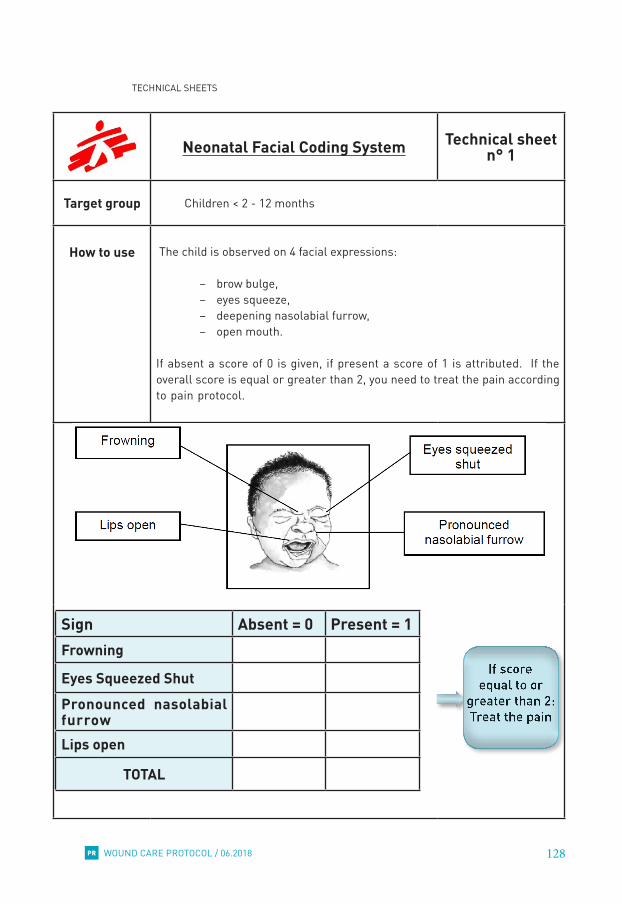
128WOUND CARE PROTOCOL / 06.2018
TECHNICAL SHEETS
PR
Neonatal Facial Coding System Technical sheet n° 1
Target group Children < 2 - 12 months
How to use The child is observed on 4 facial expressions:
– brow bulge, – eyes squeeze, – deepening nasolabial furrow, – open mouth.
If absent a score of 0 is given, if present a score of 1 is attributed. If the overall score is equal or greater than 2, you need to treat the pain according to pain protocol.
Sign Absent = 0 Present = 1
�
Frowning
Eyes Squeezed ShutPronounced nasolabial furrowLips open
TOTAL

129WOUND CARE PROTOCOL / 06.2018
TECHNICAL SHEETS
PR
EVENDOL pain scale Technical sheet n° 2
Target group Children aged 12 months to 4 years (and older if the child unable to self-evaluate the pain because of cognitive impairment or other condition).
How to useThe EVENDOL pain scale is an observational scale assessing the following five features, considering a range of scores from 0 (no pain) to 3 (strong or permanent pain) for each:
• Vocal or verbal expression• Facial expression• Movements• Postures• Interaction with the environment
The scores for each of the five features are summed, and pain severity is determined as follows (maximum score = 15):
• Score of 1–3: Mild pain• Score of 4-7: Moderate pain• Score of 8–15: Severe pain
It is important to repeat the pain assessment regularly after medication and/or analgesia are administered to assess whether the treatment was adequate.
1 At rest: observe the child from a distance, before performing any examination or procedure, under the best possible conditions of safety and
comfort (for example, with their parents, when they are playing).
2 During examination or mobilisation: assess pain during examination or mobilisation or palpation of the painful area by the nurse or doctor.
3 Reassess pain regularly after analgesic administration: wait 30 – 45 minutes (if analgesic is administered by oral or rectal route) or 10 – 15
minutes (if administered IV). Note whether the child is at rest (R) or mobilised (M).

130WOUND CARE PROTOCOL / 06.2018
TECHNICAL SHEETS
PR
Simple Verbal Scale - Visual Analogue Scale
Technical sheet n° 3
Target groupChildren > 5 yearsAdults
How to useSimple Verbal ScaleA series of 4 or 5 adjectives to describe the pain is proposed to the patient. The patient’s answer is converted into a numeric value (e.g. 0 for absent to 3 for maximal pain).
Intensity of pain No pain Mild pain Moderate
painSevere
painScoring 0 1 2 3
Write down 0 + ++ +++
Visual Analogue ScaleA 10cm line which indicates a continuum between two extremes, for example “no pain” to “worst pain”. Patients are asked to point a position on the line that best represents their level of pain. This score is then measured and recorded.
The way of questioning is important:• Correct: “Do you have pain?” Wrong: “Do you need a painkiller?”
(= suggestive)• Ask only in the actual time:
– Where/when? – How the pain does become better/worse? – During rest/during care/…?

131WOUND CARE PROTOCOL / 06.2018
TECHNICAL SHEETS
PR
Pain management for wound care Technical sheet n° 4
Pain medication
Analgesic medications for wound care should be planned well in advance: adequate time has to be allowed for the medication to work/act. Absorption after oral or subcutaneous administration is slow and a delay of 1-2 hours must be respected between giving the medication and the procedure. The procedure should be completed within 3 hours after administration.
Because of the delay in onset of analgesic action, it is not possible to ‘top-up’ analgesia once the procedure has started. Therefore adequate analgesia must be given at the outset.
For patients repeatedly undergoing procedures it may be helpful to keep a diary of the analgesia given and its effect. In this way inadequate analgesia coverage can be modified before subsequent procedures.
Level One
Paracetamol and/or NSAID (Ibuprofen or Diclofenac)
Level Two
Paracetamol and/or NSAID (Ibuprofen or Diclofenac)plus
Weak Opioid (Dihydrocodeine or Tramadol)
Level Three
Paracetamol and/or NSAID (Ibuprofen or Diclofenac)plus
Strong Opioid (Morphine)
– Start at Level One if mild to moderate procedural pain is anticipated – Record and document pain scores before, during and after dressings change
�
Pain not controlled by Level One analgesia
Pain not controlled by Level Two analgesia
�

132WOUND CARE PROTOCOL / 06.2018
TECHNICAL SHEETS
PR
– Progress to Level Two if moderate to severe pain is anticipated, including patients with moderate to severe pain during previous dressings change with Level One analgesia.
– Progress to Level Three in patients who experience moderate to severe pain during dressings changes despite Level Two Analgesia.
– In patients taking regular, prescribed analgesia check that the prescribed drugs have been taken but do not give an extra dose of the same drug.
PAEDIATRIC* Analgesic
Doses
Medication Administration DosageParacetamol oral 20 mg/kgIbuprofen oral 10 mg/kgDiclofenac oral 1 mg/kgDihydrocodeine oral 1 mg/kg (max 30 mg)Tramadol oral 1-2 mg/kgTramadol subcutaneous 1-2 mg/kgMorphine (immediate release) oral 0,5 mg/kg
Morphine subcutaneous 0,1 – 0,2 mg/kg
*Excluding neonates and Severe Acute Malnourished children
ADULT Analgesic
Doses
Medication Administration DosageParacetamol oral 1 gramIbuprofen oral 600 mgDiclofenac oral 75 mgDihydrocodeine oral 30-60 mgCodeine oral 30-60 mgTramadol oral 100 mgTramadol subcutaneous 50-100 mgMorphine (immediate release) oral 10-20 mg
Morphine subcutaneous 0,1 - 0,2 mg/kg

133WOUND CARE PROTOCOL / 06.2018
TECHNICAL SHEETS
PR
Autolytic debridement with hydrogel Technical sheet n° 5
Definition
A selective debridement by release of the patients’ activation of phagocytes and endogenous proteolytic enzymes. These enzymes will soften, break down and dissolve necrotic or sloughy tissue, enabling it to be digested by macrophages.
Indication
– Healing wounds without signs of infection that are dry or moderately moist (exudate 0, + or ++) and that contain necrosis or fibrin.
– Can be used to soften devitalized tissue prior to another type of debridement.
Contra indication
– Allergy to ingredients (e.g. preservative propylene glycol). – Highly exudative wounds. – Infected wounds and wounds with a high potential for anaerobic infections. – Bleeding wounds, fistulae or body cavities. – Patients with necrotic feet/digits because of ischemia and/or neuropathy → these wounds should be kept dry due to the risk of infection.
– Be careful in case of oncologic ulcers and patients with blood coagulation problems or anticoagulant therapy (bleeding risk when tissues are removed).
Advantage
– Little or no pain – No damage to healthy tissue – Easy to use – Can be performed by the majority of health care workers (after training in wound assessment)
Disadvantage – Time consuming (the debridement process) – Can lead to malodour – Can lead to maceration of the periwound skin (if not used correctly)
Practiced by – Nurse, nurse aid or – Medical doctor
Specific material or
product
– HYDROGEL, amorphous, 25g, tube, sterile (SDREWHYGA25A), if the wound is relatively dry. It is useless if the wound is very exudative.
Attention points
– Perform a thorough assessment of the wound bed, wound edges and periwound skin, the deeper structures and the location to define the type of debridement.
– Need for a trained health care worker with skills to perform debridement. – Do not forget to assess the pain and give pain medication when needed.

134WOUND CARE PROTOCOL / 06.2018
TECHNICAL SHEETS
PR
Sharp and surgical debridement Technical sheet n° 6
The surgical debridement falls out of the scope of the wound care protocol. The technical sheet provides general information. We refer to the surgical guidelines for more detailed instructions.
Definition
Sharp debridement: Minor surgical bedside procedure, involving cutting away non-viable tissue using a scalpel, scissors, forceps, and/or curette.Surgical debridement: Procedure performed under general and/or local anaesthesia, using various surgical instruments in a facility dedicated to surgical interventions (OT or treatment room).
Indication
Sharp and surgical debridement – Solid layer of necrotic tissue – Exudative wounds – Wound infection – When there is a clear demarcation line between viable and non-viable tissue.
– Preparation of reconstructive techniques, such as skin grafting / flap.Surgical debridement
– Very often the first treatment for traumatic highly contaminated wounds (mainly war wounds and road traffic accidents).
– If other techniques are ineffective – When the presence of devitalized tissue becomes life threatening for the patient (e.g. when large amounts of necrosis or septic tissue need to be urgently removed, for example in cases of bacterial sepsis and necrotizing fasciitis).
– Wound in close proximity of large blood vessels, tendons or nerves. – In specific areas, such as temporal areas, neck, axilla, groin and other areas where neurovascular bundles pass superficially and damage to the vitally and functionally important structures (major blood vessels, nerves and tendons) may occur.
Contra indication
Sharp and surgical debridement• Oncologic ulcer• Arterial insufficiency (e.g. arterial ulcer, diabetic foot with angiopathy)• Anticoagulant therapy/ problems with blood coagulation• In palliative wound care (the final objective is the comfort of the patient).

135WOUND CARE PROTOCOL / 06.2018
TECHNICAL SHEETS
PR
Advantage
Sharp debridement versus surgical debridement – Few resources needed – Few materials needed.
Disadvantage
Sharp debridement and surgical debridement – Risk of infection if sterility is compromised – Risk of removing healthy tissue (over-excision).
Surgical debridement – Higher need for resources ( OT , surgical staff, anaesthesia, surgical material).
Practiced by
Sharp debridement: – Nurse, nurse aid, medical doctor trained in sharp debridement, or a surgeon.
– Check the regulations, legislation and existing guidelines to define who is allowed to perform debridement.
Surgical debridement• Surgeon.

136WOUND CARE PROTOCOL / 06.2018
TECHNICAL SHEETS
PR
Wound infection with P. aeruginosa and the use of acetic acid
Technical sheet n° 7
Definition
Pseudomonas aeruginosa is a Gram-negative opportunistic pathogen with innate resistance to many antibiotics.Confirmation of P. aeruginosa in the wound infection:
– Based on bacteriological confirmation (culture) – Clinical (in absence of culture): presence of bluish-green pus/exudate, typical sweet smell.
Acetic acid
Open wounds that are infected by P. aeruginosa are difficult to treat and even more when they are hospital acquired.Acetic acid was found to have bacteriostatic activities against P. aeruginosa. Acetic acid (diluted vinegar) kills (bactericide action) and inhibits (bacteriostatic action) the growth of pseudomonas aeruginosa by lowering the acidity (pH) in the wound.Acetic acid treatment has to be kept in mind as one of the alternatives when infection is caused by multiple antibiotic resistant strains of P. aeruginosa in which there is shortage of therapeutic options and when there is no improvement with PVI 10% as antiseptic.
Side effects (possible)
– Skin reactions – Burning feeling – Pain.
Incompatible with
All other antiseptics Remark: the wound needs to be cleansed prior with PVI soap and rinsed thoroughly with NaCl 0,9%.
Dilution table
To obtain a 1% solution (STANDARD): – Take 20 ml of acetic acid 5% with a sterile syringe – Add 80 ml of distilled water.
To obtain a 0,5% solution (if 1% too painful): – Take 10 ml of acetic acid 5% with a sterile syringe – Add 90 ml of distilled water.
This solution is single use and should not be stored ⇒ Discard any rest of solution after the procedure.

137WOUND CARE PROTOCOL / 06.2018
TECHNICAL SHEETS
PR
Procedure
– Prepare acetic acid solution in a sterile (to avoid cross-contamination) kidney dish.
– Soak sterile compresses in the acetic acid solution, wring out (the compresses have to be moist but not dripping) and apply these wet compresses on the wound.
– Cover with dry compresses and fixate loosely with a bandage (to avoid the acetic acid compresses fall off the wound).
– Remove the acetic acid compresses after 15-30 minutes – Rinse the wound properly with NaCl 0,9% – Continue the wound care according to the wound care protocol.
Frequency 2 times a day.
Specific material or
product
– Acetic acid 5% (= 5º) (vinegar) (if local purchase please verify the quality of the product with your mission pharmacist).
– Distilled water (SLASWATE1B1) (= Water free of minerals and micro-organisms produced by distillation).
– Sterile kidney dish – An important amount of sterile compresses.
Attention points
– The acetic acid dressing is painful – Avoid contact of the acetic acid compresses with the surrounding skin (to prevent maceration).

138WOUND CARE PROTOCOL / 06.2018
TECHNICAL SHEETS
PR
External fixator wound care procedure
Technical sheet n° 8
Background information on pin site infection can be found in annex 3.2.
DefinitionExternal fixation is a process by which sterile pins are inserted into bone fragments through small incisions in the skin, and then held together with an external clamp or framework.
Practiced by Nurse, nurse aid, medical doctor, surgeon.
Procedure
– Keep the dressing on the pin sites – Cleanse the frame with chlorhexidine 2% in alcohol 70% or with PVI soap (remove the soap with NaCl 0,9% and gauze compresses afterwards). Use a new gauze compress for each pin. Proceed from cleanest to dirtiest.
– Remove the dressing on the pin sites – Cleanse the wound at each pin according to the general wound care protocol. Proceed from cleanest to dirtiest. Rub around the fixator with unfold sterile compress of which the opposite corners are fixated with tweezers (or sterile gloves) and stay as close as possible to the skin.
– Rinse – if necessary – and pat dry – Examine the pin sites, based on the TIME principle as well as the surrounding skin.
– Disinfection with PVI 10% if indicated: put PVI 10% on the sterile compress and leave on the wound during 1 minute; remove the compress; use a new compress for each pin.
– Cover the pin sites with a dry non-woven sterile compress.
Frequency and
disinfection
Intraoperative dressings are removed after 48–72h
Timing Disinfection IndicationFirst week post-op Daily All
Second week post op
Every 3 days Healing and no signs of infection
Daily
Signs of infectionPatient in ICUUpon medical advice in patients with increased risk
Third week post-op According to the general wound care protocol
We only use PVI 10% solution; we don’t apply PVI 10% gel on the pin sites!

139WOUND CARE PROTOCOL / 06.2018
TECHNICAL SHEETS
PR
Attention points
In the immediate post-op period slight compression with figure-of-eight bandaging between fixator wires may help to prevent an excessive skin movement around the pin and wire and to prevent the formation of a haematoma.
Specific material or
productChlorhexidine 2% in alcohol 70% or povidone iodine 7,5% soap.
Limb hygiene, showering
and bathing
As soon as pin sites are healed, showering is allowed on the day of dressing changes. This depends also on the presence of other wounds and the mobility of the patient. Laying in a bath to wash should be avoided to prevent the limb from becoming immersed in bacteria-laden water.Once showered, the frame and skin should be dried and the pin sites should be dressed according to the protocol. Limb hygiene can be maintained in between dressing changes using a clean or disposable flannel with soap and water, avoiding contact with the pin site dressings.
Management of crusts
– Maintenance: • Under normal (dry pin sites) circumstances
Rationale: these crusts provide a biological, physical barrier to the development of pin site infection.
– Removal:• When there is a collection of fluid or risk of infection• When the crust becomes detached from the pin site.
– How?• By gentle cleansing.
Surrounding skin
The surrounding skin can become rough or scaly: apply a moisturizer to the surrounding skin, avoiding direct contact with the pin/wires.
Pin site care after discharge from the hospital
Since most patients with external bone fixators are discharged from hospital before healing of the pin sites has occurred, compliance with a prescribed pin site care regimen is an important factor that is likely to influence the rate of pin site complications and infections. Patients and their families should be taught pin site care before discharge from the hospital. Material to ensure proper care could be given to the patient at discharge and in between OPD visits.As pin sites are unable to heal due to the presence of the metal work, they remain a portal for infection. The pin sites should be dressed until the fixator is removed. The patient should avoid staying in the sun, because this enhances the risk of irritation due to increased perspiration. Direct sunlight on the metal frame increases the likelihood of skin burns.

140WOUND CARE PROTOCOL / 06.2018
TECHNICAL SHEETS
PR
When to inform the
doctor
– Signs of infection. – Signs of osteomyelitis. – Necrosis around the pin sites. – Pressure wounds around the pin sites. – Symptoms of complications associated with the injury or immobility, e.g.:
• Compartment syndrome (e.g. severe oedema, marked increase in pain, inability to actively move joints, increased pain on passive movement).
• For lower limb external fixators: nerve damage - e.g. peroneal nerve: foot drop with inability to evert and dorsiflex foot.
• For upper limb external fixators: radial/median nerve at wrist: inability to adduct the thumb with numbness of the ring and small finger (ulnar), inability to extend the wrist and fingers with numbness over the dorsal (radial) and inability to extend the wrist and fingers with numbness over the dorsal (median).
• Fat embolism syndrome (common in pelvic and long-bone fractures).(symptoms include: hypoxia, restlessness, mental changes, tachycardia, tachypnea, dyspnoea, hypotension, petechial rash over upper chest and neck).
• Deep vein thrombosis with possible pulmonary embolus (symptoms include: dyspnoea, chest pain, tachypnea, apprehension, tachycardia, cyanosis, circulatory collapse).

141WOUND CARE PROTOCOL / 06.2018
TECHNICAL SHEETS
PR
Wound follow up sheet Technical sheet n° 9
WO
UN
D F
OLL
OW
UP
SHEE
T→
N.B
. Use
1 s
heet
per
wou
nd ←
……
/……
/……
…...
……
/……
/……
…...
……
/……
/……
…...
……
/……
/……
…...
……
/……
/……
…...
……
/……
/……
…...
……
/……
/……
…...
Nec
rosi
s
Fibr
in
Gra
nula
tion
Epi
thel
ializ
atio
n
Hyp
ergr
anul
atio
n
Leng
ht -
Wid
th -
Dep
th..…
. cm
; ...…
cm
; …...
cm
..…. c
m; .
..… c
m; .
.…. c
m…
... c
m; .
..… c
m; .
.…. c
m…
... c
m; .
..… c
m; .
.…. c
m..…
. cm
; ...…
cm
; …...
cm
..…. c
m; .
..… c
m; …
... c
m..…
. cm
; ...…
cm
; …...
cm
Hea
ling
- no
infe
ctio
n si
gns
Non
hea
ling
Infe
cted
Exs
udat
e (0
/ +
/ ++
/ +++
)
Odo
r
Exu
date
asp
ect
-
Ser
ous
-
San
guin
olen
t
-
Ser
osan
guin
ous
-
Pur
ulen
t
Nor
mal
-->
heal
ing
Mac
erat
ed
Und
erm
ined
(+ c
m)
Ker
atin
ized
Irrita
ted
- Itc
hy
Hea
lthy
skin
Dry
Mac
erat
ed
Ecz
ema
- Ery
them
a
Oed
ema
Birth
dat
e :
Patie
nt fi
le n
umbe
r :
Patie
nt n
ame
:
Wou
nd lo
catio
n :
M
Nam
e an
d si
gnat
ure
heal
th c
are
wor
ker
Dat
e of
dre
ssin
g ch
ange
Dat
e of
wou
nd o
rigin
:
/
/
R
isk
fact
ors
:
Peri-
wou
nd
aspe
ctT
War
d
EI
Dat
e fir
st d
ress
ing
:
/
/
Sam
plin
g da
te +
type
+ re
sults
+ a
ntib
iotic
s
Dat
e en
d of
trea
tmen
t:
/
/
⃝
defa
ulte
r
⃝
follo
w u
p tre
atm
ent i
n O
PD
⃝
refe
red
to a
noth
er w
ard
/ hea
lth s
truct
ure
⃝
heal
ed w
ound
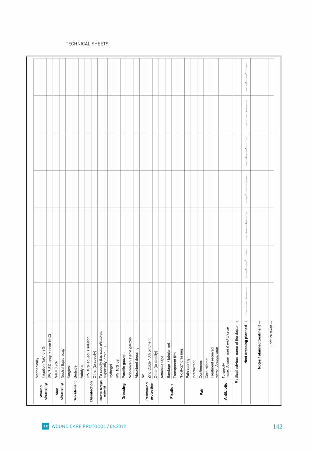
142WOUND CARE PROTOCOL / 06.2018
TECHNICAL SHEETS
PR
Mec
hani
cally
Irrig
atio
n N
aCl 0
,9%
IPV
7,5%
soa
p +
rinse
NaC
l
NaC
l 0,9
%
Neu
tral l
iqui
d so
ap
Surg
ical
Beds
ide
Auto
lytic
IPV
10%
aqu
eous
sol
utio
n
Oth
er (t
o sp
ecify
)
Rem
oval
fore
ign
mat
eria
lTo
spe
cify
(i.e
. sut
ure/
stap
les:
al
l/par
tially
; dra
in;..
.)
Hyd
roge
l
IPV
10%
gel
Para
ffin
gauz
es
Non
-wov
en s
teril
e ga
uzes
Abso
rben
t dre
ssin
g
No
Zinc
Oxi
de 1
0% o
intm
ent
Oth
er (t
o sp
ecify
)
Adhe
sive
tape
Band
age
/ tu
bula
r net
Tran
spar
ent f
ilm
"Pos
t-op"
dre
ssin
g
Pain
sco
ring
Inte
rmitt
ent
Con
tinuo
us
Car
e-re
late
dTr
eatm
ent r
ecei
ved
na
me,
dos
age,
tim
e
Antib
iotic
To s
peci
fy
nam
e, d
osag
e, s
tart
& e
nd o
f cyc
le
…...
…/…
...…
/……
…...
…...
…/…
...…
/……
…...
…...
…/…
...…
/……
…...
…...
…/…
...…
/……
…...
…...
…/…
...…
/……
…...
…...
…/…
...…
/……
…...
…...
…/…
...…
/……
…...
Pain
Pict
ure
take
n →
Med
ical
adv
ice:
nam
e of
the
doct
or →
Nex
t dre
ssin
g pl
anne
d →
Dre
ssin
g
Not
es /
plan
ned
trea
tmen
t →
Wou
nd
clea
nsin
g
Deb
ridem
ent
Periw
ound
pr
otec
tion
Fixa
tion
Skin
clea
nsin
g
Dis
infe
ctio
n

143WOUND CARE PROTOCOL / 06.2018
TECHNICAL SHEETS
PR
Wound care procedure check-list Technical sheet n° 10

144WOUND CARE PROTOCOL / 06.2018
TECHNICAL SHEETS
PR
Illustrations for T Technical sheet n° 11
Necrosis
Fibrin

145WOUND CARE PROTOCOL / 06.2018
TECHNICAL SHEETS
PR
Granulation
Epithelialization

146WOUND CARE PROTOCOL / 06.2018
TECHNICAL SHEETS
PR
Hypergranulation
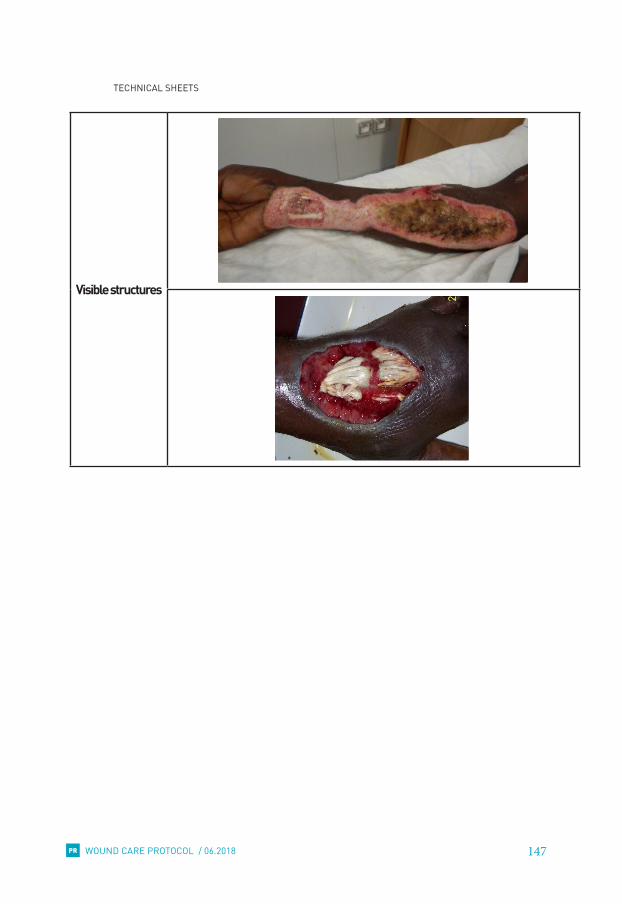
147WOUND CARE PROTOCOL / 06.2018
TECHNICAL SHEETS
PR
Visible structures

148WOUND CARE PROTOCOL / 06.2018
TECHNICAL SHEETS
PR
Illustrations for I Technical sheet n° 12
Infection

149WOUND CARE PROTOCOL / 06.2018
TECHNICAL SHEETS
PR
Illustrations for M Technical sheet n° 13
Moderately moist wound:
exudate ++
Wet wound: exudate +++

150WOUND CARE PROTOCOL / 06.2018
TECHNICAL SHEETS
PR
Illustrations for EWound edges
Technical sheet n° 14
Normal wound edges
Macerated wound edges

151WOUND CARE PROTOCOL / 06.2018
TECHNICAL SHEETS
PR
Undermined wound edges
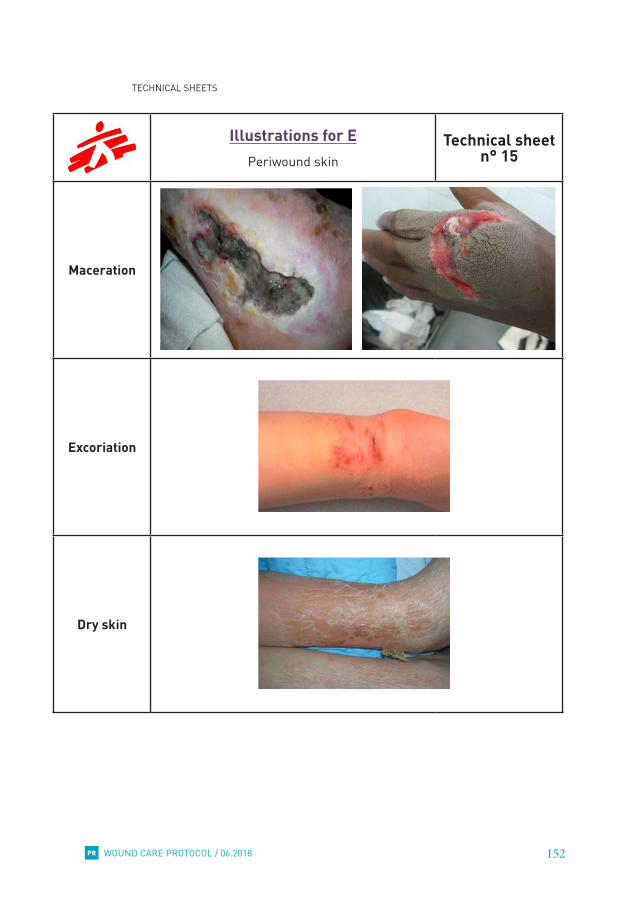
152WOUND CARE PROTOCOL / 06.2018
TECHNICAL SHEETS
PR
Illustrations for EPeriwound skin
Technical sheet n° 15
Maceration
Excoriation
Dry skin

153WOUND CARE PROTOCOL / 06.2018
TECHNICAL SHEETS
PR
Callus
Hyperkeratosis
Eczema

154WOUND CARE PROTOCOL / 06.2018PR
155WOUND CARE PROTOCOL / 06.2018
REFERENCES
PR
REFERENCESAl Ghazal P, Korber A, Klode J, Schmid EN, Buer J, Dissemond J. Evaluation of the Essen Rotary as a new technique for bacterial swabs: results of a prospective controlled clinical investigation in 50 patients with chronic leg ulcers. Int Wound J 2014; 11:44–49
Anderson, I. (2010). Key principles involved in applying and removing wound dressings. Nursing Standard, 25(10), 51-57.
Beeckman, D. et al. (2014). Digitaal Kennisbestand Wondzorg. Retrieved from http://www.digiwond.ugent.be
Biswas, A., Bharara, M, Hurst, C., Gruessner, R., Armstrong, D., & Rilo, H. (2010). Use of sugar on the healing of diabetic ulcers: a review. Journal of diabetes science and technology, 4(5), 1139-1145.
Cam R., & Korkmaz F.D. (2014).The effect of long-term care and follow-up on complications in patients with external fixators. International Journal of Nursing Practice, 20: 89–96.
Duprez, V. et al. (2014). Verpleegkundige diagnostiek en interventies: chirurgie [cursus]. Gent: Artevelde hogeschool, Bachelor in de Verpleegkunde.
Dowsett, C., & Newton, H. (2005). Wound bed preparation: TIME in practice. Wounds UK, 1(3), 58-70.
Dowsett , C., Protz, K., Drouard, M, & Harding, K.G. (2015). Triangle of wound assessment made easy. Wounds International. Retrieved from http://www.woundsinternational.com/made-easys/view/triangle-wound-assessment
European Wound Management Association (EWMA). (2006). Position Document: Management of wound infection. London: MEP Ltd.
European Wound Management Association (EWMA). (2008). Position Document: Hard-to-Heal wounds: a holistic approach. London: MEP Ltd.
European Wound Management Association (EWMA). (2015). Wound glossary. Retrieved from http://ewma.org/resources/for-patients-and-relatives/wound-glossary/
Flanagan, M.(2003). Improving accuracy of wound measurement in clinical practice. Ostomy Wound Manage. ;49(10):28-40.
Fletcher, J. (2005). Wound bed preparation and the TIME principles. Nursing Standard, 20(12), 57-65.
Fletcher, J. (2008). Differences between acute and chronic wounds and the role of wound bed preparation. Nursing Standard, 22(24), 62-68.
Fonder, M.A., Lazarus, G.S., Cowman, D.A., Aronson-Cook, B., Kholi, A.R., & Mamelak, A.J. (2008). Treating the chronic wound: a practical approach to the care of nonhealing wounds and wound care dressings. Journal of the American Academy of Dermatology, 58(2), 185-206.

156WOUND CARE PROTOCOL / 06.2018
REFERENCES
PR
Jennison, T., McNally, M., & Pandit, H. (2014). Prevention of infection in external fixator pin sites. Acta Biomaterialia, 10, 595–603.
Jones, V., Grey, J.E., & Harding, K.G. (2006). ABC of wound healing: wound dressings. British Medical Journal, 332, 777-780.
Jull, A.B., Cullum N., Dumville, J.C., Westby, M.J., Deshpande, S., & Walker, N. (2015). Honey as a topical treatment for wounds (review). The Cochrane Database of Systematic Reviews, 3. Retrieved from http://onlinelibrary.wiley.com/enhanced/doi/10.1002/14651858.CD005083.pub4#readcube-epdf:cochrane=full
Kilminster, Sue, Cottrell, David, Grant, Janet and Jolly, Brian (2007) ‘AMEE Guide No. 27: Effective educational and clinical supervision’, Medical Teacher, 29:1, 2 – 19
Hampton S (2015) Wound management 4: Accurate documentation and wound measurement. Nursing Times; 111: 48, 16-19.
Harrogate and District NHS Foundation Trust. (2015). Aseptic Technique. Community Infection Prevention and Control Guidance for Health and Social Care. Version 1.0. Retrieved from http://www.infectionpreventioncontrol.co.uk/resources/aseptic-technique/
Hess C., Wound Care Documentation, Compliance, and Revenue Checklist. Advances in Skin & Wound Care: March 2014 - Volume 27 - Issue 3 - p 144
Hiroshi N., Masashi Y., Ryoji T. et al, 2006, Mixture of sugar and povidone–iodine stimulates wound healing by activating keratinocytes and fibroblast functions.
Ktistakis, I., Guerado, E., & Giannoudis, P.V. (2015). Pin-site care: can we reduce the incidence of infections? Injury. International Journal of the Care of the Injured, 46, 35-39.
Leak, K. (2012). Ten top tips for wound debridement. Wounds International, 3(1), 21-23. Retrieved form http://www.woundsinternational.com/journal-content/view/how-to-ten-top-tips-for-wound-debridement
Leaper, D.J., Schultz, G., Carville, K., Fletcher, J., Swanson, T., & Drake, R. (2014). Extending the TIME concept: what have we learned in the past 10 years? International Wound Journal, 9(2), 1-19.
Lethaby, A., Temple, J., & Santy-Tomlinson, J. (2013). Pin site care for preventing infections associated with external bone fixators and pins (review). The Cochrane Database of Systematic Reviews, 12. Retrieved from http://onlinelibrary.wiley.com/doi/10.1002/14651858.CD004551.pub3/epdf
Madhusudhan, Vl. (2016). Efficacy of 1% acetic acid in the treatment of chronic wounds infected with Pseudomonas aeruginosa: prospective randomized controlled clinical trial. Int Wound J; 13:1129–1136.

157WOUND CARE PROTOCOL / 06.2018
REFERENCES
PR
Madoery, D., Bobbink, P. (2010). Improvement in the quality of treatment of lesions needs to be controlled and adapted. [Unpublished thesis]. Haute Ecole de Santé, Geneva
Macdonald, John M, Geyer, Mary Jo & World Health Organization. ( 2010) . Wound and lymphoedema management / edited by John M. Macdonald and Mary Jo Geyer. Geneva : World Health Organization. http://www.who.int/iris/handle/10665/44279
McGrath, A. (2011). Overcoming the challenge of overgranulation. Wounds UK, 7 (1). Retrieved from http://www.wounds-uk.com/journal-articles/overcoming-the-challenge-of-overgranulation
MSF, Providing Care for an Influx of Wounded – War Wounded/Natural Disasters - October 2007. Technical form n° 23 - Protocol for treatment of wounds, burns and fractures
MSF-OCB. (2017). Good pharmacy practice for end-user pharmacies: essential points. Matthew Nicholson & Marjolein De Bruycker.
MSF-OCB. (2015). Neonatal Care - Clinical & Therapeutic Guideline. [unpublished internal document]. Belgium: Médecins Sans Frontières – OCB.
MSF-OCB Sage Unit. (2011).Pain evaluation. [unpublished internal document]. Belgium: Médecins Sans Frontières – OCB.
MSF-OCB Sage Unit. (2015). Surgical Site Infection Data Collection [unpublished internal document]. Belgium: Médecins Sans Frontières – OCB.
MSF-OCP. (2014). Nursing guidelines. [unpublished internal document]. France: Médecins Sans Frontières – OCP.
MSF-OCP. (2012). Burn Open Wound Sampling. [unpublished internal document]. France: Médecins Sans Frontières – OCP.
Nagoba, BS., Selkar, Sp., Wadher, BJ., Gandhi, RC. (2013). Acetic acid treatment of pseudomonal wound infections--a review. J Infect Public Health. Dec;6(6):410-5.
Nicks, B.A., Ayello, E.A., Woo, K., Sibbalt, R.G., & Nitzki-George, D. (2010). Acute wound management: revisiting the approach to assessment, irrigation, and closure considerations. International Journal of Emergency Medicine, 3, 399–407.
Nicol, M., Payne, A. & Edwards, D. (2008). Aseptic Dressing Technique. Retrieved from http://www.cetl.org.uk/learning/print/aseptic-dressing-print.pdf
Phillips, P.L., Wolcott, R.D., Fletcher, J., & Schultz, G.S. (2010). Biofilms made easy. Wounds International, 1(3), 1-6. Retrieved from: http://www.woundsinternational.com/media/issues/288/files/content_8851.pdf
Principles of best practice: Minimising pain at wound dressing-related procedures. A consensus document. World Union of Wound Healing Societies. London: MEP Ltd, 2004

158WOUND CARE PROTOCOL / 06.2018
REFERENCES
PR
Slater R.A. et al. (2004). Swab cultures in diabetic foot ulcers. Diabetes UK. Diabetic Medicine, 21, 705–709
Smet S. (2013). From TIME to ITEM: a new approach in wound care decision-making. Wound care poster. Gent: Universitair Ziekenhuis.
Smith, F., Dryburgh, N., Donaldson, J., Mitchell, M. (2013). Debridement for surgical wounds. Cochrane Database of Systematic Reviews, Issue 9.
Stephen-Haynes, J. (2013). Woundcare Today Special Report: Managing overgranulation. Retrieved from http://woundcare-today.com/support/uploads/1381862137managing%20overgranulat ion.pdf
Strohal, R., Dissemond, J., Jordan O’Brien, J., Piaggesi, A., Rimdeika, R., Young, T., & Apelqvist, J. (2013). EWMA Document: Debridement. An updated overview and clarification of the principle role of debridement. Journal of Wound Care, 22 (Suppl. 1), S1-S52. Retrieved from http://ewma.org/fileadmin/user_upload/EWMA.org/Project_Portfolio/EWMA_Documents/EWMA_Debridement_Document_JWCfinal.pdf
Sue S., Tom M. (2003). Wound swabbing technique. Wound care research supplement. Vol 99 No 5. www.nursingtimes.net
Sundaramurthi S. (2017). Does Fine Needle Aspiration Microbiology Offer Any Benefit Over Wound Swab in Detecting the Causative Organisms in Surgical Site Infections?. Wounds. 29(9):255–261.
Thomas, B. (2001). Manual of Dietetic Practice. Blackwell Science. London
Timms, A., & Pugh, H. (2010). From British consensus to Russian protocol: how we justified our journey. International Journal of Orthopaedic and Trauma Nursing, 14, 109–115.
Timms, A., & Pugh, H. (2012). Pin site care: guidance and key recommendations. Nursing Standard, 27 (1), 50-55.
Topham, J., Phil, M. (1996). Sugar paste and povidone-iodine - J Wound Care, 09 364-365
Vowden, K., & Vowden, P. (2014). Wound dressings: principles and practice. Surgery, 32(9), 462-467.
Vowden, P., Vowden, K., & Carville, K. (2011). Antimicrobial dressings made easy. Wounds International, 2(1). Retrieved from http://www.woundsinternational.com/other-resources/view/antimicrobial-dressings-made-easy-1
Vuolo, J. (2010). Hypergranulation: exploring possible management options. British Journal of Nursing, 19 (6), S5-S8.
Walker, J. (2012). Pin site infection in orthopaedic external fixation devices. British Journal of Nursing, 21(3), 148-151.

159WOUND CARE PROTOCOL / 06.2018
REFERENCES
PR
WHO (2004 reformatted. 2007). Surgical Care at the District Hospital 2003 - Management of Burns. WHO/EHT/CPR
Wicker and O’ Neill (2010). Caring for the Perioperative Patient, 2nd Edition. Wiley-Blackwell.
World Union of Wound Healing Societies (WUWHS )(2004). Minimising pain at wound dressing-related procedures. A consensus document. London: MEP Ltd, 2004. Retrieved from http://www.woundsinternational.com/media/issues/79/files/content_39.pdf
World Union of Wound Healing Societies (WUWHS) (2008). Principles of best practice: wound infection in clinical practice. An international consensus. London: MEP Ltd, 2008. Retrieved from http://www.woundsinternational.com/media/issues/71/files/content_31.pdf
Wounds UK (WUK BPS) (2013). Best Practice Statement. The use of topical antimicrobial agents in wound management. London: Wounds UK, 2013 (third edition). Retrieved from http://www.woundsinternational.com/other-resources/view/best-practice-statement-the-use-of-topical-antimicrobial-agents-in-wound-management
World Wide Wounds; http://www.worldwidewounds.com/2006/september/White/Modern-Exudate-Mgt.html
Yasuhiro, N., Kiori, F., Satoshi F. (2009). Critical evaluation of cadexomer-iodine ointment and povidone-iodine sugar ointment. International Journal of Pharmaceutics 372 (2009) 85–90
Pictures
Pictures are retrieved from:
Personal pictures from: – Erika Wagner and Joel Zbinden, Haiti, 2016 – Kris Bernaerts, UZ Leuven
Centre International de Recherches, d’Enseignements et de Soins http://cires.club/phototheque/index.php
MSF pictures data base
World Union of Wound Healing Societies website
Wounds International website:Dowsett , C., Protz, K., Drouard, M, & Harding, K.G. (2015). Triangle of wound assessment made easy. Wounds International. Retrieved from http://www.woundsinternational.com/made-easys/view/triangle-wound-assessment

160WOUND CARE PROTOCOL / 06.2018PR

161WOUND CARE PROTOCOL / 06.2018
LIST OF EXTRA READINGS
PR
LIST OF EXTRA READINGSGood quanity information on a broad range of wound care documents: European Pressure Ulcer Advisory Panel (EPUAP): http://www.epuap.orgEuropean Wound Management Association (EWMA): http://www.ewma.orgJournal of wound care (JWC): https://www.magonlinelibrary.com/toc/jowc/currentNational Pressure Ulcer Advisory Panel (NPUAP): http://www.npuap.orgWound Healing Society: http://www.woundheal.orgWound UK: https://www.wounds-uk.com
Anatomy and physiology of the skin and wound healing
English – MSF-OCP (2014). Nursing guidelines. Paris: Médecins Sans Frontières, 113 – https://www.bbraun.com/en/patients/wound-healing/knowledge-series.html – http://cnx.org/contents/[email protected] , unit 2, point 5.1 and 5.3 (Open-
Stax College, Anatomy and Physiology. OpenStax CNX) – Dealey, C., (1994-2012), The care of wounds: a guide for nurses. Wiley-Blackwell – https://www.amazon.co.uk/Care-Wounds-Guide-Nurses/dp/140519569X
French – MSF-OCP (2014). Soins infirmiers, Paris: Médecins Sans Frontières, 115 – http://www.cancer.ca/fr-ca/cancer-information/cancer-type/skin-melanoma/
a n a t o m y - a n d - p h y s i o l o g y / ? r e g i o n = o n – Perrot, C. www.opmedica.fr/publications/telecharger/1 – http://www.soins-infirmiers.com/peau.php – http://guide-ide.com/peau/
Antiseptics
• Antimicrobials and non-healing wounds – http://ewma.org/fileadmin/user_upload/EWMA.org/Project_Portfolio/EWMA_Documents/
Antimicrobial.pdf
• Povidone iodine – http://www.woundsinternational.com/made-easys/view/iodine-made-easy (free account)
• Exudate management – http://www.woundsinternational.com/media/issues/82/files/content_42.pdf – http://www.woundsinternational.com/media/issues/272/files/content_8812.pdf
Biofilm
– http://www.woundsinternational.com/media/issues/288/files/content_8851.pdf
Burns
– See OCP guidelines on burns.
Debridement
French – http://ewma.org/fileadmin/user_upload/EWMA.org/Project_Portfolio/EWMA_Documents/EWMA_
Debridement_Document_JWCfinal.pdf
Italian – http://ewma.org/fileadmin/user_upload/EWMA.org/Project_Portfolio/EWMA_Documents/EWMA_
Debridement_Italian_AISLEC.pdf

162WOUND CARE PROTOCOL / 06.2018
LIST OF EXTRA READINGS
PR
Diabetic foot ulcer
– https://www.hindawi.com/journals/isrn/2013/608313/ – https://www.uptodate.com/contents/management-of-diabetic-foot-ulcers?source=search_result&-
search=wagner%20classification&selectedTitle=1~35 – https://www.ncbi.nlm.nih.gov/pubmed/26171906
Disability and buruli ulcers
• Buruli ulcer: a manual on how to prevent disability – http://www.who.int/buruli/resources/9241546816/en/
• Prevention of disability in Buruli ulcer: basic rehabilitation – http://www.who.int/buruli/resources/who_htm_ntd_idm_gbui_2008.1/en/
General principles of wound care
• Holistic approach
English – http://ewma.org/fileadmin/user_upload/EWMA.org/Position_documents_2002-2008/EWMA_08_Eng_
final.pdf
French, German, Italian (click on the language you want to view in) – http://ewma.org/resources/for-professionals/ewma-documents-and-joint-publications/
ewma-position-documents-2002-2008/
• Wound bed preparation – TIME – http://www.woundsinterna.com/media/issues/122/files/content_86.pdf – http://ewma.org/fileadmin/user_upload/EWMA.org/Position_documents_2002-2008/pos_doc_English_
final_04.pdf
French, German, Italian, Spanish (click on the language you want to view in) – http://ewma.org/resources/for-professionals/ewma-documents-and-joint-publications/
ewma-position-documents-2002-2008/
• Wound assessment – http://www.wounds-uk.com/pdf/content_10469.pdf – http://www.ncbi.nlm.nih.gov/pmc/articles/PMC1360405/ – http://www.woundsinternational.com/made-easys/view/triangle-wound-assessment (free account)
Hyperkeratosis
• Hyperkeratosis: management of the lower limb – http://www.wounds-uk.com/pdf/content_11413.pdf
Lymphedema
• Management: – http://ewma.org/fileadmin/user_upload/EWMA.org/Clinical_Guidance/ILF_Best_practice_Consensus.
• Lymphedema bandaging: – http://ewma.org/fileadmin/user_upload/EWMA.org/Position_documents_2002-2008/English_focus_
doc_05.pdf
French, German (click on the language you want to view in) – http://ewma.org/resources/for-professionals/ewma-documents-and-joint-publications/
ewma-position-documents-2002-2008/

163WOUND CARE PROTOCOL / 06.2018
LIST OF EXTRA READINGS
PR
Neglected tropical Diseases
• Ten Steps: A guide for health promotion and empowerment of people affected by Neglected Tropical Diseases – https://www.leprosy.org/ten-steps/
Pain management
• Pain at wound dressing changes – http://ewma.org/fileadmin/user_upload/EWMA.org/Position_documents_2002-2008/position_
doc2002_ENGLISH.pdf
French, German, Italian, Spanish (click on the language you want to view in) – http://ewma.org/resources/for-professionals/ewma-documents-and-joint-publications/
ewma-position-documents-2002-2008/
• Minimizing pain at wound dressing-related procedure – http://www.woundsinternational.com/media/issues/79/files/content_39.pdf
• Wound infection and pain management – http://www.woundsinternational.com/media/issues/290/files/content_8902.pdf
Venous leg ulcers
• Management of chronic venous leg ulcers: – http://www.sign.ac.uk/pdf/sign120.pdf – http://ewma.org/fileadmin/user_upload/EWMA.org/Project_Portfolio/EWMA_Documents/
Management_of_patients_with_venous_leg_ulcers_FINAL_2016.pdf
• Understanding compression therapy – http://ewma.org/fileadmin/user_upload/EWMA.org/Position_documents_2002-2008/Compression.pdf
French, German, Italian, Spanish and Japanese (click on the language you want to view in) – http://ewma.org/resources/for-professionals/ewma-documents-and-joint-publications/
ewma-position-documents-2002-2008/
Wound infection
• Criteria for wound infection – http://ewma.org/fileadmin/user_upload/EWMA.org/Position_documents_2002-2008/English_pos_doc_
final.pdf
French, German, Italian, Spanish (click on the language you want to view in) – http://ewma.org/resources/for-professionals/ewma-documents-and-joint-publications/
ewma-position-documents-2002-2008/
• Management of wound infection – http://www.woundsinternational.com/other-resources/view/best-practice-statement-the-use-of-topical-
antimicrobial-agents-in-wound-management – http://www.woundsinternational.com/media/issues/71/files/content_31.pdf
French – http://www.woundinfection-institute.com/wp-content/uploads/2014/04/wound_inf_french.pdf – http://ewma.org/fileadmin/user_upload/EWMA.org/Position_documents_2002-2008/English_pos_
doc_2006.pdf
French, German, Italian, Spanish and Japanese (click on the language you want to view in) – http://ewma.org/resources/for-professionals/ewma-documents-and-joint-publications/
ewma-position-documents-2002-2008/

164WOUND CARE PROTOCOL / 06.2018PR
WOUND CARE PROTOCOL MEDICAL DEPT - OCB / 06.2018
This WOUND CARE PROTOCOL is the product of the collaboration of many different contributors who all worked substantially in the definition of the technical content, the writing and the revision/proofreading.
AUTHORS Andrea Marelli, An Calwuaerts, Erika Wagner, Karolien D’hollander, Sofia Goudmaeker
ACKNOWLEDGMENTS MSF contributors
OCB Medical Department: Annick Antierens, Annick Filot, Ernestina Repetto, Innocent Baluhe Muke, Innocent Nyaruhirira, Isabella Panunzi, James Lee, Jeffry Edwards, Judith Kendell, Kirrily de Polnay, Lynette Dominguez, Marie Evrard, Matthew Nicholson, Michel Van Herp, Miguel Trelles, Olivier Denis, Silvia Pineda, Yves WaillyOCB field projects: Erika Wagner, Joel Zbinden and all national and international staff present in Haiti from December 2015 till November 2016 for making the realisation of the field test possible.MSF Nursing Care Contact Group: Ana De La Osada, François Xavier Daoudal, Isabelle Lessard, Vicky Treacy-Wong MSF Surgical Working Group: Caio Mario De Vecchis, Christian Heck,Mohana Amirtharajah, Patrick Herard, MSF IPC Contact Group: Chiara Martino, Miriam AliaMSF Supply: Diana Avellaneda, Marta OnievaMSF IO: Sonia De MeyereExternal experts / contributorsAnna Josepha- Halden, Anselme Derese, Carine Vandendries, Carolyn Wyndham-White, Christian Thyse, Els Jonckheere, Erik Roovers, Evelien De Cock, Evelien Touriany, Henk Hoeksema, Jan Vandeputte, Jeannette de Vries, Jeannine Spinnael, Jonathan Elias, Karin Timm, Kathryn Ruth Vowden, Luc Gryson, Peter Brooks, Peter Vowden, Rene Lessing, Veerle Luyens, Zena MooreSpecial thanks toHubert Vuagnat (Hôpitaux Universitaires de Genève), Kris Bernaerts (UZ Leuven), Steven Smet (UZ Gent)
GRAPHIC & LAYOUT: Philippe Maillot Bibop Code : L029NURM02E-P