Embed Size (px)
Citation preview
David V. Feliciano, M.D., F.A.C.S.
10 INJURIES TO THE GREAT VESSELSOF THE ABDOMEN
In patients who have injuries to the great vessels of the abdomen,the findings on physical examination generally depend on whether acontained hematoma or active hemorrhage is present.1 In the caseof contained hematomas around the vascular injury in the retro-peritoneum, the base of the mesentery, or the hepatoduodenal liga-ment, the patient often has only modest hypotension in transit or onarrival at the emergency center; the hypotension can be temporarilyreversed by the infusion of fluids and may not recur until thehematoma is opened at the time of laparotomy.This is usually thesituation when an abdominal venous injury is present. In the case ofactive intraperitoneal hemorrhage, the patient typically has signifi-cant hypotension and may have a distended abdomen on arrival.Another physical finding that is occasionally noted in associationwith an injury to the common or external iliac artery is intermittentor complete loss of a pulse in the ipsilateral femoral artery; this find-ing in a patient with a transpelvic gunshot wound is pathognomon-ic of an injury to the iliac artery.
Injuries to the great vessels of the abdomen are caused by pene-trating wounds in 90% to 95% of cases; accordingly, they are oftenaccompanied by injuries to multiple intra-abdominal organs, in-cluding those in the gastrointestinal tract.2-5The general principlesgoverning the sequencing of repairs of injuries to the great vesselsand the GI tract are outlined elsewhere [see 7;6 Operative Exposure ofAbdominal Injuries and Closure of the Abdomen].
A hematoma [see Figures 1 and 2] or hemorrhage associatedwith an injury to a great vessel of the abdomen occurs in one of
the three zones of the retroperitoneum or in the portal-retrohe-patic area of the right upper quadrant [see 7:6 Operative Exposureof Abdominal Injuries and Closure of the Abdomen].The magnitudeof injury is usually described according to the AbdominalVascular Organ Injury Scale, devised in 1992 by the AmericanAssociation for the Surgery of Trauma [see Table 1].6
Injuries in Zone 1
SUPRAMESOCOLIC
It is helpful to divide midline retroperitoneal hematomas intothose that are supramesocolic and those that are inframesocolic.1Hematoma or hemorrhage in the midline supramesocolic area ofzone 1 is cause to suspect the presence of an injury to the suprarenalaorta, the celiac axis, the proximal superior mesenteric artery, orthe proximal renal artery.
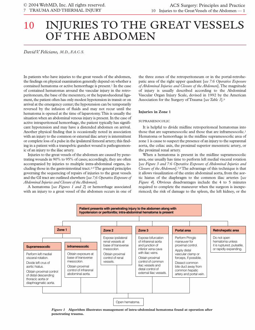
When a hematoma is present in the midline supramesocolicarea, one usually has time to perform left medial visceral rotation[see Figure 3 and 7:6 Operative Exposure of Abdominal Injuries andClosure of the Abdomen].7,8 The advantage of this technique is thatit allows visualization of the entire abdominal aorta, from the aor-tic hiatus of the diaphragm to the common iliac arteries [seeFigure 4]. Obvious disadvantages include the 4 to 5 minutesrequired to complete the maneuver when the surgeon is inexpe-rienced; the risk of damage to the spleen, the left kidney, or the
Patient presents with penetrating injury to the abdomen along with hypotension or peritonitis; intra-abdominal hematoma is present
Zone 1
Supramesocolic
Perform left medial visceral rotation.
Divide left crus of aortic hiatus.
Obtain proximal control of distal descending thoracic aorta or diaphragmatic aorta.
Inframesocolic
Obtain exposure at base of transverse mesocolon.
Obtain proximal control of infrarenal abdominal aorta.
Retrohepatic area
Do not open hematoma unless it is ruptured, pulsatile, or rapidly expanding.
Zone 2
Expose ipsilateralrenal vessels at base of transverse mesocolon.
Obtain proximal control of renalvessels.
Zone 3
Expose bifurcation of infrarenal aorta and junction of inferior vena cava with iliac veins.
Obtain proximal control of common iliac vessels and distal control of external iliac vessels.
Portal area
Perform Pringle maneuver for proximal control.
Apply distal vascular clamp or forceps, if possible.
Dissect common bile duct away from common hepatic artery and portal vein.
Open hematoma.
Figure 1 Algorithm illustrates management of intra-abdominal hematoma found at operation afterpenetrating trauma.
© 2004 WebMD, Inc. All rights reserved.7 TRAUMA AND THERMAL INJURY
ACS Surgery: Principles and Practice10 Injuries to the Great Vessels of the Abdomen — 1

© 2004 WebMD, Inc. All rights reserved.7 TRAUMA AND THERMAL INJURY
ACS Surgery: Principles and Practice10 Injuries to the Great Vessels of the Abdomen — 2
posterior left renal artery as the maneuver is performed; and theanatomic distortion that results when the left kidney and the leftrenal artery are rotated anteriorly. When the hematoma is nearthe aortic hiatus of the diaphragm, it may be advisable to leavethe left kidney in its fossa, thereby eliminating potential damage
to the structure as well as the distortion resulting from rotation.Because of the density of the celiac ganglia and nerve plexus andthe lymphatic vessels surrounding the upper abdominal aorta,this portion of the aorta is difficult to visualize even when leftmedial visceral rotation has been performed. It is frequently help-
Patient presents with blunt injury to the abdomen along with hypotension or peritonitis; intra-abdominal hematoma is present
Zone 1
Supramesocolic
Proceed as for penetrating injury[see Figure 1].
Inframesocolic
Proceed as for penetrating injury[see Figure 1].
Retrohepatic area
Proceed as for penetrating injury[see Figure 1].
Zone 2
Do not open hematomaif kidney appears normal on preoperative CT or arteriography.
If kidney does not appear normal, still do not open hematoma unless it is ruptured, pulsatile, or rapidly expanding.
Zone 3
Do not open hematoma unless it is ruptured, pulsatile, orrapidly expanding or unless ipsilateral iliacpulse is absent.
Portal area
Proceed as for penetrating injury[see Figure 1].
Figure 2 Algorithm illustrates management of intra-abdominal hematoma found at operationafter blunt trauma.
Table 1—AAST Abdominal Vascular Organ Injury Scale
Grade
I
II
III*
IV*†
V†
OIS Grade
IIIIIII
IIIIIIIIIIIIII
IIIIIIIIIIIIIII
IVIVIVIV
VV
VV
AIS-90
NSNSNSNSNSNSNS
3333333
33333
3334
33 (hepatic vein)5 (liver + veins)54
ICD-9
902.20/902.39902.27/902.32902.89902.89902.89902.81/902.82902.90
902.22902.23/902.34902.21902.24902.27/902.32902.26/902.31902.89
902.31902.41/902.42902.53/902.54902.51/902.52902.10
902.25902.24902.10902.00
902.33902.11
902.19902.00
Characteristics of Injury
Unnamed superior mesenteric artery or superior mesenteric vein branchesUnnamed inferior mesenteric artery or inferior mesenteric vein branchesPhrenic artery or veinLumbar artery or veinGonadal artery or veinOvarian artery or veinOther unnamed small arterial or venous structures requiring ligation
Right, left, or common hepatic arterySplenic artery or veinRight or left gastric arteriesGastroduodenal arteryInferior mesenteric artery, trunk, or inferior mesenteric vein, trunkPrimary named branches of mesenteric artery (e.g., ileocolic artery) or mesenteric veinOther named abdominal vessels requiring ligation or repair
Superior mesenteric vein, trunkRenal artery or veinIliac artery or veinHypogastric artery or veinVena cava, infrarenal
Superior mesenteric artery, trunkCeliac axis, properVena cava, suprarenal and infrahepaticAorta, infrarenal
Portal veinExtraparenchymal hepatic vein
Vena cava, retrohepatic or suprahepaticAorta, suprarenal and subdiaphragmatic
Note: This classification is applicable to extraparenchymal vascular injuries. If the vessel injury is within 2 cm of the parenchyma of a specific organ, one should refer to the injuryscale for that organ.*Increase grade by I if there are multiple injuries involving > 50% of vessel circumference.†Reduce grade by I if laceration is < 25% of vessel circumference.AAST—American Association for the Surgery of Trauma—AIS—Abbreviated Injury Scale—ICD—International Classification of Diseases
© 2004 WebMD, Inc. All rights reserved.7 TRAUMA AND THERMAL INJURY
ACS Surgery: Principles and Practice10 Injuries to the Great Vessels of the Abdomen — 3
ful to transect the left crus of the aortic hiatus in the diaphragmat the 2 o’clock position to allow exposure of the distal descend-ing thoracic aorta above the hiatus.Visualization of this portion ofthe vessel is much easier to achieve than visualization of thediaphragmatic or visceral abdominal aorta below, and an aorticcross-clamp can be applied much more quickly at this level.
Active hemorrhage from the midline supramesocolic area is con-trolled temporarily by packing with laparotomy pads or using anaortic compression device [see Figure 5].9,10 A definitive approach isto divide the lesser omentum manually, retract the stomach andesophagus to the left, and manually dissect in the area just below theaortic hiatus of the diaphragm to obtain rapid exposure of thesupraceliac abdominal aorta.11 An aortic cross-clamp can then beapplied. Distal control of the upper abdominal aorta is difficult toobtain because of the presence of the anteriorly located celiac axisand superior mesenteric artery. In young trauma patients who are
otherwise healthy, ligation and division of the celiac axis allow easierapplication of the distal aortic clamp and better exposure of thesupraceliac area for subsequent vascular repair.12
Small penetrating wounds to the supraceliac abdominal aortaare repaired with a continuous 3-0 or 4-0 polypropylene suture.If two small perforations are adjacent to each other, they can beconnected and the defect closed in a transverse fashion. If closureof a perforation would result in significant narrowing of the aortaor if a portion of the aortic wall is missing, patch aortoplasty withpolytetrafluoroethylene (PTFE) is indicated. On rare occasions,in patients with extensive injuries to the diaphragmatic or supra-celiac aorta, resection of the area of injury and insertion of a vas-cular conduit are indicated. Even though many of these patientshave associated gastric, enteric, or colonic injuries, the most ap-propriate prosthesis with such a life-threatening injury is a 12 mmor 14 mm Dacron or PTFE graft [see Figure 6].13 Provided thatvigorous intraoperative irrigation is performed after repair of GItract perforations, that proper graft coverage is ensured, and thatperioperative antibiotics are appropriately employed, it is extraor-dinarily rare for a prosthesis inserted in the healthy aorta of ayoung trauma patient to become infected.
The aortic prosthesis is sewn in place with a continuous 3-0 or4-0 polypropylene suture. Both ends of the aorta are flushedbefore the distal anastomosis is completed, and the distal aorticcross-clamp is removed before the final knot is tied to eliminateair from the system.The proximal aortic cross-clamp is removedvery slowly as the anesthesiologist rapidly infuses fluids and intra-
Figure 3 Left medial visceral rotation is performed by means ofsharp and blunt dissection with elevation of the left colon, the leftkidney, the spleen, the tail of the pancreas, and the gastric fundus.
Figure 4 Shown is an autopsy view of the supraceliac aorta andthe celiac axis, the proximal superior mesenteric artery, and themedially rotated left renal artery after removal of lymphatic andnerve tissue.
Figure 5 An aortic compression device is used to control hem-orrhage from the visceral portion of the abdominal aorta.
© 2004 WebMD, Inc. All rights reserved.7 TRAUMA AND THERMAL INJURY
ACS Surgery: Principles and Practice10 Injuries to the Great Vessels of the Abdomen — 4
venous bicarbonate to reverse so-called washout acidosis from thepreviously ischemic lower extremities. The retroperitoneum isthen irrigated with an antibiotic solution and closed over the graftin a watertight fashion with an absorbable suture.
Cross-clamping of the diaphragmatic or supraceliac abdominalaorta in a patient with hemorrhagic shock results in severeischemia of the legs. Restoration of flow through the repairedabdominal aorta may then cause a reperfusion injury in additionto the ischemic edema that develops in the muscle compartmentsbelow the knee. In a patient who is hemodynamically stable afterrepair of the suprarenal abdominal aorta and other injuries, mea-surement of compartmental pressures below the knees should beperformed before the patient is moved from the operating room.Pressures in the range of 30 to 35 mm Hg are likely to rise in theintensive care unit; accordingly, at many centers, bilateral below-the-knee two-incision four-compartment fasciotomies would beperformed in this situation.
The survival rate in patients with injuries to the suprarenalabdominal aorta had been 30% to 35% but was lower than 10%in one 2001 review.4,13
Injuries to branches of the celiac axis are often difficult to re-pair because of the amount of dissection required to remove thedense neural and lymphatic tissue in this area. Because most pa-tients have excellent collateral flow in the upper abdomen, majorinjuries to either the left gastric or the proximal splenic arterygenerally should be ligated. Because the common hepatic arterymay have a larger diameter than either of these two arteries, aninjury to this vessel can occasionally be repaired by means of lat-eral arteriorrhaphy, an end-to-end anastomosis, or the insertionof a saphenous vein graft. One should not worry about ligatingthe common hepatic artery proximal to the origin of the gastro-duodenal artery: there is extensive collateral flow to the liverfrom the midgut.When the entire celiac axis is injured, it is bestto ligate all three vessels and forgo any attempt at repair.
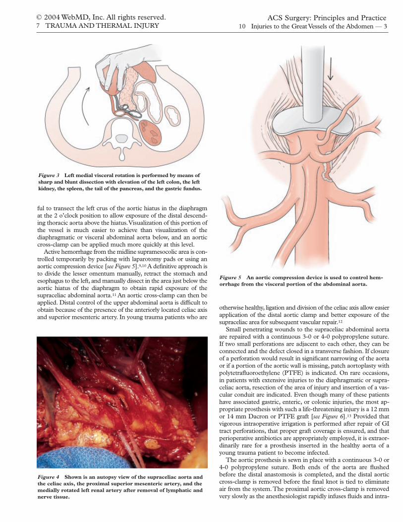
Injuries to the superior mesenteric artery are managed accordingto the anatomic level of the perforation or thrombosis.14 On rare oc-casions, in patients with injuries beneath the neck of the pancreas,one may have to transect the pancreas to obtain proximal control.
Another option is to perform left medial visceral rotation (seeabove) and apply a clamp directly to the origin of the superiormesenteric artery. Injuries to the superior mesenteric artery in thisarea or just beyond the base of the mesocolon are often associatedwith injuries to the pancreas.The potential for a postoperative leakfrom the injured pancreas near the arterial repair has led numerousauthors to suggest that any extensive injury to the artery at this loca-tion should be ligated [see Figure 7].
Because of the intense vasoconstriction of the distal superiormesenteric artery in patients who have sustained exsanguinatinghemorrhage from more proximal injuries treated with ligation,the collateral flow from the foregut and hindgut is often inade-quate to maintain the viability of the organs in the distal midgut,especially the cecum and the ascending colon. Therefore, it issafest to place a saphenous vein or PTFE graft on the distalinfrarenal aorta, away from the pancreatic injury and any otherupper abdominal injuries.15 Such a graft can be tailored to reachthe side or the anterior aspect of the superior mesenteric artery,or it can be attached to the transected distal superior mesentericartery in an end-to-end fashion without significant tension [seeFigure 8]. Soft tissue must be approximated over the aortic sutureline of the graft to prevent the development of an aortoenteric fis-tula in the postoperative period.
In patients with severe shock from exsanguination caused by acomplex injury to the superior mesenteric artery, damage-controllaparotomy is indicated [see Damage-Control Laparotomy, below]:the injured area should be resected and a temporary intraluminalArgyle, Javid, or Pruitt-Inahara shunt inserted to maintain flow tothe midgut during resuscitation in the surgical intensive care unit.16
Figure 6 A 22-year-old man with a gunshot wound to the rightupper quadrant had injuries to the prepyloric area of the stom-ach and to the supraceliac abdominal aorta. The aortic injury wasmanaged by means of segmental resection and replacement witha 16 mm polytetrafluoroethylene (PTFE) graft. The patient wenthome 46 days after injury.
Figure 7 An 18-year-old man experienced a gunshot wound tothe head of the pancreas and the proximal superior mesentericartery. A Whipple procedure was performed, and a 6 mm PTFEgraft was placed in the artery. The artery-graft suture linedehisced secondary to a pancreatic leak on day 30 after injury,and the patient died on day 42.

© 2004 WebMD, Inc. All rights reserved.7 TRAUMA AND THERMAL INJURY
ACS Surgery: Principles and Practice10 Injuries to the Great Vessels of the Abdomen — 5
When ligation is indicated for more distal injuries to the superiormesenteric artery, segments of the ileum or even the right colonmay have to be resected because of ischemia.
The survival rate in patients with penetrating injuries to thesuperior mesenteric artery is approximately 50% to 55% overall[see Table 2] but only 20% to 25% when any form of repair morecomplex than lateral arteriorrhaphy is necessary.1-4,15,17
An injury to the proximal renal artery may also be presentunder a supramesocolic hematoma or bleeding area.When activehemorrhage is present, control of the supraceliac abdominal aortain or just below the aortic hiatus must be obtained.When only ahematoma or a known thrombosis of the proximal renal artery ispresent, proximal vascular control can be obtained by elevatingthe transverse mesocolon and dissecting the vessel from the later-al aspect of the abdominal aorta. Options for repair of either theproximal or the distal renal artery are described elsewhere [seeInjuries in Zone 2, below].
The superior mesenteric vein is the other great vessel of theabdomen that may be injured in the supramesocolic or retrome-socolic area of the midline retroperitoneum. Because of the over-lying pancreas, the proximity of the uncinate process, and theclose association of this vessel with the superior mesenteric artery,repair of the superior mesenteric vein is quite difficult. As withinjuries to the superior mesenteric artery (see above), one mayhave to transect the neck of the pancreas between noncrushingvascular or intestinal clamps to gain access to a perforation of thesuperior mesenteric vein. An injury to this vein below the inferi-or border of the pancreas can be managed by compressing itmanually between one’s fingers as an assistant places a continu-ous 5-0 polypropylene suture to complete the repair.When a pen-etrating injury to the vein has a posterior component, one mustligate multiple collateral vessels entering the vein in this area toachieve proper visualization.
There is excellent evidence that young trauma patients tolerateligation of the superior mesenteric vein well when vigorous postop-erative fluid resuscitation is performed to reverse the peripheral hy-povolemia that results from splanchnic hypervolemia.18,19Typically,ligation is followed almost immediately by swelling of the midgutand discoloration suggestive of impending ischemia. In such cases,temporary coverage of the midgut with a silo, followed by early re-operation, may be necessary to reassure the operating surgeon thatthe ischemia has not become permanent.
The survival rate in patients with injuries to the superiormesenteric vein ranges from 36% to 71%, depending on whetherother vascular injuries are present [see Table 3].1-4,18
INFRAMESOCOLIC
The lower area of the midline retroperitoneum in zone 1 isknown as the midline inframesocolic area. Injuries to either theinfrarenal abdominal aorta or the inferior vena cava occur in thisarea.
An injury to the inframesocolic abdominal aorta that is undera hematoma is controlled by performing the same maneuversused to gain proximal control of an infrarenal abdominal aorticaneurysm. The infrarenal abdominal aorta is exposed by pullingthe transverse mesocolon up toward the patient’s head, eviscerat-ing the small bowel to the right side of the abdomen, and open-ing the midline retroperitoneum until the left renal vein is ex-posed. A proximal aortic cross-clamp is then placed immediatelyinferior to this vessel [see Figure 9].When the entire inframesoco-lic area is distorted by the presence of a large pulsatile hematoma,the inexperienced trauma surgeon should remember that the holein the infrarenal abdominal aorta is under the highest point of thehematoma (the so-called Mt. Everest phenomenon).When thereis active hemorrhage from this area, rapid proximal control isobtained in the same fashion or, if necessary because of the need
Figure 8 (a) When complex grafting procedures to the superior mesenteric artery are necessary,it may be dangerous to place the proximal suture line near an associated pancreatic injury. (b)The proximal suture line should be on the lower aorta, away from the upper abdominal injuries,and should be covered with retroperitoneal tissue.
a b

to apply compression, by dividing the lesser omentum and apply-ing the cross-clamp just below the aortic hiatus of the diaphragm.Distal control of the infrarenal abdominal aorta is obtained bydividing the midline retroperitoneum down to the aortic bifurca-tion, taking care to avoid the origin of the inferior mesentericartery on the left side.
Injuries to the infrarenal aorta are repaired by means of lateralaortorrhaphy, patch aortoplasty, an end-to-end anastomosis, orinsertion of a Dacron or PTFE graft. Much as with injuries to thesuprarenal abdominal aorta in young trauma patients, it is rarelypossible to place a tube graft larger than 12 or 14 mm. Becausethe retroperitoneal tissue is often thin at this location in youngpatients, an important adjunctive measure after the aortic repairis to mobilize the gastrocolic omentum, flip it into the lesser sacsuperiorly, and then bring it down over the infrarenal aortic graftthrough a hole in the left transverse mesocolon. An alternative isto mobilize the gastrocolic omentum away from the right side ofthe colon and then swing the mobilized tissue into the left lateralgutter just below the ligament of Treitz to cover the graft. Witheither technique, it is mandatory to suture the viable omentalpedicle superior to the aortic suture line to prevent a postopera-tive aortoduodenal fistula.20,21
The survival rate in patients with injuries to the infrarenalabdominal aorta had been approximately 45% but was 34% in a2001 review [see Table 2].1-4
Injury to the inferior vena cava below the liver should be sus-pected when the aorta is found to be intact underneath aninframesocolic hematoma, when such a hematoma appears to bemore extensive on the right side of the abdomen than on the left,or when there is active hemorrhage coming through the base ofthe mesentery of the ascending colon or the hepatic flexure. It iscertainly possible to visualize the inferior vena cava through themidline retroperitoneal exposure just described (see above); how-ever, most surgeons are more comfortable with visualizing thevessel by mobilizing the right half of the colon and the C loop ofthe duodenum.1 With this right medial visceral rotation maneu-ver, the right kidney is left in situ unless there is an associatedinjury to the posterior aspect of the right renal vein, to thesuprarenal vena cava, or to the right kidney itself. Right medialvisceral rotation, in conjunction with the Kocher maneuver, per-mits visualization of the entire vena caval system from the con-fluence of the iliac veins to the suprarenal vena cava below theliver [see 7:6 Operative Exposure of Abdominal Injuries and Closureof the Abdomen]. Local exposure of the iliac vein–vena cava junc-tion in the lower abdomen and of the renal vein–vena cava junc-tion in the upper abdomen is appropriate before completion ofright medial visceral rotation.This measure allows rapid applica-tion of proximal and distal vascular clamps on the inferior venacava in the event that exsanguinating hemorrhage results whenthe caval injury is exposed.
© 2004 WebMD, Inc. All rights reserved.7 TRAUMA AND THERMAL INJURY
ACS Surgery: Principles and Practice10 Injuries to the Great Vessels of the Abdomen — 6
Table 2—Survival after Injuries to Arteries in the Abdomen
Injured Artery
Abdominal aorta as a whole
Pararenal to diaphragm
Infrarenal
Superior mesenteric artery
Renal artery
Iliac artery
Common
External
With Other Arterial Injury
17.6% (3/17)
—
—
28.6% (2/7)
33.3% (2/6)
—
—
Davis et al3 (2001)*
39.1% (25/64)
—
—
53.3% (8/15)
56.2% (9/16)
55.5% (5/9)
65.2% (30/46)
Isolated Injury
21.7% (10/46)
—
—
52.4% (11/21)
62.5% (5/8)
—
—
*Excludes patients who exsanguinated before repair or ligation.
Asensio et al2 (2000)Tyburski et al4 (2001)
21.1% (15/71)
8.3% (3/36)
34.2% (12/35)
48.8% (20/41)
73.7% (14/19)
44.7% (17/38)
62.5% (20/32)
Table 3—Survival after Injuries to Veins in the Abdomen
Injured Vein
Inferior vena cava as a whole
Pararenal to diaphragm
Infrarenal
Superior mesenteric vein
Renal vein
Iliac vein (all)
Common
External
With Other Venous Injury
22.2% (8/36)†
—
—
35.7% (5/14)
44.1% (15/34)
33.3% (5/15)
—
—
Davis et al3 (2001)*
56% (47/84)
—
—
71.4% (15/21)
70% (21/30)
—
81% (17/21)
74.5% (41/55)
Isolated Injury
29.3% (12/41)†
—
—
47.4% (9/19)
—
62.2% (23/37)
—
—
*Excludes patients who exsanguinated before repair or ligation.†Excludes retrohepatic vena cava.
Asensio et al2 (2000)Tyburski et al4 (2001)
43% (61/142)
40.3% (31/77)
46.2% (30/65)
56.3% (18/32)
68.8% (22/32)
—
49% (24/49)
66.7% (16/24)

For proper exposure of a hole in a large vein such as the inferiorvena cava, the loose retroperitoneal fatty tissue must be dissectedaway from the wall of the vessel. Active hemorrhage coming fromthe anterior surface of the inferior vena cava is best controlled byapplying a Satinsky vascular clamp. If it is difficult to apply thisclamp, one should try grasping the area of the perforation with apair of vascular forceps or several Judd-Allis clamps; this step mayfacilitate safe application of the Satinsky clamp.22When the perfora-tion in the inferior vena cava is more lateral or posterior, it is oftenhelpful to compress the vessel proximally and distally around theperforation, using gauze sponges placed in straight sponge sticks.On occasion, an extensive injury to the inferior vena cava can becontrolled only by completely occluding the entire inferior venacava with large DeBakey aortic cross-clamps.This maneuver inter-rupts much of the venous return to the right side of the heart and ispoorly tolerated by hypotensive patients unless the infrarenal ab-dominal aorta is cross-clamped simultaneously.
There are two anatomic areas in which vascular control of aninjury to the inferior vena cava below the liver is difficult toobtain: (1) the confluence of the common iliac veins and (2) thejunction of the renal veins with the inferior vena cava. One inter-esting approach to an injury to the inferior vena cava at the con-fluence of the iliac veins is temporary division of the overlyingright common iliac artery, coupled with mobilization of the aor-tic bifurcation to the patient’s left.23 This approach yields a betterview of the common iliac veins and the proximal inferior venacava and makes repair considerably easier than it would be if theaortic bifurcation were left in place. Once the vein is repaired, theright common iliac artery is reconstituted via an end-to-endanastomosis. The usual approach to injuries to the inferior venacava at its junction with the renal veins involves clamp or sponge-stick compression of the infrarenal and suprarenal vena cava, as
well as control of both renal veins with Silastic loops to facilitatethe direct application of angled vascular clamps. As noted, medi-al mobilization of the right kidney may permit the application ofa partial occlusion clamp across the inferior vena cava at its junc-tion with the right renal vein as an alternative approach to aninjury in this area. Another useful technique for controlling hem-orrhage from the inferior vena cava at any location is to insert a5 ml or 30 ml Foley balloon catheter into the caval laceration andthen inflate it in the lumen.24, 25 Once the bleeding is controlled,vascular clamps are positioned around the perforation, and theballoon catheter is removed before repair of the vessel.
Anterior perforations of the inferior vena cava are managed bymeans of transverse repair with a continuous 4-0 or 5-0 poly-propylene suture. Much has been written about visualizing pos-terior perforations by extending anterior perforations, but in myexperience, opportunities to apply this approach have been rare.It is often easier to roll the vena cava to one side to complete acontinuous suture repair of a posterior perforation. When bothanterior and posterior perforations have been repaired, there isusually a significant degree of narrowing of the inferior vena cava,which may lead to slow postoperative occlusion. If the patient’scondition is unstable and a coagulopathy has developed, no fur-ther attempt should be made to revise the repair. If the patient isstable, there may be some justification for applying a large PTFEpatch to prevent this postoperative occlusion [see Figure 10].
Ligation of the infrarenal inferior vena cava is appropriate foryoung patients who are exsanguinating and in whom a complexrepair of the vessel would be necessary. After the damage-controlabdominal procedure has been completed and a silo has beenused to cover the midgut, it is, once again, worthwhile to measurethe compartmental pressures below the knees before the patientis moved from the OR. Below-the-knee two-incision four-com-partment fasciotomies are performed when pressures exceed 30to 35 mm Hg. Three-compartment fasciotomies in the thighshave proved necessary in some surviving patients after caval liga-
© 2004 WebMD, Inc. All rights reserved.7 TRAUMA AND THERMAL INJURY
ACS Surgery: Principles and Practice10 Injuries to the Great Vessels of the Abdomen — 7
Figure 9 Shown is a gunshot wound to the infrarenal abdominalaorta viewed through standard inframesocolic exposure. Patient’shead is at the bottom of the photograph.
Figure 10 Shown is PTFE patch repair of an injury to theinfrarenal inferior vena cava.

tion. Patients who have undergone ligation of the infrarenal infe-rior vena cava require vigorous resuscitation with crystalloid solu-tions in the postoperative period; in addition, both lower extrem-ities should be wrapped with elastic compression wraps and ele-vated for 5 to 7 days after operation. Patients who have someresidual edema during the later stages of hospitalization despitethe elastic compression wraps should be fitted with full-lengthcustom-made support hose. Ligation of the suprarenal inferiorvena cava is occasionally necessary when the patient has an exten-sive injury at this location and appears to be in an irreversibleshock state during operation.26 If the patient’s condition improvesduring a brief period of resuscitation in the SICU, reoperationand reconstruction with an externally supported PTFE graft areusually necessary to prevent renal failure.
The survival rate in patients with injuries to the inferior venacava depends on the location of the injury; in the past, it rangedfrom 60% for the suprarenal vena cava to 78% for the infrarenalvena cava but decreased to approximately 33% to 56% if injuriesto the retrohepatic vena cava were included. Current studies indi-cate survival rates of 22% to 56% for inferior vena cava injuriestaken as a whole.1-3,26-28 A 2001 review reported survival rates of46.1% for the infrarenal inferior vena cava and 40.3% for moresuperior injuries.4
Injuries in Zone 2
Hematoma or hemorrhage in zone 2 is cause to suspect the pres-ence of injury to the renal artery, the renal vein, or the kidney.
In patients who have sustained blunt abdominal trauma but inwhom preoperative intravenous pyelography, renal arteriography,or abdominal CT confirms that a reasonably intact kidney is pres-ent, there is no justification for opening a perirenal hematoma ifone is found at a subsequent operation [see Figure 11].29
In highly selected stable patients with penetrating wounds to theflank, there are some data to justify performing preoperative CT.On occasion, documentation of an isolated minor renal injury in theabsence of peritoneal findings on physical examination makes itpossible to manage such patients nonoperatively.29 In all other pa-tients with penetrating wounds, when a perirenal hematoma isfound during initial exploration, the hematoma should be unroofedand the wound tract explored. If the hematoma is not rapidly ex-panding and there is no active hemorrhage from the perirenal area,
one may control the ipsilateral renal artery with a Silastic loop in themidline of the retroperitoneum at the base of the mesocolon [seeFigure 12].30,31 Control of the left renal vein can be obtained at thesame location; however, control of the proximal right renal vein re-quires mobilization of the C loop of the duodenum and dissectionof the vena cava at its junction with this vessel.
If there is active hemorrhage from the kidney through Gerota’sfascia or from the retroperitoneum overlying the renal vessels, nocentral renal vascular control is necessary. In such a situation, theretroperitoneum lateral to the injured kidney should be opened,and the kidney should be manually elevated directly into theabdominal incision. A large vascular clamp should then beapplied directly to the hilar vessels of the elevated kidney to con-trol any further bleeding until a decision is reached on repair ver-sus nephrectomy.
Occasionally, a small perforation of the renal artery can berepaired by lateral arteriorrhaphy or resection with an end-to-endanastomosis.32 Interposition grafting and replacement of therenal artery with either the hepatic artery (on the right) or thesplenic artery (on the left) have been used on rare occasions, butsuch approaches ordinarily are not indicated unless the injuredkidney is the only one the patient has. In patients who have sus-tained multiple intra-abdominal injuries from penetratingwounds or have undergone a long period of ischemia while otherinjuries were being repaired, nephrectomy is the appropriatechoice for a major renovascular injury, provided that intraopera-tive palpation has confirmed the presence of a normal contralat-eral kidney.
The role of renal revascularization in patients who have intimaltears in the renal arteries as a result of deceleration-type traumaremains controversial. If a circumferential intimal tear is noted onpreoperative arteriography but flow to the kidney is preserved, thedecision whether to repair the artery depends on whether laparot-omy is necessary for other injuries and whether the opportunityfor anticoagulation is available. If there are no other significantinjuries and flow to the kidney is preserved despite the presenceof an intimal tear, anticoagulation and a repeat isotope renogramwithin the first several days may be justified. An alternativeapproach involves insertion of an endovascular stent in the renal
© 2004 WebMD, Inc. All rights reserved.7 TRAUMA AND THERMAL INJURY
ACS Surgery: Principles and Practice10 Injuries to the Great Vessels of the Abdomen — 8
Figure 11 A right perirenal hematoma was not opened at opera-tion, because preoperative abdominal CT documented a reason-ably intact kidney.
Figure 12 Midline looping of respective renal vessels is per-formed before entry into any perirenal hematoma.

artery, followed by a period of anticoagulation.33 If occlusion ofthe proximal renal artery from blunt deceleration-type trauma isdocumented, the critical factor for renal salvage is the time fromocclusion to revascularization. Renal artery occlusion from decel-eration-type trauma that is detected within 6 hours of injury maybe treated with immediate operation, resection of the area of inti-mal damage, and an end-to-end anastomosis performed by anexperienced vascular trauma team. Given proper exposure andmedial mobilization of the kidney, this operation is not technical-ly difficult in a young trauma patient whose renal artery is other-wise normal. In a 1998 review, fewer than 20% of kidneys revas-cularized in this manner regained any significant degree of func-tion.34 Hypertension develops in 40% to 45% of patients whoundergo observation only after thrombosis is detected.
The survival rate in patients with isolated injuries to the renalarteries ranges from 56% to 74% [see Table 2].1,3,4
Many patients with penetrating wounds to the renal veins arequite stable as a result of the retroperitoneal tamponade describedearlier (see above). Once vascular control is obtained with the directapplication of clamps, lateral venorrhaphy is the preferred tech-
nique of repair. If ligation of the right renal vein is necessary to con-trol hemorrhage, nephrectomy should be performed, either at initiallaparotomy or at reoperation after a damage-control laparotomy;the medial left renal vein may be ligated as long as the left adrenaland gonadal veins are intact. It should be noted, however, that insome series, more postoperative renal complications were notedwhen this maneuver was used on the left side.35
The survival rate in patients with isolated injuries to the renalveins is approximately 70% [see Table 3].1,3,4
Injuries in Zone 3
Hematoma or hemorrhage in either lateral pelvic area is sug-gestive of injury to the iliac artery or the iliac vein. When lateralpelvic hematoma or hemorrhage is noted after penetrating trau-ma, compression with a laparotomy pad or the fingers should bemaintained as proximal and distal vascular control is obtained.The proximal common iliac arteries are exposed by evisceratingthe small bowel to the right and dividing the midline retroperi-toneum over the aortic bifurcation. In young trauma patients, thecommon iliac artery usually is not adherent to the common iliacvein, and Silastic loops can be passed rapidly around these ves-sels to provide proximal vascular control. Distal vascular controlis most easily obtained where the external iliac artery and veincome out of the pelvis proximal to the inguinal ligament. Evenwith proximal and distal control of the common or the externaliliac artery and vein, there is often continued back-bleeding fromthe internal iliac artery. Such bleeding is controlled by elevatingthe Silastic loops on the proximal and distal iliac artery and theneither clamping or looping the internal iliac artery, which is theonly major branch vessel that descends into the pelvis.
For transpelvic bilateral iliac vascular injuries resulting from apenetrating wound, a technique of total pelvic vascular isolationhas been described. Proximally, the abdominal aorta and theinferior vena cava are cross-clamped just above their bifurcations,and distally, both the external iliac artery and the external iliacvein are cross-clamped, with one clamp on each side of the distalpelvis. Back-bleeding from the internal iliac vessels is minimalwith this approach.
Ligation of either the common or the external iliac artery in a hy-potensive trauma patient leads to a 40% to 50% amputation rate inthe postoperative period; consequently, injuries to these vesselsshould be repaired if at all possible.The standard options for re-pair—lateral arteriorrhaphy, completion of a partial transectionwith an end-to-end anastomosis, and resection of the injured areawith insertion of a conduit—are feasible in most situations [see Fig-ure 13].36 On rare occasions, it may be preferable either to mobilizethe ipsilateral internal iliac artery to serve as a replacement for theexternal iliac artery or to transpose one iliac artery to the side of thecontralateral iliac artery.37 When a patient is in severe shock fromexsanguination caused by a complex injury to the common or theexternal iliac artery, damage control laparotomy [see Damage Con-trol Laparotomy, below] is indicated.38 The injured area should beresected and a temporary intraluminal Argyle, Javid, or Pruitt-Ina-hara shunt inserted to maintain flow to the ipsilateral lower extrem-ity during resuscitation in the SICU.1
One unique problem associated with repair of the common or theexternal iliac artery is the choice of technique when significant en-teric or fecal contamination is present in the pelvis. In such cases,there is a substantial risk of postoperative pelvic cellulitis, a pelvic ab-scess, or both, which may lead to blowout of any type of repair.When extensive contamination is present, it is appropriate to divide
© 2004 WebMD, Inc. All rights reserved.7 TRAUMA AND THERMAL INJURY
ACS Surgery: Principles and Practice10 Injuries to the Great Vessels of the Abdomen — 9
Figure 13 A 24-year-old man experienced a gunshot wound tothe left external iliac artery and vein. The arterial injury wasrepaired with segmental resection and insertion of an 8 mmPTFE graft; the venous injury was repaired with segmental resec-tion and an end-to-end anastomosis.
Figure 14 Failure to properly dissect out the structures in theporta hepatis after a penetrating wound led to the creation of aniatrogenic hepatic artery–portal vein fistula, which was correctedafter the arrival of the attending surgeon.

the common or external iliac artery above the level of injury, closethe injury with a double row of continuous 4-0 or 5-0 polypropylenesutures, and bury the stump underneath uninjured retroperitoneum.If a stable patient has obvious ischemia of the ipsilateral lower ex-tremity at the completion of this proximal ligation, one may performan extra-anatomic femorofemoral crossover bypass with an 8 mmexternally supported PTFE graft to restore arterial flow to the ex-tremity.1 If the patient is unstable, one should take several minutes toperform an ipsilateral four-compartment below-knee fasciotomy;this step will counteract the ischemic edema that inevitably leads to acompartment syndrome and compromises the early survival of theleg.After adequate resuscitation in the SICU, the patient should bereturned to the OR for the femorofemoral graft within 4 to 6 hours.
Injuries to the internal iliac arteries are usually ligated even ifthey occur bilaterally, because young trauma patients typicallyhave extensive collateral flow through the pelvis.
The survival rate in patients with isolated injuries to the exter-nal iliac artery exceeds 80% when tamponade is present. If theinjury is large and free bleeding has occurred preoperatively, how-ever, the survival rate is only 45%. Current studies report overallsurvival rates of approximately 45% to 55% for injuries to thecommon iliac artery and 62% to 65% for injuries to the externaliliac artery [see Table 2].1,3,4,39
Hemorrhage from injuries to the iliac veins can usually be con-trolled by means of compression with either sponge sticks or thefingers. As noted, division of the right common iliac artery maybe necessary for proper visualization of an injury to the right com-mon iliac vein. Similarly, ligation and division of the internal iliacartery on the side of the pelvis yield improved exposure of aninjury to an ipsilateral internal iliac vein.40
Injuries to the common or the external iliac vein are best treat-ed by means of lateral venorrhaphy with continuous 4-0 or 5-0polypropylene sutures. Significant narrowing often results, and anumber of reports have demonstrated occlusion on postoperativevenography. For patients with narrowing or occlusion, as well asfor those in whom ligation was necessary to control exsanguinat-ing hemorrhage, the use of elastic compression wraps and eleva-tion for the first 5 to 7 days after operation is mandatory.41
In some centers, once the patient’s perioperative coagulopathyhas resolved, anticoagulation with a low-molecular-weight hep-arin is initiated to prevent progression or migration of a venousthrombus. The patient is then discharged on a regimen of oralwarfarin sodium, and serial measurement of the internationalnormalized ratio (INR) is continued for 3 months.
The survival rate in patients with injuries to the iliac veinsranges from 33% to 81%, depending on whether associated vas-cular injuries are present [see Table 3].1-4,38,42
Injuries in the Porta Hepatis or Retrohepatic Area
PORTA HEPATIS
Hematoma or hemorrhage in the area of the portal triad in theright upper quadrant is cause to suspect the presence of injury tothe portal vein or the hepatic artery or of vascular injury com-bined with an injury to the common bile duct.
If a hematoma is present, the proximal hepatoduodenal liga-ment should be occluded with a vascular clamp (the Pringlemaneuver [see 7:7 Injuries to the Liver, Biliary Tract, Spleen, andDiaphragm]) before the hematoma is entered.43 If the hematomais centrally located in the porta, one may also be able to apply anangled vascular clamp to the distal end of the portal structures attheir entrance into the liver.
If hemorrhage is occurring, compression of the bleeding vesselswith the fingers should suffice until the vascular clamp is in place.Once proximal and distal vascular control is obtained, the threestructures in the hepatoduodenal ligament must be dissected outvery carefully because of the danger of blindly placing sutures inproximity to the common bile duct [see Figure 14].
Injuries to the hepatic artery in this location are occasionallyamenable to lateral repair, though ligation without reconstructionis ordinarily well tolerated because of the extensive collateral arte-rial flow to the liver.44-47 If an associated hepatic injury calls forextensive suturing or debridement, ligation of the commonhepatic artery or artery to the injured lobe will certainly lead toincreased postoperative necrosis of the hepatic repair. Moreover,ligation of the common hepatic artery, the right or left hepaticartery supplying the injured lobe, and the portal vein branch tothat lobe will lead to necrosis of the lobe and will necessitatehepatectomy. Finally, ligation of the right hepatic artery to con-trol hemorrhage should be followed by cholecystectomy.
Because of the large size of the portal vein and its posteriorposition in the hepatoduodenal ligament, injuries to this vessel areparticularly lethal. Once the Pringle maneuver has been per-formed, mobilization of the common bile duct to the left and ofthe cystic duct superiorly, coupled with an extensive Kochermaneuver, allows excellent visualization of any injury to this veinabove the superior border of the pancreas.When the perforationextends underneath the neck of the pancreas, it may be necessaryto have an assistant compress the superior mesenteric vein belowthe pancreas and then to divide the pancreas between noncrush-ing intestinal clamps to obtain exposure of the junction of thesuperior mesenteric vein and the splenic vein.
The preferred technique for repairing an injury to the portal veinis lateral venorrhaphy with continuous 4-0 or 5-0 polypropylenesutures.48,49 Complex repairs that have been successful on rare occa-sions include end-to-end anastomosis, interposition grafting withexternally supported PTFE, transposition of the splenic vein, and avenovenous shunt from the superior mesenteric vein to the distalportal vein or the inferior vena cava. Such vigorous attempts atrestoration of blood flow are not justified in patients who are insevere hypovolemic shock, for whom ligation of the portal vein ismore appropriate. In addition, if a portosystemic shunt is performedin such a patient, hepatic encephalopathy will result becausehepatofugal flow will be present in the rerouted or bypassed portalvein. As with ligation of the superior mesenteric vein, it is necessaryto infuse tremendous amounts of crystalloids to reverse the transientperipheral hypovolemia that occurs secondary to the splanchnichypervolemia resulting from ligation of the portal vein.18,19,49
Since the early 1980s, the survival rate in patients with injuriesto the portal vein has been approximately 50%.1,3,48,49
RETROHEPATIC AREA
Retrohepatic hematoma or hemorrhage is cause to suspect thepresence of injury to the retrohepatic vena cava, a hepatic vein, ora right renal blood vessel. In addition, hemorrhage in this areamay signal injury to the overlying liver [see 7:7 Injuries to the Liver,Biliary Tract, Spleen, and Diaphragm].
If there is a hematoma that is not expanding or ruptured andclearly has no association with the right perirenal area, a tampon-aded injury to the retrohepatic vena cava or a hepatic vein is pres-ent. Perihepatic packing around the right lobe of the liver for 24to 48 hours has been shown to prevent further expansion andshould be considered.
If hemorrhage is occurring that does not appear to be comingfrom the overlying liver, the right lobe of the liver should be com-
© 2004 WebMD, Inc. All rights reserved.7 TRAUMA AND THERMAL INJURY
ACS Surgery: Principles and Practice10 Injuries to the Great Vessels of the Abdomen — 10

pressed posteriorly to tamponade the caval perforation. ThePringle maneuver is then applied, and the surgical and nursingteam, the anesthesiologist, and the blood blank are notified. Oncethe proper instruments and banked blood are in the OR, theoverlying injured hepatic lobe or the lobe closest to the presumedsite of injury is mobilized by dividing the triangular and coronaryligaments and then lifted out of the subdiaphragmatic area.50 Onoccasion, an obvious perforation of the retrohepatic or suprahe-patic vena cava or an obvious area where a hepatic vein was avulsedfrom the vena cava may be grasped with a forceps or a series ofJudd-Allis clamps; a Satinsky clamp may then be applied.4 Be-cause of the copious bleeding that occurs as the liver is lifted andthe hole in the vena cava sought, the anesthesiologist should startblood transfusions as the lobe is being mobilized.
If the retrohepatic hemorrhage is not controlled after one ortwo direct attempts, another technique must be tried. The mostcommon choice is the insertion of a 36 French chest tube or a 9mm endotracheal tube as an atriocaval shunt [see 7;6 OperativeExposure of Abdominal Injuries and Closure of the Abdomen and 7:7Injuries to the Liver, Biliary Tract, Spleen, and Diaphragm].51 Theshunt can reduce bleeding by 40% to 60%, but vigorous hemor-rhage persists until full control of the perforation is obtained withclamps or sutures. An alternative approach is to isolate the liverand the vena cava by cross-clamping the supraceliac aorta, theporta hepatis, the suprarenal inferior vena cava, and the intraperi-cardial inferior vena cava.52 Because profoundly hypovolemicpatients usually cannot tolerate cross-clamping of the inferior venacava, this approach is rarely employed. Some experienced hepaticsurgeons have successfully used extensive hepatotomy to exposeand repair the retrohepatic vena cava.53
The retrohepatic vena cava is repaired with continuous 4-0 or5-0 polypropylene sutures.When the atriocaval shunt is removedfrom the heart after the vessel has been repaired, the right atrialappendage is ligated with a 2-0 silk tie.
The survival rate in patients not in cardiac arrest who under-go atriocaval shunting and repair of the retrohepatic vena cavahas ranged from 33% to 50%.51,52
Damage-Control Laparotomy
Patients with injuries to the great vessels of the abdomen areideal candidates for damage-control laparotomy54: they are uni-formly hypothermic, acidotic, and coagulopathic on completionof the vascular repair, and a prolonged operation would lead totheir demise. In such patients, packing of minor or moderateinjuries to solid organs, packing of the retroperitoneum, staplingand rapid resection of multiple injuries to the GI tract, and con-sideration of diffuse intra-abdominal packing are all appropriate,as is silo coverage of the open abdomen, in which a temporarysilo made from a urologic irrigation bag is sewn to the skin edgeswith a continuous 2-0 polypropylene or nylon suture.
The patient is then rapidly moved to the SICU for furtherresuscitation. Priorities in the SICU include rapid restoration ofnormal body temperature, reversal of shock, infusion of intra-venous bicarbonate to correct a persistent pH lower than 7.2, andadministration of fresh frozen plasma, platelets, and cryoprecipi-tate when indicated. It is usually possible to return the patient tothe OR for removal of clot and packs, reconstruction of the GItract, irrigation, and reapplication of silo coverage or applicationof a vacuum-assisted closure device within 48 to 72 hours.55,56
When massive distention of the midgut persists after 7 days of intensive care and use of the vacuum-assisted closure device(15% to 25% of patients), the safest approach is to convert thepatient to an open abdomen (i.e., without closure of the midlineincision) and cover the midgut with a double-thickness layer ofabsorbable mesh.With proper nutritional support and occasion-al use of Dakin solution to minimize bacterial contamination of the absorbable mesh, most patients are ready for the applica-tion of a split-thickness skin graft to the eviscerated midgut within 3 to 4 weeks of the original operation for an injury to agreat vessel.
Complications
Besides those already mentioned, major complications associ-ated with repair of injuries to the great vessels in the abdomeninclude thrombosis, dehiscence of the suture line, and infection.Because of the risk of occlusion of repairs in small vasoconstrict-ed vessels (e.g., the superior mesenteric artery), it may be worth-while to perform a second-look operation within 12 to 24 hoursif the patient’s metabolic state suggests that ischemia of themidgut is present. Early correction of an arterial thrombosis inthe superior mesenteric artery may permit salvage of the midgut.
As noted [see Injuries in Zone 1, above], dehiscence of an end-to-end anastomosis or a vascular conduit inserted in the proximalsuperior mesenteric artery when there is an injury to the adjacentpancreas may be prevented or the incidence lowered by insertinga distal aorta–superior mesenteric artery bypass graft.To preventadjacent loops of small bowel from adhering to the vascularsuture lines, both lines should be covered with soft tissue(retroperitoneal tissue for the aortic suture line and mesenterictissue for the superior mesenteric arterial suture line). Also asnoted [see Injuries in Zone 3, above], when an extensive injury toeither the common or the external iliac artery occurs in the pres-ence of significant enteric or fecal contamination in the pelvis, li-gation and extra-anatomic bypass may be necessary.
On occasion, vascular-enteric fistulas occur after repair of theanterior aorta or the insertion of grafts in either the abdominalaorta or the superior mesenteric artery.6 In my experience, suchfistulas all occur at suture lines; hence, once again, proper cover-age of suture lines with soft tissue should eliminate or lower theincidence of this complication.20,57
© 2004 WebMD, Inc. All rights reserved.7 TRAUMA AND THERMAL INJURY
ACS Surgery: Principles and Practice10 Injuries to the Great Vessels of the Abdomen — 11
References
1. Feliciano DV: Abdominal vascular injury.Trauma, 5th ed. Moore EE, Feliciano DV,Mattox KL, Eds. McGraw-Hill, New York, 2004
2. Asensio JA, Chahwan S, Hanpeter D, et al:Operative management and outcome of 302abdominal vascular injuries. Am J Surg 180:528,2000
3. Davis TP, Feliciano DV, Rozycki GS, et al:Results with abdominal vascular trauma in the
modern era. Am Surg 67:565, 2001
4. Tyburski JG, Wilson RF, Dente C, et al: Factorsaffecting mortality rates in patients with abdom-inal vascular injuries. J Trauma 50:1020, 2001
5. Wilson RF, Dulchavsky S: Abdominal vasculartrauma. Management of Trauma: Pitfalls andPractice. Wilson RF, Walt AJ, Eds. Williams &Wilkins, Baltimore, 1996
6. Moore EE, Cogbill TH, Jurkovich GJ, et al: Organinjury scaling III: Chest wall, abdominal vascular,ureter, bladder, and urethra. J Trauma 33:337, 1992
7. Creech O Jr, DeBakey ME, Morris GS Jr:Aneurysm of thoracoabdominal aorta involvingthe celiac, superior mesenteric, and renal arter-ies: report of four cases treated by resection andhomograft replacement. Ann Surg 144:549,1956

© 2004 WebMD, Inc. All rights reserved.7 TRAUMA AND THERMAL INJURY
ACS Surgery: Principles and Practice10 Injuries to the Great Vessels of the Abdomen — 12
8. Elkins R, DeMeester TR, Brawley RK: Surgicalexposure of the upper abdominal aorta and itsbranches. Surgery 70:622, 1971
9. Conn J Jr, Trippel OH, Bergan JJ: A new atrau-matic aortic occluder. Surgery 64:1158, 1968
10. Mahoney BD, Gerdes D, Roller B, et al: Aorticcompressor for aortic occlusion in hemorrhagicshock. Ann Emerg Med 13:29, 1984
11. Veith FJ, Gupta S, Daly V:Technique for occlud-ing the supraceliac aorta through the abdomen.Surg Gynecol Obstet 151:426, 1980
12. Kavic SM, Atweh N, Ivy ME, et al: Celiac axisligation after gunshot wound to the abdomen:Case report and literature review. J Trauma50:738, 2001
13. Accola KD, Feliciano DV, Mattox KL, et al:Management of injuries to the suprarenal aorta.Am J Surg 154:613, 1987
14. Fullen WD, Hunt J, Altemeier WA: The clinicalspectrum of penetrating injury to the superiormesenteric arterial circulation. J Trauma 12:656,1972
15. Accola KD, Feliciano DV, Mattox KL, et al:Management of injuries to the superior mesen-teric artery. J Trauma 26:313, 1986
16. Reilly PM, Rotondo MF, Carpenter JP, et al:Temporary vascular continuity during damagecontrol: intraluminal shunting for proximalsuperior mesenteric artery injury. J Trauma39:757, 1995
17. Asensio JA, Britt LD, Borzotta A, et al:Multiinstitutional experience with the manage-ment of superior mesenteric artery injuries. JAm Coll Surg 193:354, 2001
18. Stone HH, Fabian TC, Turkleson ML: Woundsof the portal venous system.World J Surg 6:335,1982
19. Donahue TK, Strauch GO: Ligation as definitivemanagement of injury to the superior mesen-teric vein. J Trauma 28:541, 1988
20. Bunt TJ, Doerhoff CR, Haynes JL: Retrocolicomental pedicle flap for routine plication ofabdominal aortic grafts. Surg Gynecol Obstet158:591, 1984
21. Nothmann A, Tung TC, Simon B: Aorto-duodenal fistula in the acute trauma setting:case report. J Trauma 53:106, 2002
22. Henry SM, Duncan AO, Scalea TM: IntestinalAllis clamps as temporary vascular control formajor retroperitoneal venous injury. J Trauma51:170, 2001
23. Salam AA, Stewart MT: New approach towounds of the aortic bifurcation and inferiorvena cava. Surgery 98:105, 1985
24. Ravikumar S, Stahl WM: Intraluminal ballooncatheter occlusion for major vena cava injuries. JTrauma 25:458, 1985
25. Feliciano DV, Burch JM, Mattox KL, et al:Balloon catheter tamponade in cardiovascularwounds. Am J Surg 160:583, 1990
26. Ivy ME, Possenti P, Atweh N, et al: Ligation ofthe suprarenal vena cava after a gunshot wound.J Trauma 45:630, 1998
27. Wiencek RG, Wilson RF: Abdominal venousinjuries. J Trauma 26:771, 1986
28. Klein SR, Baumgartner FJ, Bongard FS:Contemporary management strategy for majorinferior vena caval injuries. J Trauma 37:35,1994
29. Carroll PR, McAninch JW, Klosterman P, et al:Renovascular trauma: risk assessment, surgicalmanagement, and outcome. J Trauma 30:547,1990
30. McAninch JW, Carroll PR: Renal trauma: kid-ney preservation through improved vascularcontrol—a refined approach. J Trauma 22:285,1982
31. Gonzalez RP, Falimirski M, Holevar MR, et al:Surgical management of renal trauma: is vascu-lar control necessary? J Trauma 47:1039, 1999
32. Brown MF, Graham JM, Mattox KL, et al:Renovascular trauma. Am J Surg 140:802, 1980
33. Villas PA, Cohen G, Putnam SG III, et al:Wallstent placement in a renal artery after bluntabdominal trauma. J Trauma 46:1137, 1999
34. Haas CA, Dinchman KH, Nasrallah PF, et al:Traumatic renal artery occlusion: a 15-yearreview. J Trauma 45:557, 1998
35. Rastad J, Almgren B, Bowald S, et al: Renalcomplications to left renal vein ligation inabdominal aortic surgery. J Cardiovasc Surg25:432, 1984
36. Landreneau RJ, Lewis DM, Snyder WH:Complex iliac arterial trauma: autologous orprosthetic vascular repair? Surgery 114:9, 1993
37. Landreneau RJ, Mitchum P, Fry WJ: Iliac arterytransposition. Arch Surg 124:978, 1989
38. Cushman JG, Feliciano DV, Renz BM, et al:Iliac vessel injury: operative physiology related tooutcome. J Trauma 42:1033, 1997
39. Burch JM, Richardson RJ, Martin RR, et al:Penetrating iliac vascular injuries: recent experi-ence with 233 consecutive patients. J Trauma30:1450, 1990
40. Vitelli CE, Scalea TM, Phillips TF, et al: A tech-nique for controlling injuries of the iliac vein inthe patient with trauma. Surg Gynecol Obstet166:551, 1988
41. Mullins RJ, Lucas CE, Ledgerwood AM: Thenatural history following venous ligation forcivilian injuries. J Trauma 20:737, 1980
42. Wilson RF, Wiencek RG, Balog M: Factorsaffecting mortality rate with iliac vein injuries. JTrauma 30:320, 1990
43. Busuttil RW, Kitahama A, Cerise E, et al:Management of blunt and penetrating injuriesto the porta hepatis. Ann Surg 191:641, 1980
44. Mays ET,Wheeler CS: Demonstration of collat-eral arterial flow after interruption of hepaticarteries in man. N Engl J Med 290:993, 1974
45. Mays ET, Conti S, Fallahzadeh H: Hepaticartery ligation. Surgery 86:536, 1979
46. Flint LM Jr, Polk HC Jr: Selective hepatic arteryligation: limitations and failures. J Trauma19:319, 1979
47. Bryant DP, Cooney RN, Smith JS: Traumaticproper hepatic artery occlusion: case report. JTrauma 50:735, 2001
48. Petersen SR, Sheldon GF, Lim RC Jr:Management of portal vein injuries. J Trauma19:616, 1979
49. Pachter HL, Drager S, Godfrey N, et al:Traumatic injuries of the portal vein.The role ofacute ligation. Ann Surg 189:383, 1979
50. Feliciano DV, Mattox KL, Jordan GL Jr, et al:Management of 1000 consecutive cases ofhepatic trauma (1979–1984). Ann Surg 204:438, 1986
51. Burch JM, Feliciano DV, Mattox KL: The atrio-caval shunt: facts and fiction. Ann Surg 207:555,1988
52. Yellin AE, Chaffee CB, Donovan AJ: Vascularisolation in treatment of juxtahepatic venousinjuries. Arch Surg 102:566, 1971
53. Pachter HL, Spencer FC, Hofstetter SR, et al:The management of juxtahepatic venous injurieswithout an atriocaval shunt: preliminary clinicalobservations. Surgery 99:569, 1986
54. Feliciano DV, Moore EE, Mattox KL: Traumadamage control. Trauma, 5th ed. Moore EE,Feliciano DV, Mattox KL, Eds. McGraw-Hill,New York, 2004
55. Feliciano DV, Burch JM: Towel clips, silos, andheroic forms of wound closure. Advances inTrauma and Critical Care. Maull KI, ClevelandHC, Feliciano DV, et al, Eds.Year Book MedicalPublishers, Chicago, 1991, vol 6, p 231
56. Tremblay LN, Feliciano DV, Schmidt J, et al:Skin only or silo closure in the critically illpatient with an open abdomen. Am J Surg182:670, 2001
57. Feliciano DV: Management of infected graftsand graft blowout in vascular trauma patients.Civilian Vascular Trauma. Flanigan DP, SchulerJJ, Meyer JP, Eds. Lea & Febiger, Philadelphia,1992, p 44
AcknowledgmentsFigures 1 and 2 Marcia Kammerer Figures 3, 5, 8, 10, 12, and 14 Tom Moore.Figures 4 and 6 From “Abdominal Vascular Injury,” byD. V. Feliciano in Trauma, 5th ed., edited by E. E.Moore, D.V. Feliciano, and K. L. Mattox, McGraw-Hill,New York, 2004. Reproduced by permission.Figure 9 From “Abdominal Vascular Injury,” by D. V.Feliciano, J. M. Burch, and J. M. Graham, in Trauma,3rd ed., edited by D. V. Feliciano, E. E. Moore, and K.L. Mattox, Appleton & Lange, Stamford, Connecticut,1996. Reproduced by permission.


![First Case of Hepatic Polycystic Echinococcosis Involving ...The involvement of liver and mesentery [5] and exclusively . the mesentery [10] are the most reported PE clinical presentation](https://img.pdfslide.us/doc/110x75/5fbdc5051c35c657811004d0/first-case-of-hepatic-polycystic-echinococcosis-involving-the-involvement-of.jpg)