Embed Size (px)
Citation preview
J. H. Scatliff' A. L. Williams'
M. R. Krigman 2
R. A. Whaley'
Received June 2, 1980; accepted after revision July 29, 1980.
Presented at the annual meeting of the American Society of Neuroradiology, Los Angeles, March 1980.
I Department of Radiology, University of North Carolina, School of Medicine, Chapel Hi ll, NC 27514. Address reprin t requests to J. H. Scatliff.
2 Department of Pathology, University of North Carolina, School of Medicine, Chapel Hill , NC 27514.
AJNR 2:49-53, January/ February 1981 0195- 6108/ 81/ 0021-0049 $00.00 © American Roentgen Ray Society
CT Recognition of Subcortical Hematomas
49
Subcortical hematomas develop in brain trauma and less commonly in hypertensive intracerebral hemorrhage. Six cases are reported that exhibit a spectru m of computed tomographic (eT) findings in this entity. Pathologic correlates in four cases are presented. It is theorized that subcortical hematomas form in trauma secondary to shearing stresses in the brain . Differential movement of gray and white matter may disrupt cortical medullary vessels. Rupture of degenerative vessels at t his junct ion may account for hypertensive hematomas.
Neuropathologic literature over a period of 60 years has documented subcortical intracerebral bleeding secondary to trauma [1] and hypertension [2]. It has also been recognized in computed tomography (CT) of brain trauma [3]. Continued technical improvement in CT is promoting better resolution of gray and white matter. An understanding of the eti ology and sit ing of these lesions would seem useful in neuro-CT interpretati on. In thi s paper, we examine info rmation about the gray-white matter juncti on in th is reference. Selected case reports place the subject in a c linical context.
Case Reports
Case 1
An 18-year-old man sustained mul tiple injuries, inc luding c losed head trauma, a lacerat ion of the liver, and right hip and rib fractures, in an automobile acc ident. Neuro log ic examination revealed stupor and left hemiparesis. A non enhanced CT scan showed a subcortical hematoma (fig. 1) in the right frontal reg ion, with surrounding edema. There was also a small adjacent epidural hematoma. A mildly depressed right frontal skull fracture was present.
Repeat scan 2 days after admission and treatment for his inju ries again delineated the subcortical hematoma. He gradually improved and was discharged neurologically in tact 6 weeks after admission.
Case 2
A 57-year-old man was found unconscious after a fall . CT (fig. 2A) showed a rig ht frontal hematoma and a small , ri ght subdural blood collect ion. A higher CT sect ion (fig. 28) brought out a 2 cm diameter hematoma with some surrou nd ing edema at the junction of the right temporal and parietal lobes. The hematoma did not extend to the brain surface. The right frontal hematoma and a part of the fronta l lobe that was necrotic were removed. The remaining right frontal lobe developed an abscess . Pulmonary complications occurred , and
the patient also had liver decompensation. At postmortem, a moderate-sized abscess was present in the right fronta l lobe. A 2 .5 cm
d iameter contusion was present in the superoposterior aspect of the right temporal lobe (fig. 2C). The hematoma had expanded the white matter of the infe rior temporal gyrus and extended vert ica lly to the white matter of the midd le temporal gyrus . The gray matter
50 SCATLIFF ET AL. AJ NR:2 , January / February 1981
A
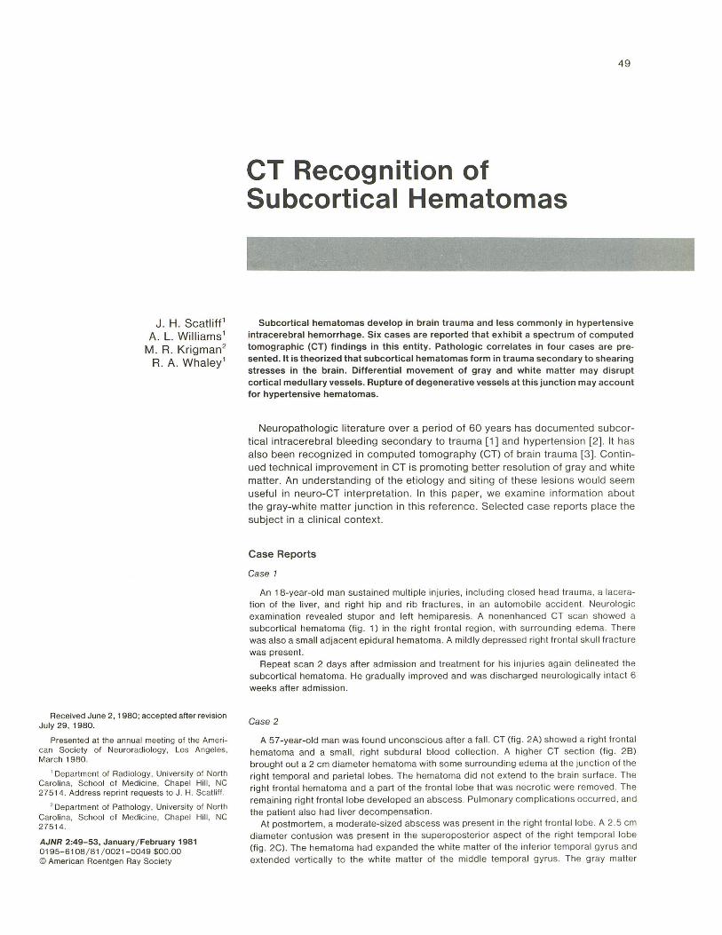
Fig. 1.-Case 1. Posttraumatic subcortical hematoma in right frontal region. Small adjacent epidural hematoma (arrow). Frontal fracture also present.
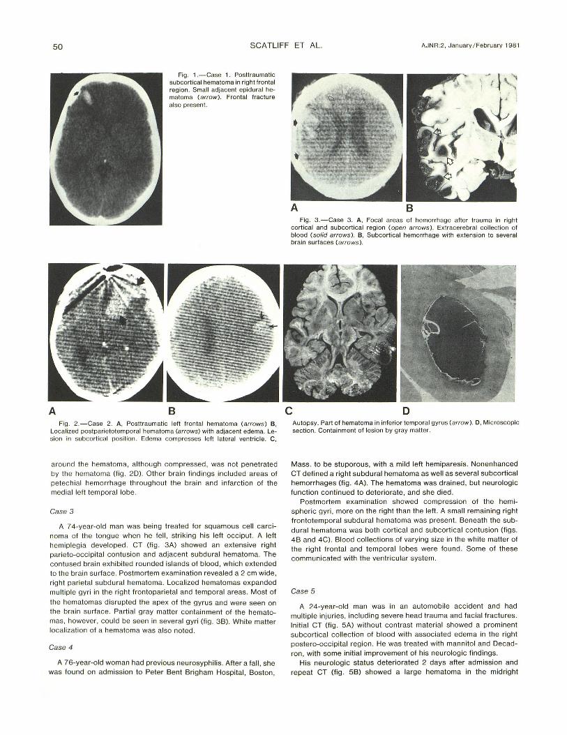
B Fig. 2. - Case 2. A, Posttraumatic left frontal hematoma (arrows) B,
Loca lized postparietotemporal hematoma (arrows) with adjacent edema. Lesion in subcortical position. Edema compresses left lateral ventric le. C,
around the hematoma, although compressed , was not penetrated by the hematoma (fig. 20) . Other brain findings inc luded areas of petech ial hemorrh age throughout the brain and infarc tion of the med ial left temporal lobe.
Case 3
A 74-year-old man was being treated for squamous cell carcinoma of th e tongue when he fell , striking his left occiput. A left hemiplegia developed. CT (fig . 3A) showed an extensive right parieto-occipital con tu sion and adjacent subdural hematoma. The contused brain exhibited rounded islands of blood, which extended to the brain surface. Postmortem examination revealed a 2 cm wide, right parietal subdural hematoma. Localized hematomas expanded multiple gyri in the right frontoparietal and temporal areas. Most of
the hematomas disrupted the apex of the gyrus and were seen on the brain surface . Partial gray matter containment of th e hematomas, however, could be seen in several gyri (fig . 3B) . White matter localization of a hematoma was also noted.
Case 4
A 76-year-old woman had previous neurosyphilis. After a fall , she was found on admission to Peter Bent Brigham Hospital, Boston,
Fig. 3.-Case 3 . A , Focal areas of hemorrh age after trauma in right cortical and subcortical region ( open arrows ). Extracerebral collection of blood ( solid arrows ). B, Subcortical hemorrhage with extension to several brain surfaces (arrows).
c D Autopsy. Part of hematoma in inferior temporal gyrus (arrow). D, Microscopic section . Containment of lesion by gray matter.
Mass. to be stuporous, with a mi ld left hemiparesis. Nonenhanced CT defined a right subdural hematoma as well as several subcortical hemorrhages (fig . 4A). The hematoma was drained , but neurologic function continued to deteriorate, and she died.
Postmortem examination showed compression of the hemispheric gyri, more on th e right than the left . A small remaining right frontotemporal subdural hematoma was present. Beneath the subdural hematoma was both cortical and subcortical contusion (figs. 4B and 4C). Blood collections of vary ing size in the white matter of the right frontal and temporal lobes were found. Some of these communicated with the ventricular system.
Case 5
A 24-year-old man was in an automobile acc ident and had multiple injuries, including severe head trauma and facial fractures. Initial CT (fig . 5A) without contrast material showed a prominent subcorti cal collection of blood with assoc iated edema in the right postero-occipital region . He was treated with mannitol and Oecadron, with some initial improvement of his neurologic findings.
His neurologic status deteriorated 2 days after admission and repeat CT (fig. 5B) showed a large hematoma in the midright
AJNR:2, January / February 1981 CT OF SUBCORTICAL HEMATOMAS 51
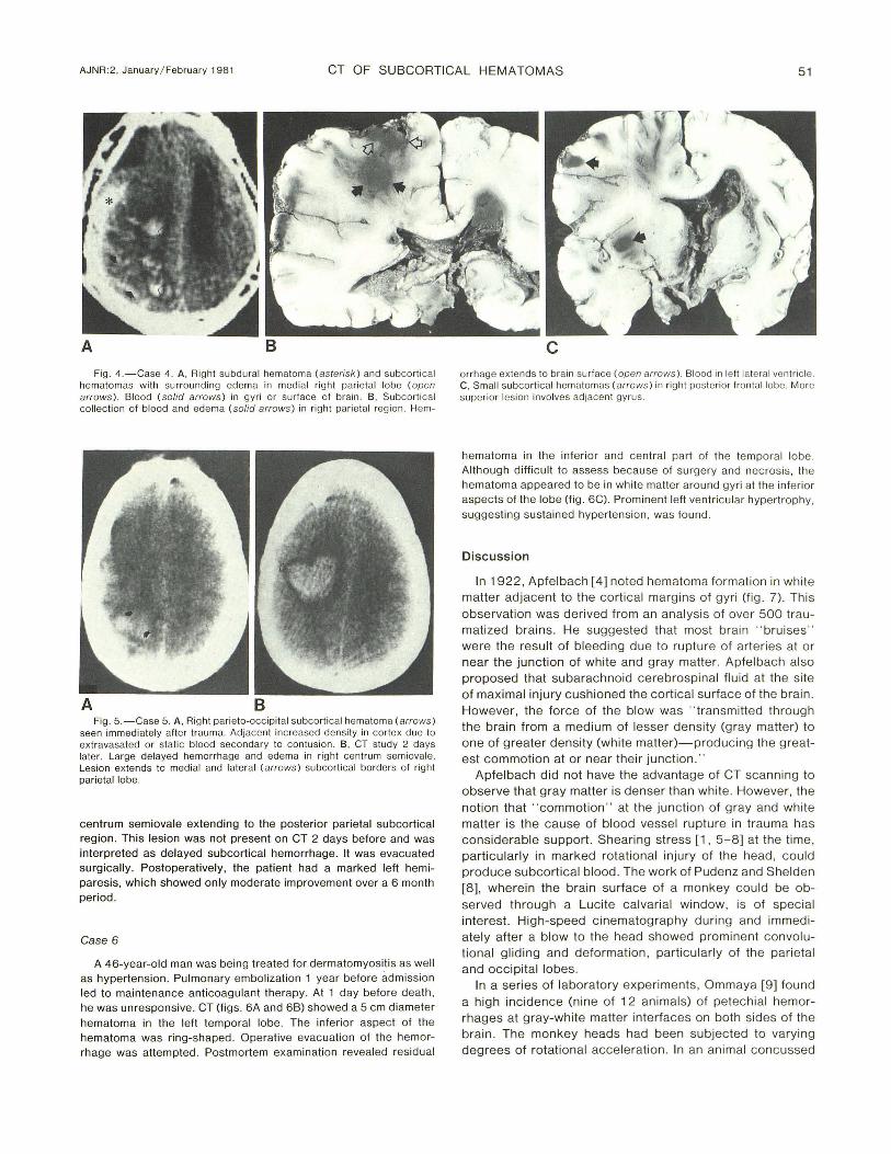
Fig. 4 .-Case 4. A, Right subdural hematoma (as terisk ) and subcortica l hematomas with surrounding edema in medial ri ght pari etal lobe ( open arrows ). Blood ( solid arrows) in gyri or surface of brain . B, Subcortical co llection of blood and edema (solid arrows ) in right parietal reg ion . Hem-
Fig. 5. - Case 5. A , Right pari eto-occ ipital subcortica l hematoma (arrows ) seen immediately after trauma. Adjacent inc reased density in co rt ex due to ex travasated or static blood secondary to contusion. B, CT study 2 days later. Larg e delayed hemorrhage and edema in ri ght centrum semiovale. Lesion extends to medial and lateral (arrows ) subcortical borders of ri ght pari etal lobe.
centrum semiovale extending to the posterior parietal subcortical region. This lesion was not present on CT 2 days before and was interpreted as delayed subcortical hemorrhage. It was evacuated surgically. Postoperatively, the patient had a marked left hemiparesis, which showed only moderate improvement over a 6 month period.
Case 6
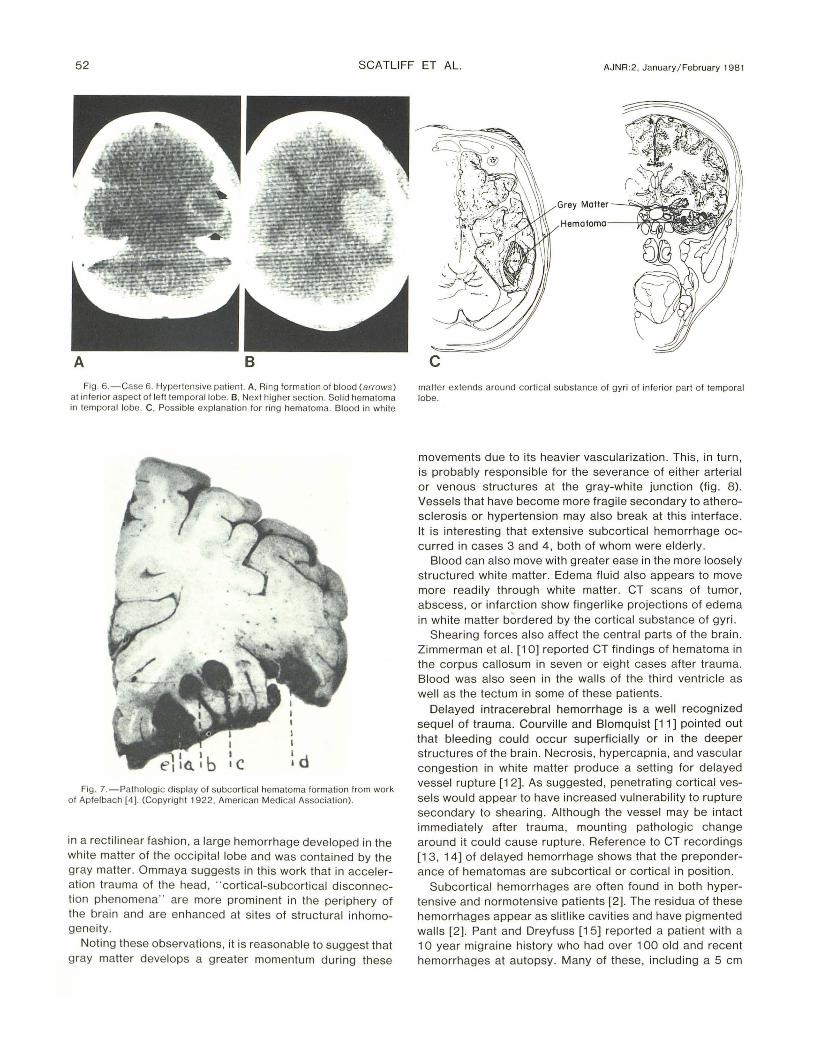
A 46-year-old man was being treated for dermatomyositis as well as hypertension . Pulmonary embolization 1 year before ad mission led to maintenance anticoagulant therapy. At 1 day before death , he was unresponsive. CT (figs. 6A and 68) showed a 5 cm diameter hematoma in the left temporal lobe. The inferior aspect of the hematoma was ring-shaped . Operative evacuation of the hemorrhage was attempted . Postmortem examination revealed residual
orrhage ex tends to brain surface (open arrows ). Blood in left lateral ventricle. C, Small subcortical hematomas (arrows ) in right posteri or frontal lobe. More superior les ion involves adjacent gyrus.
hematoma in the infe rior and central part of the temporal lobe. Although difficul t to assess because of surgery and necrosis, the hematoma appeared to be in whi te matter around gyri at the in ferior aspects of the lobe (fig . 6C). Prominent left ventri cular hypertrophy, suggesting sustained hypertension, was found.
Discussion
In 19 22 , Apfelbach [4] noted hematoma formati on in white matter adjacent to the corti cal margins of gyri (fi g. 7). Thi s observati on was derived from an analys is of over 500 trau matized brains. He suggested that most brain " bruises " were the result of bleeding due to rupture of arteries at or near the junction of wh ite and g ray matte r. Apfelbach also proposed that subarachno id cerebrospinal fluid at the si te of maximal injury cushioned the corti cal surface of the brain . However , the force of the blow was " transmitted th rough the brain from a medium of lesser density (gray matter) to one of greater density (white matter)-prod uc ing the g reatest commotion at or near their juncti on."
Apfelbach did not have the advantage of CT scanning to observe that gray matte r is denser than wh ite. However , the notion that "commotion " at the junction of gray and whi te matter is the cause of blood vessel rupture in trauma has considerabl e support. Shearing stress [1 , 5-8] at the time, parti c ularly in marked rotational injury of the head , could produce subcortical blood . The work of Pudenz and Shelden [8] , wherein the brain surface of a monkey could be observed through a Luc ite calvari al w indow, is of spec ial interest. High-speed c inematography during and immed iately after a blow to the head showed prominent convolut ional gliding and deform ati on, particularl y of the pari etal and occ ipital lobes.
In a seri es of laboratory experiments , Om maya [9] fo und a high inc idence (nine of 12 animals) of petechial hemorrhages at gray-white matter interfaces on both sides of the brain . The monkey heads had been subjected to varying degrees of ro tati onal acceleration. In an animal conc ussed
52 SCATLIFF ET AL. AJNR:2, January / February 1981
A B
Fig. 6.-Case 6. Hypertensive patien t. A , Ring form at ion of blood (arrows ) at in fe rior aspec t o f left temporal lobe. S, Nex t hig her secti on. Solid hematoma in temporal lobe. C, Possible explanat ion for ring hematoma. Blood in white
Fig. 7. -Patholog ic display o f subcortical hematoma fo rm ation from work of Apfe lbach [4]. (Copyrig ht 1922, American Medical Associat ion).
in a rectilinear fashion, a large hemorrhage developed in the white matter of the occipital lobe and was contained by the gray matter . Ommaya suggests in this work that in acceleration trauma of the head , " cortical-subcortical disconnecti on phenomena" are more prominent in the periphery of the brain and are enhanced at sites of structural inhomogeneity.
Noting these observations, it is reasonable to suggest that gray matter develops a greater momentum during these
Hemotnrr\o---~~~
c matter ex tends around corti ca l substance of gyri o f inferior part of temporal lobe.
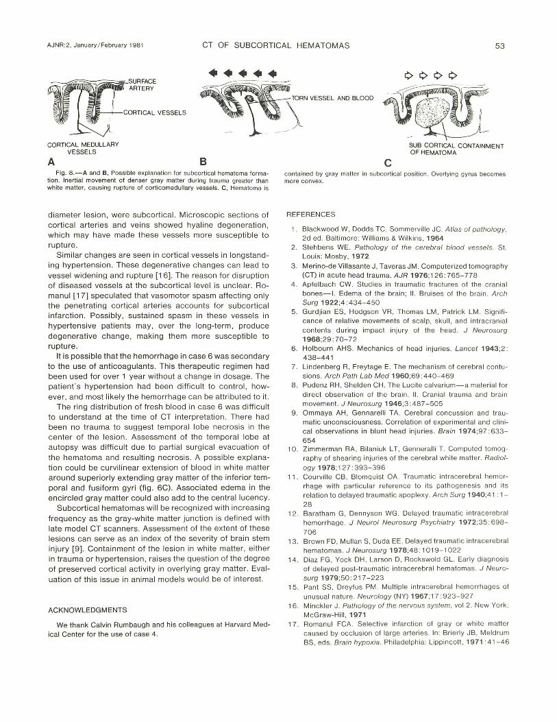
movements due to its heavier vascularization. This, in turn, is probably responsible for the severance of either arterial or venous structures at the gray-white junction (fig . 8). Vessels that have become more fragile secondary to atherosclerosis or hypertension may also break at this interface . It is interesting that extensive subcortical hemorrhage occurred in cases 3 and 4, both of whom were elderly.
Blood can also move with greater ease in the more loosely structured white matter. Edema fluid also appears to move more readily through white matter. CT scans of tumor, abscess, or infarction show fingerlike projections of edema in white matter bordered by the cortical substance of gyri.
Shearing forces also affect the central parts of the brain . Zimmerman et al. [10] reported CT findings of hematoma in the corpus callosum in seven or eight cases after trauma. Blood was also seen in the walls of the third ventricle as well as the tectum in some of these patients.
Delayed intracerebral hemorrhage is a well recognized sequel of trauma. Courville and Blomquist [11] pointed out that bleeding could occur superficially or in the deeper structures of the brain. Necrosis, hypercapnia, and vascular congestion in white matter produce a setting for delayed vessel rupture [12]. As suggested, penetrating cortical vessels would appear to have increased vulnerability to rupture secondary to shearing . Although the vessel may be intact immediately after trauma, mounting pathologic change around it could cause rupture. Reference to CT recordings [13, 14] of delayed hemorrhage shows that the preponderance of hematomas are subcortical or cortical in position .
Subcortical hemorrhages are often found in both hypertensive and normotensive patients [2]. The residua of these hemorrhages appear as slitlike cavities and have pigmented walls [2]. Pant and Dreyfuss [15] reported a patient with a 10 year migraine history who had over 1 00 old and recent hemorrhages at autopsy. Many of these, including a 5 cm
AJNR:2 , January / February 1981 CT OF SUBCORTICAL HEMATOMAS 53
CORTICAL VESSELS
\ '" ",50" ANO BWDO
- ---.-CORTICAL MEDULLARY
VESSELS
A 8 Fig . 8.-A and B, Possible explanation for subcortical hematoma forma
tion. Inertial movement of denser gray matter during trauma greater than white matter, causing rupture of corti comedullary vessels. C, Hematoma is
diameter lesion, were subcorti cal. Microscopic sections of cortical arteri es and veins showed hyaline degeneration , which may have made these vessels more susceptible to rupture.
Similar changes are seen in cortical vessels in longstanding hypertension. These degenerative changes can lead to vesse l widening and rupture [16]. The reason for disruption of diseased vesse ls at the subcortical leve l is unclear. Romanul [17] speculated that vasomotor spasm affecting on ly the penetrating cortical arte ries accounts for subcortical infarction. Possibly , sustained spasm in these vessels in hypertensive patients may , over the long-term, produce degenerative change, making them more susceptible to rupture.
It is possible that the hemorrhage in case 6 was secondary to the use of anticoagulants. This therapeutic regimen had been used for over 1 year without a change in dosage. The patient's hypertension had been difficu lt to control, however, and most likely the hemorrhage can be attributed to it.
The ring distribution of fresh blood in case 6 was difficult to understand at the time of CT interpretation. There had been no trauma to suggest temporal lobe necrosis in the center of the lesion. Assessment of the temporal lobe at autopsy was difficult due to partial surg ica l evacuation of the hematoma and resulting nec rosis. A possible exp lanation could be curvilinear extension of blood in white matter around superiorly extending gray matter of the inferior temporal and fusiform gyri (fig. 6C). Associated edema in the encircled gray matter could also add to the central lucency.
Subcortical hematomas will be recog nized with increasing frequency as the gray-white matter junction is defined with late model CT scanners . Assessment of the extent of these lesions can serve as an index of the severity of brain stem injury [9]. Containment of the lesion in white matter, either in trauma or hypertension , raises the question of the degree of preserved cort ical activity in overlying gray matter. Evaluation of this issue in animal models would be of interest.
ACKNOWLEDGMENTS
We thank Calvin Rumbaugh and his co lleagues at Harvard Med
ica l Center for the use of case 4.
c SUB CORTICAL CONTAINMENT OF HEMATOMA
contained by gray matter in subcortical position . Overlying gyrus becomes more convex.
REFERENCE S
1. Blackwood W , Dodds TC , Sommerville JC. Atlas of pathology, 2d ed. Baltimore: Wi lliams & Wi lkins, 1964
2. Stehbens WE. Pathology o f the cerebral blood vessels. St. Louis: Mosby , 1972
3. Merino-de Villasante J , Taveras JM. Computerized tomography (CT) in acute head trauma. AJR 1976;126 : 765-778
4. Apfe lbach CWo Studies in traumatic fractures of the c ran ial bones- I. Edema of the brain ; II . Bru ises of the brain. Arch Surg 1922;4: 434-450
5. Gurdjian ES, Hodgson VR , Thomas LM , Patrick LM . Sign ifi cance of re lative movements of scalp , sku ll , and intracran ial
contents during impact injury of the head. J Neurosurg 1968;29: 70-72
6 . Holbourn AHS. Mechanics of head injuries. Lancet 1943;2: 438-441
7. Lindenberg R, Freytage E. The mechanism of ce rebral contusions. Arch Path Lab M ed 1960;69: 440-469
8. Pudenz RH, Shelden CH . The Luc ite calvarium-a material for
d irect observat ion of the brain . II . Cran ial trauma and brain movement. J Neurosurg 1946;3: 487 -505
9. Ommaya AH , Gennarelli TA . Cerebral concussion and traumatic unconsciousness. Correlation of experimental and c linica l observations in blunt head injuries . Brain 1974;97 : 633-654
10. Z immerman RA , Bilaniuk L T, Genneralli T . Computed tomography of shear ing injuries of the cerebra l white matter. Radiol
ogy 1978; 127: 393-396 11 . Courville CB , Blomquist OA. Traumatic intracerebral hemor
rh age with particular reference to its pathogenesis and its relatio n to delayed trau mat ic apoplexy. Arch Surg 1940;41 : 1-
28 12. Baratham G, Denn yson WG . Delayed traumatic intracerebral
hemorrhage. J Neurol Neurosurg Psychiatry 1972;35: 698-
706 13. Brown FD, Mullan S, Duda EE . Delayed traumatic int racerebral
hematomas. J Neurosurg 1978;48: 1 019-1 022 14. Diaz FG , Yock DH , Larson 0 , Rockswold GL. Earl y diagnosis
of delayed post-traumatic intracerebral hematomas. J Neuro
surg 1979;50:217 - 223 15. Pant SS, Dreyfus PM . Multiple intracerebral hemorrhages of
unusual natu re. Neurology (NY) 1967; 17 : 923-927 16. Minckler J . Pathology of the nervous sys tem , vol 2. New York:
McG raw-Hili , 1971 17 . Romanul FCA. Select ive infarct ion of gray or wh ite matter
caused by occlu sion of large arteries. In : Brier ly JB, Meld rum BS, eds. Brain hypoxia . Philadelphia: Lippinco tt , 1971 : 41-46