Embed Size (px)
Citation preview
Journal of Interdisciplinary Histopathology
DOI: 10.5455/jihp.20140612073245www.scopmed.org
J Interdiscipl Histopathol 2014; 2(4): 228-231 228
INTRODUCTION
Low grade fibromyxoid sarcomas (LGFMS) (Evan’s tumor, hyalinizing spindle cell tumor with giant rosettes) are uncommon low grade malignant deep soft-tissue neoplasms initially described by Evans in 1987, with a deceptive benign histological appearance [1]. With the incidence being 0.18/million, these tumors represent 0.6% of all soft-tissue sarcomas [2]. These fibroblastic tumors typically occur in the lower extremities, particularly the thigh but can occur sporadically in other deep soft-tissues [3]. The other sites include the neck, chest wall, axilla, mediastinum, inguinal region, buttock, and even the brain [4]. Abdominal LGFMS are extremely rare. They have been reported to arise from anterior abdominal wall, small bowel mesentery, colon, falciform ligament, and retroperitoneum [4-6]. These tumors most commonly occur in young or middle-aged adults. However, occurrence over a wide age range has been noted [3]. Although these are indolent tumors, the potential for late metastasis suggests a need for prolonged follow-up [7,8].
CASE REPORT
A 62-year-old male presented with dull aching abdominal pain since 15 days. There was no history of fever, nausea, vomiting or change in the bowel and bladder habits. On examination, abdominal tenderness was present, a mass was palpable
at the hypogastric region which was hard and measured about 6 cm × 8 cm. On investigation, the hemoglobin was 13.7 g/dl and total white blood cells count was 9.2 × 10³/μL. Liver function tests were normal.
Computed tomography scan showed a circumscribed, lobulated homogenously enhancing abdomino-pelvic mass measuring about 11.6 cm × 9.5 cm × 8 cm. Clinically, differential diagnosis of mesenteric desmoid tumor/ileo-cecal gastrointestinal stromal tumor was considered. Hence ileo-colectomy with resection of the tumor was performed and the specimen sent for histopathological examination. Post-operative period was uneventful and sutures were removed on the 11th post-operative day. He was stable on discharge and is lost to follow-up.
HISTOPATHOLOGICAL FINDINGS
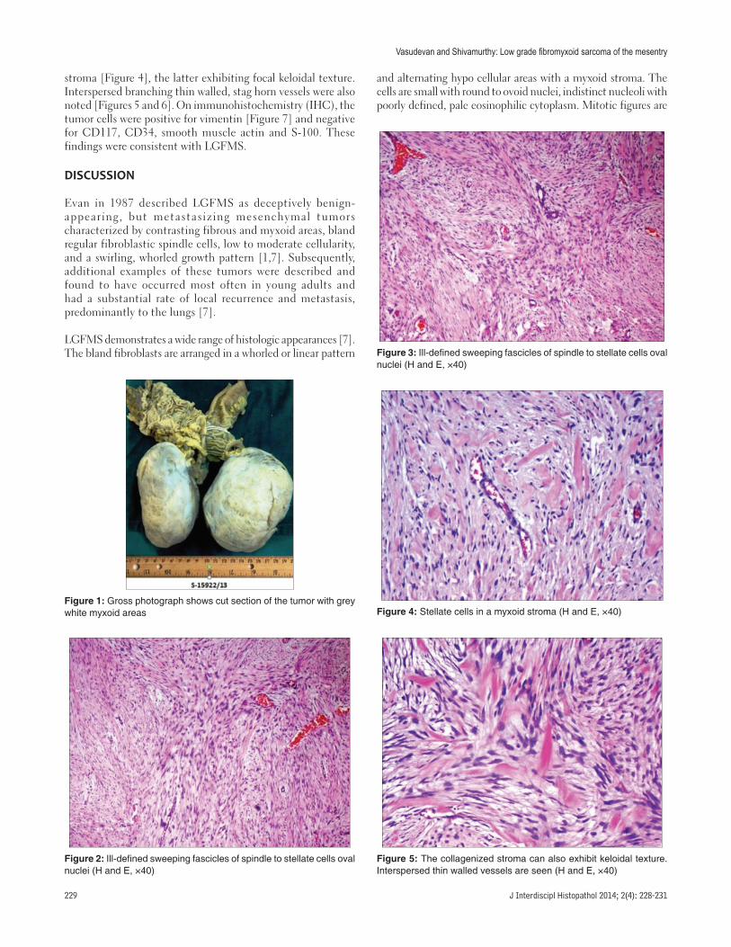
The ileo-colectomy specimen with the tumor mass weighed 652 g. Grossly, a well-circumscribed mesenteric mass was identified, which measured 11.5 cm × 9 cm × 5 cm. The cut section revealed homogenous grey-white areas with specks of hemorrhage [Figure 1].
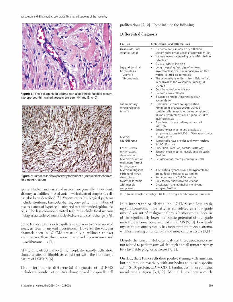
On microscopy, the tumor was composed of ill-defined sweeping fascicles of spindle to stellate cells with oval nuclei [Figures 2 and 3], some with small nucleoli and occasional mitosis (1/10 hpf), in an edematous, myxoid and collagenized
Low grade fi bromyxoid sarcoma of the mesentery, an under recognised entity: A case reportGeetha Vasudevan1, Archana Shivamurthy2
Case Report
Department of Pathology, 1Kasturba Medical College, 2Melaka Manipal Medical College, Manipal University, Manipal, Karnataka, India
Address for correspondence:Address for correspondence:Archana Shivamurthy, Melaka Manipal Medical College, Manipal University, Manipal, Karnataka, India. E-mail: [email protected]
Received: Received: May 11, 2014
Accepted: Accepted: June 12, 2014
Published: Published: June 25, 2014
ABSTRACTLow grade fibromyxoid sarcoma (LGFMS) is an indolent, rare soft tissue tumor having potential of late metastasis, but high local recurrence rate despite its low grade histologic findings. The morphologic diagnosis may be challenging, owing to its typically low cellularity, abundant collagen, and relatively bland cytology. We present here a rare case report of a mesenteric mass in an elderly male. The mass was excised. On microscopy, the tumor was composed of sweeping fascicles of spindle cells, which were positive for vimentin and negative for CD117, CD34, smooth muscle antigen and S-100. Herein we report a rare, under recognized case of LGFMS arising in the mesentery.
KEY WORDS: Low grade fibromyxoid sarcoma, mesentery
Vasudevan and Shivamurthy: Low grade fi bromyxoid sarcoma of the mesentry
229 J Interdiscipl Histopathol 2014; 2(4): 228-231
stroma [Figure 4], the latter exhibiting focal keloidal texture. Interspersed branching thin walled, stag horn vessels were also noted [Figures 5 and 6]. On immunohistochemistry (IHC), the tumor cells were positive for vimentin [Figure 7] and negative for CD117, CD34, smooth muscle actin and S-100. These findings were consistent with LGFMS.
DISCUSSION
Evan in 1987 described LGFMS as deceptively benign-appearing, but metastasizing mesenchymal tumors characterized by contrasting fibrous and myxoid areas, bland regular fibroblastic spindle cells, low to moderate cellularity, and a swirling, whorled growth pattern [1,7]. Subsequently, additional examples of these tumors were described and found to have occurred most often in young adults and had a substantial rate of local recurrence and metastasis, predominantly to the lungs [7].
LGFMS demonstrates a wide range of histologic appearances [7]. The bland fibroblasts are arranged in a whorled or linear pattern
and alternating hypo cellular areas with a myxoid stroma. The cells are small with round to ovoid nuclei, indistinct nucleoli with poorly defined, pale eosinophilic cytoplasm. Mitotic figures are
Figure 1: Gross photograph shows cut section of the tumor with grey white myxoid areas
Figure 2: Ill-defi ned sweeping fascicles of spindle to stellate cells oval nuclei (H and E, ×40)
Figure 3: Ill-defi ned sweeping fascicles of spindle to stellate cells oval nuclei (H and E, ×40)
Figure 4: Stellate cells in a myxoid stroma (H and E, ×40)
Figure 5: The collagenized stroma can also exhibit keloidal texture. Interspersed thin walled vessels are seen (H and E, ×40)
Vasudevan and Shivamurthy: Low grade fi bromyxoid sarcoma of the mesentry
J Interdiscipl Histopathol 2014; 2(4): 228-231 230
sparse. Nuclear anaplasia and necrosis are generally not evident, although a dedifferentiated variant with sheets of anaplastic cells has also been described [3]. Various other histological patterns include storiform, fascicular-herringbone pattern, formation of rosettes, areas of hyper cellularity and foci of rounded epithelioid cells. The less commonly noted features include focal osseous metaplasia, scattered multinucleated cells and cystic change [7,8].
Some tumors have a rich capillary vascular network in myxoid areas, as seen in myxoid liposarcoma. However, the vascular channels seen in LGFMS are usually curvilinear, thicker and coarser than those seen in myxoid liposarcoma and myxofibrosarcoma [9].
At the ultra-structural level the neoplastic spindle cells show characteristics of fibroblasts consistent with the fibroblastic nature of LGFMS [6].
The microscopic differential diagnosis of LGFMS includes a number of entities characterized by spindle cell
proliferations [3,10]. These include the following:
Differential diagnosis
Entities Architectural and IHC features
Gastrointestinal stromal tumor
• Predominantly spindled or epithelioid, seldom show broad zones of collagenization,
• Vaguely neural-appearing cells with fibrillar cytoplasm
• CD117, CD34: PositiveIntra-abdominal fibromatosis
Desmoid fibromatosis
• Long, sweeping fascicles of uniform myofibroblastic cells arranged around thin walled, dilated blood vessels
• The cellularity is uniform from field to field in contrast to the variable cellularity of LGFMS
• Cells have vesicular nucleus• Contain more collagen• β-catenin protein: Aberrant nuclear
accumulationInflammatory myofibroblastic tumors
• Prominent stromal collagenization reminiscent of areas within LGFMS, contain cellular spindled zones composed of plump myofibroblasts and “ganglion-like” myofibroblasts
• Prominent chronic inflammatory cell infiltrate
• Smooth muscle actin and anaplastic lymphoma kinase (ALK-1) : Strong positivity
Myxoid neurofibroma
• Encapsulated• Tumor cells have slender and wavy nucleus• S-100: Positive
Fasciitis with myxomatous degeneration
• Superficial location, Similar histology• Smooth muscle actin, muscle specific actin:
PositiveMyxoid variant of malignant fibrous histiocytoma
• Cellular areas, more pleomorphic cells
Myxoid malignant peripheral nerve sheath tumor
• Alternating hypocellular and hypercellular areas, focal peripheral palisading
• Some tumors are S-100 positiveSynovial sarcoma with myxoid component
• Only focally shows myxoid change• Cytokeratin and epithelial membrane
antigen: Positive
IHC: Immunohistochemistry, LGFMS: Low grade fibromyxoid sarcoma
It is important to distinguish LGFMS and low grade myxofibrosarcoma. The latter is considered as a low grade myxoid variant of malignant fibrous histiocytoma, because of the significantly lower metastatic potential of low grade myxofibrosarcoma compared with LGFMS [9,10]. Low grade myxofibrosarcoma typically has more uniform myxoid stroma, with less swirling of tumor cells and more cellular atypia [3,11].
Despite the varied histological features, these appearances are not related to patient survival although a small tumor size may be a favorable prognostic factor [7,11].
On IHC, these tumor cells show positive staining with vimentin, but no immune-reactivity with antibodies to muscle specific actin, S-100 protein, CD34, CD31, keratin, desmin or epithelial membrane antigen [3,4,12]. Mucin 4 has been recently
Figure 6: The collagenized stroma can also exhibit keloidal texture. Interspersed thin walled vessels are seen (H and E, ×40)
Figure 7: Tumor cells show positivity for vimentin (immunohistochemical for vimentin, ×100)

Vasudevan and Shivamurthy: Low grade fi bromyxoid sarcoma of the mesentry
231 J Interdiscipl Histopathol 2014; 2(4): 228-231
described to be a highly sensitive and quite specific IHC marker for LGFMS [13,14].
A chromosomal translocation (t (7;16) (q34;p11)) which results in a novel fusion gene (Fused in Sarcoma/cyclic adenosine monophosphate responsive element binding protein 3-like 2) is well characterized and constitutes an excellent tool in the differential diagnosis of LGFMS as it accounts for more than 95% of the cases. The demonstration of a ring chromosome may be associated with an increased risk of tumor progression [4,8].
In LGFMS, surgery is the only treatment resulting in disease-free periods. LGFMS is not very chemo- or radiosensitive due to the low grade of malignancy with low mitotic rate. Atypical metastatic potential, with atypical sites of metastases poses a problem in deciding the optimal duration of follow-up in these patients [2].
CONCLUSION
LGFMS are rare soft tissue tumors characterized by a deceptively bland appearance and potential for late metastases. At present, surgery is the only treatment that results in disease-free periods. Diagnosis of LGFMS remains problematic because of its bland histologic features that can be potentially confused with other benign or low grade soft-tissue tumors. Careful consideration of the morphological and IHC features of these tumors would render a positive diagnosis.
REFERENCES
1. Evans HL. Low-grade fibromyxoid sarcoma. A report of two metastasizing neoplasms having a deceptively benign appearance. Am J Clin Pathol 1987;88:615-9.
2. Maretty-Nielsen K, Baerentzen S, Keller J, Dyrop HB, Safwat A. Low-Grade Fibromyxoid Sarcoma: Incidence, Treatment Strategy of Metastases, and Clinical Significance of the FUS Gene. Sarcoma 2013;2013:256280.
3. Vernon SE, Bejarano PA. Low-grade fibromyxoid sarcoma: A brief review. Arch Pathol Lab Med 2006;130:1358-60.
4. Alatise OI, Oke OA, Olaofe OO, Omoniyi-Esan GO, Adesunkanmi AR. A huge low-grade fibromyxoid sarcoma of small bowel mesentery simulating hyper immune splenomegaly syndrome: A case report and review of literature. Afr Health Sci 2013;13:736-40.
5. Harish K, Ashok AC, Alva NK. Low grade fibromyxoid sarcoma of the falciform ligament: A case report. BMC Surg 2003;3:7.
6. Singh K, Singh S, Pal N, Sampley SK, Chhabra K. Low-grade fibromyxoid sarcoma of anterior abdominal wall. Indian J Surg 2012;74:351-3.
7. Evans HL. Low-grade fibromyxoid sarcoma: A clinicopathologic study of 33 cases with long-term follow-up. Am J Surg Pathol 2011;35:1450-62.
8. Mertens F, Fletcher CD, Antonescu CR, Coindre JM, Colecchia M, Domanski HA, et al. Clinicopathologic and molecular genetic characterization of low-grade fibromyxoid sarcoma, and cloning of a novel FUS/CREB3L1 fusion gene. Lab Invest 2005;85:408-15.
9. Dawamneh MF, Amra NK, Amr SS. Low grade fibromyxoid sarcoma: Report of a case with fine needle aspiration cytology and histologic correlation. Acta Cytol 2006;50:208-12.
10. Dvornik G, Barbareschi M, Gallotta P, Dallapalma P. Low grade fibromyxoid sarcoma. Histopathology 1997;30:274-6.
11. Lindberg GM, Maitra A, Gokaslan ST, Saboorian MH, Albores-Saavedra J. Low grade fibromyxoid sarcoma: Fine-needle aspiration cytology with histologic, cytogenetic, immunohistochemical, and ultrastructural correlation. Cancer 1999;87:75-82.
12. Laurini JA, Zhang L, Goldblum JR, Montgomery E, Folpe AL. Low-grade fibromyxoid sarcoma of the small intestine: Report of 4 cases with molecular cytogenetic confirmation. Am J Surg Pathol 2011;35:1069-73.
13. Reid R, de Silva MV, Paterson L, Ryan E, Fisher C. Low-grade fibromyxoid sarcoma and hyalinizing spindle cell tumor with giant rosettes share a common t (7;16)(q34;p11) translocation. Am J Surg Pathol 2003;27:1229-36.
14. Doyle LA, Möller E, Dal Cin P, Fletcher CD, Mertens F, Hornick JL. MUC4 is a highly sensitive and specific marker for low-grade fibromyxoid sarcoma. Am J Surg Pathol 2011;35:733-41.
© GESDAV; licensee GESDAV. This is an open access article licensed under the terms of the Creative Commons Attribution Non-Commercial License (http://creativecommons.org/licenses/by-nc/3.0/) which permits unrestricted, non-commercial use, distribution and reproduction in any medium, provided the work is properly cited.
Source of Support: Nil, Confl ict of Interest: None declared.


![First Case of Hepatic Polycystic Echinococcosis Involving ...The involvement of liver and mesentery [5] and exclusively . the mesentery [10] are the most reported PE clinical presentation](https://img.pdfslide.us/doc/110x75/5fbdc5051c35c657811004d0/first-case-of-hepatic-polycystic-echinococcosis-involving-the-involvement-of.jpg)