Embed Size (px)
Citation preview
2714 | J. S. King et al. Molecular Biology of the Cell
MBoC | ARTICLE
WASH is required for lysosomal recycling and efficient autophagic and phagocytic digestionJason S. Kinga,*, Aurélie Guehob, Monica Hagedornc, Navin Gopaldassb,†, Florence Leubab, Thierry Soldatib, and Robert H. InsallaaBeatson Institute for Cancer Research, Bearsden, Glasgow G61 1BD, United Kingdom; bDepartment of Biochemistry, University of Geneva, CH-1211 Geneva, Switzerland; cBernhard Nocht Institute for Tropical Medicine, 20359 Hamburg, Germany
ABSTRACT Wiskott-Aldrich syndrome protein and SCAR homologue (WASH) is an important regulator of vesicle trafficking. By generating actin on the surface of intracellular vesicles, WASH is able to directly regulate endosomal sorting and maturation. We report that, in Dictyostelium, WASH is also required for the lysosomal digestion of both phagocytic and autophagic cargo. Consequently, Dictyostelium cells lacking WASH are unable to grow on many bacteria or to digest their own cytoplasm to survive starvation. WASH is required for efficient phagosomal proteolysis, and proteomic analysis demonstrates that this is due to reduced delivery of lysosomal hydrolases. Both protease and lipase delivery are disrupted, and lipid catabolism is also perturbed. Starvation-induced autophagy therefore leads to phospholipid accumulation within WASH-null lysosomes. This causes the formation of multila-mellar bodies typical of many lysosomal storage diseases. Mechanistically, we show that, in cells lacking WASH, cathepsin D becomes trapped in a late endosomal compartment, unable to be recycled to nascent phagosomes and autophagosomes. WASH is therefore required for the maturation of lysosomes to a stage at which hydrolases can be retrieved and reused.
INTRODUCTIONThe Wiskott-Aldrich syndrome protein and SCAR homologue (WASH) is an evolutionarily conserved regulator of the Arp2/3 complex. Like other members of the Wiskott-Aldrich syndrome protein (WASP)
family, the primary role of WASH is to activate Arp2/3 and regulate the spatial and temporal formation of actin networks (Linardopoulou et al., 2007). Specifically, WASH generates actin subdomains on en-docytic vesicles and is important for several sorting and maturation steps. These include retrograde transport from endosomes to the trans-Golgi via direct regulation of the retromer (Derivery et al., 2009; Gomez and Billadeau, 2009; Duleh and Welch, 2010; Harbour et al., 2010) and the sorting and trafficking of other signaling mole-cules, such as integrins, the epidermal growth factor (EGF), and transferrin receptors (Derivery et al., 2009, Zech et al., 2011).
A comparable role has also been described in Dictyostelium, whereby WASH-generated actin sequesters the vacuolar (v)-ATPase to late endosomal subdomains, leading to v-ATPase removal via recycling vesicles (Carnell et al., 2011). The v-ATPase is the proton pump responsible for establishing and maintaining an acidic pH in the lumen of lysosomes. Therefore its WASH-mediated removal leads to the neutralization and maturation of lysosomes into postlysosomes prior to exocytosis (Neuhaus et al., 2002). In Dictyostelium, the loss of WASH therefore arrests lysosome matu-ration before v-ATPase removal, blocking both neutralization and exocytosis (Carnell et al., 2011).
Like other WASP-family proteins, WASH acts as part of a com-plex (Jia et al., 2010; Veltman and Insall, 2010). Our recent work
Monitoring EditorCarole ParentNational Institutes of Health
Received: Feb 14, 2013Revised: Jun 5, 2013Accepted: Jul 1, 2013
This article was published online ahead of print in MBoC in Press (http://www .molbiolcell.org/cgi/doi/10.1091/mbc.E13-02-0092) on July 24, 2013.Present addresses: *Department of Biomedical Sciences, Firth Court, Sheffield University, Sheffield S10 2TN, United Kingdom; †Département de Biochimie, Université de Lausanne, Chemin des boveresses 155, CH-1066 Epalinges, Switzerland.Address correspondence to: Jason S. King ([email protected]).Abbreviations used: BSA, bovine serum albumin; catD, cathepsin D; CD, cation dependent; CI, cation independent; CID, collision-induced dissociation; EGF, epidermal growth factor; EGTA, ethylene glycol tetraacetic acid; FITC, fluorescein isothiocyanate; GFP, green fluorescent protein; HCD, high-energy C-trap disso-ciation; M6PR, mannose-6-phosphate receptor; MLB, multilamellar body; MS, mass spectrometry; NA, numerical aperture; PBS, phosphate-buffered saline; PDI, protein disulfide isomerase; siRNA, small interfering RNA; TCEP, Tris-(2-carboxyethyl) phosphine hydrochloride; TEAB, triethylammonium hydrogen carbonate buffer; TEM, transmission electron microscopy; TMT, tandem mass tag; v-ATPase, vacuolar ATPase; WASH, Wiskott-Aldrich syndrome protein and scar homologue; WASP, Wiskott-Aldrich syndrome protein.© 2013 King et al. This article is distributed by The American Society for Cell Biol-ogy under license from the author(s). Two months after publication it is available to the public under an Attribution–Noncommercial–Share Alike 3.0 Unported Creative Commons License (http://creativecommons.org/licenses/by-nc-sa/3.0).“ASCB®,” “The American Society for Cell Biology®,” and “Molecular Biology of the Cell®” are registered trademarks of The American Society of Cell Biology.

Volume 24 September 1, 2013 WASH and lysosomal recycling | 2715
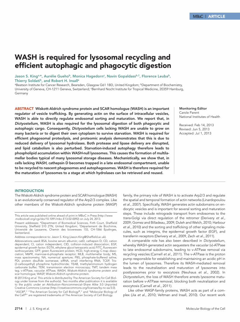
exposed GFP-atg8 is cleaved by Atg4, the pH-sensitive GFP fluores-cence on the interior is quenched by acidification, and the GFP signal is lost (Kirisako et al., 2000). Using cellular compression to both induce autophagy and improve imaging (King et al., 2011), we observed no gross defects in autophagosome formation or morphology in WASH-null cells compared with wild-type (Ax2) controls (Figure 1A and Supplemental Movies S1 and S2).
Previous work in Dictyostelium has demonstrated an important role for the WASH complex in v-ATPase trafficking and, conse-quently, the regulation of vesicular pH (Carnell et al., 2011). During maturation, autophagosomes are also acidified, due to delivery of the v-ATPase from the lysosomal compartment. The rate of acidifi-cation and maturation is represented by the loss of GFP-Atg8 fluo-rescence after phagophore completion, allowing quantification (see Movies S1 and S2). When we compared the time taken for the fluo-rescent signal to be removed in Ax2 and WASH mutants (n > 50, across three independent experiments), no difference was observed, which indicated there were no defects in acidification (Figure 1B).
In the above assays, autophagy was induced by mechanical compression, via pathways that are poorly understood (King, 2012). We therefore also tested the role of WASH under amino acid starva-tion. When WASH-null cells were placed in the defined SIH medium lacking arginine and lysine (SIH-Arg/Lys, which induces autophagy, but not the development of Dictyostelium; King et al., 2011) we found no differences in either the rate of induction or steady-state levels of autophagosomes after 24 h (Figure 1, C and D). We there-fore conclude that WASH is not required for the induction, forma-tion, or maturation of autophagosomes.
WASH is required for cytoplasmic degradation and surviving starvationAlthough not required for autophagosome formation, we hypothe-sized that WASH may be necessary for subsequent processing of autophagosomes. The canonical role for autophagy is during starva-tion, when the degradation and recycling of cytoplasmic compo-nents is essential for cells to maintain nutrient levels and survive. To test whether the autophagic pathway was fully functional in WASH-null cells, we determined their ability to survive Arg/Lys starvation. While Ax2 cells were able to survive for almost 7 d before their via-bility decreased, WASH mutants died significantly faster, losing almost 50% viability after only 4 d (Figure 1E). Their survival was, however, better than that of Atg1 mutants, which are completely deficient in autophagy and started dying after just 24 h. WASH is therefore required for effective survival of Arg/Lys starvation.
WASH exists in a complex with four other proteins. We recently determined that one of the complex subunits, FAM21, is required to recycle WASH from endosomes. Loss of FAM21 therefore leads to persistent WASH activity, blocking the endocytic pathway at a later stage, after v-ATPase removal (Park et al., 2013). FAM21 mutants were also unable to survive starvation, to the same degree as WASH mutants (Figure 1E), indicating that the defect in survival is due to a trafficking event downstream of WASH and v-ATPase recycling.
The function of autophagy under starvation is to degrade the cytosol, which results in reduction of both cell size and mass (Otto et al., 2003). Total protein levels were decreased by 15% in Ax2 cells upon 24 h Arg/Lys starvation, but remained constant in Atg1-null cells (Figure 1G). Consistent with their inability to sur-vive starvation, both WASH and FAM21 mutants also had no re-duction in total cellular protein and retained their size over sev-eral days (Figure 1, F and G). Therefore, while autophagosomes form and acidify normally in WASH mutants, total protein is not reduced, and the mutant cells are unable to survive starvation.
showed that, while disruption of the Strumpellin, ccdc53, or SWIP subunits leads to total loss of WASH activity, the FAM21 subunit has a unique role, driving the recycling of WASH (Park et al., 2013). Therefore loss of FAM21 gives hyperactive WASH, and, although cells retain the ability to recycle v-ATPase, endocytic maturation is blocked at a later stage, at which WASH is itself recycled. Impor-tantly, these contrasting phenotypes of WASH and FAM21 mutants now allow us to dissect the pathway in more detail and discriminate direct and indirect effectors of WASH.
In addition to its specific trafficking roles, WASH appears to be more generally important in maintaining endosomal and lysosomal integrity. In mammalian cells, small interfering RNA (siRNA) leads to altered endosome morphology (Derivery et al., 2009; Duleh and Welch, 2010), while full deletion causes lysosomal collapse (Gomez et al., 2012; Piotrowski et al., 2012). We therefore hypothesized that WASH may also be important for normal lysosomal function.
Lysosomes are responsible for the degradation of endosomal contents. They are therefore essential for breaking down endocytic cargo, such as membrane receptors, as well as the contents of autophagosomes and bacteria captured within phagosomes. Autophagy is the name given to a family of pathways leading to the capture of cytoplasmic constituents and their delivery to lysosomes. This includes both microautophagy, wherein selected proteins are directly transported across the lysosomal membrane, and macroau-tophagy, wherein double-membrane vesicles are formed de novo, capturing parts of the cytoplasm and fusing with lysosomes (Xie and Klionsky, 2007). In this study, we have specifically studied macroau-tophagy, which we will simply refer to as autophagy for clarity.
Cells use autophagy for a number of purposes, and defective autophagy is associated with a number of medical conditions, in-cluding aging, cancer, infection, and neurodegeneration (Mizushima et al., 2008). The best-understood function of autophagy is during starvation, when degradation of the cytosol provides nutrients and energy to the cell, enabling survival. In professional phagocytes such as Dictyostelium, this is similar to the phagocytic pathway, in which the cell uses the same digestive pathway to liberate nutrients from captured bacteria (Deretic, 2008). Autophagy and phagocyto-sis therefore both require lysosomal delivery and digestion in order to feed the cell.
The WASH complex has been assigned a number of roles in ve-sicular trafficking. In this study, we assess the importance of WASH in lysosomal function within the context of both the autophagic and phagocytic pathways. We show that WASH is required for both pro-cesses in Dictyostelium and describe a new role for WASH in main-taining lysosomal flux.
RESULTSWASH is not required for autophagosome formationPrevious work has shown that the WASH complex is involved in mul-tiple stages of the endocytic pathway. Autophagy intersects with the endocytic pathway and relies on many of the same processes, such as v-ATPase and lysosomal hydrolase trafficking. We therefore di-rectly assessed the role of WASH in autophagy, using the model system Dictyostelium discoideum, for which there are well-defined assays for autophagosome induction and formation (Calvo-Garrido et al., 2010, King et al., 2011).
To directly observe autophagosome formation, we used a green fluorescent protein (GFP)-Atg8 fusion probe. During autophago-some formation, GFP-Atg8 is lipidated and incorporated into the expanding phagophore membrane (Ichimura et al., 2000) and can therefore be used to label autophagosomes until they are fully formed. When the autophagosomes are completed, the cytosolically
2716 | J. S. King et al. Molecular Biology of the Cell
using beads coated with self-quenching fluorescently labeled DQ-BSA (bovine se-rum albumin), which becomes dequenched upon proteolysis (Gopaldass et al., 2012). With this assay, the rate of proteolysis was reduced by ∼50% in both WASH and FAM21 mutants compared with Ax2, and rescued by reexpression of GFP-WASH in WASH-null cells (Figure 2A). WASH is therefore re-quired for both phagocytic and autophagic digestion, indicating a general role for WASH in vesicular degradation. Importantly, this cannot be due to a defect in acidifica-tion, as this is unaffected in both WASH and FAM21 mutants (Figure 4E; Carnell et al., 2011; Park et al., 2013).
Efficient phagocytic digestion is essen-tial for Dictyostelium to survive on bacteria as a food source. We therefore tested the ability of WASH-null Dictyostelium to grow on several bacterial strains. While WASH- and FAM21-null cells grew normally on our laboratory strains of Klebsiella aerogenes and Bacillus subtilis, both mutants had dra-matically reduced growth on the majority of the other strains tested (Figure 2, B and C), demonstrating a physiological requirement for WASH in growth on bacteria.
Lysosomal components are reduced in the phagocytic compartmentPhagosome maturation is a highly regu-lated and organized process during which lysosomal components are sequentially de-livered and retrieved (Clarke et al., 2002; Gotthardt et al., 2002, 2006a). To establish the mechanism underlying the defects in degradation, we used quantitative com-parative proteomics to identify differentially represented proteins in the isolated pha-gosomes of Ax2, WASH, and FAM21 mu-tants (Dieckmann et al., 2012; Gotthardt et al., 2006a). To confirm this approach, we first looked at the levels of v-ATPase. In Dictyostelium, WASH is required for v-ATPase recycling, and WASH mutants re-tain v-ATPase on their endosomes (Carnell
et al., 2011). Consistent with this, all nine subunits of the v-ATPase complex were elevated in the WASH-null phagocytic compartment compared with wild-type (Figure 3B). In contrast, FAM21 mutants retain WASH activity (Park et al., 2013), and several of the v-ATPase subunits were identified as decreased in FAM21-null phagosomes relative to Ax2. This validates the data set and confirms that we are able to identify differentially trafficked proteins with this method.
Although the loss of WASH or FAM21 blocks the endocytic path-way at different points, both mutants have comparable defects in phagocytosis and autophagy. We therefore looked for differences in the phagocytic pathway common to both mutants. Of 85 proteins that were decreased by at least 30% in both mutants, 11 (13%) were lysosomal proteins (Figure 3A and Supplemental Table S1). Several additional lysosomal proteins were also decreased in either WASH or FAM21 mutants, indicating a general loss of lysosomal enzymes from
This indicates a defect in autophagic degradation and nutrient recycling.
Phagocytic proteolysis is reduced in WASH mutantsAutophagy is not the only pathway that breaks down vesicular cargo. In soil, Dictyostelium is a professional phagocyte, relying on the capture and digestion of bacteria within phagosomes to provide nutrients. After formation, phagosomes and autophagosomes are processed in a similar manner, with the same requirement for acidi-fication and hydrolytic enzymes in order to release nutrients (Deretic, 2008). We therefore tested whether WASH is also required for phagocytic digestion.
Like autophagosome formation, loss of either WASH or FAM21 did not affect the rate of phagocytosis (Supplemental Figure S1A). We were therefore able to directly measure phagocytic proteolysis
FIGURE 1: Autophagy in WASH mutants. (A) High-resolution imaging of autophagosome formation in GFP-atg8–expressing cells compressed under agar. Stills are taken from Movies S1 and S2. (B) Quantification of the time taken for the GFP signal of individual autophagosomes in cells treated as in (A) to fade after autophagosome formation was complete. (n > 50 autophagosomes in total for each strain). (C) Autophagy was induced by placing cells expressing GFP-atg8 in SIH-Arg/Lys. The number of autophagosomes was counted at each time point. (D) The steady-state levels of autophagosomes after 24 h of starvation. (E) Survival of mutants under Arg/Lys starvation. At each time point, viability was measured by the ability to reform colonies on bacterial plates. (F) Morphology of cells under starvation. DIC images of cells after 3 d in full medium or SIH-Arg/Lys. (G) Total protein of cells after 24 h of Arg/Lys starvation. All values plotted are the means ± SD of three independent experiments. *, p < 0.05; **, p < 0.01; ***, p < 0.005 (Student’s t-test). Scale bars: 5 μm.
0
10
20
30
40
50
Ax2 WASH
Aci
dific
atio
n tim
e (s
)
C D
0
1
2
3
4
0 10 20 30 40
Pun
cta
per
cell
Time (min)
A
0
1
2
3 Ax2WASH
0h 24h
Pun
cta
per
cell
BAx2 WASH
E
Ax2 WASH FAM21 Atg1
Ful
l Med
ium
-Arg
/lys
72h
F
% T
otal
pro
tein
120
100
80
Ax2 WASH FAM21 Atg1
0h24h
***
0
25
50
75
100
125
0 2 4 6 8 10
Via
bilit
y (%
)
Days starvation
****
*
*
G
Ax2WASH
Ax2 WASH FAM21 Atg1

Volume 24 September 1, 2013 WASH and lysosomal recycling | 2717
the phagocytic compartment (Figure 3B). These include several pro-teases, such as cathepsins D and Z, as well as a cysteine protease, explaining the measured reduction in proteolytic activity. A form of lysozyme (DDB_G0274181) was also reduced in both mutant
FIGURE 2: Phagocytic defects in WASH mutant cells (A) Proteolytic activity is decreased in WASH mutants, as measured by feeding cells DQ-BSA–labeled beads and measuring unquenching of the fluorophore over time. (B) WASH- and FAM21-null cells are unable to grow on certain bacteria. The indicated number of cells were seeded on the bacterial lawn, and Dictyostelium growth is indicated by the formation of dark plaques where the bacteria have been consumed. (C) Summary of mutant growth on various bacteria. The color code refers to the number of wells in (B) in which Dictyostelium were able to grow, indicating the severity of the growth defect.
A
B
C
100 0
2
4
6
0 25 50 75
Rat
io A
488/
594
Time (min)
Ax2 WASH FAM21 WASH::GFP-WASH
10K
1K
100
10
Cells Ax2 WASH FAM21 Ax2 WASH FAM21
K. aerogenes K. pneumoniae 52145
K. a
erog
enes
K. p
neum
onia
e
5214
5
M. l
eutu
s
B. s
ubti
lis
E.c
oli
B/r
E.c
oli
DH
5
S. a
ureu
s
P. a
erug
inos
as
PT
5 P
. aer
ugin
osas
P
T53
1
Ax2
WASH
FAM21
4 3 2 1 0
E. a
erog
enes
FIGURE 3: Proteomic analysis of mutant phagosomes. (A) The relative proportion of proteins from different compartments is reduced by at least 30% in both WASH and FAM21 mutant phagosomes. Eighty-five proteins in all were identified by this criteria. (B) Identity and relative abundance of lysosomal and secreted (below dashed line) proteins identified in this screen. Lysosomal proteins only identified in one mutant are also listed. The relative abundance of v-ATPase subunits is included in the lower section. *, p < 0.05; **, p < 0.005; ***, p < 0.001 (Student’s t-test).
Transmembrane Endosomal membrane
Cytoskeleton
Nucleus
ER
Lysosome
Secreted
Ribosome Cytoplasm
Mitochondria
Unknown
B
A
Gene Protein Gene ID WASH/Ax2 Fam21/Ax2
ctsD
Cathepsin D
DDB_G0279411
0.46***
ctsZ Cathepsin Z; DDB_G0283401 0.44*** 0.37**
V4-7
peptidase S8 and S53 domain-containing protein
DDB_G0281823 0.40** 0.36*
Cysteine protease;
DDB_G0291191 0.57***
0.45**
nagA
Beta-hexosaminidase subunit A1
DDB_G0287033 0.60*
nagB
Beta-hexosaminidase subunit A2
DDB_G0282539 0.45* 0.23**
manA
Alpha-mannosidase DDB_G0292206 0.72*** 0.52***
m6pr Mannose-6-phosphate receptor
DDB_G0279059 0.62*** 0.62**
lmpA
Lysosome membrane protein 2-A
DDB_G0267406 0.75** 0.40***
lmpB
Lysosome membrane protein 2-B
DDB_G0287035 0.64***
aprA Autocrine Proliferation Repressor
0.56*** 0.39***
sgmA Sphingomyelinase A DDB_G0270834 0.64* 0.66**
Carboxylesterase, type B DDB_G0279717 0.57*** 0.38*** vatA V-type ATPase subunit DDB_G0287127 1.52*** 0.89** vatB DDB_G0277401 1.56*** 1.36*** vatC DDB_G0284473 1.25*** vatD-1 DDB_G0273071 1.68*** 0.54*** vatE DDB_G0275701 1.42*** vatF DDB_G0271882 1.61*** 0.78* vatG DDB_G0277971 1.24* vatH DDB_G0274553 1.41* vatM DDB_G0291858 1.97*** 0.64***
DDB_G0281663
GH-Family 25 lysozyme 2 DDB_G0274181 0.65*** 0.47***
2718 | J. S. King et al. Molecular Biology of the Cell
phagosomes. As lysozymes are necessary to break down bacterial cell walls, this reduction would also contribute to the defective growth of the mutants on bacteria.
In addition to known endo/lysosomal proteins, our proteomic analysis also identi-fied differences in a number of mitochon-drial and ribosomal components that would not normally be expected to be present in phagosomes. As the data show relative changes, these components are unlikely to be contaminants, and their absolute abun-dance is not known. Instead, we speculate that these proteins indicate the conver-gence of the autophagic and phagocytic pathways and represent either changes in autophagosome trafficking or global tran-scriptional changes.
Interestingly, the reduction in hydrolytic enzymes was accompanied by a decreased mannose-6-phosphate receptor (M6PR) in both mutants. It should be noted, however, that this M6PR is most similar to the cation-dependent (CD)-M6PR family. Unlike the cation-independent (CI)-M6PRs, CD-M6PRs do not interact with the retromer (Arighi et al., 2004; Seaman, 2004) and are there-fore unlikely to be directly mediated by WASH, as recently described (Gomez and Billadeau, 2009).
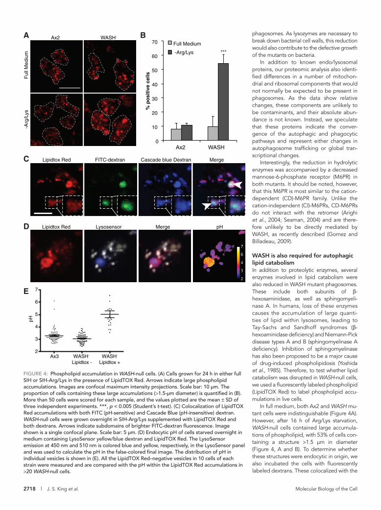
WASH is also required for autophagic lipid catabolismIn addition to proteolytic enzymes, several enzymes involved in lipid catabolism were also reduced in WASH mutant phagosomes. These include both subunits of β-hexosaminidase, as well as sphingomyeli-nase A. In humans, loss of these enzymes causes the accumulation of large quanti-ties of lipid within lysosomes, leading to Tay-Sachs and Sandhoff syndromes (β-hexoaminidase deficiency) and Niemann-Pick disease types A and B (sphingomyelinase A deficiency). Inhibition of sphin gomyelinase has also been proposed to be a major cause of drug-induced phospholipidosis (Yoshida et al., 1985). Therefore, to test whether lipid catabolism was disrupted in WASH-null cells, we used a fluorescently labeled phospholipid (LipidTOX Red) to label phospholipid accu-mulations in live cells.
In full medium, both Ax2 and WASH mu-tant cells were indistinguishable (Figure 4A). However, after 16 h of Arg/Lys starvation, WASH-null cells contained large accumula-tions of phospholipid, with 53% of cells con-taining a structure >1.5 μm in diameter (Figure 4, A and B). To determine whether these structures were endocytic in origin, we also incubated the cells with fluorescently labeled dextrans. These colocalized with the
FIGURE 4: Phospholipid accumulation in WASH-null cells. (A) Cells grown for 24 h in either full SIH or SIH-Arg/Lys in the presence of LipidTOX Red. Arrows indicate large phospholipid accumulations. Images are confocal maximum intensity projections. Scale bar: 10 μm. The proportion of cells containing these large accumulations (>1.5-μm diameter) is quantified in (B). More than 50 cells were scored for each sample, and the values plotted are the mean ± SD of three independent experiments. ***, p < 0.005 (Student’s t-test). (C) Colocalization of LipidTOX Red accumulations with both FITC (pH-sensitive) and Cascade Blue (pH-insensitive) dextran. WASH-null cells were grown overnight in SIH-Arg/Lys supplemented with LipidTOX Red and both dextrans. Arrows indicate subdomains of brighter FITC-dextran fluorescence. Image shown is a single confocal plane. Scale bar: 5 μm. (D) Endocytic pH of cells starved overnight in medium containing LysoSensor yellow/blue dextran and LipidTOX Red. The LysoSensor emission at 450 nm and 510 nm is colored blue and yellow, respectively, in the LysoSensor panel and was used to calculate the pH in the false-colored final image. The distribution of pH in individual vesicles is shown in (E). All the LipidTOX Red–negative vesicles in 10 cells of each strain were measured and are compared with the pH within the LipidTOX Red accumulations in >20 WASH-null cells.
0
10
20
30
40
50
60
70
Ax2 WASH %
po
siti
ve c
ells
Full Medium
-Arg/Lys
BAF
ull M
ediu
m-A
rg/L
ys
Ax2 WASH-
C Lipidtox Red FITC-dextran Cascade blue Dextran Merge
***
D Lipidtox Red Lysosensor Merge pH
Ax3 WASH- Lipidtox -
WASH-
Lipidtox +
2
3
4
5
6
7
pH
E
Volume 24 September 1, 2013 WASH and lysosomal recycling | 2719
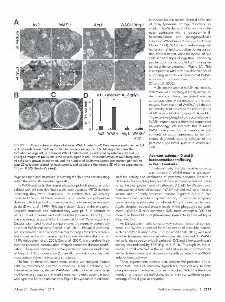
lar bodies (MLBs) are the classical hallmarks of many lysosomal storage disorders, in-cluding Tay-Sachs and Niemann-Pick dis-ease, consistent with a reduction in β-hexosaminidase and sphingomyelinase activity in WASH mutant cells (Schmitz and Muller, 1991). WASH is therefore required for lysosomal lipid catabolism during starva-tion. Note also that, while the cytosol of Ax2 cells showed signs of digestion, becoming patchy upon starvation, WASH mutants re-tained a dense cytoplasm (Figure 5A). This is comparable with previous observations of autophagy mutants, confirming that WASH-null cells do not lose mass upon starvation (Otto et al., 2003).
MLBs are induced in WASH-null cells by starvation. As autophagy is highly active un-der these conditions, we tested whether autophagy directly contributed to this phe-notype. Examination of WASH/Atg1 double mutants by TEM indicated the accumulation of MLBs was blocked (Figure 5, A and D). The extensive phospholipid accumulation in WASH mutant cells is therefore dependent on autophagy. We interpret this to mean WASH is required for the membranes and contents of autophagosomes to be effi-ciently degraded, causing collapse of the perturbed lysosomal system in WASH-null cells.
Aberrant cathepsin D and β-hexosaminidase trafficking in WASH mutantsTo establish why the degradative capacity was reduced in WASH mutants, we exam-
ined the activity and localization of lysosomal enzymes. Despite a 50% reduction in the phagosomal compartment, when we mea-sured the total protein level of cathepsin D (catD) by Western blot, there was no difference between WASH-null and Ax2 cells, nor any accumulation of partly processed enzyme (Figure 6, A and B). We then measured the total enzymatic activity of lysosomal enzymes, using fluorogenic substrates for cathepsin D/E and β-hexosaminidase. Again, despite reduced protein levels in the phagocytic compart-ment, WASH-null cells contained 50% more cathepsin D/E and more than threefold more β-hexosaminidase activity than wild-type (Figure 6, C–E).
As Dictyostelium cells constitutively secrete lysosomal compo-nents, and WASH is essential for the secretion of insoluble material such as dextran (Dimond et al., 1981; Carnell et al., 2011), we asked whether lysosomal enzyme secretion was also blocked. In WASH-null cells, the secretion of both cathepsin D/E and β-hexosaminidase activity was reduced by 50% (Figure 6, F–H). This explains the in-crease in total activities in the mutant but also demonstrates that, unlike dextran, lysosomal enzymes are partly secreted by a WASH-independent pathway.
These experiments indicate that, despite the presence of ele-vated total levels of lysosomal hydrolases, their delivery to both phagosomes and autophagosomes is blocked. WASH is therefore needed for the correct trafficking, rather than the synthesis or pro-cessing, of the digestive enzymes.
FIGURE 5: Ultrastructural analysis of starved WASH mutants. (A) Cells were placed in either full or Arg/Lys-deficient medium for 36 h before processing for TEM. Micrographs show the formation of large MLBs in starved WASH mutant cells, as indicated by asterisks. (B) and (C) Enlarged images of MLBs, (B) is the boxed region in (A). (D) Quantification of MLB frequency. At cells were grown as indicated, and the number of MLBs was scored per section, per cell. At least 20 cells were scored for each sample, and values are the mean ± SD of three experiments. ***, p < 0.005 (Student’s t-test).
A
B D
0
1
2
3
4
Ax2 WASH Atg1 WASH/Atg1
MLB
's p
er c
ell
Full medium - Arg/lys
Ax2 WASH- Atg1- WASH-/Atg1-
Ful
l Med
ium
-Arg
/Lys
*
500nm
1µm
**
*C
*** ***
large phospholipid structures, indicating the lipid was accumulating within the endocytic system (Figure 4C).
In WASH-null cells, the largest phospholipid-rich structures colo-calized with pH-sensitive fluorescein isothiocyanate (FITC)-dextran, indicating they were neutralized. To confirm this, we directly measured the pH of these vesicles using LysoSensor yellow/blue dextran, which has both pH-sensitive and pH-insensitive emission peaks (Diwu et al., 1999). This again accumulated in the phospho-lipid-rich structures and indicated they were pH 5, in contrast to pH 3.1 found in normal endocytic vesicles (Figure 4, D and E). This was surprising, because WASH is essential for v-ATPase recycling in Dictyostelium, and neutral compartments are normally completely absent in WASH-null cells (Carnell et al., 2011). Elevated lysosomal pH has, however, been reported in macrophages forced to accumu-late cholesterol and in several lipid storage disorders (Bach et al., 1999; Holopainen et al., 2001; Cox et al., 2007). It is therefore likely that the excessive accumulation of lipids somehow disrupts acidifi-cation. These compartments also frequently contained a subdomain of brighter FITC-fluorescence (Figure 4C, arrows), indicating they must contain some intravesicular structures.
To look at these structures more closely, we analyzed mutant cells by transmission electron microscopy (TEM). Confirming our live-cell experiments, starved WASH-null cells contained many large multilamellar structures that were almost completely absent in both wild-type and full medium controls (Figure 5). Lysosomal multilamel-

2720 | J. S. King et al. Molecular Biology of the Cell
(Park et al., 2013). When we expressed FAM21-GFP in WASH-null cells, it clearly localized to a vesicular membrane surrounding the catD structure, indicating that the catD accumulates where WASH would normally be active.
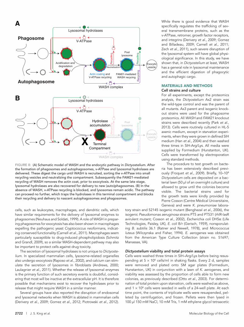
Previous analysis of Dictyostelium phagosome maturation has shown that the lysosomal hydrolases, including catD, are recovered from the phagosome at a late stage, just prior to exocytosis of the indigestible material (Gotthardt et al., 2002). Loss of WASH must therefore block the pathway before this recycling can take place. Lysosomal proteins therefore accumulate in a terminal compart-ment, unable to fuse with or be delivered to nascent phagosomes and autophagosomes. Furthermore, the identical results obtained in FAM21 -null cells (in which the pathway proceeds past v-ATPase re-cycling and neutralization before stalling) indicate that hydrolase re-cycling occurs at the same time as or after FAM21-mediated removal of WASH and is independent of v-ATPase retrieval (Figure 8).
DISCUSSIONThe WASH complex, and the actin it polymerizes on vesicles, has been associated with an increasing number of trafficking roles. In this paper, we demonstrate that disruption of WASH causes the ac-cumulation of lysosomal hydrolases in a late endocytic compart-ment, where they get trapped and are thus unavailable for recycling to nascent degradative compartments.
The reduced delivery of lysosomal hydrolases in WASH mutants has several physiological implications. The primary defect in these mutants is a decreased capacity to degrade lipids and proteins. This means that both phagocytosis and autophagy are compromised, and the cell is unable to use either bacteria or its own cytoplasm to provide nutrients. Therefore, while WASH mutants grow normally on our standard laboratory strain of K. aerogenes, they have much more difficulty growing on other bacteria. It should be noted that both the Dictyostelium and laboratory Klebsiella have been strongly selected for optimum growth over decades of coculture in the laboratory, and, although K. aerogenes, Klebsiella pneumoniae, and Enterobacter aerogenes are now classified as the same bacteria (Brisse et al., 2006), WASH mutants could grow only on our standard laboratory strain. In the wild, amoebae need to consume a wide range of bacteria, and so WASH function will be crucial for survival. While there is no obvious pattern in the types of bacteria that WASH mutants cannot use, it is likely that they have particularly high levels of fat or lipid or are simply more difficult to digest. In addition to enzymes required for protein and lipid catabolism, we also identi-fied lysozyme as reduced in WASH mutant phagosomes. As lysozyme is required to break down the bacterial cell wall, this re-duction will also disrupt the digestive process and explain this phenotype.
Consistent with a general lysosomal defect, WASH is also critical for autophagic degradation. While there are no overt defects in au-tophagosome formation or acidification, WASH-null cells are unable to digest their cytoplasm and survive starvation. In addition to known endocytic proteins, our proteomics assay also identified a number of components, such as mitochondrial and ribosomal pro-teins, that would not normally be expected to be present in phago-somes. As these data are ratiometric, it is unlikely that these are contaminants, and they instead may be the products of autophagy, indicative of altered autophagic trafficking and fusion with the endo/phagocytic pathway.
Interestingly, we also show that induction of autophagy leads to the accumulation of large quantities of phospholipid within lyso-somal multilamellar bodies. This is characteristic of many lysosomal storage disorders and implies that autophagy is a major source of
WASH is required for lysosomal enzyme recyclingTo determine where lysosomal trafficking was disrupted, we exam-ined the localization of catD in mutant cells by immunofluorescence. As previously reported, in Ax2 cells fixed with ultracold methanol, the catD antibody labeled spots on several large ring structures (endocytic vesicles) and numerous smaller puncta (Figure 7A; Neu-haus et al., 2002). In both WASH- and FAM21-null cells however, catD frequently accumulated in a single large structure, with a sig-nificant reduction in other small puncta (Figure 7, A and C). This was confirmed using an alternative probe, fluorescently labeled pepsta-tin A, which specifically labels active catD and gave identical results (Figure 7B; Chen et al., 2000).
These observations indicate that catD is retained within a spe-cific cellular compartment in WASH mutants. These concentrated catD structures, however, neither colocalized with the Golgi marker golvesin-GFP (Schneider et al., 2000) nor with the endoplasmic re-ticulum marker protein disulfide isomerase (PDI; Figure 7, D and E). Recently we showed that FAM21-GFP is able to localize to maturing endosomes independently of the other WASH complex members
FIGURE 6: Hydrolytic activities in WASH mutants. (A) Western blot of catD (green) with 3-methylcrotonyl-CoA carboxylase α (MCCC1) as loading control in red. Quantitation of three independent blots shown in (B). (C–E) hydrolytic activity in Ax2 (circles) and WASH-null (crosses) whole-cell lysates using fluorogenic substrates for (C) cathepsin D/E and (D) β-hexosaminidase. Activity is indicated by the increase in fluorescence over time. (E) The relative activity of WASH-null cells (dark gray) compared with Ax2 (light gray). (F–H) Secreted activity of (F) catD and (G) β-hexosaminidase. Cells were placed in fresh media for the times indicated, and the activity in the media was measured as above. (H) Relative rates of secretion are plotted in (H). All values are the means ± SD of three independent experiments. *, p < 0.05; **, p < 0.01; ***, p < 0.005 (Student’s t-test).
200
400
600
0 20 40 60
Flu
ores
cenc
e (A
.U.)
Time (min)
0
400
800
1200
1600
µmol
MU
/ 10
3 ce
lls
0 20 40 60 Time (min)
Rel
ativ
e ac
tivity
Rel
ativ
e ac
tivity
0
1
2
3
4
5
Cathepsin Hex.
Time (Hrs)
0 0 1 2 3 4
Sec
rete
d ac
tivity
(%
)
0 1 2 3 4 Time (Hrs)
Sec
rete
d ac
tivity
(%
)
0
0.2
0.4
0.6
0.8
1
Cathepsin Hex.
Rel
ativ
e ac
tivity
Ax2 WASH0
0.5
1.0
Cat
heps
in D
pro
tein
BA
C D E
F G H
Ax2 WASH
10070
50
40
35
kDa
Hexosaminidase
Cathepsin Hexosaminidase
*
**
****
25
50
75
100
0
25
50
75
100
0
CathepsinAx2
WASHAx2WASH

Volume 24 September 1, 2013 WASH and lysosomal recycling | 2721
(Carnell et al., 2011), so we were surprised to find the largest accumulations of lipid within neutral vesicles (Figure 4C). However, lyso-somal pH is elevated in several lipid storage diseases (Bach et al., 1999; Holopainen et al., 2001), and the autophagy-mediated accumulation of lysosomal cholesterol in Niemann-Pick type C disease also inhibits proteolytic activity (Elrick et al., 2012). It is therefore likely that the excessive lipid ac-cumulation in WASH-null lysosomes dis-rupts lysosomal function in multiple ways, exacerbating the underlying defect in enzyme delivery.
In a number of respects, our observa-tions contrast with the work of others on mammalian epithelial cells, indicating a novel mechanism by which WASH influ-ences lysosomal trafficking. In HeLa cells, WASH was shown to directly regulate ret-romer-mediated retrograde transport of the CI-M6PR from endosomes to the Golgi. WASH knockdown therefore leads to CI-M6PR accumulation on endosomes (Gomez and Billadeau, 2009). In contrast, our pro-teomic data show a putative CD-M6PR de-creased on both WASH- and FAM21-null phagosomes. Loss of FAM21 causes the constitutive activation of WASH in both Dictyostelium and mammalian cells (Park et al., 2013), and the FAM21-free complex remains able to mediate v-ATPase recycling from late endosomes. In this respect, WASH and FAM21 mutants have opposing pheno-types. Despite the contrasting defects in v-ATPase trafficking, the putative CD-M6PR is reduced in both mutant phagosomes. This implies either indirect regulation by WASH or that the hydrolases are recycled with the WASH complex independently of its actin polymerization activity.
Our data indicate that lysosomal hydro-lases are retrieved after WASH-mediated neutralization. This is in good agreement with previous analysis of Dictyostelium pha-gosome maturation, which showed that dif-ferent hydrolases are delivered to and re-covered from phagosomes at distinct stages in their maturation (Gotthardt et al., 2002). It is not known how the soluble enzymes are selectively retrieved, or how different lyso-somal populations are maintained, but WASH is clearly important to define distinct
phases of phagosome maturation. Binding of M6PRs to lysosomal hydrolases is pH dependent, resulting in their release when they enter acidified lysosomes (Seaman, 2004). It is therefore plausible that a similar mechanism occurs during hydrolase recycling, requir-ing WASH-mediated neutralization before enzyme retrieval.
While the general mechanisms are conserved, there are clearly substantial differences in the trafficking pathways of Dictyostelium and mammalian epithelial and fibroblast cell lines. As a professional phagocyte, Dictyostelium is more representative of other phagocytic
lipid delivery to the lysosome. Autophagy is also required for the biogenesis of physiological MLBs, such as those secreted by lung alveolar cells as a surfactant, indicating a general role for autophagy in the transport of lipids (Hariri et al., 2000). Autophagy is therefore a conserved source of lysosomal phospholipid and may therefore be a viable therapeutic target for the treatment of lysosomal storage disorders.
Previous work has shown that WASH is absolutely required for v-ATPase recycling and vesicle neutralization in Dictyostelium
FIGURE 7: Localization of catD in WASH mutant cells. (A) Ultracold methanol fixed cells stained with anti-catD antibody. (B) Paraformaldehyde-fixed cells stained with Bodipy-pepstatin A. (C) Quantification of large aggregate frequency in cells stained as in (A); ***, p < 0.005 (Student’s t-test). For further characterization of these structures, colocalization of catD with (D) golvesin-GFP (Golgi), (E) anti-PDI antibody staining, and (F) FAM21-GFP was examined in fixed, WASH-null cells. Deconvolved wide-field images shown in all panels are all maximum intensity projections, except (E) and (F), which are single planes. All scale bars: 5 μm.
A CAx2 WASH FAM21
B
D
E
F
Cat D golvesin-GFP Merge
Cat D α-PDI Merge
Cat D FAM21-GFP Merge
Bod
ipy-
Pep
Aan
ti-ca
tD
Ax2 WASH FAM21 0
20
40
60
% c
ells
with
larg
e st
ruct
ures
***
***
2722 | J. S. King et al. Molecular Biology of the Cell
While there is good evidence that WASH specifically regulates the trafficking of sev-eral transmembrane proteins, such as the v-ATPase, retromer, growth factor receptors, and integrins (Derivery et al., 2009; Gomez and Billadeau, 2009; Carnell et al., 2011; Zech et al., 2011), such severe disruption of the lysosomal system will have global physi-ological significance. In this study, we have shown that, in Dictyostelium at least, WASH has a general role in lysosomal maintenance and the efficient digestion of phagocytic and autophagic cargo.
MATERIALS AND METHODSCell strains and cultureFor all experiments, except the proteomics analysis, the Dictyostelium Ax2 strain was the wild-type control and was the parent of all mutants. Ax3 parent and isogenic knock-out strains were used for the phagosome proteomics. All WASH and FAM21 knockout strains were described recently (Park et al., 2013). Cells were routinely cultured in HL-5 axenic medium, except in starvation experi-ments, when they were grown in defined SIH medium (Han et al., 2004) and then washed three times in SIH-Arg/Lys. All media were supplied by Formedium (Hunstanton, UK). Cells were transformed by electroporation using standard methods.
The procedure to test growth on bacte-ria has been extensively described previ-ously (Froquet et al., 2009). Briefly, 10–104 Dictyostelium cells are deposited on a bac-terial lawn (50 μl of an overnight culture) and allowed to grow until the colonies become visible. The bacterial strains used for the growth tests were kindly provided by Pierre Cosson (Centre Médical Universitaire, Geneva) and were K. pneumoniae labora-
tory strain and 52145 isogenic mutant (Benghezal et al., 2006), the isogenic Pseudomonas aeruginosa strains PT5 and PT531 (rhlR-lasR avirulent mutant; Cosson et al., 2002), Escherichia coli DH5α (Life Technologies, Paisley, UK), E. coli B/r (Gerisch, 1959), nonsporulat-ing B. subtilis 36.1 (Ratner and Newell, 1978), and Micrococcus luteus (Wilczynska and Fisher, 1994). E. aerogenes was obtained from the American Type Culture Collection (strain no. 51697; Manassas, VA).
Dictyostelium viability and total protein assaysCells were washed three times in SIH–Arg/Lys before being resus-pending at 5 × 10⁶ cells/ml in shaking flasks. Every 2 d, samples were removed and plated onto SM agar plates (Formedium, Hunstanton, UK) in conjunction with a lawn of K. aerogenes, and viability was assessed by the proportion of cells able to form new colonies, as previously described (Otto et al., 2003). For determi-nation of total protein upon starvation, cells were washed as above, and 1 × 10⁶ cells were seeded in wells of a 24-well plate. At each time point, the contents of triplicate wells were resuspended, pel-leted by centrifugation, and frozen. Pellets were then lysed in 100 μl 150 mM NaCl, 10 mM Tris, 1 mM ethylene glycol tetraacetic
cells, such as leukocytes, macrophages, and dendritic cells, which have similar requirements for the delivery of lysosomal enzymes to phagosomes (Neuhaus and Soldati, 1999). A role of WASH in prepar-ing phagosomes for exocytosis has also been shown in macrophages expelling the pathogenic yeast Cryptococcus neoformans, indicat-ing conserved functionality (Carnell et al., 2011). Macrophages seem particularly susceptible to drug-induced phospholipidosis (Schmitz and Grandl, 2009), so a similar WASH-dependent pathway may also be important to protect cells against drug toxicity.
The secretion of lysosomal hydrolases is not unique to Dictyoste-lium. In specialized mammalian cells, lysosome-related organelles also undergo exocytosis (Raposo et al., 2002), and calcium can stim-ulate the secretion of lysosomes in fibroblasts (Andrews, 2000; Laulagnier et al., 2011). Whether the release of lysosomal enzymes is the primary function of such secretory events is doubtful, consid-ering that most will be inactive at the extracellular pH. It is therefore possible that mechanisms exist to recover the hydrolases prior to release that might require WASH in a similar manner.
Several groups have also reported the disruption of endosomal and lysosomal networks when WASH is ablated in mammalian cells (Derivery et al., 2009; Gomez et al., 2012; Piotrowski et al., 2012).
FIGURE 8: (A) Schematic model of WASH and the endocytic pathway in Dictyostelium. After the formation of phagosomes and autophagosomes, v-ATPase and lysosomal hydrolases are delivered. These digest the cargo until WASH is recruited, sorting the v-ATPase into small recycling vesicles and neutralizing the compartment. Subsequently the FAM21-mediated recycling of WASH removes the actin coat, prior to exocytosis. At the same late stage, lysosomal hydrolases are also recovered for delivery to new (auto)phagosomes. (B) In the absence of WASH, v-ATPase recycling is blocked, and lysosomes remain acidic. The pathway can proceed no further, which traps the hydrolases in this terminal compartment and blocks their recycling and delivery to nascent autophagosomes and phagosomes.
Phagosome
Autophagosome
V
V-ATPase
Lysosomal hydrolases
V
VVV
VV
V
Acidification and degradation
W
V
VVV
VV
V
W
W
WWW
WW
Actin coating and V-ATPase recycling
V
W
W
WW
W
WW
Neutralisation
FAM21-mediated WASH recycing
Postlysosome
Rec
yclin
g
Rec
yclin
gR
ecyc
ling
WASH complex
Exocyticvesicle
VV
VVV
V
V-ATPase
Lysosomal hydrolases
V
VVV
VV
V
WWW
V
VVV
VV
V
VVV
WWWW
WWW
WWWWWWW
WWWW
WWWWWWWW
Neutralisation
Postlysosome
Rec
yclin
g
Rec
yclin
gR
ecyc
ling
WASH complexWW
Exocyticvesicle
VV
VVV
A
B
Hydrolaseaccumulation
X TerminalCompartment

Volume 24 September 1, 2013 WASH and lysosomal recycling | 2723
manually outlining each vesicle and measuring the mean intensity of each channel.
ImmunohistochemistryTwo methods of fixation were used. All antibody staining was done using cells fixed in ultracold methanol, as described by Hagedorn et al. (2006). Briefly, cells were seeded onto thin (no. 0) acid-washed coverslips and left to adhere. They were then plunged into a beaker of −80°C methanol on dry ice and left to fix for 30 min. After several washes in phosphate-buffered saline (PBS), coverslips were blocked with 3% bovine serum albumin (BSA) and stained. The rabbit polyclonal anti-catD antibody was a kind gift from Agnes Journet (Journet et al., 1999), and PDI was stained using a cocktail of five monoclonal antibodies. For Bodipy-pepstatin A staining, cells were fixed with 4% paraformaldehyde, 0.1% Brij35 in citrate buffer (50 mM sodium citrate, pH 4.1) for 20 min before being washed and then stained with 1 μM Bodipy-pepstatin A (Life Technologies; Chen et al., 2000). Coverslips were kept in citrate buffer at all stages to maintain cathepsin in the active conformation. All coverslips were mounted in ProLong Gold with 4′,6-diamidino-2-phenylindole (Life Technologies). Images were captured on an Olympus IX81 wide-field fluorescence microscope with a 100×/1.4 NA objective and an additional 1.6× optovar magnification. For deconvolution, a Z-stack of images (0.25-μm step size) was captured with a CoolSNAPHQ camera (Photometrics). Images were then deconvolved by Volocity software (Perkin Elmer-Cetus, Waltham, MA) using calculated point-spread functions.
Phagocytosis and in vivo proteolysis assayBead uptake was measured as previously described (Sattler et al., 2013). Fluorescent beads of 1-μm diameter (fluorescent YG-carbox-ylated beads; Polysciences, Warrington, PA) were added to cells at a 200:1 dilution; at each time point, cells were washed free of un-bound beads, and the number of ingested beads was determined by flow cytometry. Proteolysis experiments were adapted from Yates et al. (2005) and carried out as described previously (Gopaldass et al., 2012). Briefly, cells were fed beads coupled to Alexa Fluor 594 succinimidyl ester (Life Technologies) and BSA labeled with DQ Green at a self-quenching concentration (Molecular Probes). On BSA proteolysis, DQ Green fluorescence increases due to de-quenching of the fluorophore. Results were normalized to bead up-take by plotting the ratio of DQ Green to Alexa Fluor 594 fluores-cence as a function of time.
TEMFor investigating subcellular structures using TEM, adherent Dictyos-telium cells were fixed in HL5c medium with 2% glutaraldehyde and 1% paraformaldehyde for 1 h. Subsequently the cells were scraped off the dish, washed with buffer, and incubated for 1 h with 0.5% OsO4. The samples were washed several times; this was followed by an incubation step with 1% tannin for 20 min at room temperature. The cells were washed again, dehydrated, embedded in Epon resin, and processed for conventional electron microscopy. Grids were ex-amined with a Tecnai Spirit transmission electron microscope (FEI, Eindhoven, Netherlands). Images were quantified by counting the number of MLBs in >20 cells for each strain/condition.
Cathepsin and hexosaminidase assaysFor enzymatic assays, cells growing in HL5 were resuspended, washed twice in KK2 buffer (0.1 M potassium phosphate, pH 6.1), and lysed in 50 mM sodium citrate buffer (pH 4.2) with 1% Triton X100 at 5 × 10⁴ cells/ml. For cathepsin activity, lysates were
acid (EGTA), 1 mM EDTA, 1% NP40 (pH 7.5), and total protein measures were taken using Precision Red reagent (Cytoskeleton, Denver, CO). Protein levels were then normalized to the unstarved protein measurements for each strain.
Cloning and gene disruptionFor generating an Atg1 knockout construct, the 5′ and 3′ frag-ments of the gene were amplified using the primers AAACAA-ATGAACCCTTTGCC/aagcttTTGGATCCCATAAGGAAGTGAA-GAGGCG and GGATCCaaaagcttTATTCATCACCAACCGAGGC/TGAGTTCTCACTTCAAATGC. These were then combined by PCR, and the floxed blasticidin cassette from pLPBLP (Faix et al., 2004) was inserted as a BamHI/HindIII fragment to generate the final Atg1 knockout construct pJSK471. The Bsr cassette from WASH-null cells was removed using Cre recombinase (Faix et al., 2004), and pJSK471 was used to make both a WASH/Atg1 double and an Ax2 Atg1 single knockout. The FAM21-GFP construct used was described by Park et al. (2013), and the golvesin-GFP plasmid was generated by amplifying the full-length golvesin gene from cDNA, adding 5′BamHI/3′XbaI sites, and subcloning into the GFP-fusion vector pDM450 (Veltman et al., 2009).
Live-cell imaging and pH determinationAutophagosomes were observed using the GFP-Atg8 plasmid pDM430 (King et al., 2011). High-resolution images were obtained by overlaying cells with a layer of 1% agarose and compressing them by capillary action, as previously described (Yumura et al., 1984; King et al., 2011). Images were obtained on a Nikon (Tokyo, Japan) Eclipse TE 2000-U microscope equipped with a 100×/1.45 numerical aperture (NA) Nikon total internal reflection fluorescence oil-immersion objective, 473-nm diode laser illumination (Omicron, Rodgau, Germany), and a Cascade 512F EMCCD camera (Photo-metrics, Tucson, AZ).
For determination of autophagosome induction upon starvation, Z-series of images (0.25-μm step size) of uncompressed cells were taken at each time point, using an Olympus IX81 inverted wide-field microscope equipped with a 60×/1.42 NA Plan-ApoN objective. The number of visible puncta in maximum intensity projections was scored for >100 cells at each point. Differential interference contrast (DIC) images were captured on an inverted microscope fitted with a 100×/1.40 NA oil-immersion objective (Nikon).
For phospholipid labeling, cells were incubated with HCS LipidTOX Red (Molecular Probes) diluted 2000× in SIH or SIH–Arg/Lys for 24 h. The endocytic pathway was labeled by also including 0.2 mg/ml Cascade Blue and 0.4 mg/ml FITC-dextran (Sigma-Aldrich, St. Louis, MO). Live-cell images were captured on an Olympus FV1000 confocal microscope, using a Plan-ApoN 60×/1.4 NA objective. Ax2 control cells were used to calibrate the microscope laser intensities such that the pH-sensitive FITC fluoresced only in a subset of vesicles, and identical settings were used with all strains and conditions. For direct determination of vesicular pH, cells were incubated overnight with 0.2 mg/ml LysoSensor yellow/blue dextran (Life Technologies; Diwu et al., 1999). Confocal images were then collected, using 365-nm excitation and detecting the emission at both 450 and 510 nm. pH was determined by comparing the ratio of 450/510-nm emission to a standard curve generated by imaging LysoSensor in SIH medium at different pHs (Figure S1B). The false-colored ve-sicular pH images were then generated using ImageJ (http://rsbweb.nih.gov/ij) by thresholding the 450-nm channel to gener-ate a mask and converting the 450/510-nm emission ratios to pH values. The emission ratio of individual vesicles was determined by
2724 | J. S. King et al. Molecular Biology of the Cell
and maximum injection time of 750 ms. The normalized collision energies were set to 35% for CID and 60% for HCD.
Protein identification was performed with the EasyProt platform (Gluck et al., 2012). After peak list generation, the CID and HCD spectra were merged for simultaneous identification and quantifica-tion (Dayon et al., 2010). The parent ion tolerance was set to 10 ppm. TMT-sixplex amino terminus and TMT-sixplex lysine (229.1629 Da), carbamidomethylation of cysteines were set as fixed modifications. All data sets were searched once in the forward and once in the re-verse Dictyostelium Uniprot_sprot database. For identification, only proteins matching two different peptide sequences were kept.
Isobaric quantification was performed using the IsoQuant mod-ule of EasyProt. TMT sixplex was selected as the reporter, and a mass tolerance of 0.05 m/z was used to calculate WASH/Ax2 and FAM21/Ax2 protein ratios. Statistics were performed using EasyProt’s Mascat and Libra statistical methods to generate a list of proteins that significantly differed between samples.
Western blottingCells growing in HL5 were resuspended, washed in KK2 buffer, and lysed in 150 mM NaCl, 10 mM Tris, 1 mM EGTA, 1 mM EDTA, 1% NP40 (pH 7.5). Samples were then normalized to total protein con-tent and boiled in SDS buffer (Life Technologies) before being loaded on a 10% Bis-Tris acrylamide Nu-PAGE gel (Life Technolo-gies). Gels were then transferred onto nitrocellulose membrane be-fore being probed with anti-catD antibody diluted 1:5000 in PBS (Journet et al., 1999) and a DyLight 800 anti-rabbit fluorescent sec-ondary antibody (Pierce, Rockford, IL). As a loading control, we used Alexa Fluor 680–labeled streptavidin (Life Technologies), which, in Dictyostelium, recognizes a single protein at ∼77 kDa correspond-ing to the mitochondrial methylcrotonyl-CoA carboxylase (Davidson et al., 2013). Blots were analyzed on a fluorescent gel imager, and images were quantified using ImageJ.
incubated at 37°C with 50 μM cathepsin fluorogenic substrate (BML-P145-001; Enzo Life Sciences, Exeter, UK) in 50 mM citrate buffer (pH 4.2), and samples were removed and quenched by being diluted at 1:10 in 10% trichloroacetic acid. Fluorescence at 330-nm excitation/410-nm emission was then detected in a fluorometer. For β-hexoaminidase activity, 2 mM 4-methylumbelliferyl-β-d-N-acetylglucosamine (Calbiochem, San Diego, CA) was used as sub-strate, and reactions were quenched by dilution at 1:10 in 0.2 M glycine, 0.2 M Na2CO3. Fluorescence was measured at 365-nm excitation/445-nm emission. For calculation of β-hexoaminidase activity, 4-methylumbelliferone was used to generate a standard curve.
For measuring secreted enzyme activity, cells were washed three times in fresh media and seeded at 2 × 10⁶ cells per well of a 24-well plate in 500 μl of fresh medium. At each time point, the medium was carefully removed and spun for 30 s at 3000 rpm in a benchtop mi-crofuge to pellet any cells, and 400 μl of the supernatant was taken. Activity was determined by incubating samples of the media with the appropriate substrates as above for 1 h at 37°C.
Mass spectrometry and analysisPhagosomes were isolated by density centrifugation after cells were fed latex beads, as described previously, using a pulse–chase of 15 min/2 h 45 min (Gotthardt et al., 2006b). The reduction, alkyla-tion, digestion, and tandem mass tag (TMT) labeling was mainly performed as described by Dayon et al. (2008). Briefly, phagosome pellets were resuspended at 100 μg proteins per 33 μl of triethylam-monium hydrogen carbonate buffer (TEAB: 0.1 M, pH 8.5, 6 M urea). After addition of 50 mM Tris-(2-carboxyethyl) phosphine hydrochloride (TCEP), samples were reduced for 1 h at 37°C. Alkyla-tion was then performed at room temperature in the dark for 30 min after addition of 1 μl of 400 mM iodoacetamide. Then 67 μl of TEAB and 2 μg trypsin were added, and digestion was performed over-night at 37°C. Each sample was then labeled with a TMT reagent according to the manufacturer’s instructions (Proteome Sciences, Frankfurt, Germany) and evaporated under a speed-vacuum.
Off-gel electrophoresis was performed according to the manu-facturer’s instructions (Agilent, Santa Clara, CA). After being de-salted, pooled samples were reconstituted in OFFGEL solution. Isoelectric focusing was performed using a 12-well frame on an Immobiline DryStrip (pH 3–10, 13 cm) and run at 8000 V, 50 μA, 200 mW, until 20 kVh was reached. The 12 fractions were then recovered and desalted using C18 MicroSpin columns.
ESI LTQ-OT (electrospray ionization linear trap quadropole orbi-trap) mass spectrometry (MS) was performed on a LTQ Orbitrap Ve-los (Thermo Electron, Waltham, MA) equipped with a NanoAcquity system (Waters). Peptides were trapped on a 5-μm, 200-Å Magic C18 AQ (Michrom) 0.1-mm × 20-mm precolumn and separated on a 5-μm, 100-Å Magic C18 AQ (Michrom, Auburn, CA) 0.75-mm × 150-mm column with a gravity-pulled emitter. Analytical separation was run for 65 min using a gradient of H2O/FA 99.9%/0.1% (solvent A) and CH3CN/FA 99.9%/0.1% (solvent B). The gradient was run as follows: 95% A and 5% B at 0–1 min, then to 65% A and 35% B at 55 min, and 20% A and 80% B at 65 min at a flow rate of 220 nl/min. For MS survey scans, the OT resolution was set to 60,000 and the ion population was set to 5 × 10⁵, with an m/z window of 400–2000. A maximum of three precursors were selected for both collision-in-duced dissociation (CID) in the LTQ and high-energy C-trap disso-ciation (HCD) with analysis in the OT. For tandem MS (MS/MS) in the LTQ, the ion population was set to 7000 (isolation width of 2 m/z), while for MS/MS detection in the OT, it was set to 2 × 10⁵ (isolation width of 2.5 m/z), with resolution of 7500, first mass at m/z = 100,
ACKNOWLEDGMENTSWe thank Agnes Journet for kindly providing us with catD antibody; Pierre Cosson for sharing the bacterial strains; David Strachan for help with image processing; and Alex Scherl, Carla Pasquarello, Patrizia Arboit, and Alexandre Hainard from the Proteomic Core Facility (CMU, Geneva) for their proteomics expertise and help. We are also very grateful to Pete Thomason, Tobias Zech, and Laura Machesky for many helpful discussions and critiques of the manu-script and to Pete Watson for informed suggestions. This work was supported by Cancer Research–UK core funding to R.H.I. and a grant from the Swiss National Science Foundation to the T.S. laboratory.
REFERENCESAndrews NW (2000). Regulated secretion of conventional lysosomes. Trends
Cell Biol 10, 316–321.Arighi CN, Hartnell LM, Aguilar RC, Haft CR, Bonifacino JS (2004). Role of
the mammalian retromer in sorting of the cation-independent mannose 6-phosphate receptor. J Cell Biol 165, 123–133.
Bach G, Chen CS, Pagano RE (1999). Elevated lysosomal pH in Mucolipido-sis type IV cells. Clin Chim Acta 280, 173–179.
Benghezal M et al. (2006). Specific host genes required for the killing of Klebsiella bacteria by phagocytes. Cell Microbiol 8, 139–148.
Brisse S, Grimont F, Grimont PAD (2006). The genus Klebsiella. In: The Prokaryotes, eds. M Dworkin, S Falkow, E Rosenberg, K-H Schleifer, and E Stackebrandt, New York: Springer, 159–196.
Calvo-Garrido J, Carilla-Latorre S, Kubohara Y, Santos-Rodrigo N, Mesquita A, Soldati T, Golstein P, Escalante R (2010). Autophagy in Dictyostelium: genes and pathways, cell death and infection. Autophagy 6, 686–701.
Carnell M, Zech T, Calaminus SD, Ura S, Hagedorn M, Johnston SA, May RC, Soldati T, Machesky LM, Insall RH (2011). Actin polymerization

Volume 24 September 1, 2013 WASH and lysosomal recycling | 2725
Hagedorn M, Neuhaus EM, Soldati T (2006). Optimized fixation and immu-nofluorescence staining methods for Dictyostelium cells. Methods Mol Biol 346, 327–338.
Han SI, Friehs K, Flaschel E (2004). Cultivation of Dictyostelium discoideum on an improved synthetic medium in a conventional bioreactor. Process Biochem 39, 925–930.
Harbour ME, Breusegem SY, Antrobus R, Freeman C, Reid E, Seaman MN (2010). The cargo-selective retromer complex is a recruiting hub for protein complexes that regulate endosomal tubule dynamics. J Cell Sci 123, 3703–3717.
Hariri M, Millane G, Guimond MP, Guay G, Dennis JW, Nabi IR (2000). Bio-genesis of multilamellar bodies via autophagy. Mol Biol Cell 11, 255–268.
Holopainen JM, Saarikoski J, Kinnunen PK, Jarvela I (2001). Elevated lyso-somal pH in neuronal ceroid lipofuscinoses (NCLs). Eur J Biochem 268, 5851–5856.
Ichimura Y et al. (2000). A ubiquitin-like system mediates protein lipidation. Nature 408, 488–492.
Jia D, Gomez TS, Metlagel Z, Umetani J, Otwinowski Z, Rosen MK, Billadeau DD (2010). WASH and WAVE actin regulators of the Wiskott-Aldrich syndrome protein (WASP) family are controlled by analogous structurally related complexes. Proc Natl Acad Sci USA 107, 10442–10447.
Journet A, Chapel A, Jehan S, Adessi C, Freeze H, Klein G, Garin J (1999). Characterization of Dictyostelium discoideum cathepsin D. J Cell Sci 112, 3833–3843.
King JS (2012). Mechanical stress meets autophagy: potential implications for physiology and pathology. Trends Mol Med 18, 583–588.
King JS, Veltman DM, Insall RH (2011). The induction of autophagy by mechanical stress. Autophagy 7, 1490–1499.
Kirisako T, Ichimura Y, Okada H, Kabeya Y, Mizushima N, Yoshimori T, Ohsumi M, Takao T, Noda T, Ohsumi Y (2000). The reversible modifica-tion regulates the membrane-binding state of Apg8/Aut7 essential for autophagy and the cytoplasm to vacuole targeting pathway. J Cell Biol 151, 263–276.
Laulagnier K, Schieber NL, Maritzen T, Haucke V, Parton RG, Gruenberg J (2011). Role of AP1 and Gadkin in the traffic of secretory endo-lyso-somes. Mol Biol Cell 22, 2068–2082.
Linardopoulou EV, Parghi SS, Friedman C, Osborn GE, Parkhurst SM, Trask BJ (2007). Human subtelomeric WASH genes encode a new subclass of the WASP family. PLoS Genet 3, e237.
Mizushima N, Levine B, Cuervo AM, Klionsky DJ (2008). Autophagy fights disease through cellular self-digestion. Nature 451, 1069–1075.
Neuhaus EM, Almers W, Soldati T (2002). Morphology and dynamics of the endocytic pathway in Dictyostelium discoideum. Mol Biol Cell 13, 1390–1407.
Neuhaus EM, Soldati T (1999). Molecular mechanisms of membrane traf-ficking. What do we learn from Dictyostelium discoideum. Protist 150, 235–243.
Otto GP, Wu MY, Kazgan N, Anderson OR, Kessin RH (2003). Macroau-tophagy is required for multicellular development of the social amoeba Dictyostelium discoideum. J Biol Chem 278, 17636–17645.
Park L, Thomason PA, Zech T, King JS, Veltman DM, Carnell M, Ura S, Machesky LM, Insall RH (2013). Cyclical action of the WASH complex: FAM21 and capping protein drive WASH recycling, not initial recruit-ment. Dev Cell 24, 169–181.
Piotrowski JT, Gomez TS, Schoon RA, Mangalam AK, Billadeau DD (2012). WASHout T cells demonstrate defective receptor trafficking, prolifera-tion and effector function. Mol Cell Biol 33, 958–973.
Raposo G, Fevrier B, Stoorvogel W, Marks MS (2002). Lysosome-related organelles: a view from immunity and pigmentation. Cell Struct Funct 27, 443–456.
Ratner DI, Newell PC (1978). Linkage analysis in Dictyostelium discoideum using multiply marked tester strains: establishment of linkage group VII and the reassessment of earlier linkage data. J Gen Microbiol 109, 225–236.
Sattler N, Monroy R, Soldati T (2013). Quantitative analysis of phagocytosis and phagosome maturation. Methods Mol Biol 983, 383–402.
Schmitz G, Grandl M (2009). Endolysosomal phospholipidosis and cytosolic lipid droplet storage and release in macrophages. Biochim Biophys Acta 1791, 524–539.
Schmitz G, Muller G (1991). Structure and function of lamellar bodies, lipid-protein complexes involved in storage and secretion of cellular lipids. J Lipid Res 32, 1539–1570.
Schneider N, Schwartz JM, Kohler J, Becker M, Schwarz H, Gerisch G (2000). Golvesin-GFP fusions as distinct markers for Golgi and post-Golgi vesicles in Dictyostelium cells. Biol Cell 92, 495–511.
driven by WASH causes V-ATPase retrieval and vesicle neutralization before exocytosis. J Cell Biol 193, 831–839.
Chen CS, Chen WN, Zhou M, Arttamangkul S, Haugland RP (2000). Prob-ing the cathepsin D using a BODIPY FL-pepstatin A: applications in fluorescence polarization and microscopy. J Biochem Biophys Methods 42, 137–151.
Clarke M, Köhler J, Arana Q, Liu T, Heuser J, Gerisch G (2002). Dynamics of the vacuolar H+-ATPase in the contractile vacuole complex and the endosomal pathway of Dictyostelium cells. J Cell Sci 115, 2893–2905.
Cosson P, Zulianello L, Join-Lambert O, Faurisson F, Gebbie L, Benghezal M, Van Delden C, Curty LK, Kohler T (2002). Pseudomonas aeruginosa virulence analyzed in a Dictyostelium discoideum host system. J Bacte-riol 184, 3027–3033.
Cox BE, Griffin EE, Ullery JC, Jerome WG (2007). Effects of cellular choles-terol loading on macrophage foam cell lysosome acidification. J Lipid Res 48, 1012–1021.
Davidson AJ, King JS, Insall RH (2013). The use of streptavidin conjugates as immunoblot loading controls and mitochondrial markers for use with Dictyostelium discoideum. Biotechniques 55, 39–41.
Dayon L, Hainard A, Licker V, Turck N, Kuhn K, Hochstrasser DF, Burkhard PR, Sanchez JC (2008). Relative quantification of proteins in human cerebrospinal fluids by MS/MS using 6-plex isobaric tags. Anal Chem 80, 2921–2931.
Dayon L, Pasquarello C, Hoogland C, Sanchez JC, Scherl A (2010). Combin-ing low- and high-energy tandem mass spectra for optimized peptide quantification with isobaric tags. J Proteomics 73, 769–777.
Deretic V (2008). Autophagosome and phagosome. Methods Mol Biol 445, 1–10.
Derivery E, Sousa C, Gautier JJ, Lombard B, Loew D, Gautreau A (2009). The Arp2/3 activator WASH controls the fission of endosomes through a large multiprotein complex. Dev Cell 17, 712–723.
Dieckmann R, Gueho A, Monroy R, Ruppert T, Bloomfield G, Soldati T (2012). The balance in the delivery of ER components and the vacuolar proton pump to the phagosome depends on myosin IK in Dictyoste-lium. Mol Cell Proteomics 11, 886–900.
Dimond RL, Burns RA, Jordan KB (1981). Secretion of lysosomal enzymes in the cellular slime mold, Dictyostelium discoideum. J Biol Chem 256, 6565–6572.
Diwu Z, Chen CS, Zhang C, Klaubert DH, Haugland RP (1999). A novel acidotropic pH indicator and its potential application in labeling acidic organelles of live cells. Chem Biol 6, 411–418.
Duleh SN, Welch MD (2010). WASH and the Arp2/3 complex regulate endosome shape and trafficking. Cytoskeleton (Hoboken) 67, 193–206.
Elrick MJ, Yu T, Chung C, Lieberman AP (2012). Impaired proteolysis under-lies autophagic dysfunction in Niemann-Pick type C disease. Hum Mol Genet 21, 4876–4887.
Faix J, Kreppel L, Shaulsky G, Schleicher M, Kimmel AR (2004). A rapid and efficient method to generate multiple gene disruptions in Dictyostelium discoideum using a single selectable marker and the Cre-loxP system. Nucleic Acids Res 32, e143.
Froquet R, Lelong E, Marchetti A, Cosson P (2009). Dictyostelium discoi-deum: a model host to measure bacterial virulence. Nat Protoc 4, 25–30.
Gerisch G (1959). Ein submerskulturver fahren fur entwicklungsphysiologis-che untersuchungen an Dictyostelium discoideum. Naturwissenschaften 46, 654–656.
Gluck F et al. (2012). EasyProt—an easy-to-use graphical platform for pro-teomics data analysis. J Proteomics 79, 146–160.
Gomez TS, Billadeau DD (2009). A FAM21-containing WASH complex regu-lates retromer-dependent sorting. Dev Cell 17, 699–711.
Gomez TS, Gorman JA, de Narvajas AA, Koenig AO, Billadeau DD (2012). Trafficking defects in WASH-knockout fibroblasts originate from collapsed endosomal and lysosomal networks. Mol Biol Cell 23, 3215–3228.
Gopaldass N et al. (2012). Dynamin A, myosin IB and Abp1 couple phago-some maturation to F-actin binding. Traffic 13, 120–130.
Gotthardt D, Blancheteau V, Bosserhoff A, Ruppert T, Delorenzi M, Soldati T (2006a). Proteomics fingerprinting of phagosome maturation and evidence for the role of a Gα during uptake. Mol Cell Proteomics 5, 2228–2243.
Gotthardt D, Dieckmann R, Blancheteau V, Kistler C, Reichardt F, Soldati T (2006b). Preparation of intact, highly purified phagosomes from Dictyos-telium. Methods Mol Biol 346, 439–448.
Gotthardt D, Warnatz HJ, Henschel O, Bruckert F, Schleicher M, Soldati T (2002). High-resolution dissection of phagosome maturation reveals distinct membrane trafficking phases. Mol Biol Cell 13, 3508–3520.

2726 | J. S. King et al. Molecular Biology of the Cell
Yates RM, Hermetter A, Russell DG (2005). The kinetics of phagosome maturation as a function of phagosome/lysosome fusion and acquisition of hydrolytic activity. Traffic 6, 413–420.
Yoshida Y, Arimoto K, Sato M, Sakuragawa N, Arima M, Satoyoshi E (1985). Reduction of acid sphingomyelinase activity in human fibroblasts induced by AY-9944 and other cationic amphiphilic drugs. J Biochem 98, 1669–1679.
Yumura S, Mori H, Fukui Y (1984). Localization of actin and myosin for the study of ameboid movement in Dictyostelium using improved immuno-fluorescence. J Cell Biol 99, 894–899.
Zech T, Calaminus SD, Caswell P, Spence HJ, Carnell M, Insall RH, Norman J, Machesky LM (2011). The Arp2/3 activator WASH regulates α5β1-integrin-mediated invasive migration. J Cell Sci 124, 3753–3759.
Seaman MN (2004). Cargo-selective endosomal sorting for retrieval to the Golgi requires retromer. J Cell Biol 165, 111–122.
Veltman DM, Akar G, Bosgraaf L, Van Haastert PJ (2009). A new set of small, extrachromosomal expression vectors for Dictyostelium discoideum. Plasmid 61, 110–118.
Veltman DM, Insall RH (2010). WASP family proteins: their evolution and its physiological implications. Mol Biol Cell 21, 2880–2893.
Wilczynska Z, Fisher PR (1994). Analysis of a complex plasmid insertion in a phototaxis-deficient transformant of Dictyostelium discoideum selected on a Micrococcus luteus lawn. Plasmid 32, 182–194.
Xie Z, Klionsky DJ (2007). Autophagosome formation: core machinery and adaptations. Nat Cell Biol 9, 1102–1109.