Embed Size (px)
Citation preview
https://doi.org/10.33805/2573-3877.134
Volume 4 Issue 1 | PDF 134 | Pages 6 Volume 1 . Issue 1 | PDF 101 | Page 1 of x
Nursing and Health Care
Citation: Khaled A and Faiza A. The prevalence of left ventricular thrombus among heart failure patients admitted to Kuwait teaching hospital in Sana’a City between January 2014 -2017 (2019) Nursing and Health Care 4: 29-34.
29
Research Article ISSN: 2573-3877
The Prevalence of Left Ventricular Thrombus among Heart
Failure Patients Admitted to Kuwait Teaching Hospital in
Sana’a City between January 2014 -2017 Alaghbari Khaled
* and Askar Faiza
Affiliation: Internal Medicine, Sana’a University, Yemen
*Corresponding author: Alaghbari Khaled, Associated professor of internal Medicine, Sana’a University, Yemen, Tel: +96-7711118376,
Email: [email protected]
Citation: Khaled A and Faiza A. The prevalence of left ventricular thrombus among heart failure patients admitted to Kuwait teaching hospital
in Sana’a City between January 2014 -2017 (2019) Nursing and Health Care 4: 29-34.
Received: May 17, 2019
Accepted: June 03, 2019
Published: June 11, 2019
Copyright: © 2019 Khaled A, et al., This is an open-access article distributed under the terms of the Creative Commons Attribution License,
which permits unrestricted use, distribution, and reproduction in any medium, provided the original author and source are credited.
Abstract Background: Heart failure is a major Public Health problem due to its high morbidity and mortality rates .The Left Ventricular Thrombus (LVT)
is more frequently seen in acute heart failure as a complication of Left Ventricular (LV) systolic dysfunction.
Objectives: The objective of this study was to determine the prevalence of LVT and its outcome among Yemeni patients presented with heart
failure to Kuwait teaching hospital in Sana’a. This cross sectional retrospective study made during the period of January 2014-January 2017 study
for all patients admitted to the hospital with Heart Failure (HF).
Results: During study period 1856 patients with cardiac diseases were admitted to the hospital. Of this 217 were in (F. Among patient which
61(28.1%) had LVT. The mean age of patient presented with LVT was 51 years ± 8.1 Most cases were male (90%) while only (10%), were
females. Ischemic Heart Disease (IHD), Dilated Cardiac Myopathy (DCMP) and Hypertension, found to be an associated risk factors of LVT
represented (51%, 34.2% and 34.2%) respectively. However 8 (13%) of patients with LVT had embolic complications. The mortality rate during
hospitalization was 4 (6.6%).
Conclusion: The Ischemic heart disease was the leading cause of left ventricular thrombus.
Keywords: Left ventricular thrombus, Heart failure, Yemeni patients.
Abbreviations: LVT-Left Ventricular Thrombus, LV-Left Ventricular, HF-Heart failure, IHD-Ischemic Heart Disease, DCMP-Dilated Cardiac
Myopathy, AMI-Acute Myocardial Infarction, PCI-Percutaneous Intervention, TTE-Transthoracic Echocardiography, ACE-Angiotensin
Converting Enzymes, SEC-Spontaneous Echo Contrast, LVEF-Left Ventricular Ejection Fraction, ASE-American Society of Echo, MI-
Myocardial Infarction, HHD-Hypertensive Heart Disease, RHD-Rheumatic Heart Disease, LVEDD-Left Ventricular End Diastolic Dimensions,
EF-Ejection Fraction, DM-Diabetes Mellitus, LBBB-Left Bundle Branch Block, RBBB-Right Bundle Branch Block, SVT-Supra Ventricular
Tachycardia, AF-Atrial Fibrillation, HTN-Hypertension.
Introduction
Heart failure represents a major and growing public health problem
because of its prevalence, incidence, morbidity, mortality and
economic costs. The prevalence of HF is 2% to 3% of general
population [1]. Five million Americans are affected, with more than
530000 cases diagnosed each year [2]. The mortality rate from severe
HF remains >60% within 5 years of diagnosis and that of 50% of
hospitalized patients with HF required readmission to hospital within 6
months of discharge. The estimated costs of HF amounted to >35
billion $ per year in the USA [3]. The development of LVT is a well-
known complication in various cardiac conditions with the highest rate
observed in acute anterior myocardial infarction and congestive HF
reached to 10-30% [4,5]. As a result of severe left ventricular systolic
dysfunction [6,7]. Rabbani et al found that the incidence of LVT
remain persistently high reached to (35%) for Acute Myocardial
Infarction (AMI) involving the anterior wall [8]. The prevalence of
LVT, especially in early Percutaneous Intervention (PCI) facilities are
found to be reduced with estimation ranging between 5% and 15%
[9,10]. Solheim et al, reported an incidence of LVT within 3 months of
AMI in selected patients managed with primary PCI was 15% [7]. The
constellation of endothelial injury, hypercoagubility and blood
stagnation, which are well described previously as Virchow’s triad, for
formation of the thrombus [11,12]. In AMI, other predisposing factors
also play a role in the development of LVT such as large infarct size,
severe apical a synergy, LV aneurysm and anteroposterior myocardiac
infarction. The early recognition of LVT is vital to prevent the
unwanted sequel of systemic thromboembolic events [6,11,12].
Currently in Yemen the well-known diagnostic system applied in
majority health center and hospitals is Transthoracic Echocardiography
(TTE) which is easily accessible and believed to have over 85%
accuracy in proper imaging results [13-15].
However, care must be taken to exclude false positive results which
occasionally may occurred [16,17]. Improved LV cavity assessment
and thrombi detection using TTE contrast studies were noted to be
better than non-contrast TTE, especially for mural (Laminar) and
Khaled A, et al. Nursing and Health Care, 2019 PDF: 134, 4:1
Citation: Khaled A and Faiza A. The prevalence of left ventricular thrombus among heart failure patients admitted to Kuwait teaching hospital in Sana’a City between January 2014 -2017 (2019) Nursing and Health Care 4: 29-34.
30
smaller thrombus [18]. Although several therapies as B-blockers,
Angiotensin Converting Enzymes (ACE) inhibitors and cardiac
resynchronization therapy have been proven effective in improving HF
outcomes, many questions about optimal treatment remain yet un-
answered. The magnitudes of heart failure and left ventricular
thrombus have not been yet studied in Yemen. We carried out this
study to determine the prevalence of heart failure with LVT among
Yemeni patients admitted into Kuwait Teaching Hospital in Sana’a
City.
Methodology
We reviewed all files of patients admitted to the hospital between
January 2014 to 2017 whom had heart failure based on Framingham
clinical major and minor criteria for the diagnosis of HF [19].
Major criteria include the following:
Paroxysmal nocturnal dyspnea.
Weight loss of 4.5 kg in 5 days in response to treatment.
Neck vein distention.
Rales.
Acute pulmonary edema.
Hepatojugular reflux.
S3 gallop.
Central venous pressure greater than 16 cm water.
Circulation time of 25 seconds.
Radiographic cardiomegaly.
Pulmonary edema, visceral congestion, or cardiomegaly at
autopsy.
Minor criteria are as follows:
Nocturnal cough
Dyspnea on ordinary exertion
A decrease in vital capacity by one third the maximal value
recorded
Pleural effusion
Tachycardia (rate of 120 bpm)
Bilateral ankle edema
The diagnosis of HF was by 2 major or 1 major and 2 minor criteria.
Special form was designed to record demographic data clinical
presentation and all investigations including echocardiogram, chest X-
ray and ECG. The Echo procedure was performed using vivid 3 GE
machine with adult prop transducer with frequency from 1.5-5 to
ensure adequate imaging analysis. The diagnosis of LVT was made
using the following criteria (20):
A distinct echogenic mass within the left ventricle cavity
(may be sessile/layered or protruding/mobile) that is
contiguous with, but acoustically distinct from the
underlying endocardial surface [20].
It is seen throughout the cardiac cycle and visualized on at
least 2 orthogonal views, an associated underlying region of
severe wall motion abnormality, usually severe
hypokinesis,akinesis, dyskinesis, or aneurysmal dilatation
[14].
Rarely, LVT forms in regions of stunned myocardium that
has recovered normal wall motion at the time of detection
[21].
Spontaneous Echo Contrast (SEC) or “smoke” is commonly
seen within the left ventricle of patients with Intracardiac
thrombi and is believed to be due to the interaction of red
cells and plasma proteins in situations of low, stagnant flow
[22].
The presence of SEC in association with marked wall
motion abnormalities should warrant a high suspicion for
the presence of left ventricle thrombus [21].
Given the propensity for thrombi to form at the apex of the
left ventricle, the best imaging planes to visualize left
ventricle thrombus are the apical views, where the
transducer is closest to the region of interest [21].
Certain normal anatomic structures (papillary muscles, false
tendons, and trabeculations) and technical artifacts
(reverberations, near-field artifacts) will result in false
positive diagnoses of left ventricle thrombus [23].
The use of higher frequency transducers has been shown to
overcome some of these limitations due to higher spatial
resolution and reduced artifacts [21].
Left ventricular dimensions were determined by the leading edge to
leading edge method [6,16]. Left Ventricular Ejection Fraction (LVEF)
was determined based on the recommendation of American Society of Echo
(ASE) and European Society of Echo [24]. Dilated cardiomyopathy was diagnosed in the presence of globular LV dilatation with LVDD>56 mm
and EF <40% [25]. Diagnosis of Myocardial Infarction (MI) was based on
combination of documented history of chest pain, ECG abnormalities and
segmental wall motion abnormalities [26]. Peripartum cardiomyopathy was
diagnosed on the basis of temporal relation of HF to last pregnancy and
delivery as proposed in ESC guideline [27]. Hypertensive Heart Disease
(HHD) was diagnosed in hypertensive patients documented by history
and the presence to concentric or eccentric LV hypertrophy or
concentric LV remodeling, left atrial dilatation and/or systolic and/or
diastolic dysfunction [28]. Diagnosis of Rheumatic Heart Disease
(RHD) was made using the World Heart Federation criteria [29].
Data analysis
Data was verified and Interred to PC, and analyzed using SPSS V16.0,
AP. Variables were presented as proportions, and the differences were
tested using Pearson’s chi-square test. P value of ≤ 0.05 was considered
significant.
Results
The total numbers of patients admitted into the medical wards with
heart diseases during the study period were 1856 patients. Of them 217
patients (11.7%) were suffering from heart failure. Among those
patients of heart failure there were 61 (28.1%) patients had left
ventricular thrombosis. Distribution of patients with heart failure
according to age and sex is shown in table 1. There were significance
differences between age and sex of both groups of patients with LVT
and those without LVT. The mean age of patients with LVT was 51 ±
8.1 years and that without LVT was 60 ± 2.3. In there were only 7
(11.4%) below 30 years old and 30 (49%) were >50 years in heart
failure with LVT. Heart failure was more frequent in male’s patients
than in females in both groups (HF with LVT and HF without LFT).
Characters HF with LVT HF Without
LVT
P. value
Age
Mean age 51 ± 8.2 60 ± 2.3 0.002
<30 7 (12%) 4 (2.5%)
30-50 24 (39%) 37 (23.5)
>50 30 (49%) 115 (74%)
Sex
Female 06(10%) 54 (33%) <0.001
Male 55 (90%) 102 (65%)
Table 1: Distribution of patients with heart failure according to age
and sex in both groups (with LVT and without LVT).
We analyzed several risk factors that may play significant role in the
development of left ventricular thrombus among patients with heart
failure, we found that Ischemic heart diseases is the significant risk
factor for development LVT with Value of (<0.0001) see table 2.
Risk factors With LVT Without P. value
Khaled A, et al. Nursing and Health Care, 2019 PDF: 134, 4:1
Citation: Khaled A and Faiza A. The prevalence of left ventricular thrombus among heart failure patients admitted to Kuwait teaching hospital in Sana’a City between January 2014 -2017 (2019) Nursing and Health Care 4: 29-34.
31
LVT
Smoker 43 (70%) 107 (69%) 0.785
Kat chewer 46 (75%) 117 (75%) 0.95
DM 08 (13%) 36 (23%) 0.101
Causes of HF
IHD 31 (51.0%) 85 (54%) <0.0001
DCMP 21 (34.2%) 16 (10%) -
HTN 07 (11.5%) 24 (15.5%) -
RHD 02 (03.0) 12 (8%) -
Presence of
embolic
8 (13%) 11 (7%) 0.078
complication
Mortality rate 4 (6.6%) 4 (2.6%) 0.161 DM-Diabetes Mellitus, IHD-Ischemic Heart Disease, DCMP-Dilated Cardiomyopathy, HTN-
Hypertension, RHD-Rheumatic Heart Disease.
Table 2: Characteristic manifestation and Risk factors among heart failure
patients with LVT and those without LVT.
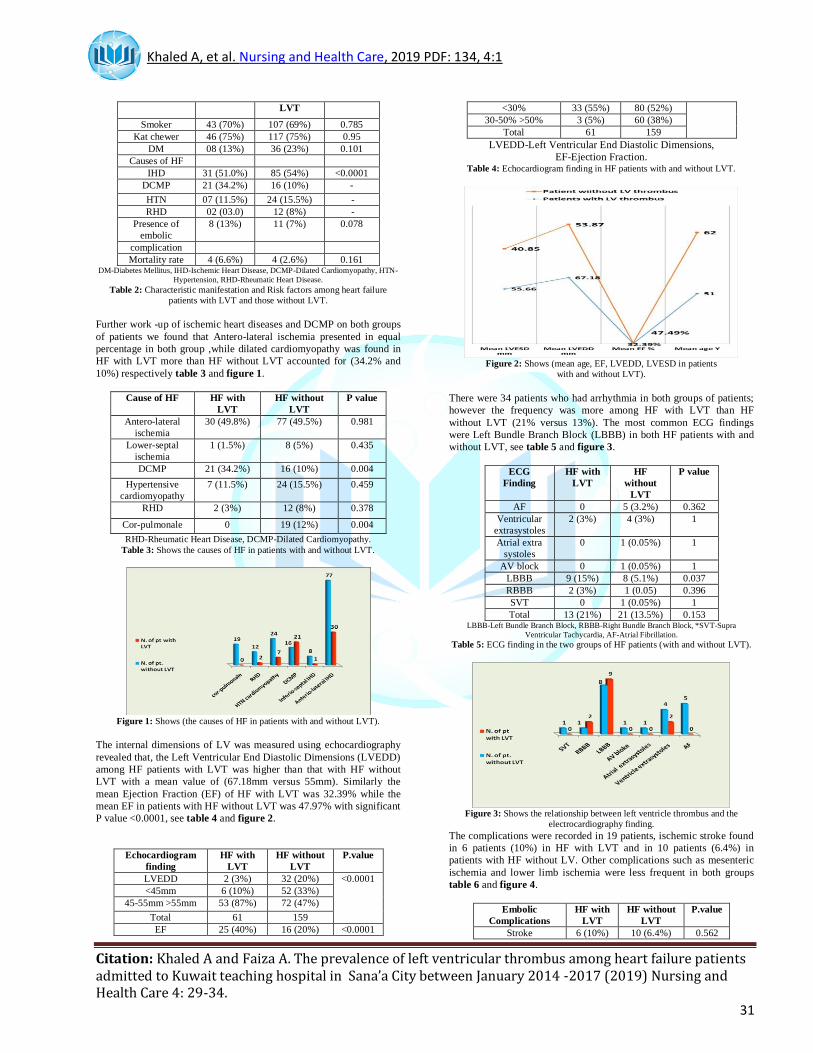
Further work -up of ischemic heart diseases and DCMP on both groups
of patients we found that Antero-lateral ischemia presented in equal
percentage in both group ,while dilated cardiomyopathy was found in
HF with LVT more than HF without LVT accounted for (34.2% and
10%) respectively table 3 and figure 1.
Cause of HF HF with
LVT
HF without
LVT
P value
Antero-lateral
ischemia
30 (49.8%) 77 (49.5%) 0.981
Lower-septal
ischemia
1 (1.5%) 8 (5%) 0.435
DCMP 21 (34.2%) 16 (10%) 0.004
Hypertensive
cardiomyopathy
7 (11.5%) 24 (15.5%) 0.459
RHD 2 (3%) 12 (8%) 0.378
Cor-pulmonale 0 19 (12%) 0.004
RHD-Rheumatic Heart Disease, DCMP-Dilated Cardiomyopathy.
Table 3: Shows the causes of HF in patients with and without LVT.
Figure 1: Shows (the causes of HF in patients with and without LVT).
The internal dimensions of LV was measured using echocardiography
revealed that, the Left Ventricular End Diastolic Dimensions (LVEDD)
among HF patients with LVT was higher than that with HF without
LVT with a mean value of (67.18mm versus 55mm). Similarly the
mean Ejection Fraction (EF) of HF with LVT was 32.39% while the
mean EF in patients with HF without LVT was 47.97% with significant
P value <0.0001, see table 4 and figure 2.
Echocardiogram
finding
HF with
LVT
HF without
LVT
P.value
LVEDD 2 (3%) 32 (20%) <0.0001
<45mm 6 (10%) 52 (33%)
45-55mm >55mm 53 (87%) 72 (47%)
Total 61 159
EF 25 (40%) 16 (20%) <0.0001
<30% 33 (55%) 80 (52%)
30-50% >50% 3 (5%) 60 (38%)
Total 61 159
LVEDD-Left Ventricular End Diastolic Dimensions,
EF-Ejection Fraction. Table 4: Echocardiogram finding in HF patients with and without LVT.
Figure 2: Shows (mean age, EF, LVEDD, LVESD in patients
with and without LVT).
There were 34 patients who had arrhythmia in both groups of patients;
however the frequency was more among HF with LVT than HF
without LVT (21% versus 13%). The most common ECG findings
were Left Bundle Branch Block (LBBB) in both HF patients with and
without LVT, see table 5 and figure 3.
ECG
Finding
HF with
LVT
HF
without
LVT
P value
AF 0 5 (3.2%) 0.362
Ventricular
extrasystoles
2 (3%) 4 (3%) 1
Atrial extra
systoles
0 1 (0.05%) 1
AV block 0 1 (0.05%) 1
LBBB 9 (15%) 8 (5.1%) 0.037
RBBB 2 (3%) 1 (0.05) 0.396
SVT 0 1 (0.05%) 1
Total 13 (21%) 21 (13.5%) 0.153 LBBB-Left Bundle Branch Block, RBBB-Right Bundle Branch Block, *SVT-Supra
Ventricular Tachycardia, AF-Atrial Fibrillation.
Table 5: ECG finding in the two groups of HF patients (with and without LVT).
Figure 3: Shows the relationship between left ventricle thrombus and the
electrocardiography finding.
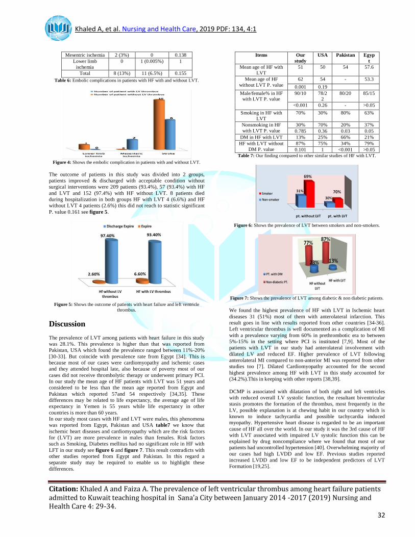
The complications were recorded in 19 patients, ischemic stroke found
in 6 patients (10%) in HF with LVT and in 10 patients (6.4%) in
patients with HF without LV. Other complications such as mesenteric
ischemia and lower limb ischemia were less frequent in both groups
table 6 and figure 4.
Embolic
Complications
HF with
LVT
HF without
LVT
P.value
Stroke 6 (10%) 10 (6.4%) 0.562
Khaled A, et al. Nursing and Health Care, 2019 PDF: 134, 4:1
Citation: Khaled A and Faiza A. The prevalence of left ventricular thrombus among heart failure patients admitted to Kuwait teaching hospital in Sana’a City between January 2014 -2017 (2019) Nursing and Health Care 4: 29-34.
32
Mesentric ischemia 2 (3%) 0 0.138
Lower limb
ischemia
0 1 (0.005%) 1
Total 8 (13%) 11 (6.5%) 0.155
Table 6: Embolic complications in patients with HF with and without LVT.
Figure 4: Shows the embolic complication in patients with and without LVT.
The outcome of patients in this study was divided into 2 groups,
patients improved & discharged with acceptable condition without
surgical interventions were 209 patients (93.4%), 57 (93.4%) with HF
and LVT and 152 (97.4%) with HF without LVT. 8 patients died
during hospitalization in both groups HF with LVT 4 (6.6%) and HF
without LVT 4 patients (2.6%) this did not reach to statistic significant
P. value 0.161 see figure 5.
Figure 5: Shows the outcome of patients with heart failure and left ventricle
thrombus.
Discussion
The prevalence of LVT among patients with heart failure in this study
was 28.1%. This prevalence is higher than that was reported from
Pakistan, USA which found the prevalence ranged between 11%-20%
[30-33]. But coincide with prevalence rate from Egypt [34]. This is
because most of our cases were cardiomyopathy and ischemic cases
and they attended hospital late, also because of poverty most of our
cases did not receive thrombolytic therapy or underwent primary PCI.
In our study the mean age of HF patients with LVT was 51 years and
considered to be less than the mean age reported from Egypt and
Pakistan which reported 57and 54 respectively [34,35]. These
differences may be related to life expectancy, the average age of life
expectancy in Yemen is 55 years while life expectancy in other
countries is more than 60 years.
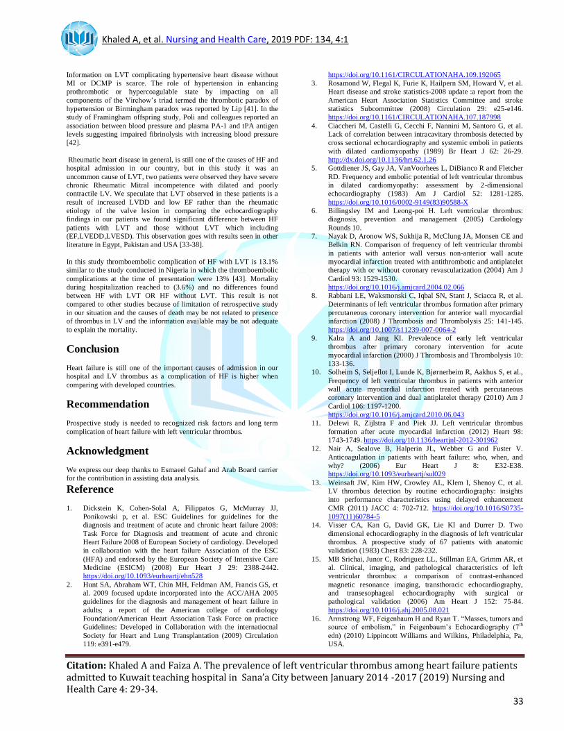
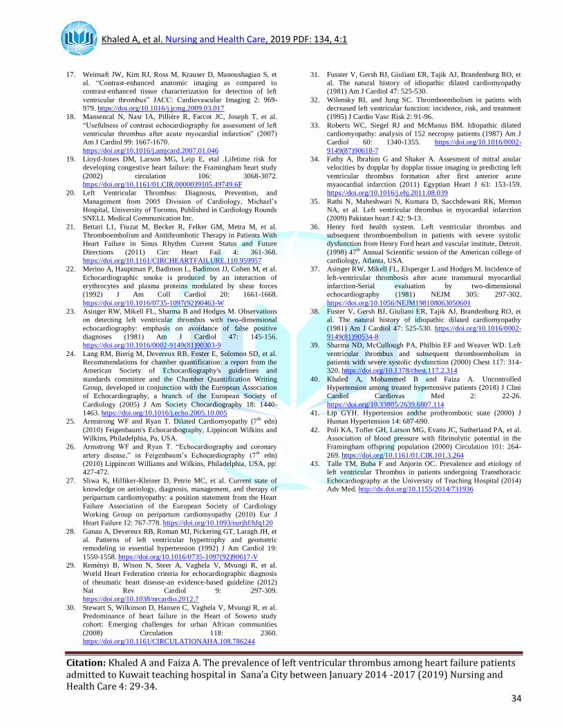
In our study most cases with HF and LVT were males, this phenomena
was reported from Egypt, Pakistan and USA table7 we know that
ischemic heart diseases and cardiomyopathy which are the risk factors
for (LVT) are more prevalence in males than females. Risk factors
such as Smoking, Diabetes mellitus had no significant role in HF with
LFT in our study see figure 6 and figure 7. This result contradicts with
other studies reported from Egypt and Pakistan. In this regard a
separate study may be required to enable us to highlight these
differences.
Items Our
study
USA Pakistan Egyp
t
Mean age of HF with
LVT
51 50 54 57.6
Mean age of HF
without LVT P. value
62 54 - 53.3
0.001 0.19
Male/female% in HF with LVT P. value
90/10 78/22
80/20 85/15
<0.001 0.26 - >0.05
Smoking in HF with
LVT
70% 30% 80% 63%
Nonsmoking in HF
with LVT P. value
30% 70% 20% 37%
0.785 0.36 0.03 0.05
DM in HF with LVT 13% 25% 66% 21%
HF with LVT without
DM P. value
87% 75% 34% 79%
0.101 1 <0.001 >0.05
Table 7: Our finding compared to other similar studies of HF with LVT.
Figure 6: Shows the prevalence of LVT between smokers and non-smokers.
Figure 7: Shows the prevalence of LVT among diabetic & non diabetic patients.
We found the highest prevalence of HF with LVT in Ischemic heart
diseases 31 (51%) most of them with anterolateral infarction. This
result goes in line with results reported from other countries [34-36].
Left ventricular thrombus is well documented as a complication of MI
with a prevalence varying from 60% in prethrombotic era to between
5%-15% in the setting where PCI is instituted [7,9]. Most of the
patients with LVT in our study had anterolateral involvement with
dilated LV and reduced EF. Higher prevalence of LVT following
anterolateral MI compared to non-anterior MI was reported from other
studies too [7]. Dilated Cardiomyopathy accounted for the second
highest prevalence among HF with LVT in this study accounted for
(34.2%).This in keeping with other reports [38,39].
DCMP is associated with dilatation of both right and left ventricles
with reduced overall LV systolic function, the resultant biventricular
stasis promotes the formation of the thrombus, most frequently in the
LV, possible explanation is at chewing habit in our country which is
known to induce tachycardia and possible tachycardia induced
myopathy. Hypertensive heart disease is regarded to be an important
cause of HF all over the world. In our study it was the 3rd cause of HF
with LVT associated with impaired LV systolic function this can be
explained by drug noncompliance where we found that most of our
patients had uncontrolled hypertension [40]. Overwhelming majority of
our cases had high LVDD and low EF. Previous studies reported
increased LVDD and low EF to be independent predictors of LVT
Formation [19,25].
Khaled A, et al. Nursing and Health Care, 2019 PDF: 134, 4:1
Citation: Khaled A and Faiza A. The prevalence of left ventricular thrombus among heart failure patients admitted to Kuwait teaching hospital in Sana’a City between January 2014 -2017 (2019) Nursing and Health Care 4: 29-34.
33
Information on LVT complicating hypertensive heart disease without
MI or DCMP is scarce. The role of hypertension in enhancing
prothrombotic or hypercoagulable state by impacting on all
components of the Virchow’s triad termed the thrombotic paradox of
hypertension or Birmingham paradox was reported by Lip [41]. In the
study of Framingham offspring study, Poli and colleagues reported an
association between blood pressure and plasma PA-1 and tPA antigen
levels suggesting impaired fibrinolysis with increasing blood pressure
[42].
Rheumatic heart disease in general, is still one of the causes of HF and
hospital admission in our country, but in this study it was an
uncommon cause of LVT, two patients were observed they have severe
chronic Rheumatic Mitral incompetence with dilated and poorly
contractile LV. We speculate that LVT observed in these patients is a
result of increased LVDD and low EF rather than the rheumatic
etiology of the valve lesion in comparing the echocardiography
findings in our patients we found significant difference between HF
patients with LVT and those without LVT which including
(EF,LVEDD,LVESD). This observation goes with results seen in other
literature in Egypt, Pakistan and USA [33-38].
In this study thromboembolic complication of HF with LVT is 13.1%
similar to the study conducted in Nigeria in which the thromboembolic
complications at the time of presentation were 13% [43]. Mortality
during hospitalization reached to (3.6%) and no differences found
between HF with LVT OR HF without LVT. This result is not
compared to other studies because of limitation of retrospective study
in our situation and the causes of death may be not related to presence
of thrombus in LV and the information available may be not adequate
to explain the mortality.
Conclusion
Heart failure is still one of the important causes of admission in our
hospital and LV thrombus as a complication of HF is higher when
comparing with developed countries.
Recommendation
Prospective study is needed to recognized risk factors and long term
complication of heart failure with left ventricular thrombus.
Acknowledgment
We express our deep thanks to Esmaeel Gahaf and Arab Board carrier
for the contribution in assisting data analysis.
Reference
1. Dickstein K, Cohen-Solal A, Filippatos G, McMurray JJ,
Ponikowski p, et al. ESC Guidelines for guidelines for the
diagnosis and treatment of acute and chronic heart failure 2008:
Task Force for Diagnosis and treatment of acute and chronic
Heart Failure 2008 of European Society of cardiology. Developed
in collaboration with the heart failure Association of the ESC
(HFA) and endorsed by the European Society of Intensive Care
Medicine (ESICM) (2008) Eur Heart J 29: 2388-2442.
https://doi.org/10.1093/eurheartj/ehn528
2. Hunt SA, Abraham WT, Chin MH, Feldman AM, Francis GS, et
al. 2009 focused update incorporated into the ACC/AHA 2005
guidelines for the diagnosis and management of heart failure in
adults; a report of the American college of cardiology
Foundation/American Heart Association Task Force on practice
Guidelines: Developed in Collaboration with the internatiocnal
Society for Heart and Lung Transplantation (2009) Circulation
119: e391-e479.
https://doi.org/10.1161/CIRCULATIONAHA.109.192065
3. Rosamond W, Flegal K, Furie K, Hailpern SM, Howard V, et al.
Heart disease and stroke statistics-2008 update :a report from the
American Heart Association Statistics Committee and stroke
statistics Subcommittee (2008) Circulation 29: e25-e146.
https://doi.org/10.1161/CIRCULATIONAHA.107.187998
4. Ciaccheri M, Castelli G, Cecchi F, Nannini M, Santoro G, et al.
Lack of correlation between intracavitary thrombosis detected by
cross sectional echocardiography and systemic emboli in patients
with dilated cardiomyopathy (1989) Br Heart J 62: 26-29.
http://dx.doi.org/10.1136/hrt.62.1.26
5. Gottdiener JS, Gay JA, VanVoorhees L, DiBianco R and Fletcher
RD. Frequency and embolic potential of left ventricular thrombus
in dilated cardiomyopathy: assessment by 2-dimensional
echocardiography (1983) Am J Cardiol 52: 1281-1285.
https://doi.org/10.1016/0002-9149(83)90588-X
6. Billingsley IM and Leong-poi H. Left ventricular thrombus:
diagnosis, prevention and management (2005) Cardiology
Rounds 10.
7. Nayak D, Aronow WS, Sukhija R, McClung JA, Monsen CE and
Belkin RN. Comparison of frequency of left ventricular thrombi
in patients with anterior wall versus non-anterior wall acute
myocardial infarction treated with antithrombotic and antiplatelet
therapy with or without coronary revascularization (2004) Am J
Cardiol 93: 1529-1530.
https://doi.org/10.1016/j.amjcard.2004.02.066
8. Rabbani LE, Waksmonski C, Iqbal SN, Stant J, Sciacca R, et al.
Determinants of left ventricular thrombus formation after primary
percutaneous coronary intervention for anterior wall myocardial
infarction (2008) J Thrombosis and Thrombolysis 25: 141-145.
https://doi.org/10.1007/s11239-007-0064-2
9. Kalra A and Jang KI. Prevalence of early left ventricular
thrombus after primary coronary intervention for acute
myocardial infarction (2000) J Thrombosis and Thrombolysis 10:
133-136.
10. Solheim S, Seljeflot I, Lunde K, Bjørnerheim R, Aakhus S, et al.,
Frequency of left ventricular thrombus in patients with anterior
wall acute myocardial infarction treated with percutaneous
coronary intervention and dual antiplatelet therapy (2010) Am J
Cardiol 106: 1197-1200.
https://doi.org/10.1016/j.amjcard.2010.06.043
11. Delewi R, Zijlstra F and Piek JJ. Left ventricular thrombus
formation after acute myocardial infarction (2012) Heart 98:
1743-1749. https://doi.org/10.1136/heartjnl-2012-301962
12. Nair A, Sealove B, Halperin JL, Webber G and Fuster V.
Anticoagulation in patients with heart failure: who, when, and
why? (2006) Eur Heart J 8: E32-E38.
https://doi.org/10.1093/eurheartj/sul029
13. Weinsaft JW, Kim HW, Crowley AL, Klem I, Shenoy C, et al.
LV thrombus detection by routine echocardiography: insights
into performance characteristics using delayed enhancement
CMR (2011) JACC 4: 702-712. https://doi.org/10.1016/S0735-
1097(11)60784-5
14. Visser CA, Kan G, David GK, Lie KI and Durrer D. Two
dimensional echocardiography in the diagnosis of left ventricular
thrombus. A prospective study of 67 patients with anatomic
validation (1983) Chest 83: 228-232.
15. MB Srichai, Junor C, Rodriguez LL, Stillman EA, Grimm AR, et
al. Clinical, imaging, and pathological characteristics of left
ventricular thrombus: a comparison of contrast-enhanced
magnetic resonance imaging, transthoracic echocardiography,
and transesophageal echocardiography with surgical or
pathological validation (2006) Am Heart J 152: 75-84.
https://doi.org/10.1016/j.ahj.2005.08.021
16. Armstrong WF, Feigenbaum H and Ryan T. “Masses, tumors and
source of embolism,” in Feigenbaum’s Echocardiography (7th
edn) (2010) Lippincott Williams and Wilkins, Philadelphia, Pa,
USA.
Khaled A, et al. Nursing and Health Care, 2019 PDF: 134, 4:1
Citation: Khaled A and Faiza A. The prevalence of left ventricular thrombus among heart failure patients admitted to Kuwait teaching hospital in Sana’a City between January 2014 -2017 (2019) Nursing and Health Care 4: 29-34.
34
17. Weinsaft JW, Kim RJ, Ross M, Krauser D, Manoushagian S, et
al. “Contrast-enhanced anatomic imaging as compared to
contrast-enhanced tissue characterization for detection of left
ventricular thrombus” JACC: Cardiovascular Imaging 2: 969-
979. https://doi.org/10.1016/j.jcmg.2009.03.017
18. Mansencal N, Nasr IA, Pillière R, Farcot JC, Joseph T, et al.
“Usefulness of contrast echocardiography for assessment of left
ventricular thrombus after acute myocardial infarction” (2007)
Am J Cardiol 99: 1667-1670.
https://doi.org/10.1016/j.amjcard.2007.01.046
19. Lioyd-Jones DM, Larson MG, Leip E, etal .Lifetime risk for
developing congestive heart failure: the Framingham heart study
(2002) circulation 106: 3068-3072.
https://doi.org/10.1161/01.CIR.0000039105.49749.6F
20. Left Ventricular Thrombus: Diagnosis, Prevention, and
Management from 2005 Division of Cardiology, Michael’s
Hospital, University of Toronto, Published in Cardiology Rounds
SNELL Medical Communication Inc.
21. Bettari L1, Fiuzat M, Becker R, Felker GM, Metra M, et al.
Thromboembolism and Antithrombotic Therapy in Patients With
Heart Failure in Sinus Rhythm Current Status and Future
Directions (2011) Circ Heart Fail 4: 361-368.
https://doi.org/10.1161/CIRCHEARTFAILURE.110.959957
22. Merino A, Hauptman P, Badimon L, Badimon JJ, Cohen M, et al.
Echocardiographic smoke is produced by an interaction of
erythrocytes and plasma proteins modulated by shear forces
(1992) J Am Coll Cardiol 20: 1661-1668.
https://doi.org/10.1016/0735-1097(92)90463-W
23. Asinger RW, Mikell FL, Sharma B and Hodges M. Observations
on detecting left ventricular thrombus with two-dimensional
echocardiography: emphasis on avoidance of false positive
diagnoses (1981) Am J Cardiol 47: 145-156.
https://doi.org/10.1016/0002-9149(81)90303-9
24. Lang RM, Bierig M, Devereux RB, Foster E, Solomon SD, et al.
Recommendations for chamber quantification: a report from the
American Society of Echocardiography's guidelines and
standards committee and the Chamber Quantification Writing
Group, developed in conjunction with the European Association
of Echocardiography, a branch of the European Society of
Cardiology (2005) J Am Society Chocardiography 18: 1440-
1463. https://doi.org/10.1016/j.echo.2005.10.005
25. Armstrong WF and Ryan T. Dilated Cardiomyopathy (7th edn)
(2010) Feigenbaum's Echocardiography, Lippincott Wilkins and
Wilkins, Philadelphia, Pa, USA.
26. Armstrong WF and Ryan T. “Echocardiography and coronary
artery disease,” in Feigenbaum’s Echocardiography (7th edn)
(2010) Lippincott Williams and Wilkins, Philadelphia, USA, pp:
427-472.
27. Sliwa K, Hilfiker-Kleiner D, Petrie MC, et al. Current state of
knowledge on aetiology, diagnosis, management, and therapy of
peripartum cardiomyopathy: a position statement from the Heart
Failure Association of the European Society of Cardiology
Working Group on peripartum cardiomyopathy (2010) Eur J
Heart Failure 12: 767-778. https://doi.org/10.1093/eurjhf/hfq120
28. Ganau A, Devereux RB, Roman MJ, Pickering GT, Laragh JH, et
al. Patterns of left ventricular hypertrophy and geometric
remodeling in essential hypertension (1992) J Am Cardiol 19:
1550-1558. https://doi.org/10.1016/0735-1097(92)90617-V
29. Reményi B, Wison N, Steer A, Vaghela V, Mvungi R, et al.
World Heart Federation criteria for echocardiographic diagnosis
of rheumatic heart disease-an evidence-based guideline (2012)
Nat Rev Cardiol 9: 297-309.
https://doi.org/10.1038/nrcardio.2012.7
30. Stewart S, Wilkinson D, Hansen C, Vaghela V, Mvungi R, et al.
Predominance of heart failure in the Heart of Soweto study
cohort: Emerging challenges for urban African communities
(2008) Circulation 118: 2360.
https://doi.org/10.1161/CIRCULATIONAHA.108.786244
31. Fusster V, Gersh BJ, Giuliani ER, Tajik AJ, Brandenburg RO, et
al. The natural history of idiopathic dilated cardiomyopathy
(1981) Am J Cardiol 47: 525-530.
32. Wilensky RL and Jung SC. Thromboembolism in patints with
decreased left ventricular function: incidence, risk, and treatment
(1995) J Cardio Vasc Risk 2: 91-96.
33. Roberts WC, Siegel RJ and McManus BM. Idiopathic dilated
cardiomyopathy: analysis of 152 necropsy patients (1987) Am J
Cardiol 60: 1340-1355. https://doi.org/10.1016/0002-
9149(87)90618-7
34. Fathy A, Ibrahim G and Shaker A. Assesment of mitral anular
velocities by dopplar by dopplar tissue imaging in predicting left
ventricular thrombus formation after first anterior acute
myaocardial infarction (2011) Egyptian Heart J 63: 153-159.
https://doi.org/10.1016/j.ehj.2011.08.039
35. Rathi N, Maheshwari N, Kumara D, Sacchdewani RK, Memon
NA, et al. Left ventricular thrombus in myocardial infarction
(2009) Pakistan heart J 42: 9-13.
36. Henry ford health system. Left ventricular thrombus and
subsequent thromboembolism in patients with severe systolic
dysfunction from Henry Ford heart and vascular institute, Detroit.
(1998) 47th Annual Scientific session of the American college of
cardiology, Atlanta, USA.
37. Asinger RW, Mikell FL, Elsperger L and Hodges M. Incidence of
left-ventricular thrombosis after acute transmural myocardial
infarction-Serial evaluation by two-dimensional
echocardiography (1981) NEJM 305: 297-302.
https://doi.org/10.1056/NEJM198108063050601
38. Fuster V, Gersh BJ, Giuliani ER, Tajik AJ, Brandenburg RO, et
al. The natural history of idiopathic dilated cardiomyopathy
(1981) Am J Cardiol 47: 525-530. https://doi.org/10.1016/0002-
9149(81)90534-8
39. Sharma ND, McCullough PA, Philbin EF and Weaver WD. Left
ventricular thrombus and subsequent thromboembolism in
patients with severe systolic dysfunction (2000) Chest 117: 314-
320. https://doi.org/10.1378/chest.117.2.314
40. Khaled A, Mohammed B and Faiza A. Uncontrolled
Hypertension among treated hypertensive patients (2018) J Clini
Cardiol Cardiovas Med 2: 22-26.
https://doi.org/10.33805/2639.6807.114
41. Lip GYH. Hypertension andthe prothrombotic state (2000) J
Human Hypertension 14: 687-690.
42. Poli KA, Tofler GH, Larson MG, Evans JC, Sutherland PA, et al.
Association of blood pressure with fibrinolytic potential in the
Framingham offspring population (2000) Circulation 101: 264-
269. https://doi.org/10.1161/01.CIR.101.3.264
43. Talle TM, Buba F and Anjorin OC. Prevalence and etiology of
left ventricular Thrombus in patients undergoing Transthoracic
Echocardiography at the University of Teaching Hospital (2014)
Adv Med. http://dx.doi.org/10.1155/2014/731936