Embed Size (px)
Citation preview
1607
*Correspondence to: Haga, T.: [email protected](Supplementary material: https://www.ncbi.nlm.nih.gov/pmc/journals/2350/)©2020 The Japanese Society of Veterinary Science
This is an open-access article distributed under the terms of the Creative Commons Attribution Non-Commercial No Derivatives (by-nc-nd) License. (CC-BY-NC-ND 4.0: https://creativecommons.org/licenses/by-nc-nd/4.0/)
FULL PAPERVirology
Genomic characterization and distribution of bovine foamy virus in JapanMari OKAMOTO1), Keisuke OGUMA2), Nanako YAMASHITA-KAWANISHI 1), Toshihiro ICHIJO3), Shinichi HATAMA4), Maiko ENDO5), Maya ISHIKAWA6) and Takeshi HAGA1)*
1)Division of Infection Control and Disease Prevention, Department of Veterinary Medical Science, The University of Tokyo, 1-1-1 Yayoi, Bunkyo-ku, Tokyo 113-8657, Japan
2)Laboratory of Veterinary Epizootiology, Department of Veterinary Medicine, Nihon University, 1866 Kameino, Fujisawa, Kanagawa 252-0880, Japan
3)Iwate University Faculty of Agriculture Veterinary Medicine, 3-18-8 Ueda, Morioka, Iwate 020-8550, Japan4)National Institute of Animal Health, National Agriculture and Food Research Organization, 3-1-5 Kannondai,
Tsukuba, Ibaraki 305-0856, Japan5)Animal Resource Science Center, Graduate School of Agricultural and Life Science, The University of Tokyo,
3145 Yorii, Kasama, Ibaraki 319-0206, Japan6)Private investigator, Izumimachi, Shikaoi, Katogun, Hokkaido 081-0212, Japan
ABSTRACT. Bovine foamy virus (BFV) is distributed through worldwide cattle herds. Although the biological features of BFV are not well understood, appearance of clinical manifestation by superinfection with other microorganisms is inferred. In Japan, reports of genomic characterizations and epidemiology of this virus are limited. In this study, we performed whole genomic sequencing of BFV strains Ibaraki and No.43, which were isolated in this country. Additionally, we investigated BFV in geographically distant four daily farms in Japan, to estimate the distribution of BFV and its correlation to bovine leukemia virus (BLV). BFV was distributed throughout Japan; the average positive rate was 12.7%. The nucleotide sequence identities of the isolates were 99.6% when compared with BFV strain isolated in the USA. The phylogenetic tree using env gene sequence showed strains Ibaraki, No.43 and Kagoshima were sorted in the same cluster including the USA and Chinese strains, while Hokkaido strain was in the other cluster including European strains. Although no clear correlation between BFV and BLV could be found, BFV and BLV infections were likely to increase with ages. Our data on epidemiology and characteristics of BFV will provide important information to reveal biological features of BFV.
KEY WORDS: bovine foamy virus, bovine leukemia virus, epidemiology
Foamy viruses (FVs) belong to the subfamily Spumaretrovirinae which is a member of the family Retroviridae [19]. The length of viral genome is about 12 kb. FV was first isolated and identified using rhesus monkey kidney cells in 1955 [33]. FVs have been identified in many vertebrates, including mammals [1, 16, 24, 30, 35, 36], amphibians [2] and fish [2, 9, 10, 32]. FVs infect several kinds of primary and lined cells and form multinucleated giant cells. The foamy aspect is confirmed by microscopic observation of cultured cells, and this feature became the root of the name of FV [34]. The FV genome encodes three structural, gag, pol, env, and two nonstructural, bel-1and bel-2, genes. Tas proteins, the bel-1 products, play a role in trans activator, while the function of Bet proteins, complex of the bel-2 products and a part of bel-1 products, has not yet been determined [23, 34].
BFVs have been isolated in Western countries and China, and considered to be common in cows [5, 12, 14, 15, 29]. This implies they may be spreading over a wide region. In Japan, BFV strain No.43 was isolated from peripheral blood leukocytes (PBLs) of a clinically healthy cow in 2018 [8], by co-culturing BFV-infected PBLs with fetal bovine muscle (FBM) cells. In that report, the prevalence of BFV infection was shown to be 8.8% (five out of 57 cattle) in Kanagawa prefecture, Japan, and a part of the long terminal repeat (LTR), env and bel-1-bel-2 sequences were identified. Another recent study showed that BFV was detected among cattle in the Kanto region in Japan showing that the average BFV-positive rate was 16.7% (91 out of 545 cattle) [13]. However, the full-length genome sequence of strain No.43 was undetermined. Moreover, the information on the prevalence of BFV infection was limited to the Kanto region. Consequently, epidemiology and genomic characteristics of BFV in Japan is still limited.
FVs are considered to be nonpathogenic alone [34]. However, there are some reports indicating the possibility of FV
Received: 16 July 2020Accepted: 2 September 2020Advanced Epub: 14 September 2020
J. Vet. Med. Sci. 82(11): 1607–1613, 2020doi: 10.1292/jvms.20-0429
M. OKAMOTO ET AL.
1608J. Vet. Med. Sci. 82(11): 1607–1613, 2020
pathogenicity. Prototype FV activates human immunodeficiency virus type 1 (HIV-1) via LTR activation [18]. Correlation of feline foamy virus (FFV) and feline leukemia virus (FeLV) infection has been described, in particular, cats infected with progressive FeLV are more susceptible to FFV [6, 28]. Also, buccal FFV viral load measured by quantitative PCR of FFV/FeLV co-infected cats was higher than among FFV mono-infected cats [6]. In addition, the cats infected experimentally appeared clinically normal but histopathological changes were observed in their lungs and kidneys [7]. Considering the possibility that FVs affect the disease manifestation with other retroviruses, in the present study, we investigated the relationship between BFV infection and bovine leukemia virus (BLV). BLV Tax protein was shown to activate HIV-1 transcription in a previous study [18], suggesting that BLV infection may have a potential to alter BFV infection. The seropositive rate of BLV in Japan recently reached about 40% among dairy cattle [26], and infection control of BLV is a matter of utmost concern in Japan. However, there are few reports focusing on relationship between BFV and BLV.
In this study, to further understand the characteristics of BFV in Japan, detection, isolation, and genomic characterization of BFV were conducted. In addition, the association between the BLV infection and BFV prevalence among dairy cattle was examined.
MATERIALS AND METHODS
AnimalsThe whole blood samples analyzed in this study were obtained from a total of 181 Holstein-Friesian or F1 (crossbreed of
purebreds) cattle kept in the respective farms of Ibaraki, Kagoshima, Hokkaido, and Miyagi prefectures (Supplementary Fig. 1) aged between 0 to 10 years old (Table 1). The whole blood was collected from the jugular vein of each cattle with EDTA-coated capillary tubes. In addition to the whole blood, serum was isolated from the cows kept in Ibaraki.
BFV surveillanceThe prevalence of BFV in Ibaraki, Kagoshima, Hokkaido and Miyagi was determined by conventional PCR. Genomic DNA was
directly extracted from 181 cattle blood samples (Table 1) with QIAamp DNA Mini Kit (QIAGEN, Hilden, Germany) following the manufacturer’s instructions. A primer pair amplifying a 6,781 bp fragment from pol to bel-1 region referring to the BFV reference sequence (GenBank accession number: U94514) was designed and applied. The sequences of the primer pair were 5′- AATACCAAG CTCTATGGCAATGTT -3′ (8F) and 5′- ACCAGGGCTGTACTGGTTCT -3′ (8R). PCR was performed using DNA polymerase, KOD Fx Neo (TOYOBO, Osaka, Japan). The cycling parameters were: initial denaturation was at 94°C for 2 min, and 45 cycles of the following 2 steps: at 98°C for 10 sec, 68°C for 3.5 min. The PCR products were electrophoresed on 1% agarose gels to confirm the presence of 6,781 bp band.
BFV isolation and preparationThirty-four cattle serum samples collected from a farm in Ibaraki prefecture were tested by agarose gel immunodiffusion (AGID)
test to determine the BFV infection. AGID tests were performed according to the methods described elsewhere [8]. Positive serum and BFV antigen were kindly provided by Prof. Sentsui (Nihon University). Cows showing a clear precipitation line in AGID test were selected, and three strains (No. 5266, 5270, and 7800) were isolated from the cattle in Ibaraki. The full genome sequences of the isolated viruses were determined.
Blood was collected with EDTA tubes from the jugular vein of three cattle showing AGID test-positive. To isolate BFV, the peripheral blood mononuclear cells (PBMC) from three AGID-positive cases were separated from the blood stored at 4°C by Lymphopep (Cosmo Bio, Carlsbad, CA, USA) within 24 hr from the blood collection. PBMC derived from cows reared in Ibaraki were co-cultured with BHK21 cells in 6-well plates at 37°C in Dulbecco’s modified eagle medium (DMEM) (Sigma-Aldrich Co., LLC, St. Louis, MO, USA) containing 10% fetal bovine serum (FBS). After 48 hr, the cells were washed with DMEM and fresh medium was added. Two days after, cells were subcultured by trypsinization. Blind passage was carried out at intervals of two or three days until cytopathic effect (CPE) was observed. Two negative controls were prepared as: BHK21 cells only and BHK21 cells cocultured with PBMC of BFV-negative cows. In addition to the three AGID-positive BFV samples, strain No.43, which was preciously isolated and kindly provided by Prof. Sentsui, was co-cultured with BHK21 cells and subcultured at passage five. When CPE was observed, the cells were stained with Giemsa after being fixed with methanol to observe the multinucleated giant cells.
Whole and partial genome sequence analysisDNA was extracted from BHK21 cells cocultured with BFV-infected PBMC derived from cows reared in Ibaraki at passage
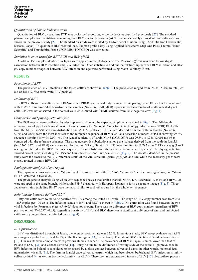
Table 1. Sample information
Prefecture Breed Age (years) Number of cows tested BFV prevalenceIbaraki Holstein-Friesian/ F1 0–10 34 11.8% (4/34)Kagoshima Holstein-Friesian 0–2 29 13.8% (4/29)Hokkaido Holstein-Friesian 0–6 98 15.4% (15/98)Miyagi Holstein-Friesian 0–1 20 0.0% (0/20)Total 0–10 181 12.7% (23/181)BFV: bovine foamy virus. F1: crossbreed of purebreds.

BOVINE FOAMY VIRUS IN JAPAN
J. Vet. Med. Sci. 82(11): 1609
12 and strain No.43 at passage five. Prior to the DNA extraction, cells were washed with PBS twice and peeled off with trypsin-EDTA. The suspension was processed using QIAamp DNA Mini Kit (QIAGEN), following the manufacturer’s instructions. Three pairs of primers described in Fig. 1 were used for detecting each gene. All primer sets were designed referred to the BFV reference sequence (GenBank accession number: U94514). The primer sequence used for amplifying an 1,869 bp fragment of gag to pol region were as follows: 5′- GGCAACCCTTGCCTATTCCT -3′ (gagpolF) and 5′- CTCAATACGCCGATGTCCGA -3′ (gagpolR). A 6,781-bp fragment comprising of pol, env and part of bel-1 was amplified using the 8F/R primers. The other 3,665-bp fragment was amplified with primer named ipcrF 5′- GAACACCCGGACAGCA-TACACTC -3′ and ipcrR 5′- CCATCGGAGCAGAGACCACT -3′ by inverse PCR method. The PCR cycle to amplify from bel-1 to gag region was carried as follows: initial denaturation was at 94°C for 2 min, and 45 cycles of the following 2 steps: at 98°C for 10 sec, 68°C 4.5 min. The PCR condition of 8F/R primers was the same protocol as described above in the BFV surveillance section. The other PCR condition with gagpolF/R primers was performed as follows: 94°C for 2 min; followed by 45 cycles of 98°C for 10 sec, 56°C for 30 sec and 68°C for 1 min. The PCR products were electrophoresed on 1% agarose gels and purified using NucleoSpin Gel and PCR Clean-up (MACHEREY-NAGEL, Düren, Germany). The sequence except pol to bel-1 region of Ibaraki and the whole sequence of the strain No.43 were determined by direct sequencing. With regard to pol to bel-1 (env) region, purified PCR products of strain Ibaraki, BH47 and K7 were cloned using TOPO XL PCR Cloning Kit (Thermo Fisher Scientific, Waltham, MA, USA) or Zero Blunt TOPO PCR cloning Kit (Thermo Fisher Scientific). The identity of the direct and cloned sequence of env region of Ibaraki strain was confirmed. The other purified PCR products, corresponding to gag to pol region and bel-1 to gag region of strain Ibaraki and strain No.43 were cloned using TOPO XL PCR Cloning Kit (Thermo Fisher Scientific) or Zero Blunt TOPO PCR cloning Kit (Thermo Fisher Scientific). Primer sets for PCR and sequence analysis were shown in Supplementary Table 1. Sequences were analyzed with BigDye Terminator v3.1 Cycle Sequencing Kit (Thermo Fisher Scientific) and Applied Biosystems 3130 Genetic Analyzer (Thermo Fisher Scientific). The obtained sequences were registered in GenBank with the following accession numbers noted in parenthesis: Ibaraki (LC510606), No.43 (LC510607), K7 (LC510608), BH47 (LC510609).
Phylogenetic analysisMultiple alignment and phylogenetic analysis were performed using Clustal W in MEGA7 software [21]. Phylogenetic trees
based on the nucleotide sequences of each gene or genome were generated by the Maximum Likelihood method based on the Hasegawa-Kishino-Yano model (for gag, pol, env and whole genome) and Kimura 2-parameter model (for bel-1 and bel-2) with bootstrap replicates of 1,000 [11, 20]. The analysis was performed including the sequences registered in GenBank, including named BFVbta_BSV11 (USA, U94514), BFVbta_100 (Poland, JX307861), BFVbta_Riems (Germany, JX307862) and BFV3026 (China, AY134750).
1607–1613, 2020
Fig. 1. Bovine foamy virus (BFV) isolation and primer design for full-length sequence analysis. (A) BHK21 cells cocultured with peripheral blood mononuclear cells (PBMC) from BFV positive cow (No. 5266). Blind passage was performed nine times and then stained with Gimza. Characteristic multinucleated giant cells were formed (arrows). (B) Control BHK21 cells. Control BHK21 cells were cocultured with PBMC from BFV-negative cow. Cells were stained with Gimza at nine passages. (C) BFV genomic structure and primer target. BFV has the three structural genes and two regulatory genes at the 3′ end of the provirus. The positions of each primer target and expected PCR amplicon size referred to the BFV reference sequence (GenBank accession number: U94514) are shown.
M. OKAMOTO ET AL.
1610J. Vet. Med. Sci. 82(11): 1607–1613, 2020
Quantitation of bovine leukemia virusQuantitation of BLV by real time PCR was performed according to the methods as described previously [27]. The standard
plasmid samples for quantitation containing both BLV pol and beta-actin (ACTB) at an accurately equivalent molecular ratio were shown in the previous study [27]. The standard plasmids were diluted by 10-fold serial dilution using EASY Dilution (Takara Bio, Kusatsu, Japan). To quantitate BLV proviral load, Taqman probe assay using Applied Biosystems Step One Plus (Thermo Fisher Scientific) and Thunderbird Probe qPCR Mix (TOYOBO) was carried out.
Statistics in cows tested for BFV PCR and BLV qPCRA total of 153 samples identified in Japan were applied in the phylogenetic tree. Pearson’s χ2 test was done to investigate
association between BFV infection and BLV infection. Other statistics to find out the relationship between BFV infection and BLV pol copy number or age, or between BLV infection and age were performed using Mann–Whitney U test.
RESULTS
Prevalence of BFVThe prevalence of BFV infection in the tested cattle are shown in Table 1. The prevalence ranged from 0% to 15.4%. In total, 23
out of 181 (12.7%) cattle were BFV positive.
Isolation of BFVBHK21 cells were cocultured with BFV-infected PBMC and passed until passage 12. At passage nine, BHK21 cells cocultured
with PBMC from three AGID-positive cattle samples (No.5266, 5270, 7800) represented characteristic of multinucleated giant cells. CPE was not observed in the control wells co-cultured with PBMC derived from BFV-negative cow (Fig. 1).
Comparison and phylogenetic analysisThe PCR results were confirmed by electrophoresis showing the expected amplicon size noted in Fig. 1. The full-length
sequence homology of each isolate was determined using the National Center for Biotechnology Information (NCBI) BLASTN from the NCBI BLAST software distribution and MEGA7 software. The isolates derived from the cattle in Ibaraki (No.5266, 5270, and 7800) were the most identical to the reference sequence of BFV (GenBank accession number: U94514) showing 99.6% sequence identity (11,949/12,001 nt). The sequence identity of strain No.43 (LC510607) was 99.5% (11,945/12,001 nt) when compared with the reference sequence. The three nucleotide substitutions among the isolates derived from the cattle in Ibaraki (No.5266, 5270, and 7800) were observed, located in LTR (1,099 nt in 5′ LTR corresponding to 11,792 nt in 3′ LTR) or gag (1,699 nt) region referred to the BFV reference sequence. These substitutions did not affect amino acid sequences. The phylogenetic tree showed two clusters, including the USA and Chinese cluster and European cluster (Fig. 2). The isolates identified in the present study were the closest to the BFV reference strain of the viral structural genes, gag, pol, and env, while the accessory genes were closely related to strain BFV3026.
Phylogenetic analysis of env regionThe Japanese strains were named “strain Ibaraki” derived from cattle No.5266, “strain K7” detected in Kagoshima, and “strain
BH47” detected in Hokkaido.The phylogenetic analysis using whole env sequence showed that strains Ibaraki, No.43, K7, Reference U94514, and BFV3026
were grouped in the same branch, while strain BH47 clustered with European isolates to form a separate lineage (Fig. 3). Three Japanese strains excluding BH47 were the most similar to each other based on the whole env sequence.
Relationship between BFV and BLVFifty-one cattle were found to be positive for BLV among the tested 153 cattle. The range of BLV copy number was from 2 to
1,188 copies per 100 cells. The infection status of BFV and BLV is shown in Table 2. No correlation was found between the two viral infections by Pearson’s χ2 test (P>0.05, data not shown). There was no difference of BLV copy number regardless of BFV positive or not (P=0.597 >0.05). Regarding positivity of BFV and BLV, there was a significant difference of age, and uninfected cattle were younger than the infected ones (Fig. 4).
DISCUSSION
BFV prevalenceBFV was distributed throughout Japan; the average positive rate was 12.7%. In previous study, BFV seroprevalence was 8.8%
in Kanagawa prefecture [8] and 16.7% in the Kanto region [13], respectively. The rate of BFV infection differed between farms [13]. Our results were compatible with previous studies in Japan. The prevalence of BFV in Japan is much lower than that of Poland (41.5%) [31] and Canada (39.6%) [14]. It may be due to the difference of rearing style of the cattle. High prevalence in BFV infection in Poland is considered to be caused by a close contact between calves and dams, in other words, maternal-fetal transmission via milk [31]. The farm in Ibaraki gave calves colostrum which had been frozen beforehand. BFV infection is tightly cell-associated [4] as well as bovine leukemia virus (BLV). Therefore, as demonstrated in case of BLV [17], freeze-thaw process

BOVINE FOAMY VIRUS IN JAPAN
J. Vet. Med. Sci. 82(11): 16111607–1613, 2020
Fig. 2. Phylogenetic analysis for the isolates based on each region. The isolates identified in the present study are noted by closed circles. Phylogenetic tree based on gag (A), pol (B), env (C), bel-1 (D), bel-2 (E) and whole genome (F).
Fig. 3. Phylogenetic analysis of env region among bovine foamy virus (BFV) strains including the Japanese strains. Phylogenetic tree was generated using whole env sequence. The isolates iden-tified in the present study are noted by closed circles.
Table 2. Summary of bovine foamy virus (BFV) and bovine leukemia virus (BLV) PCR results
BLVTotal
+ −BFV + 6 16 22
− 45 86 131Total 51 102 153One hundred fifty three samples were used for BFV prevalence test. PCR and real time PCR were used to detect BFV and BLV respectively.

M. OKAMOTO ET AL.
1612J. Vet. Med. Sci. 82(11): 1607–1613, 2020
may reduce the risk of BFV transmission through milk. In addition, separation of a newborn calf from its mother immediately after birth may also reduce a risk.
Phylogenetic analysisPhylogenetic analysis based on partial and whole sequence indicated that there were clearly two BFV clusters. The isolates
identified in this study were the closest to the reference data of BFV, indicating that the isolates from Ibaraki (No.5266, 5270, and 7800) were closer to the USA and Chinese isolates than that of another Japanese strain No.43 when compared with pol, env and whole sequences. According to the recent review on the taxonomy and nomenclature, an alignment comprising the pol and env gene sequences can be utilized for FV classification [19]. Our results also showed that the phylogenetic tree of whole genome, pol and env were similar to each other. Therefore, the analysis based on BFV pol and env nucleotide sequences may reflect the BFV full length relationship. In our study, the phylogenetic tree using env gene sequence showed strains Kagoshima and No.43 were sorted in the same cluster including the USA and Chinese strains, while Hokkaido strain was in the other cluster including European strains. These results may suggest that there are several routs of the introduction of BFV into Japan. Further studies are needed to evaluate how BFV was introduced and spread in Japan.
BFV isolates identified in Ibaraki farm showed three nucleotide mutations in gag and LTR region, but amino acid sequence was identical. This result indicated that BFV sequence in the farm level is likely to be conserved. In the present study, the nucleotide similarity of gag region (96.0%) was higher than that of the env region (94.6%). This result suggests that env region of BFV is more variable than its gag region, which is similar to other retroviruses. Regarding amino acid level sequence identity of each protein, Gag was the most conserved (98.2%) among the viral proteins while Tas (94.8%) was the least. Gag was reported to be the most conserved protein in retroviruses [25, 31]. Although all FVs produce Tas-Bel2 fusion proteins [22], Tas was found to be less conserved in this study. Further studies are needed to find out whether these substitutions affect viral replication, infectivity and pathogenicity.
Relationship among BFV, BLV and ageIn this study, the number of BFV-infected cows was less than BLV-infected cows, in accordance with the previous report [13].
No correlation was found between the BFV and BLV infections, suggesting that BFV and BLV infection routes were independent. In addition, no correlation was found between BFV infection and BLV copy number (P=0.597 >0.05). Significant difference was found between the age of cattle and BFV or BLV positivity. BFV or BLV positive cattle were older than negative animals, suggesting horizontal transmission of both viruses. Vertical transmission from mothers to calves was reported to be the main cause of BFV transmission [3, 14, 31], and BFV infection was considered to occur in young cattle [3] or age-independent [31]. However, our results show that BFV infection is more common in older cows. This may suggest that horizontal transmission is important as a BFV transmission route in Japan.
Taken together, BFVs are supposed to be widely distributed in Japan from north to south with each region harboring various genomic characteristics. Although no clear correlation between BFV and BLV infection was shown, the present study showed that the infection rate increased with age. This is the first study to clarify the full-length information of BFV in Japan. Our study contributes to the accumulation of BFV genome information and the characteristics of BFV in Japan.
ACKNOWLEDGMENTS. We would like to give heartful thanks to Prof. Sentsui for generously providing us with strain No.43. This work was partly supported by JSPS KAKENHI Grant Number 20H03142.
Fig. 4. Relationship among bovine foamy virus (BFV), bovine leukemia virus (BLV) and age. (A) BLV pol copy number of BFV infected or unin-fected cattle. The y-axis shows the BLV copy number targeting pol in 100 cells by qPCR method. There was not significant difference between BLV copy number and BFV infection (P=0.597 >0.05). (B) Age of BFV infected or uninfected cattle. (C) Age of BLV infected or uninfected cattle. The y-axis shows the age of cattle in month. Significant difference of age on BLV positivity (P=0.011 <0.05) as well as on BFV positivity (P=0.0332 <0.05).

BOVINE FOAMY VIRUS IN JAPAN
J. Vet. Med. Sci. 82(11): 1613
REFERENCES
1. Achong, B. G., Mansell, P. W., Epstein, M. A. and Clifford, P. 1971. An unusual virus in cultures from a human nasopharyngeal carcinoma. J. Natl. Cancer Inst. 46: 299–307. [Medline]
2. Aiewsakun, P. and Katzourakis, A. 2017. Marine origin of retroviruses in the early Palaeozoic Era. Nat. Commun. 8: 13954. [Medline] [CrossRef] 3. Appleby, R. C. 1979. Antibodies to bovine syncytial virus in dairy cattle. Vet. Rec. 105: 80–81. [Medline] [CrossRef] 4. Bao, Q., Hipp, M., Hugo, A., Lei, J., Liu, Y., Kehl, T., Hechler, T. and Löchelt, M. 2015. In vitro evolution of bovine foamy virus variants with
enhanced cell-free virus titers and transmission. Viruses 7: 5855–5874. [Medline] [CrossRef] 5. Bing, T., Yu, H., Li, Y., Sun, L., Tan, J., Geng, Y. and Qiao, W. 2014. Characterization of a full-length infectious clone of bovine foamy virus 3026.
Virol. Sin. 29: 94–102. [Medline] [CrossRef] 6. Cavalcante, L. T. F., Muniz, C. P., Jia, H., Augusto, A. M., Troccoli, F., Medeiros, S. O., Dias, C. G. A., Switzer, W. M., Soares, M. A. and Santos, A. F.
2018. Clinical and molecular features of feline foamy virus and feline leukemia virus co-infection in naturally-infected cats. Viruses 10: 702. [Medline] [CrossRef]
7. German, A. C., Harbour, D. A., Helps, C. R. and Gruffydd-Jones, T. J. 2008. Is feline foamy virus really apathogenic? Vet. Immunol. Immunopathol. 123: 114–118. [Medline] [CrossRef]
8. Hachiya, Y., Kimura, K., Oguma, K., Ono, M., Horikita, T. and Sentsui, H. 2018. Isolation of bovine foamy virus in Japan. J. Vet. Med. Sci. 80: 1604–1609. [Medline] [CrossRef]
9. Han, G. Z. 2015. Extensive retroviral diversity in shark. Retrovirology 12: 34. [Medline] [CrossRef] 10. Han, G. Z. and Worobey, M. 2012. An endogenous foamy-like viral element in the coelacanth genome. PLoS Pathog. 8: e1002790. [Medline] [CrossRef] 11. Hasegawa, M., Kishino, H. and Yano, T. 1985. Dating of the human-ape splitting by a molecular clock of mitochondrial DNA. J. Mol. Evol. 22:
160–174. [Medline] [CrossRef] 12. Hechler, T., Materniak, M., Kehl, T., Kuzmak, J. and Löchelt, M. 2012. Complete genome sequences of two novel European clade bovine foamy
viruses from Germany and Poland. J. Virol. 86: 10905–10906. [Medline] [CrossRef] 13. Iwasaki, R., Nakagiri, Y., Yaguchi, Y., Oguma, K., Ono, M., Horikita, T. and Sentsui, H. 2020. Survey of bovine foamy virus infection among cattle in
Japan and comparison with bovine leukemia virus infection. J. Vet. Med. Sci. 82: 615–618. [Medline] [CrossRef] 14. Jacobs, R. M., Pollari, F. L., McNab, W. B. and Jefferson, B. 1995. A serological survey of bovine syncytial virus in Ontario: associations with bovine
leukemia and immunodeficiency-like viruses, production records, and management practices. Can. J. Vet. Res. 59: 271–278. [Medline] 15. Jacobs, R. M., Smith, H. E., Gregory, B., Valli, V. E. and Whetstone, C. A. 1992. Detection of multiple retroviral infections in cattle and cross-reactivity
of bovine immunodeficiency-like virus and human immunodeficiency virus type 1 proteins using bovine and human sera in a western blot assay. Can. J. Vet. Res. 56: 353–359. [Medline]
16. Johnston, P. B. 1961. A second immunologic type of simian foamy virus: monkey throat infections and unmasking by both types. J. Infect. Dis. 109: 1–9. [Medline] [CrossRef]
17. Kanno, T., Ishihara, R., Hatama, S., Oue, Y., Edamatsu, H., Konno, Y., Tachibana, S. and Murakami, K. 2014. Effect of freezing treatment on colostrum to prevent the transmission of bovine leukemia virus. J. Vet. Med. Sci. 76: 255–257. [Medline] [CrossRef]
18. Keller, A., Garrett, E. D. and Cullen, B. R. 1992. The Bel-1 protein of human foamy virus activates human immunodeficiency virus type 1 gene expression via a novel DNA target site. J. Virol. 66: 3946–3949. [Medline] [CrossRef]
19. Khan, A. S., Bodem, J., Buseyne, F., Gessain, A., Johnson, W., Kuhn, J. H., Kuzmak, J., Lindemann, D., Linial, M. L., Löchelt, M., Materniak-Kornas, M., Soares, M. A. and Switzer, W. M. 2018. Spumaretroviruses: Updated taxonomy and nomenclature. Virology 516: 158–164. [Medline] [CrossRef]
20. Kimura, M. 1980. A simple method for estimating evolutionary rates of base substitutions through comparative studies of nucleotide sequences. J. Mol. Evol. 16: 111–120. [Medline] [CrossRef]
21. Kumar, S., Stecher, G. and Tamura, K. 2016. MEGA7: Molecular Evolutionary Genetics Analysis Version 7.0 for Bigger Datasets. Mol. Biol. Evol. 33: 1870–1874. [Medline] [CrossRef]
22. Linial, M. 2000. Why aren’t foamy viruses pathogenic? Trends Microbiol. 8: 284–289. [Medline] [CrossRef] 23. Linial, M. L. 1999. Foamy viruses are unconventional retroviruses. J. Virol. 73: 1747–1755. [Medline] [CrossRef] 24. Malmquist, W. A., Van der Maaten, M. J. and Boothe, A. D. 1969. Isolation, immunodiffusion, immunofluorescence, and electron microscopy of a
syncytial virus of lymphosarcomatous and apparently normal cattle. Cancer Res. 29: 188–200. [Medline] 25. Materniak-Kornas, M., Osiński, Z., Rudzki, M. and Kuźmak, J. 2017. Development of a recombinant protein-based ELISA for detection of antibodies
against bovine foamy virus. J. Vet. Res. (Pulawy) 61: 247–252. [Medline] [CrossRef] 26. Murakami, K., Kobayashi, S., Konishi, M., Kameyama, K. and Tsutsui, T. 2013. Nationwide survey of bovine leukemia virus infection among dairy
and beef breeding cattle in Japan from 2009–2011. J. Vet. Med. Sci. 75: 1123–1126. [Medline] [CrossRef] 27. Oguma, K., Suzuki, M. and Sentsui, H. 2017. Enzootic bovine leukosis in a two-month-old calf. Virus Res. 233: 120–124. [Medline] [CrossRef] 28. Powers, J. A., Chiu, E. S., Kraberger, S. J., Roelke-Parker, M., Lowery, I., Erbeck, K., Troyer, R., Carver, S. and VandeWoude, S. 2018. Feline
leukemia virus (FeLV) disease outcomes in a domestic cat breeding colony: Relationship to endogenous FeLV and other chronic viral infections. J. Virol. 92: e00649–e18. [Medline] [CrossRef]
29. Renshaw, R. W. and Casey, J. W. 1994. Transcriptional mapping of the 3′ end of the bovine syncytial virus genome. J. Virol. 68: 1021–1028. [Medline] [CrossRef]
30. Riggs, J. L., Oshirls., Taylor, D. O. and Lennette, E. H. 1969. Syncytium-forming agent isolated from domestic cats. Nature 222: 1190–1191. [Medline] [CrossRef]
31. Romen, F., Backes, P., Materniak, M., Sting, R., Vahlenkamp, T. W., Riebe, R., Pawlita, M., Kuzmak, J. and Löchelt, M. 2007. Serological detection systems for identification of cows shedding bovine foamy virus via milk. Virology 364: 123–131. [Medline] [CrossRef]
32. Ruboyianes, R. and Worobey, M. 2016. Foamy-like endogenous retroviruses are extensive and abundant in teleosts. Virus Evol. 2: vew032. [Medline] [CrossRef]
33. Rustigian, R., Johnston, P. and Reihart, H. 1955. Infection of monkey kidney tissue cultures with virus-like agents. Proc. Soc. Exp. Biol. Med. 88: 8–16. [Medline] [CrossRef]
34. Saïb, A. 2003. Non-primate foamy viruses. pp. 197–212. In: Current Topics in Microbiology and Immunology 27, Springer, Berlin. 35. Tobaly-Tapiero, J., Bittoun, P., Neves, M., Guillemin, M. C., Lecellier, C. H., Puvion-Dutilleul, F., Gicquel, B., Zientara, S., Giron, M. L., de Thé, H.
and Saïb, A. 2000. Isolation and characterization of an equine foamy virus. J. Virol. 74: 4064–4073. [Medline] [CrossRef] 36. Wu, Z., Ren, X., Yang, L., Hu, Y., Yang, J., He, G., Zhang, J., Dong, J., Sun, L., Du, J., Liu, L., Xue, Y., Wang, J., Yang, F., Zhang, S. and Jin, Q. 2012.
Virome analysis for identification of novel mammalian viruses in bat species from Chinese provinces. J. Virol. 86: 10999–11012. [Medline] [CrossRef]
1607–1613, 2020