Embed Size (px)
Citation preview
Willemsen et al. Virol J (2020) 17:167 https://doi.org/10.1186/s12985-020-01440-9
RESEARCH
Genomic and phylogenetic characterization of ChPV2, a novel goat PV closely related to the Xi‑PV1 species infecting bovinesAnouk Willemsen2,6, Alexander van den Boom1, Julienne Dietz1, Seval Bilge Dagalp3, Firat Dogan4, Ignacio G. Bravo2,5, Anja Ehrhardt1 and Eric Ehrke‑Schulz1*
Abstract
Background: Papillomaviruses (PVs) infecting artiodactyls are very diverse, and only second in number to PVs infect‑ing primates. PVs associated to lesions in economically important ruminant species have been isolated from cattle and sheep.
Methods: Potential PV DNA from teat lesions of a Damascus goat was isolated, cloned and sequenced. The PV genome was analyzed using bioinformatics approaches to detect open reading frames and to predict potential fea‑tures of encoded proteins as well as putative regulatory elements. Sequence comparison and phylogenetic analyses using the concatenated E1E2L2L1 nucleotide and amino acid alignments was used to reveal the relationship of the new PV to the known PV diversity and its closest relevants.
Results: We isolated and characterized the full‑genome of novel Capra hircus papillomavirus. We identified the E6, E7, E1, E2, L2, L1 open reading frames with protein coding potential and putative active elements in the ChPV2 proteins and putative regulatory genome elements. Sequence similarities of L1 and phylogenetic analyses using concatenated E1E2L2L1 nucleotide and amino acid alignments suggest the classification as a new PV type designated ChPV2 with a phylogenetic position within the XiPV genus, basal to the XiPV1 species. ChPV2 is not closely related to ChPV1, the other known goat PV isolated from healthy skin, although both of them belong confidently into a clade composed of PVs infecting cervids and bovids. Interestingly, ChPV2 contains an E6 open reading frame whereas all closely related PVs do not
Conclusion: ChPV2 is a novel goat PV closely related to the Xi‑PV1 species infecting bovines. Phylogenetic relation‑ships and genome architecture of ChPV2 and closely related PV types suggest at least two independent E6 losses within the XiPV clade.
Keywords: Novel goat papillomavirus, ChPV2, Genome characterization, Phylogenetic analysis
© The Author(s) 2020. Open Access This article is licensed under a Creative Commons Attribution 4.0 International License, which permits use, sharing, adaptation, distribution and reproduction in any medium or format, as long as you give appropriate credit to the original author(s) and the source, provide a link to the Creative Commons licence, and indicate if changes were made. The images or other third party material in this article are included in the article’s Creative Commons licence, unless indicated otherwise in a credit line to the material. If material is not included in the article’s Creative Commons licence and your intended use is not permitted by statutory regulation or exceeds the permitted use, you will need to obtain permission directly from the copyright holder. To view a copy of this licence, visit http://creat iveco mmons .org/licen ses/by/4.0/. The Creative Commons Public Domain Dedication waiver (http://creat iveco mmons .org/publi cdoma in/zero/1.0/) applies to the data made available in this article, unless otherwise stated in a credit line to the data.
BackgroundPapillomaviruses (PVs) are small epitheliotropic viruses infecting mammals, reptiles, birds and fish. They are found in healthy skin and mucosa [1], benign prolifera-tive epithelial lesions, and malignant cancers [2–4]. PVs virions are comprised of a naked capsid containing a cir-cular, double-stranded DNA genome of approximately 8 kb organized into an upstream regulatory region
Open Access
*Correspondence: eric.ehrke‑schulz@uni‑wh.de1 Chair for Virology and Microbiology, Center for Biomedical Education and Research (ZBAF), Department for Human Medicine, Faculty of Health, Witten/Herdecke University, Stockumer Strasse 10, 58453 Witten, GermanyFull list of author information is available at the end of the article
Page 2 of 11Willemsen et al. Virol J (2020) 17:167
(URR), an early gene region, and a late gene region [5]. The URR contains regulatory sequences for initiation of viral replication, genome maintenance and regulation of gene expression. The early region contains up to seven open reading frames (ORFs) encoding regulatory pro-teins (E6, E7, E5, E1, E2 and E4, nested in E2). The late region contains two ORFs encoding the capsid proteins L1 and L2. According to recent analyses, the ancestral PV genome consisted of the E1, E2, L2 and L1 genes, whereas the PV oncogenes (E6, E7 and E5) were acquired later during PV evolution [6, 7]. Although the E6 and E7 onco-genes in PVs infecting mammals appear to have a com-mon ancestor, several extant PV genome do lack either E6 or E7 [6], suggesting repeated loss of these genes [8]. PV classification is based on the nucleotide sequence similarities within L1, being the most conserved gene. Sequence differences of more than 10% define a new PV type if the complete genome has been cloned and sequenced. Even though most PV types share less than 60% of L1 nucleotide identity with PVs from other gen-era, their assignment to species and genera requires the analysis of phylogenetic position, genome organization, biology and pathophysiology[9]. Papillomaviridae are divided into the First- and Secondpapillomavirinae sub-families. Firstpapillomavirinae consists of only one PV, Sparus aurata Papillomavirus 1 (SaPV1), the only clas-sified fish PV so far. SaPV1 is very divergent from other PVs, and has a unique genome organization containing only the URR, E1, E2, L2, and L1 genes [10], shared by other PVs genomes isolated from other fish species (Gen-Bank accessions MH510267, MH616908, MH617143, and MH617579). The Secondpapillomavirinae consist of 52 genera named after the Greek alphabet and variations thereof. Within this clade, genera can be grouped into crown-groups: four well-defined clades spanning Alpha-OmikronPVs, Beta-XiPVs, Lambda-MuPVs, Delta-ZetaPVs, an additional, ill-defined clade of PVs infecting other mammals, and a yet unclassified clade, consisting of PVs infecting birds and turtles [6].
PVs infecting cetartiodactyls are plentiful, only second in number to PVs infecting primates. They do not consti-tute a monophyletic group, but are scattered instead into several crown-groups. PVs infecting ruminants belong within the Delta-, Xi-, Epsilon-, Dyoxi, Dyokappa-, Phi- and DyolambdaPV genera within the Beta-XiPV and Delta-ZetaPV crown groups. In this manuscript we have focused on the description of a novel PV, Capra hircus papillomavirus 2 (ChPV2), previously identified in teat-papillomas of a Damascus goat in Turkey [12]. A previ-ously described goat PV, Capra hircus papillomavirus 1 (ChPV1) [11] was classified as the only member of the Phipapillomavirus, sister taxon of a RtPV1 infect-ing Timor deer, and closely related to Xipapillomavirus.
Here, we describe the genetic characterization and phy-logenetic analysis of the novel goat PV ChPV2.
Materials and methodsCloning and sequencing the complete ChPV2 genomeOrigin of animal samples [12] and DNA extraction [13] were described before. The host animal was a Damascus goat (Shami goat) in a herd comprising about 60 animals in Hatay province in southern Turkey. Clinically, only one animal in the herd had a papilloma. According to the animals’ owners, the goats had been allowed to graze in a mountainous area, where goats and cattle used the same grazing land. The clinical sample was obtained from a teat papillomatosis case (Additional file 1: Figure S8) and sent to the laboratory at the department of Virol-ogy at Ankara University by veterinary Ali Haciömeroglu and DNA was extracted upon arrival of the specimen. No experiments with living animals were performed, nor were animals harmed or killed during any procedure related to this article.
The clinical sample was minced in phosphate-buffered saline (PBS) supplemented with streptomycin and peni-cillin using a scalpel and stored at − 80 °C until tested. DNA extraction from the papilloma sample was carried out according to Sambrook et al. [13].
Based on the previously published, partial L1-nucle-otide sequence (MG523274, HTY-goat-TR2016) [12], we designed primers (5′GAC TGC CCT CCT TTA CAG CTT3′ and 5′GCT TTC CTG AAC TTG GTA GCC3′) directed towards the edges of this fragment. The remaining part of the PV genome was amplified using Phusion DNA Polymerase kit using with standard buffer (New England Biolabs) and 100 ng of the origi-nal sample DNA as template. The resulting PCR-prod-uct was purified using the Double Pure kit (PEQLAB) according to manufacturers’ instructions and cloned into the Zero Blunt TOPO PCR Cloning plasmid (Inv-itrogen) according to manufacturer’s instructions. The nucleotide sequence of the cloned genome fragment was determined by primer walking from both direc-tions using conventional Sanger sequencing (Eurofins). A ~ 2.8 kb fragment spanning the 3′ end of E2 and the 5′ end of L2 ORF was amplified using newly designed new primers (5′GCA AAT ATG CTT CCC TCC ATTAG3′ and 5′CTG CAT AAT TAC ACT GTC TGCAG 3′). As amplification of this fragment failed when using Phu-sion DNA Polymerase we amplified the missing part of the PV genome using One-Taq Polymerase 2 × Mas-termix (New England Biolabs) with standard buffer and 100 ng of DNA from the original sample as a tem-plate according to manufacturer’s instructions. The resulting ~ 2.8 kb PCR fragment was gel purified using the Double Pure kit (PEQLAB) and cloned into the
Page 3 of 11Willemsen et al. Virol J (2020) 17:167
pGEM-T-easy plasmid (Promega) according to manu-facturers’ instructions. To rule out biases resulting from the use of non-proofreading polymerase three individual clones were sequenced by primer walk-ing (Eurofins). Resulting sequences were assembled together with the previously available genome parts and conflicts were corrected after manual inspec-tion of sequencing results. We amplified the genomic region covering the original sequence communicated by Dogan and coworkers using the specific primers (5′TAG CTT GCT TCG CAA ATT C 3′, and 5′ ATT TCG TGG CTT GCA AAG C 3′) using One-Taq Polymerase 2 × Mastermix (New England Biolabs) with standard buffer and 100 ng of DNA from the original sample as a template according to manufacturer’s instructions. The resulting PCR product was gel purified using the Double Pure kit (PEQLAB) and cloned into the pGEM-T-easy plasmid (Promega) according to manufacturers’ instructions. Three individual clones were sequenced (Eurofins) and assembled with the previously available genome parts. Conflicts with the previously published sequence from Dogan and coworkers were corrected after comparative alignment and manual inspection. Finally we performed rolling circle amplification (RCA) using the TempliPhi amplification kit (GE healthcare). Resulting RCA products were subjected to MfeI restric-tion digest to the linearize PV genomes into single genome copies. The resulting restriction fragments of ~ 7 kb was gel-purified and cloned into pShV plas-mid. A positive clone was sequenced by primer walking and the previously assembled sequence was confirmed.
Sequence analysisORF analysis was performed using the ORF Finder tool implemented in SnapGene (https ://www.snapg ene.com/) and reviewed manually by comparative align-ment to closely related PV genotypes. Potential splicing patterns for the E1^E4 ORF were predicted using the Softberry Fsplice program (https ://www.softb erry.com/berry .phtml ?topic =fspli ce&group =progr ams&subgr oup=gfind ) with the Capra hircus genome as a refer-ence. Sequence similarities were calculated based on the single gene alignments. The molecular weight of the putative proteins was calculated using the ExPASy (Expert Protein Analysis System) Compute pI/Mw tool (https ://au.expas y.org/tools /pi_tool.html) [14]. Protein motifs and domains were identified manually or predicted using prosite tool (https ://prosi te.expas y.org/) [15, 16]. TATA Box, pA signals as well as E2 and E1 binding sites were identified manually. Poten-tial E2 binding sites were reconfirmed by screening the TRANSFAC Database [17] using the Match tool (https
://gene-regul ation .com/pub/progr ams.html#match ) [18] as described previously [19].
Phylogenetic analysisWe collected 376 full-length PV genomes from the PaVE database (pave.niaid.nih.gov, accessed 28 May 2019) and the ChPV2 genome was added to this data set (Addi-tional file 1: Table S6). The E1, E2, L2 and L1 genes were extracted from the collected genomes. Genes were aligned individually at the amino acid level using MAFFT v.7.310 [20], corrected manually, and backtranslated to nucleotides using PAL2NAL v.14 [21]. The alignments were filtered using Gblocks v.0.91b [22].
The full 377 PV data set contained recombinant PVs infecting Cetaceans [23–25], known to have undergone a recombination event between the early and the late gene regions. Therefore initial tree construction was performed on the concatenated E1E2 and L2L1 align-ments separately. Maximum Likelihood (ML) phylo-genetic inference was done using RAxML v8.2.11 [26], under the GTR + Γ4 model for the nucleotide alignments using six partitions (three for each gene corresponding to each codon position), or under the LG + Γ model for the amino acid alignment using two partitions (one for each gene), and using 1000 bootstrap replicates. The trees were rooted using the SaPV1 sequence.
Subsequently, the recombinant and unresolved taxa were removed from the full data set, leaving us with a reduced data set of 324 PVs. The individual genes were again aligned and filtered as described above. The con-catenated E1E2, L2L1, and E1E2L2L1 alignments were used to construct ML trees as described above. For the E1E2L2L1 alignment, twelve partitions were used at the nucleotide level, and four partitions were used at the amino acid level. The trees were rooted using the SaPV1 sequence.
Based on the constructed trees, the close relatives of ChPV2 were extracted from the full data set, leaving us with a reduced data set of 17 PVs. Besides E1, E2, L2, and L1, the E6 and E7 genes were extracted from these selected genomes. The individual genes were aligned without Gblocks filtering. The E6, E7, E1, E2, L2, L1, and the concatenated E1E2L2L1 alignments were used to construct ML trees for this reduced data set as described above. For the individual gene alignments, three parti-tions were used at the nucleotide level, and no partitions were used at the amino acid level. For the E1E2L2L1 alignment, twelve partitions were used at the nucleotide level, and four partitions were used at the amino acid level. Possible rogue taxa were identified with an algo-rithm implemented in RAxML. For the individual gene trees, majority rule consensus trees were constructed,

Page 4 of 11Willemsen et al. Virol J (2020) 17:167
and subsequently used for constructing a supernetwork using Splits Tree 4 [27].
ResultsCloning and sequence assembly PV genomeTo isolate the complete genomic DNA of a PV identi-fied in DNA samples from teat papillomas of a Damas-cus goat in Turkey [12], we performed long range PCR using a primer set directed towards the boarders of the previously published, partial L1-nucleotide sequence (MG523274, HTY-goat-TR2016). The resulting PCR-product was cloned and sequenced by primer walking. Based on the resulting sequence assembly we finally amplified the genome region spanning the partial L1 sequences published by Dogan and coworkers 2018 from the original DNA sample. The resulting PCR frag-ment was cloned, sequenced and assembled with the previously available genome parts. Conflicts with the previously published sequence were corrected after man-ual inspection of a comparative alignment. MfeI restric-tion digest of rolling circle amplified (RCA) concatenated linearized PV genome copies generated full genome fragments that were cloned respectively. Sequencing confirmed the previously assembled sequence. Plasmids are available from Eric Ehrke-Schulz upon request. The nucleotide sequence of the ChPV2 genome is accessible in GenBank under accession number MN148899.
Genome characterizationThe genome of ChPV2 spans 7295 bp with an aver-age GC content of 43% and has the typical organiza-tion of a PV genome, containing an upstream regulatory region (URR), an early gene region and a late gene region (Additional file 1: Figure S1, Table S1). The early region contains five putative partially overlapping ORFs, E6 (417 bp), E7 (297 bp), E1 (1809 bp), E2 (1293 bp), and E4 nested within E2 (336 bp). Splice site prediction sug-gested three different potential E1^E4 splice patterns, with nucleotide position 712 as donor and positions 3052, 3088 or 3226 as putative acceptors (Additional file 1: Table S1). Nucleotide and amino acid sequence similarities of ChPV2 genes to their counterparts of closely related PV types were determined based on sin-gle gene alignments of ChPV2 and 16 closely related PV types (Additional file 1: Table S2 and Table S3).
Prediction of potential protein featuresWithin the translated ORFs of ChPV2 several pro-tein domains/motifs were predicted (Additional file 1: Table S4). ChPV2-E6 contains two zinc-binding motifs (C-X2-C-X28-C-X2-C), but it does not seem to contain a standard PDZ-binding motif. ChPV2-E7 contains a casein kinase II phosphorylation site followed by a
retinoblastoma protein (pRB) binding motif (LXCXE) and a zinc binding motif (C-X2-C-X28-C-X2-C). In ChPV2-E1 a SF3 helicase 1 domain was identified. ChPV2-E2 contains a DNA binding Helix (GCANTLKC-FRRRTSHSHPHK). The late proteins ChPV2-L2 and ChPV2-L1 contain lysine and arginine rich nuclear local-ization signals (RKFKRKTK) (Additional file 1: Table S4).
Prediction of potential regulatory elementsA number of non-coding regulatory elements were found throughout the ChPV2 genome (Additional file 1: Table S5, Additional file 1: Figure S2). Within the URR a potential TATA box (TAT AAA ) is located from bp 7273 to 7277, 18 bp upstream of the E6 start codon. Upstream of the TATA Box, three potential E2 binding sites (E2BS), (ACC-N6-GGT) are located at nucleotide positions 7164–7175, 7242–7253, and 7258–7269. A potential E1 binding site (E1BS), (GTA GTT GTT GTT AAC AAC AAT) is located between the first and second E2BS. Two imper-fect E2BS* (ACT-N6-GGT, bp 455 to bp 466) and (ACC-N6-GTG, bp 481 to bp 492) are located within the E7 ORF, 64 bp upstream of a potential TATA box (TATAA) at nucleotide positions 555 to 559, and could constitute the late promoter. Another potential E2BS (ACC-N6-GGT, positions 6789–6796) is located close to the 3′ end of the L1 ORF. An early polyadenylation signal (pA, AAT AAA ) is located downstream of the E2 ORF (bp 3749 to bp 3754) and a late pA is located downstream of the L1 ORF (bp 6920 to bp 6925).
Phylogenetic analysisIn order to assess the phylogenetic relationships of ChPV2, we collected the available 376 full-length PV genomes from the PaVE database (pave.niaid.nih.gov, accessed 28 May 2019) (Additional file 1: Table S6). First, we constructed ML phylogenetic trees of the concat-enated early (E1E2) and late (L2L1) gene sequences at the nucleotide (Additional file 1: Figure S2) and amino acid (Additional file 1: Figure S3) levels. Based on these four trees, we observe that ChPV2 clusters within the Beta-XiPV crown group, and is closely related to XiPVs infecting cetartiodactyles. The position of ChPV2 is well-supported (bootstrap support of 97 to 100) and is basal to XiPVs, 1 species (infecting bovines).
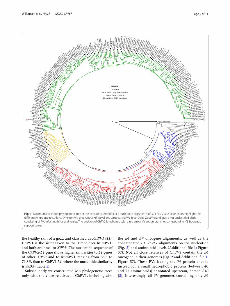
After removing the recombinant and unresolved taxa, ML phylogenetic trees were constructed of the concat-enated E1E2 and L2L1 alignments (Additional file 1: Fig-ure S4 and Additional file 1: Figure S5), as well as of the concatenated E1E2L2L1 alignments at the nucleotide (Fig. 1) and amino acid (Additional file 1: Figure S6) lev-els. The position of ChPV2 did not change and remained well supported. Interestingly, ChPV2 does not cluster with the previously described ChPV1, retrieved from
Page 5 of 11Willemsen et al. Virol J (2020) 17:167
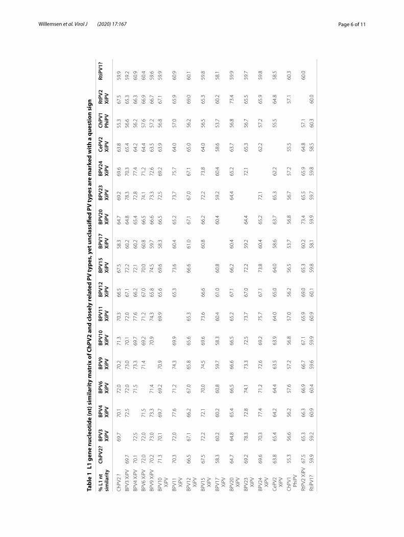
the healthy skin of a goat, and classified as PhiPV1 (11). ChPV1 is the sister taxon to the Timor deer RtimPV1, and both are basal to XiPVs. The nucleotide sequence of the ChPV2-L1 gene shows higher similarities to L1 genes of other XiPVs and to RtimPV1 ranging from 58.3 to 71.8%, than to ChPV1 L1, where the nucleotide similarity is 55.3% (Table 1).
Subsequently we constructed ML phylogenetic trees only with the close relatives of ChPV1, including also
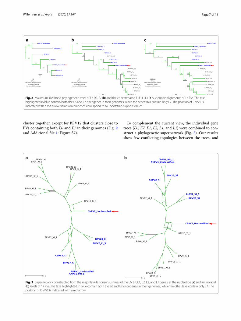
the E6 and E7 oncogene alignments, as well as the concatenated E1E2L2L1 alignments on the nucleotide (Fig. 2) and amino acid levels (Additional file 1: Figure S7). Not all close relatives of ChPV2 contain the E6 oncogene in their genomes (Fig. 2 and Additional file 1: Figure S7). These PVs lacking the E6 protein encode instead for a small hydrophobic protein (between 40 and 75 amino acids) annotated upstream, named E10 [8]. Interestingly, all PV genomes containing only E6
Fig. 1 Maximum likelihood phylogenetic tree of the concatenated E1E2L2L1 nucleotide alignments of 324 PVs. Clade color codes highlight the different PV groups: red, Alpha‑OmikronPVs; green, Beta‑XiPVs; yellow, Lambda‑MuPVs; blue, Delta‑ZetaPVs; and gray, a yet unclassified clade consisting of PVs infecting birds and turtles. The position of ChPV2 is indicated with a red arrow. Values on branches correspond to ML bootstrap support values
Page 6 of 11Willemsen et al. Virol J (2020) 17:167
Tabl
e 1
L1 g
ene
nucl
eoti
de (n
t) s
imila
rity
mat
rix
of C
hPV
2 an
d cl
osel
y re
late
d PV
type
s, y
et u
ncla
ssifi
ed P
V ty
pes
are
mar
ked
wit
h a
ques
tion
sig
n
% L
1 nt
si
mila
rity
ChPV
2?BP
V3
XiPV
BPV4
Xi
PVBP
V6
XiPV
BPV9
Xi
PVBP
V10
XiPV
BPV1
1 Xi
PVBP
V12
XiPV
BPV1
5 Xi
PVBP
V17
XiPV
BPV2
0 Xi
PVBP
V23
XiPV
BPV2
4 Xi
PVCe
PV2
XiPV
ChPV
1 Ph
iPV
RtPV
2 Xi
PVRt
iPV1
?
ChP
V2 ?
69.7
70.1
72.0
70.2
71.3
70.3
66.5
67.5
58.3
64.7
69.2
69.6
63.8
55.3
67.5
59.9
BPV3
XiP
V69
.772
.572
.073
.070
.172
.067
.172
.260
.264
.878
.370
.365
.456
.665
.359
.2
BPV4
XiP
V70
.172
.571
.573
.369
.777
.666
.272
.160
.265
.472
.877
.464
.256
.266
.360
.9
BPV6
XiP
V72
.072
.071
.571
.469
.271
.267
.070
.060
.866
.574
.171
.264
.457
.666
.960
.4
BPV9
XiP
V70
.273
.073
.371
.470
.974
.365
.874
.559
.766
.673
.372
.663
.557
.266
.759
.6
BPV1
0 Xi
PV71
.370
.169
.769
.270
.969
.965
.669
.658
.366
.572
.569
.263
.956
.867
.159
.9
BPV1
1 Xi
PV70
.372
.077
.671
.274
.369
.965
.373
.660
.465
.273
.775
.764
.057
.065
.960
.9
BPV1
2 Xi
PV66
.567
.166
.267
.065
.865
.665
.366
.661
.067
.167
.067
.165
.056
.269
.060
.1
BPV1
5 Xi
PV67
.572
.272
.170
.074
.569
.673
.666
.660
.866
.272
.273
.864
.056
.565
.359
.8
BPV1
7 Xi
PV58
.360
.260
.260
.859
.758
.360
.461
.060
.860
.459
.260
.458
.653
.760
.258
.1
BPV2
0 Xi
PV64
.764
.865
.466
.566
.666
.565
.267
.166
.260
.464
.465
.263
.756
.873
.459
.9
BPV2
3 Xi
PV69
.278
.372
.874
.173
.372
.573
.767
.072
.259
.264
.472
.165
.356
.765
.559
.7
BPV2
4 Xi
PV69
.670
.377
.471
.272
.669
.275
.767
.173
.860
.465
.272
.162
.257
.265
.959
.8
CePV
2 Xi
PV63
.865
.464
.264
.463
.563
.964
.065
.064
.058
.663
.765
.362
.255
.564
.858
.5
ChP
V1
PhiP
V55
.356
.656
.257
.657
.256
.857
.056
.256
.553
.756
.856
.757
.255
.557
.160
.3
RtPV
2 Xi
PV67
.565
.366
.366
.966
.767
.165
.969
.065
.360
.273
.465
.565
.964
.857
.160
.0
RtiP
V1?
59.9
59.2
60.9
60.4
59.6
59.9
60.9
60.1
59.8
58.1
59.9
59.7
59.8
58.5
60.3
60.0
Page 7 of 11Willemsen et al. Virol J (2020) 17:167
cluster together, except for BPV12 that clusters close to PVs containing both E6 and E7 in their genomes (Fig. 2 and Additional file 1: Figure S7).
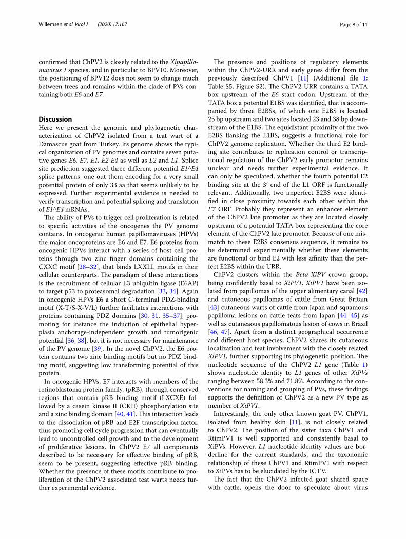
To complement the current view, the individual gene trees (E6, E7, E1, E2, L1, and L1) were combined to con-struct a phylogenetic supernetwork (Fig. 3). Our results show few conflicting topologies between the trees, and
a b c
Fig. 2 Maximum likelihood phylogenetic trees of E6 (a), E7 (b) and the concatenated E1E2L2L1 (c nucleotide alignments of 17 PVs. The taxa highlighted in blue contain both the E6 and E7 oncogenes in their genomes, while the other taxa contain only E7. The position of ChPV2 is indicated with a red arrow. Values on branches correspond to ML bootstrap support values
a b
Fig. 3 Supernetwork constructed from the majority rule consensus trees of the E6, E7, E1, E2, L2, and L1 genes, at the nucleotide (a) and amino acid (b) levels of 17 PVs. The taxa highlighted in blue contain both the E6 and E7 oncogenes in their genomes, while the other taxa contain only E7. The position of ChPV2 is indicated with a red arrow
Page 8 of 11Willemsen et al. Virol J (2020) 17:167
confirmed that ChPV2 is closely related to the Xipapillo-mavirus 1 species, and in particular to BPV10. Moreover, the positioning of BPV12 does not seem to change much between trees and remains within the clade of PVs con-taining both E6 and E7.
DiscussionHere we present the genomic and phylogenetic char-acterization of ChPV2 isolated from a teat wart of a Damascus goat from Turkey. Its genome shows the typi-cal organization of PV genomes and contains seven puta-tive genes E6, E7, E1, E2 E4 as well as L2 and L1. Splice site prediction suggested three different potential E1^E4 splice patterns, one out them encoding for a very small potential protein of only 33 aa that seems unlikely to be expressed. Further experimental evidence is needed to verify transcription and potential splicing and translation of E1^E4 mRNAs.
The ability of PVs to trigger cell proliferation is related to specific activities of the oncogenes the PV genome contains. In oncogenic human papillomaviruses (HPVs) the major oncoproteins are E6 and E7. E6 proteins from oncogenic HPVs interact with a series of host cell pro-teins through two zinc finger domains containing the CXXC motif [28–32], that binds LXXLL motifs in their cellular counterparts. The paradigm of these interactions is the recruitment of cellular E3 ubiquitin ligase (E6AP) to target p53 to proteasomal degradation [33, 34]. Again in oncogenic HPVs E6 a short C-terminal PDZ-binding motif (X-T/S-X-V/L) further facilitates interactions with proteins containing PDZ domains [30, 31, 35–37], pro-moting for instance the induction of epithelial hyper-plasia anchorage-independent growth and tumorigenic potential [36, 38], but it is not necessary for maintenance of the PV genome [39]. In the novel ChPV2, the E6 pro-tein contains two zinc binding motifs but no PDZ bind-ing motif, suggesting low transforming potential of this protein.
In oncogenic HPVs, E7 interacts with members of the retinoblastoma protein family, (pRB), through conserved regions that contain pRB binding motif (LXCXE) fol-lowed by a casein kinase II (CKII) phosphorylation site and a zinc binding domain [40, 41]. This interaction leads to the dissociation of pRB and E2F transcription factor, thus promoting cell cycle progression that can eventually lead to uncontrolled cell growth and to the development of proliferative lesions. In ChPV2 E7 all components described to be necessary for effective binding of pRB, seem to be present, suggesting effective pRB binding. Whether the presence of these motifs contribute to pro-liferation of the ChPV2 associated teat warts needs fur-ther experimental evidence.
The presence and positions of regulatory elements within the ChPV2-URR and early genes differ from the previously described ChPV1 [11] (Additional file 1: Table S5, Figure S2). The ChPV2-URR contains a TATA box upstream of the E6 start codon. Upstream of the TATA box a potential E1BS was identified, that is accom-panied by three E2BSs, of which one E2BS is located 25 bp upstream and two sites located 23 and 38 bp down-stream of the E1BS. The equidistant proximity of the two E2BS flanking the E1BS, suggests a functional role for ChPV2 genome replication. Whether the third E2 bind-ing site contributes to replication control or transcrip-tional regulation of the ChPV2 early promotor remains unclear and needs further experimental evidence. It can only be speculated, whether the fourth potential E2 binding site at the 3′ end of the L1 ORF is functionally relevant. Additionally, two imperfect E2BS were identi-fied in close proximity towards each other within the E7 ORF. Probably they represent an enhancer element of the ChPV2 late promoter as they are located closely upstream of a potential TATA box representing the core element of the ChPV2 late promoter. Because of one mis-match to these E2BS consensus sequence, it remains to be determined experimentally whether these elements are functional or bind E2 with less affinity than the per-fect E2BS within the URR.
ChPV2 clusters within the Beta-XiPV crown group, being confidently basal to XiPV1. XiPV1 have been iso-lated from papillomas of the upper alimentary canal [42] and cutaneous papillomas of cattle from Great Britain [43] cutaneous warts of cattle from Japan and squamous papilloma lesions on cattle teats from Japan [44, 45] as well as cutaneaous papillomatous lesion of cows in Brazil [46, 47]. Apart from a distinct geographical occurrence and different host species, ChPV2 shares its cutaneous localization and teat involvement with the closely related XiPV1, further supporting its phylogenetic position. The nucleotide sequence of the ChPV2 L1 gene (Table 1) shows nucleotide identity to L1 genes of other XiPVs ranging between 58.3% and 71.8%. According to the con-ventions for naming and grouping of PVs, these findings supports the definition of ChPV2 as a new PV type as member of XiPV1.
Interestingly, the only other known goat PV, ChPV1, isolated from healthy skin [11], is not closely related to ChPV2. The position of the sister taxa ChPV1 and RtimPV1 is well supported and consistently basal to XiPVs. However, L1 nucleotide identity values are bor-derline for the current standards, and the taxonomic relationship of these ChPV1 and RtimPV1 with respect to XiPVs has to be elucidated by the ICTV.
The fact that the ChPV2 infected goat shared space with cattle, opens the door to speculate about virus

Page 9 of 11Willemsen et al. Virol J (2020) 17:167
circulation between species. Although ChPV2 is closely related to bovine PVs, sequence similarities between ChPV2 and its closest relatives are probably too small to claim, that ChPV2 arose from an interspecies trans-mission event. We believe that, ChPV2 and/or closely related Xi PVs, that infect bovids, ovids and cervids might rather represent broad spectrum PVs, that could be able to infect different host species. Therefore, ChPV2 and closely related PVs might rather mimic the case of BPV1, which can infect cattle, sheep, several deer spe-cies, horses, zebras and tapirs. Currently, we do not know how prevalent this virus is among goats, or whether it is present in cattle alongside. Further studies are necessary to investigate the relative prevalence of the virus in the different species, as well as the efficiency of transmis-sion within and between species to allow for differentia-tion between the broad spectrum PV and the interspecies transmission hypothesis.
Both the E6 and E7 oncogenes in PVs infecting mam-mals appear to have a common ancestor [6]. It has been suggested previously that E6 may have been lost two separate times within the XiPV clade [8]. Among the closest relatives of ChPV2 all PV genomes containing only E7 cluster together, except for BPV12 that clusters within PVs containing both E6 and E7 (Fig. 2 and Addi-tional file 1: Figure S7), supporting the repeated loss of E6 hypothesis [6, 8]. However, it is possible that the separate E6 and E7, and concatenated E1E2L2L1 gene trees do not accurately describe the evolutionary history of these PVs. Nonetheless, we did not identify any rogue taxa for the constructed trees.
ConclusionBroadening the spectrum of known PVs infecting arti-odactyls will probably improve future phylogenetic inference allowing for a more detailed resolution and interpretation of the phylogenetic relationships of the artiodactyl PVs within Phi- and Xi-PVs.
Supplementary informationSupplementary information accompanies this paper at https ://doi.org/10.1186/s1298 5‑020‑01440 ‑9.
Additional file 1: Figure S1. Genome organization of ChPV2; Upstream regulatory region, yellow; early genes, green late genes, blue, potential E4 gene and potential E1^E4 splicing patterns, ochre. Figure S2. Presence and position of putative regulatory elements within URR and early gene region of ChPV1 and ChPV2, E1‑ binding sites (E1BS) are marked by yellow boxes, E2‑ binding sites (E2BS) perfectly matching the E2BS consensus sequence are marked by red boxes, E2BS with 1 nucleotide mismatch to the E2BS consensus sequence are marked in orange. nucleotide positions of early genes and regulatory elements are depicted below or above the respective element. Figure S3. Maximum likelihood phylogenetic trees of the concatenated E1E2 and concatenated L2L1 nucleotide align‑ments of 377 PVs. Clade color codes highlight the different PV groups
and recombinant taxa: red, Alpha‑OmikronPVs; green, Beta‑XiPVs; yellow, Lambda‑MuPVs; blue, Delta‑ZetaPVs; gray, a yet unclassified clade consist‑ing of PVs infecting birds and turtles; aqua‑blue, a yet unclassified clade consisting of PVs infecting Caniformia; orange, known recombinant PVs infecting Cetaceans; and pink, possible recombinant PVs. The position of ChPV2 is indicated with a red arrow. Values on branches correspond to ML bootstrap support values. Figure S4. Maximum likelihood phylogenetic trees of the concatenated E1E2 and concatenated L2L1 nucleotide align‑ments of 324 PVs. Clade color codes highlight the different PV groups: red, Alpha‑OmikronPVs; green, Beta‑XiPVs; yellow, Lambda‑MuPVs; blue, Delta‑ZetaPVs; and gray, a yet unclassified clade consisting of PVs infecting birds and turtles. The position of ChPV2 is indicated with a red arrow. Values on branches correspond to ML bootstrap support values. Figure S5. Maximum likelihood phylogenetic trees of the concatenated E1E2 and concatenated L2L1 amino acid alignments of 324 PVs. Clade color codes highlight the different PV groups: red, Alpha‑OmikronPVs; green, Beta‑XiPVs; yellow, Lambda‑MuPVs; blue, Delta‑ZetaPVs; and gray, a yet unclas‑sified clade consisting of PVs infecting birds and turtles. The position of ChPV2 is indicated with a red arrow. Values on branches correspond to ML bootstrap support values. Figure S6. Maximum likelihood phylogenetic tree of the concatenated E1E2L2L1 amino acid alignments of 324 PVs. Clade color codes highlight the different PV groups: red, Alpha‑Omikro‑nPVs; green, Beta‑XiPVs; yellow, Lambda‑MuPVs; blue, Delta‑ZetaPVs; and gray, a yet unclassified clade consisting of PVs infecting birds and turtles. The position of ChPV2 is indicated with a red arrow. Values on branches correspond to ML bootstrap support values. Figure S7. Maximum likeli‑hood phylogenetic trees of E6 (A), E7 (B) and the concatenated E1E2L2L1 (C) amino acid alignments of 17 PVs. The taxa highlighted in blue contain both the E6 and E7 oncogenes in their genomes, while the other taxa contain only E7. The position of ChPV2 is indicated with a red arrow. Values on branches correspond to ML bootstrap support values. Figure S8. Mac‑roscopic view of teat papilloma of a Damascus (Shami) goat, taken from (12). Table S1. Genome organization of ChPV2 presenting orf positions, orf length, GC content and amino acid content of encoded potential proteins and their molecular weight. Table S2. Nucleotide (nt) identity of ChPV2 genes with the respective genes of closely related PV types. Table S3. Amino acid (aa) identity of ChPV2 genes with the respective genes of closely related PV types. Table S4. Potential motifs and domains identified in amino acid sequences of ChPV2 proteins. Table S5. Potential regulatory elements identified throughout the ChPV2 genome, elements marked with a * have a mismatch to the published consensus sequence for the respective element. Table S6. List of PV genomes collected from PaVE (pave.niaid.nih.gov) plus ChPV2.
AbbreviationsPVs: Papillomaviruses; ORFs: Open reading frames; RCA : Rolling circle amplification; ML: Maximum likelihood; URR : Upstream regulatory region; pRB: Retinoblastoma protein; E2BS: E2 binding site; E1BS: E1 binding site; pA: Polyadenylation signal; HPV: Human papillomavirus; E6AP: E3 ubiquitin ligase; CKII: Casein kinase II.
AcknowledgementsWe thank Vet. Ali Haciömeroglu for providing the clinical sample used to isolate ChPV2. We are grateful to the Genotoul bioinformatics platform Tou‑louse Midi‑Pyrenees (Bioinfo Genotoul) for providing computing and storage resources. The authors acknowledge the IRD itrop HPC (South Green Platform) at IRD Montpellier for providing HPC resources that have contributed to the research results reported within this paper.
Authors’ contributionsConceptualization: EE‑S; data curation: EE‑S, AW, IGB, AE; formal analysis: AW, EE‑S; funding acquisition: EE‑S; investigation: AW, EE‑S, JD, AvdB, SBD; project administration: EE‑S; resources: SBD, FDAE, supervision: EES, validation: EE‑S, AW, IGB, AE, Visualization: AW, EE‑S; writing—original draft: AW, EE‑S; writ‑ing—review and editing: EE‑S, AW. All authors read and approved the final manuscript.
Page 10 of 11Willemsen et al. Virol J (2020) 17:167
FundingOpen Access funding enabled and organized by Projekt DEAL. Funding was provided to Eric Ehrke‑Schulz by the “Alexander Karl‑ Preis 2018” from the Stif‑tung Tumorforschung Kopf‑ Hals. AW was supported by the European Union Horizon 2020 Marie Sklodowska‑Curie research and innovation program grant ONCOGENEVOL (Contract Number 750180).
Availability of data and materialsThe nucleotide sequence of the ChPV2 genome is accessible in GeneBank under accession number MN148899 under https ://www.ncbi.nlm.nih.gov/nucco re/MN148 899: The authors confirm all supporting data, code and protocols have been provided within the article or through supplementary data files.
Ethics approval and consent to participateNo experiments with humans or living animals were performed, nor were animals harmed or killed during any procedure related to this article.
Consent for publicationNot applicable.
Competing interestsThe authors declare that they have no competing interests.
Author details1 Chair for Virology and Microbiology, Center for Biomedical Education and Research (ZBAF), Department for Human Medicine, Faculty of Health, Wit‑ten/Herdecke University, Stockumer Strasse 10, 58453 Witten, Germany. 2 Cen‑tre National de La Recherche Scientifique (CNRS), Laboratory MIVEGEC (CNRS IRD Uni Montpellier), Montpellier, France. 3 Faculty of Veterinary Medicine, Department of Virology, Ankara University, Ankara, Turkey. 4 Faculty of Vet‑erinary Medicine, Department of Virology, Hatay Mustafa Kemal University, Hatay, Turkey. 5 Center for Research on the Ecology and Evolution of Diseases (CREES), Montpellier, France. 6 Centre for Microbiology and Environmental Systems Science, University of Vienna, Vienna, Austria.
Received: 10 June 2020 Accepted: 21 October 2020
References 1. Antonsson A, Hansson BG. Healthy skin of many animal species harbors
papillomaviruses which are closely related to their human counterparts. J Virol. 2002;76(24):12537–42. https ://doi.org/10.1128/jvi.76.24.12537 ‑12542 .2002.
2. Nindl I, Gottschling M, Stockfleth E. Human papillomaviruses and non‑melanoma skin cancer: basic virology and clinical manifestations. Dis Mark. 2007;23(4):247–59.
3. Hubbers CU, Akgul B. HPV and cancer of the oral cavity. Virulence. 2015;6(3):244–8. https ://doi.org/10.1080/21505 594.2014.99957 0.
4. zur Hausen H. Papillomaviruses and cancer: from basic studies to clinical application. Nat Rev Cancer. 2002;2(5):342–50. https ://doi.org/10.1038/nrc79 8.
5. Bravo IG, Felez‑Sanchez M. Papillomaviruses: viral evolution, cancer and evolutionary medicine. Evol Med Public Health. 2015;2015(1):32–51. https ://doi.org/10.1093/emph/eov00 3.
6. Willemsen A, Bravo IG. Origin and evolution of papillomavirus (onco)genes and genomes. Philos Trans R Soc Lond B Biol Sci. 2019;374(1773):20180303. https ://doi.org/10.1098/rstb.2018.0303.
7. Willemsen A, Felez‑Sanchez M, Bravo IG. Genome plasticity in papillo‑maviruses and de novo emergence of E5 oncogenes. Genome Biol Evol. 2019;11(6):1602–17. https ://doi.org/10.1093/gbe/evz09 5.
8. Van Doorslaer K, McBride AA. Molecular archeological evidence in sup‑port of the repeated loss of a papillomavirus gene. Sci Rep. 2016;6:33028. https ://doi.org/10.1038/srep3 3028.
9. Bernard HU, Burk RD, Chen Z, van Doorslaer K, zur Hausen H, de Villiers EM. Classification of papillomaviruses (PVs) based on 189 PV types and
proposal of taxonomic amendments. Virology. 2010;401(1):70–9. https ://doi.org/10.1016/j.virol .2010.02.002.
10. Lopez‑Bueno A, Mavian C, Labella AM, Castro D, Borrego JJ, Alcami A, et al. Concurrence of iridovirus, polyomavirus, and a unique member of a new group of fish papillomaviruses in lymphocystis disease‑affected gilthead sea bream. J Virol. 2016;90(19):8768–79. https ://doi.org/10.1128/JVI.01369 ‑16.
11. Van Doorslaer K, Rector A, Vos P, Van Ranst M. Genetic characterization of the Capra hircus papillomavirus: a novel close‑to‑root artiodactyl papil‑lomavirus. Virus Res. 2006;118(1–2):164–9. https ://doi.org/10.1016/j.virus res.2005.12.007.
12. Dogan F, Dorttas SD, Bilge Dagalp S, Ataseven VS, Alkan F. A teat papillo‑matosis case in a Damascus goat (Shami goat) in Hatay Province, Turkey: a new putative papillomavirus? Arch Virol. 2018. https ://doi.org/10.1007/s0070 5‑018‑3781‑2.
13. Maniatis T, Fritsch EF, Sambrook J. Molecular cloning: a laboratory manual. New York: Cold Spring Harbor Laboratory; 2001.
14. Gasteiger E, Hoogland C, Gattiker A, Duvaud S, Wilkins MR, Appel RD, et al. Protein identification and analysis tools on the ExPASy server. In: Walker JM, editor., et al., The proteomics protocols handbook. Totowa: Humana Press; 2005. p. 571–607.
15. Sigrist CJ, de Castro E, Cerutti L, Cuche BA, Hulo N, Bridge A, et al. New and continuing developments at PROSITE. Nucleic Acids Res. 2013;41(1):D344–7. https ://doi.org/10.1093/nar/gks10 67.
16. Sigrist CJ, Cerutti L, Hulo N, Gattiker A, Falquet L, Pagni M, et al. PROSITE: a documented database using patterns and profiles as motif descriptors. Brief Bioinform. 2002;3(3):265–74. https ://doi.org/10.1093/bib/3.3.265.
17. Wingender E, Dietze P, Karas H, Knuppel R. TRANSFAC: a database on transcription factors and their DNA binding sites. Nucleic Acids Res. 1996;24(1):238–41. https ://doi.org/10.1093/nar/24.1.238.
18. Kel AE, Gossling E, Reuter I, Cheremushkin E, Kel‑Margoulis OV, Wing‑ender E. MATCH: a tool for searching transcription factor binding sites in DNA sequences. Nucleic Acids Res. 2003;31(13):3576–9. https ://doi.org/10.1093/nar/gkg58 5.
19. Schulz E, Gottschling M, Bravo IG, Wittstatt U, Stockfleth E, Nindl I. Genomic characterization of the first insectivoran papillomavirus reveals an unusually long, second non‑coding region and indicates a close rela‑tionship to Betapapillomavirus. J Gen Virol. 2009;90(Pt 3):626–33. https ://doi.org/10.1099/vir.0.00801 1‑0.
20. Katoh K, Standley DM. MAFFT multiple sequence alignment software version 7: improvements in performance and usability. Mol Biol Evol. 2013;30(4):772–80. https ://doi.org/10.1093/molbe v/mst01 0.
21. Suyama M, Torrents D, Bork P. PAL2NAL: robust conversion of protein sequence alignments into the corresponding codon alignments. Nucleic Acids Res. 2006;34(Web Server issue):W609–12. https ://doi.org/10.1093/nar/gkl31 5.
22. Castresana J. Selection of conserved blocks from multiple alignments for their use in phylogenetic analysis. Mol Biol Evol. 2000;17(4):540–52. https ://doi.org/10.1093/oxfor djour nals.molbe v.a0263 34.
23. Rector A, Stevens H, Lacave G, Lemey P, Mostmans S, Salbany A, et al. Genomic characterization of novel dolphin papillomaviruses provides indications for recombination within the Papillomaviridae. Virology. 2008;378(1):151–61. https ://doi.org/10.1016/j.virol .2008.05.020.
24. Gottschling M, Bravo IG, Schulz E, Bracho MA, Deaville R, Jepson PD, et al. Modular organizations of novel cetacean papillomaviruses. Mol Phylogenet Evol. 2011;59(1):34–42. https ://doi.org/10.1016/j.ympev .2010.12.013.
25. Robles‑Sikisaka R, Rivera R, Nollens HH, St Leger J, Durden WN, Stolen M, et al. Evidence of recombination and positive selection in cetacean papil‑lomaviruses. Virology. 2012;427(2):189–97. https ://doi.org/10.1016/j.virol .2012.01.039.
26. Stamatakis A. RAxML version 8: a tool for phylogenetic analysis and post‑analysis of large phylogenies. Bioinformatics. 2014;30(9):1312–3. https ://doi.org/10.1093/bioin forma tics/btu03 3.
27. Huson DH, Bryant D. Application of phylogenetic networks in evolution‑ary studies. Mol Biol Evol. 2006;23(2):254–67. https ://doi.org/10.1093/molbe v/msj03 0.
28. Glaunsinger BA, Lee SS, Thomas M, Banks L, Javier R. Interactions of the PDZ‑protein MAGI‑1 with adenovirus E4‑ORF1 and high‑risk papillo‑mavirus E6 oncoproteins. Oncogene. 2000;19(46):5270–80. https ://doi.org/10.1038/sj.onc.12039 06.
Page 11 of 11Willemsen et al. Virol J (2020) 17:167
• fast, convenient online submission
•
thorough peer review by experienced researchers in your field
• rapid publication on acceptance
• support for research data, including large and complex data types
•
gold Open Access which fosters wider collaboration and increased citations
maximum visibility for your research: over 100M website views per year •
At BMC, research is always in progress.
Learn more biomedcentral.com/submissions
Ready to submit your researchReady to submit your research ? Choose BMC and benefit from: ? Choose BMC and benefit from:
29. Thomas M, Laura R, Hepner K, Guccione E, Sawyers C, Lasky L, et al. Onco‑genic human papillomavirus E6 proteins target the MAGI‑2 and MAGI‑3 proteins for degradation. Oncogene. 2002;21(33):5088–96. https ://doi.org/10.1038/sj.onc.12056 68.
30. Lee SS, Glaunsinger B, Mantovani F, Banks L, Javier RT. Multi‑PDZ domain protein MUPP1 is a cellular target for both adenovirus E4‑ORF1 and high‑risk papillomavirus type 18 E6 oncoproteins. J Virol. 2000;74(20):9680–93. https ://doi.org/10.1128/jvi.74.20.9680‑9693.2000.
31. Nakagawa S, Huibregtse JM. Human scribble (Vartul) is targeted for ubiq‑uitin‑mediated degradation by the high‑risk papillomavirus E6 proteins and the E6AP ubiquitin‑protein ligase. Mol Cell Biol. 2000;20(21):8244–53. https ://doi.org/10.1128/mcb.20.21.8244‑8253.2000.
32. Gardiol D, Kuhne C, Glaunsinger B, Lee SS, Javier R, Banks L. Oncogenic human papillomavirus E6 proteins target the discs large tumour suppres‑sor for proteasome‑mediated degradation. Oncogene. 1999;18(40):5487–96. https ://doi.org/10.1038/sj.onc.12029 20.
33. Huibregtse JM, Scheffner M, Howley PM. A cellular protein mediates asso‑ciation of p53 with the E6 oncoprotein of human papillomavirus types 16 or 18. EMBO J. 1991;10(13):4129–35.
34. Scheffner M, Huibregtse JM, Vierstra RD, Howley PM. The HPV‑16 E6 and E6‑AP complex functions as a ubiquitin‑protein ligase in the ubiquitina‑tion of p53. Cell. 1993;75(3):495–505. https ://doi.org/10.1016/0092‑8674(93)90384 ‑3.
35. Jing M, Bohl J, Brimer N, Kinter M, Vande Pol SB. Degradation of tyrosine phosphatase PTPN3 (PTPH1) by association with oncogenic human pap‑illomavirus E6 proteins. J Virol. 2007;81(5):2231–9. https ://doi.org/10.1128/JVI.01979 ‑06.
36. Spanos WC, Hoover A, Harris GF, Wu S, Strand GL, Anderson ME, et al. The PDZ binding motif of human papillomavirus type 16 E6 induces PTPN13 loss, which allows anchorage‑independent growth and synergizes with ras for invasive growth. J Virol. 2008;82(5):2493–500. https ://doi.org/10.1128/JVI.02188 ‑07.
37. White EA, Howley PM. Proteomic approaches to the study of papil‑lomavirus‑host interactions. Virology. 2013;435(1):57–69. https ://doi.org/10.1016/j.virol .2012.09.046.
38. Kiyono T, Foster SA, Koop JI, McDougall JK, Galloway DA, Klingelhutz AJ. Both Rb/p16INK4a inactivation and telomerase activity are required to immortalize human epithelial cells. Nature. 1998;396(6706):84–8. https ://doi.org/10.1038/23962 .
39. Lorenz LD, Rivera Cardona J, Lambert PF. Inactivation of p53 rescues the maintenance of high risk HPV DNA genomes deficient in expression of E6. PLoS Pathog. 2013;9(10):e1003717. https ://doi.org/10.1371/journ al.ppat.10037 17.
40. Dyson N, Howley PM, Munger K, Harlow E. The human papilloma virus‑16 E7 oncoprotein is able to bind to the retinoblastoma gene product. Sci‑ence. 1989;243(4893):934–7.
41. Munger K, Werness BA, Dyson N, Phelps WC, Harlow E, Howley PM. Com‑plex formation of human papillomavirus E7 proteins with the retinoblas‑toma tumor suppressor gene product. EMBO J. 1989;8(13):4099–105.
42. Saveria Campo M, Moar MH, Jarrett WF, Laird HM. A new papillomavirus associated with alimentary cancer in cattle. Nature. 1980;286(5769):180–2. https ://doi.org/10.1038/28618 0a0.
43. Jarrett WF, Campo MS, O’Neil BW, Laird HM, Coggins LW. A novel bovine papillomavirus (BPV‑6) causing true epithelial papillomas of the mam‑mary gland skin: a member of a proposed new BPV subgroup. Virology. 1984;136(2):255–64.
44. Hatama S, Ishihara R, Ueda Y, Kanno T, Uchida I. Detection of a novel bovine papillomavirus type 11 (BPV‑11) using xipapillomavirus consensus polymerase chain reaction primers. Arch Virol. 2011;156(7):1281–5. https ://doi.org/10.1007/s0070 5‑011‑0970‑7.
45. Hatama S, Nobumoto K, Kanno T. Genomic and phylogenetic analysis of two novel bovine papillomaviruses, BPV‑9 and BPV‑10. J Gen Virol. 2008;89(Pt 1):158–63. https ://doi.org/10.1099/vir.0.83334 ‑0.
46. da Silva FR, Cibulski SP, Daudt C, Weber MN, Guimaraes LL, Streck AF, et al. Novel Bovine papillomavirus type discovered by rolling‑circle amplification coupled with next‑generation sequencing. PLoS ONE. 2016;11(9):e0162345. https ://doi.org/10.1371/journ al.pone.01623 45.
47. Daudt C, da Silva FRC, Cibulski SP, Streck AF, Laurie RE, Munday JS, et al. Bovine papillomavirus 24: a novel member of the genus Xipapillomavirus detected in the Amazon region. Arch Virol. 2019;164(2):637–41. https ://doi.org/10.1007/s0070 5‑018‑4092‑3.
Publisher’s NoteSpringer Nature remains neutral with regard to jurisdictional claims in pub‑lished maps and institutional affiliations.