Embed Size (px)
Citation preview
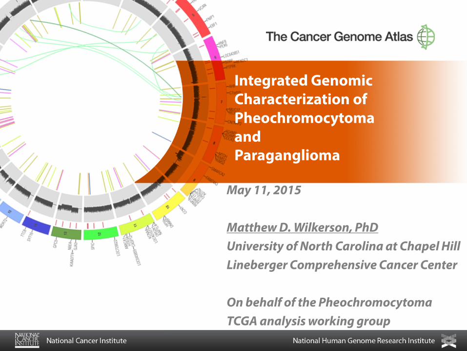
Integrated Genomic Characterization of Pheochromocytoma and Paraganglioma
May 11, 2015 Matthew D. Wilkerson, PhD University of North Carolina at Chapel Hill Lineberger Comprehensive Cancer Center On behalf of the Pheochromocytoma TCGA analysis working group
2
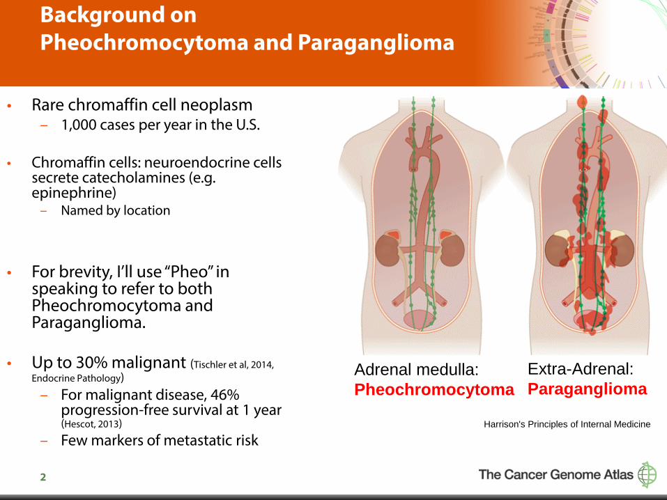
Background on Pheochromocytoma and Paraganglioma
• Rare chromaffin cell neoplasm – 1,000 cases per year in the U.S.
• Chromaffin cells: neuroendocrine cells
secrete catecholamines (e.g. epinephrine)
– Named by location
• For brevity, I’ll use “Pheo” in speaking to refer to both Pheochromocytoma and Paraganglioma.
• Up to 30% malignant (Tischler et al, 2014,
Endocrine Pathology) – For malignant disease, 46%
progression-free survival at 1 year (Hescot, 2013)
– Few markers of metastatic risk
Adrenal medulla: Pheochromocytoma
Extra-Adrenal: Paraganglioma
Harrison's Principles of Internal Medicine

3
Background on Pheo Genetics and Genomics
• Underlying inherited mutations in ~ 40% (highest of any tumor type) – (Dahia, 2014 Nature Genetics, Fishbein et al, 2013 Ann Surg Oncol)
• 19 susceptibility genes – NF1, RET, SDHB, SDHD, SDHA, SDHC, MAX, other less frequent genes.
• Pheo can be familial or sporadic.
• mRNA Expression Clusters – Pseudohypoxia and Kinase signaling (Dahia, 2005, Plos Genetics) – Potentially up to five clusters (Burnichon) – Associate with different susceptibility genes.
4
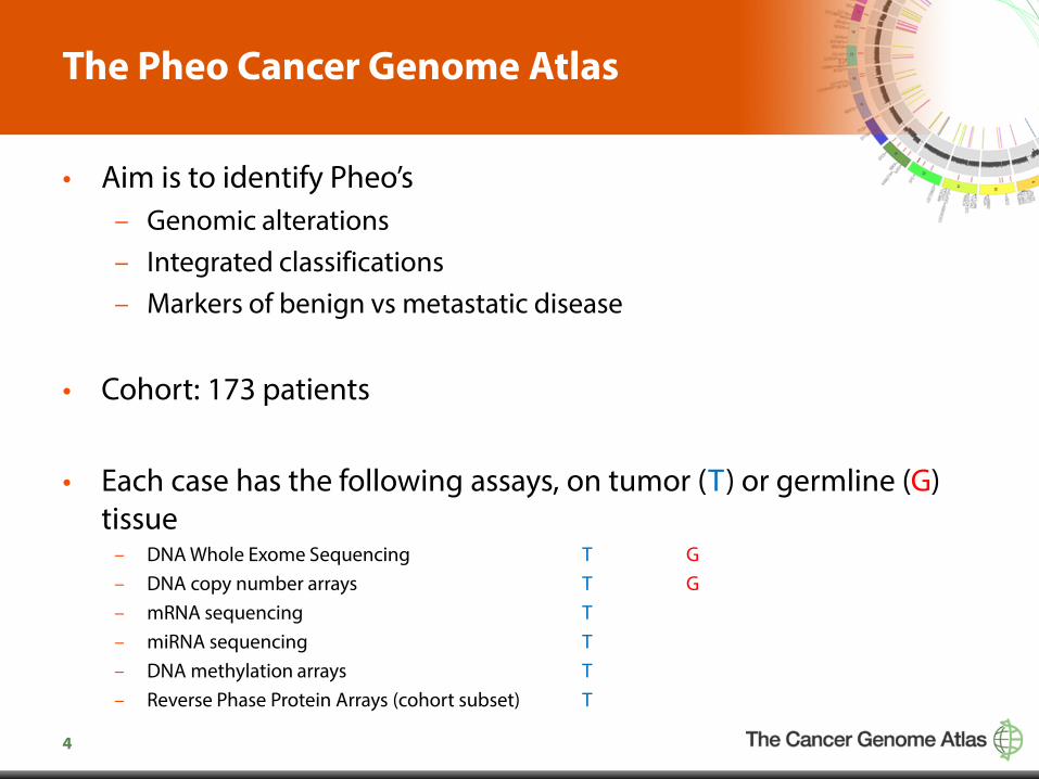
The Pheo Cancer Genome Atlas
• Aim is to identify Pheo’s – Genomic alterations – Integrated classifications – Markers of benign vs metastatic disease
• Cohort: 173 patients
• Each case has the following assays, on tumor (T) or germline (G) tissue
– DNA Whole Exome Sequencing T G – DNA copy number arrays T G – mRNA sequencing T – miRNA sequencing T – DNA methylation arrays T – Reverse Phase Protein Arrays (cohort subset) T
5
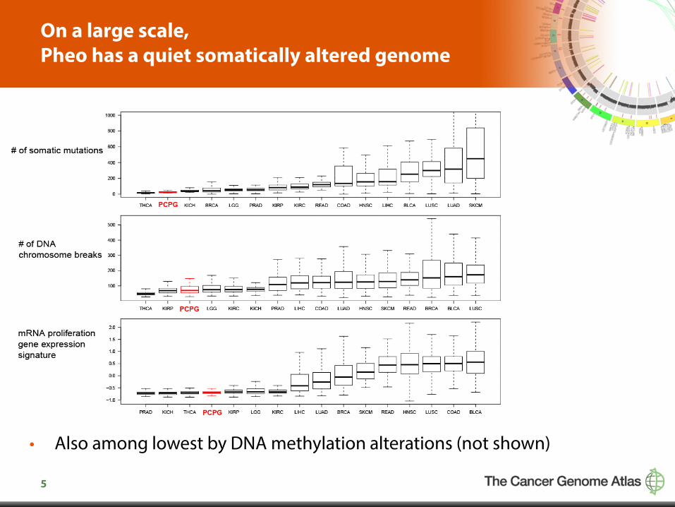
On a large scale, Pheo has a quiet somatically altered genome
• Also among lowest by DNA methylation alterations (not shown)
6
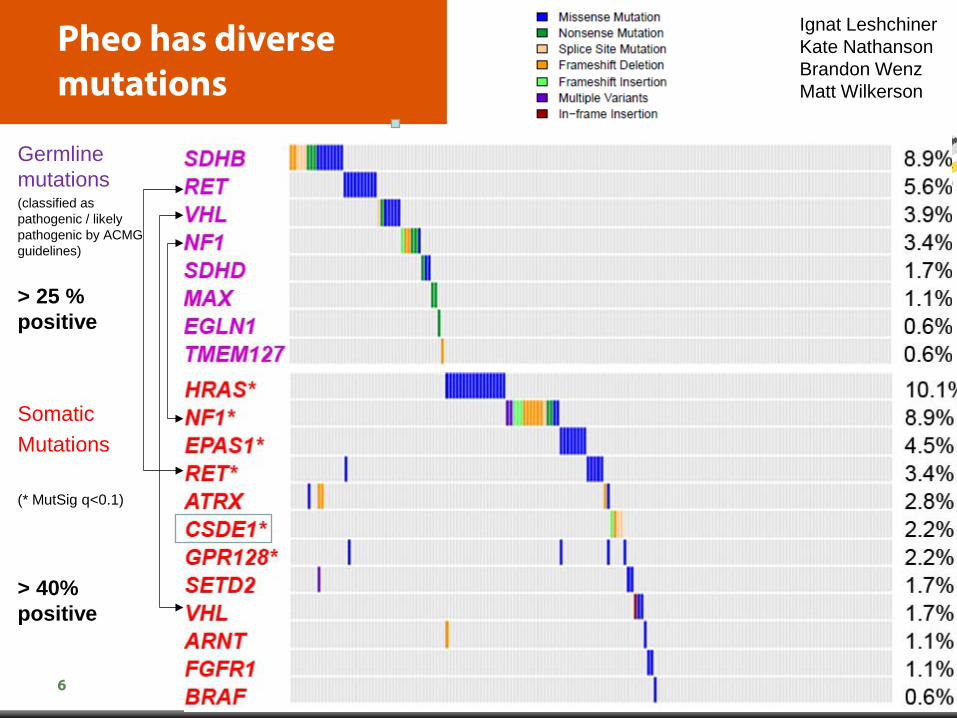
Pheo has diverse mutations
Germline mutations (classified as pathogenic / likely pathogenic by ACMG guidelines)
> 25 % positive Somatic Mutations (* MutSig q<0.1)
> 40% positive
Ignat Leshchiner Kate Nathanson Brandon Wenz Matt Wilkerson
7
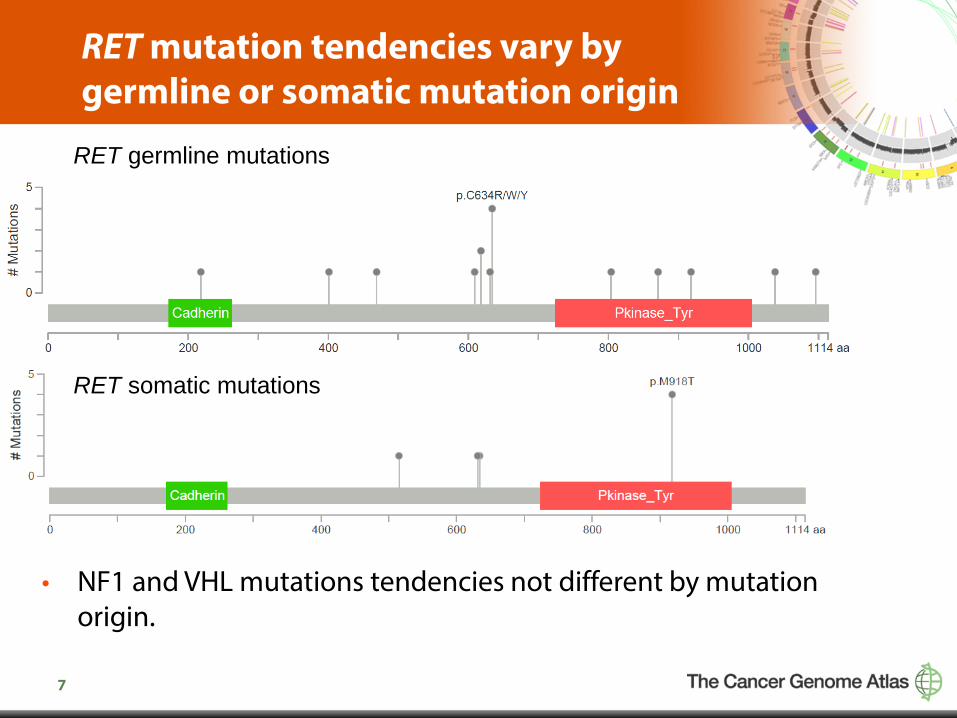
RET mutation tendencies vary by germline or somatic mutation origin
• NF1 and VHL mutations tendencies not different by mutation origin.
RET germline mutations RET somatic mutations

8
CSDE1 – new driver gene in Pheo
• Cold Shock Domain Containing E1, RNA-Binding
• CSDE1 mutant tumors co-occur with DNA copy number deletion and extreme low expression
• Supports loss of function role for CSDE1
• Kobyashi et al. (2013) Neuroscience: Knock out of CSDE1 causes irregular neuronal migration in brain development
9
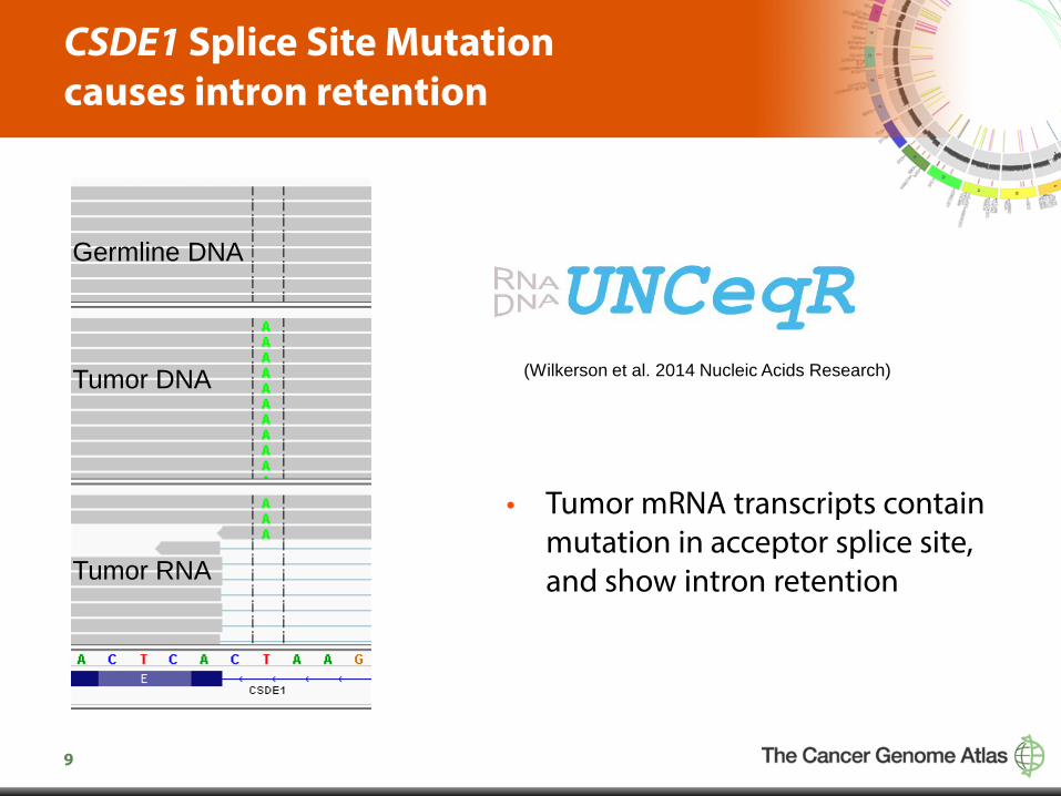
CSDE1 Splice Site Mutation causes intron retention
• Tumor mRNA transcripts contain mutation in acceptor splice site, and show intron retention
(Wilkerson et al. 2014 Nucleic Acids Research)
Germline DNA Tumor DNA Tumor RNA
10
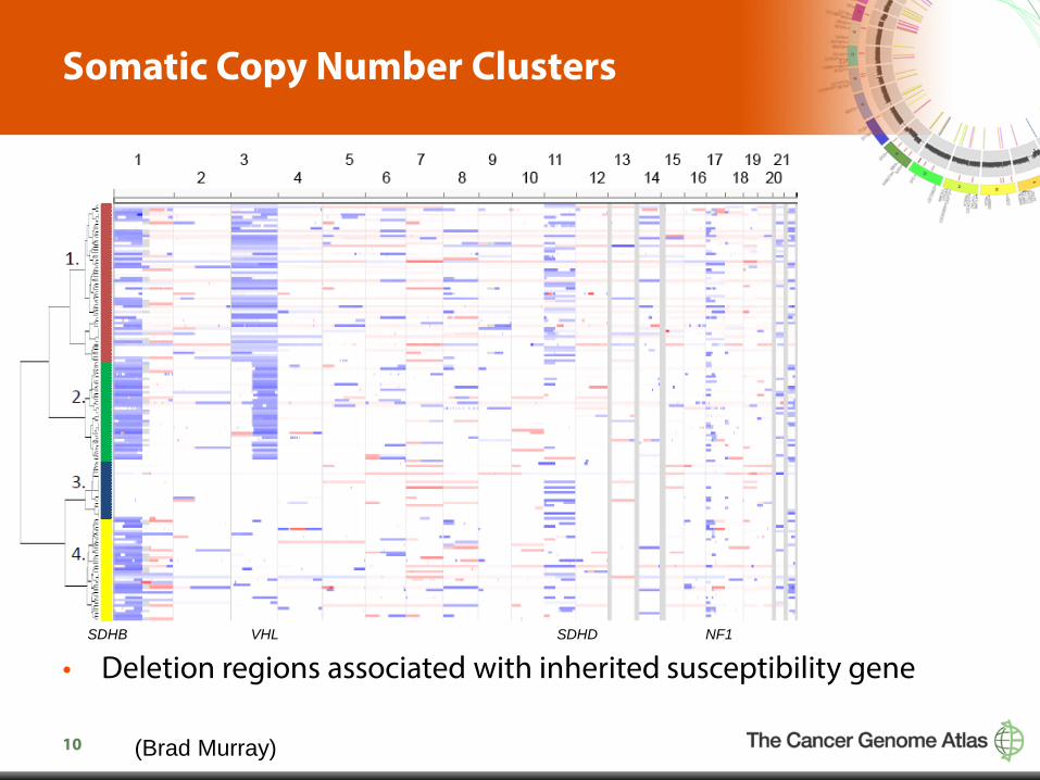
Somatic Copy Number Clusters
• Deletion regions associated with inherited susceptibility gene
(Brad Murray)
SDHB VHL SDHD NF1

11
Focal DNA copy number alterations
• Analysis of recurrent somatic copy number alterations (GISTIC)
CSDE1
NF1
Statistical significance increasing
Statistical significance
Focal copy number deletions
Focal copy number amplifications
4q31, 17q21
Brad Murray
12
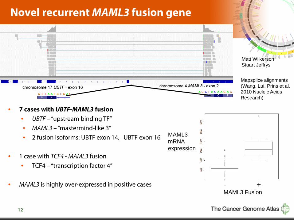
Novel recurrent MAML3 fusion gene
• 7 cases with UBTF-MAML3 fusion • UBTF – “upstream binding TF” • MAML3 – “mastermind-like 3” • 2 fusion isoforms: UBTF exon 14, UBTF exon 16
• 1 case with TCF4 - MAML3 fusion • TCF4 – “transcription factor 4”
• MAML3 is highly over-expressed in positive cases
MAML3 Fusion - +
MAML3 mRNA expression
Mapsplice alignments (Wang, Lui, Prins et al. 2010 Nucleic Acids Research)
Matt Wilkerson Stuart Jeffrys
13
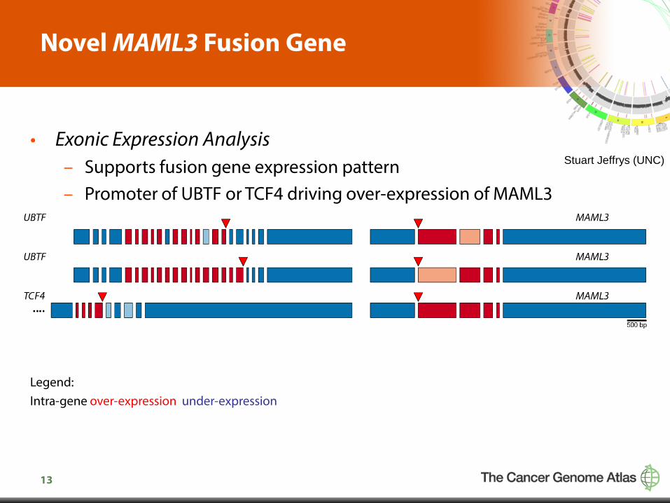
Novel MAML3 Fusion Gene
• Exonic Expression Analysis – Supports fusion gene expression pattern – Promoter of UBTF or TCF4 driving over-expression of MAML3
Legend: Intra-gene over-expression under-expression
Stuart Jeffrys (UNC)
14
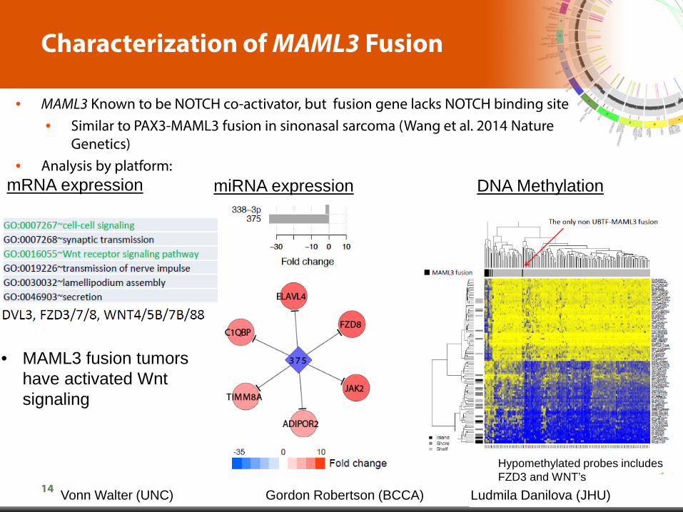
Characterization of MAML3 Fusion
• MAML3 Known to be NOTCH co-activator, but fusion gene lacks NOTCH binding site • Similar to PAX3-MAML3 fusion in sinonasal sarcoma (Wang et al. 2014 Nature
Genetics) • Analysis by platform:
mRNA expression miRNA expression DNA Methylation
• MAML3 fusion tumors have activated Wnt signaling
Hypomethylated probes includes FZD3 and WNT’s
Vonn Walter (UNC) Gordon Robertson (BCCA) Ludmila Danilova (JHU)
15
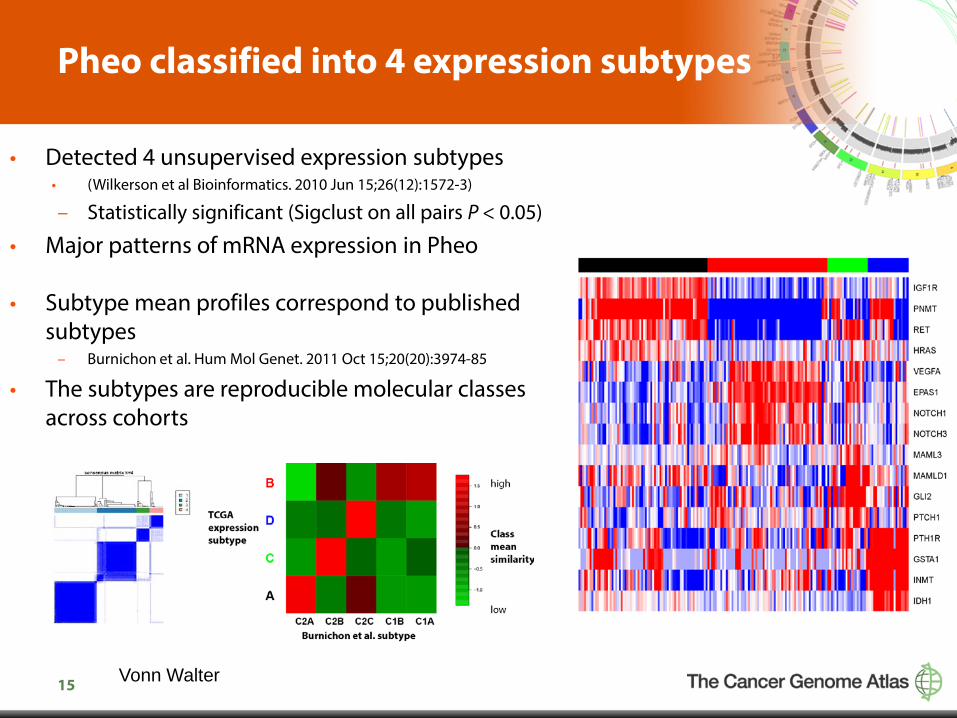
Pheo classified into 4 expression subtypes
• Detected 4 unsupervised expression subtypes • (Wilkerson et al Bioinformatics. 2010 Jun 15;26(12):1572-3)
– Statistically significant (Sigclust on all pairs P < 0.05)
• Major patterns of mRNA expression in Pheo
• Subtype mean profiles correspond to published subtypes
– Burnichon et al. Hum Mol Genet. 2011 Oct 15;20(20):3974-85
• The subtypes are reproducible molecular classes across cohorts
Vonn Walter
16
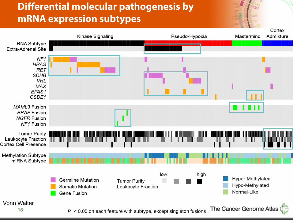
Differential molecular pathogenesis by mRNA expression subtypes
P < 0.05 on each feature with subtype, except singleton fusions Vonn Walter
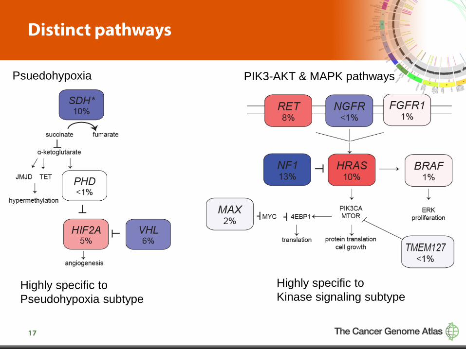
17
Distinct pathways
Psuedohypoxia PIK3-AKT & MAPK pathways
Highly specific to Pseudohypoxia subtype
Highly specific to Kinase signaling subtype
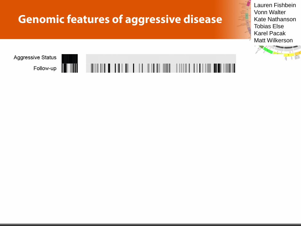
18
Genomic features of aggressive disease Lauren Fishbein Vonn Walter Kate Nathanson Tobias Else Karel Pacak Matt Wilkerson
* P < 0.05

19
Summary of the new discoveries of the TCGA Pheo Study
1. ~65 % cases have a driving germline or somatic mutation.
2. First recurrent fusion gene in Pheo (MAML3) – Associates with clinically aggressive disease – Found in one expression subtype of sporadic Pheo – Overexpresses Wnt signaling pathway
3. First reports of other alterations – CSDE1 somatic mutations – Fusion genes in (NGFR, BRAF, NF1)
20
Acknowledgements
• Anonymous patients
University of North Carolina Vonn Walter Matt Wilkerson Stuart Jeffrys Lisle Mose Joel Parker Patrick Kimes Neil Hayes Kimryn Rathmell Amy Johnson Tony Amelio Katie Hoadley University of Pennsylvania Lauren Fishbein Brandon Wenz Katherine Nathanson NIH Karel Pacak Broad Institute Ignat Leshchiner Brad Murray Carrie Sougnez Johns Hopkins Ludmila Danilova
BC Cancer Agency Gordon Robertson MDACC Rehan Akbani UCSC Sophie Olena Morozova Ian Fiddes Theo Knijnenburg BCR Tara Lichtenburg Kristen Leeras Program Office Ina Felau Kenna Shaw Jean Claude Zenklusen Carolyn Hutter
Disease experts Art Tischler Jim Powers Anne-Paule Gimenez-Roqueplo Charis Eng Tom Giordano Tobias Else Hans Ghayee Esther Korpershoek Sylvia Asa Ronald de Krijger Richard Auchus