Embed Size (px)
Citation preview
CHARACTERIZATION AND GENOMIC ANALYSIS OF A NOVEL
BACTERIOPHAGE AGAINST METHICILLIN-RESISTANT
STAPHYLOCOCCUS AUREUS
A THESIS SUBMITTED TO
THE GRADUATE SCHOOL OF NATURAL AND APPLIED SCIENCES
OF
MIDDLE EAST TECHNICAL UNIVERSITY
BY
MEDINE ÇOTAK
IN PARTIAL FULFILLMENT OF THE REQUIREMENTS
FOR
THE DEGREE OF DOCTOR OF PHILOSOPHY
IN
BIOTECHNOLOGY
FEBRUARY 2019

Approval of the thesis:
CHARACTERIZATION AND GENOMIC ANALYSIS OF A NOVEL
BACTERIOPHAGE AGAINST METHICILLIN-RESISTANT
STAPHYLOCOCCUS AUREUS
submitted by MEDINE ÇOTAK in partial fulfillment of the requirements for the
degree of Doctor of Philosophy in Biotechnology Department, Middle East
Technical University by,
Prof. Dr. Halil Kalıpçılar
Dean, Graduate School of Natural and Applied Sciences
Assoc. Prof. Dr. Can Özen
Head of Department, Biotechnology
Prof. Dr. Mahinur S. Akkaya
Supervisor, Biotechnology, METU
Prof. Dr. Zeynep Ceren Karahan
Co-Supervisor, Medical Microbiology, Ankara University
Examining Committee Members:
Assoc. Prof. Dr. Çağdaş Devrim Son
Biology, METU
Prof. Dr. Mahinur S. Akkaya
Biotechnology, METU
Assoc. Prof. Dr. Bala Gür Dedeoğlu
Biotechnology, Ankara University
Prof. Dr. Erdoğan Eşref Hakkı
Soil Science and Nutrition, Selçuk University
Dr. Mohd Kamran Khan
Soil Science and Nutrition, Selçuk University
Date: 05.02.2019
iv
I hereby declare that all information in this document has been obtained and
presented in accordance with academic rules and ethical conduct. I also declare
that, as required by these rules and conduct, I have fully cited and referenced all
material and results that are not original to this work.
Name, Surname:
Signature:
Medine Çotak
v
ABSTRACT
CHARACTERIZATION AND GENOMIC ANALYSIS OF A NOVEL
BACTERIOPHAGE AGAINST METHICILLIN-RESISTANT
STAPHYLOCOCCUS AUREUS
Çotak, Medine
Doctor of Philosophy, Biotechnology
Supervisor: Prof. Dr. Mahinur S. Akkaya
Co-Supervisor: Prof. Dr. Zeynep Ceren Karahan
February 2019, 75 pages
Methicillin resistant Staphylococcus aureus (MRSA) is one of the most frequent
hospital and community acquired infectious agents causing severe diseases.
Bacteriophages may offer a solution to treat the bacterial infections that are not
responding to classical antibiotics. In this context, a new lytic phage, named as
vB_SauM-MikSA913, was isolated from sewage treatment center in Samsun
(Turkey). MRSA clinical strains obtained from a local hospital were used as
propagating hosts while searching for the lytic phages. Genomic analysis suggest that
the isolated vB_SauM-MikSA913 belongs to the Myoviridiae family like the most
characterized phage K. Phage vB_SauM-MikSA913 has a genome size of 134193 bp
double-stranded DNA, encoding 206 open reading frames (ORFs) and 4 tRNAs. There
was a high similarity between our phage to others described in the literature such as
qdsa002, GH15, vB_Sau_CG, and phiSA039 with more than 90% query cover and
identity. vB_SauM-MikSA913 (shortly called MikSA913) has a wide range of host
and high lytic activity on MRSA strains. The bacteriolytic activity of the phage was
tested over a range of multiplicity of infection (MOI) and the optimal MOI was found
to be 0.001, which indicates its good lytic efficiency even at low concentration.
MikSA913 was stable at a wide range of pH and temperatures. One-step growth curve
vi
analysis showed that the eclipse and latent periods of MikSA913 was 15 min and 20
min, respectively and the burst size is 112 plaque forming units/infected cell.
Bacteriophage therapy is now seriously on the table as an alternative treatment to
combat with antibiotic resistance crisis. The high lytic activity, its wide host range and
lack of virulence factors and antibiotic resistance genes deduced from bioinformatics
analysis suggest that the phage vB_SauM-MikSA913 could be an option for treating
S.aureus infections including MRSA infections.
Keywords: Staphylococcus aureus, Methicillin-Resistant Staphylococcus aureus,
MRSA, Bacteriophage, Phage, Antibiotic Resistance, Lytic Phage, Bacteriophage
Therapy
vii
ÖZ
METHİSİLİN DİRENÇLİ STAPHYLOCOCCUS AUREUS’A KARŞI YENİ
BAKTERİYOFAJ KARAKTERİZASYONU VE GENOMİK
TANIMLANMASI
Çotak, Medine
Doktora, Biyoteknoloji
Tez Danışmanı: Prof. Dr. Mahinur S. Akkaya
Ortak Tez Danışmanı: Prof. Dr. Zeynep Ceren Karahan
Şubat 2019, 75 sayfa
Methisiline dirençli Staphylococcus aureus (MRSA), ciddi hastalıklara neden olan en
yaygın hastane ve toplum kökenli bulaşıcı ajanlardan biridir. Bakteriyofajlar, klasik
antibiyotiklere cevap vermeyen bakteriyel enfeksiyonları tedavi etmek için bir çözüm
sunabilir. Bu bağlamda, vB_SauM-Mik913 olarak adlandırılan yeni bir litik faj,
Samsun'daki (Türkiye) atık su arıtma merkezinden izole edildi. Yerel bir hastaneden
elde edilen MRSA klinik suşları litik fajlar aranırken konakçı olarak kullanıldı.
Genomik analiz sonuçları, izole edilmiş vB_SauM-Mik913'ün, literatürde en çok
çalışılan faj K gibi Myoviridiae familyasına ait olduğunu göstermektedir. vB_SauM-
MikSA913 genomu 134193 bp çift sarmallı DNA'ya sahiptir ve 206 ORF ve 4
tRNA’yı kodlamaktadır. Faj vB_SauM-MikSA913, MRSA suşlarında geniş konakçı
aralığı vardır ve yüksek litik aktivite göstermektedir. Fajın bakteriyolitik aktivitesi,
çok sayıda MOI (fajın bakteriye oranı) aralığında test edildi ve optimal MOI'nin, 0.001
olduğu bulundu; bu durum, düşük konsantrasyonda bile yüksek litik etkinliğini
göstermektedir. Bakteriyofaj terapisi şimdi ciddi biçimde antibiyotik direnci kriziyle
mücadele için alternatif bir tedavi olarak kabul edilmektedir. Yüksek litik aktivite,
geniş spektrum aralığı ve biyoinformatik analizlerden elde edilen sonuçlara göre
virülans faktörü ve antibiyotik direnç genleri eksikliği faj vB_SauM-Mik913'ün
viii
MRSA enfeksiyonları dahil S.aureus enfeksiyonlarının tedavisi için bir seçenek
olabileceğini düşündürmektedir.
Anahtar Kelimeler: Staphylococcus aureus, Methisilin Dirençli Staphylococcus
aureus, MRSA, Bakteriyofaj, Faj, Antibiyotik Dirençliliği, Litik Faji, Faj Terapi

ix
To my family and my love
x
ACKNOWLEDGEMENTS
First, I am deeply grateful to my supervisor, Prof. Dr. Mahinur S. Akkaya for giving
me the endless support and sincere approach.
I express my deepest gratitude to Prof. Dr. Zeynep Ceren Karahan for being my co-
supervisor.
I would like to thank to all the personnel of central clinical microbiology laboratory
of Ibn-i Sina Hospital for their help during collection of clinical samples.
Finally, I would like to thank all of the committee members for their time and valuable
feedbacks
xi
TABLE OF CONTENTS
ABSTRACT ................................................................................................................. v
ÖZ.... ......................................................................................................................... vii
ACKNOWLEDGEMENTS ......................................................................................... x
TABLE OF CONTENTS ........................................................................................... xi
LIST OF TABLES ................................................................................................... xiv
LIST OF FIGURES ................................................................................................... xv
LIST OF ABBREVIATIONS .................................................................................. xvi
1. INTRODUCTION ................................................................................................ 1
1.1. Staphylococcus aureus ...................................................................................... 1
1.1.1. S. aureus Cell Wall ..................................................................................... 2
1.1.2. S. aureus Genome ....................................................................................... 3
1.2. Antibiotic Resistance in S. aureus ..................................................................... 3
1.2.1. Penicillin Resistance ................................................................................... 4
1.2.2. Methicillin Resistance................................................................................. 4
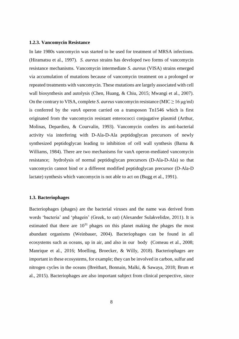
1.2.3. Vancomycin Resistance .............................................................................. 8
1.3. Bacteriophages .................................................................................................. 8
1.3.1. Bacteriophage Classification ...................................................................... 9
1.3.2. Bacteriophage Life Cycle ......................................................................... 10
1.3.2.1. Lytic Phages ....................................................................................... 11
1.3.2.2. Temperate Phages .............................................................................. 11
1.4. Bacteriophage Therapy .................................................................................... 12
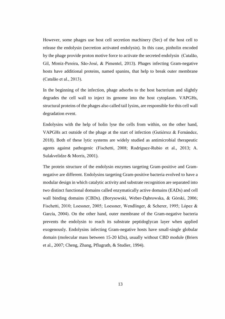
1.5. Bacteriophage Lytic Enzymes ......................................................................... 12
xii
1.6. Staphylococcal Phages .................................................................................... 15
1.7. Endolysins targeting S.aureus ......................................................................... 15
1.8. Aim of This Study ........................................................................................... 18
2. MATERIALS AND METHODS ....................................................................... 19
2.1. Culture media and growth conditions ............................................................. 19
2.2. Bacterial Strains .............................................................................................. 19
2.3. Double Plaque Assay ...................................................................................... 20
2.4. Bacteriophage Isolation................................................................................... 22
2.5. Phage Propagation and Concentration ............................................................ 23
2.6. Phage Titer Calculation ................................................................................... 24
2.7. Multiplicity of Infection (MOI) Assay ............................................................ 24
2.8. Phage Host Range Analysis (Spot Testing) .................................................... 25
2.9. Effect of Calcium on Adsorption Kinetics ...................................................... 25
2.10. One-Step Growth Curve ................................................................................ 26
2.11. Thermal and pH stability ............................................................................... 27
2.12. Phage Genomic DNA Isolation ..................................................................... 27
2.13. Whole Genome Sequencing .......................................................................... 28
2.14. Bioinformatics Analysis ................................................................................ 29
3. RESULTS ........................................................................................................... 31
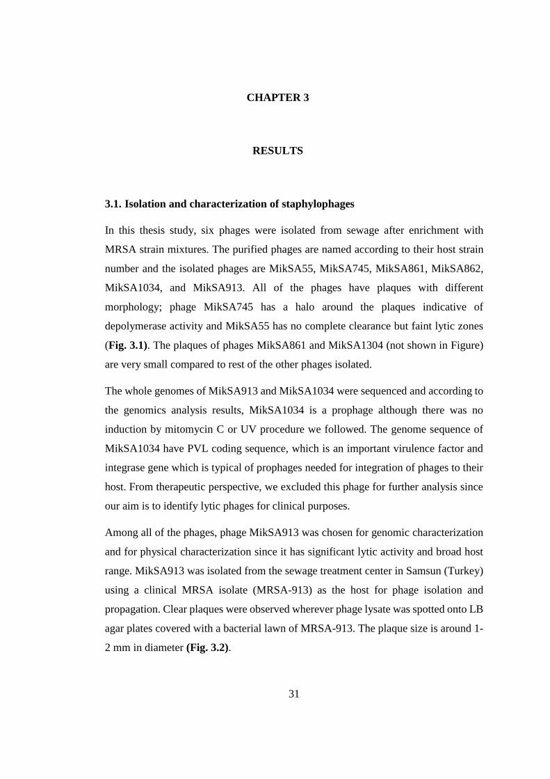
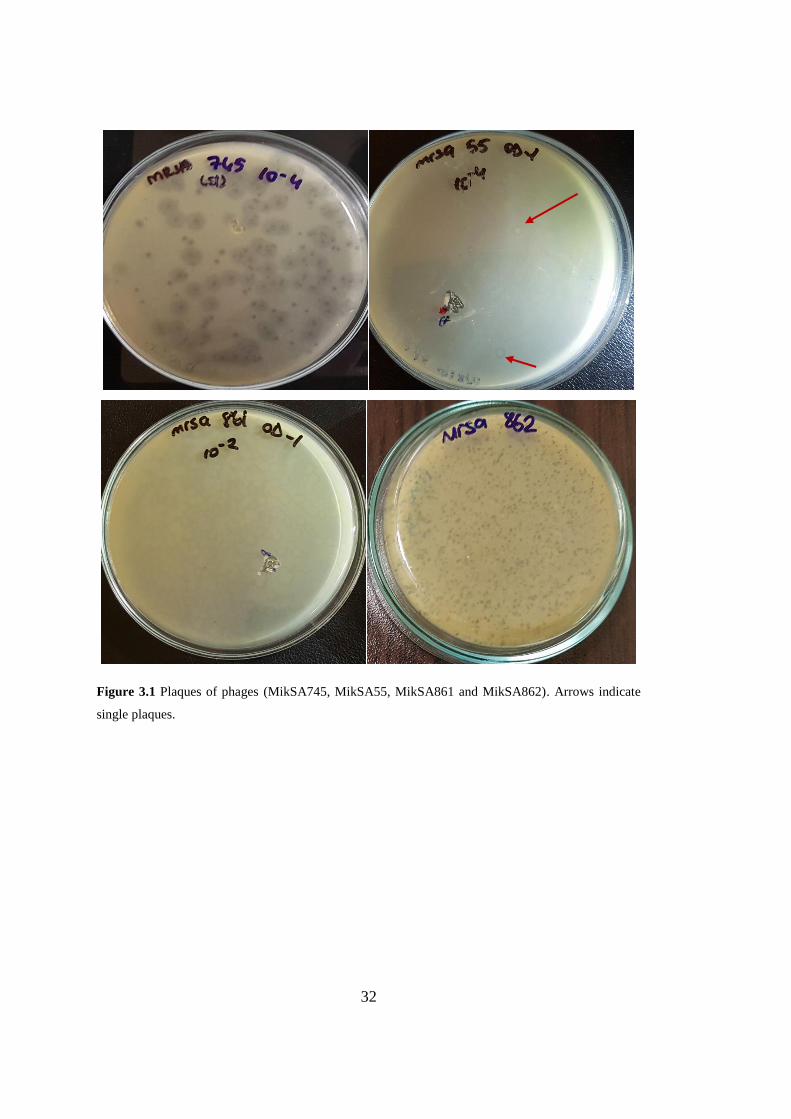
3.1. Isolation and characterization of staphylophages ........................................... 31
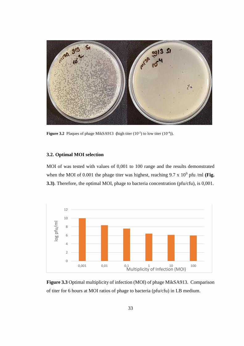
3.2. Optimal MOI selection.................................................................................... 33
3.3. Effect of Calcium on Adsorption Kinetics ...................................................... 34
3.4. MikSA913 Host Range ................................................................................... 35
3.5. Burst Size and Latent Period ........................................................................... 38
xiii
3.6. Thermal and pH stability ................................................................................. 39
3.7. MikSA913 Genome Analysis .......................................................................... 40
3.7.1. Genome Overview .................................................................................... 40
3.7.2. Lytic Proteins ............................................................................................ 45
3.7.3. Comparative Genomics............................................................................. 48
4. DISCUSSION ..................................................................................................... 51
5. CONCLUSION ................................................................................................... 55
REFERENCES ........................................................................................................... 57
A. Table A1 ............................................................................................................. 69
CURRICULUM VITAE ............................................................................................ 75
xiv
LIST OF TABLES
TABLES
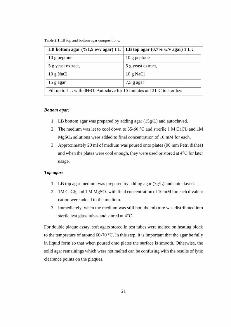
Table 2.1 LB top and bottom agar compositions. ..................................................... 21
Table 2.2 Saline-Magnesium (SM) buffer 1L ........................................................... 24
Table 3.1 Percentages of free phages with or without calcium. ............................... 34
Table 3.2 Phage MikSA913 host range analysis by spot testing. ............................. 36
Table 3.3 Functional modules of MikSA913 genome. ............................................. 42
Table 3.4 tRNAs encoded by phage MikSA913 genome. ........................................ 44
Table 3.5 Comparison of endolysin of phage MikSA913 with other similar lysins . 46
Table 3.6 Comparison of phage MikSA913 with its mostly closed phages. ............ 48

xv
LIST OF FIGURES
FIGURES
Figure 1.1 S. aureus drug resistance development timeline ........................................ 4
Figure 1.2 MRSA global prevalence map. .................................................................. 6
Figure 1.3 SSCmec types (I-VIII) identified in S. aureus. .......................................... 7
Figure 1.4 Morphology of three families of the tailed bacteriophages ....................... 9
Figure 1.5 Bacteriophage life cycles: Lytic and lysogenic cycles ........................... 10
Figure 1.6 Activities of phage lytic proteins on Gram-positive cell wall. ................ 14
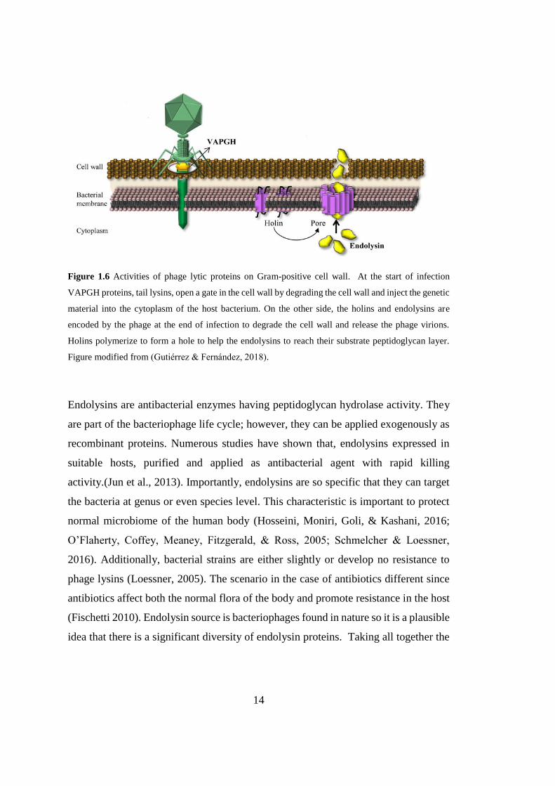
Figure 1.7 S.aureus peptidoglycan and the enzymatic activities of the endolysins .. 16
Figure 1.8 Endolysin and VAPGH domains. ............................................................ 17
Figure 3.1 Plaques of phages .................................................................................... 32
Figure 3.2 Plaques of phage MikSA913 .................................................................. 33
Figure 3.3 Optimal multiplicity of infection (MOI) of phage MikSA913. ............... 33
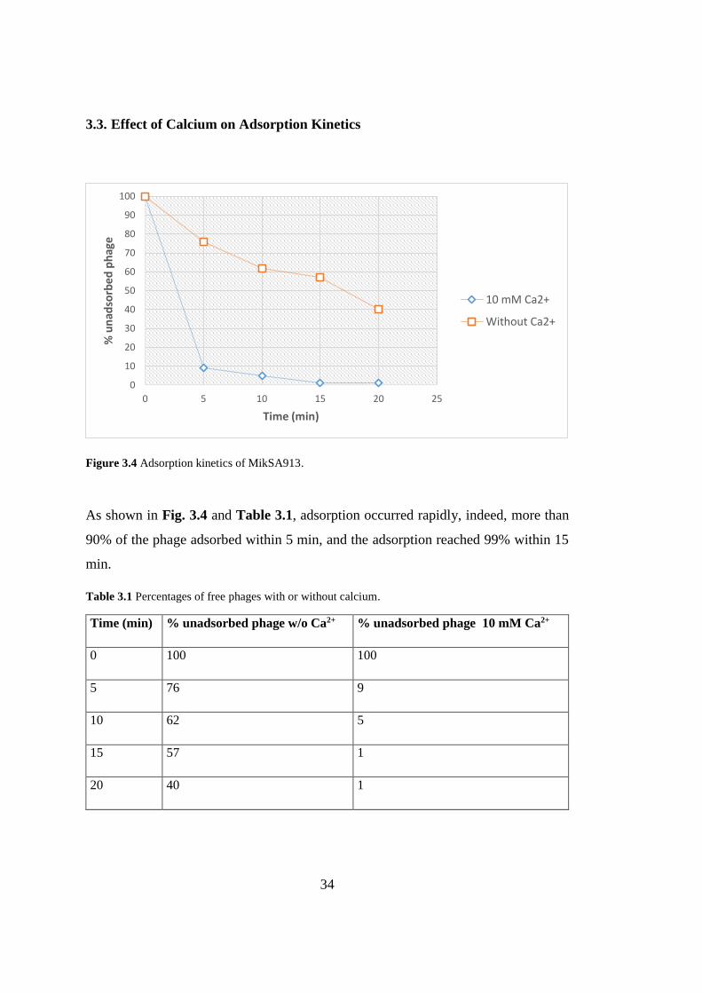
Figure 3.4 Adsorption kinetics of MikSA913. .......................................................... 34
Figure 3.5 One-step growth curve of phage MikSA913. ......................................... 38
Figure 3.6 pH stability assay of phage MikSA913. .................................................. 39
Figure 3.7 Stability of phage MikSA913 at different temperatures. ........................ 40
Figure 3.8 Phage MikSA913 endolysin domains. ..................................................... 45
Figure 3.9 Amino acid sequence of endolysin MikSA913 ...................................... 45
Figure 3.10 Phage MikSA913 holin domains. .......................................................... 47
Figure 3.11 VAPHGs of MikSA913. ........................................................................ 47
Figure 3.12 Phylogenetic comparison of Staphylococcus aureus ............................. 48
Figure 3.13 Nucleotide BLAST of phage MikSA913.............................................. 49
xvi
LIST OF ABBREVIATIONS
CA-MRSA Community-associated MRSA
CBD Cell wall Binding Domain
ccr cassette chromosome recombinase
EAD Enzymatically Active Domain
ICTV International Committee on Taxonomy of Viruses
LA-MRSA Livestock associated MRSA
LB Luria Bertani
MOI Multiplicity of Infection
MRSA
Methicillin resistant Staphylococcus aureus
NAG N-acteylglucosamine
NAM N-acetylmuramic acid
ORF Open Reading Frame
PBP Penicillin-binding protein
pfu plaque forming unit
PRSA Penicillin resistant Staphylococcus aureus
PVL Panton-Valentine Leucocidin
SCCmec Staphylococcal Cassette Chromosome
SM Saline-Magnesium
VAPGH Virion-Associated Peptidoglycan Hydrolase
VISA Vancomycin intermediate Staphylococcus aureus
1
CHAPTER 1
1. INTRODUCTION
1.1. Staphylococcus aureus
Staphylococcus bacterium is an important human firstly discovered by surgeon Sir
Alexander Ogston in a pus from a surgical abscess in 1881. He named it
Staphylococcus due to its shape resemblance to grape clusters (Greek, ‘staphyle’ -
bunch of grapes and ‘kokkos’-berry). Rosenbach gave the formal name of
Stapyhylococcus aureus in 1884. He differentiated the Staphylococcus aureus (Latin
‘aurum’-golden) from Staphylococcus albus (now called as S. epidermidis) (Latin,
‘epidermidis’ -white) since S. aureus grows golden-yellow colonies on bacterial
media (Giancarlo Licitra, 2013; Stryjewski & Corey, 2014).
S. aureus is a Gram-positive bacterium with the features of non-motility, catalase- and
coagulase-positive and facultative anaerobic. S. aureus has a high adaptation capacity
to its host and the environmental conditions and can survive in a wide range of pH and
temperature (Feng et al., 2008; Le Loir, Baron, & Gautier, 2003; Schmitt, Schuler-
Schmid, & Schmidt-Lorenz, 1990).
S. aureus is an opportunistic bacterium which can be both pathogen and commensal
carried on healthy individuals especially in nasal cavity (Peacock, De Silva, & Lowy,
2001; Sakwinska et al., 2010; van Belkum et al., 2009; Wertheim et al., 2005). As a
pathogen, S. aureus can infect any site of body and it is responsible for minor or lethal
infections; skin and soft tissue infections, food poisoning, endocarditis, chronic
osteomyletitis, penumonia, bacteremia, toxic shock syndrome, meningitis, septicemia
(Bassetti et al., 2014; McGuinness, Malachowa, & DeLeo, 2017; Nickerson, West,
Day, & Peacock, 2009; Todar, 2005; WHO, 2014).
2
S. aureus is a highly successful pathogen having a wide range of virulence factors.
These virulence factors include surface proteins, toxins, enzymes promoting tissue
damage and factors for evading the host immunity (Foster & Höök, 1998; Gill et al.,
2005). Mobile genetic elements called pathogenicity islands and prophages are
responsible for expressing the important toxins and other virulence determinants.
Panton-Valentine leucocidin (PVL) is a common virulence factor carried by
prophages in S. aureus strains (Jarraud et al., 2001; Malachowa & Deleo, 2010).
Antibiotic resistance in healthcare settings and in community is on the rise and
represents a global health burden. Multi-drug resistant S. aureus is one of the most
common cause of nosocomial infections with high rates of morbidity and mortality
(Salge, Vera, Antons, & Cimiotti, 2017).
1.1.1. S. aureus Cell Wall
In S. aureus cell, the outermost layer is the polysaccharide capsule which is an
important virulence factor (O’Riordan & Lee, 2004). Underneath the capsule is the
cell wall and it is essential for cell integrity and host-pathogen interactions (Dmitriev,
Toukach, Holst, Rietschel, & Ehlers, 2004). Peptidoglycan layer (20-40 nm) is the
primary component of the cell wall comprising of alternating glycan chains NAG (N-
acteylglucosamine) and NAM (N-acetylmuramic acid) cross-linked by pentaglycine
bridges and stem pentapeptides (Giesbrecht, Kersten, Maidhof, & Wecke, 1998).
Teichoic acids embedded in peptidoglycan layer function as phage receptors, epitopes
or as communication tools in pathogenicity with the environment (Navarre &
Schneewind, 1999; Szweda et al., 2012). The overall S. aureus cell wall structure is
shown in Fig. 1.7.
3
1.1.2. S. aureus Genome
S. aureus genome size is around 2.8 Mb and the genome have core and accessory
regions. The core genome genome is highly conserved among S. aureus strains and
contains essential genetic components relating to cell metabolism and replication. On
the other hand, 25% of the S. aureus genome is the accessory part comprising of
mobile genetic elements for example, prophages, pathogenicity islands, chromosomal
cassettes, plasmids and transposons. These mobile genetic elements are responsible
for the virulence, immune escape from host and for acquiring drug resistance (Lindsay
& Holden, 2004).
1.2. Antibiotic Resistance in S. aureus
The antibiotic era started with the discovery of penicillin in 1940s and many bacterial
pathogens including S. aureus were treatable with antibiotics since then (Aminov,
2010). However, many bacteria are able to adapt to changing environmental
conditions including antibiotic treatment. Mobile genetic elements play significant
role in acquiring resistance. S. aureus is also developing resistance rapidly day by day
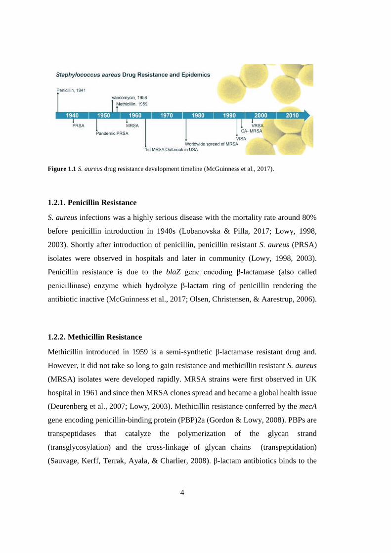
to different antibiotics introduced which is demonstrated in Fig. 1.1 (McGuinness et
al., 2017). Plasmids, transposons, bacteriophages, pathogenicity islands and
staphylococcal cassette chromosomes are the kinds of mobile genetic elements in S.
aureus conferring to the antibiotic resistance (Gill et al., 2005; Holden et al., 2004;
Lindsay, 2010; McGuinness et al., 2017).
4
Figure 1.1 S. aureus drug resistance development timeline (McGuinness et al., 2017).
1.2.1. Penicillin Resistance
S. aureus infections was a highly serious disease with the mortality rate around 80%
before penicillin introduction in 1940s (Lobanovska & Pilla, 2017; Lowy, 1998,
2003). Shortly after introduction of penicillin, penicillin resistant S. aureus (PRSA)
isolates were observed in hospitals and later in community (Lowy, 1998, 2003).
Penicillin resistance is due to the blaZ gene encoding β-lactamase (also called
penicillinase) enzyme which hydrolyze β-lactam ring of penicillin rendering the
antibiotic inactive (McGuinness et al., 2017; Olsen, Christensen, & Aarestrup, 2006).
1.2.2. Methicillin Resistance
Methicillin introduced in 1959 is a semi-synthetic β-lactamase resistant drug and.
However, it did not take so long to gain resistance and methicillin resistant S. aureus
(MRSA) isolates were developed rapidly. MRSA strains were first observed in UK
hospital in 1961 and since then MRSA clones spread and became a global health issue
(Deurenberg et al., 2007; Lowy, 2003). Methicillin resistance conferred by the mecA
gene encoding penicillin-binding protein (PBP)2a (Gordon & Lowy, 2008). PBPs are
transpeptidases that catalyze the polymerization of the glycan strand
(transglycosylation) and the cross-linkage of glycan chains (transpeptidation)
(Sauvage, Kerff, Terrak, Ayala, & Charlier, 2008). β-lactam antibiotics binds to the
5
PBPs on the cell wall and interferes with synthesis of peptidoglycan layer leading to
cell death. In the presence of PBP2a, which has a low affinity for all beta-lactams
including methicillin, oxacillin and third-generation cephalosporins, the cell wall
synthesis continues and the cell survives (Chambers, 1997; Deurenberg et al., 2007;
Ito et al., 2009). MRSA isolates are not not only resistant to methicillin but they also
carry multiple resistance genes to other antibiotics (Lowy, 2003).
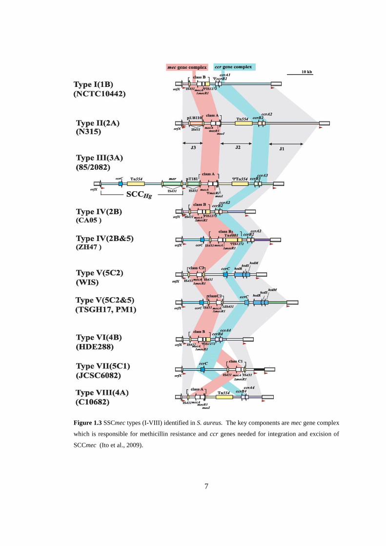
Staphylococcal Cassette Chromosome (SCCmec) is the mobile region of S. aureus
chromosome (Ito, Okuma, Ma, Yuzawa, & Hiramatsu, 2003; Saber, Jasni,
Jamaluddin, & Ibrahim, 2017). SCCmec consists of two fundamental components: the
mec and ccr gene complexes (Zong, Peng, & Lü, 2011). Cassette chromosome
recombinase (ccr) genes codes for recombinases those are responsible integration and
excision of SCCmec into and out of chromosome (Saber et al., 2017). Thereby,
SCCmec with its resistance elements can be transferred horizontally and vertically
intraspecies and interspecies (Stojanov, Sakwinska, & Moreillon, 2013).
To date, there are eleven SCCmec types (I-XI) (Fig 1.3) identified in staphylococci
having size range of 20.9 to 66.9 kb (Saber et al., 2017). SCCmec carries various
multiple resistance genes such as for macrolides, tetracycline and these genes are
carried via transposons, insertion sequences and transposons (Deurenberg et al.,
2007). In addition, the antibiotic resistance genes can be found on other sites of S.
aureus chromosome and on plasmids (Deurenberg et al., 2007).
Community-associated MRSA (CA-MRSA) causing infections in public, outside of
healthcare settings, is also a serious concern worldwide (Chambers & DeLeo, 2009;
Herold et al., 1998). In addition, MRSA is an important issue in food industry since
livestock associated MRSA (LA-MRSA) causes infections in livestock husbandry
(Cuny, Wieler, & Witte, 2015).
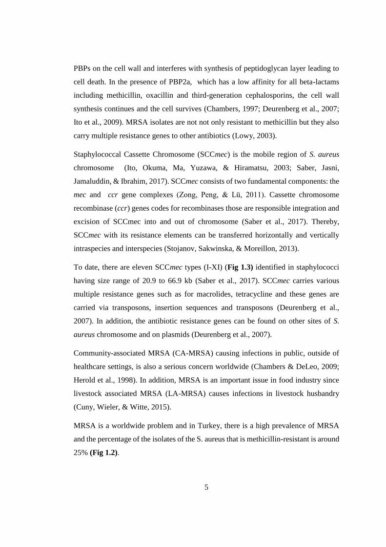
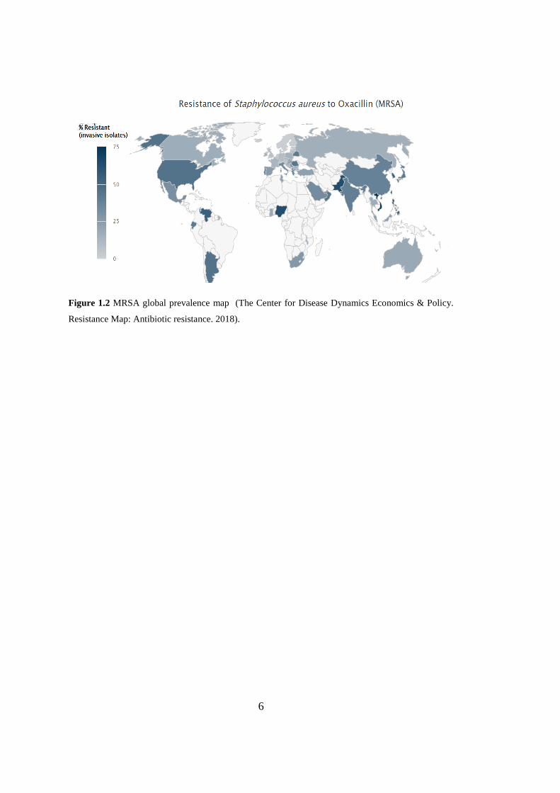
MRSA is a worldwide problem and in Turkey, there is a high prevalence of MRSA
and the percentage of the isolates of the S. aureus that is methicillin-resistant is around
25% (Fig 1.2).
6
Figure 1.2 MRSA global prevalence map (The Center for Disease Dynamics Economics & Policy.
Resistance Map: Antibiotic resistance. 2018).
7
Figure 1.3 SSCmec types (I-VIII) identified in S. aureus. The key components are mec gene complex
which is responsible for methicillin resistance and ccr genes needed for integration and excision of
SCCmec (Ito et al., 2009).
8
1.2.3. Vancomycin Resistance
In late 1980s vancomycin was started to be used for treatment of MRSA infections.
(Hiramatsu et al., 1997). S. aureus strains has developed two forms of vancomycin
resistance mechanisms. Vancomycin intermediate S. aureus (VISA) strains emerged
via accumulation of mutations because of vancomycin treatment on a prolonged or
repeated treatments with vancomycin. These mutations are largely associated with cell
wall biosynthesis and autolysis (Chen, Huang, & Chiu, 2015; Mwangi et al., 2007).
On the contrary to VISA, complete S. aureus vancomycin resistance (MIC ≥ 16 μg/ml)
is conferred by the vanA operon carried on a transposon Tn1546 which is first
originated from the vancomycin resistant enterococci conjugative plasmid (Arthur,
Molinas, Depardieu, & Courvalin, 1993). Vancomycin confers its anti-bacterial
activity via interfering with D-Ala-D-Ala peptidoglycan precursors of newly
synthesized peptidoglycan leading to inhibition of cell wall synthesis (Barna &
Williams, 1984). There are two mechanisms for vanA operon-mediated vancomycin
resistance; hydrolysis of normal peptidoglycan precursors (D-Ala-D-Ala) so that
vancomycin cannot bind or a different modified peptidoglycan precursor (D-Ala-D
lactate) synthesis which vancomycin is not able to act on (Bugg et al., 1991).
1.3. Bacteriophages
Bacteriophages (phages) are the bacterial viruses and the name was derived from
words ‘bacteria’ and ‘phagein’ (Greek, to eat) (Alexander Sulakvelidze, 2011). It is
estimated that there are 1031 phages on this planet making the phages the most
abundant organisms (Weinbauer, 2004). Bacteriophages can be found in all
ecosystems such as oceans, up in air, and also in our body (Comeau et al., 2008;
Manrique et al., 2016; Moelling, Broecker, & Willy, 2018). Bacteriophages are
important in these ecosystems, for example; they can be involved in carbon, sulfur and
nitrogen cycles in the oceans (Breitbart, Bonnain, Malki, & Sawaya, 2018; Brum et
al., 2015). Bacteriophages are also important subject from clinical perspective, since

9
they can carry disease-causing genes or the way around bacteriolytic phages can be
considered as therapeutic agents of multi-resistant pathogenic bacteria.
1.3.1. Bacteriophage Classification
Bacteriophages are classified based on genome type and phage morphology by
International Committee on Taxonomy of Viruses (ICTV). The genome of phages
consists of RNA or DNA and can be both single-stranded and double-stranded. The
genome size ranges from 3.5 kb (ssRNA phage MS2) to 500 kb (dsDNA Bacillus
phage G) (Salmond & Fineran, 2015). Different phage morphologies are identified;
tailed, polyhedral, and filamentous or pleomorphic and some have lipid or lipoprotein
capsids. The most characterized, around 96% of, bacteriophages belong to the order
Caudovirales (Latin ‘cauda’-tail). Caudovirales phages are tailed with dsDNA
genome. The families of this order are Myoviridae characterized by a straight
contractile tail, Podoviridae having a short tail and Siphoviridae with a contractile and
flexible tail (Fig. 1.4).
Figure 1.4 Morphology of three families of the tailed bacteriophages (Caudovirales) (Harper,
Anderson, & Enright, 2011).
10
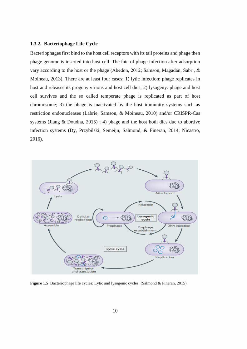
1.3.2. Bacteriophage Life Cycle
Bacteriophages first bind to the host cell receptors with its tail proteins and phage then
phage genome is inserted into host cell. The fate of phage infection after adsorption
vary according to the host or the phage (Abedon, 2012; Samson, Magadán, Sabri, &
Moineau, 2013). There are at least four cases: 1) lytic infection: phage replicates in
host and releases its progeny virions and host cell dies; 2) lysogeny: phage and host
cell survives and the so called temperate phage is replicated as part of host
chromosome; 3) the phage is inactivated by the host immunity systems such as
restriction endonucleases (Labrie, Samson, & Moineau, 2010) and/or CRISPR-Cas
systems (Jiang & Doudna, 2015) ; 4) phage and the host both dies due to abortive
infection systems (Dy, Przybilski, Semeijn, Salmond, & Fineran, 2014; Nicastro,
2016).
Figure 1.5 Bacteriophage life cycles: Lytic and lysogenic cycles (Salmond & Fineran, 2015).
11
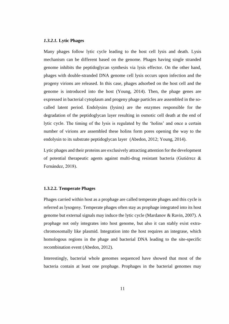
1.3.2.1. Lytic Phages
Many phages follow lytic cycle leading to the host cell lysis and death. Lysis
mechanism can be different based on the genome. Phages having single stranded
genome inhibits the peptidoglycan synthesis via lysis effector. On the other hand,
phages with double-stranded DNA genome cell lysis occurs upon infection and the
progeny virions are released. In this case, phages adsorbed on the host cell and the
genome is introduced into the host (Young, 2014). Then, the phage genes are
expressed in bacterial cytoplasm and progeny phage particles are assembled in the so-
called latent period. Endolysins (lysins) are the enzymes responsible for the
degradation of the peptidoglycan layer resulting in osmotic cell death at the end of
lytic cycle. The timing of the lysis is regulated by the ‘holins’ and once a certain
number of virions are assembled these holins form pores opening the way to the
endolysin to its substrate peptidoglycan layer (Abedon, 2012; Young, 2014).
Lytic phages and their proteins are exclusively attracting attention for the development
of potential therapeutic agents against multi-drug resistant bacteria (Gutiérrez &
Fernández, 2018).
1.3.2.2. Temperate Phages
Phages carried within host as a prophage are called temperate phages and this cycle is
referred as lysogeny. Temperate phages often stay as prophage integrated into its host
genome but external signals may induce the lytic cycle (Mardanov & Ravin, 2007). A
prophage not only integrates into host genome, but also it can stably exist extra-
chromosomally like plasmid. Integration into the host requires an integrase, which
homologous regions in the phage and bacterial DNA leading to the site-specific
recombination event (Abedon, 2012).
Interestingly, bacterial whole genomes sequenced have showed that most of the
bacteria contain at least one prophage. Prophages in the bacterial genomes may
12
responsible for host genome evolution or pathogenicity. These phages have shown to
carry genes for toxins, virulence factors, and antibiotic resistance (Kropinski &
Martha, 2009).
1.4. Bacteriophage Therapy
The discovery of the phages was credited to two scientists; Frederick William Twort
Felix in 1915 and d’Herelle in 1917 independently described the bacteriophages.
D’Herelle and George Eliava founded Eliava Institute for Phage Therapy in Georgia,
which is still active, in 1923. Phage therapy was used against open wound infections
of soldiers during the Winter War between the former Soviet Union and Finland
(Moelling et al., 2018). Phages were started to be visualized by the invention of
electron microscopy. With the introduction of antibiotics in 1940s, antibiotics
especially in western countries supersede phage therapy.
Bacteriophages are specific for their host bacteria and do not affect the mammalian
cells. The application of phages has been studied as therapeutic agents to treat acute
and chronic infections especially caused by the multidrug-resistant bacteria
(Wittebole, De Roock, & Opal, 2014). Nowadays, the use of lytic bacteriophages and
their enzymes to deal with antibiotic resistance crisis is getting renewed attraction by
the researchers and also by pharmaceutical companies (Fischetti, 2008; Rodríguez-
Rubio, Martínez, Donovan, Rodríguez, & García, 2013; A. Sulakvelidze & Morris,
2001). There are phages, phage cocktails and their lytic enzymes currently on the way
through clinical trials.
1.5. Bacteriophage Lytic Enzymes
Lytic enzymes derived from bacteriophages can be classified as endolysins and virion-
associated peptidoglycan hydrolases (VAPGHs) (Fig. 1.6). Lysis cassette containing
the two proteins; endolysin and holin are common in dsDNA bacteriophages.
13
However, some phages use host cell secretion machinery (Sec) of the host cell to
release the endolysin (secretion activated endolysin). In this case, pinholin encoded
by the phage provide proton motive force to activate the secreted endolysin (Catalão,
Gil, Moniz-Pereira, São-José, & Pimentel, 2013). Phages infecting Gram-negative
hosts have additional proteins, named spanins, that help to break outer membrane
(Catalão et al., 2013).
In the beginning of the infection, phage adsorbs to the host bacterium and slightly
degrades the cell wall to inject its genome into the host cytoplasm. VAPGHs,
structural proteins of the phages also called tail lysins, are responsible for this cell wall
degradation event.
Endolysins with the help of holin lyse the cells from within, on the other hand,
VAPGHs act outside of the phage at the start of infection (Gutiérrez & Fernández,
2018). Both of these lytic systems are widely studied as antimicrobial therapeutic
agents against pathogenic (Fischetti, 2008; Rodríguez-Rubio et al., 2013; A.
Sulakvelidze & Morris, 2001).
The protein structure of the endolysin enzymes targeting Gram-positive and Gram-
negative are different. Endolysins targeting Gram-positive bacteria evolved to have a
modular design in which catalytic activity and substrate recognition are separated into
two distinct functional domains called enzymatically active domains (EADs) and cell
wall binding domains (CBDs). (Borysowski, Weber-Da̧browska, & Górski, 2006;
Fischetti, 2010; Loessner, 2005; Loessner, Wendlinger, & Scherer, 1995; López &
García, 2004). On the other hand, outer membrane of the Gram-negative bacteria
prevents the endolysin to reach its substrate peptidoglycan layer when applied
exogenously. Endolysins infecting Gram-negative hosts have small-single globular
domain (molecular mass between 15-20 kDa), usually without CBD module (Briers
et al., 2007; Cheng, Zhang, Pflugrath, & Studier, 1994).
14
Figure 1.6 Activities of phage lytic proteins on Gram-positive cell wall. At the start of infection
VAPGH proteins, tail lysins, open a gate in the cell wall by degrading the cell wall and inject the genetic
material into the cytoplasm of the host bacterium. On the other side, the holins and endolysins are
encoded by the phage at the end of infection to degrade the cell wall and release the phage virions.
Holins polymerize to form a hole to help the endolysins to reach their substrate peptidoglycan layer.
Figure modified from (Gutiérrez & Fernández, 2018).
Endolysins are antibacterial enzymes having peptidoglycan hydrolase activity. They
are part of the bacteriophage life cycle; however, they can be applied exogenously as
recombinant proteins. Numerous studies have shown that, endolysins expressed in
suitable hosts, purified and applied as antibacterial agent with rapid killing
activity.(Jun et al., 2013). Importantly, endolysins are so specific that they can target
the bacteria at genus or even species level. This characteristic is important to protect
normal microbiome of the human body (Hosseini, Moniri, Goli, & Kashani, 2016;
O’Flaherty, Coffey, Meaney, Fitzgerald, & Ross, 2005; Schmelcher & Loessner,
2016). Additionally, bacterial strains are either slightly or develop no resistance to
phage lysins (Loessner, 2005). The scenario in the case of antibiotics different since
antibiotics affect both the normal flora of the body and promote resistance in the host
(Fischetti 2010). Endolysin source is bacteriophages found in nature so it is a plausible
idea that there is a significant diversity of endolysin proteins. Taking all together the
15
advantages of endolysins, they are now considered as potential antimicrobial agents
to cope with multidrug resistant bacteria.
1.6. Staphylococcal Phages
In literature, all of the characterized S. aureus phages fall into the order of
Caudovirales; tailed phages with an icosahedral capsid and dsDNA genome
(Deghorain & Van Melderen, 2012; Xia & Wolz, 2014). Anti-staphylococcal phages
can be classified into three groups: 1) podoviruses with <20kb genomes; 2)
siphoviruses with around 40 kb genomes ; and 3) myoviruses with >125 kb genomes
(Deghorain & Van Melderen, 2012). Siphoviruses are mainly temperate phages and
contain virulence genes (Xia & Wolz, 2014). Unlike siphoviruses, podoviruses
infecting S.aureus are strictly lytic but unfortunately, they are not common
(Kaźmierczak, Górski, & Dabrowska, 2014). Myoviruses are also lytic phages,
therefore myoviruses and podoviruses are appropriate for therapeutic usages.
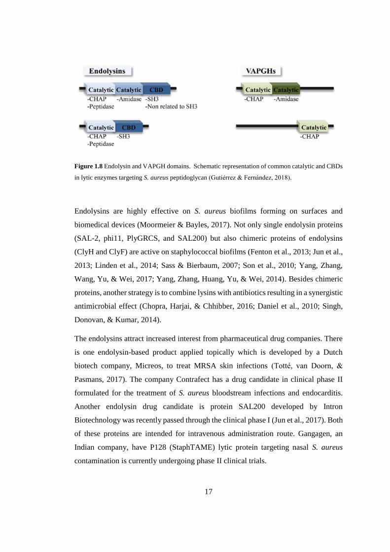
1.7. Endolysins targeting S.aureus
S.aureus bacteriophage derived endolysins have a modular structure possessing one
or more N-terminal enzymatically active domain (EAD) and a C-terminal cell wall
binding domain (CBD) conferring substrate specificity (Oliveira et al., 2013).
VAPGHs lack CBD but have a similar modular structure consisting of one or two
catalytic domains (Donovan, Lardeo, & Foster-Frey, 2006; Obeso, Martínez,
Rodríguez, & García, 2008). There are at least six enzymatically active catalytic
domain types of phage endolysins, the cleavage sites of these proteins on the
peptidoglycan layer are illustrated in (Fig 1.7).
16
Figure 1.7 S.aureus peptidoglycan and the enzymatic activities of the endolysins (indicated with an
arrow and a number). 1) N-Acetylmuramoyl-L alanine amidase; 2) interpeptide bridge endopeptidase;
3) L-alanoyl-D-glutamate endopeptidase; 4) N-acetyl-β-D-muramidase; 5) transglycosylase; 6) N-
acetyl-β-D-glucosaminidase (modified from Gutiérrez & Fernández, 2018)
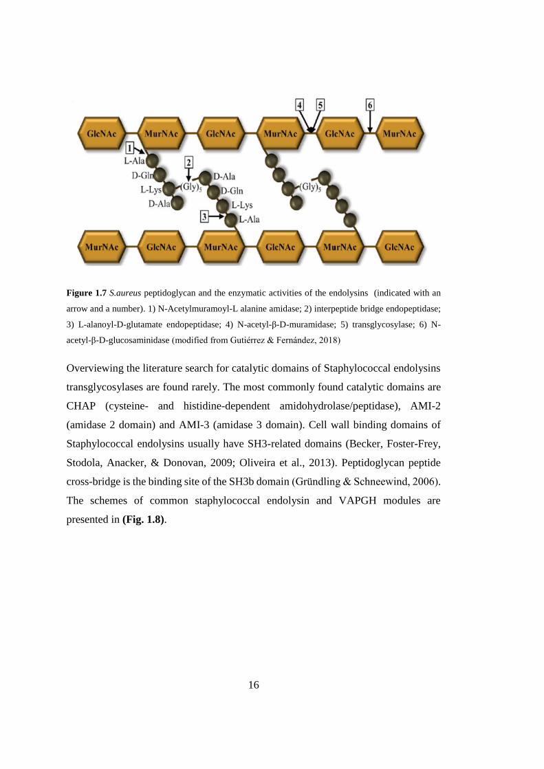
Overviewing the literature search for catalytic domains of Staphylococcal endolysins
transglycosylases are found rarely. The most commonly found catalytic domains are
CHAP (cysteine- and histidine-dependent amidohydrolase/peptidase), AMI-2
(amidase 2 domain) and AMI-3 (amidase 3 domain). Cell wall binding domains of
Staphylococcal endolysins usually have SH3-related domains (Becker, Foster-Frey,
Stodola, Anacker, & Donovan, 2009; Oliveira et al., 2013). Peptidoglycan peptide
cross-bridge is the binding site of the SH3b domain (Gründling & Schneewind, 2006).
The schemes of common staphylococcal endolysin and VAPGH modules are
presented in (Fig. 1.8).
17
Figure 1.8 Endolysin and VAPGH domains. Schematic representation of common catalytic and CBDs
in lytic enzymes targeting S. aureus peptidoglycan (Gutiérrez & Fernández, 2018).
Endolysins are highly effective on S. aureus biofilms forming on surfaces and
biomedical devices (Moormeier & Bayles, 2017). Not only single endolysin proteins
(SAL-2, phi11, PlyGRCS, and SAL200) but also chimeric proteins of endolysins
(ClyH and ClyF) are active on staphylococcal biofilms (Fenton et al., 2013; Jun et al.,
2013; Linden et al., 2014; Sass & Bierbaum, 2007; Son et al., 2010; Yang, Zhang,
Wang, Yu, & Wei, 2017; Yang, Zhang, Huang, Yu, & Wei, 2014). Besides chimeric
proteins, another strategy is to combine lysins with antibiotics resulting in a synergistic
antimicrobial effect (Chopra, Harjai, & Chhibber, 2016; Daniel et al., 2010; Singh,
Donovan, & Kumar, 2014).
The endolysins attract increased interest from pharmaceutical drug companies. There
is one endolysin-based product applied topically which is developed by a Dutch
biotech company, Micreos, to treat MRSA skin infections (Totté, van Doorn, &
Pasmans, 2017). The company Contrafect has a drug candidate in clinical phase II
formulated for the treatment of S. aureus bloodstream infections and endocarditis.
Another endolysin drug candidate is protein SAL200 developed by Intron
Biotechnology was recently passed through the clinical phase I (Jun et al., 2017). Both
of these proteins are intended for intravenous administration route. Gangagen, an
Indian company, have P128 (StaphTAME) lytic protein targeting nasal S. aureus
contamination is currently undergoing phase II clinical trials.
18
1.8. Aim of This Study
In this study, the aim is to find new bacteriophages against local MRSA strains. We
report the isolation and analysis of a Kayvirus genus S. aureus phage. Here, we show
the analysis of phage MikSA913 at a genetic and proteome level. Additionally, we
provide an insight into the reasons why this phage might be well suited for clinical
applications by testing its lytic efficiency and host range with a broad range of human
MRSA isolates and its safety at genomic level lacking of virulence factors or antibiotic
resistance genes. We also present an evaluation of the biophysical parameters: pH and
temperature, with intention to select optimal conditions to work with the phage
MikSA913.
19
CHAPTER 2
2. MATERIALS AND METHODS
2.1. Culture media and growth conditions
The bacteria were cultured in Luria Bertani (LB) broth or on LB agar plates and
incubated at 37°C. Phage propagation with host strains in liquid media were incubated
at 30°C. For phage isolation, double plaque assay method was used. Double plaque
assay was performed with two LB agar mediums with different concentrations: LB
medium with 1.5% or 0.7% agar was used for the standard agar (top layer) and for soft
agar (bottom layer), respectively. Bacteriophage enrichment assays were carried out
with 10 x strength LB. LB broth was used for the storage of bacteria with 20% glycerol
at -20°C.
Bacterial growth was measured by optical density at 565 nm by densitometer turbidity
detector (DEN-1, Biosan) where the bacterial cell concentration of 3 x 10⁸ cells/ml is
approximately equal to the 1 McFarland Standard Unit.
2.2. Bacterial Strains
All the strains of MRSA and other clinical isolates were taken from a local hospital
from the samples of patients (Ibn-i Sina Hospital- Ankara University). The isolates
used in this study are MRSA (n=50), MSSA (n=5) , Enterococcus faecalis (n=2),
Staphylococcus lugdunensis (n=2), Klebsiella pneumoniae (n=2) and Bacillus subtilis
(n=1). In addition, standard strains were used: Staphylococcus aureus ATCC 25923,
Escherichia coli ATCC 25922, Pseudomonas aeroginosa ATCC 27853. All of the
strains are listed for host range analysis in Table 3.2.
20
Clinical bacterial strains were identified by BD Phoenix (Becton Dickinson)
automated systems. Antimicrobial susceptibilities of these isolates were tested by disk
diffusion method and the results were assigned following the Clinical & Laboratory
Standards Institute (CLSI) guidelines. The tested antibiotics are erythromycin,
gentamicin, clindamycin, penicillin, oxacillin, cefotoxitin, vancomycin, rifampin,
linezolid, teicoplanin, ciprofloxacin, quinopristi / dalfopristin, chloromphenicol,
tetracycline and trimethoprim-sulfamethoxazole (Clinical and Laboratory Standards
Institute, 2015). The strains that were oxacillin- and/or cefoxitin resistant were
considered as methicillin resistant.
MikSA913 was isolated using a clinical MRSA strain 913 as a host and the same strain
was used as a standard host strain for the phage.
2.3. Double Plaque Assay
The double agar overlay plaque assay method was used to isolate the phages and for
phage tittering. In this method, phage suspensions were mixed with the host bacteria,
the top agar (also called soft agar or molten top agar), and the mixture was poured
onto the standard bottom agar. After enough overnight incubation at appropriate
temperature, phage plaques were visualized on the bacterial lawns. The phage titration
and propagation experiments were carried out with solid or liquid LB medium
supplemented with 10 mM CaCl2 and 10 mM MgSO4. The bottom and and top agar
components are listed and preparation of the layers are described as below:
21
Table 2.1 LB top and bottom agar compositions.
LB bottom agar (%1,5 w/v agar) 1 L LB top agar (0,7% w/v agar) 1 L :
10 g peptone 10 g peptone
5 g yeast extract, 5 g yeast extract,
10 g NaCl 10 g NaCl
15 g agar 7,5 g agar
Fill up to 1 L with dH2O. Autoclave for 15 minutes at 121°C to sterilize.
Bottom agar:
1. LB bottom agar was prepared by adding agar (15g/L) and autoclaved.
2. The medium was let to cool down to 55-60 °C and strerile 1 M CaCl2 and 1M
MgSO4 solutions were added to final concentration of 10 mM for each.
3. Approximately 20 ml of medium was poured onto plates (90 mm Petri dishes)
and when the plates were cool enough, they were used or stored at 4°C for later
usage.
Top agar:
1. LB top agar medium was prepared by adding agar (7g/L) and autoclaved.
2. 1M CaCl2 and 1 M MgSO4 with final concentration of 10 mM for each divalent
cation were added to the medium.
3. Immediately, when the medium was still hot, the mixture was distributed into
sterile test glass tubes and stored at 4°C.
For double plaque assay, soft agars stored in test tubes were melted on heating block
to the tempreture of around 60-70 °C. In this step, it is important that the agar be fully
in liquid form so that when poured onto plates the surface is smooth. Otherwise, the
solid agar remainings which were not melted can be confusing with the results of lytic
clearance points on the plaques.
22
2.4. Bacteriophage Isolation
The raw sewage water was collected from two waste treatment centers: at Middle East
Technical University, Ankara and in Samsun and the phage enrichment procedure was
followed as below:
1. Cenrifuge the sewage suspension at 9000 rpm, 10 min to remove particulates.
2. Add 45 ml of the supernatant (the clarified sewage water) and 5 ml of 10x
strength LB broth containing 100 mM MgSO4 and 100 CaCl2 into a 500 ml
Erlenmeyer flask.
3. Add 0.1 ml of 10 randomly chosen overnight grown MRSA clinical strains and
incubate at 30°C with shaking at 100 rpm in a bigger size flask for enough
aerobic respiration.
4. After overnight incubation, add 2,5 ml of chloroform to the flask contents and
let 30 min with gentle mixing for 5-6 times at room temperature (RT).
5. Centrifuge the flask contents at 9000 rpm 10 min 4°C to discard cell debris
and decant the supernatant into sterile tubes.
6. Sterilize the phage supernatant through a 0,45 µm pore size membrane.
7. Phages were isolated by double agar layer method. 0,1 ml of each overnight
MRSA strain was mixed with 1,5 ml of the resultant phage supernatant (from
step 6). 2.5 mL of heated (60-70 °C) soft agar was added to this mixture and
the mixture was poured evenly onto the bottom agar plates. Allow the overlays
harden for about 15 min under laminar flow.
8. The plates were incubated overnight at 37 °C.
9. Next day, check for the cleared zones on the plates where phage lysis occurs.
10. Plates in which bacteriophages shown with clear zones were chosen for single
plaque isolation. Using sterile pipette tip, the top layer with clear zone was
picked up and inoculated into 2 ml LB with 0,1 ml of corresponding host
MRSA strain and incubated for 6 hours to enrich the potential phages.
11. The 2 mL enriched phage filtrate was tenfold serially diluted (1 and 10-1 to 10-
9) in 1 ml of LB broth.
23
12. Add 0,1 ml of overnight grown MRSA host strain to each dilution and mix
with 3,5 ml of heated (60-70 °C) top agar medium and pour onto the standard
agar plate with bottom agar layer and allow hardening for 15 min.
13. Incubate the plates at 37 °C overnight and the next day check for separated
plaques on the dilution series.
14. The well-separated single plaque was taken with a pipette tip and suspended
in LB medium and again serially diluted (1 and 10-1 to 10-4). This single plaque
isolation procedure was repeated three times to ensure single phage isolation.
2.5. Phage Propagation and Concentration
The bacteriophages isolated with single plaque isolation method as described in 2.4
were propagated with their hosts for large volume and concentrated.
1. The single plaques on the plates (section 2.4 step 14) were picked up from the
plates with pipette tips and inoculated into a 500 ml Erlenmeyer flask with its
host of MRSA strain (0,5 ml) in 50 ml LB broth. The mixture was incubated
overnight at 30 °C with 100 rpm shaking.
2. 2,5 ml of chloroform was added to lyse the bacteria in the mixture and waited
30 min with gentle shaking for 5-6 times.
3. Centrifuge the flask contents at 9000 rpm 10 min 4 °C to get rid of cell debris
and decant the supernatant into sterile tubes.
4. Filter sterilize the phage supernatant through a 0,45 um pore size membrane
5. Add 6 ml of 10% (w/v) PEG 6000 solution (containing 50% ,1M NaCl) to the
phage filtrate and incubate overnight at 4 °C to precipitate phages
6. Next day, centrifuge at 15000 rpm 20 min at 4°C. Remove the supernatant and
resuspend the pellet containing the phages in 1 ml Saline-Magnesium (SM)
buffer and store at 4 °C. Phage lysate was supplemented with 20% glycerol
and stored at -20 °C for long term.
24
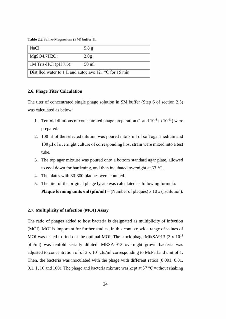
Table 2.2 Saline-Magnesium (SM) buffer 1L
NaCl: 5,8 g
MgSO4.7H2O: 2,0g
1M Tris-HCl (pH 7.5): 50 ml
Distilled water to 1 L and autoclave 121 °C for 15 min.
2.6. Phage Titer Calculation
The titer of concentrated single phage solution in SM buffer (Step 6 of section 2.5)
was calculated as below:
1. Tenfold dilutions of concentrated phage preparation (1 and 10-1 to 10-11) were
prepared.
2. 100 µl of the selected dilution was poured into 3 ml of soft agar medium and
100 µl of overnight culture of corresponding host strain were mixed into a test
tube.
3. The top agar mixture was poured onto a bottom standard agar plate, allowed
to cool down for hardening, and then incubated overnight at 37 °C.
4. The plates with 30-300 plaques were counted.
5. The titer of the original phage lysate was calculated as following formula:
Plaque forming units /ml (pfu/ml) = (Number of plaques) x 10 x (1/dilution).
2.7. Multiplicity of Infection (MOI) Assay
The ratio of phages added to host bacteria is designated as multiplicity of infection
(MOI). MOI is important for further studies, in this context; wide range of values of
MOI was tested to find out the optimal MOI. The stock phage MikSA913 (3 x 10¹¹
pfu/ml) was tenfold serially diluted. MRSA-913 overnight grown bacteria was
adjusted to concentration of of 3 x 10⁸ cfu/ml corresponding to McFarland unit of 1.
Then, the bacteria was inoculated with the phage with different ratios (0.001, 0.01,
0.1, 1, 10 and 100). The phage and bacteria mixture was kept at 37 °C without shaking
25
allowing 15 min adsorption time. Afterwards, unadsorbed free phages were removed
by centrifugation at 9000 rpm for 5 min and pellets were resuspended in LB medium.
The resuspended phage samples were incubated for 6 hours at 37 °C and the phage
titer was determined.
2.8. Phage Host Range Analysis (Spot Testing)
The host range of isolated phages were checked by spot testing:
1. Put the overlay mediums in test tubes (0,7% w/v, LB agar containing 10 mM
CaCl2 and 10 mM MgSO4) onto heat block and melt to 60-70°C.
2. Add 0,1 ml tested overnight bacteria into the overlay top medium (1,5% w/v, LB
agar containing 10mM CaCl2 and 10 mM MgSO4) and mix gently and quickly
pour onto standard agar plate avoiding any crystallization of agar.
3. After hardening 15 min, spot 10 µl of each bacteriophage (around 3 x 10⁷ pfu/ml
per spot) onto the bacterial lawn using a new sterile pipette for each spot and
incubate the plates overnight at 37 °C.
4. The next day, the clear zones on the plates were examined and the spots were
classified as suggested by Kutter (Kutter, 2009). The classification scheme ranges
from complete lysis (++++) to no lysis (-).
2.9. Effect of Calcium on Adsorption Kinetics
Divalent cations, especially calcium ion was shown to affect adsorption of phages to
their hosts. The effect of calcium concentration on adsorption of the MikSA913 to its
host cell was assessed by the method described by Chibber et al. (Chhibber, Kaur, &
Kaur, 2014) with some changes. Phage MikSA913 was inoculated with host cells (3
x108 cfu/ml) at MOI 0,001. Phage adsorption was assessed in the presence and absence
of 10 mM CaCl2 and the samples containing the mixtures were incubated at 37°C with
shaking at 160 rpm. 100 µl aliquots were removed at 5 min intervals and the number
26
of the free unadsorbed phages was determined by double plaque assay phage titration.
The percentage of the unabsorbed phages in each case was shown in Table 3.1. From
this data adsorption rate constants were calculated.
2.10. One-Step Growth Curve
The growth characteristics of the phage; burst size, latent, rise and eclipse phases of
the MikSA913 were calculated using one-step growth curve method. One step growth
experiment was carried out following the procedure adapted from the previous study
(Casey et al., 2015).
1. Firstly, MRSA host strains were grown in 50 mL of LB to a McFarland unit
of 1.0, which corresponds to approximately 3 x 10⁸ cfu/ml.
2. Cells of host MRSA then harvested by 9000 rpm, 5 min and resuspended in
500 µl of LB broth.
3. 500 µl of phage suspension was added at MOI 0.0001 to the bacteria and
allowed 5 min at 37°C for phage adsorption. In order to remove unadsorbed
phages, the mixture was centrifuged at 9000 rpm, 5 min.
4. The final pellet was resuspended in 50 mL of LB and incubated at 37°C. Two
aliquot samples (100 µl) was taken 5 min intervals for 1 hour for 1 hour and
centrifugation at 9000 rpm for 1 min. Phages in the resultant supernatant was
serially diluted and titered by double-layer agar plate assay. One of the samples
were plated immediately without any treatment while the other samples were
treated with 1% (vol/vol) chloroform to burst the host cell and release the
phages inside the cell.
5. Burst size was calculated as:
Burst size= (phage titer following burst –initial titer)/ (phage added-initial
titer)
27
2.11. Thermal and pH stability
Thermostability of the bacteriophages were tested by the protocol as described earlier
(Kwiatek et al., 2012) with some modifications. Samples of the isolated
bacteriophages were incubated at various temperatures ranging from 30°C to 80 °C as
shown in Fig. 3.7 and aliquots were taken after 5, 15, 30, 60, 90, and 120 minutes and
double plaque assay was performed to calculate phage titer (section 2.6).
For pH stability assay, phage lysates were inoculated into LB medium in Eppendorf
tubes and NaOH or HCl were used to adjust pH values ranging from 2 to 12. Following
incubation at 37 °C for 1 hour, phage titer was calculated by double plaque assay
(sections 2.3 and 2.6).
2.12. Phage Genomic DNA Isolation
Phage DNA was extracted from the isolated phage stock solution (3x 10¹¹ pfu/ml).
DNA isolation of phage particles was performed with DNA isolation kit (DNA, RNA,
and Protein Purification Kit, NucleoSpin Tissue ™, Macherey-Nagel) following to the
manufacturer’s instructions. Phage DNA isolation was performed as described below:
1. 100 µl of phage stock solution in SM buffer was taken into Eppendorf tube
and 2µg/µl of DNase I (Promega) was added and incubated at 37°C for 45 min
on heating block.
2. Then, 2 u/µl DNase Stop Solution (Promega) was added and followed by
incubation on heating block at 65 C° for 10 min.
3. 300 µl of T1 solution was added and vortexed. Proteinase K (50 µg/ml)
(Macherey Nagel) and 400 µl B3 solution were added. Then, the mixture was
vortexed briefly and incubated at 55°C for 15 min.
4. The next step is to stop the reaction by 15 min incubation at 70°C.
5. 420 µl of pure ethanol (70%) was added.

28
6. 700 µl of sample was loaded onto the DNA binding column and centrifuged at
8000 rpm for 1 min and flowthrough was discarded.
7. Another 700 µl of the sample was added and loaded onto the column followed
by centrifugation at 8000 rpm for 1 min and discard flow through.
8. 600 µl BW buffer was addedd and centrifuged at 8000 rpm for 1 min. The flow
through was discarded.
9. 600 µl B5 solution was added and again centrifuged at 8000 rpm for 1 min.
The flow through was discarded.
10. 600 µl B5 solution and again centrifuge at 13000 rpm for 2 min. The flow
through was discarded.
11. The column was air dried at 70 °C for 10 min on heating block.
12. For elution of the DNA, the column was put onto the new fresh Eppendorf
tube and 100 µl of TE buffer was loaded onto the column and centrifuged at
3000 rpm for 1 min.
13. The eluted DNA was stored at -20°C for further experiments.
2.13. Whole Genome Sequencing
The isolated bacteriophage genome concentration was measured with
spectrophotometer NanoDrop (Thermofisher) and the DNA concentration of phage
MikSA913 was 9 ng/ml. Phage DNA was sequenced at a commercial local firm. For
next generation sequencing, the DNA library was constructed with Nextera sample
prep kit (Illumina). Paired-end sequencing was performed by Illumina MiSeq PE300
sequencer (Illumina) with the 300 nucleotide read length.
29
2.14. Bioinformatics Analysis
The assembled whole genome sequence was first searched by BLAST for comparative
analysis to identify the phage. The prediction of open reading frames (ORFs) were
identified by GeneMarkS optimized for phage genome (Besemer & Borodovsky,
2005). The putative ORFs were annotated by BLAST and structural predictions and
motif searches were performed with InterPro and by the Conserved Domain database
of NCBI. The putative genes and the amino acid sequences were searched by BLASTn
and BLASTp databases, respectively. The nucleotide sequence was scanned in all
reading frames with start codons ‘ATG and alternative start codons with a threshold
of 75 nucleotide.
tRNA-encoding genes was searched with the tRNAscan-SE software (Lowe & Eddy,
1996) and ARAGORN (Laslett & Canback, 2004). Rho-independent transcription
terminators was identified by ARNold (Naville, Ghuillot-Gaudeffroy, Marchais, &
Gautheret, 2011). Genome was scanned for virulence factor with the virulence factor
database (VFDB) (http://www.mgc.ac.cn/VFs/main.htm).

30
31
CHAPTER 3
3. RESULTS
3.1. Isolation and characterization of staphylophages
In this thesis study, six phages were isolated from sewage after enrichment with
MRSA strain mixtures. The purified phages are named according to their host strain
number and the isolated phages are MikSA55, MikSA745, MikSA861, MikSA862,
MikSA1034, and MikSA913. All of the phages have plaques with different
morphology; phage MikSA745 has a halo around the plaques indicative of
depolymerase activity and MikSA55 has no complete clearance but faint lytic zones
(Fig. 3.1). The plaques of phages MikSA861 and MikSA1304 (not shown in Figure)
are very small compared to rest of the other phages isolated.
The whole genomes of MikSA913 and MikSA1034 were sequenced and according to
the genomics analysis results, MikSA1034 is a prophage although there was no
induction by mitomycin C or UV procedure we followed. The genome sequence of
MikSA1034 have PVL coding sequence, which is an important virulence factor and
integrase gene which is typical of prophages needed for integration of phages to their
host. From therapeutic perspective, we excluded this phage for further analysis since
our aim is to identify lytic phages for clinical purposes.
Among all of the phages, phage MikSA913 was chosen for genomic characterization
and for physical characterization since it has significant lytic activity and broad host
range. MikSA913 was isolated from the sewage treatment center in Samsun (Turkey)
using a clinical MRSA isolate (MRSA-913) as the host for phage isolation and
propagation. Clear plaques were observed wherever phage lysate was spotted onto LB
agar plates covered with a bacterial lawn of MRSA-913. The plaque size is around 1-
2 mm in diameter (Fig. 3.2).
32
Figure 3.1 Plaques of phages (MikSA745, MikSA55, MikSA861 and MikSA862). Arrows indicate
single plaques.
33
Figure 3.2 Plaques of phage MikSA913 (high titer (10-2) to low titer (10-4)).
3.2. Optimal MOI selection
MOI of was tested with values of 0,001 to 100 range and the results demonstrated
when the MOI of 0.001 the phage titer was highest, reaching 9.7 x 10⁹ pfu /ml (Fig.
3.3). Therefore, the optimal MOI, phage to bacteria concentration (pfu/cfu), is 0,001.
Figure 3.3 Optimal multiplicity of infection (MOI) of phage MikSA913. Comparison
of titer for 6 hours at MOI ratios of phage to bacteria (pfu/cfu) in LB medium.
0
2
4
6
8
10
12
0,001 0,01 0,1 1 10 100
log
pfu
/ml
Multiplicity of Infection (MOI)
34
3.3. Effect of Calcium on Adsorption Kinetics
Figure 3.4 Adsorption kinetics of MikSA913.
As shown in Fig. 3.4 and Table 3.1, adsorption occurred rapidly, indeed, more than
90% of the phage adsorbed within 5 min, and the adsorption reached 99% within 15
min.
Table 3.1 Percentages of free phages with or without calcium.
Time (min) % unadsorbed phage w/o Ca2+ % unadsorbed phage 10 mM Ca2+
0 100 100
5 76 9
10 62 5
15 57 1
20 40 1
0
10
20
30
40
50
60
70
80
90
100
0 5 10 15 20 25
% u
nad
sorb
ed
ph
age
Time (min)
10 mM Ca2+
Without Ca2+
35
Samples were removed at 5 min intervals and the number of free infectious phage
particles was calculated by phage titration. The absorption rate constant was calculated
as following formula where k is the adsorption rate constant (ml/min), B is the
concentration of bacterial cells, and t is the time interval in which the titer falls from
P0 to P (final).
Adsorption rate of phage MikSA913 with 10 mM Ca2+ as calculated from the data
within the interval 5 min to 15 min:
k= 2.3 / ( (3x 108) x 10 min) x log (9/1) = 7,32 x 10⁻¹⁰ ml/min
Adsorption rate of phage MikSA913 without Ca2+ as calculated from the data within
the interval 5 min to 15 min:
k=2.3/ ((3x 108) x10 min) x log (76/57) = 9,57 x 10⁻¹¹ ml/min
3.4. MikSA913 Host Range
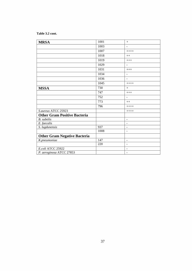
Of the tested isolates, the phage MikSA913 was lytic against 35 out of 50 strains
(70%) of MRSA and 4 MSSA strains tested. The phage also lysed the standard strain
S. aureus ATCC 25923. However, the phage could not lyse the other species of Gram-
negative or Gram-positive bacteria (Table 3.2).
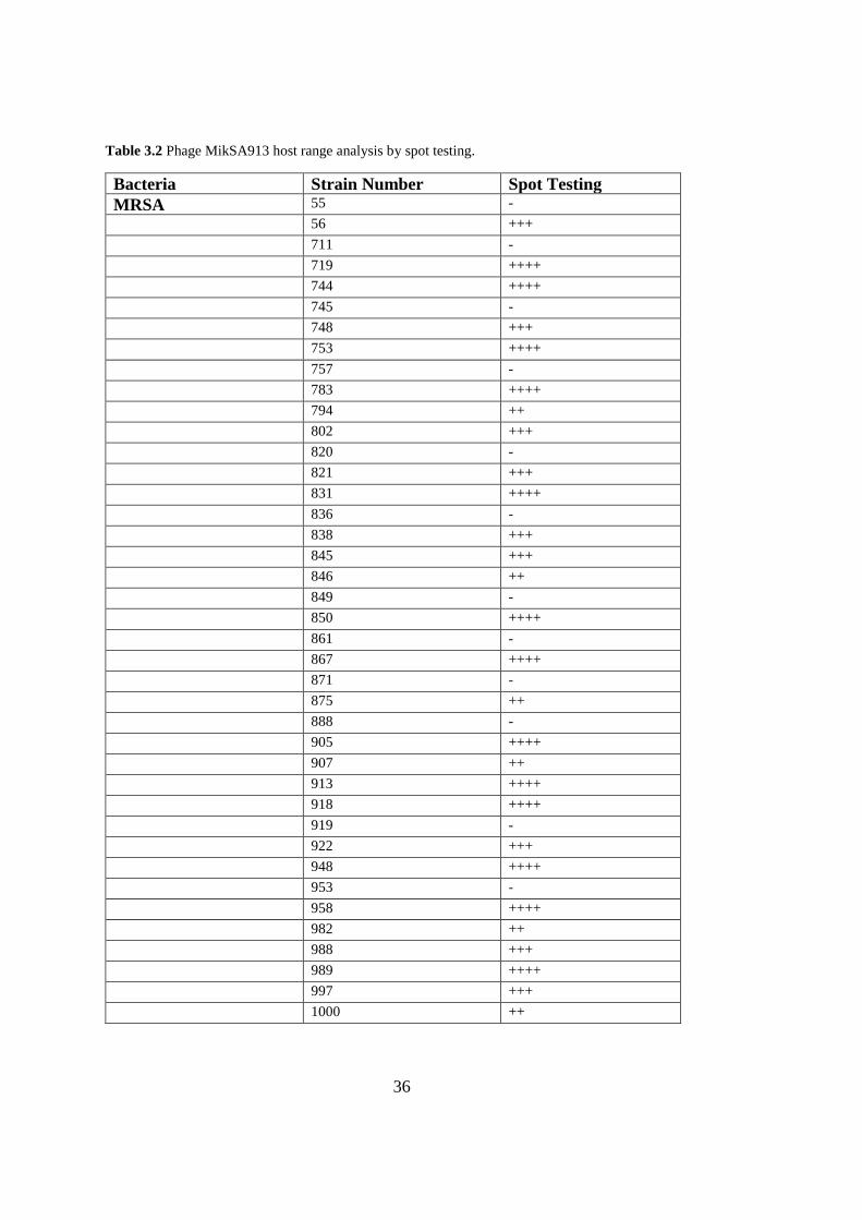
36
Table 3.2 Phage MikSA913 host range analysis by spot testing.
Bacteria Strain Number Spot Testing
MRSA 55 -
56 +++
711 -
719 ++++
744 ++++
745 -
748 +++
753 ++++
757 -
783 ++++
794 ++
802 +++
820 -
821 +++
831 ++++
836 -
838 +++
845 +++
846 ++
849 -
850 ++++
861 -
867 ++++
871 -
875 ++
888 -
905 ++++
907 ++
913 ++++
918 ++++
919 -
922 +++
948 ++++
953 -
958 ++++
982 ++
988 +++
989 ++++
997 +++
1000 ++
37
Table 3.2 cont.
MRSA 1001 +
1003 -
1007 ++++
1018 ++
1019 +++
1029 -
1031 +++
1034 -
1036 -
1045 ++++
MSSA 730 +
747 +++
752 -
773 ++
796 ++++
S.aureus ATCC 25923 ++++
Other Gram Positive Bacteria
B. subtilis -
E. faecalis -
S. lugdunensis 937 -
1008 -
Other Gram Negative Bacteria
K.pneumoniae 147 -
220 -
E.coli ATCC 25922 -
P. aeroginosa ATCC 27853 -
38
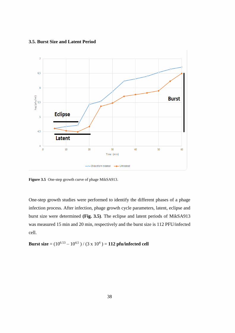
3.5. Burst Size and Latent Period
Figure 3.5 One-step growth curve of phage MikSA913.
One-step growth studies were performed to identify the different phases of a phage
infection process. After infection, phage growth cycle parameters, latent, eclipse and
burst size were determined (Fig. 3.5). The eclipse and latent periods of MikSA913
was measured 15 min and 20 min, respectively and the burst size is 112 PFU/infected
cell.
Burst size = (106.53 – 104,5 ) / (3 x 10⁴ ) = 112 pfu/infected cell
39
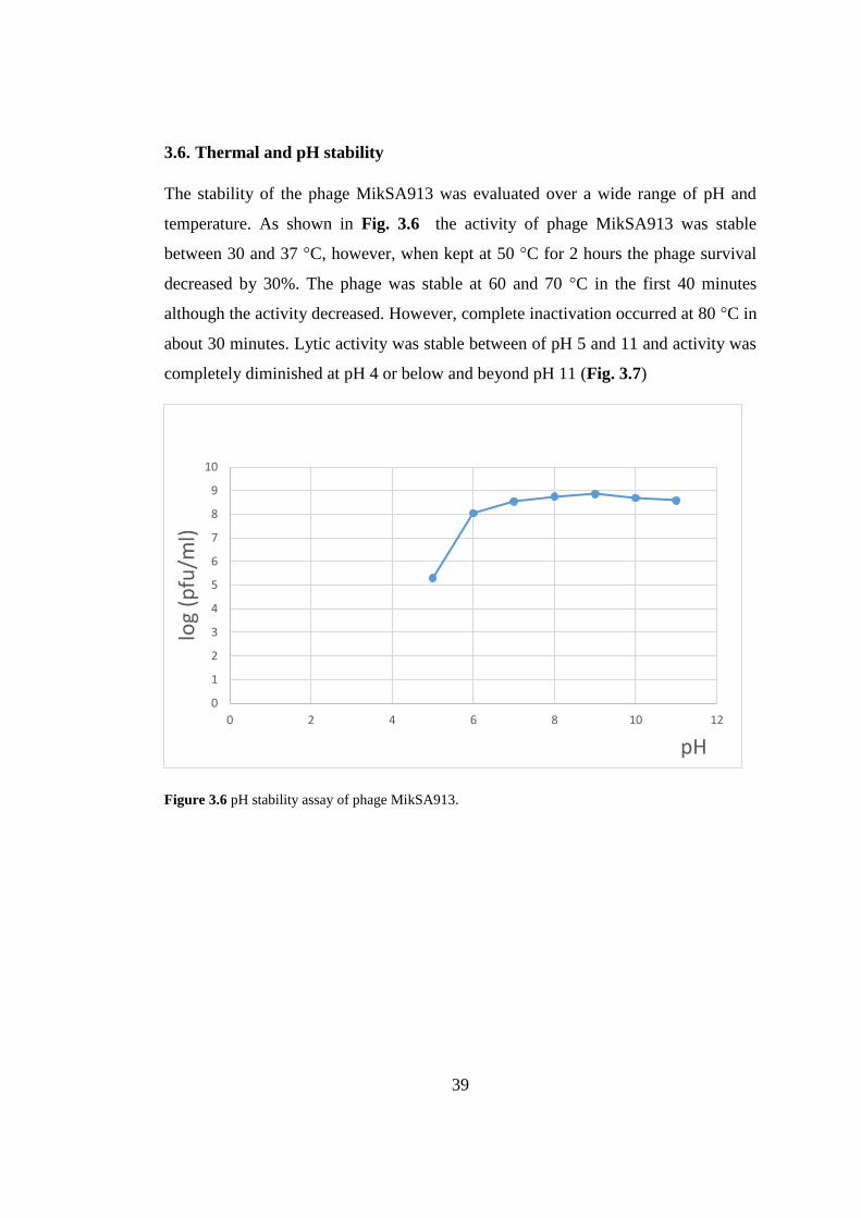
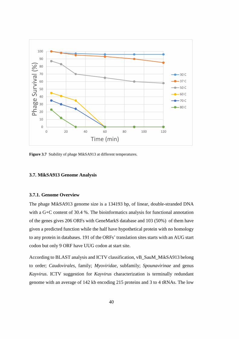
3.6. Thermal and pH stability
The stability of the phage MikSA913 was evaluated over a wide range of pH and
temperature. As shown in Fig. 3.6 the activity of phage MikSA913 was stable
between 30 and 37 °C, however, when kept at 50 °C for 2 hours the phage survival
decreased by 30%. The phage was stable at 60 and 70 °C in the first 40 minutes
although the activity decreased. However, complete inactivation occurred at 80 °C in
about 30 minutes. Lytic activity was stable between of pH 5 and 11 and activity was
completely diminished at pH 4 or below and beyond pH 11 (Fig. 3.7)
Figure 3.6 pH stability assay of phage MikSA913.
0
1
2
3
4
5
6
7
8
9
10
0 2 4 6 8 10 12
log
(pfu
/ml)
pH
40
Figure 3.7 Stability of phage MikSA913 at different temperatures.
3.7. MikSA913 Genome Analysis
3.7.1. Genome Overview
The phage MikSA913 genome size is a 134193 bp, of linear, double-stranded DNA
with a G+C content of 30.4 %. The bioinformatics analysis for functional annotation
of the genes gives 206 ORFs with GeneMarkS database and 103 (50%) of them have
given a predicted function while the half have hypothetical protein with no homology
to any protein in databases. 191 of the ORFs’ translation sites starts with an AUG start
codon but only 9 ORF have UUG codon at start site.
According to BLAST analysis and ICTV classification, vB_SauM_MikSA913 belong
to order; Caudovirales, family; Myoviridae, subfamily; Spounavirinae and genus
Kayvirus. ICTV suggestion for Kayvirus characterization is terminally redundant
genome with an average of 142 kb encoding 215 proteins and 3 to 4 tRNAs. The low
0
10
20
30
40
50
60
70
80
90
100
0 20 40 60 80 100 120
Ph
age
Surv
ival
(%
)
Time (min)
30 C
37 C
50 C
60 C
70 C
80 C
41
percentage of G+C (30.3%) is also a characteristic of Kayvirus genus. No GATC site
was found in this genus so far (Adriaenssens et al., 2018). All of these characteristics
are in coherent with the phage MikSA913 genome confirming that it belongs to the
Kayvirus genus.
Overall, genes of the MikSA913 organized into functional modules of structural,
DNA/RNA manipulation, lysis and some other additional functions (Table 3.3). The
large terminase subunit (orf81 and orf83) of MikSA913 contains a group I intron
protein called a VRS endonuclease (orf82).
The ends of Spounavirinae phages are long terminal repeat genes, which are encoding
small proteins functioning in takeover of the host cell metabolism and redirect to
phage propagation (Stewart, Yip, Myles, & Laughlin, 2009). Long terminal repeats
are first parts of the injected genome of the Twort-like phages. This region size is
different in each Staphylococcal Spounavirinae family. Long terminal repeat encoded
proteins are written as tre and in MikSA913 genome 15 tre genes were observed (tre,
treH ,tre,treJ, treK, treN, treP, tre, treS, treT, treA, treC, treD, treE and treF) in a
region of 9930 bp. The core genome region is suggested to be between the boundary
between TreA (orf202) and BofL (orf22).
Using the ARNOLD web server, the existence and location of the rho-independent
transcription terminators were predicted and the total number of predicted
transcription terminators was found to be 70 for phage MikSA913 genome.
Phage MikSA913 genome was searched with the virulence factor database (VFDB)
and antibiotic resistance genes database (ARDB) and there was no hit to any known
virulence or resistance gene.
42
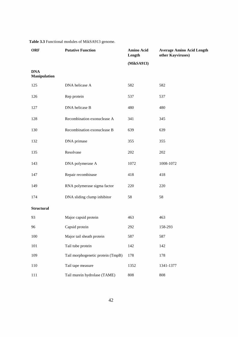
Table 3.3 Functional modules of MikSA913 genome.
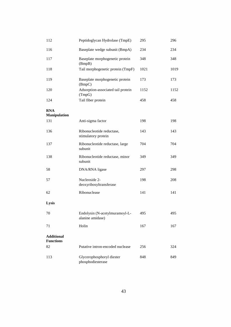
ORF Putative Function Amino Acid
Length
(MikSA913)
Average Amino Acid Length
other Kayviruses)
DNA
Manipulation
125 DNA helicase A 582 582
126 Rep protein 537 537
127 DNA helicase B 480 480
128 Recombination exonuclease A 341 345
130 Recombination exonuclease B 639 639
132 DNA primase 355 355
135 Resolvase 202 202
143 DNA polymerase A 1072 1008-1072
147 Repair recombinase 418 418
149 RNA polymerase sigma factor 220 220
174 DNA sliding clump inhibitor 58 58
Structural
93 Major capsid protein 463 463
96 Capsid protein 292 158-293
100 Major tail sheath protein 587 587
101 Tail tube protein 142 142
109 Tail morphogenetic protein (TmpB) 178 178
110 Tail tape measure 1352 1341-1377
111 Tail murein hydrolase (TAME) 808 808
43
112 Peptidoglycan Hydrolase (TmpE) 295 296
116 Baseplate wedge subunit (BmpA) 234 234
117 Baseplate morphogenetic protein
(BmpB)
348 348
118 Tail morphogenetic protein (TmpF) 1021 1019
119 Baseplate morphogenetic protein
(BmpC)
173 173
120 Adsorption-associated tail protein
(TmpG)
1152 1152
124 Tail fiber protein 458 458
RNA
Manipulation
131 Anti-sigma factor 198 198
136 Ribonucleotide reductase,
stimulatory protein
143 143
137 Ribonucleotide reductase, large
subunit
704 704
138 Ribonucleotide reductase, minor
subunit
349 349
58 DNA/RNA ligase 297 298
57 Nucleoside 2-
deoxyribosyltransferase
198 208
62 Ribonuclease 141 141
Lysis
70 Endolysin (N-acetylmuramoyl-L-
alanine amidase)
495 495
71 Holin 167 167
Additional
Functions
82 Putative intron-encoded nuclease 256 324
113 Glycerophosphoryl diester
phosphodiesterase
848 849
44
140 Thioredoxin 106 106
142 DNA binding protein 101 101
150 Ig like protein 210 210
82 Group I intron endonuclease 256 245
56 HNH homing endonuclease 261 162-194
22 BofL 82 78-82
31 Serine/Threonine protein
phosphatase
233 235
46 AAA family ATPase 372 372
60 PhoH related protein 246 246
65 Trancriptional regulator 76 70-76
67 Transglycosylase 230 210-232
Four tRNA genes were found in the phage MikSA913 genome coding for tRNA-Met,
tRNA-Trp, tRNA-Phe and tRNA-Asp. The G+C content of the tRNA genes ranges
from 38.9% to 51.3% (Table 3.4). The three of these tRNAs are located next to each
other between orf72 and orf73 region.
Table 3.4 tRNAs encoded by phage MikSA913 genome.
tRNA type Number of Bases Genome region G+C %
tRNA-Met (cat) 72 [12982,13053] 45.8
tRNA-Trp (cca) 72 [35358,35429] 38.9
tRNA-Phe (gaa) 73 [35436,35508] 41.1
tRNA-Asp (gtc) 76 [35514,35589] 51.3
45
3.7.2. Lytic Proteins
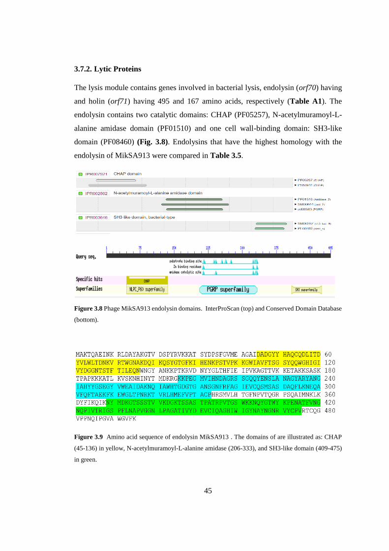
The lysis module contains genes involved in bacterial lysis, endolysin (orf70) having
and holin (orf71) having 495 and 167 amino acids, respectively (Table A1). The
endolysin contains two catalytic domains: CHAP (PF05257), N-acetylmuramoyl-L-
alanine amidase domain (PF01510) and one cell wall-binding domain: SH3-like
domain (PF08460) (Fig. 3.8). Endolysins that have the highest homology with the
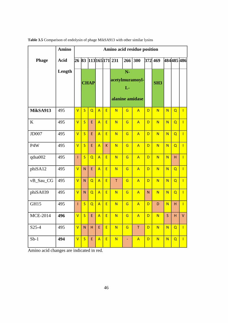
endolysin of MikSA913 were compared in Table 3.5.
Figure 3.8 Phage MikSA913 endolysin domains. InterProScan (top) and Conserved Domain Database
(bottom).
Figure 3.9 Amino acid sequence of endolysin MikSA913 . The domains of are illustrated as: CHAP
(45-136) in yellow, N-acetylmuramoyl-L-alanine amidase (206-333), and SH3-like domain (409-475)
in green.
46
Table 3.5 Comparison of endolysin of phage MikSA913 with other similar lysins
Phage
Amino
Acid
Length
Amino acid residue position
26 83 113 165 171 231 266 300 372 469 484 485 486
CHAP
N-
acetylmuramoyl-
L-
alanine amidase
SH3
MikSA913 495 V S Q A E N G A D N N Q I
K 495 V S E A E N G A D N N Q I
JD007 495 V S E A E N G A D N N Q I
P4W 495 V S E A K N G A D N N Q I
qdsa002 495 I S Q A E N G A D N N H I
phiSA12 495 V N E A E N G A D N N Q I
vB_Sau_CG 495 V N Q A E T G A D N N Q I
phiSA039 495 V N Q A E N G A N N N Q I
GH15 495 I S Q A E N G A D D N H I
MCE-2014 496 V S E A E N G A D N S H V
S25-4 495 V N H E E N G T D N N Q I
Sb-1 494 V S E A E N - A D N N Q I
Amino acid changes are indicated in red.
47
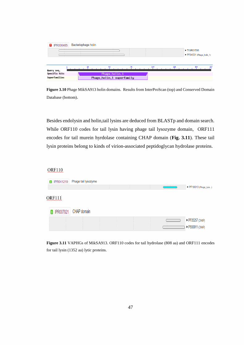
Figure 3.10 Phage MikSA913 holin domains. Results from InterProScan (top) and Conserved Domain
Database (bottom).
Besides endolysin and holin,tail lysins are deduced from BLASTp and domain search.
While ORF110 codes for tail lysin having phage tail lysozyme domain, ORF111
encodes for tail murein hyrdolase containing CHAP domain (Fig. 3.11). These tail
lysin proteins belong to kinds of virion-associated peptidoglycan hydrolase proteins.
Figure 3.11 VAPHGs of MikSA913. ORF110 codes for tail hydrolase (808 aa) and ORF111 encodes
for tail lysin (1352 aa) lytic proteins.
48
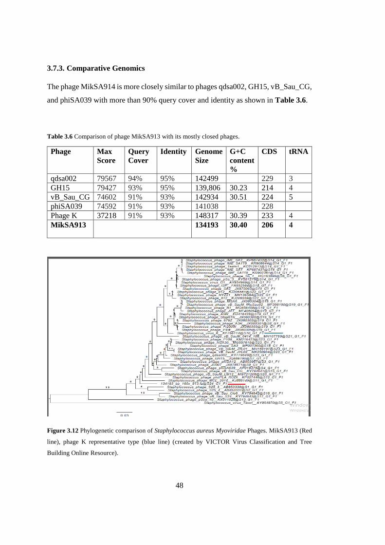
3.7.3. Comparative Genomics
The phage MikSA914 is more closely similar to phages qdsa002, GH15, vB_Sau_CG,
and phiSA039 with more than 90% query cover and identity as shown in Table 3.6.
Table 3.6 Comparison of phage MikSA913 with its mostly closed phages.
Phage Max
Score
Query
Cover
Identity Genome
Size
G+C
content
%
CDS tRNA
qdsa002 79567 94% 95% 142499 229 3
GH15 79427 93% 95% 139,806 30.23 214 4
vB_Sau_CG 74602 91% 93% 142934 30.51 224 5
phiSA039 74592 91% 93% 141038 228
Phage K 37218 91% 93% 148317 30.39 233 4
MikSA913 134193 30.40 206 4
Figure 3.12 Phylogenetic comparison of Staphylococcus aureus Myoviridae Phages. MikSA913 (Red
line), phage K representative type (blue line) (created by VICTOR Virus Classification and Tree
Building Online Resource).
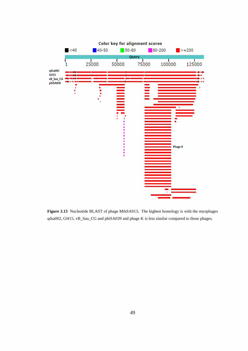
49
Figure 3.13 Nucleotide BLAST of phage MikSA913. The highest homology is with the myophages
qdsa002, GH15, vB_Sau_CG and phiSA039 and phage K is less similar compared to those phages.


51
CHAPTER 4
4. DISCUSSION
S. aureus is one of the most common pathogen causing serious infections and lytic
bacteriophages can provide a solution to this problem. There is a lack of information
about genomes of bacteriophages in our region. Thus, in this study, we aimed to find
phages active against MRSA strains circulating in Turkey. We searched sewage
waters, isolated a few bacteriophages, and chose only one to characterize with high
lytic efficacy and broad host range. The isolated phage named as vB_SauM-
MikSA913 was classified into Myoviridae family, and Kayvirus genus. Based on
genome size and organization vB_SauM-MikSA913 belongs to the class III
staphylococcal phages classified by Kwan (Kwan, Liu, DuBow, Gros, & Pelletier,
2005). Myoviridae class III phages generally contain introns in the genes of lysin,
DNA polymerase, ribonucleotide reductase, large terminase and DNA helicase (Kwan
et al., 2005). MikSA913 large terminase subunit has a group I intron protein coding
for VRS endonuclease and there seems no intron in other genes.
The eclipse and latent periods of MikSA913 was measured 15 min and 20 min,
respectively and the burst size is 112 pfu/infected cell in one-step growth experiments.
Calcium ion was reported to increase adsorption rate constant of the phage. MikSA913
is stable at a wide range of pH and temperature.
Phage MikSA913 has a broad host range (lysing 75% of MRSA strains) like other
Kayviruses known for their broad host range. This phenomenon is mainly accounted
to the presence of multiple receptor binding proteins in the viral capsid that allow them
to utilize at least two adsorption apparatuses and recognize different structures
(Takeuchi et al., 2016).
52
Currently, Kayvirus genus contains 13 members, including phage K, GH15, JD007,
MCE-2014, P108, S25-3, S25-4, G1, Team1, philPLA-RODI, phiSA12, 812 and Sb-
1 on NCBI. A typical feature for Kayvirus genus is the presence of long terminal
repeats, several thousand base pair-long direct repeats at the ends of the genome (King
et al. 2011). The ends of the phage genome are flanked by LTRs. The region between
TreA and Bofl encoding genes is considered LTR and this is 9930 bp in phage
MikSA913. The nucleotide sequence and length of LTR regions differ among the
representatives of the genus and may influence the host range. (Łobocka et al., 2012).
According to nucleotide BLAST analysis, the most variable regions are the LTRs
between MikSA913 and its closed phages.
S. aureus Myoviridae phages contain a large deal of promoters recognized by S. aureus
σ70. In addition, anti-σ factors and an alternative σ factors are located in this phage
class. MikSA913 ORF131 codes for the anti-sigma factor while ORF 149 encodes
sigma factor.
MikSA913 genome has no ‘GATC’ sites, which are the targets of S. aureus restriction
enzymes thus allowing the phage to avoid bacterial defensive immune system. The
safety assessment based on genome sequence showed that phage MikSA913 is strictly
lytic with no integrase that is responsible for recombination event, which is
characteristics of temperate phages. In addition, there is no virulence factor, toxin or
antibiotic resistance coding genes.
The in silico analysis of the phage derived lytic enzymes was also performed with
BLAST analysis and domain search. As seen in most Myoviridae staphylococcal
phages, MikSA913 contain a lysis cassette containing two proteins: endolysin and
holin. The endolysin modular structure have three domains: N-terminal CHAP
domain, and internal N-acetylmuramoyl-L-alanine amidase domain and a C-terminal
SH3-like domain. In addition, the other lytic proteins, virion associated peptidoglycan
hyrdolases are shown in the MikSA913 genome. ORF110 and ORF111 have lytic
domains of lysozyme and CHAP domain, respectively.
53
MikSA913 endolysin BLAST analysis results showed that it is highly homologous to
other isolated phage endolysins with only one or a few amino acid changes. Jun et al.
showed that despite the high degree of similarity SAL-1 and the phage endolysin
LysK, SAL-1 has higher cell wall hydrolyzing activity than LysK. The authors
suggested that this enhanced enzymatic change is due to the glutamic acid to glutamine
at the residue at 114. However, I believe that this reported residue is 113 and
erroneously reported as 114 since LysK has glutamic acid at residue 113 and
glutamine at residue 114. Based on BLASTp analysis, the MikSA913 endolysin
differs from LysK and endolysin derived from JD007 only at one residue: glutamine
instead of glutamic acid at the 113th residue. According to the this previous study, we
can claim that endolysin of MikSA913 have higher staphylolytic activity than LysK
and endolysin of JD007 phage (Jun et al., 2011). In structural studies of the LysK,
Cys54-His117-Glu134 residues are shown to be proteolytic site of the CHAP domain
(Keary et al., 2016). These residues are also conserved in CHAP domain of MikSA913
endolysin spanning the residues 45-136.
The gene sequences and organization of Myoviridae phages isolated in different
geographical regions show high similarity and this suggests that the evolutionary
forces lead to maintain core functional activities on S. aureus isolates.

55
CHAPTER 5
5. CONCLUSION
In this thesis, we isolated a new bacteriophage active against local MRSA strains and
carried out biophysical and genomic characterization. To sum up all of the
characteristics: strictly lytic cycle, and lack of virulence or resistance genes, highly
lytic activity, wide host range on MRSA strains and specificity to S. aureus strains
make the phage MikSA913 suitable for clinical applications to eradicate MRSA.
Further work on the activities of the phage and its purified recombinant endolysin
should be carried out to validate their efficacy in vivo.

57
REFERENCES
Abedon, S. T. (2012). Bacteriophages in health and disease. Bacteriophages in health
and disease. https://doi.org/10.1079/9781845939847.0000
Adriaenssens, E. M., Wittmann, J., Kuhn, J. H., Turner, D., Sullivan, M. B., Dutilh,
B. E., … Rodney Brister, J. (2018). Taxonomy of prokaryotic viruses: 2017
update from the ICTV Bacterial and Archaeal Viruses Subcommittee. Archives
of Virology. https://doi.org/10.1007/s00705-018-3723-z
Aminov, R. I. (2010). A brief history of the antibiotic era: Lessons learned and
challenges for the future. Frontiers in Microbiology.
https://doi.org/10.3389/fmicb.2010.00134
Arthur, M., Molinas, C., Depardieu, F., & Courvalin, P. (1993). Characterization of
Tn1546, a Tn3-related transposon conferring glycopeptide resistance by
synthesis of depsipeptide peptidoglycan precursors in Enterococcus faecium
BM4147. Journal of Bacteriology. https://doi.org/10.1128/jb.175.1.117-
127.1993
Barna, J. C. J., & Williams, D. H. (1984). The Structure and Mode of Action of
Glycopeptide Antibiotics of the Vancomycin Group. Annual Review of
Microbiology. https://doi.org/10.1146/annurev.mi.38.100184.002011
Bassetti, M., Baguneid, M., Bouza, E., Dryden, M., Nathwani, D., & Wilcox, M.
(2014). European perspective and update on the management of complicated skin
and soft tissue infections due to methicillin-resistant Staphylococcus aureus after
more than 10 years of experience with linezolid. Clinical Microbiology and
Infection. https://doi.org/10.1111/1469-0691.12463
Becker, S. C., Foster-Frey, J., Stodola, A. J., Anacker, D., & Donovan, D. M. (2009).
Differentially conserved staphylococcal SH3b_5 cell wall binding domains
confer increased staphylolytic and streptolytic activity to a streptococcal
prophage endolysin domain. Gene. https://doi.org/10.1016/j.gene.2009.04.023
Besemer, J., & Borodovsky, M. (2005). GeneMark: Web software for gene finding in
prokaryotes, eukaryotes and viruses. Nucleic Acids Research.
https://doi.org/10.1093/nar/gki487
Borysowski, J., Weber-Da̧browska, B., & Górski, A. (2006). Bacteriophage
endolysins as a novel class of antibacterial agents. Experimental Biology and
Medicine. https://doi.org/10.1177/153537020623100402
Breitbart, M., Bonnain, C., Malki, K., & Sawaya, N. A. (2018). Phage puppet masters
of the marine microbial realm. Nature Microbiology.
https://doi.org/10.1038/s41564-018-0166-y
58
Briers, Y., Volckaert, G., Cornelissen, A., Lagaert, S., Michiels, C. W., Hertveldt, K.,
& Lavigne, R. (2007). Muralytic activity and modular structure of the endolysins
of Pseudomonas aeruginosa bacteriophages φKZ and EL. Molecular
Microbiology. https://doi.org/10.1111/j.1365-2958.2007.05870.x
Brum, J. R., Cesar Ignacio-Espinoza, J., Roux, S., Doulcier, G., Acinas, S. G., Alberti,
A., … Weissenbach, J. (2015). Patterns and ecological drivers of ocean viral
communities. Science. https://doi.org/10.1126/science.1261498
Bugg, T. D. H., Wright, G. D., Walsh, C. T., Dutka-Malen, S., Arthur, M., &
Courvalin, P. (1991). Molecular Basis for Vancomycin Resistance in
Enterococcus faecium BM4147: Biosynthesis of a Depsipeptide Peptidoglycan
Precursor by Vancomycin Resistance Proteins VanH and VanA. Biochemistry.
https://doi.org/10.1021/bi00107a007
Casey, E., Mahony, J., Neve, H., Noben, J. P., Bello, F. D., & van Sinderen, D. (2015).
Novel phage group infecting Lactobacillus delbrueckii subsp. lactis, as revealed
by genomic and proteomic analysis of bacteriophage Ldl1. Applied and
Environmental Microbiology. https://doi.org/10.1128/AEM.03413-14
Catalão, M. J., Gil, F., Moniz-Pereira, J., São-José, C., & Pimentel, M. (2013).
Diversity in bacterial lysis systems: Bacteriophages Show the way. FEMS
Microbiology Reviews. https://doi.org/10.1111/1574-6976.12006
Chambers, H. F. (1997). Methicillin resistance in staphylococci: Molecular and
biochemical basis and clinical implications. Clinical Microbiology Reviews.
https://doi.org/10.1128/CMR.10.4.781
Chambers, H. F., & DeLeo, F. R. (2009). Waves of resistance: Staphylococcus aureus
in the antibiotic era. Nature Reviews Microbiology.
https://doi.org/10.1038/nrmicro2200
Chen, C. J., Huang, Y. C., & Chiu, C. H. (2015). Multiple pathways of cross-resistance
to glycopeptides and daptomycin in persistent MRSA bacteraemia. Journal of
Antimicrobial Chemotherapy. https://doi.org/10.1093/jac/dkv225
Cheng, X., Zhang, X., Pflugrath, J. W., & Studier, F. W. (1994). The structure of
bacteriophage T7 lysozyme, a zinc amidase and an inhibitor of T7 RNA
polymerase. Proceedings of the National Academy of Sciences.
https://doi.org/10.1073/pnas.91.9.4034
Chhibber, S., Kaur, T., & Kaur, S. (2014). Essential role of calcium in the infection
process of broad-spectrum methicillin-resistant Staphylococcus aureus
bacteriophage. Journal of Basic Microbiology.
https://doi.org/10.1002/jobm.201300051
Chopra, S., Harjai, K., & Chhibber, S. (2016). Potential of combination therapy of
endolysin MR-10 and minocycline in treating MRSA induced systemic and
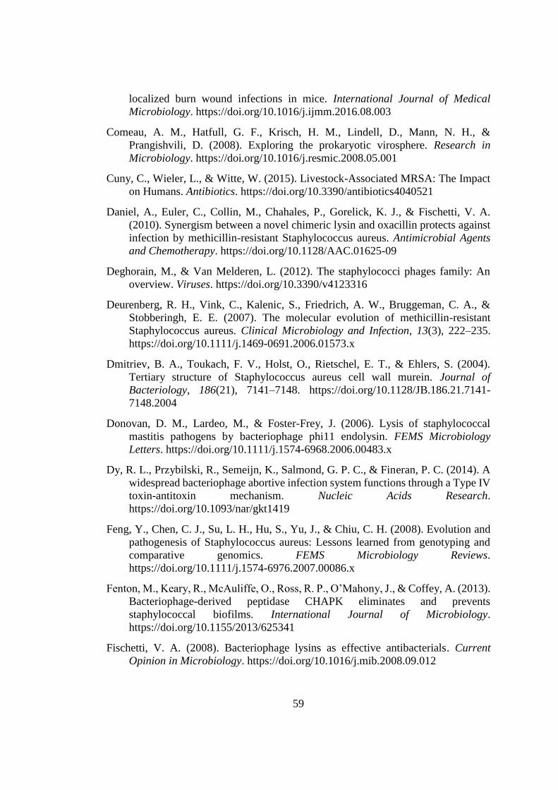
59
localized burn wound infections in mice. International Journal of Medical
Microbiology. https://doi.org/10.1016/j.ijmm.2016.08.003
Comeau, A. M., Hatfull, G. F., Krisch, H. M., Lindell, D., Mann, N. H., &
Prangishvili, D. (2008). Exploring the prokaryotic virosphere. Research in
Microbiology. https://doi.org/10.1016/j.resmic.2008.05.001
Cuny, C., Wieler, L., & Witte, W. (2015). Livestock-Associated MRSA: The Impact
on Humans. Antibiotics. https://doi.org/10.3390/antibiotics4040521
Daniel, A., Euler, C., Collin, M., Chahales, P., Gorelick, K. J., & Fischetti, V. A.
(2010). Synergism between a novel chimeric lysin and oxacillin protects against
infection by methicillin-resistant Staphylococcus aureus. Antimicrobial Agents
and Chemotherapy. https://doi.org/10.1128/AAC.01625-09
Deghorain, M., & Van Melderen, L. (2012). The staphylococci phages family: An
overview. Viruses. https://doi.org/10.3390/v4123316
Deurenberg, R. H., Vink, C., Kalenic, S., Friedrich, A. W., Bruggeman, C. A., &
Stobberingh, E. E. (2007). The molecular evolution of methicillin-resistant
Staphylococcus aureus. Clinical Microbiology and Infection, 13(3), 222–235.
https://doi.org/10.1111/j.1469-0691.2006.01573.x
Dmitriev, B. A., Toukach, F. V., Holst, O., Rietschel, E. T., & Ehlers, S. (2004).
Tertiary structure of Staphylococcus aureus cell wall murein. Journal of
Bacteriology, 186(21), 7141–7148. https://doi.org/10.1128/JB.186.21.7141-
7148.2004
Donovan, D. M., Lardeo, M., & Foster-Frey, J. (2006). Lysis of staphylococcal
mastitis pathogens by bacteriophage phi11 endolysin. FEMS Microbiology
Letters. https://doi.org/10.1111/j.1574-6968.2006.00483.x
Dy, R. L., Przybilski, R., Semeijn, K., Salmond, G. P. C., & Fineran, P. C. (2014). A
widespread bacteriophage abortive infection system functions through a Type IV
toxin-antitoxin mechanism. Nucleic Acids Research.
https://doi.org/10.1093/nar/gkt1419
Feng, Y., Chen, C. J., Su, L. H., Hu, S., Yu, J., & Chiu, C. H. (2008). Evolution and
pathogenesis of Staphylococcus aureus: Lessons learned from genotyping and
comparative genomics. FEMS Microbiology Reviews.
https://doi.org/10.1111/j.1574-6976.2007.00086.x
Fenton, M., Keary, R., McAuliffe, O., Ross, R. P., O’Mahony, J., & Coffey, A. (2013).
Bacteriophage-derived peptidase CHAPK eliminates and prevents
staphylococcal biofilms. International Journal of Microbiology.
https://doi.org/10.1155/2013/625341
Fischetti, V. A. (2008). Bacteriophage lysins as effective antibacterials. Current
Opinion in Microbiology. https://doi.org/10.1016/j.mib.2008.09.012
60
Fischetti, V. A. (2010). Bacteriophage endolysins: A novel anti-infective to control
Gram-positive pathogens. International Journal of Medical Microbiology.
https://doi.org/10.1016/j.ijmm.2010.04.002
Foster, T. J., & Höök, M. (1998). Surface protein adhesins of Staphylococcus aureus.
Trends in Microbiology. https://doi.org/10.1016/S0966-842X(98)01400-0
Giancarlo Licitra. (2013). Etymologia: Staphylococcus. Emerging Infectious
Diseases. https://doi.org/http://doi.org/10.3201/eid1909.ET1909
Giesbrecht, P., Kersten, T., Maidhof, H., & Wecke, J. (1998). Staphylococcal cell
wall: morphogenesis and fatal variations in the presence of penicillin.
Microbiology and Molecular Biology Reviews : MMBR.
https://doi.org/10.1080/00016480601075357
Gill, S. R., Fouts, D. E., Archer, G. L., Mongodin, E. F., DeBoy, R. T., Ravel, J., …
Fraser, C. M. (2005). Insights on evolution of virulence and resistance from the
complete genome analysis of an early methicillin-resistant Staphylococcus
aureus strain and a biofilm-producing methicillin-resistant Staphylococcus
epidermidis strain. Journal of Bacteriology.
https://doi.org/10.1128/JB.187.7.2426-2438.2005
Gründling, A., & Schneewind, O. (2006). Cross-linked peptidoglycan mediates
lysostaphin binding to the cell wall envelope of Staphylococcus aureus. Journal
of Bacteriology. https://doi.org/10.1128/JB.188.7.2463-2472.2006
Gutiérrez, D., & Fernández, L. (2018). crossm Are Phage Lytic Proteins the Secret
Weapon To Kill, 1–17. https://doi.org/10.1128/mBio.01923-17
Harper, D. R., Anderson, J., & Enright, M. (2011). Phage therapy: Delivering on the
promise. Therapeutic Delivery. https://doi.org/10.4155/tde.11.64
Herold, B. C., Immergluck, L. C., Maranan, M. C., Lauderdale, D. S., Gaskin, R. E.,
Boyle-Vavra, S., … Daum, R. S. (1998). Community-acquired methicillin-
resistant staphylococcus aureus in children with no identified predisposing risk.
Journal of the American Medical Association.
https://doi.org/10.1001/jama.279.8.593
Hiramatsu, K., Aritaka, N., Hanaki, H., Kawasaki, S., Hosoda, Y., Hori, S., …
Kobayashi, I. (1997). Dissemination in Japanese hospitals of strains of
Staphylococcus aureus heterogeneously resistant to vancomycin. Lancet.
https://doi.org/10.1016/S0140-6736(97)07324-8
Holden, M. T. G., Feil, E. J., Lindsay, J. A., Peacock, S. J., Day, N. P. J., Enright, M.
C., … Parkhill, J. (2004). Complete genomes of two clinical Staphylococcus
aureus strains: Evidence for the rapid evolution of virulence and drug resistance.
Proceedings of the National Academy of Sciences.
https://doi.org/10.1073/pnas.0402521101
61
Hosseini, E. S., Moniri, R., Goli, Y. D., & Kashani, H. H. (2016). Purification of
Antibacterial CHAPKProtein Using a Self-Cleaving Fusion Tag and Its Activity
Against Methicillin-Resistant Staphylococcus aureus. Probiotics and
Antimicrobial Proteins. https://doi.org/10.1007/s12602-016-9236-8
Ito, T., Hiramatsu, K., Oliveira, D. C., De Lencastre, H., Zhang, K., Westh, H., …
Soderquist, B. (2009). Classification of staphylococcal cassette chromosome mec
(SCCmec): Guidelines for reporting novel SCCmec elements. Antimicrobial
Agents and Chemotherapy. https://doi.org/10.1128/AAC.00579-09
Ito, T., Okuma, K., Ma, X. X., Yuzawa, H., & Hiramatsu, K. (2003). Insights on
antibiotic resistance of Staphylococcus aureus from its whole genome: Genomic
island SCC. Drug Resistance Updates. https://doi.org/10.1016/S1368-
7646(03)00003-7
Jarraud, S., Narita, S., Kaneko, J., Chiba, J., Etienne, Y. P. J., & Kamio, Y. (2001).
Phage conversion of Panton-Valentine leukocidin in Staphylococcus aureus:
molecular analysis of a PVL-converting phage, fSLT. Gene, 268.
https://doi.org/10.1016/S0070-4571(08)70081-5
Jiang, F., & Doudna, J. A. (2015). The structural biology of CRISPR-Cas systems.
Current Opinion in Structural Biology. https://doi.org/10.1016/j.sbi.2015.02.002
Jun, S. Y., Jang, I. J., Yoon, S., Jang, K., Yu, K. S., Cho, J. Y., … Kang, S. H. (2017).
Pharmacokinetics and Tolerance of the Phage endolysin-based candidate drug
SAL200 after a single intravenous administration among healthy volunteers.
Antimicrobial Agents and Chemotherapy. https://doi.org/10.1128/AAC.02629-
16
Jun, S. Y., Jung, G. M., Son, J. S., Yoon, S. J., Choi, Y. J., & Kang, S. H. (2011).
Comparison of the antibacterial properties of phage endolysins SAL-1 and LysK.
Antimicrobial Agents and Chemotherapy. https://doi.org/10.1128/AAC.01097-
10
Jun, S. Y., Jung, G. M., Yoon, S. J., Oh, M. D., Choi, Y. J., Lee, W. J., … Kang, S. H.
(2013). Antibacterial properties of a pre-formulated recombinant phage
endolysin, SAL-1. International Journal of Antimicrobial Agents.
https://doi.org/10.1016/j.ijantimicag.2012.10.011
Kaźmierczak, Z., Górski, A., & Dabrowska, K. (2014). Facing antibiotic resistance:
Staphylococcus aureus phages as a medical tool. Viruses, 6(7), 2551–2570.
https://doi.org/10.3390/v6072551
Keary, R., Paul Ross, R., Hill, C., O’ Mahony, J., J. van Raaij, M., Fenton, M., …
McAuliffe, O. (2016). Characterization of a Bacteriophage-Derived Murein
Peptidase for Elimination of Antibiotic-Resistant Staphylococcus aureus.
Current Protein & Peptide Science.
https://doi.org/10.2174/1389203716666151102105515
62
King E.J, Andrew MQ, ADAMS, Michael J., et LEFKOWITZ, Elliot J. (2011). Ninth
Report of the International Committee on Taxonomy of Viruses. Virus
Taxonomy. https://doi.org/10.1007/s13398-014-0173-7.2
Kropinski, A. M., & Martha, C. R. J. (2009). Introduction. In Bacteriophages.
Methods and Protocols. Volume 1: Isolation, Characterization, and Interactions.
3rd ed. https://doi.org/10.1007/978-1-60327-164-6
Kutter, E. (2009). Phage host range and efficiency of plating. In Bacteriophages.
Methods and Protocols. Volume 1: Isolation, Characterization, and Interactions.
3rd ed. (pp. 141–149). https://doi.org/10.1007/978-1-60327-164-6_14
Kwan, T., Liu, J., DuBow, M., Gros, P., & Pelletier, J. (2005). The complete genomes
and proteomes of 27 Staphylococcus aureus bacteriophages. Proceedings of the
National Academy of Sciences, 102(14), 5174–5179.
https://doi.org/10.1073/pnas.0501140102
Kwiatek, M., Parasion, S., Mizak, L., Gryko, R., Bartoszcze, M., & Kocik, J. (2012).
Characterization of a bacteriophage, isolated from a cow with mastitis, that is
lytic against Staphylococcus aureus strains. Archives of Virology, 157(2), 225–
234. https://doi.org/10.1007/s00705-011-1160-3
Labrie, S. J., Samson, J. E., & Moineau, S. (2010). Bacteriophage resistance
mechanisms. Nature Reviews Microbiology.
https://doi.org/10.1038/nrmicro2315
Laslett, D., & Canback, B. (2004). ARAGORN, a program to detect tRNA genes and
tmRNA genes in nucleotide sequences. Nucleic Acids Research.
https://doi.org/10.1093/nar/gkh152
Le Loir, Y., Baron, F., & Gautier, M. (2003). Staphylococcus aureus and food
poisoning. In Genetics and Molecular Research. https://doi.org/S02 [pii]
Linden, S. B., Zhang, H., Heselpoth, R. D., Shen, Y., Schmelcher, M., Eichenseher,
F., & Nelson, D. C. (2014). Biochemical and biophysical characterization of
PlyGRCS, a bacteriophage endolysin active against methicillin-resistant
Staphylococcus aureus. Applied Microbiology and Biotechnology.
https://doi.org/10.1007/s00253-014-5930-1
Lindsay, J. A. (2010). Genomic variation and evolution of Staphylococcus aureus.
International Journal of Medical Microbiology.
https://doi.org/10.1016/j.ijmm.2009.08.013
Lindsay, J. A., & Holden, M. T. G. (2004). Staphylococcus aureus: Superbug, super
genome? Trends in Microbiology. https://doi.org/10.1016/j.tim.2004.06.004
Lobanovska, M., & Pilla, G. (2017). Penicillin’s discovery and antibiotic resistance:
Lessons for the future? Yale Journal of Biology and Medicine.
https://doi.org/10.1109/TEMC.2011.2143719
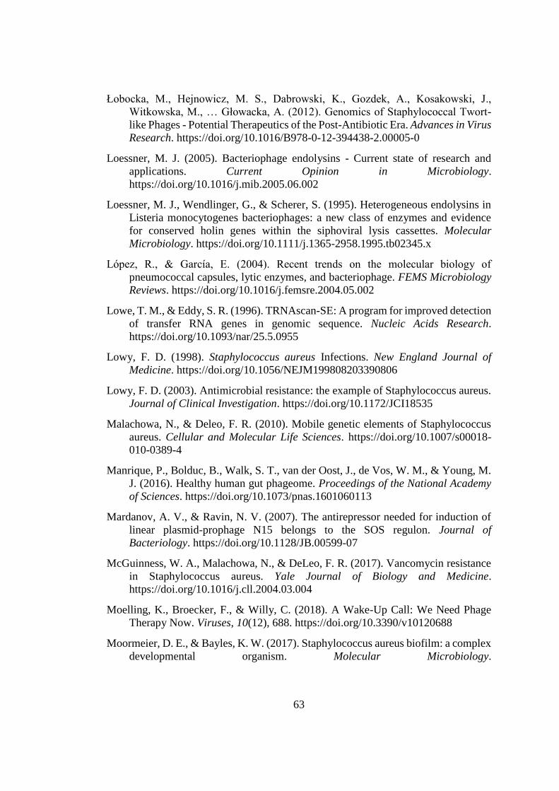
63
Łobocka, M., Hejnowicz, M. S., Dabrowski, K., Gozdek, A., Kosakowski, J.,
Witkowska, M., … Głowacka, A. (2012). Genomics of Staphylococcal Twort-
like Phages - Potential Therapeutics of the Post-Antibiotic Era. Advances in Virus
Research. https://doi.org/10.1016/B978-0-12-394438-2.00005-0
Loessner, M. J. (2005). Bacteriophage endolysins - Current state of research and
applications. Current Opinion in Microbiology.
https://doi.org/10.1016/j.mib.2005.06.002
Loessner, M. J., Wendlinger, G., & Scherer, S. (1995). Heterogeneous endolysins in
Listeria monocytogenes bacteriophages: a new class of enzymes and evidence
for conserved holin genes within the siphoviral lysis cassettes. Molecular
Microbiology. https://doi.org/10.1111/j.1365-2958.1995.tb02345.x
López, R., & García, E. (2004). Recent trends on the molecular biology of
pneumococcal capsules, lytic enzymes, and bacteriophage. FEMS Microbiology
Reviews. https://doi.org/10.1016/j.femsre.2004.05.002
Lowe, T. M., & Eddy, S. R. (1996). TRNAscan-SE: A program for improved detection
of transfer RNA genes in genomic sequence. Nucleic Acids Research.
https://doi.org/10.1093/nar/25.5.0955
Lowy, F. D. (1998). Staphylococcus aureus Infections. New England Journal of
Medicine. https://doi.org/10.1056/NEJM199808203390806
Lowy, F. D. (2003). Antimicrobial resistance: the example of Staphylococcus aureus.
Journal of Clinical Investigation. https://doi.org/10.1172/JCI18535
Malachowa, N., & Deleo, F. R. (2010). Mobile genetic elements of Staphylococcus
aureus. Cellular and Molecular Life Sciences. https://doi.org/10.1007/s00018-
010-0389-4
Manrique, P., Bolduc, B., Walk, S. T., van der Oost, J., de Vos, W. M., & Young, M.
J. (2016). Healthy human gut phageome. Proceedings of the National Academy
of Sciences. https://doi.org/10.1073/pnas.1601060113
Mardanov, A. V., & Ravin, N. V. (2007). The antirepressor needed for induction of
linear plasmid-prophage N15 belongs to the SOS regulon. Journal of
Bacteriology. https://doi.org/10.1128/JB.00599-07
McGuinness, W. A., Malachowa, N., & DeLeo, F. R. (2017). Vancomycin resistance
in Staphylococcus aureus. Yale Journal of Biology and Medicine.
https://doi.org/10.1016/j.cll.2004.03.004
Moelling, K., Broecker, F., & Willy, C. (2018). A Wake-Up Call: We Need Phage
Therapy Now. Viruses, 10(12), 688. https://doi.org/10.3390/v10120688
Moormeier, D. E., & Bayles, K. W. (2017). Staphylococcus aureus biofilm: a complex
developmental organism. Molecular Microbiology.
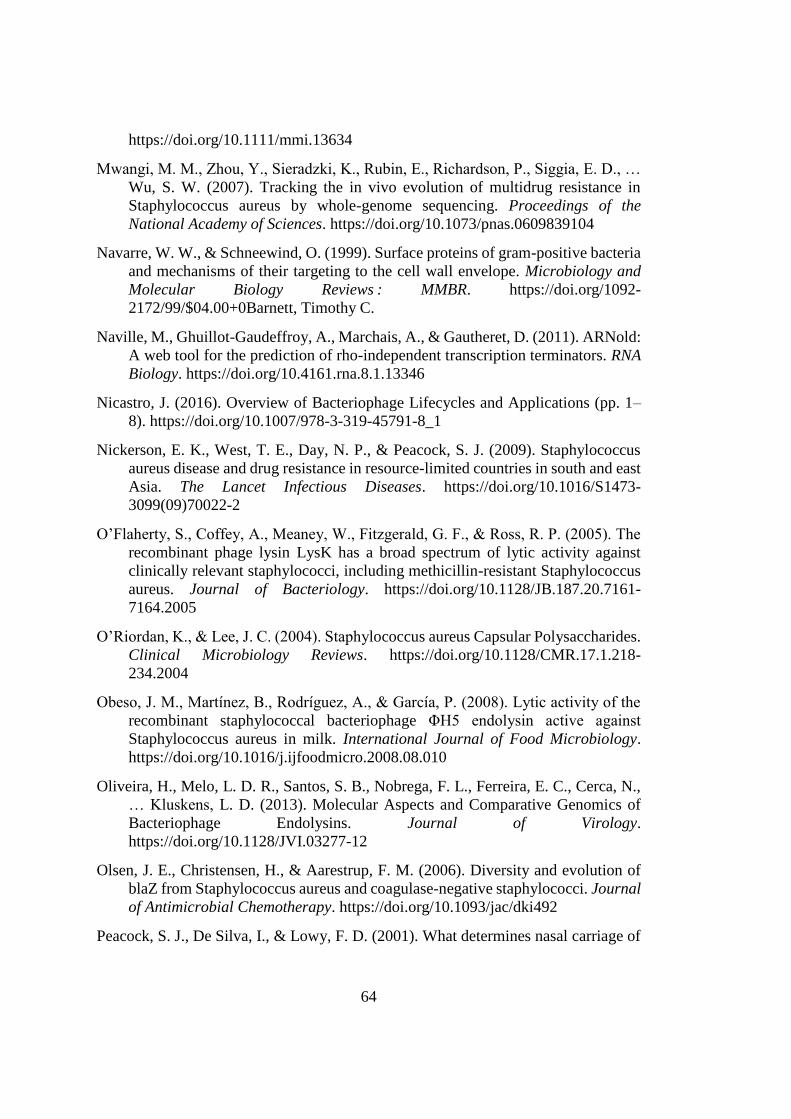
64
https://doi.org/10.1111/mmi.13634
Mwangi, M. M., Zhou, Y., Sieradzki, K., Rubin, E., Richardson, P., Siggia, E. D., …
Wu, S. W. (2007). Tracking the in vivo evolution of multidrug resistance in
Staphylococcus aureus by whole-genome sequencing. Proceedings of the
National Academy of Sciences. https://doi.org/10.1073/pnas.0609839104
Navarre, W. W., & Schneewind, O. (1999). Surface proteins of gram-positive bacteria
and mechanisms of their targeting to the cell wall envelope. Microbiology and
Molecular Biology Reviews : MMBR. https://doi.org/1092-
2172/99/$04.00+0Barnett, Timothy C.
Naville, M., Ghuillot-Gaudeffroy, A., Marchais, A., & Gautheret, D. (2011). ARNold:
A web tool for the prediction of rho-independent transcription terminators. RNA
Biology. https://doi.org/10.4161.rna.8.1.13346
Nicastro, J. (2016). Overview of Bacteriophage Lifecycles and Applications (pp. 1–
8). https://doi.org/10.1007/978-3-319-45791-8_1
Nickerson, E. K., West, T. E., Day, N. P., & Peacock, S. J. (2009). Staphylococcus
aureus disease and drug resistance in resource-limited countries in south and east
Asia. The Lancet Infectious Diseases. https://doi.org/10.1016/S1473-
3099(09)70022-2
O’Flaherty, S., Coffey, A., Meaney, W., Fitzgerald, G. F., & Ross, R. P. (2005). The
recombinant phage lysin LysK has a broad spectrum of lytic activity against
clinically relevant staphylococci, including methicillin-resistant Staphylococcus
aureus. Journal of Bacteriology. https://doi.org/10.1128/JB.187.20.7161-
7164.2005
O’Riordan, K., & Lee, J. C. (2004). Staphylococcus aureus Capsular Polysaccharides.
Clinical Microbiology Reviews. https://doi.org/10.1128/CMR.17.1.218-
234.2004
Obeso, J. M., Martínez, B., Rodríguez, A., & García, P. (2008). Lytic activity of the
recombinant staphylococcal bacteriophage ΦH5 endolysin active against
Staphylococcus aureus in milk. International Journal of Food Microbiology.
https://doi.org/10.1016/j.ijfoodmicro.2008.08.010
Oliveira, H., Melo, L. D. R., Santos, S. B., Nobrega, F. L., Ferreira, E. C., Cerca, N.,
… Kluskens, L. D. (2013). Molecular Aspects and Comparative Genomics of
Bacteriophage Endolysins. Journal of Virology.
https://doi.org/10.1128/JVI.03277-12
Olsen, J. E., Christensen, H., & Aarestrup, F. M. (2006). Diversity and evolution of
blaZ from Staphylococcus aureus and coagulase-negative staphylococci. Journal
of Antimicrobial Chemotherapy. https://doi.org/10.1093/jac/dki492
Peacock, S. J., De Silva, I., & Lowy, F. D. (2001). What determines nasal carriage of
65
Staphylococcus aureus? Trends in Microbiology. https://doi.org/10.1016/S0966-
842X(01)02254-5
Rodríguez-Rubio, L., Martínez, B., Donovan, D. M., Rodríguez, A., & García, P.
(2013). Bacteriophage virion-associated peptidoglycan hydrolases: Potential new
enzybiotics. Critical Reviews in Microbiology.
https://doi.org/10.3109/1040841X.2012.723675
Saber, H., Jasni, A. S., Jamaluddin, T. Z. M. T., & Ibrahim, R. (2017). A review of
staphylococcal cassette chromosome mec (SCCmec) types in coagulase-negative
staphylococci (CoNS) species. Malaysian Journal of Medical Sciences.
https://doi.org/10.21315/mjms2017.24.5.2
Sakwinska, O., Blanc, D. S., Lazor-Blanchet, C., Moreillon, M., Giddey, M., &
Moreillon, P. (2010). Ecological temporal stability of Staphylococcus aureus
nasal carriage. Journal of Clinical Microbiology.
https://doi.org/10.1128/JCM.02091-09
Salge, T. O., Vera, A., Antons, D., & Cimiotti, J. P. (2017). Fighting MRSA Infections
in Hospital Care: How Organizational Factors Matter. Health Services Research.
https://doi.org/10.1111/1475-6773.12521
Salmond, G. P. C., & Fineran, P. C. (2015). A century of the phage: Past, present and
future. Nature Reviews Microbiology. https://doi.org/10.1038/nrmicro3564
Samson, J. E., Magadán, A. H., Sabri, M., & Moineau, S. (2013). Revenge of the
phages: Defeating bacterial defences. Nature Reviews Microbiology.
https://doi.org/10.1038/nrmicro3096
Sass, P., & Bierbaum, G. (2007). Lytic activity of recombinant bacteriophage ??11
and ??12 endolysins on whole cells and biofilms of Staphylococcus aureus.
Applied and Environmental Microbiology. https://doi.org/10.1128/AEM.01616-
06
Sauvage, E., Kerff, F., Terrak, M., Ayala, J. A., & Charlier, P. (2008). The penicillin-
binding proteins: Structure and role in peptidoglycan biosynthesis. FEMS
Microbiology Reviews. https://doi.org/10.1111/j.1574-6976.2008.00105.x
Schmelcher, M., & Loessner, M. J. (2016). Bacteriophage endolysins: Applications
for food safety. Current Opinion in Biotechnology.
https://doi.org/10.1016/j.copbio.2015.10.005
Schmitt, M., Schuler-Schmid, U., & Schmidt-Lorenz, W. (1990). Temperature limits
of growth, TNase and enterotoxin production of Staphylococcus aureus strains
isolated from foods. International Journal of Food Microbiology.
https://doi.org/10.1016/0168-1605(90)90036-5
Singh, P. K., Donovan, D. M., & Kumar, A. (2014). Intravitreal injection of the
chimeric phage endolysin Ply187 protects mice from Staphylococcus aureus
66
endophthalmitis. Antimicrobial Agents and Chemotherapy.
https://doi.org/10.1128/AAC.00126-14
Son, J. S., Lee, S. J., Jun, S. Y., Yoon, S. J., Kang, S. H., Paik, H. R., … Choi, Y. J.
(2010). Antibacterial and biofilm removal activity of a podoviridae
Staphylococcus aureus bacteriophage SAP-2 and a derived recombinant cell-
wall-degrading enzyme. Applied Microbiology and Biotechnology, 86(5), 1439–
1449. https://doi.org/10.1007/s00253-009-2386-9
Stewart, C. R., Yip, T. K. S., Myles, B., & Laughlin, L. (2009). Roles of genes 38, 39,
and 40 in shutoff of host biosyntheses during infection of Bacillus subtilis by
bacteriophage SPO1. Virology. https://doi.org/10.1016/j.virol.2009.06.046
Stojanov, M., Sakwinska, O., & Moreillon, P. (2013). Expression of sccmec cassette
chromosome recombinases in methicillin-resistant Staphylococcus aureus and
Staphylococcus epidermidis. Journal of Antimicrobial Chemotherapy.
https://doi.org/10.1093/jac/dks494
Stryjewski, M. E., & Corey, G. R. (2014). Methicillin-resistant staphylococcus aureus:
An evolving pathogen. Clinical Infectious Diseases.
https://doi.org/10.1093/cid/cit613
Sulakvelidze, A. (2011). Bacteriophage: A new journal for the most ubiquitous
organism on Earth. Bacteriophage. https://doi.org/10.4161/bact.1.1.15030
Sulakvelidze, A., & Morris, J. (2001). Bacteriophages as therapeutic agents. Annals
of Medicine. https://doi.org/10.3109/07853890108995959
Szweda, P., Schielmann, M., Kotlowski, R., Gorczyca, G., Zalewska, M., & Milewski,
S. (2012). Peptidoglycan hydrolases-potential weapons against Staphylococcus
aureus. Applied Microbiology and Biotechnology.
https://doi.org/10.1007/s00253-012-4484-3
Takeuchi, I., Osada, K., Azam, A. H., Asakawa, H., Miyanaga, K., & Tanji, Y. (2016).
The presence of two receptor-binding proteins contributes to the wide host range
of staphylococcal twort-like phages. Applied and Environmental Microbiology.
https://doi.org/10.1128/AEM.01385-16
Todar, K. (2005). Todar ’ s Online Textbook of Bacteriology. In Overview of
Bacteriology. https://doi.org/10.1128/CMR.00052-09
Totté, J. E. E., van Doorn, M. B., & Pasmans, S. G. M. A. (2017). Successful
Treatment of Chronic Staphylococcus aureus-Related Dermatoses with the
Topical Endolysin Staphefekt SA.100: A Report of 3 Cases. Case Reports in
Dermatology. https://doi.org/10.1159/000473872
van Belkum, A., Verkaik, N. J., de Vogel, C. P., Boelens, H. A., Verveer, J., Nouwen,
J. L., … Wertheim, H. F. L. (2009). Reclassification of Staphylococcus aureus
Nasal Carriage Types. The Journal of Infectious Diseases.
67
https://doi.org/10.1086/599119
Weinbauer, M. G. (2004). Ecology of prokaryotic viruses. FEMS Microbiology
Reviews. https://doi.org/10.1016/j.femsre.2003.08.001
Wertheim, H. F. L., Melles, D. C., Vos, M. C., Van Leeuwen, W., Van Belkum, A.,
Verbrugh, H. A., & Nouwen, J. L. (2005). The role of nasal carriage in
Staphylococcus aureus infections. Lancet Infectious Diseases.
https://doi.org/10.1016/S1473-3099(05)70295-4
WHO. (2014). Antimicrobial resistance: Global Health Report on Surveillance.
Bulletin of the World Health Organization, 1–256.
https://doi.org/10.1007/s13312-014-0374-3
Wittebole, X., De Roock, S., & Opal, S. M. (2014). A historical overview of
bacteriophage therapy as an alternative to antibiotics for the treatment of bacterial
pathogens. Virulence, 5(1), 209–218. https://doi.org/10.4161/viru.25991
Xia, G., & Wolz, C. (2014). Phages of Staphylococcus aureus and their impact on host
evolution. Infection, Genetics and Evolution, 21, 593–601.
https://doi.org/10.1016/j.meegid.2013.04.022
Yang, H., Zhang, H., Wang, J., Yu, J., & Wei, H. (2017). A novel chimeric lysin with
robust antibacterial activity against planktonic and biofilm methicillin-resistant
Staphylococcus aureus. Scientific Reports. https://doi.org/10.1038/srep40182
Yang, H., Zhang, Y., Huang, Y., Yu, J., & Wei, H. (2014). Degradation of methicillin-
resistant Staphylococcus aureus biofilms using a chimeric lysin. Biofouling.
https://doi.org/10.1080/08927014.2014.905927
Young, R. (2014). Phage lysis: Three steps, three choices, one outcome. Journal of
Microbiology. https://doi.org/10.1007/s12275-014-4087-z
Zong, Z., Peng, C., & Lü, X. (2011). Diversity of SCCmec elements in methicillin-
resistant coagulase-negative staphylococci clinical isolates. PLoS ONE.
https://doi.org/10.1371/journal.pone.0020191

69
APPENDICES
A. Table A1 ORF annotation of phage MikSA913 genome by BLASTp and tRNAs
Phage
MikSA
ORF
Strand
Amino
Acid
Length
Gene
Length
LeftEnd
RightEnd
BLASTp Best Hit
1 + 81 246 32 -277
terminal repeat-encoded protein
[Staphylococcus phage phiSA12]
2 + 114 345 288 -632
TreH [Staphylococcus phage vB_SauM_LM12]
3 - 112 339 833 -1171
terminal repeat-encoded protein
[Staphylococcus phage phiSA12]
4 + 102 309 1480 - 1788
TreJ [Staphylococcus phage vB_Sau_CG]
5 + 94 285 1994 -2278
TreK [Staphylococcus phage A5W]
7 + 63 159 3066 -3224
TreN [Staphylococcus phage JD007]
8 + 52 324 3392 -3715
TreP [Staphylococcus phage B1]
16 + 161 486 6417- 6902
terminal repeat-encoded protein
[Staphylococcus phage phiSA12]
18 + 54 174 7158 - 7331
TreS [Staphylococcus phage vB_Sau_CG]
19 + 89 270 7331 -7600
TreT [Staphylococcus phage phiIPLA-RODI]
22 - 82 249 8586 - 8834
BofL [Staphylococcus phage vB_Sau_S24]
24 81 246 9094 - 9339
putative DUF1024 domain protein
[Staphylococcus virus K]
25 - 63 192 9339 - 9530
membrane protein [Staphylococcus phage
vB_SauM_0414_108]
26 - 161 486 9527 -10012
membrane protein [Staphylococcus phage
phiSA12]
28 - 164 495 10461-
10955
GTP cyclohydrolase II [Staphylococcus phage
phiSA_BS1]
31 - 233 702 11843 -
12544
serine/threonine protein phosphatase
[Staphylococcus phage MCE-2014]
32 - 182 549 13333 -
13881
putative non-cytoplasmic protein
[Staphylococcus phage 812h1]
39 - 57 174 15928 -
16101
transglycosylase [Staphylococcus caprae]
42 - 175 528 17216 -
17743
peptidase membrane protein [Staphylococcus
phage VB_SavM_JYL01]
43 - 54 165 17746 -
17910
putative membrane protein [Staphylococcus
phage vB_SauM_LM12]
70
44 - 92 279 17913 -
18191
putative membrane protein [Staphylococcus
phage qdsa002]
45 - 281 846 18191 -
19036
AAA family ATPase [Staphylococcus phage
VB_SavM_JYL01]
46 - 372 1119 19049 -
20167
AAA family ATPase [Staphylococcus phage
P108]
49 - 100 303 21185 -
21487 NTP pyrophosphohydrolase [Staphylococcus
phage Twillingate]
52 - 683 2052 21881 -
23932
diguanylate cyclase/phosphodiesterase
[Staphylococcus phage vB_SauM_0414_108]
54 - 57 174 24291 -
24464
LysM domain-containing protein
[Staphylococcus virus SA11]
55 - 192 579 24471 -
25049
MbpB [Staphylococcus phage Staph1N]
56 - 261 786 25042 -
25827
putative HNH endonuclease [Staphylococcus
phage GH15]
57 - 198 597 25802 -
26398
nucleoside 2-deoxyribosyltransferase
[Staphylococcus phage phiIPLA-RODI]
58 - 297 894 26391 -
27284
RNA ligase [Staphylococcus phage phiIPLA-
RODI]
putative DNA ligase [Staphylococcus phage
P108]
60 - 246 741 27581 -
28321
PhoH-related protein [Staphylococcus phage
phiIPLA-RODI]
62 - 141 426 29003 -
29428
putative ribonuclease [Staphylococcus phage
GH15]
ribonuclease H [Staphylococcus phage
vB_SauM_Romulus]
65 - 76 231 30263 -
30493
transcriptional regulator [Staphylococcus phage
JD007]
67 - 230 693 30832 -
31524
putative transglycosylase IsaA [Staphylococcus
phage GH15]
immunodominant staphylococcal antigen A
precursor [Staphylococcus phage MCE-2014]
68 - 264 795 31722 -
32516
putative membrane protein [Staphylococcus
phage GH15]
70 - 495 1488 32938 -
34425
autolysin (n-acetylmuramoyl-l-alanine amidase)
[Staphylococcus phage JD007]
71 - 167 504 34425 -
34928
holin [Staphylococcus phage phiIPLA-RODI]
76 - 108 327 38002 -
38328
putative membrane protein [Staphylococcus
phage GH15]
78 + 88 267 38889 -
39155 membrane protein [Staphylococcus phage
phiIPLA-RODI]
71
81 + 116 351 39833 -
40183
terminase large subunit [Staphylococcus phage
MCE-2014]
82 + 256 771 40423 -
41193
VSR homing endonuclease [Staphylococcus
phage phiIPLA-RODI]
group I intron protein [Staphylococcus phage
vB_SauM_Romulus]
83 + 486 1461 41260 -
42720
Ter [Staphylococcus phage MSA6]
putative terminase large subunit [Staphylococcus
phage GH15]
84 + 273 822 42713 -
43534
putative structural protein [Staphylococcus
phage S25-4]
87 + 392 1179 44263 -
45441
membrane protein [Staphylococcus phage
phiIPLA-RODI]
88 + 116 351 45517 -
45867
putative membrane protein [Staphylococcus
phage GH15]
89 + 123 372 45886 -
46257
putative portal protein [Staphylococcus phage
GH15]
90 + 563 1692 46261 -
47952
portal protein [Staphylococcus phage
vB_Sau_CG]
91 + 257 774 48148 -
48921
prohead protease [Staphylococcus phage pSco-
10]
93 + 463 1392 50003 -
51394
major capsid protein [Staphylococcus phage
JD007]
96 + 292 879 52717 -
53595
capsid protein [Staphylococcus phage JD007]
100 + 587 1764 55315 -
57078
major tail sheath protein [Staphylococcus phage
phiIPLA-RODI]
101 + 142 429 57151 -
57579
putative tail tube protein [Staphylococcus phage
phiSA12]
105 + 64 195 58470 -
58664
putative membrane protein [Staphylococcus
phage phiSA12]
108 + 152 459 59343 -
59801
tail tape measure chaperone [Staphylococcus
phage Terranova]
109 + 178 537 59845 -
60381
tail morphogenetic protein [Staphylococcus
phage P108]
110 + 1352 4059 60434 -
64492
tail tape measure [Staphylococcus phage
vB_SauM_LM12]
putative tail lysin [Staphylococcus phage GH15]
111 + 808 2427 64570 -
66996
N-acetylmuramoyl-L-alanine amidase
[Staphylococcus phage SA3]
tail murein hydrolase [Staphylococcus phage
vB_SauM_LM12]
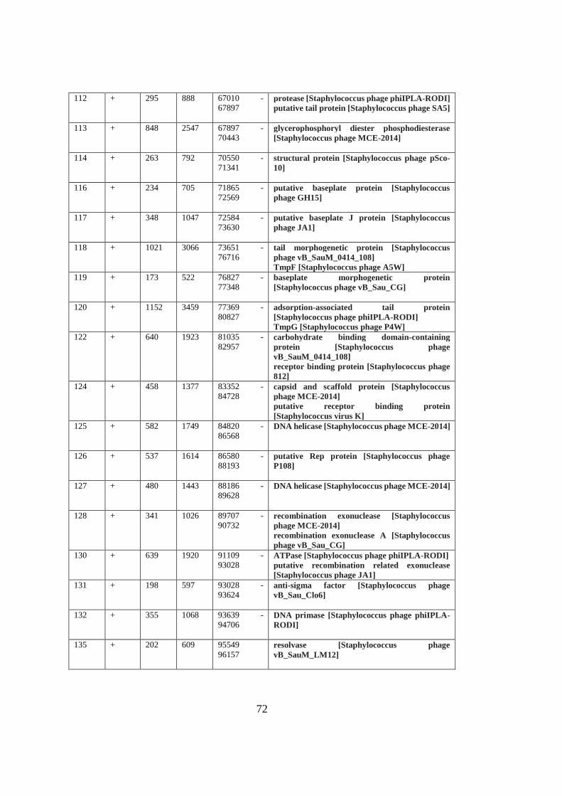
72
112 + 295 888 67010 -
67897
protease [Staphylococcus phage phiIPLA-RODI]
putative tail protein [Staphylococcus phage SA5]
113 + 848 2547 67897 -
70443
glycerophosphoryl diester phosphodiesterase
[Staphylococcus phage MCE-2014]
114 + 263 792 70550 -
71341
structural protein [Staphylococcus phage pSco-
10]
116 + 234 705 71865 -
72569
putative baseplate protein [Staphylococcus
phage GH15]
117 + 348 1047 72584 -
73630
putative baseplate J protein [Staphylococcus
phage JA1]
118 + 1021 3066 73651 -
76716
tail morphogenetic protein [Staphylococcus
phage vB_SauM_0414_108]
TmpF [Staphylococcus phage A5W]
119 + 173 522 76827 -
77348
baseplate morphogenetic protein
[Staphylococcus phage vB_Sau_CG]
120 + 1152 3459 77369 -
80827
adsorption-associated tail protein
[Staphylococcus phage phiIPLA-RODI]
TmpG [Staphylococcus phage P4W]
122 + 640 1923 81035 -
82957
carbohydrate binding domain-containing
protein [Staphylococcus phage
vB_SauM_0414_108]
receptor binding protein [Staphylococcus phage
812]
124 + 458 1377 83352 -
84728
capsid and scaffold protein [Staphylococcus
phage MCE-2014]
putative receptor binding protein
[Staphylococcus virus K]
125 + 582 1749 84820 -
86568
DNA helicase [Staphylococcus phage MCE-2014]
126 + 537 1614 86580 -
88193
putative Rep protein [Staphylococcus phage
P108]
127 + 480 1443 88186 -
89628
DNA helicase [Staphylococcus phage MCE-2014]
128 + 341 1026 89707 -
90732
recombination exonuclease [Staphylococcus
phage MCE-2014]
recombination exonuclease A [Staphylococcus
phage vB_Sau_CG]
130 + 639 1920 91109 -
93028
ATPase [Staphylococcus phage phiIPLA-RODI]
putative recombination related exonuclease
[Staphylococcus phage JA1]
131 + 198 597 93028 -
93624
anti-sigma factor [Staphylococcus phage
vB_Sau_Clo6]
132 + 355 1068 93639 -
94706
DNA primase [Staphylococcus phage phiIPLA-
RODI]
135 + 202 609 95549
96157
resolvase [Staphylococcus phage
vB_SauM_LM12]
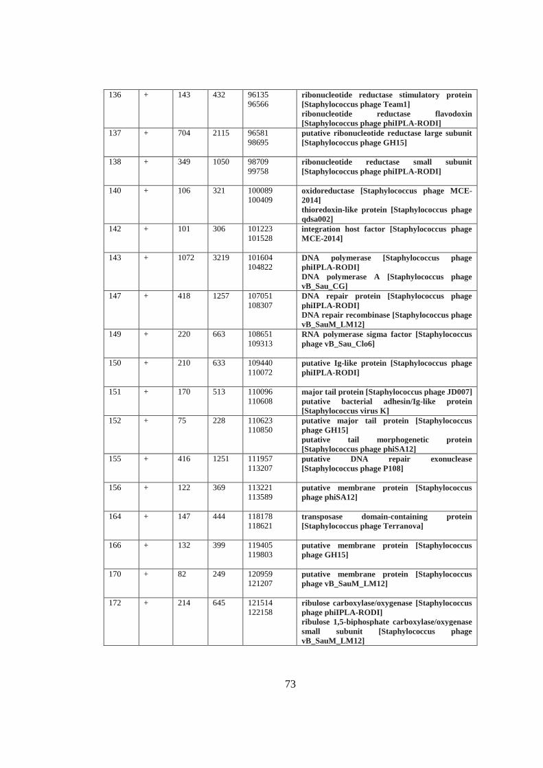
73
136 + 143 432 96135
96566
ribonucleotide reductase stimulatory protein
[Staphylococcus phage Team1]
ribonucleotide reductase flavodoxin
[Staphylococcus phage phiIPLA-RODI]
137 + 704 2115 96581
98695
putative ribonucleotide reductase large subunit
[Staphylococcus phage GH15]
138 + 349 1050 98709
99758
ribonucleotide reductase small subunit
[Staphylococcus phage phiIPLA-RODI]
140 + 106 321 100089
100409
oxidoreductase [Staphylococcus phage MCE-
2014]
thioredoxin-like protein [Staphylococcus phage
qdsa002]
142 + 101 306 101223
101528
integration host factor [Staphylococcus phage
MCE-2014]
143 + 1072 3219 101604
104822
DNA polymerase [Staphylococcus phage
phiIPLA-RODI]
DNA polymerase A [Staphylococcus phage
vB_Sau_CG]
147 + 418 1257 107051
108307
DNA repair protein [Staphylococcus phage
phiIPLA-RODI]
DNA repair recombinase [Staphylococcus phage
vB_SauM_LM12]
149 + 220 663 108651
109313
RNA polymerase sigma factor [Staphylococcus
phage vB_Sau_Clo6]
150 + 210 633 109440
110072
putative Ig-like protein [Staphylococcus phage
phiIPLA-RODI]
151 + 170 513 110096
110608
major tail protein [Staphylococcus phage JD007]
putative bacterial adhesin/Ig-like protein
[Staphylococcus virus K]
152 + 75 228 110623
110850
putative major tail protein [Staphylococcus
phage GH15]
putative tail morphogenetic protein
[Staphylococcus phage phiSA12]
155 + 416 1251 111957
113207
putative DNA repair exonuclease
[Staphylococcus phage P108]
156 + 122 369 113221
113589
putative membrane protein [Staphylococcus
phage phiSA12]
164 + 147 444 118178
118621
transposase domain-containing protein
[Staphylococcus phage Terranova]
166 + 132 399 119405
119803
putative membrane protein [Staphylococcus
phage GH15]
170 + 82 249 120959
121207
putative membrane protein [Staphylococcus
phage vB_SauM_LM12]
172 + 214 645 121514
122158
ribulose carboxylase/oxygenase [Staphylococcus
phage phiIPLA-RODI]
ribulose 1,5-biphosphate carboxylase/oxygenase
small subunit [Staphylococcus phage
vB_SauM_LM12]
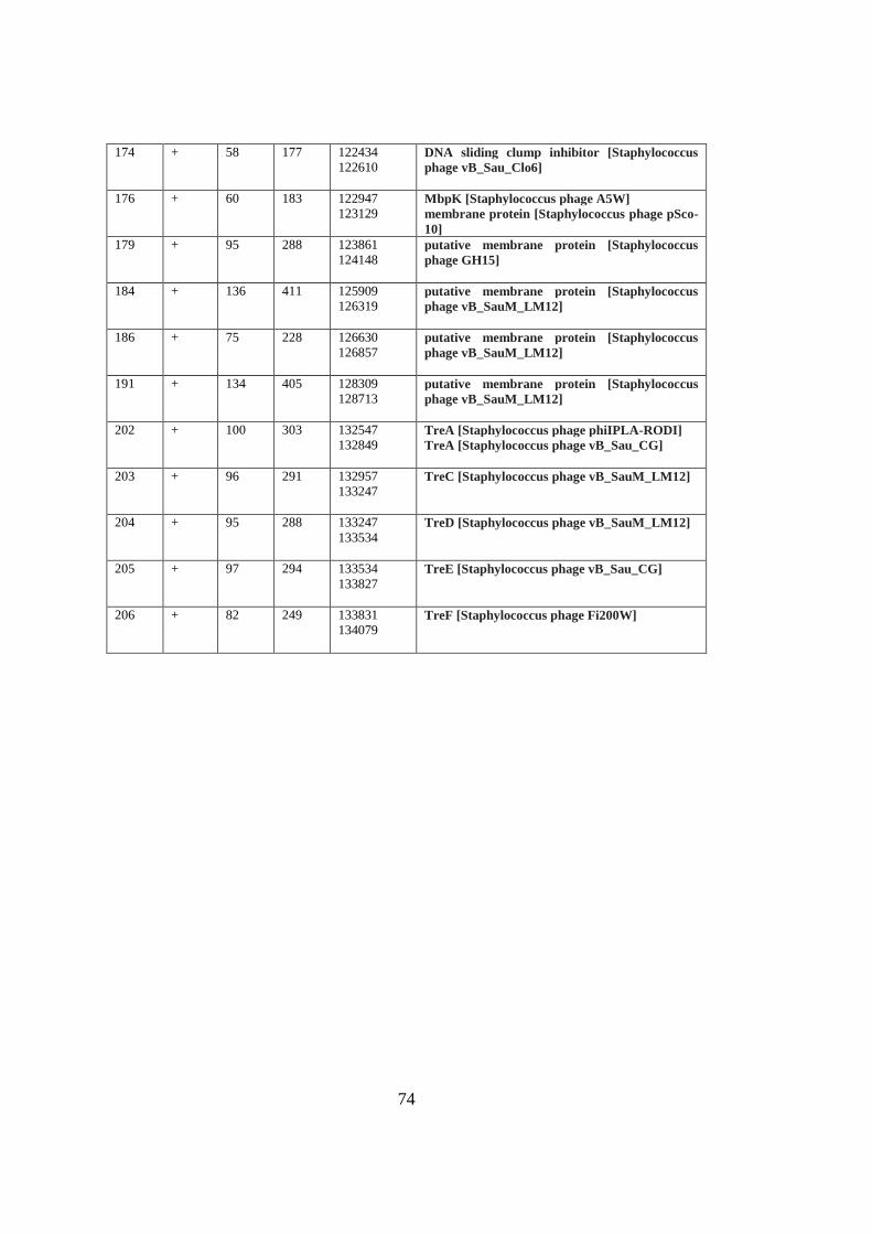
74
174 + 58 177 122434
122610
DNA sliding clump inhibitor [Staphylococcus
phage vB_Sau_Clo6]
176 + 60 183 122947
123129
MbpK [Staphylococcus phage A5W]
membrane protein [Staphylococcus phage pSco-
10]
179 + 95 288 123861
124148
putative membrane protein [Staphylococcus
phage GH15]
184 + 136 411 125909
126319
putative membrane protein [Staphylococcus
phage vB_SauM_LM12]
186 + 75 228 126630
126857
putative membrane protein [Staphylococcus
phage vB_SauM_LM12]
191 + 134 405 128309
128713
putative membrane protein [Staphylococcus
phage vB_SauM_LM12]
202 + 100 303 132547
132849
TreA [Staphylococcus phage phiIPLA-RODI]
TreA [Staphylococcus phage vB_Sau_CG]
203 + 96 291 132957
133247
TreC [Staphylococcus phage vB_SauM_LM12]
204 + 95 288 133247
133534
TreD [Staphylococcus phage vB_SauM_LM12]
205 + 97 294 133534
133827
TreE [Staphylococcus phage vB_Sau_CG]
206 + 82 249 133831
134079
TreF [Staphylococcus phage Fi200W]
75
CURRICULUM VITAE
PERSONAL INFORMATION
Surname, Name : Çotak, Medine
Nationality : Turkish (TC)
Date and Place of Birth : 3 January 1986, Afyon
Phone : +90 5550098603
E-mail : [email protected]
EDUCATION
Degree Institution Year of Graduation
MS Bonn University Molecular Biotechnology 2014
BS METU Molecular Biology and Genetics 2010
High School Kütahya Science High School, Kütahya 2006
WORK EXPERIENCE
Year Place Enrollment
2015-Present Mikroliz Biotech Founder R&D
FOREIGN LANGUAGES
Advanced English, Fluent German
PUBLICATIONS
HOBBIES
Gardening, Crime Science Investigation