Embed Size (px)
Citation preview

U.S. D E P A R T M E N T OF H E A L T H A N D H U M A N S E R V I C E S Pub l i c H e a l t h S e r v i c e N a t i o n a l Ins t i tu tes of Hea l t h

T a b l e o f C o n t e n t s
Vol. 69, No. 5 November 1982
Investigations on Man
989 T h e s e r u m c h o l e s t e r o l - c a n c e r r e l a t i o n s h i p : A n analys is o f t i m e t rends i n the F r a m i n g h a m S t u d y
997 R e a c t i v i t y i n n e o p l a s i a , p r e n e o p l a s i a , a n d p r e g n a n c y o f l y m p h o c y t e s aga ins t fetal extracts: Cross - reac t ions b e t w e e n m a n a n d mouse
P a u l D . Sorlie, M a n n i n g F e i n l e i b
Günter Pasternak, B e r n h a r d Schlott, Jürgen Reinhöf er, Günter Gryschek, Bodo von B r o e n , Sybille Albrecht
1005 C a n c e r s o f the g a l l b l a d d e r a n d b i l i a r y t rac t i n A l a s k a n N a t i v e s : 1970 -79
Leslie P . Boss, A n n e P . L a n i e r , Peter H . D o h a n , Thomas R . Bender
1009 C o l l a g e n p o l y m o r p h i s m i n e x t r a c e l l u l a r m a t r i x o f h u m a n os t eosa rcoma
F r e d e r i c D . S h a p i r o , D a v i d R . E y r e
1017 E x o g e n o u s estrogens a n d o the r r i sk factors for breast Cancer i n w o m e n w i t h b e n i g n breast diseases
D a v i d B . Thomas, Joyce P . Per sing, W i l l i a m B . H u t c h i n s o n
1027 S e c o n d Cancers f o l l o w i n g r a d i o t h e r a p y for c e r v i c a l c a n c e r
1035 A s s o c i a t i o n o f breast c a n c e r r isk w i t h age at first a n d subsequent b i r t h s : A s tudy i n the p o p u l a t i o n o f the E s t o n i a n R e p u b l i c
1039 In v i t r o g r o w t h S t i m u l a t i o n o f h u m a n o v a r i a n cance r cells b y x e n o g e n e i c p e r i t o n e a l m a c r o p h a g e s
R u t h A . K l e i n e r m a n , Rochelle E . Curtis,John D . Boke, Jr., John T F l a n n e r y , Joseph F . F r a u m e n i ,
Jr-
B r i a n M a c M a h o n , M a r e t P u r d e , D a n i e l C r a m e r , E v i H i n t
Charles E . Welander, R o n a l d B . N a t a l e , John L . Lewis, Jr.
1049 Effect o f n a t u r a l protease i n h i b i t o r s a n d a c h e m o a t t r a c t a n t o n t u m o r c e l l i n v a s i o n i n v i t r o
U. P . Thorgeirsson, L . A . L i o t t a , T. Kalebic, I . M . M a r g u l i e s , K. Thomas, M . Rios-Candelore, R . G. Russo
1055 A n d r o g e n conjugates i n h u m a n breast cyst f lu ids
1059 P a r i t y a n d c o l o r e c t a l c a n c e r r isk i n w o m e n
1063 C o r r e l a t i o n b e t w e e n Cancers o f the u t e r i ne c e r v i x a n d penis i n C h i n a
1067 U s e o f c i r c u l a t i n g p r e g n a n c y - s p e c i f i c ßi g l y c o p r o t e i n as a m a r k e r i n C a r c i n o m a o f the breast i n w o m e n
W. R . M i l l e r , M . M . Roberts, R . J . Creel, P . L . Yap, R . W. Kelly, A . P M . F o r r e s t
T i m Byers, Saxon G r a h a m , M y a Swanson
J u n - Yao L i , F r e d e r i c k P . L i , W i l l i a m J. Blot, Robert W. M i l l e r , Joseph F . F r a u m e n i , Jr.
S a u l W. Rosen, M i t c h e l l H . G a i l , Douglass C. Tormey
J N C I , V O L . 69, N O . 5, N O V E M B E R 1982
Investigations on Nonhuman Systems
1073 C a r c i n o e m b r y o n i c a n t i g e n i n n o n h u m a n p r ima te s
1077 S p o n t a n e o u s l e u k e m i a viruses: L y m p h o m a g e n i c eco t rop i c viruses o f A K R m i c e
1083 P r o s t a g l a n d i n - i n d u c e d d i f f e r en t i a t i on o r d i m e t h y l s u l f o x i d e - i n d u c e d d i f f e r e n t i a t i o n : R e d u c t i o n o f the neop la s t i c p o t e n t i a l o f a rat m a m m a r y t u m o r s tem-ce l l l i ne
1095 I m m u n o t h e r a p y b y i n t r a l e s i o n a l i n j e c t i o n o f B C G ce l l w a l l s o r l i ve B C G i n b o v i n e o c u l a r s q u a m o u s ce l l C a r c i n o m a : A p r e l i m i n a r y repor t
1105 E x p r e s s i o n o f f i b r o n e c t i n a n d l a m i n i n i n the rat l i ve r after p a r t i a l h e p a t e c t o m y , d u r i n g carc inogenes is , a n d i n t r a n s p l a n t a b l e h e p a t o c e l l u l a r c a r c i n o m a s
1115 Effects o f i sop ro te reno l o n m a m m a r y g l a n d t u m o r s i n d u c e d b y Af -n i t roso -Af -methy lu rea a n d s a l i v a r y g l a n d t u m o r s i n d u c e d b y 7,12-d i m e t h y l b e n z [ ö ] a n t h r a c e n e
1121 I m m u n o t h e r a p e u t i c effects o f p a r t i a l l y p u r i f i e d tumor - spec i f i c t r a n s p l a n t a t i o n an t igens o n p u l m o n a r y metastases o f a 3 - m e t h y l c h o l a n -t h r e n e - i n d u c e d s a r c o m a i n m i c e : A p r e l i m i n a r y repor t
1127 Dose-response s tudies w i t h n i t r o s o h e p t a m e t h y l -e n e i m i n e a n d its a - d e u t e r i u m - l a b e l e d d e r i v a t i v e i n F 3 4 4 rats
1135 I n d u c t i o n o f m a m m a r y neop la sms i n the A C I rat by 4 3 0 - k e V neu t rons , X - r a y s , a n d d i e thy l s t i l be s t ro l
1147 I n h i b i t i o n b y t u m o r - p r o m o t i n g p h o r b o l esters o f p r o c o l l a g e n synthesis i n p r o m o t a b l e J B 6 mouse e p i d e r m a l cells
1155 Effect o f c a r c i n o g e n release rate o n the i n c i d e n c e o f p r eneop la s t i c a n d neop la s t i c lesions o f the r e sp i r a to ry t ract e p i t h e l i u m i n rats
D a r r o w E . Haagensen, Jr., R i c h a r d S. Metzgar, B r e n t Swenson, W i l l i a m G. D i l l e y , Charles E . Cox, S a r a h Davis, Janet M u r d o c h , N o r m a n Zamcheck, Samuel A . Wells, Jr.
Esther F . H a y s , N a d i n e M a r g a r e t t e n , Stephen K Swanson
P h i l i p S. Rudland, A n n a Twiston Davies, M i c h a e l J. W a r b u r t o n
W i m R . K l e i n , E . Joost Ruitenberg, Peter A . Steerenberg, W i m H . de Jong, W i m K r u i z i n g a , W i m M i s d o r p , Jürgen Bier, Rudy H . Tiesjema,
Johan G. Kreeftenberg, Jacob S. Teppema, H e r b e r t J. Rapp
Stewart S e l L E r k k i Ruoslahti
T i b o r B a r k a
Tsuguo T a n a k a , N e a l R . Pellis, B a r r y D . K a h a n
W. Lijinsky, M . D . Reuber, T. S. Davies, C W.
C i a i r e J. Shellabarger, D a n i e l l e Chmelevsky, Albrecht M . Kellerer, J. P a t r i c k Stone, Seymour H o l t z m a n
L . D a v i d D i o n , Jenifer Bear, John B a t e m a n , L u i g i M . D e L u c a , Nancy H . C o l b u r n
Mitsutoshi S h i b a , Andres J. P . Klein-Szanto, A n n C. M a r c h o k , B i m a l C P a l , P a u l Nettesheim
1163 E x c l u s i v e b i n d i n g o f i m m u n o g l o b u l i n to F c y receptors o n m a c r o p h a g e s i n 3 - m e t h y l c h o l a n -t h r e n e - i n d u c e d m u r i n e t u m o r s
John M . Lindsay, L i n d a M a n n i n g , Gary W. Wood
J N C I , V O L . 69, N O . 5, N O V E M B E R 1982
1175 I n d e p e n d e n t express ion o f c h e m i c a l c a r c i n o g e n -i n d u c e d p h e n o t y p i c proper t ies f r equen t ly assoc ia ted w i t h the neop las t i c State i n a c u l t u r e d g u i n e a p i g ce l l l ine
1183 C e r u l o p l a s m i n , c o p p e r ions , a n d angiogenes is
1189 I s o l a t i o n o f t u m o r i c i d a l m a c r o p h a g e s f r o m l u n g m e l a n o m a metastases o f m i c e t r ea ted s y s t e m i c a l l y w i t h l i posomes c o n t a i n i n g a l i p o p h i l i c d e r i v a t i v e o f m u r a m y l d i p e p t i d e
1199 I n d u c t i o n o f m a m m a r y t u m o r s i n v i r g i n f emale B A L B / c m i c e b y s ing le l o w doses o f 7,12-d i m e t h y l b e n z [ a ] a n t h r a c e n e
Charles I L Evans, Joseph A . D i Paolo
K a t a r i S. R a j u , G i u l i o Alessandri, M a r i n a Z i e h e , Pietro M . G u l l i n o
M . E . Key,J. E . Talmadge, W. E . F o g l e r , C B u c a n a , I . J. F i d l e r
Stephen P . E t hier, Robert L . U l l r i c h
Announcements
1205 N e w N I H B i o t e c h n o l o g y H i g h V o l t a g e E l e c t r o n M i c r o s c o p e ( H V E M ) R e s o u r c e A v a i l a b l e to B i o l o g i c a l a n d M e d i c a l Researchers
1205 H a r v a r d M e d i c a l S c h o o l C o n t i n u i n g E d u c a t i o n P r o g r a m
1205 T h e 1983 O n c o l o g y U p d a t e S y m p o s i u m
1205 S i x t y - f i f t h A n n u a l M e e t i n g o f the A m e r i c a n R a d i u m Soc ie ty
1205 E i g h t h S u m m e r P r o g r a m i n M e t h o d s o f I m m u n o l o g i e R e s e a r c h a n d D i a g n o s i s
1206 E i g h t h I n t e r n a t i o n a l Congre s s o f C y t o l o g y
1206 E . O . R . T . C . S y m p o s i u m o n T r e a t m e n t o f A d v a n c e d G a s t r o i n t e s t i n a l C a n c e r
J N C I , V O L . 69, N O . 5, N O V E M B E R 1982

Induction of Mammary Neoplasms in the ACI Rat by 430-keV Neutrons, X-Rays, and Diethylst i lbestrol 1 2 3
Cla i re J . S h e l l a b a r g e r , 4 Dan ie l le C h m e l e v s k y , 5 A l b r e c h t M. K e l l e r e r , 5 J . Pa t r i ck S t o n e , 4
and S e y m o u r H o l t z m a n 4 6
ABSTRACT—Mammary tumorigenesis was studied in female ACI rats after treatment with X-irradiation or neutron-irradiation, with or without diethylstilbestrol (DES) treatment. The mortality-corrected cumulative tumor rate based on all mammary neoplasms and the mortality-corrected incidence based on the first neoplasms only have been derived. In non-DES-treated animals, at the relatively high radiation doses studied, all dose-effect relationships were consistent with relative biological effectiveness (RBE) values slightly in excess of 10. In DES-treated rats definite findings were observed at neutron doses as low as 0.01 Gy (1 rad). The dose-effect relationship in DES-treated rats showed a strong sublinearity (dose exponent <1 ) at low neutron doses. RBE values in DES-treated rats increased in inverse Proportion to the Square root of the neutron dose, and exceeded 100 at a neutron dose ofO.01 Gy (1 rad) .—JNCM 982; 69 :1135 -1146 .
In female rats of the A C I strain, both low L E T and h igh L E T radiations induce m a m m a r y A C and m a m m a r y F A (/, 2 ) . In this strain of rat, a form of synthetic estrogen, D E S , induces mammary A C , but no mammary F A , when given in the form of a 20-mg pellet of 25% D E S and 75% cholesterol ( 3 ) . Furthermore, in A C I rats, the effect of D E S and ion iz ing radiations appears to be synergistic for m a m mary A C formation, as shown in studies wi th X- rays by Segaloff ( 4 ) and later by Stone et a l . (2), and in studies w i t h neutrons by Shellabarger et a l . ( 5 ) .
T h e present work is p r imar i ly a imed not at the further elucidation of the synergistic effect but at the comparison of the effects o f X-rays and neutrons at low doses. Therefore, graded X - r a y and neutron doses were appl ied and only one dose of D E S was uti l ized. T h e interest in this investigation is due to the theoretical prediction ( 6 ) and the experimental Observation in various Systems ( 7 - 9 ) of high-neutron R B E , which may be of considerable impact to radiat ion protection because they support the Suggestion that the quality factor of 10, now used for neutrons, should be reconsidered ( 1 0 ) . T h e only source of human data for neutrons, the Japanese atomic b o m b survivors, may be lost due to the revision of neutron dosimetry ( / / ) ; thus data from an imal studies wi th low doses o f neutrons have gained importance.
In an earlier experiment w i th Sprague-Dawley rats, very low doses o f neutrons were used, and for a neutron dose of 1 m G y an R B E in excess of 100 was found ( 1 2 , 1 3 ) . However , it remained unclear whether this finding was specific to the Sprague-Dawley rats only, w i th the h igh spontaneous inc i dence and w i t h a considerably larger number of F A than A C characteristic to this strain. Therefore, it appeared de-sirable to conduct analogous studies w i th A C I rats that have a very low spontaneous incidence of mammary neoplasms and show predominant ly A C when treated wi th D E S .
A second aspect of the earlier findings on Sprague-Dawley rats was of par t icular importance and has been an addi t ional motivat ion o f the present study. T h i s was the f inding of a
substantial sublinearity, i.e., a negative curvature of the dose-effect relationship, at small neutron doses. Such a sublinearity implies that the effect per unit dose is largest at smallest doses and is therefore of evident concern in radiation protection, for which risk assessments are commonly based on the linear hypothesis. Subl ineari ty is also of interest because it occurs at doses that are sufficiently small that only few cells are traversed by any neutron recoil, and the probabi l i ty of several recoils can be disregarded ( 1 4 , 1 5 ) . In this dose r ä n g e the tendency is to postulate linear dose dependences for neutrons, because the number of cells that receive any energy deposition is merely proport ional to dose. T h e failure to find a linear relation has therefore been interpreted as evidence of a mul t ice l iu lar reaction or a radiat ion-induced tissue factor ( 1 5 ) . Examina t ion of the l inearity or nonline-arity of the dose-effect relationship for neutrons was accord-ingly another ma in goal in the present investigation.
M A T E R I A L S A N D M E T H O D S
Rats and treatment schedules.—Approximately 800 weani ing, virgin, female A C I rats were purchased from M . A . B iop rod-ucts, Walkersvi l le , M d . Rats were delivered to this laboratory in eight shipments of at least 50 rats at weekly intervals. F r o m each shipment 3 rats were assigned to each of 14 groups that were to be irradiated, and 4 rats were assigned to each of 2 groups that were reserved as nonirradiated controls. Th i s procedure was repeated wi th another 400 rats. Thus all 16 groups were matched closely for age both in regard to the average age and the r ä n g e of ages. Neutron irradiat ion was given to rats wi th an average age of 88 days, and X- i r r ad i a t i on was given to rats wi th an average age of
ABBRF.VIATIONS USED: AC=adcnoca?cinoma(s): DES—dicihylsi übest rol:
FA=fibroadcnoma(s): Gy (gray)=unit ofabsorbed dose in the International
System of Units (Gy=J/kg or ]()() rad); LET—linear energy transfer;
RBE=relativc biological effectiveness.
] Received February 17, 1982: aeeepted July 6, 1982. 2 Supported by Public Health Service contract X01CP-60219 from the
Division of Cancer Cause and Prevention, National Cancer Institute; by
contract DE-AC02-76CH00016 from the U.S. Department of Energy; and
by Euratom Research contract BIO-286-81 D (B). 3 Animals were maintained under the guidelines set forth by the Animal
Weifare Act in facilities aecredited by the American Association for Ac-
creditation of Laboratory Animal Care. 4 Medical Department, Brookhaven National Laboratory, Associated
Universities, Inc., Upton, N .Y . 11973.
"Institut für Medizinische Strahlenkunde, Univers i tä t W ü r z b u r g , Vers
bacher Str. 5, 8700 W ü r z b u r g , Federal Republic of Germany. 6 W r e thank Ms. Lindora Boyd, Ms. Elizabeth M . Jellett, M r . John P.
Shanley, M r . Leon J . Goodman, and Ms. Mary R. Snead for technical
assistance.
1 1 3 5 JNCI, VOL. 69, NO. 5, NOVEMBER 1982

1 1 3 6 She l l aba rge r , C h m e l e v s k y , Ke l l e re r , e t a l .
89 days. A l l irradiations were done as described previously ( 5 ) . D E S (5 mg) was given, in pellet form, 2 days before irradiation, w i th methods described earlier ( 4 ) . A l l da ta were recorded in days after the date of D E S adminis t ra t ion and, for s impl ic i ty , a l l ages, life-spans, and times quoted in this article refer to the date of D E S adminis t ra t ion as t ime 0.
A l l rats were kept i n temperature-controlled ( 2 1 - 2 3 ° C ) and humidi ty-control led (45-65%) an ima l rooms wi th 12 hours (8 a.m.-8 p.m.) o f fluorescent l ight. T h e animals were maintained on commercia l rat chow and water ad l i b i t u m . Each rat was examined at least weekly for the presence o f mammary tumors, and a l l m a m m a r y tumors were studied microscopically and given a pathologic Classification o f either A C or F A according to cri ter ia consistent wi th those of Y o u n g and Hal lowes ( 1 6 ) .
T u m o r methodology.—Each rat was identified by a n u m -bered ear tag. T h e anatomic location of each m a m m a r y tumor was recorded w i t h the nipples as reference points. Because two distinct types o f m a m m a r y tumor responses were found, i nd iv idua l or mul t ip le , each was recorded sep-arately. Ind iv idua l m a m m a r y tumors were removed under ether anesthesia at a size of about 2 cm. I f a second tumor was found at the site o f a previously removed tumor, it was not recorded as a second ind iv idua l tumor unless a 10-week period had elapsed between removal o f the first tumor a n d the detection of the subsequent tumor. In the other type o f mammary tumor response, wh ich was confined to m a m m a r y A C in DES-t rea ted rats, mul t ip le A C were often detected wi th in a single quadrant of mammary tissue over about 1 week. Because too many tumors existed to be counted by palpation alone, the entire quadrant was removed, fixed, defatted, stained wi th hematoxyl in , cleared, and stored i n methyl salicylate. E a c h quadrant was examined under a dissecting microscope at magnification 10X, and al l pre-sumed pathologic areas were removed ind iv idua l ly for sec-tioning. T h e m i n i m u m number of these mul t ip le A C was never less than 4 per quadrant. Even though there were often too many mul t ip le A C per quadrant to be counted accurately, an ind iv idua l quadrant was assigned a m a x i -m u m number of 4 A C . T h u s an ind iv idua l rat could then have a m a x i m u m number of 16 A C . T h e mul t ip le A C response wi th in a single quadrant was usually found w i t h i n a 1-3-week period. However , quadrants were removed not more often than twice per week. Rats were k i l l ed when they showed a mul t ip le tumor response in al l four quadrants, when they became mor ibund , or when tumor removal was no longer feasible. A l l remaining DES-t rea ted rats were ki l led at days 523-525. O f the non-DES-treated rats s t i l l alive, approximately ha l f of each treatment group was k i l l ed either on day 712 or on days 726-730. A t autopsy, confir-mation of D E S treatment was made by locat ing the D E S pellet. A n abnormal pi tui tary gland was recorded as a pituitary tumor i f the pi tui tary was hemorrhagic and fragile and exceeded a weight of 50 mg. Pi tu i tary tumors were found in 30% of the untreated control rats, i n 39% of the irradiated only rats, i n 90% of the D E S only rats, and in 79% of the irradiated plus DES-t rea ted rats. T h e combined incidence of pi tui tary tumors in a l l nonirradiated rats was 58% compared to 52% in a l l irradiated rats, and the c o m
bined incidence in a l l non-DES-t rea ted rats was 38% compared to 81% in a l l DES-t rea ted rats. These resuits are in agreement wi th those of Stone et a l . ( 3 ) .
RESULTS
O f a total of 789 rats studied, 348 rats received D E S and 441 d i d not receive D E S . O f the rats that received D E S , only 6.3% developed F A . Because of the small y ie ld of F A in DES-t rea ted rats (a total o f 32 F A vs. 2,474 A C ) , the F A response in DES-t rea ted animals was not analyzed. A m o n g the non-DES-treated animals, 47% showed m a m m a r y neoplasms, 35% developed F A , and 26% developed A C ; in view of their l imi ted number , both types of neoplasms were pooled in the analysis.
S u m m a r y o f O b s e r v a t i o n s
T h e resuits o f this study w i l l first be presented i n elemen-tary form; the more rigorous actuarial analysis w i l l be given
0 200 400 600 T I M E d a y s
T E X T - H G U R F . 1.—Fraciion of the non-OES-treatcd animals dcad or alive with and vvithout F A and/or A C as a funetion oft imc after irradiation. Group sizes are given in table '.\. Fraction of animals dead withoul neoplasm: E3. fraction of animals dead with at least 1 neoplasm: SS. fraction of animals alive with at least 1 neoplasm: El. fraction of animals alive withoul a neoplasm.
JNCI, VOL. 69, NO. 5, NOVEMBER 1982
N e u t r o n RBE of Rat M a m m a r y T u m o r i g e n e s i s 1 1 3 7
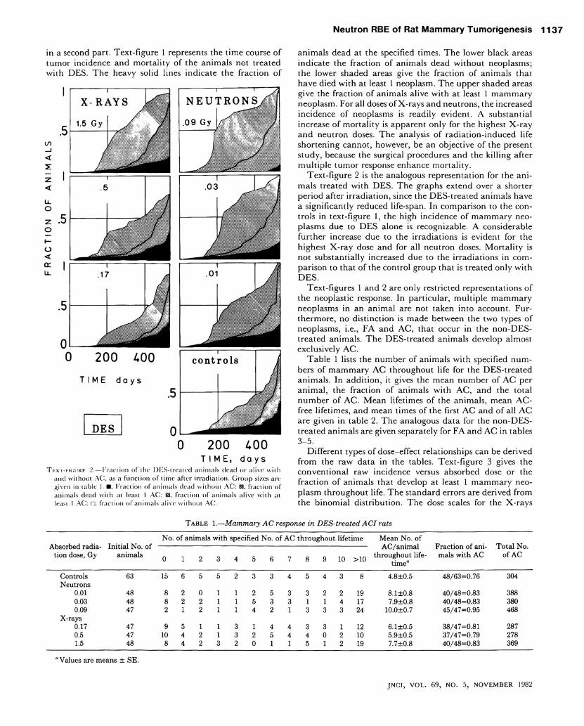
in a second part. Text-figure 1 represents the time course o f tumor incidence and mortal i ty of the animals not treated wi th D E S . T h e heavy solid lines indicate the fraction o f
0 200 400 T I M E , d a y s
TF.XT-HCUIRF. '2.—Fraciion of ihc DES-treated animals dead or alive with and withoul A G . as a f u n c ü o n of time after irradiation. Group si/.es are given in table 1. Fraction of animals dead withoul A C : fraciion of animals dead wiih at least 1 A C : fraction of animals alive with al least 1 A C : fraciion of animals alive without A C .
animals dead at the specified times. T h e lower black areas indicate the fraction of animals dead without neoplasms; the lower shaded areas give the fraction of animals that have died wi th at least 1 neoplasm. T h e upper shaded areas give the fraction of animals alive wi th at least 1 m a m m a r y neoplasm. For al l doses of X-rays and neutrons, the increased incidence of neoplasms is readily evident. A substantial increase of mortal i ty is apparent only for the highest X - r a y a n d neutron doses. T h e analysis of radiat ion-induced life shortening cannot, however, be an objective of the present study, because the surgical procedures and the k i l l i ng after mul t ip le tumor response enhance mortal i ty.
Text-figure 2 is the analogous representation for the animals treated wi th D E S . T h e graphs extend over a shorter period after i r radiat ion, since the DES-t rea ted animals have a significantly reduced life-span. In comparison to the con-trols in text-figure 1, the high incidence of mammary neoplasms due to D E S alone is recognizable. A considerable further increase due to the irradiations is evident for the highest X - r a y dose and for al l neutron doses. M o r t a l i t y is not substantially increased due to the irradiations in comparison to that of the control group that is treated only w i t h D E S .
Text-figures 1 and 2 are only restricted representations of the neoplastic response. In part icular , mul t ip le m a m m a r y neoplasms in an an imal are not taken into account. Fur -thermore, no dist inction is made between the two types o f neoplasms, i.e., F A and A C , that occur in the n o n - D E S -treated animals. T h e DES-t rea ted animals develop almost exclusively A C .
T a b l e 1 lists the number of animals w i th specified num-bers of mammary A C throughout life for the DES-t rea ted animals. In addi t ion, it gives the mean number of A C per a n i m a l , the fraction of animals wi th A C , and the total number o f A C . M e a n lifetimes of the animals, mean A C -free lifetimes, and mean times of the first A C and of al l A C are given in table 2. T h e analogous data for the n o n - D E S -treated animals are given separately for F A and A C in tables 3-5 .
Different types of dose-effect relationships can be derived from the raw data in the tables. Text-figure 3 gives the conventionai raw incidence versus absorbed dose or the fraction of animals that develop at least 1 mammary neoplasm throughout life. T h e Standard errors are derived from the b inomia l distr ibution. T h e dose scales for the X- rays
T A B L E 1 . — M a m m a r y A C response i n D E S - t r e a t e d A C I rats
No. of animals with specified No. of AC throughout lifetime Mean No. of Absorbed radia- Initial No. of AC/animal Fraction of ani- Total No.
tion dose, Gy animals Q 1 2 3 4 5 6 7 8 9 10 >10 t n r o u & n o u t n^ e" rnals with AC of AC time"
Controls 63 Neutrons
0.01 48 0.03 48 0.09 47
X-rays 0.17 47 0.5 47 1.5 48
15 6 5 5 2
8 2 0 1 1 8 2 2 1 1 2 1 2 1 1
9 5 1 1 3 10 4 2 1 3 8 4 2 3 2
3 3 4 5 4
2 5 3 3 2 5 3 3 1 1 4 2 1 3 3
1 4 4 3 3 2 5 4 4 0 0 1 1 5 1
3 8 4.8±0.5
2 19 8.1±0.8 4 17 7.9±0.8 3 24 10.0±0.7
1 12 6.1±0.5 2 10 5.9±0.5 2 19 7.7±0.8
48/63=0.76 304
40/48=0.83 388 40/48=0.83 380 45/47=0.95 468
38/47=0.81 287 37/47=0.79 278 40/48=0.83 369
a Values are means ± SE.
J N C I , V O L . 69, N O . 5, N O V E M B E R 1982
1 1 3 8 S h e l l a b a r g e r , C h m e l e v s k y , Ke l le re r , e t a l .
and the neutrons in this and in the subsequent text-figures differ by a factor of 10. Da ta for the non-DES-treated animals indicate an R B E of neutrons somewhat larger than 10. For the DES-t rea ted animals, R B E is substantially larger and may apparently exceed 100 at the lowest dose; however, it is difficult to infer accurate values of R B E from this crude analysis.
A n RBE-versus-dose relationship for neutrons w i l l be given at the end of this paper. T h e curves d rawn through the da ta i n text-figure 3 correspond to this R B E - d o s e relat ionship. T h e same applies to the dose-effect relationships
T A B L E 2 . — T e m p o r a l mammary A C response i n D E S - t r e a t e d A C I ratsa
Absorbed radiation dose, Gy Lifetime AC-free
lifetime
Time of appearance of first AC
Time of appear
ance of all AC
Controls 456±13 374±18 372±19 434±6 Neutrons
0.01 446±17 305±20 314±22 396±6 0.03 435±17 268±17 253±16 374±6 0.09 455±12 260±19 251±19 354±6
X-rays 0.17 471±13 378±19 381±21 448±6 0.5 445±16 341+23 338±26 405±7 1.5 432±16 301±18 292±20 373±6
" Values are mean No. of days ± SE.
given in subsequent text-figures. These curves w i l l not a l -ways be the best fits to the ind iv idua l data, but they are always consistent wi th them, and they are inserted to permit a judgment o f the R B E - d o s e relationship of the last text-figure as an overall fit to the experimental data.
Text-figure 4 gives the mean number o f m a m m a r y neoplasms per an ima l throughout life. T h e approximate coin-cidence of the two curves for X- rays and neutrons for the non-DES-treated animals indicates again R B E values close to 10. For the DES-t rea ted animals the R B E appears to be well in excess of 100 at small doses.
S imi l a r conclusions can be drawn from the values of the mean tumor-free life-span in tables 2 and 5. T h i s w i l l be substantiated by a competing-risk-corrected analysis in a later section.
Text-figures 3 and 4 show, for the D E S experiments, not only R B E values in excess o f 100 but also sublinear dose-effect relationships (dose exponent <1) for neutron i r radiat ion. S imi la r ly , high R B E values and sublinear dose-effect relations were also obtained in earlier experiments w i th Sprague-Dawley rats ( 1 2 , 1 3 ) . In view of the evident importance of such observations for general considerations on the effects of small doses of ion iz ing radiations, it is essential to submit the data to a rigorous analysis. Such an analysis corrects for differences in life-span. It also permits an eval-uation of the time course of the appearance of m a m m a r y neoplasms; this is par t icular ly important in the experiments
T A B L E 3 . — M a m m a r y FA response i n n o n - D E S - t r e a t e d A C I rats
No. of animals with specified No. of FA throughout ^ q ^ FA/animal Absorbed radia- Initial No. of lifetime throu hout life Fraction of animals Total No.
tion dose, Gy animals 5 « " with FA of FA f \ T c % o A u . f F 7 t i m e
Controls 61 59 0 2 0 0.07±0.05 2/61=0.03 4 Neutrons
0.045 46 32 11 1 1 0 1 0.46±0.14 14/46=0.30 21 0.09 48 24 11 7 5 0 1 0.94±0.17 24/48=0.50 45 0.18 48 23 12 7 3 0 1 1 1 1.10±0.23 25/48=0.52 53 0.36 48 19 11 5 6 0 4 2 1 1.63±0.29 29/48=0.60 78
X-rays 0.37 48 43 4 0 1 0.15±0.07 5/48=0.10 7 0.75 48 29 13 5 1 0.54±0.11 19/48=0.40 26 1.5 46 27 8 4 5 1 0 1 0.89±0.20 19/46=0.41 41 3 48 29 8 9 1 0 1 0.71±0.15 19/48=0.40 34
' Values are means ± SE.
T A B L E 4 . — M a m m a r y neoplastic response i n n o n - D E S - t r e a t e d rats
Absorbed radiation dose, Gy
Initial No. of an
imals
No. of animals with specified No. of AC throughout lifetime
8 >8
No. of A C / animal
throughout lifetime"
No. of AF and AC"
Fraction of animals with
AC
Fraction of animals with AC and/or
FA
Total No. of
AC
Controls 61 56 5 0 0.08±0.04 0.15±0.06 5/61=0.08 6/61=0.10 5 Neutrons
0.045 46 40 4 0 1 0 0 0 0 1 0 0.33±0.18 0.78±0.26 6/46=0.13 16/46=0.35 15 0.09 48 39 4 2 1 2 0.40±0.14 1.33±0.23 9/48=0.19 27/48=0.56 19 0.18 48 32 8 2 3 3 0.69±0.17 1.79±0.30 16/48=0.33 31/48=0.65 33 0.36 48 26 8 7 1 1 2 1 0 1 1 1.31±0.24 2.94±0.42 22/48=0.46 36/48=0.75 63
X-rays 0.37 48 42 4 1 0 0 1 0.23±0.11 0.37±0.15 6/48=0.12 10/48=0.21 11 0.75 48 38 5 2 1 1 1 0.44±0.15 0.98±0.20 10/48=0.21 25/48=0.52 21 1.5 46 25 12 6 0 1 1 0 1 0.87±0.21 1.76±0.32 21/46=0.46 29/46=0.63 40 3 48 29 10 6 1 0 0 0 1 0 1 0.90±0.25 1.60±0.32 19/48=0.40 27/48=0.56 43
" Values are means ± SE.
JNCI, VOL. 69, NO. 5, NOVEMBER 1982
N e u t r o n RBE of Rat M a m m a r y T u m o r i g e n e s i s 1 1 3 9
T A B L E 5 . — T e m p o r a l mammary neoplastic response i n n o n - D E S - t r e a t e d A C I rats"
Absorbed radiation dose, Gy Lifetime AC-free
lifetime FA-free lifetime
Tumorfree life
time
Time of appearance of: Time of appearance of:
First AC First FA First tumor All AC All FA All tu
mors
Controls Neutrons
0 .045
0 .09
0 . 1 8
0 .36
X-rays 0 . 3 7
0 . 7 5
1.5
3.
6 3 0 ± 2 1
6 5 5 ± 1 3
6 2 9 ± 2 0
5 8 0 ± 2 6
5 9 0 ± 1 8
6 1 4 ± 2 5
6 4 6 ± 2 0
6 1 7 ± 2 4
5 3 7 ± 2 4
6 2 7 ± 2 1
6 4 4 ± 1 4
6 1 7 ± 1 9
5 0 1 ± 2 6
4 9 6 ± 2 6
6 0 3 ± 2 5
6 2 1 ± 2 0
5 7 7 ± 2 3
4 9 8 ± 2 2
6 2 6 ± 2 1
6 1 9 ± 1 6
5 6 0 ± 2 1
5 3 3 ± 2 4
5 0 0 ± 1 5
6 0 2 ± 2 5
6 0 3 ± 1 9
5 5 8 ± 2 3
4 8 7 ± 2 2
6 2 4 ± 2 1
6 1 4 ± 1 6
5 8 6 ± 2 0
4 7 2 ± 2 8
4 4 4 ± 2 1
5 9 1 ± 2 5
5 8 5 ± 1 9
5 3 5 ± 2 2
4 6 0 ± 2 0
6 9 0 ± 1 1
6 0 3 ± 3 5
5 7 4 ± 2 1
4 2 9 ± 5 3
4 2 6 ± 4 3
5 4 2 ± 3 6
5 6 0 ± 2 9
6 0 3 ± 1 8
5 2 3 ± 2 0
5 9 0 ± 1 1
5 7 2 ± 2 8
5 3 4 ± 2 2
5 7 2 ± 1 9
5 0 7 ± 1 9
5 8 0 ± 6 0
5 9 5 ± 1 5
5 4 5 ± 2 4
5 0 5 ± 2 2
6 5 8 ± 2 3
5 6 6 ± 2 6
5 3 3 ± 2 0
4 9 4 ± 3 2
4 3 7 ± 2 6
5 4 9 ± 3 4
5 7 4 ± 1 5
5 5 5 ± 1 8
4 9 8 ± 1 8
6 9 0 ± 1 1
61 .5+19
6 0 2 ± 1 3
5 2 3 ± 3 1
5 0 9 ± 2 5
5 8 5 ± 2 7
5 8 5 ± 2 2
6 1 3 + 1 2
5 7 3 ± 1 3
631 ± 2 4
5 8 7 ± 2 2
5 6 7 ± 1 5
6 1 6 ± 1 3
5 7 6 ± 1 2
5 7 3 ± 4 3
6 1 6 ± 1 4
5 8 8 ± 1 5
5 2 9 ± 1 4
6 6 3 ± 1 5
5 9 9 ± 1 5
5 7 7 ± 1 1
5 8 0 ± 1 5
5 4 6 ± 1 3
5 8 0 ± 2 3
6 0 2 ± 1 2
6 0 0 ± 1 0
5 5 4 ± 1 0
' Values are mean No. of days ± SE.
D x, Gy 2 3
D E S (AC)
no D E S
FA+AC)
N E U T R O N S
X - R A Y S
.2 .3 X DN, Gy
T K X T - F I G U R F . '.).—Fraction of animals with mammary neoplasms as a func-lion of ihc neutron or the X-ray dose. Scales for the neutron (DN) and the X-ray (D x ) doses differ by a factor of 10. Data are given with S t a n d a r d e r r o r s . Solid lines r e f e r to the n e u t r o n irradiations; dashed lines,
10 the X-irradiations.
with D E S where the raw incidence is always close to 100% regardlcss o f radiat ion dose, and where, accordingly, dose-effect curves can be constructed only on the basis of the temporal dis tr ibut ion of neoplasms.
Ac tua r i a l A n a l y s i s
E s t i m a t e s o f C u m u l a t i v e T u m o r R a t e a n d C u m u l a t i v e T u m o r I n c i d e n c e
T h e same basic quantities w i l l be ut i l ized that have been employed i n the preceding article on the induct ion of m a m mary neoplasms in the Sprague-Dawley rat ( 1 3 , 1 7 ) . T h e tumor rate r ( t ) is the probabi l i ty per unit t ime interval for an animal to develop a neoplasm at t ime /. Because the actual time of origin of a neoplasm is unknown, it is iden-tified with the time of first Observation. T h e cumula t ive tumor rate R ( t ) is defined as time integral of the tumor rate:
R ( t ) = f r ( t ' ) d t ' . Jo
[ i ]
It is difficult to estimate the tumor rate wi th sufficient precision, and its integral R ( t ) is therefore a quant i ty more suitable to characterize the time course of the incidence of neoplasms. R ( t ) can be considered as the expected number of neoplasms up to time / for animals that are at risk throughout the whole time period from 0 to /. However , R ( t ) may differ from the actual mean number of neoplasms in those animals that survive up to time /; the reason is that R ( t ) is estimated on the basis of the complete Information, inc lud ing tumor incidence in those animals that do not survive up to time /.
As shown in our preceding article ( 1 3 ) , the cumula t ive tumor rate can be estimated from the formula:
[2]
where Ii are the times of appearance of the i n d i v i d u a l neoplasms up to time /, and N(t
t) are the numbers of animals
still at risk at these times. T h e numerical values and the Standard errors resulting from equation 2 are given in text-figure 5 for the DES-treated animals.
CO
CO < _ J Ü_ o UJ
Dx,Gy 2 3
10
< Li- 2
° z 6 or < UJ
? ÜJ 5 o_
< UJ 2:
i - DES (AC)
. 1 -
>NEUTRONS
X-RAYS
no D E S (FA + AC) }
+ - + 5
0 .1 .2 .3 .4
DN,Gy T E X T - F I G U R E 4 .—Mean number of mammary neoplasms as a funetion of
the neutron or the X-ray dose. Scales for the neutron (DN) and the X -ray (Dx) doses differ by a factor of 10. Data a r e g i v e n with S t a n d a r d
errors. Solid lines refer to n e u t r o n irradiations: dashed lines, to X-irradia
tions.
JNCI, VOL. 6 9 , NO. 5 , NOVEMBER 1982
1140 She l l aba rge r , C h m e l e v s k y , Ke l l e re r , e t a l .
CO
CO
< & 2
£ 6 CD
1 z i n *•
CO
<
o <r LÜ CO
Z
Z <
X-RAYS
1.5 Gy ,
f i
E r l DES
.5 J
iiiif-j
NEUTRONS J
.09 Gy ]||r
/ /
DES
Li .03 1
#
r T 200 £00 600 0 200 400 600
T I M E , d a y s T I M E , d a y s
T E X T - F I G U R E 5.—Cumulative tumor rates /?(/) also designated as mean number of neoplasms, as a function of time after exposure to X-rays left paneh or to neutrons r i g h t panels for DES-treated animals. Curves are given with their Standard errors. Absorbed doses are noted in the individual p a n e l s . ' Y h z . c u r v e for the unirradiated but DES-treated animals is rcpeated in each p a n e l .
T h e resuits in text-figure 5 are obta ined on the basis o f a l l neoplasms, i.e., animals are not removed from the analysis after the occurrence of a first neoplasm but remain at risk and are not distinguished from animals that had no previous neoplasms. A comparative analysis in the subsequent section shows that the tumor rate is above average in those animals that had previous neoplasms. Therefore, estimates based on first neoplasms only wi l l be lower than those represented i n text-figure 5. Furthermore, the fo rmula for the Standard error applies strictly only to independent ly occur r ing neoplasms; the actual Standard errors w i l l therefore be some-what larger than those indicated i n text-figure 5.
A Synopsis of the cumulat ive tumor rates at a l l doses bo th for the DES- t rea ted and the non-DES- t rea ted animals is given in text-figure 6. T h e spontaneous incidence o f m a m mary neoplasms is very low in non-DES- t rea ted an imals , and the incidence is greatly enhanced in DES- t rea ted an i mals. A l l radiat ion doses except the lowest X - r a y dose enhance markedly the incidence of m a m m a r y neoplasms i n DES-treated animals.
T h e most s t r iking Observation is the very substantial enhancement of tumor incidence due to 0.01 G y (1 rad) o f
neutrons in DES-t rea ted animals. Even without further detailed analysis, it is evident that the dose dependence for neutron i r radia t ion of DES- t rea ted animals is sublinear; i.e., it corresponds to a dose exponent <1 . T h i s w i l l be substan-tiated in the section deal ing wi th dose-effect relationships.
T h e cumulat ive tumor rates are not given separately for the two types of neoplasms in DES- t rea ted animals. T h e absolute numbers of these neoplasms are too small to permit the identification of possible differences in the t ime dependence for the two types of neoplasms. Instead of the cumulative tumor rate /?(/)> o n e c a n also derive the cumula t ive incidence / ( / ) , i.e., the mortality-corrected probabi l i ty per an imal to have at least 1 tumor. T h i s quant i ty is derived by the K a p l a n - M e i e r product l imi t estimate ( 1 8 ) :
/(0 = i - n 1 — 1
± [ 1 - / ( 0 ] i
[3]
O n e may note that very nearly the same numer ica l values are obtained from the sum l imi t estimate (see equation 2):
1 / ( / ) = l - e x p -2«
N ( t i ) [4]
In equations 3 and 4 the numbers N ( t i ) of animals at risk include only those animals that have not incurred an earlier
CO
CO
< _J Q_ O LÜ
O
or LÜ CD
z>
< LÜ
T I M E , d a y s T E X T - F I G U R E 6.—Cumulative tumor rates R ( t ) also designated as mean
number of neoplasms, as a function of time after exposure to X-rays Upper p a n e l or to neutrons l o w e r p a n e l . Curves and their Standard errors are given both for DES-treated and non-DES-treated animals. Absorbed doses are noted on the c u r v e s . C designates controls, i.e., unirradiated animals. For non-DES-treated animals, both F A and A C are included; for DES-treated animals, onlv A C are included.
JNCI, VOL. 69, NO. 5, NOVEMBER 1982
N e u t r o n RBE o f Rat M a m m a r y T u m o r i g e n e s i s 1 1 4 1
neoplasm. If a l l neoplasms were statistically independent, and i f one were to deal wi th an entirely homogeneous populat ion, one could include al l animals and wou ld then obtain somewhat better Statistical accuracy, and, in partic-ular, one would then have the relation:
/(/) = l - e x p [ - Ä ( / ) ] . [5]
However , it w i l l be seen in the subsequent section that these conditions are not fulfilled in the present experiment. /(/) is therefore determined from equation 3 by considering only those animals that had no earlier neoplasms.
Text-figures 7 and 8 represent the time course of the cumulat ive incidence /(/) for non-DES-treated and for D E S -treated animals. In both text-figures the control curve is repeated i n each panel.
T h e difference between the estimates of R ( t ) from a l l neoplasms and first neoplasms only w i l l be considered next.
D i f f e r e n c e o f C u m u l a t i v e T u m o r R a t e s O b t a i n e d F r o m A l l N e o p l a s m s , F r o m F i r s t N e o p l a s m s p e r Q u a d r a n t , a n d F r o m F i r s t N e o p l a s m p e r A n i m a l
Text-figure 9 gives the estimates of the cumula t ive tumor rates for DES-treated animals that are obtained i f a l l m a m mary neoplasms, i f only the first neoplasms per quadrant o f the mammary tissue, or i f only the first m a m m a r y neoplasms per an imal are ut i l ized in the analysis. As stated, one should obtain essentially the same estimates i f the tumor rate in the animals, or in the quadrants wi th previous neoplasms, were equal to the tumor rate without previous neoplasms. T h e difference would merely be that taking al l neoplasms into
LÜ o z Lü 0
1— 1 1 r
X - R A Y S
3 Gy
JpF no DES .
account w o u l d result i n a somewhat better Statistical accuracy. In reali ty, the estimates are considerably higher i f a l l neoplasms are inc luded , i.e., i f the animals wi th previous neoplasms are retained in the analysis. T h e estimates are m u c h lower i f on ly the first neoplasm per an imal is taken into account, i.e., i f an an ima l is removed from the analysis as soon as a first neoplasm has occurred. If only first neoplasms per quadrant are considered, one obtains inter-mediate values of the cumula t ive tumor rate. In this latter type of analysis, an a n i m a l is taken to be p a r t i a l l y at risk accord ing to the fraction of quadrants still unaffected.
T h e substantial differences in the estimates of R ( t ) in the D E S experiments indicate large differences of the tumor rates between animals w i t h and without previous neoplasms. A s imi la r but less substantial effect has been found in the earlier experiment w i t h the Sprague-Dawley rats ( 1 3 ) . It has also been poin ted out earlier that the effect could either be due to an increase o f the tumor rate after the incidence of neoplasms or, and this may be more l ikely, to inherent differences in sensitivity w i th in the strain.
Text-f igure 10 shows that there is also a certain decrease o f the cumula t ive tumor rate in non-DES-treated animals i f first neoplasms only are considered. However, the difference is smaller, and , in view of the modest size of the effect, only the curves for a l l neoplasms and for the first neoplasm per a n i m a l are plot ted.
D o s e - E f f e c t a n d R B E - D o s e Re la t i onsh ips
T h e resuits from the actuarial analysis can be used in several ways for the construction of dose-effect relations; it
T E X T - F I G U R E 7.—Cumulative incidcnces /(/) a n d their S t a n d a r d errors as a function o f time after exposure to X-rays left panels o r to n e u t r o n s r i g h t panels for n o n -
D E S - t r e a t e d animals. Resuits are based both o n A C and F A . Absorbed doses are given in the individual panels. T h e c u r v e for the controls is repeated in each panel.
600
TIME, days
200 £00
TIME, days
JNCI, VOL. 69, NO. 5, NOVEMBER 1982
1 1 4 2 S h e l l a b a r g e r , C h m e l e v s k y , Ke l le re r , e t a l .
T i m e , d a y s T i m e , d a y s
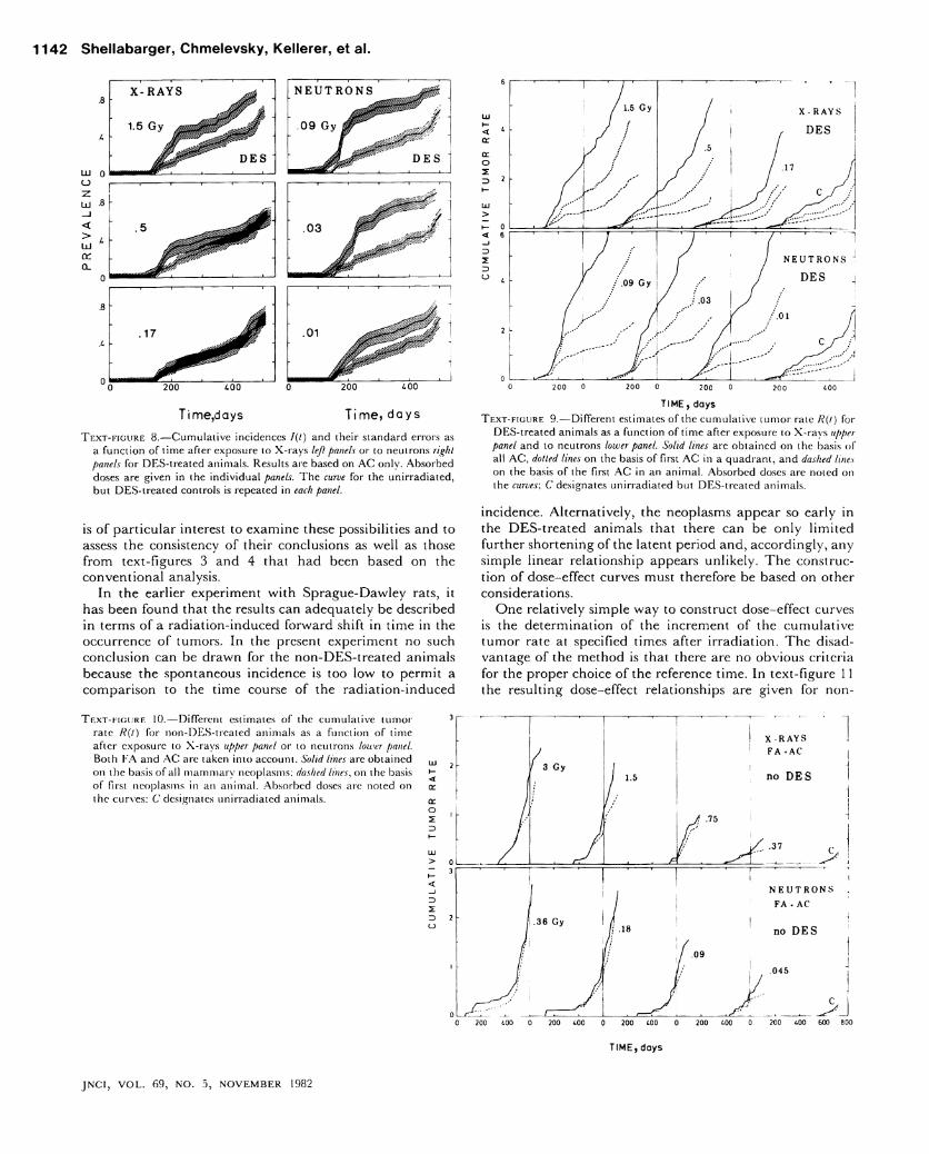
T E X T - F I G U R E 8.—Cumulative incidenccs /(/) and their Standard errors as a function of time after exposure to X-rays left panels or to neutrons r i g h t panels for DES-treated animals. Resuits are based on A C only. Absorbed doses are given in the individual panels. Th e curve for the unirradiated, but DES-treated controls is repeated in each panel.
is o f par t icular interest to examine these possibilities and to assess the consistency of their conclusions as well as those from text-figures 3 and 4 that had been based on the convent ional analysis.
In the earlier experiment wi th Sprague-Dawley rats, it has been found that the resuits can adequately be described in terms of a radiat ion-induced forward shift in t ime in the occurrence of tumors. In the present experiment no such conclusion can be drawn for the non-DES-treated animals because the spontaneous incidence is too low to permit a comparison to the time course of the radiat ion-induced
o
z>
TIME, days
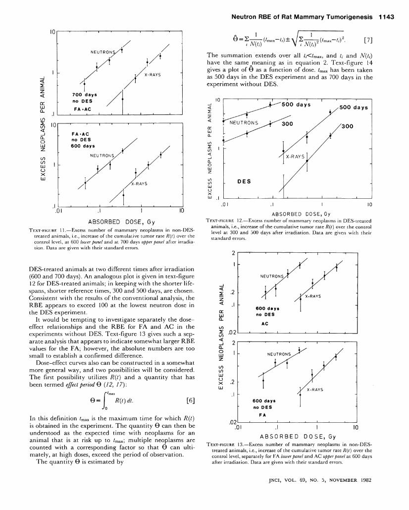
T E X T - F I G U R E 9.—Different estimates of the cumulative tumor rate R { t ) for DES-treated animals as a function of time after exposure to X-rays Upper panel and to neutrons lower panel. S o l i d lines are obtained on the basis of all A C , dotted lines on the basis of first A C in a quadrant, and dashed lines on the basis of the first A C in an animal. Absorbed doses are noted on the curves: C designates unirradiated but DES-treated animals.
incidence. Al ternat ively , the neoplasms appear so early in the DES-t rea ted animals that there can be only l imi ted further shortening of the latent period and, accordingly, any simple l inear relationship appears unl ikely. T h e construc-tion of dose-effect curves must therefore be based on other considerations.
O n e relatively simple way to construet dose-effect curves is the determination of the increment of the cumulat ive tumor rate at specified times after i r radiat ion. T h e disad-vantage of the method is that there are no obvious criteria for the proper choice of the reference time. In text-figure 11 the resulting dose-effect relationships are given for non-
T E X T - F I G U R E 10.—Different estimates of the cumulative tumor rate /?(/) for non-DES-treated animals as a function of time after exposure to X-rays Upper p a n e l or to neutrons l o w e r p a n e l . Both F A and A C are taken into account. S o l i d l i n e s are obtained on the basis of all mammary neoplasms; dashed l i n e s , on the basis of first neoplasms in an animal. Absorbed doses are noted on the curves; C designates unirradiated animals.
z>
JNCI, VOL. 69, NO. 5, NOVEMBER 1982
N e u t r o n RBE of Rat M a m m a r y T u m o r i g e n e s i s 1 1 4 3
or LÜ o_ CO 2 : CO < _ J Q_ O
00 00 LÜ O X LÜ
N E U T R O N S / ^
X - R A Y S
700 days no DES FA+AC
FA^AC no DES 600 days
.Ol 10
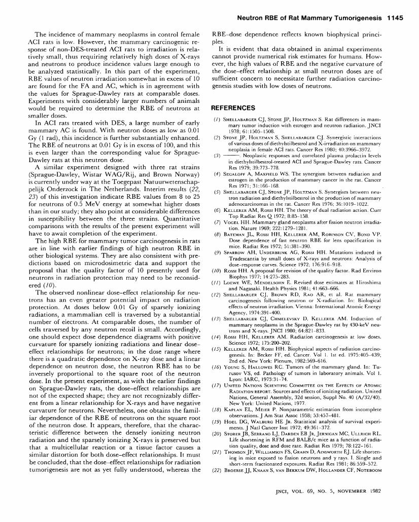
A B S O R B E D D O S E , G y T E X T - F I G U R E 11.—Excess number of mammary neoplasms in non-DES-
treated animals, i.e., increase of the cumulative tumor rate R { t ) over the control levei, at 600 lower panel and at 700 days Upper panel after irradiation. Data are given with their Standard errors.
DES-treated animals at two different times after i r radiat ion (600 and 700 days). A n analogous plot is given in text-figure 12 for DES-treated animals; in keeping wi th the shorter life-spans, shorter reference times, 300 and 500 days, are chosen. Consistent wi th the resuits of the conventional analysis, the R B E appears to exceed 100 at the lowest neutron dose in the D E S experiment.
It would be tempting to investigate separately the dose-effect relationships and the R B E for F A and A C in the experiments without D E S . Text-figure 13 gives such a separate analysis that appears to indicate somewhat larger R B E values for the F A ; however, the absolute numbers are too small to establish a confirmed difference.
Dose-effect curves also can be constructed i n a somewhat more general way, and two possibilities w i l l be considered. T h e first possibili ty utilizes R ( t ) and a quant i ty that has been termed effect period 0 ( 1 2 , 1 7 ) :
e = | R ( t ) d t . [6]
In this definition / m a x is the m a x i m u m time for wh ich R ( t ) is obtained i n the experiment. T h e quant i ty 0 can then be understood as the expected time wi th neoplasms for an an imal that is at risk up to / m a x ; mul t ip le neoplasms are counted w i t h a corresponding factor so that 0 can u l t i -mately, at h igh doses, exceed the period of Observation.
T h e quanti ty 0 is estimated by
0 = 2-N(ti)
(/n K/max-/,-)1 [7]
T h e summat ion extends over al l / i</ m a x, and Ii and N ( t i ) have the same j n e a n i n g as in equation 2. Text-f igure 14 gives a plot of 9 as a function of dose. / m a x has been taken as 500 days in the D E S experiment and as 700 days in the experiment without D E S .
10
<
or tu
Q_
0 0
CO
< _ J D_ O UJ z oo oo UJ o X UJ
1 1 - i ' ' i j ^ - ^ 5 0 0 d a y s
/ 5 0 0 d a y s
' N E U T R O N S 4 ^ 3 0 0 / / 3 0 0
-X - R A Y S 1 /
-
D E S
/ i i i t
-
. 0 1 10
A B S O R B E D D O S E , G y
T E X T - F I G U R E 12.—Excess number of mammary neoplasms in DES-treated animals, i.e., increase of the cumulative tumor rate R ( t ) over the control level at 300 and 500 days after irradiation. Data are given with their Standard errors.
<
2
or LÜ CL
CO 2 : CO < _ i Q_ O
CO CO LÜ O X LÜ
.2
600 days no DES
AC
.02
2
I
.02 .0
N E U T R O N S
i I i r
600 days no DES
FA i
.1 10
A B S O R B E D D O S E , Gy T E X T - F I G U R E 13.—Excess number of mammary neoplasms in non-DES-
treated animals, i.e., increase of the cumulative tumor rate R ( t ) over the control level, separately for F A lower panel and A C Upper panel at 600 days after irradiation. Data are given with their Standard errors.
JNCI, VOL. 69, NO. 5, NOVEMBER 1982

1 1 4 4 S h e l l a b a r g e r , C h m e l e v s k y , Ke l le re r , e t a l .
A n alternative construction of a dose-effect plot is based on a quant i ty that is s imilar to Ö but differs from it in depending on first neoplasms only. H o e l and W a l b u r g ( 1 9 ) have defined a quanti ty
i = l [ l -I ( t ) / l ( c c ) ] d t [8]
that is frequently appl ied in studies of life shortening, i.e., in the analysis of lethal diseases ( 2 0 , 2 1 ) . In these studies it is cal led "mean age at death," and I ( t ) is either the overall cumula t ive mortal i ty or the cumulat ive mortal i ty due to a par t icu lar risk. However , it is evident that the quant i ty proposed by H o e l and W a l b u r g can also be appl ied i n the analysis o f nonlethal diseases. In the present case 1(1) is the cumula t ive incidence of m a m m a r y neoplasms (see equation 5), /(oo) is set equal to 1 and, as in the determinat ion of 0, the Integration is terminated at / m a x = 5 0 0 or 700 days. T h e formula for the estimation of \k is then:
ß = H [ I ( t i ) - I ( t i - i ) ] • U± J{M[-im/N(ti)--ß2}/(n-\) [9]
(summation over a l l ti up to / m a x ; n is the number of animals w i th neoplasm). T h e Standard error in equation 9 corre-sponds to the formula given by H o e l and W a l b u r g ( 1 9 ) .
T h e symbol ju, Stands for a competing-risk-corrected mean t ime wi thout mammary neoplasm for the animals. T h e influence o f the correction is substantial, as seen from a compar ison of text-figure 15 w i th the mean tumor-free lifetimes in tables 2 and 5. T h e dose dependence of fi in text-figure 15 is yet another confirmation of the sublinearity in the D E S experiment at small neutron doses; it also confirms the h igh R B E values in excess of 100 for the D E S experiment and close to 10 for the experiment without D E S .
Text-f igure 16 gives, both for the experiments w i th and without D E S , the R B E of neutron i rradiat ion as a function of dose. These curves have been derived from the mortal i ty-corrected resuits, i.e., from text-figures 11, 12, 14, and 15. T h e curves inserted in text-figures 3 and 4 are in agreement wi th these R B E - d o s e relationships. T h e R B E - d o s e relationship from the earlier experiment w i th Sprague-Dawley rats ( 1 3 ) is inserted for comparison.
I D x , Gy
2 * 3
1600 , \ D E S ( A C ) • N E U T R O N S
o X - R A Y S
D N > G V T E X T - F I G U R E 14.—Effect p e r i o d Ö and its Standard error (see equation 7) for
DES-treated and non-DES-treated animals. Scales for the neutron (DN) and the X-ray (Dx) doses differ by a factor of 10. S o l i d l i n e s refer to neutron irradiations; d a s h e d l i n e s , to X-irradiations.
N E U T R O N S }
X - R A Y S
N '
.3
Gy T E X T - F I G U R E 15.—Mean time without tumor and its Standard error (see
equation 9) for DES-treated and non-DES-treated animals. Scales for the neutron (DN) and X-ray (Dx) doses differ by a factor of 10. S o l i d l i n e s refer to neutron irradiations; d a s h e d l i n e s , to X-irradiations.
R B E
100
. 1 1 1 i l i i
A C I R A T S ^ D E S
- S P R A G U E - D A W L E Y ^ A C I R A T S -
R A T S Z ^ L _ _ _ _ _ _ J I O D E S
1 > I
. 0 0 1 . 0 1 .1
NEUTRON DOSE, Gy T E X T - F I G U R E 16.—Dependence of the R B E of 430-kcV neutrons on dose
for the induction of mammary neoplasms. Resuits for the Sprague-Dawley rats are from an earlier study (13) (rats were not treated with DES) . Resuits for A C I rats are from the present investigation. Line segments for A C I rats correspond to the curves in lext-figs. 3, 4, 11, 12, 14, and 15.
DISCUSSION
Previous experiments ( 1 2 , 1 3 ) w i t h female Sprague-Dawley rats had led to the conclusion that the R B E of neutron i r radiat ion for the induc t ion o f m a m m a r y neoplasms ex-ceeds 100 at an absorbed dose o f 1 m G y (0.1 rad). In agreement w i t h earlier microdosimetr ic considerations ( 6 ) , it had also been found that the R B E of neutron i r radia t ion increases in inverse propor t ion to the S q u a r e root of the neutron dose. A further f ind ing was a strong sublineari ty (dose exponent ~ 0 . 5 ) o f the dose-effect relat ionship at small neutron doses. In view o f the potent ia l impl ica t ions o f such findings for radiat ion protect ion, it seemed desirable to i n q u i r e whether s imi lar resuits are ob ta ined in other strains. T h e present experiment has been designed for this purpose.
JNCI, VOL. 69, NO. 5, NOVEMBER 1982
N e u t r o n RBE o f Rat M a m m a r y T u m o r i g e n e s i s 1 1 4 5
T h e incidence of m a m m a r y neoplasms in control female A C I rats is low. However , the m a m m a r y carcinogenic response of non-DES- t rea ted A C I rats to i r radiat ion is rela-tively smal l , thus requ i r ing relatively h igh doses of X- r ays and neutrons to produce incidence values iarge enough to be analyzed statistically. In this part o f the experiment, R B E values of neutron i r rad ia t ion somewhat in excess of 10 are found for the F A a n d A C , w h i c h is in agreement w i th the values for Sprague-Dawley rats at comparable doses. Experiments w i t h considerably larger numbers o f animals wou ld be required to determine the R B E of neutrons at smaller doses.
In A C I rats treated w i t h D E S , a large number of early mammary A C is found. W i t h neutron doses as low as 0.01 G y (1 rad), this incidence is further substantial ly enhanced. T h e R B E of neutrons at 0.01 G y is i n excess of 100, and this is even larger than the corresponding value for Sprague-Dawley rats at this neutron dose.
A s imilar experiment designed w i t h three rat strains (Sprague-Dawley, Wis t a r W A G / R i j , a n d B r o w n Norway) is currently under way at the Toegepast Natuurwetenschap-pelijk Onderzock in T h e Netherlands. Inter im resuits ( 2 2 , 2 3 ) o f this investigation indicate R B E values from 8 to 25 for neutrons of 0.5 M e V energy at somewhat higher doses than in our study; they also point at considerable differences in susceptibility between the three strains. Quant i ta t ive comparisons w i t h the resuits o f the present experiment w i l l have to await comple t ion o f the experiment.
T h e high R B E for m a m m a r y tumor carcinogenesis in rats are in line w i th earlier findings of h igh neutron R B E in other biological Systems. T h e y are also consistent w i th pre-dictions based on microdosimetr ic da ta and support the proposal that the qual i ty factor of 10 presently used for neutrons in radia t ion protect ion may need to be reconsid-ered ( 1 0 ) .
T h e observed nonl inear dose-effect relationship for neutrons has an even greater potent ial impact on radiat ion protection. A t doses below 0.01 G y o f sparsely ion iz ing radiations, a m a m m a l i a n cell is traversed by a substantial number of electrons. A t comparable doses, the number of cells traversed by any neutron recoil is smal l . Accord ing ly , one should expect dose dependence diagrams wi th positive curvature for sparsely i on i z ing radiat ions and linear dose-effect relationships for neutrons; in the dose r ä n g e where there is a q u a d r a t i c dependence on X - r a y dose and a linear dependence on neutron dose, the neutron R B E has to be inversely propor t ional to the S q u a r e root o f the neutron dose. In the present experiment, as w i t h the earlier findings on Sprague-Dawley rats, the dose-effect relationships are not of the expected shape; they are not recognizably different from a linear relat ionship for X - r a y s and have negative curvature for neutrons. Nevertheless, one obtains the familiär dependence of the R B E o f neutrons on the S q u a r e root of the neutron dose. It appears, therefore, that the charac-teristic difference between the densely ion iz ing neutron radiat ion and the sparsely ion iz ing X - r a y s is preserved but that a mul t ice l lu lar reaction or a tissue factor causes a s imilar distortion for bo th dose-effect relationships. It must be concluded, that the dose-effect relationships for radiat ion tumorigenesis are not as yet fully understood, whereas the
R B E - d o s e dependence reflects known biophysical p r i nc i -ples.
It is evident that data obtained in an imal experiments cannot provide numerical risk estimates for humans. H o w ever, the high values of R B E and the negative curvature of the dose-effect relationship at small neutron doses are o f sufficient concern to necessitate further radiat ion carcinogenesis studies wi th low doses of neutrons.
REFERENCES
( / ) S H E L L A B A R G E R C J , S T O N E J P , H O L T Z M A N S. Rat differences in mam
mary tumor induction with estrogen and neutron radiation. J N C I 1978; 61:1505-1508.
(2) S T O N E J P , H O L T Z M A N S, S H E L L A B A R G E R C J . Synergistic interactions
of various doses of diethylstilbestrol and X-irradiation on mammary neoplasia in female A C I rats. Cancer Res 1980; 40:3966-3972.
(3) . Neoplastic responses and corrclated plasma prolactin levels in diethylstilbestrol-treated A C I and Sprague-Dawley rats. Cancer Res 1979; 39:773-778.
(4) S E G A L O F F A , M A X F I E L D W S . The synergism between radiation and estrogen in the production of mammary cancer in the rat. Cancer Res 1971; 31:166-168.
(5) S H E L L A B A R G E R C J , S T O N E J P , H O L T Z M A N S. Synergism between neu
tron radiation and diethylstilbestrol in the production of mammary adenocarcinomas in the rat. Cancer Res 1976; 36:1019-1022.
(6) K E L L E R E R A M , Rossi H H . The theory of dual radiation action. C u r r T o p Radial Res Q 1972; 8:85-158.
(7) V O G E L H H . Mammary gland neoplasms after fission neutron irradiation. Nature 1969; 222:1279-1281.
(8) B A T E M A N J L , Rossi H H , K E L L E R E R A M , R O B I N S O N C V , B O N D V P .
Dose dependence of fast neutron R B E for lens opacification in mice. Radiat Res 1972; 51:381-390.
(9) S P A R R O W A H , U N D E R B R I N K A G , Rossi H H . Mutations induced in Tradescantia by small doses of X-rays and neutrons: Analysis of dose-response curves. Science 1972; 176:916-918.
(10) Rossi H H . A proposal for revision of the quality factor. Rad Environ Biophys 1977; 14:275-283.
( / / ) L O E W E W E , M E N D E L S O H N E . Revised dose estimates at Hiroshima and Nagasaki. Health Physics 1981; 41:663-666.
( 1 2 ) S H E L L A B A R G E R C J , B R O W N R D , R A O A R , et al. Rat mammary
carcinogenesis following neutron or X-radiation. In: Biological effects of neutron irradiation. Vienna: International Atomic Energy Agency, 1974:391-400.
( 1 3 ) S H E L L A B A R G E R C J , C H M E L E V S K Y D, K E L L E R E R A M . Induction of
mammary neoplasms in the Sprague-Dawley rat by 430-keV neutrons and X-rays. J N C I 1980; 64:821-833.
(14) Rossi H H , K E L L E R E R A M . Radiation carcinogenesis at low doses. Science 1972; 175:200-202.
(15) K E L L E R E R A M , Rossi H H . Biophysical aspects of radiation carcinogenesis. In: Becker F F , ed. Cancer. Vol 1. Ist ed. 1975:405-439; 2nd ed. New York: Plenum, 1982:569-616.
(16) Y O U N G S, H A L L O W E S R C . Tumors of the mammary gland. In: T u -rusov V S , ed. Pathology of tumors in laboratory animals. V o l 1. Lyon: I A R C , 1973:31-74.
( 1 7 ) U N I T E D N A T I O N S SCIENTIFIC C O M M I T T E E ON T H E E F F E C T S O F A T O M I C
R A D I A T I O N R E P O R T . Sources and effects of ionizing radiation. United Nations, General Assembly, 32d session, Suppl No. 40 (A/32/40). New York: United Nations, 1977.
(18) K A P L A N E L , M E I E R P. Nonparametric estimation from incomplete observations. J A m Stat Assoc 1958; 53:457-481.
(19) H O E L D G , W A L B U R G H E J R . Statistical analysis of survival experiments. J Natl Cancer Inst 1972; 49:361-372.
( 2 0 ) S T O R E R J B , S E R R A N O L J , D A R D E N E B J R , J E R N I G A N M C , U L L R I C H R L .
Life shortening in R F M and B A L B / c mice as a function of radiation quality, dose and dose rate. Radiat Res 1979; 78:122-161.
( 2 1 ) T H O M S O N J F , W I L L I A M S O N FS, G R A H N D , A I N S W O R T H E J . Life shorten
ing in mice exposed to fission neutrons and y rays. I. Single and short-term fractionated exposures. Radiat Res 1981; 86:559-572.
( 2 2 ) B R O E R S E J J , K N A A N S, V A N B E R K U M D W , H O L L A N D E R C F , N O T E B O O M
JNCI, VOL. 69, NO. 5, NOVEMBER 1982
1 1 4 6 She l l aba rge r , C h m e l e v s k y , Ke l le re r , e t a l .
A L , V A N Z W I E T E N M J . Mammary carcinogenesis in rats after X -and neutron irradiation and hormone administration. In: Proceed-ings of the international S y m p o s i u m on the late biological effects of ionizing radiation. V o l 2. Vienna: International Atomic Energy Agency, 1978:13-27.
( 2 3 ) V A N B E R K U M D W , B R O E R S E J J , V A N Z W I E T E N M J , H O L L Ä N D E R C K ,
B L A N K E N S T E I N M A . Radiation-induced mammary cancer in the rat. Okada S, Imamura H , Terashima T , Yamaguchi H , eds. Procecd-ings of the sixth international congress on radiation research, Tokyo, M a y 13-19. Tokyo: Toppan, 1979:734-752.
JNCI, VOL. 69, NO. 5, NOVEMBER 1982