Embed Size (px)
Citation preview
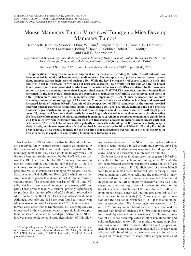
MOLECULAR AND CELLULAR BIOLOGY, Aug. 2003, p. 5738–5754 Vol. 23, No. 160270-7306/03/$08.00�0 DOI: 10.1128/MCB.23.16.5738–5754.2003Copyright © 2003, American Society for Microbiology. All Rights Reserved.
Mouse Mammary Tumor Virus c-rel Transgenic Mice DevelopMammary Tumors
Raphaelle Romieu-Mourez,1 Dong W. Kim,1 Sang Min Shin,1 Elizabeth G. Demicco,1Esther Landesman-Bollag,2 David C. Seldin,2 Robert D. Cardiff,3
and Gail E. Sonenshein1*Departments of Biochemistry1 and Medicine,2 Boston University Medical School, Boston, Massachusetts 02118, and
UCD Center for Comparative Medicine, University of California, Davis, California 956163
Received 11 November 2002/Returned for modification 20 February 2003/Accepted 20 May 2003
Amplification, overexpression, or rearrangement of the c-rel gene, encoding the c-Rel NF-�B subunit, hasbeen reported in solid and hematopoietic malignancies. For example, many primary human breast cancertissue samples express high levels of nuclear c-Rel. While the Rev-T oncogene v-rel causes tumors in birds, theability of c-Rel to transform in vivo has not been demonstrated. To directly test the role of c-Rel in breasttumorigenesis, mice were generated in which overexpression of mouse c-rel cDNA was driven by the hormone-responsive mouse mammary tumor virus long terminal repeat (MMTV-LTR) promoter, and four founder linesidentified. In the first cycle of pregnancy, the expression of transgenic c-rel mRNA was observed, and levels ofc-Rel protein were increased in the mammary gland. Importantly, 31.6% of mice developed one or moremammary tumors at an average age of 19.9 months. Mammary tumors were of diverse histology and expressedincreased levels of nuclear NF-�B. Analysis of the composition of NF-�B complexes in the tumors revealedaberrant nuclear expression of multiple subunits, including c-Rel, p50, p52, RelA, RelB, and the Bcl-3 protein,as observed previously in human primary breast cancers. Expression of the cancer-related NF-�B target genescyclin D1, c-myc, and bcl-xl was significantly increased in grossly normal transgenic mammary glands startingthe first cycle of pregnancy and increased further in mammary carcinomas compared to mammary glands fromwild-type mice or virgin transgenic mice. In transient transfection analysis in untransformed breast epithelialcells, c-Rel-p52 or -p50 heterodimers either potently or modestly induced cyclin D1 promoter activity, respec-tively. Lastly, stable overexpression of c-Rel resulted in increased cyclin D1 and NF-�B p52 and p50 subunitprotein levels. These results indicate for the first time that dysregulated expression of c-Rel, as observed inbreast cancers, is capable of contributing to mammary tumorigenesis.
Nuclear factor (NF)-�B/Rel is a structurally and evolution-ary conserved family of transcription factors distinguished bythe presence of a 300 amino acid region, termed the Relhomology domain (RHD), based on its homology with v-Rel,the transforming protein encoded by the RevT avian retrovi-rus. The RHD is responsible for DNA-binding, dimerization,nuclear translocation, and binding of Rel factors to the I�Binhibitory proteins (reviewed in reference 21). Mammals ex-press five NF-�B members that belong to two classes. The firstclass includes c-Rel, RelB, and RelA (p65), which are synthe-sized as mature products and contain a C-terminal transacti-vation domain. The second class consists of NF-�B1 and NF-�B2, which are synthesized as longer precursors, p105 andp100. Those proteins require C-terminal proteolytic processingto produce the mature p50 and p52 subunits, respectively,which contain the RHD but lack a transactivation domain.Although, both p50 and p52 have been found to transactivatewhen in association with Bcl-3 protein (7, 20). In most untrans-formed cells, other than B lymphocytes, NF-�B complexes aresequestered in the cytoplasm bound to specific inhibitory pro-teins, of which I�B-� is the paradigm. Activation of NF-�Binvolves phosphorylation and rapid degradation of I�B, allow-
ing for translocation of free NF-�B to the nucleus, where itcontrols genes involved in cell growth and survival, adhesion,and immune and inflammatory responses, including cyclin D1,c-myc, and bcl-xl (reviewed in references 47 and 56).
Evidence from several laboratories has suggested NF-�B iscritically involved in regulation of tumorigenesis. We and oth-ers demonstrated aberrant constitutive activation of NF-�Bfactors in breast cancer (43, 59). High levels of nuclear NF-�Bwere found in human breast tumor cell lines, carcinogen-trans-formed mammary epithelial cells, and the majority of primaryhuman and rodent breast tumor tissue samples. Accelerateddegradation of the I�B-� inhibitory protein was observed (34),suggesting aberrant regulation of nuclear translocation inbreast cancer cells. Inhibition of the constitutive NF-�B activ-ity in human breast cancer cell lines induced apoptosis (59) orled to reduced tumorigenicity (50). Conversely, ectopic expres-sion of c-Rel resulted in resistance to TGF-�-mediated inhibi-tion of proliferation (58). Interestingly, we observed that 21out of 25 primary human breast cancer tissues examined ex-pressed high levels of nuclear c-Rel (59); similar observationswere made by Cogswell and coworkers (15). The overexpres-sion of c-Rel has been implicated in other hematopoietic andsolid malignancies as well. For example, c-rel gene amplifica-tion was seen in �20% of non-Hodgkin’s B-cell lymphomas,including diffuse large-B-cell lymphomas (DBCL) (reviewed inreference 47). In addition, the c-rel gene was also found rear-ranged or overexpressed in some follicular lymphomas and
* Corresponding author. Mailing address: Department of Biochem-istry, Boston University School of Medicine, 715 Albany St., Boston,MA 02118. Phone: (617) 638-4120. Fax: (617) 638-4252. E-mail:[email protected].
5738
Dow
nloa
ded
from
http
s://j
ourn
als.
asm
.org
/jour
nal/m
cb o
n 20
Oct
ober
202
1 by
61.
37.1
99.2
29.
DBCL. The higher level of expression of c-Rel, plus RelA, inthe activated B-cell (ABC) form of DBCL, was found associ-ated with poorer prognosis (2). Furthermore, inhibition ofc-Rel induced apoptosis in immature B-cell lymphomas (66).
The c-rel gene encodes a 68-kDa protein which is activemostly in lymphocytes and monocytic, granulocytic, and ery-throid cells. Mice lacking c-rel are viable but show severelyimpaired lymphocyte proliferation and immune function, withimpaired interleukin-2 expression (36). The X-ray crystal struc-ture of the c-Rel homodimer bound to a DNA target site wasresolved recently (28). It confirmed that c-Rel homodimersrecognize a different set of �B element DNA sequences com-pared to c-Rel heterodimers or p50-containing dimers, sug-gesting that those complexes may have a different range oftarget genes. The v-rel gene, carried by the highly oncogenicavian reticuloendotheliosis virus strain T (Rev-T), is able tocause tumors in birds and transgenic animal models. The v-Reloncogenic protein differs from its avian progenitor c-Rel by thepresence of multiple mutations that increases its expression,nuclear localization, DNA binding, transactivation capabilityand neoplastic transformation potential (reviewed in reference22). The direct role of c-Rel in tumorigenesis is still a matter ofdebate. Overexpression of either avian or human c-Rel wasshown to transform primary chicken lymphoid cells (1, 23),although with lower efficiency than v-Rel. Retroviral deliveryof avian c-rel in young chickens gave rise to lymphoid tumors,and cell transformation involved selection of a C-terminal de-leted c-Rel protein with increased oncogenic activity (27). Allthese observations led us to test the oncogenic properties ofc-Rel in vivo, in the mammary gland. Here, we have con-structed a mouse model in which the mouse c-rel cDNA wasdriven by the hormone-dependent mouse mammary tumorvirus (MMTV) promoter. MMTV–c-rel female mice developedlate-onset mammary tumors, which were related to increasedlevels of expression of the NF-�B growth and survival targetgenes cyclin D1, c-myc, and bcl-xl. These results provide thefirst in vivo evidence for a causal role of c-Rel activation in thepathogenesis of breast cancer.
MATERIALS AND METHODS
Isolation of founder MMTV–c-rel transgenic mice. The 2.5-kb EcoRI/HindIIIfragment from the pSVSport-c-Rel vector, containing the full-length murine c-relcDNA (kindly provided by T. Gilmore, Boston University, Boston, Mass.), wasblunt end ligated into the MMTV-LTR plasmid, containing the MMTV longterminal repeat (MMTV-LTR), which directs expression chiefly to the mammaryepithelium, with ras 5� untranslated sequences provided upstream of the cDNAand a simian virus 40 (SV40) intron and polyadenylation signal downstream (38,55), yielding the pMMTV–c-rel plasmid. The direction of the insert was con-firmed by restriction mapping and DNA sequencing at the Molecular BiologyCore at Boston University Medical School. Plasmid sequences were removed byrestriction digestion at the SalI and SpeI sites, leaving the MMTV-LTR se-quence, ras 5� untranslated sequences, c-rel cDNA, and the SV40 intron andpoly(A) addition signal sequence. The excised transgene construct was gel pu-rified and microinjected into pronuclei of fertilized one-cell zygotes from FVB/Nmice. These zygotes were reimplanted into pseudopregnant foster mothers, andthe offspring were screened for presence of the transgene by Southern blotting(see below). Carriers were bred to establish four independent transgenic lines.Female transgenic mice were continuously bred to induce transgene expressionthough activation of the hormone-dependent MMTV-LTR promoter. Mice weremonitored biweekly for the appearance of tumors. Mice were housed in atwo-way barrier at the Boston University School of Medicine Transgenic mousefacility in accordance with the regulations of the American Association for theAccreditation of Laboratory Animal Care.
DNA analysis. Genomic tail DNA was isolated as described previously (46),samples (10 �g) were digested with PstI, and the resulting fragments wereseparated on a 0.8% agarose gel and transferred to GeneScreenPlus (DuPontNEN) nylon membranes. DNA was cross-linked to the membrane by UV irra-diation (Stratalinker; Stratagene, La Jolla, Calif.) at 0.12 J/cm2 for 30 s. Hybrid-ization was performed using the 2.2-kb DNA fragment, encompassing theMMTV-LTR promoter and an �1-kb fragment of the mouse c-rel cDNA, re-leased from the pMMTV–c-rel plasmid by digestion with PstI as a probe. TheDNA was radiolabeled by random priming and used as described previously (35),except that 2.0 � 106 cpm/ml 32P-labeled DNA was employed.
RNA analyses. Frozen breast tissue was pulverized in liquid nitrogen with amortar and pestle, and total RNA extracted with the Ultraspec-II RNA IsolationSystem (Biotecx Laboratories Inc., Houston, Tex.). To remove contaminatingDNA, RNA samples were digested for 30 min at 37°C with RQ1 RNase freeDNase (Promega Corporation, Madison, Wis.), according to the manufacturer’sdirections. RNA was reextracted and ethanol precipitated. For reverse transcrip-tase PCR (RT-PCR), 5 �g RNA samples were reverse transcribed with SUPER-SCRIPT RNase H-RT in the presence of 200 ng of random primers (all reagentsfrom Invitrogen Life Technologies, Carlsbad, Calif.). For PCR, a 236-bp frag-ment of the transgene was amplified with a sense oligonucleotide primer fromthe mouse c-rel cDNA coding sequence (5�-GTGACCCCTAAGGGTTTTCTG-3�) and an antisense oligonucleotide from the SV40 poly(A) tail of the vectorconstruct (5�-CCCATTCATAAGTTCCATAG-3�). PCR were performed in athermal cycler (MJ Research, Watertown, Mass.) by denaturing at 95°C for 45 s,annealing at 46°C for 90 s, and extending at 72°C for 90 s for 35 cycles. Primerpairs specific for the mouse cyclin D1 gene were 5�-CACAACGCACTTTCTTTCCA-3� and 5�-GACCAGCCTCTTCCTCCAC-3�, and amplified a 164-bp frag-ment with an annealing temperature of 55°C. The primer pairs specific for mousebcl-xl were as described previously (52). As a control for RNA quality, a 750-bpfragment of �-actin mRNA was amplified by 25 or 30 PCR cycles with thefollowing primers: 5�-ACCAGTTCGCCATGGATGACGATA-3� and 5�-AGCTCATAGCTCTTCTCCAGGGAG-3�, which were used with an annealing tem-perature of 55°C. For radiolabeled PCR, 0.85 �Ci of [�-32P]dCTP and[�-32P]dGTP were added in the PCR. For RNase protection assays (RPAs),multiprobe RPA kits (Pharmingen, San Diego, Calif.) were used according to themanufacturer’s directions. The identity of the RNase protected bands in the gelwere established using the undigested probes as markers and a control RNA formouse cyclin or apoptosis gene mRNA expression (Pharmingen). For Northernblot analysis, RNA samples (5 to 15 �g) were denatured and separated byelectrophoresis in 1.0% formaldehyde agarose gels (16). Probes included a 2.4-kbpM-c-myc54 mouse c-myc cDNA (60), the full-length human cyclin D1 cDNAsubcloned into the pBPST-R1 vector, kindly provided by R. G. Pestell (AlbertEinstein College of Medicine, New York, N.Y.), and a 750-bp fragment of themouse bcl-x cDNA amplified by PCR using the previously described primers(52). Quantitation by scanning densitometry was performed with a KDS1Ddevice (version 2.0; Kodak, New Haven, Conn.).
EMSA. Frozen tissue powders were resuspended in homogenization buffer (1g/ml) in 10 mM HEPES (pH 7.9), 10 mM KCl, 0.1 mM EDTA, 0.1 mM EGTA,50 mM sucrose, 1 mM dithiothreitol (DTT), 0.5 mM phenylmethylsulfonyl flu-oride (PMSF), leupeptin (5 �g/ml), and aprotinin (5 �g/ml) (Sigma ChemicalCo., St. Louis, Mo.). Samples were Dounce homogenized for 20 strokes with aloose pestle and then 20 strokes with a tight pestle. The KCl concentration wasthen adjusted to 100 mM and the nuclei were washed twice with the homogeni-zation buffer with 100 mM KCl. Nuclear proteins were extracted on ice for 30min in 2 packed nuclear volumes containing 10 mM HEPES (pH 7.9), 400 mMNaCl, 0.1 mM EDTA, 0.1 mM EGTA, 20% glycerol, 1 mM DTT, 0.5 mM PMSF,leupeptin (0.5 �g/ml), and aprotinin (5 �g/ml). Protein concentration was de-termined using the Bio-Rad protein assay (Bio-Rad Laboratories, Hercules,Calif.). The sequence of the URE NF-�B-containing oligonucleotide from thec-myc gene is as follows: 5�-GATCCAAGTCCGGGTTTTCCCCAACC-3� (17).The core element is underlined. The Octomer-1 (Oct-1) oligonucleotide has thefollowing sequence: 5�-TGTCGAATGCAAATCACTAGAA-3�. Nuclear ex-tracts samples (5 �g) were subjected to electrophoretic mobility shift analysis(EMSA) as described previously (59). For antibody supershift analysis, the bind-ing reaction was performed in the absence of the probe, the appropriate antibodywas added and the mixture incubated for 16 h at 4°C. The probe was then addedand the reaction incubated an additional 30 min at 25°C and the complexesresolved by gel electrophoresis, as above. Antibodies used included anti-RelA(C-20), sc-372; anti-c-Rel (N), sc-70; and anti-p50 (NLS), sc-114 (all from SantaCruz Biotechnology, Santa Cruz, Calif.). In addition, rabbit polysera 1266, 1050,and 1051 specific for c-Rel were kindly provided by N. Rice and M. Ernst(National Cancer Institute, Frederick Md.). Data were quantified by densitom-etry using a Molecular Dynamics densitometer.
VOL. 23, 2003 c-Rel AND MAMMARY GLAND TUMORIGENESIS 5739
Dow
nloa
ded
from
http
s://j
ourn
als.
asm
.org
/jour
nal/m
cb o
n 20
Oct
ober
202
1 by
61.
37.1
99.2
29.
Cell lines and transfection conditions. NMuMG, which is an untransformed,immortalized mouse mammary epithelial cell line, was cultured as describedpreviously (57). MCF-10F is a human mammary epithelial cell line establishedfrom a patient with fibrocystic disease, which does not display malignant char-acteristics (9). The RelA and p50 expression vectors have been described else-where (39). The p52 and Bcl-3 expression vectors were kindly provided by U.Siebenlist (National Institutes of Health, Bethesda, Md.). The cyclin D1 pro-moter constructs CD1 66 WT-Luc and CD1 66 Mut-Luc, with wild-type(WT) and mutant NF-�B elements, respectively, were a kind gift of R. G. Pestell(25). For luciferase assays, NMuMG cells were transfected in six-well platesusing the Fugene reagent (Roche Diagnostics Corporation, Indianapolis, Ind.),and the SV40 promoter–�-galactosidase (pSV40–�-gal) reporter vector was usedto normalize transfection efficiency, as previously described (3). For stable trans-fectants, MCF-10F cells were transfected in P100 dishes with 10 �g of pBlue-script or pSVSport-c-Rel vector along with 1 �g of pGKpuro selection plasmid,selected with puromycin (4 �g/ml; Sigma) for 4 days, and then grown in thepresence of puromycin (1 �g/ml).
Immunoblot analysis. Whole-cell extracts (WCEs) were prepared in RIPAbuffer (10 mM Tris [pH 7.5], 150 mM NaCl, 10 mM EDTA, 1% NP-40, 0.1%SDS, 1% sodium sarcosyl, 0.2 mM PMSF, leupeptin [10 �g/ml], 1 mM DTT).Nuclear extracts were prepared as described above. Samples (40 �g) were sep-arated by electrophoresis in 8% polyacrylamide–SDS gels, transferred to a 0.45-�m-pore-size polyvinylidene difluoride membrane (Millipore, Bedford, Mass.)and subjected to immunoblotting, as described (59). Antibodies included anti-RelB (C-19) sc-226, anti-p52 (K-27) sc-298; Bcl-3 (C-14) sc-185, and Sp1 (PEP 2)sc-59 (all from Santa Cruz Biotechnology) and anti-cyclin D1 monoclonal Ab-3(Oncogene, Boston, Mass.). Antibodies specific for other NF-�B subunits wereas described above.
Histology. Upon necropsy, tumors and other mammary glands, heart, lung,liver, kidney, spleen, and the adrenal gland were removed and immediately fixedin Optimal Fix (American Histology Reagent Co., Lodi, Calif.) and shipped inalcohol. The tissues were processed, embedded in paraffin, and sectioned at athickness of 7 �m. The sections were mounted on glass slides and stained withhematoxylin and eosin using routine laboratory procedures in the TransgenicCore Pathology Laboratory at the University of California, Davis. Sections werecompared with other specimens in the extensive mouse mammary tumor data-base (http://www-mp.ucdavis.edu/tgmice/firststop.html).
RESULTS
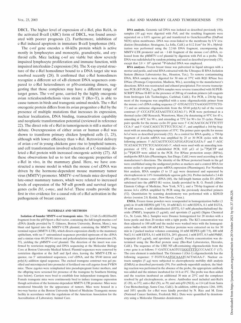
Generation and characterization of MMTV–c-rel transgenicmice. To determine the role of c-Rel in mammary tumorigen-esis, we generated a mouse model where c-rel cDNA wasexpressed under the control of the MMTV-LTR promoter, aneffective vector for expressing oncogenes and for transformingthe mammary epithelium (11, 61). A construct containing thefull-length mouse c-rel cDNA was inserted in a plasmid con-taining the MMTV-LTR promoter and an SV40 T antigenintron-poly(A) cassette to ensure efficient expression in vivo.The insert c-rel DNA was then utilized to generate transgenicmice in the FVB/N mouse strain. Integration of the constructinto the genome of potential founders was assessed by South-ern blot analysis of tail DNA with c-rel cDNA as a probe (Fig.1A and data not shown). Five founders successfully passed thetransgene through the germ line. Founder line 18 had approx-imately two to three copies of the transgene, while lines 7, 14,and 15 had four to five copies and line 16 had approximatelynine copies, as estimated by comparison with bands resultingfrom hybridization with the endogenous gene on the Southernblots. MMTV–c-rel transgenic mice of all founders developedand bred normally. Transgenic females were able to nurse theirpups.
To characterize the expression pattern of the c-rel transgene,line 14 MMTV–c-rel mice or WT FVB/N mice, as control, werebred to activate the MMTV-LTR promoter, which containshormonally responsive elements activated by progestins and
corticosteroids. At day 18.5 of the first pregnancy, total RNAwas isolated from the mammary glands and various other or-gans. RNA samples were subjected to a radiolabeled trans-gene-specific RT-PCR assay, performed with a 5� mouse c-relcDNA sense primer and a 3� SV40 poly(A) antisense primer(Fig. 1B). Expression of c-rel transgene mRNA was observed inthe mammary gland of line 14, but not the WT mouse, asexpected (Fig. 1B, left panel). RNA quality and equal loadingwas confirmed by analysis of �-actin mRNA expression profilesby RT-PCR (Fig. 1B, bottom panel), as well as in ethidiumbromide-stained gels (data not shown). At day 18.5 of preg-
FIG. 1. MMTV-LTR-driven c-rel transgene expression in FVB/Nmice. (A) Identification of founder lines. Genomic tail DNA wasprepared from the indicated potential founders MMTV–c-rel trans-genic mice, and samples (10 �g) digested with PstI and subjected toSouthern blot analysis for c-rel using the 2.2-kb fragment, encompass-ing the MMTV-LTR promoter and an �1-kb fragment of mouse c-relcDNA, released from the MMTV–c-rel plasmid digested with PstI, asa probe. The positions of the bands derived from the c-rel transgeneand the endogenous c-rel gene are as indicated. (B) Transgenic c-relexpression. Total RNA was isolated from the indicated organs of WTFVB/N or line14 MMTV–c-rel mice at day 18.5 of the first pregnancy,and subjected to DNase treatment. Samples (5 �g) were subjected toRT-PCR analysis, in the presence (�) or absence () of RT to controlfor DNA contamination, using c-rel transgene-specific oligonucleo-tides, amplifying a 236-bp fragment. Similar analysis of �-actin RNAlevels confirmed the integrity of the reverse transcription reaction.(C) Total c-Rel expression. Mammary glands were removed from WTFVB/N or the indicated transgenic line mice at day 18.5 of the firstpregnancy. Nuclear extract were prepared, and samples (20 �g) sub-jected to immunoblot analysis of c-Rel, and Sp1, as control for loading.As additional controls, nuclear extracts and WCEs from the WEHI 231immature B-lymphoma cells, which express high constitutive levels ofc-Rel (40), were similarly analyzed. The values of c-Rel normalized toSp1 level relative to the WT sample are displayed below.
5740 ROMIEU-MOUREZ ET AL. MOL. CELL. BIOL.
Dow
nloa
ded
from
http
s://j
ourn
als.
asm
.org
/jour
nal/m
cb o
n 20
Oct
ober
202
1 by
61.
37.1
99.2
29.
nancy, expression of transgene c-rel mRNA was also observedin the spleen, salivary gland, and intestine of mice of line 14(Fig. 1B) and line 16 (data not shown), while undetectable orlow levels were observed in the kidney and liver (Fig. 1B) andheart and lung (data not shown). Where indicated, reactionswere performed in the absence of RT, which confirmed theabsence of DNA contamination. Thus, the c-rel transgenemRNA is expressed mostly in glandular organs and lymphoidtissues, which is consistent with previous studies with theMMTV-LTR promoter (38, 55).
We next sought to determine total levels of c-Rel proteinexpression, which includes both endogenous and transgenicc-Rel. At day 18.5 of the first pregnancy, nuclear extracts wereprepared from mammary glands of transgenic lines 7, 14, 15,16, and 18 mice, and from a WT FVB/N mouse as control.Samples were subjected to immunoblot analysis for c-Rel andSp1 to normalize for loading (Fig. 1C). Nuclei from the WTmammary gland contained basal levels of c-Rel, as has beenreported recently (10). All of the transgenic lines displayedhigher normalized levels of nuclear c-Rel. The lowest level wasseen in line 18, consistent with its low transgene copy number,as seen above; therefore, the other four lines (i.e., lines 7, 14,15, and 16) were chosen for further study.
MMTV–c-rel transgenic mice develop late-onset mammarycarcinomas. To promote c-rel transgene overexpression,MMTV–c-rel female mice were continuously bred to inducethe MMTV-LTR promoter. A cohort of 38 multiparous femalemice from the 4 expressing lines was monitored for tumorincidence over 2 years. Mice were subjected to biweekly pal-pable examination, and when the presence of a tumor wasdetected, the mammary glands and other organs were sub-jected to histopathological analysis. Thirty-one percent of themice developed mammary carcinomas at an average age of19.9 months (Table 1). In contrast, mammary tumors developwith a very low incidence (1%) in WT female FVB/N micethat have been similarly bred (38). An identification number,with the origin of the line was attributed to each tumor, andcharacteristics of the different breast tumors are described inthe Table 1. Mice from each of the four transgenic lines de-
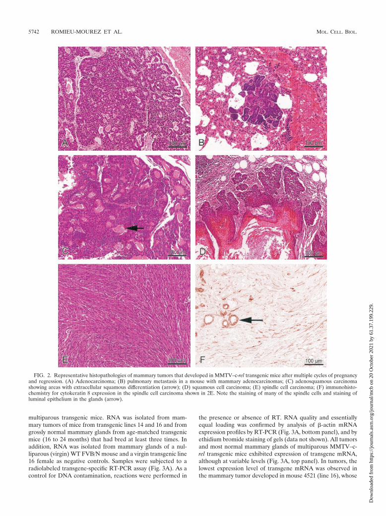
veloped mammary carcinomas. The tumor incidence was 33.3,41.7, 20, and 33.3% in MMTV–c-rel transgenic lines 7, 14, 15,and 16, respectively (data not shown), suggesting that tumordevelopment is related to c-rel transgene expression ratherthan random insertional events. In all but one case, the tumorsarose as solitary masses in a single mammary gland. Of the 12mammary tumors, 3 were pure adenocarcinomas (Fig. 2A), 3were adenosquamous carcinomas (Fig. 2C), 4 were squamouscell carcinomas (Fig. 2D), 1 was classified as a papillary ade-nocarcinoma, and 1 was a spindle cell carcinoma (Fig. 2E).Spindle cell carcinomas are often related to an epithelial tomesenchymal cell transition (EMT), which is the transforma-tion of epithelial cells into cells with features of mesenchymalcells, favoring the progression of a carcinoma towards a dedif-ferentiated and more malignant state (reviewed in reference63). To test for cells of epithelial origin, immunohistologicalstaining was carried out using antibodies specific for the epi-thelial cell marker cytokeratin 8 (Fig. 2F). The spindle cellcarcinoma stained positively for cytokeratin 8, consistent withan EMT tumor. One of the adenocarcinomas was metastatic tothe lung (Fig. 2B).
Lastly, mammary glands of three other multiparous 2-year-old transgenic mice, which were not included in the cohort,were subjected to histopathological analysis even without thepresence of a palpable tumor. This examination revealed thepresence of an adenosquamous carcinoma and a mammarysquamous cell carcinoma in two of the mice (data not shown).In addition to the tumors, poor regression of the alveolar treeof the mammary gland after pregnancy was another histolog-ical abnormality that was frequently seen (data not shown).Therefore, the histology of mammary tumors in MMTV–c-relmice appears variable, suggesting changes in mammary epithe-lial cells during c-Rel-induced tumorigenesis.
c-Rel expression is elevated in mammary glands and tumorsof MMTV–c-rel transgenic mice. Previous studies showed thatthe MMTV-LTR promoter is still active in regressing mam-mary glands, leading to sustained transgene expression overthe animal’s lifetime (38, 55). We therefore investigated trans-genic c-rel expression in mammary glands and tumors from
TABLE 1. Tumor incidence and histopathology in female MMTV–c-rel transgenic mice after multiple cycles of pregnancy and regressiona
Mam. tumor diagnosis (other tumors)b Agec
(mo)
No. of:Line Mouse
no.dMam. tumors Other tumors
Mam. squamous cell carcinoma and hyperplasia (bronchial adenocarcinoma) 22 1 1 7 4026Mam. adenosquamous carcinoma 22 1 0 7 4042Mam. adenosquamous carcinoma 18 1 0 14 3814Mam. adenosquamous carcinoma (papillary bronchial adenomas) 20 1 1 14 3983Mam. adenocarcinomas with pulmonary metastases 23 4 1 14 3996Mam. papillary carcinoma, lobular hyperplasia, squamous nodules 23 1 0 14 4936Mam. squamous cell carcinoma and hyperplasia 15 1 0 14 4556Mam. adenocarcinoma (papillary bronchial adenocarcinomas) 19 1 1 15 127Mam. squamous cell carcinoma 22 1 0 16 3872Mam. adenocarcinoma with poorly differentiated large cells 18 1 0 16 3875Mam. squamous cell carcinoma and hyperplasia (centrocytic lymphoma) 18 1 1 16 4521Spindle cell tumor, originating from a mam. adenocarcinoma 19 1 0 16 4528
a Thirty-eight multiparous female mice from the four transgenic lines (7, 14, 15, and 16) were monitored for tumor incidence by biweekly palpable examination.b Histopathological analysis of mammary (mam.) glands and other organs (heart, lung, liver, kidney, spleen, and adrenal gland) from the same animal was performed
when a tumor was detected.c Age when tumor was detected.d An identification number was given to the individual mice of the cohort for further analysis of the tumors.
VOL. 23, 2003 c-Rel AND MAMMARY GLAND TUMORIGENESIS 5741
Dow
nloa
ded
from
http
s://j
ourn
als.
asm
.org
/jour
nal/m
cb o
n 20
Oct
ober
202
1 by
61.
37.1
99.2
29.
multiparous transgenic mice. RNA was isolated from mam-mary tumors of mice from transgenic lines 14 and 16 and fromgrossly normal mammary glands from age-matched transgenicmice (16 to 24 months) that had bred at least three times. Inaddition, RNA was isolated from mammary glands of a nul-liparous (virgin) WT FVB/N mouse and a virgin transgenic line16 female as negative controls. Samples were subjected to aradiolabeled transgene-specific RT-PCR assay (Fig. 3A). As acontrol for DNA contamination, reactions were performed in
the presence or absence of RT. RNA quality and essentiallyequal loading was confirmed by analysis of �-actin mRNAexpression profiles by RT-PCR (Fig. 3A, bottom panel), and byethidium bromide staining of gels (data not shown). All tumorsand most normal mammary glands of multiparous MMTV–c-rel transgenic mice exhibited expression of transgene mRNA,although at variable levels (Fig. 3A, top panel). In tumors, thelowest expression level of transgene mRNA was observed inthe mammary tumor developed in mouse 4521 (line 16), whose
FIG. 2. Representative histopathologies of mammary tumors that developed in MMTV–c-rel transgenic mice after multiple cycles of pregnancyand regression. (A) Adenocarcinoma; (B) pulmonary metastasis in a mouse with mammary adenocarcinomas; (C) adenosquamous carcinomashowing areas with extracellular squamous differentiation (arrow); (D) squamous cell carcinoma; (E) spindle cell carcinoma; (F) immunohisto-chemistry for cytokeratin 8 expression in the spindle cell carcinoma shown in 2E. Note the staining of many of the spindle cells and staining ofluminal epithelium in the glands (arrow).
5742 ROMIEU-MOUREZ ET AL. MOL. CELL. BIOL.
Dow
nloa
ded
from
http
s://j
ourn
als.
asm
.org
/jour
nal/m
cb o
n 20
Oct
ober
202
1 by
61.
37.1
99.2
29.
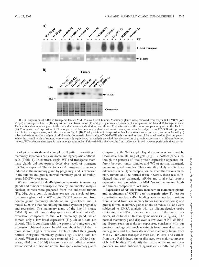
histologic analysis showed a complex cell pattern, consisting ofmammary squamous cell carcinoma and hyperplasic epithelialcells (Table 1). In contrast, virgin WT and transgenic mam-mary glands did not express detectable levels of transgenemRNA, as expected. Thus, ectopic c-rel transgene expression isinduced in the mammary gland by pregnancy, and is expressedin the tumors and grossly normal mammary glands of multip-arous MMTV–c-rel mice.
We next assessed total c-Rel protein expression in mammaryglands and tumors of transgenic mice by immunoblot analysis.Nuclear extracts were prepared from the indicated tumors(Fig. 3B). As a control, nuclear extracts were isolated frommammary glands of a WT virgin FVB/N mouse and fromnonmalignant mammary glands of an age-related line 14mouse (3000 N) that had undergone three cycles of pregnancyand regression. The mammary gland of the line 14 mouse(3000 N) and all tumors displayed elevated levels of c-Relexpression compared to the WT mammary gland, whichshowed only a low basal expression (Fig. 3B and data notshown). This is consistent with the pattern of transgenic c-relexpression obtained above. In addition, about half of the tu-mors showed higher expression levels of c-Rel than grosslynormal transgenic mammary glands (Fig. 3B and data notshown). When the results were scanned, a 3- to 150-fold (av-erage, [68.0 � 60.1]-fold) increase in nuclear c-Rel expressionwas observed in tumor and normal transgenic mammary glands
compared to the WT sample. Equal loading was confirmed byCoomassie blue staining of gels (Fig. 3B, bottom panel), al-though the patterns of total protein expression appeared dif-ferent between tumor samples and WT or normal transgenicmammary gland samples. This variability likely results fromdifferences in cell type composition between the various mam-mary tumors and the normal tissue. Overall, these results in-dicated that c-rel transgenic mRNA and total c-Rel proteinexpression are upregulated in MMTV–c-rel mammary glandsand tumors compared to WT mice.
Expression of NF-�B family members in mammary glandsand carcinomas of MMTV–c-rel transgenic mice. To test forconstitutive nuclear c-Rel binding activity, nuclear proteinswere isolated from a mammary tumor (adenocarcinoma) andgrossly normal mammary glands of line 15 mouse 127 and weresubjected to EMSA analysis with an oligonucleotide probecontaining the NF-�B element upstream of the c-myc pro-moter, which binds all Rel family members (39) (Fig. 4A). Thenormal mammary gland displayed a low level of NF-�B bind-ing (better seen on a darker exposure), consistent with ourprevious findings with nuclear extracts from normal rat mam-mary glands and histologically normal mammary tissue fromMMTV-Her-2/neu transgenic mice (34, 45, 59). The extractsfrom the c-Rel-induced tumor displayed two major complexesof NF-�B binding. To identify the nature of the subunit com-ponents, we used antibodies against either c-Rel or p50 in
FIG. 3. Expression of c-Rel in transgenic female MMTV–c-rel breast tumors. Mammary glands were removed from virgin WT FVB/N (WTVirgin) or transgenic line 16 (16 Virgin) mice and from tumor (T) and grossly normal (N) tissues of multiparous line 14 and 16 transgenic mice.The identification number given to the individual mice is indicated in parentheses. Characteristics of the tumor samples are given in the Table 1.(A) Transgenic c-rel expression. RNA was prepared from mammary gland and tumor tissues, and samples subjected to RT-PCR with primersspecific for transgenic c-rel, as in the legend to Fig. 1. (B) Total protein c-Rel expression. Nuclear extracts were prepared, and samples (40 �g)subjected to immunoblot analysis of c-Rel levels. Coomassie blue staining of SDS-PAGE gels was used as control for equal loading (bottom panel).While the overall levels of staining were essentially equivalent, the analysis revealed that the patterns of protein expression are different betweentumors, WT and normal transgenic mammary gland samples. This variability likely results from differences in cell type composition in these tissues.
VOL. 23, 2003 c-Rel AND MAMMARY GLAND TUMORIGENESIS 5743
Dow
nloa
ded
from
http
s://j
ourn
als.
asm
.org
/jour
nal/m
cb o
n 20
Oct
ober
202
1 by
61.
37.1
99.2
29.
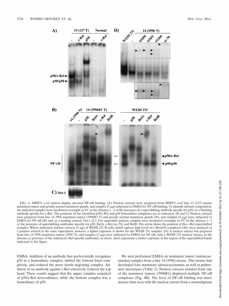
EMSA. Addition of an antibody that preferentially recognizesp50 in a homodimer complex, shifted the bottom band com-pletely, and reduced the more slowly migrating complex. Ad-dition of an antibody against c-Rel selectively reduced the topband. These results suggest that the upper complex consistedof p50/c-Rel heterodimers, while the bottom complex was ahomodimer of p50.
We next performed EMSA on mammary tumor (adenocar-cinoma) samples from a line 14 (3996) mouse. This mouse haddeveloped four mammary adenocarcinomas, as well as pulmo-nary metastases (Table 1). Nuclear extracts isolated from oneof the mammary tumors (3996R1) displayed multiple NF-�Bcomplexes (Fig. 4B). The level of NF-�B binding was moreintense than seen with the nuclear extract from a nonmalignant
FIG. 4. MMTV–c-rel tumors display elevated NF-�B binding. (A) Nuclear extracts were prepared from MMTV–c-rel line 15 (127) mousemammary tumor and grossly normal mammary glands, and samples (5 �g) subjected to EMSA for NF-�B binding. To identify subunit composition,the indicated samples were incubated overnight at 4°C in the absence () or the presence of a supershifting antibody specific for p50, or a blockingantibody specific for c-Rel. The positions of the identified p50/c-Rel and p50 homodimer complexes are as indicated. (B and C) Nuclear extractswere prepared from line 14 3996 mammary tumor (3996R1 T) and grossly normal mammary glands (N), and samples (5 �g) were subjected toEMSA for NF-�B (B) and, as a loading control, Oct-1 (C). For supershift analysis, samples were incubated overnight at 4°C in the absence ()or the presence of supershifting antibodies specific for p50, RelA, c-Rel (sc-70), and RelB. The arrow shows the position of the c-Rel supershiftedcomplex. Where indicated, nuclear extracts (5 �g) of WEHI 231 B cells, which express high levels of c-Rel/p50 complexes (40), were analyzed asa positive control in the same experiment, however a lighter exposure is shown for the WEHI 231 samples. (D) A nuclear extract was preparedfrom line 14 3996 mammary tumor (3996 T), and samples (5 �g) were subjected to EMSA for NF-�B, with a WEHI 231 nuclear extract, in theabsence or presence of the indicated c-Rel-specific antibodies, as above. Inset represents a darker exposure of the region of the supershifted bandsindicated in the figure.
5744 ROMIEU-MOUREZ ET AL. MOL. CELL. BIOL.
Dow
nloa
ded
from
http
s://j
ourn
als.
asm
.org
/jour
nal/m
cb o
n 20
Oct
ober
202
1 by
61.
37.1
99.2
29.
mammary gland from the same animal (line14 3996 N), whilelevels of control Oct-1 binding were similar in these samples(Fig. 4C). Of note, an NF-�B complex of higher mobility waspresent in the normal sample. We previously observed thepresence of such a complex in nuclear extracts isolated fromnormal human and mouse breast tissue, and supershift analysisshowed that these complexes contained predominantly p50homodimers (reference 50 and data not shown). Addition of ac-Rel-specific antibody resulted in diminished binding and theformation of one slowly migrating supershifted complex (Fig.4B, indicated by an arrowhead). Addition of an antibodyagainst c-Rel reduced the binding and yielded a similar super-shifted complex with control extracts from WEHI 231 imma-ture B-lymphoma cells, which express high levels of activatedc-Rel and p50, and lower levels of RelA (40, 48), indicatingthat the line14 3996R1 tumor sample contains p50/c-Rel com-plexes. Addition of supershifting antibodies against p50, RelA,or RelB to the line 14 3996R1 extracts resulted in shiftedcomplexes. In particular, addition of a p50 antibody greatlyreduced binding and yielded two major and a few minor su-pershifted bands. Interestingly, addition of a RelA antibodyyielded three supershifted RelA-containing complexes, whereas,the expected one RelA-containing complex (e.g., p50/RelA)was seen with the WEHI 231 extract (40). These results suggestthat RelA is present in multiple complexes in the mammarygland tumor sample. Lastly, a RelB antibody also reducedbinding with the line 14 tumor extract, yielding one super-shifted complex (Fig. 4B). Addition of the RelB antibody hadno detectable effect on the WEHI 231 cells, consistent with thelack of nuclear RelB in these cells (48). To further characterizethe c-Rel binding, we compared the effects of the anti-c-Relsc-70 from Santa Cruz with three different c-Rel antibodyrabbit polysera obtained from N. Rice and M. Ernst (sera 1050,1051, and 1266), using nuclear extracts from a second tumorfrom line 14 3996. All of the antibodies reduced the uppercomplex which comigrated with the WEHI 231 c-Rel/p50 bandto approximately the same extent (Fig. 4D). The positions ofthe supershifted complexes varied for all of the antibodies (Fig.4D, inset). These finding confirm the presence of c-Rel inNF-�B DNA binding complexes, although, it was not the pri-mary component in the nuclear extracts. Together, these re-sults indicate that multiple NF-�B subunits are binding in thetumor samples, including c-Rel, p50, RelA, and RelB.
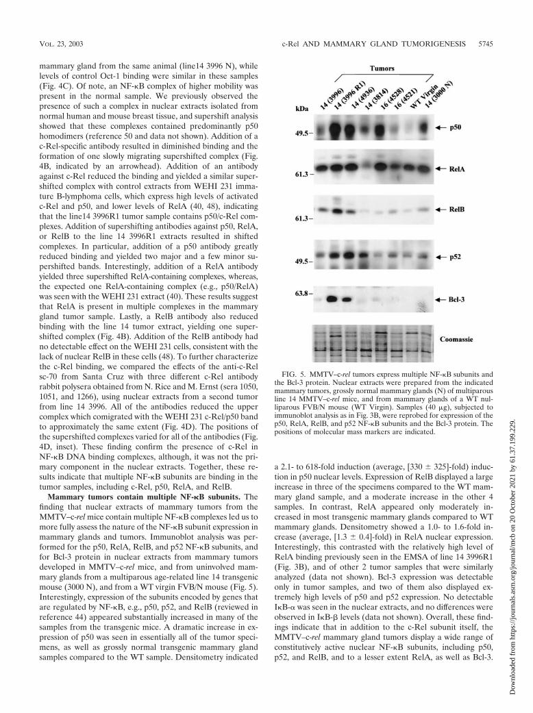
Mammary tumors contain multiple NF-�B subunits. Thefinding that nuclear extracts of mammary tumors from theMMTV–c-rel mice contain multiple NF-�B complexes led us tomore fully assess the nature of the NF-�B subunit expression inmammary glands and tumors. Immunoblot analysis was per-formed for the p50, RelA, RelB, and p52 NF-�B subunits, andfor Bcl-3 protein in nuclear extracts from mammary tumorsdeveloped in MMTV–c-rel mice, and from uninvolved mam-mary glands from a multiparous age-related line 14 transgenicmouse (3000 N), and from a WT virgin FVB/N mouse (Fig. 5).Interestingly, expression of the subunits encoded by genes thatare regulated by NF-�B, e.g., p50, p52, and RelB (reviewed inreference 44) appeared substantially increased in many of thesamples from the transgenic mice. A dramatic increase in ex-pression of p50 was seen in essentially all of the tumor speci-mens, as well as grossly normal transgenic mammary glandsamples compared to the WT sample. Densitometry indicated
a 2.1- to 618-fold induction (average, [330 � 325]-fold) induc-tion in p50 nuclear levels. Expression of RelB displayed a largeincrease in three of the specimens compared to the WT mam-mary gland sample, and a moderate increase in the other 4samples. In contrast, RelA appeared only moderately in-creased in most transgenic mammary glands compared to WTmammary glands. Densitometry showed a 1.0- to 1.6-fold in-crease (average, [1.3 � 0.4]-fold) in RelA nuclear expression.Interestingly, this contrasted with the relatively high level ofRelA binding previously seen in the EMSA of line 14 3996R1(Fig. 3B), and of other 2 tumor samples that were similarlyanalyzed (data not shown). Bcl-3 expression was detectableonly in tumor samples, and two of them also displayed ex-tremely high levels of p50 and p52 expression. No detectableI�B-� was seen in the nuclear extracts, and no differences wereobserved in I�B-� levels (data not shown). Overall, these find-ings indicate that in addition to the c-Rel subunit itself, theMMTV–c-rel mammary gland tumors display a wide range ofconstitutively active nuclear NF-�B subunits, including p50,p52, and RelB, and to a lesser extent RelA, as well as Bcl-3.
FIG. 5. MMTV–c-rel tumors express multiple NF-�B subunits andthe Bcl-3 protein. Nuclear extracts were prepared from the indicatedmammary tumors, grossly normal mammary glands (N) of multiparousline 14 MMTV–c-rel mice, and from mammary glands of a WT nul-liparous FVB/N mouse (WT Virgin). Samples (40 �g), subjected toimmunoblot analysis as in Fig. 3B, were reprobed for expression of thep50, RelA, RelB, and p52 NF-�B subunits and the Bcl-3 protein. Thepositions of molecular mass markers are indicated.
VOL. 23, 2003 c-Rel AND MAMMARY GLAND TUMORIGENESIS 5745
Dow
nloa
ded
from
http
s://j
ourn
als.
asm
.org
/jour
nal/m
cb o
n 20
Oct
ober
202
1 by
61.
37.1
99.2
29.
Similar complex patterns were seen previously in primary hu-man breast cancer specimens (15, 59).
MMTV–c-rel mammary glands and carcinomas display ele-vated expression of downstream target gene cyclin D1. TheNF-�B target gene cyclin D1 (25, 26) has been implicated inbreast cancer (reviewed in reference 32). Its expression, whichis upregulated in �50% of human breast tumors, is requiredfor proliferation of breast cancer cells in culture, and MMTV-cyclin D1 mice develop mammary adenocarcinomas (64).Therefore, we sought to test whether cyclin D1 mRNA levelswere increased during pregnancy in the mammary glands ofMMTV–c-rel versus WT mice. Lines 7, 14, 15, and 16 MMTV–c-rel mice and age-matched WT FVB/N mice (3 to 6 monthsold) were bred to activate the MMTV-LTR promoter in trans-genic mammary glands. At day 18.5 of the first pregnancy, totalRNA was isolated from the mammary glands and samplessubjected to analysis of cyclin D1 mRNA levels using either asemiquantitative RT-PCR assay specific for cyclin D1 in thepresence or absence of RT, as control, or Northern blot anal-ysis (Fig. 6A). For the RT-PCR assay, RNA quality and load-ing were normalized by evaluation of �-actin levels using 25cycles of PCR, which is within the linear phase of amplification.The level of cyclin D1 mRNA was higher in all of the trans-genic mouse mammary glands compared with the three WTmouse samples. When results of this and a duplicate experi-ment were scanned and normalized to �-actin mRNA levels, a(2.5 � 0.8)-fold increase (P 0.003) in cyclin D1 mRNA levelswas observed in transgenic compared to WT samples. Simi-larly, higher levels of cyclin D1 mRNA were observed in thetransgenic mouse mammary glands compared with WT mousesamples in the Northern blot analysis. RNA loading was nor-malized to the levels of 28S rRNA. Expression of cyclin D1mRNA could be detected in two of the five WT mice tested(WT 3 and WT 14). All transgenic mice displayed detectableexpression levels of cyclin D1 mRNA, and these levels werehigher than those of the WT 3 and WT 14 samples. Whenresults were scanned and normalized to 28S rRNA levels, a(6.8 � 5.4)-fold increase in cyclin D1 mRNA levels were ob-served in the transgenic samples compared to WT 3 and WT 14samples. Thus, overexpression of transgenic c-Rel in the mam-mary gland during the first pregnancy is sufficient to induce asubstantial increase in cyclin D1 mRNA expression.
We next evaluated cyclin D1 levels in c-Rel-induced mam-mary tumors compared to nonmalignant mammary glands intransgenic animals, again using both the semiquantitative RT-PCR assay and Northern blot analyses (Fig. 6B). Total RNAwas isolated from mammary carcinomas that had developed intransgenic mice of lines 14 and 16, as well as from grosslynormal mammary glands from multiparous age-related trans-genic mice of the same lines. As a reference for basal levels ofcyclin D1 mRNA in the mammary gland, total RNA was ex-tracted from mammary glands of five virgin adult transgenicmice of lines 16 or 15. In this and a duplicate RT-PCR assay,all of the tumors displayed higher expression levels of cyclin D1compared to the two grossly normal mammary gland samplestested. Interestingly, the normal sample 14 (5441 N) displayedhigher levels of cyclin D1 mRNA than the normal sample 16(4948 N), which correlated with their respective levels of c-reltransgene expression as seen above (Fig. 3A). In Northern blotanalysis, levels of cyclin D1 mRNA were barely detectable in
the five virgin mammary gland samples tested. In contrast, one(14 4949 N) of the three grossly normal mammary gland sam-ples and one tumor (14 3814 T) displayed detectable expres-sion levels of cyclin D1 mRNA, while the cyclin D1 mRNAlevels were sharply increased in the 3 other tumors (16 4528 T,14 3996 T, and 14 4936 T). Therefore, MMTV–c-rel mammaryglands and carcinomas display a substantial overexpression ofcyclin D1 mRNA compared to the WT mammary gland.
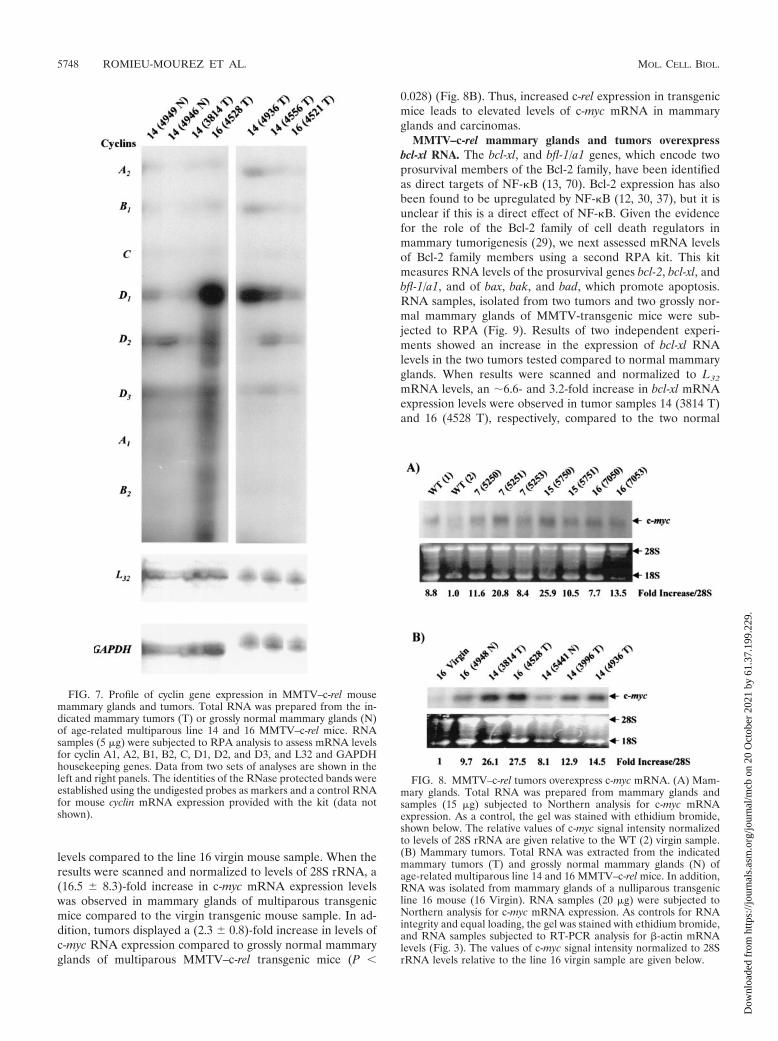
Evidence has also suggested NF-�B mediates regulation ofcyclin A (reviewed in reference 32). Thus, we compared theoverall cyclin expression profiles in the c-Rel-induced mam-mary tumors with the grossly normal mammary glands. In thefirst set, total RNA was prepared from two grossly normalmammary glands from multiparous transgenic mice line 14(4949 N and 4946 N), and two mammary tumors of line 14(3814 T) and 16 (4528 T). In a second set, total RNA wasprepared from mammary carcinomas of line 14 (4936 T and4556 T) and line 16 (4521 T). RNA samples were subjected toa multiprobe RPA kit, which assesses mRNA levels for cyclinsA1, A2, B1, B2, C, D1, D2, and D3, and L32 and GAPDHhousekeeping gene products (Fig. 7). In this and a duplicateexperiment, bands were detectable for cyclin A2, B1, D1, D2,and D3 mRNA. The mRNA from the c-Rel-induced tumorsdisplayed increased expression of the cyclin D1 gene comparedto the normal mammary glands (Fig. 7, left panel), consistentwith the Northern blot analysis above (Fig. 6B). The tumorsdisplayed variable levels of mRNA for cyclins A2, B1, D2, andD3. Analysis of the housekeeping genes L32 and GAPDHconfirmed the essentially equal loading of samples within thepanels. Thus, RPA showed that MMTV–c-rel tumors displayan increase in cyclin D1 gene expression with variable levels ofexpression of the other cyclins.
MMTV–c-rel mammary tumors overexpress c-myc. We nexttested for changes in c-myc gene expression, another NF-�Btarget gene (17, 39), which affects cell proliferation and sur-vival (reviewed in reference 5). RNA was isolated at day 18.5of the first pregnancy from mammary glands of line 7, 15, and16 MMTV–c-rel transgenic and WT FVB/N mice, all 3 to 6months old. Samples were subjected to Northern blot analysisfor c-myc RNA levels (Fig. 8A). The quality of the RNA wasevaluated by ethidium bromide staining of the gel (Fig. 8A)and by Northern blot analysis of GAPDH mRNA expressionlevels (data not shown). The level of c-myc mRNA appearedhigher in most of the mammary glands of the transgenic ani-mals compared to the two WT mice. When results werescanned and normalized to 28S rRNA levels, a (2.4 � 1.4)-foldincrease was observed in c-myc mRNA expression levels intransgenic samples compared to the average of the WT sam-ples. Comparable increase was obtained upon normalization tolevels of GAPDH mRNA ([2.8 � 0.7]-fold, data not shown).These levels of increase did not reach statistical significance.
We next compared the levels of c-myc mRNA expression inmammary glands versus carcinomas of multiparous age-relatedtransgenic mice and a virgin transgenic mouse. Quality of theRNA and equal loading was checked by ethidium bromidestaining of the gel (Fig. 8B) and by RT-PCR analysis of �-actinmRNA levels, as shown above in Fig. 3B (bottom panel). InNorthern blot analysis, all the normal and tumor mammaryglands of c-Rel-expressing multiparous transgenic mice dem-onstrated a substantial increase in c-myc mRNA expression
5746 ROMIEU-MOUREZ ET AL. MOL. CELL. BIOL.
Dow
nloa
ded
from
http
s://j
ourn
als.
asm
.org
/jour
nal/m
cb o
n 20
Oct
ober
202
1 by
61.
37.1
99.2
29.
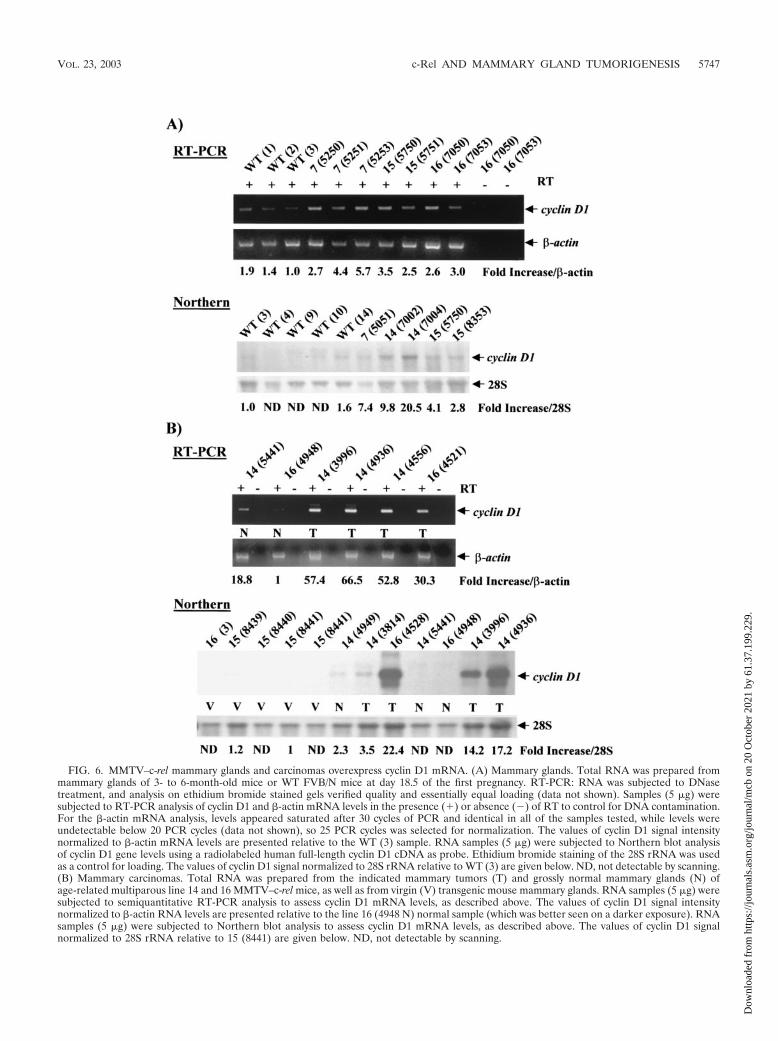
FIG. 6. MMTV–c-rel mammary glands and carcinomas overexpress cyclin D1 mRNA. (A) Mammary glands. Total RNA was prepared frommammary glands of 3- to 6-month-old mice or WT FVB/N mice at day 18.5 of the first pregnancy. RT-PCR: RNA was subjected to DNasetreatment, and analysis on ethidium bromide stained gels verified quality and essentially equal loading (data not shown). Samples (5 �g) weresubjected to RT-PCR analysis of cyclin D1 and �-actin mRNA levels in the presence (�) or absence () of RT to control for DNA contamination.For the �-actin mRNA analysis, levels appeared saturated after 30 cycles of PCR and identical in all of the samples tested, while levels wereundetectable below 20 PCR cycles (data not shown), so 25 PCR cycles was selected for normalization. The values of cyclin D1 signal intensitynormalized to �-actin mRNA levels are presented relative to the WT (3) sample. RNA samples (5 �g) were subjected to Northern blot analysisof cyclin D1 gene levels using a radiolabeled human full-length cyclin D1 cDNA as probe. Ethidium bromide staining of the 28S rRNA was usedas a control for loading. The values of cyclin D1 signal normalized to 28S rRNA relative to WT (3) are given below. ND, not detectable by scanning.(B) Mammary carcinomas. Total RNA was prepared from the indicated mammary tumors (T) and grossly normal mammary glands (N) ofage-related multiparous line 14 and 16 MMTV–c-rel mice, as well as from virgin (V) transgenic mouse mammary glands. RNA samples (5 �g) weresubjected to semiquantitative RT-PCR analysis to assess cyclin D1 mRNA levels, as described above. The values of cyclin D1 signal intensitynormalized to �-actin RNA levels are presented relative to the line 16 (4948 N) normal sample (which was better seen on a darker exposure). RNAsamples (5 �g) were subjected to Northern blot analysis to assess cyclin D1 mRNA levels, as described above. The values of cyclin D1 signalnormalized to 28S rRNA relative to 15 (8441) are given below. ND, not detectable by scanning.
VOL. 23, 2003 c-Rel AND MAMMARY GLAND TUMORIGENESIS 5747
Dow
nloa
ded
from
http
s://j
ourn
als.
asm
.org
/jour
nal/m
cb o
n 20
Oct
ober
202
1 by
61.
37.1
99.2
29.
levels compared to the line 16 virgin mouse sample. When theresults were scanned and normalized to levels of 28S rRNA, a(16.5 � 8.3)-fold increase in c-myc mRNA expression levelswas observed in mammary glands of multiparous transgenicmice compared to the virgin transgenic mouse sample. In ad-dition, tumors displayed a (2.3 � 0.8)-fold increase in levels ofc-myc RNA expression compared to grossly normal mammaryglands of multiparous MMTV–c-rel transgenic mice (P
0.028) (Fig. 8B). Thus, increased c-rel expression in transgenicmice leads to elevated levels of c-myc mRNA in mammaryglands and carcinomas.
MMTV–c-rel mammary glands and tumors overexpressbcl-xl RNA. The bcl-xl, and bfl-1/a1 genes, which encode twoprosurvival members of the Bcl-2 family, have been identifiedas direct targets of NF-�B (13, 70). Bcl-2 expression has alsobeen found to be upregulated by NF-�B (12, 30, 37), but it isunclear if this is a direct effect of NF-�B. Given the evidencefor the role of the Bcl-2 family of cell death regulators inmammary tumorigenesis (29), we next assessed mRNA levelsof Bcl-2 family members using a second RPA kit. This kitmeasures RNA levels of the prosurvival genes bcl-2, bcl-xl, andbfl-1/a1, and of bax, bak, and bad, which promote apoptosis.RNA samples, isolated from two tumors and two grossly nor-mal mammary glands of MMTV-transgenic mice were sub-jected to RPA (Fig. 9). Results of two independent experi-ments showed an increase in the expression of bcl-xl RNAlevels in the two tumors tested compared to normal mammaryglands. When results were scanned and normalized to L32
mRNA levels, an �6.6- and 3.2-fold increase in bcl-xl mRNAexpression levels were observed in tumor samples 14 (3814 T)and 16 (4528 T), respectively, compared to the two normal
FIG. 7. Profile of cyclin gene expression in MMTV–c-rel mousemammary glands and tumors. Total RNA was prepared from the in-dicated mammary tumors (T) or grossly normal mammary glands (N)of age-related multiparous line 14 and 16 MMTV–c-rel mice. RNAsamples (5 �g) were subjected to RPA analysis to assess mRNA levelsfor cyclin A1, A2, B1, B2, C, D1, D2, and D3, and L32 and GAPDHhousekeeping genes. Data from two sets of analyses are shown in theleft and right panels. The identities of the RNase protected bands wereestablished using the undigested probes as markers and a control RNAfor mouse cyclin mRNA expression provided with the kit (data notshown).
FIG. 8. MMTV–c-rel tumors overexpress c-myc mRNA. (A) Mam-mary glands. Total RNA was prepared from mammary glands andsamples (15 �g) subjected to Northern analysis for c-myc mRNAexpression. As a control, the gel was stained with ethidium bromide,shown below. The relative values of c-myc signal intensity normalizedto levels of 28S rRNA are given relative to the WT (2) virgin sample.(B) Mammary tumors. Total RNA was extracted from the indicatedmammary tumors (T) and grossly normal mammary glands (N) ofage-related multiparous line 14 and 16 MMTV–c-rel mice. In addition,RNA was isolated from mammary glands of a nulliparous transgenicline 16 mouse (16 Virgin). RNA samples (20 �g) were subjected toNorthern analysis for c-myc mRNA expression. As controls for RNAintegrity and equal loading, the gel was stained with ethidium bromide,and RNA samples subjected to RT-PCR analysis for �-actin mRNAlevels (Fig. 3). The values of c-myc signal intensity normalized to 28SrRNA levels relative to the line 16 virgin sample are given below.
5748 ROMIEU-MOUREZ ET AL. MOL. CELL. BIOL.
Dow
nloa
ded
from
http
s://j
ourn
als.
asm
.org
/jour
nal/m
cb o
n 20
Oct
ober
202
1 by
61.
37.1
99.2
29.
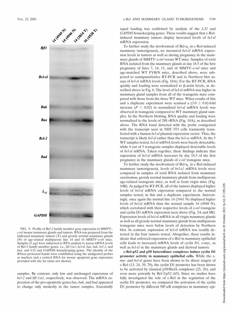
samples. By contrast, only low and unchanged expression ofbcl-2 and bfl-1/a1, respectively, was observed. The mRNA ex-pression of the pro-apoptotic genes bax, bak, and bad appearedto change only modestly in the tumor samples. Essentially
equal loading was confirmed by analysis of the L32 andGAPDH housekeeping genes. These results suggest that c-Rel-induced mammary tumors display increased levels of bcl-xlmRNA expression.
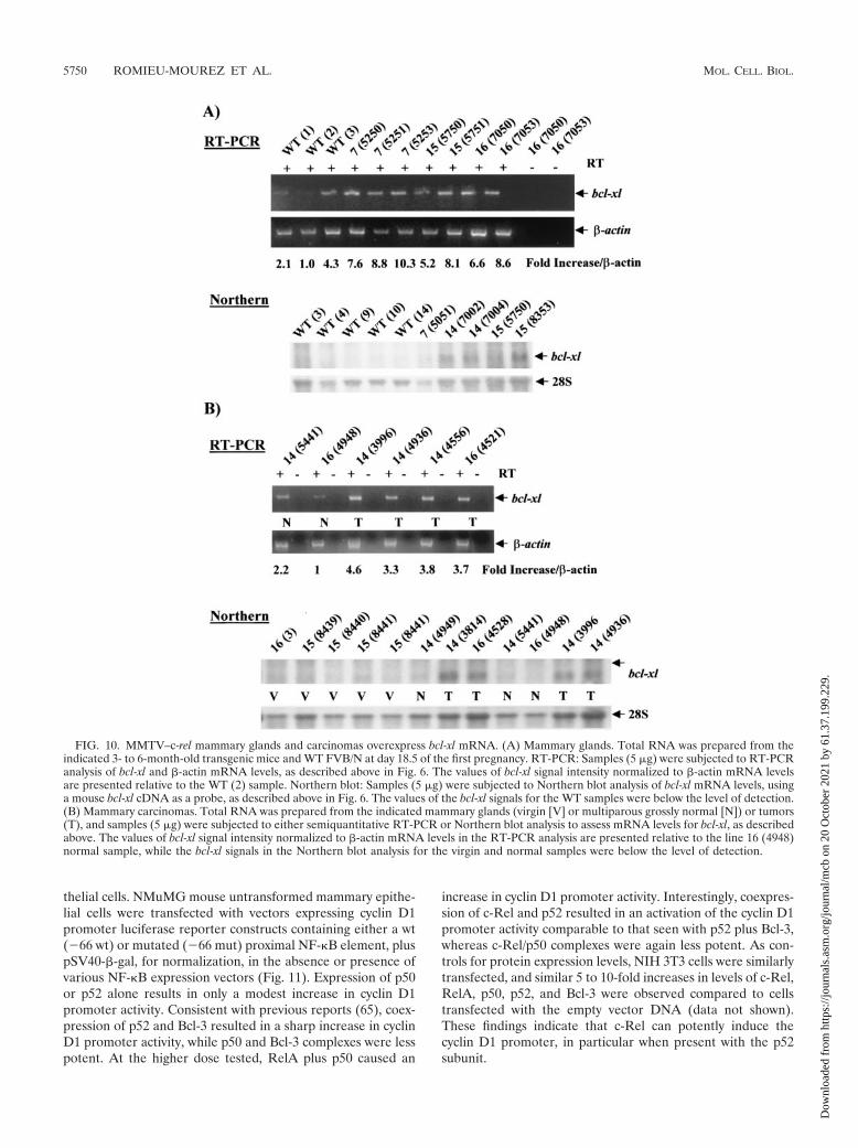
To further study the involvement of Bcl-xL in c-Rel-inducedmammary tumorigenesis, we measured bcl-xl mRNA expres-sion levels in tumors as well as during pregnancy in the mam-mary glands of MMTV–c-rel versus WT mice. Samples of totalRNA isolated from the mammary glands at day 18.5 of the firstpregnancy of lines 7, 14, 15, and 16 MMTV–c-rel mice andage-matched WT FVB/N mice, described above, were sub-jected to semiquantitative RT-PCR and to Northern blot as-says of bcl-xl mRNA levels (Fig. 10A). For the RT-PCR, RNAquality and loading were normalized to �-actin levels, as de-scribed above in Fig. 6. The level of bcl-xl mRNA was higher inmammary gland samples from all of the transgenic mice com-pared with those from the three WT mice. When results of thisand a duplicate experiment were scanned a (3.0 � 0.8)-foldincrease (P 0.02) in normalized bcl-xl mRNA levels wasobserved in transgenic compared to WT mammary gland sam-ples. In the Northern blotting, RNA quality and loading werenormalized to the levels of 28S rRNA (Fig. 10A), as describedabove. The RNA band detected with the probe comigratedwith the transcript seen in NIH 3T3 cells transiently trans-fected with a human bcl-xl plasmid expression vector. Thus, thetranscript is likely bcl-xl rather than the bcl-xs mRNA. In the 5WT samples tested, bcl-xl mRNA levels were barely detectable,while 4 out of 5 transgenic samples displayed detectable levelsof bcl-xl mRNA. Taken together, these findings indicate thatexpression of bcl-xl mRNA increases by day 18.5 of the firstpregnancy in the mammary glands of c-rel transgene mice.
To further study the involvement of Bcl-xL in c-Rel-inducedmammary tumorigenesis, levels of bcl-x1 mRNA levels werecompared in samples of total RNA isolated from mammarycarcinomas, grossly normal mammary glands from multiparousage-related transgenic mice, as well as from virgin mice (Fig.10B). As judged by RT-PCR, all of the tumors displayed higherlevels of bcl-xl mRNA expression compared to the normalsamples tested, in this and a duplicate experiment. Interest-ingly, once again the normal line 14 (5441 N) displayed higherlevels of bcl-xl mRNA than the normal sample 16 (4948 N),which correlated with their respective levels of c-rel transgeneand cyclin D1 mRNA expression seen above (Fig. 3A and 8B).Expression levels of bcl-xl mRNA in all virgin mammary glandsas well as all grossly normal mammary glands from multiparoustransgenic mice were below levels of detection by Northernblot. In contrast, expression of bcl-xl mRNA was readily de-tected in the four tumors tested. Altogether, these results in-dicate that enforced expression of c-Rel in mammary epithelialcells leads to increased mRNA levels of cyclin D1, c-myc, aswell as bcl-xl in the mammary glands and derived tumors.
c-Rel-p52 and p50 heterodimer complexes induce cyclin D1promoter activity in mammary epithelial cells. While the c-myc and bcl-xl genes have been shown to be direct targets ofc-Rel (13, 24, 39, 70), the cyclin D1 promoter has been shownto be activated by classical p50/RelA complexes (25, 26), andeven more potently by Bcl-3/p52 (65). Since no studies havefully investigated the role of c-Rel in the regulation of thecyclin D1 promoter, we compared the activation of the cyclinD1 promoter by different NF-�B complexes in mammary epi-
FIG. 9. Profile of Bcl-2 family member gene expression in MMTV–c-rel mouse mammary glands and tumors. RNA was prepared from theindicated mammary tumors (T) and grossly normal mammary glands(N) of age-related multiparous line 14 and 16 MMTV–c-rel mice.Samples (5 �g) were subjected to RPA analysis to assess mRNA levelsof Bcl-2 family member genes, i.e., bfl-1/a1, bcl-xl, bax, bak, bcl-2, andbad, and L32 and GAPDH housekeeping genes. The identity of theRNase protected bands were established using the undigested probesas markers and a control RNA for mouse apoptosis gene expressionprovided with the kit (data not shown).
VOL. 23, 2003 c-Rel AND MAMMARY GLAND TUMORIGENESIS 5749
Dow
nloa
ded
from
http
s://j
ourn
als.
asm
.org
/jour
nal/m
cb o
n 20
Oct
ober
202
1 by
61.
37.1
99.2
29.
thelial cells. NMuMG mouse untransformed mammary epithe-lial cells were transfected with vectors expressing cyclin D1promoter luciferase reporter constructs containing either a wt(66 wt) or mutated (66 mut) proximal NF-�B element, pluspSV40-�-gal, for normalization, in the absence or presence ofvarious NF-�B expression vectors (Fig. 11). Expression of p50or p52 alone results in only a modest increase in cyclin D1promoter activity. Consistent with previous reports (65), coex-pression of p52 and Bcl-3 resulted in a sharp increase in cyclinD1 promoter activity, while p50 and Bcl-3 complexes were lesspotent. At the higher dose tested, RelA plus p50 caused an
increase in cyclin D1 promoter activity. Interestingly, coexpres-sion of c-Rel and p52 resulted in an activation of the cyclin D1promoter activity comparable to that seen with p52 plus Bcl-3,whereas c-Rel/p50 complexes were again less potent. As con-trols for protein expression levels, NIH 3T3 cells were similarlytransfected, and similar 5 to 10-fold increases in levels of c-Rel,RelA, p50, p52, and Bcl-3 were observed compared to cellstransfected with the empty vector DNA (data not shown).These findings indicate that c-Rel can potently induce thecyclin D1 promoter, in particular when present with the p52subunit.
FIG. 10. MMTV–c-rel mammary glands and carcinomas overexpress bcl-xl mRNA. (A) Mammary glands. Total RNA was prepared from theindicated 3- to 6-month-old transgenic mice and WT FVB/N at day 18.5 of the first pregnancy. RT-PCR: Samples (5 �g) were subjected to RT-PCRanalysis of bcl-xl and �-actin mRNA levels, as described above in Fig. 6. The values of bcl-xl signal intensity normalized to �-actin mRNA levelsare presented relative to the WT (2) sample. Northern blot: Samples (5 �g) were subjected to Northern blot analysis of bcl-xl mRNA levels, usinga mouse bcl-xl cDNA as a probe, as described above in Fig. 6. The values of the bcl-xl signals for the WT samples were below the level of detection.(B) Mammary carcinomas. Total RNA was prepared from the indicated mammary glands (virgin [V] or multiparous grossly normal [N]) or tumors(T), and samples (5 �g) were subjected to either semiquantitative RT-PCR or Northern blot analysis to assess mRNA levels for bcl-xl, as describedabove. The values of bcl-xl signal intensity normalized to �-actin mRNA levels in the RT-PCR analysis are presented relative to the line 16 (4948)normal sample, while the bcl-xl signals in the Northern blot analysis for the virgin and normal samples were below the level of detection.
5750 ROMIEU-MOUREZ ET AL. MOL. CELL. BIOL.
Dow
nloa
ded
from
http
s://j
ourn
als.
asm
.org
/jour
nal/m
cb o
n 20
Oct
ober
202
1 by
61.
37.1
99.2
29.

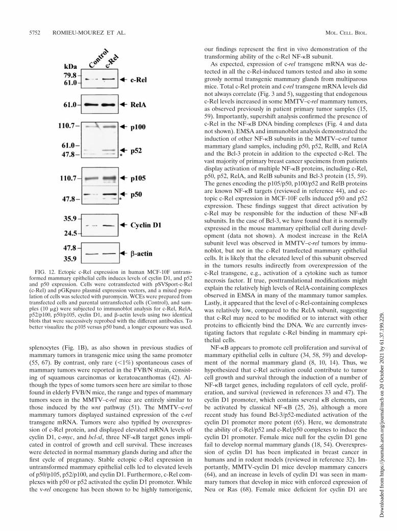
Ectopic c-Rel expression increases the levels of cyclin D1,p52, and p50 in mammary epithelial cells. Next, we investi-gated the effects of ectopic stable c-Rel overexpression oncyclin D1 and NF-�B subunit levels using MCF-10F humanuntransformed breast epithelial cells. Cells were transfectedwith a mouse c-Rel expression vector along with a puromycinselection plasmid, and grown in the presence of puromycin.Mixed populations of selected cells were used for analysis. Ascontrols, we used either MCF-10F cells transfected with thepBluescript and puromycin selection plasmids and selectedwith puromycin or MCF-10F untransfected parental cellsgrown under the same conditions. (All populations of cellsgrew at the same rate, data not shown). We first confirmedoverexpression of c-Rel in WCEs of c-Rel-transfected cellscompared to parental MCF-10F cells, using �-actin as controlfor equal loading (Fig. 12). Scanning of this and a duplicateexperiment indicated that c-Rel-transfected MCF-10F cellsdisplayed a (2.5 � 1.2)-fold increase in total level of c-Relcompared to control cells. Importantly, we observed that c-Rel-transfected MCF-10F cells displayed significant increasesin the levels of cyclin D1, as well as in the NF-�B subunits p52and p50 (and their precursors p100 and p105) compared tocontrol cells (Fig. 12). Densitometry analysis indicated thatc-Rel-transfected MCF-10F cells displayed a 3.1 and 2.7-foldincrease in levels of p52 and p50, respectively. Densitometry ofthis and two other immunoblots (data not shown) indicatedthat c-Rel-transfected MCF-10F cells displayed a (1.8 � 0.1)-fold increase in levels of cyclin D1. Essentially no change was
observed in levels of RelA in c-Rel transfected cells. Controlpuromycin-selected cells displayed levels of c-Rel and cyclinD1 comparable to the parental untransfected cells MCF-10Fcells, indicating that the selection was not responsible for thisobserved increase (data not shown). Thus, sustained overex-pression of c-Rel in untransformed mammary epithelial cellsleads to increased levels of p52, p50, and cyclin D1 expression.Overall, these findings indicate sustained c-Rel expression inmammary epithelial cells is sufficient to lead to the induction inNF-�B subunits and target genes, responsible for mammarytumorigenesis.
DISCUSSION
Here we demonstrate for the first time that c-Rel plays a causalrole in tumorigenesis of the mammary gland in an MMTV-LTR-driven mouse model. Overall, one or more mammary tumorswere detected in 31.6% of MMTV–c-rel transgenic mice at anaverage age of 19.9 months. Histological analysis of the mammarytumors in four independent lines provided evidence for a widespectrum of tumor subtypes, including adenocarcinomas, adeno-squamous carcinomas, squamous carcinomas, a spindle cell car-cinoma and a papillary carcinoma. One mouse developed pulmo-nary metastasis in addition to multiple mammary adenocarci-nomas. In addition to mammary carcinomas, several mice hadenlarged spleen or other abnormalities including lymphoid ormyeloid hyperplasia or centrocytic lymphomas in the spleen,which can be correlated to the expression of the transgene in
FIG. 11. c-Rel heterodimer complexes induce the cyclin D1 promoter. NMuMG cells were transfected, in duplicate, with 66 WT-cyclin D1or 66 Mut-cyclin D1 luciferase gene reporter constructs and 0.5 �g of pSV40-�-gal in the presence of the indicated amounts of NF-�B or Bcl-3plasmid expression vectors. After 48 h, cultures were harvested, normalized for �-Gal activity, and assayed for luciferase activity. Normalized valuesof luciferase activity are presented (error bars, standard deviations).
VOL. 23, 2003 c-Rel AND MAMMARY GLAND TUMORIGENESIS 5751
Dow
nloa
ded
from
http
s://j
ourn
als.
asm
.org
/jour
nal/m
cb o
n 20
Oct
ober
202
1 by
61.
37.1
99.2
29.
splenocytes (Fig. 1B), as also shown in previous studies ofmammary tumors in transgenic mice using the same promoter(55, 67). By contrast, only rare (1%) spontaneous cases ofmammary tumors were reported in the FVB/N strain, consist-ing of squamous carcinomas or keratoacanthomas (42). Al-though the types of some tumors seen here are similar to thosefound in elderly FVB/N mice, the range and types of mammarytumors seen in the MMTV–c-rel mice are entirely similar tothose induced by the wnt pathway (51). The MMTV–c-relmammary tumors displayed sustained expression of the c-reltransgene mRNA. Tumors were also typified by overexpres-sion of c-Rel protein, and displayed elevated mRNA levels ofcyclin D1, c-myc, and bcl-xl, three NF-�B target genes impli-cated in control of growth and cell survival. These increaseswere detected in normal mammary glands during and after thefirst cycle of pregnancy. Stable ectopic c-Rel expression inuntransformed mammary epithelial cells led to elevated levelsof p50/p105, p52/p100, and cyclin D1. Furthermore, c-Rel com-plexes with p50 or p52 activated the cyclin D1 promoter. Whilethe v-rel oncogene has been shown to be highly tumorigenic,
our findings represent the first in vivo demonstration of thetransforming ability of the c-Rel NF-�B subunit.
As expected, expression of c-rel transgene mRNA was de-tected in all the c-Rel-induced tumors tested and also in somegrossly normal transgenic mammary glands from multiparousmice. Total c-Rel protein and c-rel transgene mRNA levels didnot always correlate (Fig. 3 and 5), suggesting that endogenousc-Rel levels increased in some MMTV–c-rel mammary tumors,as observed previously in patient primary tumor samples (15,59). Importantly, supershift analysis confirmed the presence ofc-Rel in the NF-�B DNA binding complexes (Fig. 4 and datanot shown). EMSA and immunoblot analysis demonstrated theinduction of other NF-�B subunits in the MMTV–c-rel tumormammary gland samples, including p50, p52, RelB, and RelAand the Bcl-3 protein in addition to the expected c-Rel. Thevast majority of primary breast cancer specimens from patientsdisplay activation of multiple NF-�B proteins, including c-Rel,p50, p52, RelA, and RelB subunits and Bcl-3 protein (15, 59).The genes encoding the p105/p50, p100/p52 and RelB proteinsare known NF-�B targets (reviewed in reference 44), and ec-topic c-Rel expression in MCF-10F cells induced p50 and p52expression. These findings suggest that direct activation byc-Rel may be responsible for the induction of these NF-�Bsubunits. In the case of Bcl-3, we have found that it is normallyexpressed in the mouse mammary epithelial cell during devel-opment (data not shown). A modest increase in the RelAsubunit level was observed in MMTV–c-rel tumors by immu-noblot, but not in the c-Rel transfected mammary epithelialcells. It is likely that the elevated level of this subunit observedin the tumors results indirectly from overexpression of thec-Rel transgene, e.g., activation of a cytokine such as tumornecrosis factor. If true, posttranslational modifications mightexplain the relatively high levels of RelA-containing complexesobserved in EMSA in many of the mammary tumor samples.Lastly, it appeared that the level of c-Rel-containing complexeswas relatively low, compared to the RelA subunit, suggestingthat c-Rel may need to be modified or to interact with otherproteins to efficiently bind the DNA. We are currently inves-tigating factors that regulate c-Rel binding in mammary epi-thelial cells.
NF-�B appears to promote cell proliferation and survival ofmammary epithelial cells in culture (34, 58, 59) and develop-ment of the normal mammary gland (8, 10, 14). Thus, wehypothesized that c-Rel activation could contribute to tumorcell growth and survival through the induction of a number ofNF-�B target genes, including regulators of cell cycle, prolif-eration, and survival (reviewed in references 33 and 47). Thecyclin D1 promoter, which contains several �B elements, canbe activated by classical NF-�B (25, 26), although a morerecent study has found Bcl-3/p52-mediated activation of thecyclin D1 promoter more potent (65). Here, we demonstratethe ability of c-Rel/p52 and c-Rel/p50 complexes to induce thecyclin D1 promoter. Female mice null for the cyclin D1 genefail to develop normal mammary glands (18, 54). Overexpres-sion of cyclin D1 has been implicated in breast cancer inhumans and in rodent models (reviewed in reference 32). Im-portantly, MMTV-cyclin D1 mice develop mammary cancers(64), and an increase in levels of cyclin D1 was seen in mam-mary tumors that develop in mice with enforced expression ofNeu or Ras (68). Female mice deficient for cyclin D1 are
FIG. 12. Ectopic c-Rel expression in human MCF-10F untrans-formed mammary epithelial cells induces levels of cyclin D1, and p52and p50 expression. Cells were cotransfected with pSVSport-c-Rel(c-Rel) and pGKpuro plasmid expression vectors, and a mixed popu-lation of cells was selected with puromycin. WCEs were prepared fromtransfected cells and parental untransfected cells (Control), and sam-ples (10 �g) were subjected to immunoblot analysis for c-Rel, RelA,p52/p100, p50/p105, cyclin D1, and �-actin levels using two identicalblots that were successively reprobed with the different antibodies. Tobetter visualize the p105 versus p50 band, a longer exposure was used.
5752 ROMIEU-MOUREZ ET AL. MOL. CELL. BIOL.
Dow
nloa
ded
from
http
s://j
ourn
als.
asm
.org
/jour
nal/m
cb o
n 20
Oct
ober
202
1 by
61.
37.1
99.2
29.
totally resistant to breast cancers induced by Neu and Ras, butnot to those induced by Myc and Wnt-1 (68). Interestingly,several groups, including our own, have shown that activatedNeu and Ras induce functional NF-�B in mammary and liverepithelial and fibroblast cells (4, 19, 45, 69). The human andmouse c-myc promoters have been shown to be targets ofNF-�B complexes, including those containing c-Rel, in breastepithelial and other cell types (17, 31, 34, 39, 40). The c-mycgene is overexpressed in breast neoplasia samples from pa-tients (reviewed in reference 41). Targeted c-myc expression inthe mammary gland of mice using either the MMTV-LTR orthe whey acidic protein promoter leads to mammary tumordevelopment (53, 61). The promoter of the bcl-xl cell deathantagonist gene is regulated by p50-p65- or p50–c-Rel-contain-ing complexes (13, 70). In the present study, we observed thatmany of the MMTV–c-rel tumors display elevated levels ofexpression of cyclin D1, bcl-xl, and c-myc RNA. No correlationwas noted between tumor latency and cyclin D1 levels; while,some correlation was seen with c-myc mRNA levels, although,tumors numbers were too low to draw any firm conclusions.Change in expression profiles of these transcripts were alsoobserved in normal mammary glands of transgenic mice, start-ing the first cycle of gestation. Transfection analysis suggestedthat the genes encoding cyclin D1, p52 and p50 are c-Reltargets. These findings are consistent with a direct role of c-Relin the events leading to mammary gland tumorigenesis in theMMTV–c-rel mice.
Aberrant activation of nuclear NF-�B/Rel has been found tocorrelate with oncogenesis in several other systems, includingthyroid carcinoma, non-small cell lung carcinoma, colon carci-noma, ovarian carcinoma, prostate cancer (62), Hodgkin’sdisease (6), and various types of lymphomas (reviewed in ref-erence 47). In hematopoietic tumors, amplification, overex-pression, or rearrangement of the c-rel, nf-kb1, nf-kb2, or relAgenes or the bcl-3 gene; and mutations inactivating the I�B-�protein have been noted (reviewed in reference 47). Therefore,increased activation of NF-�B can occur by multiple mecha-nisms in tumor cells. Similarly, in breast cancer cells, multiplemechanisms that lead to aberrant activation of NF-�B havealso been described. We observed that many human breasttumor specimens and cell lines in culture display constitutiveIKK and protein kinase CK2 activity, which correlated withelevated levels of NF-�B binding activity (50). Products ofseveral oncogenes induce NF-�B activity in mammary epithe-lial cells, such as Her-2/neu (45, 69). Presumably these onco-genes require downstream effectors that activate either an I�Bkinase or CK2, thereby increasing the rate of I�B turnover, andbasal NF-�B nuclear translocation and binding to the DNA.Recently, we have demonstrated that activation of NF-�B byHer-2 can be reduced upon inhibition of CK2 (49). Interest-ingly, in vitro studies have suggested that Her-2/neu, Ras, andRaf preferentially induce classical p50/RelA complexes (4, 19,45, 69). The late-onset of tumor development and the variety inthe tumor histological patterns in MMTV–c-rel mice impliesthe requirement of additional pathways in the mammary glandtumorigenesis, in addition to overexpression of c-Rel. Thisphenotype is very reminiscent of the MMTV-CK2� subunittransgenic mice, which we have described recently (38). Thesemice developed late-onset mammary carcinomas of heteroge-neous histological profiles. Consistent with the biological prop-
erties of CK2, the MMTV-CK2 tumors expressed high levels ofNF-�B activity, c-Myc, and activated �-catenin (38). Alto-gether, our findings indicate that cellular c-Rel is able to trans-form mammal epithelial cells in vivo and thus represents apotential therapeutic target.
ACKNOWLEDGMENTS
R.R.-M. and D.W.K. contributed equally to this work.We thank T. Gilmore, S. Hann, N. Rice, and M. Ernst for generously
providing clones and antibodies. The excellent technical assistance ofJudy E. Walls, Lee Gazourian, Alan Lau, and Aundrea Oliver is ac-knowledged. The excellent assistance of Keisha Bratton in preparationof the manuscript is acknowledged.
This work was supported by grants from the Massachusetts De-partment of Public Health Breast Cancer Program (to E.L.-B. andR.R.-M.), the Department of Army (DAMD 17-01 10158 to R.R.-M.),and the UCD Mutant Mouse Regional Resource Center (NIH/NCRRU42-RR14905 to R.D.C) and by grants NIH RO1 CA82742 and PO5ES11624 (G.E.S. and D.C.S.).
REFERENCES
1. Abbadie, C., N. Kabrun, F. Bouali, J. Smardova, D. Stehelin, B. Vanden-bunder, and P. J. Enrietto. 1993. High levels of c-rel expression are associ-ated with programmed cell death in the developing avian embryo and inbone marrow cells in vitro. Cell 75:899–912.
2. Alizadeh, A. A., M. B. Eisen, R. E. Davis, C. Ma, I. S. Lossos, A. Rosenwald,J. C. Boldrick, H. Sabet, T. Tran, X. Yu, J. I. Powell, L. Yang, G. E. Marti,T. Moore, J. Hudson, Jr., L. Lu, D. B. Lewis, R. Tibshirani, G. Sherlock,W. C. Chan, T. C. Greiner, D. D. Weisenburger, J. O. Armitage, R. Warnke,L. M. Staudt, and et al. 2000. Distinct types of diffuse large B-cell lymphomaidentified by gene expression profiling. Nature 403:503–511.
3. Arsura, M., M. J. FitzGerald, N. Fausto, and G. E. Sonenshein. 1997.Nuclear factor-�B/Rel blocks transforming growth factor beta1-induced ap-optosis of murine hepatocyte cell lines. Cell Growth Differ. 8:1049–1059.
4. Arsura, M., F. Mercurio, A. L. Oliver, S. S. Thorgeirsson, and G. E. Sonen-shein. 2000. Role of the I�B kinase complex in oncogenic Ras- and Raf-mediated transformation of rat liver epithelial cells. Mol. Cell. Biol. 20:5381–5391.
5. Arsura, M., and G. E. Sonenshein. 2001. The roles of the c-myc and c-myboncogenes in hematopoiesis and leukemogenesis, p. 521–549. In K. Ravidand J. Licht (ed.), Transcription factors: normal and malignant developmentof blood cells. Wiley-Liss Inc., New York, N.Y.
6. Bargou, R. C., C. Leng, D. Krappmann, F. Emmerich, M. Y. Mapara, K.Bommert, H. D. Royer, C. Scheidereit, and B. Dorken. 1996. High-levelnuclear NF-kappa B and Oct-2 is a common feature of cultured Hodgkin/Reed-Sternberg cells. Blood 87:4340–4347.
7. Bours, V., G. Franzoso, V. Azarenko, S. Park, T. Kanno, K. Brown, and U.Siebenlist. 1993. The oncoprotein Bcl-3 directly transactivates throughkappa B motifs via association with DNA-binding p50B homodimers. Cell72:729–739.
8. Brantley, D. M., C. L. Chen, R. S. Muraoka, P. B. Bushdid, J. L. Bradberry,F. Kittrell, D. Medina, L. M. Matrisian, L. D. Kerr, and F. E. Yull. 2001.Nuclear factor-�B (NF-�B) regulates proliferation and branching in mousemammary epithelium. Mol. Biol. Cell 12:1445–1455.
9. Calaf, G., and J. Russo. 1996. Transformation of human breast epithelialcells by chemical carcinogens. Carcinogenesis 14:483–492.
10. Cao, Y., G. Bonizzi, T. N. Seagroves, F. R. Greten, R. Johnson, E. V. Schmidt,and M. Karin. 2001. IKK� provides an essential link between RANK sig-naling and cyclin D1 expression during mammary gland development. Cell107:763–775.
11. Cardiff, R. D., and R. J. Munn. 1995. Comparative pathology of mammarytumorigenesis in transgenic mice. Cancer Lett. 90:13–19.
12. Catz, S. D., and J. L. Johnson. 2001. Transcriptional regulation of bcl-2 bynuclear factor kappa B and its significance in prostate cancer. Oncogene20:7342–7351.
13. Chen, C., L. C. Edelstein, and C. Gelinas. 2000. The Rel/NF-�B familydirectly activates expression of the apoptosis inhibitor Bcl-xL. Mol. Cell. Biol.20:2687–2695.
14. Clarkson, R. W., J. L. Heeley, R. Chapman, F. Aillet, R. T. Hay, A. Wyllie,and C. J. Watson. 2000. NF-�B inhibits apoptosis in murine mammaryepithelia. J. Biol. Chem. 275:12737–12742.
15. Cogswell, P. C., D. C. Guttridge, W. K. Funkhouser, and A. S. Baldwin, Jr.2000. Selective activation of NF-kappa B subunits in human breast cancer:potential roles for NF-kappa B2/p52 and for Bcl-3. Oncogene 19:1123–1131.
16. Dean, M., R. B. Kent, and G. E. Sonenshein. 1983. Transcriptional activationof immunoglobulin alpha heavy-chain genes by translocation of the c-myconcogene. Nature 305:443–446.
VOL. 23, 2003 c-Rel AND MAMMARY GLAND TUMORIGENESIS 5753
Dow
nloa
ded
from
http
s://j
ourn
als.
asm
.org
/jour
nal/m
cb o
n 20
Oct
ober
202
1 by
61.
37.1
99.2
29.
17. Duyao, M. P., A. J. Buckler, and G. E. Sonenshein. 1990. Interaction of anNF-kappa B-like factor with a site upstream of the c-myc promoter. Proc.Natl. Acad. Sci. USA 87:4727–4731.
18. Fantl, V., G. Stamp, A. Andrews, I. Rosewell, and C. Dickson. 1995. Micelacking cyclin D1 are small and show defects in eye and mammary glanddevelopment. Genes Dev. 9:2364–2372.
19. Finco, T. S., J. K. Westwick, J. L. Norris, A. A. Beg, C. J. Der, and A. S.Baldwin, Jr. 1997. Oncogenic Ha-Ras-induced signaling activates NF-�Btranscriptional activity, which is required for cellular transformation. J. Biol.Chem. 272:24113–24116.
20. Fujita, T., G. P. Nolan, H. C. Liou, M. L. Scott, and D. Baltimore. 1993. Thecandidate proto-oncogene bcl-3 encodes a transcriptional coactivator thatactivates through NF-kappa B p50 homodimers. Genes Dev. 7:1354–1363.
21. Ghosh, S., and M. Karin. 2002. Missing pieces in the NF-�B puzzle. Cell109:S81–96.
22. Gilmore, T. D. 1999. Multiple mutations contribute to the oncogenicity of theretroviral oncoprotein v-Rel. Oncogene 18:6925–6937.
23. Gilmore, T. D., C. Cormier, J. Jean-Jacques, and M. E. Gapuzan. 2001.Malignant transformation of primary chicken spleen cells by human tran-scription factor c-Rel. Oncogene 20:7098–7103.
24. Grumont, R. J., A. Strasser, and S. Gerondakis. 2002. B cell growth iscontrolled by phosphatidylinosotol 3-kinase-dependent induction of Rel/NF-�B regulated c-myc transcription. Mol. Cell 10:1283–1294.
25. Guttridge, D. C., C. Albanese, J. Y. Reuther, R. G. Pestell, and A. S. Baldwin,Jr. 1999. NF-�B controls cell growth and differentiation through transcrip-tional regulation of cyclin D1. Mol. Cell. Biol. 19:5785–5799.
26. Hinz, M., D. Krappmann, A. Eichten, A. Heder, C. Scheidereit, and M.Strauss. 1999. NF-�B function in growth control: regulation of cyclin D1expression and G0/G1-to-S-phase transition. Mol. Cell. Biol. 19:2690–2698.
27. Hrdlickova, R., J. Nehyba, and E. H. Humphries. 1994. In vivo evolution ofc-rel oncogenic potential. J. Virol. 68:2371–2382.
28. Huang, D. B., Y. Q. Chen, M. Ruetsche, C. B. Phelps, and G. Ghosh. 2001.X-ray crystal structure of proto-oncogene product c-Rel bound to the CD28response element of IL-2. Structure (Cambridge) 9:669–678.
29. Jager, R., U. Herzer, J. Schenkel, and H. Weiher. 1997. Overexpression ofBcl-2 inhibits alveolar cell apoptosis during involution and accelerates c-myc-induced tumorigenesis of the mammary gland in transgenic mice. Oncogene15:1787–1795.
30. Jeay, S., G. E. Sonenshein, M. C. Postel-Vinay, and E. Baixeras. 2000.Growth hormone prevents apoptosis through activation of nuclear factor-�Bin interleukin-3-dependent Ba/F3 cell line. Mol. Endocrinol. 14:650–661.
31. Ji, L., M. Arcinas, and L. M. Boxer. 1994. NF-� B sites function as positiveregulators of expression of the translocated c-myc allele in Burkitt’s lym-phoma. Mol. Cell. Biol. 14:7967–7974.
32. Joyce, D., C. Albanese, J. Steer, M. Fu, B. Bouzahzah, and R. G. Pestell.2001. NF-�B and cell-cycle regulation: the cyclin connection. Cytokine GrowthFactor Rev. 12:73–90.
33. Karin, M., Y. Cao, F. R. Greten, and Z. W. Li. 2002. NF-�B in cancer: frominnocent bystander to major culprit. Nat. Rev. Cancer 2:301–310.
34. Kim, D. W., M. A. Sovak, G. Zanieski, G. Nonet, R. Romieu-Mourez, A. W.Lau, L. J. Hafer, P. Yaswen, M. Stampfer, A. E. Rogers, J. Russo, and G. E.Sonenshein. 2000. Activation of NF-�B/Rel occurs early during neoplastictransformation of mammary cells. Carcinogenesis 21:871–879.
35. Kindy, M. S., and G. E. Sonenshein. 1986. Regulation of oncogene expres-sion in cultured aortic smooth muscle cells. Post-transcriptional control ofc-myc mRNA. J. Biol. Chem. 261:12865–12868.
36. Kontgen, F., R. J. Grumont, A. Strasser, D. Metcalf, R. Li, D. Tarlinton, andS. Gerondakis. 1995. Mice lacking the c-rel proto-oncogene exhibit defects inlymphocyte proliferation, humoral immunity, and interleukin-2 expression.Genes Dev. 9:1965–1977.
37. Kurland, J. F., R. Kodym, M. D. Story, K. B. Spurgers, T. J. McDonnell, andR. E. Meyn. 2001. NF-�B1 (p50) homodimers contribute to transcription ofthe bcl-2 oncogene. J. Biol. Chem. 276:45380–45386.
38. Landesman-Bollag, E., R. Romieu-Mourez, D. H. Song, G. E. Sonenshein,R. D. Cardiff, and D. C. Seldin. 2001. Protein kinase CK2 in mammary glandtumorigenesis. Oncogene 20:3247–3257.
39. La Rosa, F. A., J. W. Pierce, and G. E. Sonenshein. 1994. Differentialregulation of the c-myc oncogene promoter by the NF-�B rel family oftranscription factors. Mol. Cell. Biol. 14:1039–1044.
40. Lee, H., M. Arsura, M. Wu, M. Duyao, A. J. Buckler, and G. E. Sonenshein.1995. Role of Rel-related factors in control of c-myc gene transcription inreceptor-mediated apoptosis of the murine B cell WEHI 231 line. J. Exp.Med. 181:1169–1177.
41. Liao, D. J., and R. B. Dickson. 2000. c-Myc in breast cancer. Endocr. Relat.Cancer 7:143–164.
42. Mahler, J. F., W. Stokes, P. C. Mann, M. Takaoka, and R. R. Maronpot.1996. Spontaneous lesions in aging FVB/N mice. Toxicol. Pathol. 24:710–716.
43. Nakshatri, H., P. Bhat-Nakshatri, D. A. Martin, R. J. Goulet, Jr., and G. W.Sledge, Jr. 1997. Constitutive activation of NF-�B during progression ofbreast cancer to hormone-independent growth. Mol. Cell. Biol. 17:3629–3639.
44. Pahl, H. L. 1999. Activators and target genes of Rel/NF-�B transcriptionfactors. Oncogene 18:6853–6866.
45. Pianetti, S., M. Arsura, R. Romieu-Mourez, R. J. Coffey, and G. E. Sonen-shein. 2001. Her-2/neu overexpression induces NF-�B via a PI3-kinase/Aktpathway involving calpain-mediated degradation of I�B-alpha that can beinhibited by the tumor suppressor PTEN. Oncogene 20:1287–1299.
46. Ravid, K., D. L. Beeler, M. S. Rabin, H. E. Ruley, and R. D. Rosenberg. 1991.Selective targeting of gene products with the megakaryocyte platelet factor 4promoter. Proc. Natl. Acad. Sci. USA 88:1521–1525.
47. Rayet, B., and C. Gelinas. 1999. Aberrant rel/nfkb genes and activity inhuman cancer. Oncogene 18:6938–6947.
48. Rice, N. R., and M. K. Ernst. 1993. In vivo control of NF-kappa B activationby I kappa B alpha. EMBO J. 12:4685–4695.
49. Romieu-Mourez, R., E. Landesman-Bollag, D. C. Seldin, and G. E. Sonenshein.2002. Protein kinase CK2 promotes aberrant activation of NF-�B, transformedphenotype and survival of breast cancer cells. Cancer Res. 62:6770–6778.
50. Romieu-Mourez, R., E. Landesman-Bollag, D. C. Seldin, A. M. Traish, F.Mercurio, and G. E. Sonenshein. 2001. Roles of IKK kinases and proteinkinase CK2 in activation of nuclear factor-�B in breast cancer. Cancer Res.61:3810–3818.
51. Rosner, A., K. Miyoshi, E. Landesman-Bollag, X. Xu, D. C. Seldin, A. R.Moser, C. L. MacLeod, G. Shyamala, A. E. Gillgrass, and R. D. Cardiff.2002. Pathway pathology: histological differences between ErbB/Ras andWnt pathway transgenic mammary tumors. Am. J. Pathol. 161:1087–1097.
52. Schneider, T. J., D. Grillot, L. C. Foote, G. E. Nunez, and T. L. Rothstein.1997. Bcl-x protects primary B cells against Fas-mediated apoptosis. J. Im-munol. 159:4834–4839.
53. Schoenenberger, C. A., A. C. Andres, B. Groner, M. van der Valk, M.LeMeur, and P. Gerlinger. 1988. Targeted c-myc gene expression in mam-mary glands of transgenic mice induces mammary tumours with constitutivemilk protein gene transcription. EMBO J. 7:169–175.
54. Sicinski, P., J. L. Donaher, S. B. Parker, T. Li, A. Fazeli, H. Gardner, S. Z.Haslam, R. T. Bronson, S. J. Elledge, and R. A. Weinberg. 1995. Cyclin D1provides a link between development and oncogenesis in the retina andbreast. Cell 82:621–630.
55. Sinn, E., W. Muller, P. Pattengale, I. Tepler, R. Wallace, and P. Leder. 1987.Coexpression of MMTV/v-Ha-ras and MMTV/c-myc genes in transgenicmice: synergistic action of oncogenes in vivo. Cell 49:465–475.
56. Sonenshein, G. E. 1997. Rel/NF-kappa B transcription factors and the con-trol of apoptosis. Semin. Cancer Biol. 8:113–119.
57. Song, D. H., D. J. Sussman, and D. C. Seldin. 2000. Endogenous proteinkinase CK2 participates in Wnt signaling in mammary epithelial cells. J. Biol.Chem. 275:23790–23797.
58. Sovak, M. A., M. Arsura, G. Zanieski, K. T. Kavanagh, and G. E. Sonen-shein. 1999. The inhibitory effects of transforming growth factor beta1 onbreast cancer cell proliferation are mediated through regulation of aberrantnuclear factor-�B/Rel expression. Cell Growth Differ. 10:537–544.
59. Sovak, M. A., R. E. Bellas, D. W. Kim, G. J. Zanieski, A. E. Rogers, A. M.Traish, and G. E. Sonenshein. 1997. Aberrant nuclear factor-�B/Rel expres-sion and the pathogenesis of breast cancer. J. Clin. Investig. 100:2952–2960.
60. Stanton, L. W., R. Watt, and K. B. Marcu. 1983. Translocation, breakage andtruncated transcripts of c-myc oncogene in murine plasmacytomas. Nature303:401–406.
61. Stewart, T. A., P. K. Pattengale, and P. Leder. 1984. Spontaneous mammaryadenocarcinomas in transgenic mice that carry and express MTV/myc fusiongenes. Cell 38:627–637.
62. Sumitomo, M., M. Tachibana, J. Nakashima, M. Murai, A. Miyajima, F.Kimura, M. Hayakawa, and H. Nakamura. 1999. An essential role fornuclear factor kappa B in preventing TNF-alpha-induced cell death in pros-tate cancer cells. J. Urol. 161:674–679.
63. Thiery, J. P. 2002. Epithelial-mesenchymal transitions in tumour progres-sion. Nat. Rev. Cancer 2:442–454.
64. Wang, T. C., R. D. Cardiff, L. Zukerberg, E. Lees, A. Arnold, and E. V.Schmidt. 1994. Mammary hyperplasia and carcinoma in MMTV-cyclin D1transgenic mice. Nature 369:669–671.
65. Westerheide, S. D., M. W. Mayo, V. Anest, J. L. Hanson, and A. S. Baldwin,Jr. 2001. The putative oncoprotein Bcl-3 induces cyclin D1 to stimulate G1transition. Mol. Cell. Biol. 21:8428–8436.
66. Wu, M., H. Lee, R. E. Bellas, S. L. Schauer, M. Arsura, D. Katz, M. J. Fitz-Gerald, T. L. Rothstein, D. H. Sherr, and G. E. Sonenshein. 1996. Inhibitionof NF-�B/Rel induces apoptosis of murine B cells. EMBO J. 15:4682–4690.
67. Yang, Y. A., O. Dukhanina, B. Tang, M. Mamura, J. J. Letterio, J. MacGre-gor, S. C. Patel, S. Khozin, Z. Y. Liu, J. Green, M. R. Anver, G. Merlino, andL. M. Wakefield. 2002. Lifetime exposure to a soluble TGF-beta antagonistprotects mice against metastasis without adverse side effects. J. Clin. Investig.109:1607–1615.
68. Yu, Q., Y. Geng, and P. Sicinski. 2001. Specific protection against breastcancers by cyclin D1 ablation. Nature 411:1017–1021.
69. Zhou, B. P., M. C. Hu, S. A. Miller, Z. Yu, W. Xia, S. Y. Lin, and M. C. Hung.2000. HER-2/neu blocks tumor necrosis factor-induced apoptosis via theAkt/NF-�B pathway. J. Biol. Chem. 275:8027–8031.
70. Zong, W. X., L. C. Edelstein, C. Chen, J. Bash, and C. Gelinas. 1999. Theprosurvival Bcl-2 homolog Bfl-1/A1 is a direct transcriptional target ofNF-�B that blocks TNF�-induced apoptosis. Genes Dev. 13:382–387.
5754 ROMIEU-MOUREZ ET AL. MOL. CELL. BIOL.
Dow
nloa
ded
from
http
s://j
ourn
als.
asm
.org
/jour
nal/m
cb o
n 20
Oct
ober
202
1 by
61.
37.1
99.2
29.