Embed Size (px)
Citation preview
Journal of Internal Medicine 1996; 239 : 483–488
Urolithiasis and distal renal tubular acidosis preceding
primary Sjo$ gren’s syndrome: a retrospective study 5–53
years after the presentation of urolithiasis
PER ERIKSSON,* TORSTEN DENNEBERG,† SVERKER ENESTRO> M,‡ BJO> RN JOHANSSON,¶
FOLKE LINDSTRO> M§ & THOMAS SKOGH§From the *Department of Internal Medicine, JoX nkoX ping Hospital, JoX nkoX ping ; and the Departments of †Nephrology and Urology, ‡Pathology,
§Ophthalmology, and ¶Rheumatology, University Hospital, LinkoX ping, Sweden
Abstract. Eriksson P, Denneberg T, Enestro$ m S,
Johansson B, Lindstro$ m F, Skogh T (Jo$ nko$ ping
Hospital and University Hospital Linko$ ping, Sweden).
Urolithiasis and distal renal tubular acidosis
preceding primary Sjo$ gren’s syndrome: a
retrospective study 5–53 years after the presentation
of urolithiasis. J Intern Med 1996; 239 : 483–8.
Objectives. Distal renal tubular acidosis (dRTA) can
be associated with autoimmune diseases such as
primary Sjo$ gren’s syndrome (SS). Our objective was
to study SS-associated symptoms, autoantibodies and
renal histopathology in patients with urolithiasis and
dRTA.
Setting. The patients were from the Departments of
Nephrology and Rheumatology, University Hospital
of Linko$ ping, which is a tertiary referral hospital, as
well as a secondary referral centre for the immediate
area around the city of Linko$ ping.
Subjects. Ten female patients with dRTA, who
presented with urolithiasis and not with subjective
sicca symptoms, were from the Department of Neph-
rology, University Hospital, Linko$ ping. Autoanti-
bodies were detected in eight of these patients, and
Introduction
Distal renal tubular acidosis (dRTA) is characterized
by an inability to lower urine pH despite spontaneous
or ammonium-chloride-induced metabolic acidosis.
dRTA is frequently associated with urolithiasis [1].
dRTA can be hereditary or acquired [2], and the
latter can be associated with autoimmune disease,
hypercalcaemia and different types of tubulointer-
stitial renal disease [3]. Primary Sjo$ gren’s syndrome
they were studied with respect to clinical and
laboratory evidence of SS (urolithiasis group). Fifteen
women with SS, who presented with sicca symptoms
and not with urolithiasis or dRTA, served as the
reference group.
Results. In the urolithiasis group, all of the eight
patients had anti-SS-A antibodies, and SS (or possible
SS) developed in seven of the eight patients 1–48
(mean 15) years after the onset of urolithiasis.
Histological features of tubulointerstitial nephritis
were found in four of five biopsied patients in the
urolithiasis group, and in two of four patients (with
dRTA) in the reference group.
Conclusions. Urolithiasis and dRTA can precede
subjective sicca symptoms, and patients with dRTA
may have SS in the absence of subjective sicca
symptoms. Anti-SS-A antibodies are common in
patients with urolithiasis and dRTA. Therefore, we
hypothesize the possibility of a Sjo$ gren-related renal
disease in these patients.
Keywords : primary Sjo$ gren’s syndrome, renal
tubular acidosis, urolithiasis.
(SS), SLE, hypergammaglobulinaemic purpura,
rheumatoid arthritis, and autoimmune diseases of
the liver and the thyroid gland are some of the
diseases reported in association with dRTA [1, 3–5].
Sjo$ gren’s syndrome is an autoimmune disease
characterized by xerostomia and keratoconjunctivitis
sicca. Autoantibodies, including rheumatoid factor,
anti-nuclear antibodies (ANA), anti-SS-A and anti-
SS-B antibodies are often present [6]. dRTA has been
reported in 15–50% of patients with SS, where it is
# 1996 Blackwell Science Ltd 483
484 P. ERIKSSON et al.
often associated with tubulointerstitial nephritis
(TIN) [7]. We have recently reported reduced glom-
erular filtration rate in 33% of 27 SS patients as well
as signs of distal and proximal tubular dysfunction
[8, 9]. A common finding in these patients was
hypocitraturia and}or dRTA, and the determination
of urinary excretion of citrate was found to be a
useful indicator of renal disease in SS [8].
In an abstract from 1990, Gobert et al. reported
three patients with urolithiasis, dRTA, absence of
subjective symptoms from eyes or mouth but with
objective signs of SS [10]. We wanted to extend these
observations to a larger number of dRTA}urolithiasis
patients. Our objective was to study SS-associated
symptoms, autoantibodies and renal histopathology
in a group of patients with urolithiasis and dRTA,
who presented with urolithiasis without sicca symp-
toms. For comparison, we used a reference group of
SS patients without urolithiasis or known dRTA
when they presented with sicca symptoms.
Patients and methods
In the area around Linko$ ping, patients with uroli-
thiasis are referred to and subsequently seen regu-
larly by the Department of Urology, University
Hospital of Linko$ ping, and only selected patients are
referred to the Department of Nephrology. All 10
patients with dRTA and without spontaneously
reporting sicca symptoms at the presentation of
urolithiasis, who were followed regularly for 1–23
years (mean 10±4 years) by the Department of
Nephrology, were studied after informed consent.
Two of ten patients had no detectable autoantibodies ;
they were diagnosed as having Cushing’s syndrome
(case no. 4), and hereditary dRTA (case no. 1). These
two patients were not further evaluated.
Urolithiasis group
Various autoantibodies were detected in eight female
patients, and these patients constitute the urolithiasis
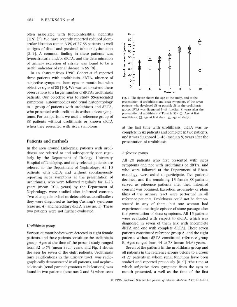
group. Ages at the time of the present study ranged
from 32 to 79 (mean 53±1) years, and Fig. 1 shows
the ages for seven of the eight patients. Urolithiasis
(any calcifications in the urinary tract) was radio-
graphically demonstrated in all patients, and nephro-
calcinosis (renal parenchymatous calcifications) was
found in two patients (case nos 2 and 3) when seen
*
Fig. 1 The figure shows the age at the study, and at the
presentation of urolithiasis and sicca symptoms, of the seven
patients who developed SS or possible SS in the urolithiasis
group. dRTA was diagnosed 1–48 (median 8) years after the
presentation of urolithiasis. (* Possible SS). D, Age at first
urolithiasis ; *, age at first sicca; ^, age at study.
at the first time with urolithiasis. dRTA was in-
complete in six patients and complete in two patients,
and it was diagnosed 1–48 (median 8) years after the
presentation of urolithiasis.
Reference groups
All 20 patients who first presented with sicca
symptoms and not with urolithiasis or dRTA, and
who were followed at the Department of Rheu-
matology, were asked to participate. Five patients
declined, and the remaining 15 female SS patients
served as reference patients after their informed
consent was obtained. Excretion urography or plain
films of the urinary tract were performed in all
reference patients. Urolithiasis could not be demon-
strated in any of them, but one woman had
experienced one single episode of stone passage after
the presentation of sicca symptoms. All 15 patients
were evaluated with respect to dRTA, which was
diagnosed in seven of them (six with incomplete
dRTA and one with complete dRTA). These seven
patients constituted reference group A, and the eight
patients without dRTA constituted reference group
B. Ages ranged from 44 to 78 (mean 64±6) years.
Seven of the patients in the urolithiasis group and
all patients in the reference groups belong to a group
of 27 patients in whom renal functions have been
studied and reported previously [8, 9]. The time at
which subjective sicca symptoms from the eyes or
mouth presented, a well as the time of the first
# 1996 Blackwell Science Ltd Journal of Internal Medicine 239 : 483–488
UROLITHIASIS–SJO> GREN’S SYNDROME 485
urolithiasis, was judged from information in the
patient records of the Departments of Nephrology
and Rheumathology, and by interviewing the
patients with a standardized questionnaire.
The classification criteria described by Daniels &
Talal [11] were used, because these criteria differ
between definite SS and possible SS. Kerato-
conjunctivitis siccas was established by a positive
Rose–Bengal staining and Schirmer test !5 mm per
5 min or reduced tear meniscus and reduced tear
film break-up time (BUT) %10 s. The oral component
of the syndrome was confirmed by labial salivary
gland biopsy demonstrating focal sialoadenitis with a
focus score above 1. Biopsy was carried out only in
patients with sialometry showing reduced, whole
unstimulated saliva (!1±5 mL 15 min−"). A diag-
nosis of possible SS was established if only the oral or
ocular component was confirmed, and if auto-
antibodies, hypergammaglobulinemia, or interstitial
nephritis}dRTA was demonstrated.
Renal acidification capacity
Complete dRTA was diagnosed in patients with
metabolic acidosis (blood base excess below
®6 mmol L−" in at least two different specimens) and
a urine pH above 5±5. An oral ammonium chloride
loading test was performed in all patients with a
urine pH above 5±5 if complete dRTA was absent
[12, 13]. Incomplete dRTA was diagnosed if urine
pH did not decrease below 5±5 during ammonium
chloride loading.
Kidney biopsy
A kidney biopsy was done after informed consent. It
was performed in patients with reduced glomerular
filtration rate (GFR) and}or tubular defects including
dRTA. The histopathological examinations, which
were performed by the same kidney pathologist
(S.E.), included immunohistochemistry for detection
of deposits of immunoglobulins (IgG, IgM, IgA),
complement factors (C3, C1q) and fibrinogen. 1–2
µm sections for light microscopy were stained
according to the periodic acid-Schiff and periodic acid-
silver methenamine methods. The nephron, vessels
and the interstitium were analysed for inflammatory,
atrophic, fibrotic and atherosclerotic lesions. The
diagnosis of chronic TIN was based on the presence
of mononuclear interstitial inflammation and varying
degrees of interstitial fibrosis and tubular atrophy in
the absence of atherosclerotic lesions.
Other tests
Serum levels of total IgG were measured by a
nephelometric method (normal range: 7±0–
15±0 g L−"). Antinuclear antibodies (ANA), anti-
mitochondrial antibodies (AMA), and anti-smooth
muscle antibodies (SMA) of IgG class were analysed
by indirect immunofluoroscence (IIF) microscopy
using unfixed cryostat sections of rat tissues (liver,
kidney and stomach) as antigen substrates [14]. Sera
containing ANA were further analysed by IIF mi-
croscopy for the presence of antibodies against
double-stranded (ds) DNA using Chritidia luciliae as
antigen substrate (Sci MedX, Denville, NJ, USA).
Anti-SS-A and anti-SS-B antibodies were detected by
means of double radial immunodiffusion (Immuno-
concepts, Sacramento, CA, USA). Anti-thyroid micro-
somal antibodies of IgG class were analysed by IIF
microscopy using acetone-fixed cryostat sections of
human thyroid, and anti-thyroglobulin antibodies
were detected by passive haemagglutination (Murex
Diagnostics Ltd, Dartford, UK). Rheumatoid factor
(RF) was determined by a latex bead agglutination
method (Roche, Basel, Switzerland). Anti-cardiolipin
antibodies of IgG class were analysed by enzyme-
linked immunosorbent assay (ELISA) [15]. Auto-
antibody tests were considered positive at titres of &1:100 for ANA, SMA, AMA, and antibodies against
cytoplasmatic thyroid antigens, and at titres of
&1:10 for anti-dsDNA antibodies and antibodies
against thyroglobulin. RF was considered positive at
values &30 WHO units, and anti-cardiolipin anti-
bodies at values "3 units. Anti-SS-A}SS-B were
considered positive when undiluted patient serum
caused precipitation of the antigens.
Statistics
Fischer’s exact test was used for comparison of
frequencies, and Student’s t-test was used for com-
parison of serum IgG in the two groups (computer
program: StatView 4.0).
Results
Eight of 10 patients with dRTA had different autoanti-
bodies, and these eight patients constituted the
# 1996 Blackwell Science Ltd Journal of Internal Medicine 239 : 483–488
486 P. ERIKSSON et al.
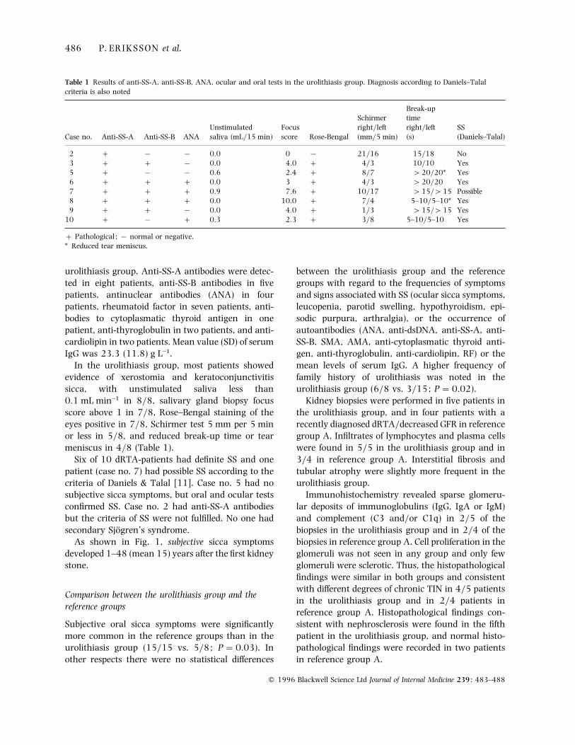
Table 1 Results of anti-SS-A, anti-SS-B, ANA, ocular and oral tests in the urolithiasis group. Diagnosis according to Daniels–Talal
criteria is also noted
Break-up
Schirmer time
Unstimulated Focus right}left right}left SS
Case no. Anti-SS-A Anti-SS-B ANA saliva (mL}15 min) score Rose-Bengal (mm}5 min) (s) (Daniels–Talal)
2 ® ® 0±0 0 ® 21}16 15}18 No
3 ® 0±0 4±0 4}3 10}10 Yes
5 ® ® 0±6 2±4 8}7 "20}20* Yes
6 0±0 3 4}3 "20}20 Yes
7 0±9 7±6 10}17 "15}"15 Possible
8 0±0 10±0 7}4 5–10}5–10* Yes
9 ® 0±0 4±0 1}3 "15}"15 Yes
10 ® 0±3 2±3 3}8 5–10}5–10 Yes
Pathological ; ® normal or negative.
* Reduced tear meniscus.
urolithiasis group. Anti-SS-A antibodies were detec-
ted in eight patients, anti-SS-B antibodies in five
patients, antinuclear antibodies (ANA) in four
patients, rheumatoid factor in seven patients, anti-
bodies to cytoplasmatic thyroid antigen in one
patient, anti-thyroglobulin in two patients, and anti-
cardiolipin in two patients. Mean value (SD) of serum
IgG was 23±3 (11±8) g L−".
In the urolithiasis group, most patients showed
evidence of xerostomia and keratoconjunctivitis
sicca, with unstimulated saliva less than
0±1 mL min−" in 8}8, salivary gland biopsy focus
score above 1 in 7}8, Rose–Bengal staining of the
eyes positive in 7}8, Schirmer test 5 mm per 5 min
or less in 5}8, and reduced break-up time or tear
meniscus in 4}8 (Table 1).
Six of 10 dRTA-patients had definite SS and one
patient (case no. 7) had possible SS according to the
criteria of Daniels & Talal [11]. Case no. 5 had no
subjective sicca symptoms, but oral and ocular tests
confirmed SS. Case no. 2 had anti-SS-A antibodies
but the criteria of SS were not fulfilled. No one had
secondary Sjo$ gren’s syndrome.
As shown in Fig. 1, subjective sicca symptoms
developed 1–48 (mean 15) years after the first kidney
stone.
Comparison between the urolithiasis group and the
reference groups
Subjective oral sicca symptoms were significantly
more common in the reference groups than in the
urolithiasis group (15}15 vs. 5}8; P¯0±03). In
other respects there were no statistical differences
between the urolithiasis group and the reference
groups with regard to the frequencies of symptoms
and signs associated with SS (ocular sicca symptoms,
leucopenia, parotid swelling, hypothyroidism, epi-
sodic purpura, arthralgia), or the occurrence of
autoantibodies (ANA, anti-dsDNA, anti-SS-A, anti-
SS-B, SMA, AMA, anti-cytoplasmatic thyroid anti-
gen, anti-thyroglobulin, anti-cardiolipin, RF) or the
mean levels of serum IgG. A higher frequency of
family history of urolithiasis was noted in the
urolithiasis group (6}8 vs. 3}15; P¯0±02).
Kidney biopsies were performed in five patients in
the urolithiasis group, and in four patients with a
recently diagnosed dRTA}decreased GFR in reference
group A. Infiltrates of lymphocytes and plasma cells
were found in 5}5 in the urolithiasis group and in
3}4 in reference group A. Interstitial fibrosis and
tubular atrophy were slightly more frequent in the
urolithiasis group.
Immunohistochemistry revealed sparse glomeru-
lar deposits of immunoglobulins (IgG, IgA or IgM)
and complement (C3 and}or C1q) in 2}5 of the
biopsies in the urolithiasis group and in 2}4 of the
biopsies in reference group A. Cell proliferation in the
glomeruli was not seen in any group and only few
glomeruli were sclerotic. Thus, the histopathological
findings were similar in both groups and consistent
with different degrees of chronic TIN in 4}5 patients
in the urolithiasis group and in 2}4 patients in
reference group A. Histopathological findings con-
sistent with nephrosclerosis were found in the fifth
patient in the urolithiasis group, and normal histo-
pathological findings were recorded in two patients
in reference group A.
# 1996 Blackwell Science Ltd Journal of Internal Medicine 239 : 483–488
UROLITHIASIS–SJO> GREN’S SYNDROME 487
Discussion
Distal renal tubular acidosis (dRTA) is sometimes
associated with primary Sjo$ gren’s syndrome [1]. We
have recently studied dRTA in 27 SS patients [8], but
in the present study the problem was approached
from a different perspective : 10 patients with uro-
lithiasis and dRTA but without subjective symptoms
of SS at the time of the first urolithiasis were
investigated. During the study period, a diagnosis of
SS or possible SS was established in 7}10 dRTA
patients. In three other reports on dRTA patients
with 44, 48 and 87 patients studied, the pathogenesis
of dRTA was judged as immune-related in 25, 23
and 39%, respectively [1, 16, 17]. Our material is
smaller but the results agree with these reports in
which immune-related dRTA was the commonest
form of dRTA seen in adults.
We found that subjective sicca symptoms presented
1–48 (mean 15) years after the first episode of
urolithiasis. Renal disease preceding the onset of
classical manifestations of SS has also been reported
by Tu et al. [18] and by Moutsopoulos et al. [19].
Objective ocular and oral tests during the period
when the patients were followed would possibly have
shown signs of Sjo$ gren’s syndrome earlier. This is
illustrated by case no. 5, who had no subjective sicca
symptoms, but SS was diagnosed according to the
Daniels–Talal criteria [11]. At least six similar
patients with dRTA and objective signs of SS, but
without subjective sicca symptoms, have been re-
ported previously [10, 20, 21]. Thus, SS can be
diagnosed in dRTA patients even in the absence of
subjective sicca symptoms. In our study, anti-SS-A
antibodies were present in eight of 10 patients with
urolithiasis and dRTA, one of which (case no. 2) did
not fulfil the SS criteria. Anti-SS-A is unusual in a
normal population [22, 23].
ANA was found in found in four patients in the
urolithiasis group, but in no instance were anti-DNA
antibodies recorded and none of the patients fulfilled
the criteria for having SLE. Also, in contrast to SS,
isolated TIN in uncommon in SLE [24].
Are there any differences between the urolithiasis
group and the reference groups of SS patients? To
look for possible differences between the two groups,
clinical and laboratory features, as well as renal
histopathology, were compared.
Except for the occurrence of oral sicca symptoms,
the frequencies of various autoantibodies and SS-
related symptoms, as well as serum levels of IgG,
were similar in the groups. The data of renal
histopathology show that chronic TIN was present
in four of five patients in the urolithiasis group and in
two of four SS patients in reference group A. Renal
histopathological findings have previously been re-
ported in dRTA patients without sicca symptoms.
Feest et al. reported that interstitial lymphocyte
infiltration and fibrosis consistent with chronic TIN
were common findings both in dRTA patients with,
and in those without immunological abnormalities
[25]. Similar results have been reported by others
[20, 21, 26]. In patients with sicca symptoms and
dRTA due to SS, a few reports of renal histo-
pathological findings exist : chronic TIN was present
in most cases [18, 27–29]. Thus, the histopatho-
logical findings seem to be similar in patients
presenting with urolithiasis}dRTA and in those
presenting with subjective sicca symptoms, and later
developing dRTA.
In summary in this study, features of SS and}or
presence of anti-SS-A antibodies were common in a
group of female patients with urolithiasis and dRTA;
we also found that dRTA and urolithiasis can precede
subjective sicca symptoms, and that patients with
dRTA may have SS in the absence of subjective sicca
symptoms. Except for oral sicca symptoms and a
family history of urolithiasis there were no clinical
differences between the urolithiasis group and the
reference groups of SS patients. We hypothesize the
possibility of a Sjo$ gren-related renal disease, charac-
terized by urolithiasis and}or dRTA and antibodies to
SS-A, whether subjective sicca symptoms are pre-
sent or not. Further investigation will be needed to
prove whether the renal disease precedes SS, or
whether it is related to subclinical SS with auto-
antibodies but without subjective or objective sicca
symptoms.
Testing for anti-SS-A is a valuable tool in the
diagnosis of SS in patients with urolithiasis and
dRTA. Patients with anti-SS-A antibodies should be
tested further for keratoconjunctivitis sicca and}or
xerostomia, even in the absence of subjective sicca
symptoms. At present, SS cannot be cured, but it
seems important to establish a correct diagnosis, as
other manifestations of SS such as vitamin B"#
deficiency [30] and thyroid hypofunction secondary
to autoimmune thyroiditis [31] can be effectively
treated.
# 1996 Blackwell Science Ltd Journal of Internal Medicine 239 : 483–488
488 P. ERIKSSON et al.
Acknowledgements
This work was supported by Grants from the County
Council of O> stergo$ tland, the University Hospital,
Linko$ ping and the Faculty of Health Science, Uni-
versity of Linko$ ping, Sweden.
The authors wish to thank medical student Peter
Christakos, College of Medicine, University of
Vermont, for correcting to the language in the
manuscript.
References
1 Caruana RJ, Buckalew VM. The syndrome of distal (type 1)
renal tubular acidosis. Medicine 1988; 67 : 84–99.
2 Buckalew VM. Calcium nephrolithiasis and renal tubular
acidosis. In : Coe FL, Favus MJ, eds. Disorders of Bone and
Mineral Metabolism, 1st edn. New York: Raven Press, 1992;
729–56.
3 Wrong OM, Feest TG. The natural history of distal renal
tubular acidosis. Contrib Nephrol 1980; 21 : 137–44.
4 Mason AMS, Golding PL. Hyperglobulinemic renal tubular
acidosis : a report of nine cases. Br Med J 1970; 3 : 143–6.
5 McCurdy DK, Cornwell GG, DePratti VJ. Hyperglobulinemic
renal tubular acidosis. Ann Intern Med 1967; 67 : 110–17.
6 Hansen B, Manthorpe R. Antibodies against SS-B}La and SS-
A}Ro antigens in patients with primary Sjo$ gren’s syndrome.
Scand J Rheumatol 1986; Suppl. 61: 93–7.
7 Winer RL. Sjo$ gren’s syndrome. In: Grishman E, Churg J,
Needle MA, Venkataseshan VS, eds. The Kidney in Collagen
Vascular Diseases. New York: Raven Press, 1993, 179–87.
8 Eriksson P, Denneberg T, Larsson L, Lindstro$ m F. Biochemical
markers of renal disease in Primary Sjo$ grens’s syndrome.
Scand J Urol Nephrol 1994 (in press).
9 Eriksson P, Denneberg T, Granerus G, Lindstro$ m F. Glomerular
filtration rate in Primary Sjo$ gren’s Syndrome with renal
disease. Scand J Urol Nephrol 1995; 29 : 383–92.
10 Gobert P, Vanhille Ph, Mougenot B, Lemaı# re V, Fleury D.
Nephrolithiasis revealing Sjo$ gren’s syndrome. Kidney Int
1990; 38 : 1237–8.
11 Daniels TE, Talal N. Diagnosis and differential diagnosis in
Sjo$ gren’s syndrome. In: Talal N, Moutsopoulos HM, Kassan
SS, eds. SjoX gren’s Syndrome : Clinical and Immunological Aspects.
Berlin : Springer Verlag, 1987; 193–9.
12 Backman U, Danielsson BG, Sothell M. A short duration renal
acidification test. Scand J Urol Nephrol 1976; Suppl. 35:
33–48.
13 Backman U, Danielsson BG, Johansson G, Ljunghall S,
Wikstro$ m S. Incidence and clinical importance of renal tubular
defects in recurrent renal stone formers. Nephron 1980; 25 :
96–101.
14 Vrethem M, Skogh T, Berlin G, Ernerudh J. Autoantibodies
versus clinical symptoms in blood donors. J Rheumatol 1992;
19 : 1919–21.
15 Vrethem M, Ernerudh J, Lindstro$ m F, Olsson JE. Cerebral
ishemia associated with anti-cardiolipin antibodies. Acta
Neurol Scand 1992; 85 : 412.
16 Harrington TM, Bunch TW, van der Berg CJ. Renal tubular
acidosis – a new look at treatment of musculoskeletal and
renal disease. Mayo Clin Proc 1983; 58 : 354–60.
17 Wrong OM, Feest TG, MacIver AG. Immune-related potassium-
losing interstitial nephritis : a comparison with distal renal
tubular acidosis. Q J Med 1993; 86 : 513–34.
18 Tu WH, Shearn MA, Lee JC, Hopper J. Interstitial nephritis in
Sjo$ gren’s syndrome. Ann Intern Med 1968; 6 : 1163–70.
19 Moutsopoulos HM, Cledes J, Skopouli FN, Elisaf M, Youinou P.
Nephrocalcinosis in Sjo$ gren’s syndrome: a late sequela of
renal tubular acidosis. J Intern Med 1991; 230 : 187–91.
20 Andrassy K, Gebest J, Tan E, Thoenes W, Ritz E. Interstitial
nephritis in a patient with atypical Sjo$ gren’s syndrome. Klin
Wochenschr 1980; 58 : 563–7.
21 Shioji R, Furuyama T, Onodera S, Sito H, Ito H, Sasaki Y.
Sjo$ gren’s syndrome and renal tubular acidosis. Am J Med
1970; 48 : 456–63.
22 Calmes M, Bartholomew B. SS-A (Ro) antibody in random
mother-infant pairs. J Clin Pathol 1985; 38 : 73–5.
23 Lindstro$ m F, Eriksson P, Tejle K, Skogh T. IgG subclasses of
anti-SS-A}Ro in patients with primary Sjo$ gren’s syndrome.
Clin Immunol Immunopathol 1994; 73 : 358–61.
24 Graninger WB, Steinberg AD, Meron G, Smolen JS. Interstitial
nephritis in patients with systemic lupus erythematodes : a
manifestation of concomitant Sjo$ gren’s syndrome? Clin Exp
Rheumatol 1991; 9 : 41–5.
25 Feest TG, Lockwood CM, Morley AR, Uff JS. Renal histology
and immunopathology in distal renal tubular acidosis. Clin
Nephrol 1978; 10 : 187–90.
26 Pasternack A, Linder E. Renal tubular acidosis : an immuno-
pathological study on four patients. Clin Exp Immunol 1970;
7 :115–23.
27 Matsumura R, Kondo Y, Sugiyama T, Sueishi M, Koike T,
Takabayashi K et al. Immunohistochemistical identification of
infiltrating mononuclear cells in tubulointerstitial nephritis
associated with Sjo$ gren’s syndrome. Clin Nephrol 1988; 30 :
335–40.
28 Siamopoulos KC, Mavridis AK, Elisaf M, Drosos AA,
Moutsopoulos HM. Kidney involvement in primary Sjo$ gren’s
syndrome. Scand J Rheumatol 1986; Suppl 61: 156–60.
29 Talal N, Zisman E, Schur PH. Renal tubular acidosis, glomeru-
lonephritis and immunologic factors in Sjo$ gren’s syndrome.
Arthritis Rheum 1968; 11 : 774–86.
30 Wegelius O, Fyhrquist F, Adner P-L. Sjo$ gren’s syndrome
associated with vitamin B"#
deficiency. Acta Rheumatol Scand
1970; 16 : 184–90.
31 Karsh J, Pavlidids N, Weintraub BD, Moutsopoulos HM.
Thyroid disease in Sjo$ gren’s syndrome. Arthritis Rheum
1980; 23 : 1326–9.
Received 15 August 1995; accepted 30 November 1995.
Correspondence : Dr Per Eriksson MD, Department of Internal
Medicine, La$ nssjukhuset Ryhov, S-551 85 Jo$ nko$ ping, Sweden.
# 1996 Blackwell Science Ltd Journal of Internal Medicine 239 : 483–488