Embed Size (px)
Citation preview

293Urolithiasis
GUIDELINES ON UROLITHIASIS
(Update March 2013)
C. Türk (chair), T. Knoll (vice-chair), A. Petrik, K. Sarica, C. Seitz, A. Skolarikos, M. Straub
Classification of stonesUrinary stones can be classified according to the following aspects: stone size, stone location, X-ray characteristics of stone, aetiology of stone formation, stone composition (mineralogy), and risk group for recurrent stone formation (Tables 1-3).
Table 1: X-ray characteristicsRadiopaque Poor radiopaque RadiolucentCalcium oxalate dihydrate
Magnesium ammonium phos-phate
Uric acid
Calcium oxalate monohydrate
Apatite Ammonium urate
Calcium phos-phates
Cystine Xanthine
2,8-dihydroxyade-nine‘Drug-stones’

294 Urolithiasis
Table 2: Stones classified according to their aetiologyNon infection stones
Infection stones
Genetic stones
Drug stones
Calcium oxalates
Magnesium ammonium phosphate
Cystine e.g. indinavir (see extended document)
Calcium phosphates
Carbonate apatite
Xanthine
Uric acid Ammonium urate
2,8-dihydroxy-adenine
Table 3: Stones classified by their compositionChemical composition MineralCalcium oxalate monohydrate whewelliteCalcium-oxalate-dihydrate wheddeliteUric acid dihydrate uriciteAmmonium urateMagnesium ammonium phosphate struviteCarbonate apatite (phosphate) dahlliteCalcium hydrogenphosphate brushiteCystineXanthine2,8-dihydroxyadenine‘Drug stones’
Risk groups for stone formationThe risk status of a stone former is of particular interest as it defines both probability of recurrence or (re)growth of stonesand is imperative for pharmacological treatment.

295Urolithiasis
Table 4: High risk stone formersGeneral factorsEarly onset of urolithiasis in life (especially children and teenagers)Familial stone formationBrushite containing stones (calcium hydrogen phosphate; CaHPO4.2H2O)
Uric acid and urate containing stonesInfection stonesSolitary kidney (The solitary kidney itself does not present an increased risk of stone formation, but prevention of stone recurrence is more important)Diseases associated with stone formationHyperparathyroidismNephrocalcinosisGastrointestinal diseases or disorders (e.g. jejuno-ileal bypass, intestinal resection, Crohn’s disease, malabsorptive conditions, enteric hyperoxaluaria after urinary diversion and bariatric surgery)SarcoidosisGenetically determined stone formationCystinuria (type A, B, AB)Primary hyperoxaluria (PH)Renal tubular acidosis (RTA) type I2,8-dihydroxyadenineXanthinuriaLesch-Nyhan-SyndromeCystic fibrosisDrugs associated with stone formation (see Chapter 11 extended text)Anatomical abnormalities associated with stone formationMedullary sponge kidney (tubular ectasia)

296 Urolithiasis
UPJ obstructionCalyceal diverticulum, calyceal cystUreteral strictureVesico-uretero-renal refluxHorseshoe kidneyUreterocele
DIAGNOSISDiagnostic imagingStandard evaluation of a patient includes taking a detailed medical history and physical examination. The clinical diagno-sis should be supported by appropriate imaging.
Recommendation LE GRWith fever or solitary kidney, and when diagno-sis is doubtful, immediate imaging is indicated.
4 A*
*Upgraded following panel consensus.
If available, ultrasonography should be used as the primary diagnostic imaging tool although pain relief, or any other emergency measures should not be delayed by imaging assessments. KUB should not be performed if non-contrast enhanced computed tomography (NCCT) is considered, but KUB can differentiate between radiolucent and radiopaque stones and serve for comparison during follow-up.
Evaluation of patients with acute flank painRecommendation LE GRNCCT should be used to confirm stone diagno-sis in patients with acute flank pain, because it is superior to IVU.
1a A

297Urolithiasis
Some drug stones like indinavir stones are not detectable on NCCT.
Recommendation LE GRA renal contrast study (enhanced CT or IVU) is indicated when planning treatment for renal stones.
3 A*
*Upgraded following panel consensus.
Biochemical work-upEach emergency patient with urolithiasis needs a succinct biochemical work-up of urine and blood besides imaging studies; no difference is made between high- and low-risk patients.
Recommendations: Basic analysis emergency stone patientUrine GRUrinary sediment/dipstick test out of spot urine sam-ple for: red cells / white cells / nitrite / urine pH level by approximation.
A*
Urine culture or microscopy. ABloodSerum blood sample creatinine / uric acid / ionized calcium / sodium / potassium / CRP.
A*
Blood cell count. A*If intervention is likely or planned: Coagulation test (PTT and INR).
A*
*Upgraded following panel consensus.
Examination of sodium, potassium, CRP, and blood coagula-tion time can be omitted in the non-emergency stone patient.
Patients at high risk for stone recurrences should undergo a

298 Urolithiasis
more specific analytical programme (see section on Metabolic Evaluation below).
Analysis of stone composition should be performed in all first-time stone formers (GR: A) and will need redoing if changes are expected. The preferred analytical procedures are:• X-ray diffraction (XRD)• Infrared spectroscopy (IRS)Wet chemistry is generally deemed to be obsolete.
Acute treatment of a patient with renal colicPain relief is the first therapeutic step in patients with an acute stone episode.
Recommendations for pain relief during and prevention of recurrent renal colic
LE GR
First choice: start with an NSAID, e.g. diclofenac*, indomethacin or ibuprofen.**
1b A
Second choice: hydromorphine, pentazocine and tramadol.
4 C
Use a-blockers to reduce recurrent colic. 1a AGFR = glomerular filtration rate; NSAID = non-steroidal anti-inflammatory drug.*Caution: Diclofenac sodium affects GFR in patients with reduced renal function, but not in patients with normal renal function (LE: 2a). ** Recommended to counteract recurrent pain after renal colic. (see extended document section 5.3)
If analgesia cannot be achieved medically, drainage, using stenting or percutaneous nephrostomy, or stone removal, should be performed.
Management of sepsis in the obstructed kidneyThe obstructed, infected kidney is a urological emergency.

299Urolithiasis
Recommendations LE GRFor sepsis with obstructing stones, the collect-ing system should be urgently decompressed, using either percutaneous drainage or ureteral stenting.
1b A
Definitive treatment of the stone should be delayed until sepsis is resolved.
1b A
In exceptional cases, with severe sepsis and/or the forma-tion of abscesses, an emergency nephrectomy may become necessary.
Recommendations - Further Measures GRCollect urine for antibiogram following decompres-sion.
A*
Start antibiotics immediately thereafter (+ intensive care if necessary).Revisit antibiotic treatment regimen following antibio-gram findings.
* Upgraded based on panel consensus.
Stone reliefWhen deciding between active stone removal and conserva-tive treatment using MET, it is important to consider the individual circumstances of a patient that may affect treat-ment decisions.
300 Urolithiasis
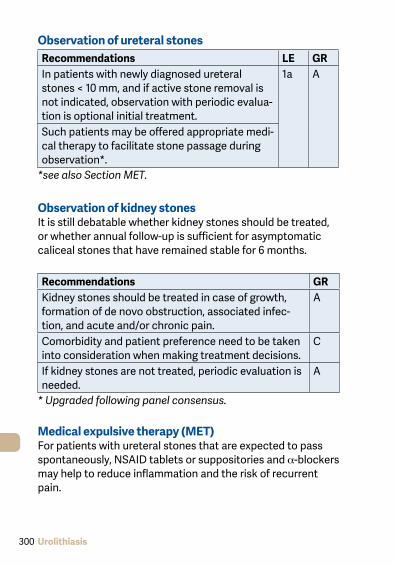
Observation of ureteral stonesRecommendations LE GRIn patients with newly diagnosed ureteral stones < 10 mm, and if active stone removal is not indicated, observation with periodic evalua-tion is optional initial treatment.
1a A
Such patients may be offered appropriate medi-cal therapy to facilitate stone passage during observation*.
*see also Section MET.
Observation of kidney stonesIt is still debatable whether kidney stones should be treated, or whether annual follow-up is sufficient for asymptomatic caliceal stones that have remained stable for 6 months.
Recommendations GRKidney stones should be treated in case of growth, formation of de novo obstruction, associated infec-tion, and acute and/or chronic pain.
A
Comorbidity and patient preference need to be taken into consideration when making treatment decisions.
C
If kidney stones are not treated, periodic evaluation is needed.
A
* Upgraded following panel consensus.
Medical expulsive therapy (MET)For patients with ureteral stones that are expected to pass spontaneously, NSAID tablets or suppositories and a-blockers may help to reduce inflammation and the risk of recurrent pain.

301Urolithiasis
Recommendations for MET LE GRFor MET, a-blockers are recommended. APatients should be informed about the attend-ant risks of MET, including associated drug side effects, and should be informed that it is admin-istered as ‘off-label’**.
A*
Patients, who elect for an attempt at sponta-neous passage or MET, should have well-con-trolled pain, no clinical evidence of sepsis, and adequate renal functional reserve.
A
Patients should be followed to monitor stone position and to assess for hydronephrosis.
4 A*
*Upgraded following panel consensus.** MET using a-blockers in children and during pregnancy can-
not be recommended due to the limited data in this specific population.
Statements LEThere is good evidence that MET accelerates spon-taneous passage of ureteral stones and fragments generated with SWL limits pain.
1
No recommendation for the use of corticosteroids in combination with a-blockers in MET can be made, due to limited data.
1b
Chemolytic dissolution of stonesOral or percutaneous irrigation chemolysis of stones can be a useful first-line therapy or an adjunct to SWL, PNL, URS, or open surgery to support elimination of residual fragments. However, its use as first-line therapy may take weeks to be effective.

302 Urolithiasis
Percutaneous irrigation chemolysis
Recommendations GRIn percutaneous chemolysis, at least two nephrosto-my catheters should be used to allow irrigation of the renal collecting system, while preventing chemolytic fluid draining into the bladder and reducing the risk of increased intrarenal pressure*.
A
Pressure- and flow-controlled systems should be used if available.
* Alternatively, one nephrostomy catheter with a JJ stent and bladder catheter can serve as a through-flow system pre-venting high pressure.
Methods of percutaneous irrigation chemolysisPercutaneous irrigation chemolysis is rarely used; it may be an option for infection stones (using 10% Hemiacidrin at a pH of 3,5 -4) and for uric acid and cystine stones (using THAM [Trihydroxymethylaminomethan], 0.3 or 0.6mol/L, pH 8.5-9.0).
For uric acid stones oral chemolysis is preferred.
Oral chemolysisOral chemolitholysis is efficient for uric acid calculi only. The urine pH should be adjusted to between 6.5 and 7.2.
Recommendations GRThe dosage of alkalising medication must be modified by the patient according to the urine pH, which is a direct consequence of the alkalising medication.
A
Dipstick monitoring of urine pH by the patient is required at regular intervals during the day. Morning urine must be included.
A

303Urolithiasis
Careful monitoring of radiolucent stones during/after therapy is imperative.
A
The physician should clearly inform the patient of the significance of compliance.
A
SWLThe success rate for SWL will depend on the efficacy of the lithotripter and on:• size, location (ureteral, pelvic or calyceal), and composition
(hardness) of the stones;• patient’s habitus;• performance of SWL.
Contraindications of SWLContraindications to the use of SWL are few, but include:• pregnancy;• bleeding diatheses;• uncontrolled urinary tract infections (UTIs);• severe skeletal malformations and severe obesity, which
prevent targeting of the stone;• arterial aneurism in the vicinity of the stone;• anatomical obstruction distal of the stone.
Stenting prior to SWLKidney stonesA JJ stent reduces the risk of renal colic and obstruction, but does not reduce formation of steinstrasse or infective compli-cations.
Recommendation - stenting & SWL LE GRRoutine stenting is not recommended as part of SWL treatment of ureteral stones.
1b A

304 Urolithiasis
Best clinical practice (best performance)PacemakerPatients with a pacemaker can be treated with SWL, provided that appropriate technical precautions are taken; patients with implanted cardioverter defibrillators must be managed with special care (firing mode temporarily reprogrammed dur-ing SWL treatment). However, this might not be necessary with new-generation lithotripters.
Recommendation - Shock wave rate LE GRThe optimal shock wave frequency is 1.0 (to 1.5) Hz.
1a A
Number of shock waves, energy setting and repeat treatment sessions• The number of shock waves that can be delivered at each
session depends on the type of lithotripter and shockwave power.• Starting SWL on a lower energy setting with step-wise
power (and SWL sequence) ramping prevents renal injury.• Clinical experience has shown that repeat sessions are
feasible (within 1 day for ureteral stones).
Procedural controlResults of treatment are operator dependent. Careful imaging control of localisation will contribute to outcome quality.
Pain controlCareful control of pain during treatment is necessary to limit pain-induced movements and excessive respiratory excur-sions.
Antibiotic prophylaxisNo standard prophylaxis prior to SWL is recommended.

305Urolithiasis
Recommendation LE GRIn case of infected stones or bacteriuria, antibiotics should be given prior to SWL.
4 C
Medical expulsive therapy (MET) after SWL can expedite expulsion and enhance stone-free rates.
Percutaneous nephrolitholapaxy (PNL)
Recommendation GRUltrasonic, ballistic and Ho:YAG devices are recom-mended for intracorporeal lithotripsy using rigid nephroscopes.
A*
When using flexible instruments, the Ho:YAG laser is currently the most effective device available.
* Upgraded following panel consensus.
Best clinical practiceContraindications:• all contraindications for general anaesthesia apply;• untreated UTI;• atypical bowel interposition;• tumour in the presumptive access tract area;• potential malignant kidney tumour; • pregnancy.
Pre-operative recommendation - imaging GRPreprocedural imaging, including contrast medium where possible or retrograde study when starting the procedure, is mandatory to assess stone comprehen-siveness, view the anatomy of the collecting system, and ensure safe access to the kidney stone.
A*
* Upgraded based on panel consensus.

306 Urolithiasis
Positioning of the patient: prone or supine?Traditionally, the patient is positioned prone for PNL, supine position is also possible, showing advantages in shorter operating time, the possibility of simultaneous ret-rograde transurethral manipulation, and easier anaesthesia. Disadvantages are limited manoeuvrability of instruments and the need of appropriate equipment.
Nephrostomy and stents after PNLRecommendation LE GRIn uncomplicated cases, tubeless (without nephrostomy tube) or totally tubeless (without nephrostomy tube and without ureteral stent) PNL procedures provide a safe alternative.
1b A
Ureterorenoscopy (URS)(including retrograde access to renal collecting system)
Best clinical practice in URSBefore the procedure, the following information should be sought and actions taken (LE: 4):• Patient history;• physical examination (i.e. to detect anatomical and con-
genital abnormalities);• thrombocyte aggregation inhibitors/anticoagulation
(anti-platelet drugs) treatment should be discontinued. However, URS can be performed in patients with bleeding disorders, with only a moderate increase in complications;
• imaging.
Recommendation GRShort-term antibiotic prophylaxis should be adminis-tered.
A*

307Urolithiasis
ContraindicationsApart from general considerations, e.g. with general anaes-thesia or untreated UTIs, URS can be performed in all patients without any specific contraindications.
Access to the upper urinary tractMost interventions are performed under general anaesthesia, although local or spinal anaesthesia are possible. Intravenous sedation with miniaturized instruments is especially suitable for female patients with distal ureteral stones. Antegrade URS is an option for large, impacted proximal ureteral calculi.
Safety aspectsFluoroscopic equipment must be available in the operating room. If ureteral access is not possible, the insertion of a JJ stent followed by URS after a delay of 7-14 days offers an appropriate alternative to dilatation.
Recommendation GRPlacement of a safety wire is recommended. A*
*Upgraded following panel consensus.
Ureteral access sheathsHydrophilic-coated ureteral access sheaths (UAS), can be inserted via a guide wire, with the tip placed in the proximal ureter. Ureteral access sheaths allow easy multiple access to the upper urinary tract and therefore significantly facilitate URS. The use of UAS improves vision by establishing a con-tinuous outflow, decrease intrarenal pressure and potentially reduce operating time.
Stone disintegration and extractionThe aim of endourological intervention is complete stone removal. ‘Smash and go’ strategies should be limited to the treatment of large renal stones. For flexible URS (RIRS) only

308 Urolithiasis
baskets made of Nitinol are suitable.
Recommendation LE GRStone extraction using a basket without endo-scopic visualisation of the stone (blind basket-ing) should not be performed.
4 A*
Ho:YAG laser lithotripsy is the preferred method for (flexible) URS.
3 B
*Upgraded following panel consensus.
Stenting before and after URSPre-stenting facilitates ureteroscopic management of stones, improves the stone-free rate, and reduces complications. Following URS, stents should be inserted in patients who are at increased risk of complications.
Recommendation LE GRIn uncomplicated URS, a stent need not be inserted.
1a A
An a-blocker can reduce stent-related symp-toms
1a
Open surgeryMost stones should be approached primarily with PNL, URS, SWL, or a combination of these techniques. Open surgery may be a valid primary treatment option in selected cases.
Indications for open surgery:• Complex stone burden• Treatment failure of SWL and/or PNL, or URS• Intrarenal anatomical abnormalities: infundibular steno-
sis, stone in the calyceal diverticulum (particularly in an anterior calyx), obstruction of the ureteropelvic junction,

309Urolithiasis
stricture if endourologic procedures have failed or are not promising
• Morbid obesity• Skeletal deformity, contractures and fixed deformities of
hips and legs• Comorbidity• Concomitant open surgery• Non-functioning lower pole (partial nephrectomy), non-
functioning kidney (nephrectomy)• Patient choice following failed minimally invasive proce-
dures; the patient may prefer a single procedure and avoid the risk of needing more than one PNL procedure
• Stone in an ectopic kidney where percutaneous access and SWL may be difficult or impossible
• For the paediatric population, the same considerations apply as for adults.
Laparoscopic surgeryIndications for laparoscopic kidney-stone surgery include:• complex stone burden;• failed previous SWL and/or endourological procedures;• anatomical abnormalities;• morbid obesity;• nephrectomy in case of non-functioning kidney.
Indications for laparoscopic ureteral stone surgery include:• large, impacted stones;• multiple ureteral stones;• in cases of concurrent conditions requiring surgery;• when other non-invasive or low-invasive procedures have
failed.
If indicated, for upper ureteral calculi, laparoscopic urolithomy has the highest stone-free rate compared to URS and SWL (LE: 1a).

310 Urolithiasis
Recommendations LE GRLaparoscopic or open surgical stone removal may be considered in rare cases where SWL, URS, and percutaneous URS fail or are unlikely to be successful.
3 C
When expertise is available, laparoscopic sur-gery should be the preferred option before pro-ceeding to open surgery. An exception is com-plex renal stone burden and/or stone location.
3 C
For ureterolithotomy, laparoscopy is recom-mended for large impact stones or when endo-scopic lithotripsy or SWL have failed.
2 B
Indication for active stone removal and selection of procedureUreter:• stones with a low likelihood of spontaneous passage;• persistent pain despite adequate pain medication;• persistent obstruction;• renal insufficiency (renal failure, bilateral obstruction, sin-
gle kidney).Kidney:• stone growth;• stones in high-risk patients for stone formation;• obstruction caused by stones;• infection;• symptomatic stones (e.g. pain, haematuria);• stones > 15 mm;• stones < 15 mm if observation is not the option of choice;• patient preference (medical and social situation);• comorbidity;• choice of treatment.The suspected stone composition might influence the choiceof treatment modality.

311Urolithiasis
Recommendations GRFor asymptomatic caliceal stones in general, active surveillance with an annual follow-up of symptoms and stone status (KUB, ultrasonography [US], NCCT) is an option for 2-3 years, whereas intervention should be considered after this period provided patients are adequately informed.
C
Observation might be associated with a greater risk of necessitating more invasive procedures.
STONE REMOVAL
Recommendations GRUrine culture or urinary microscopy is mandatory before any treatment is planned and urinary infection should be treated ahead of stone removal.
A*
Anticoagulation therapy including salicylates should be stopped before stone removal.
B
If intervention for stone removal is essential and sali-cylate therapy should not be interrupted, retrograde URS is the preferred treatment of choice.
*Upgraded based on panel consensus.
Radiolucent uric acid stones, but not sodium urate or ammo-nium urate stones, can be dissolved by oral chemolysis.

312 Urolithiasis
Selection of procedure for active removal of renal stones**
Fig. 1: Treatment algorithm for renal calculi
* Flexible URS is used less as first-line therapy for renal stones > 1.5 cm.
** The ranking of the recommendations reflects a panel majority vote.
*** see Table 19 extended document
Kidney stone (all but lower pole stone 10-20 mm)
Lower pole stone > 20 mm and < 10 mm: like above
1. PNL2. RIRS or SWL
SWL or Endourology*
SWL or Endourology
1. SWL or RIRS2. PNL
1. Endourology2. SWL
10-20 mmFavourable factors for
SWL***
< 10 mmm
> 20 mm
Yes
No
10-20 mm

313Urolithiasis
Selection of procedure for active stone removal of ureteral stones (GR: A*)
Stone location and size First choice Second choiceProximal ureter < 10 mm SWL URSProximal ureter > 10 mm URS (retrograde or antegrade)
or SWLDistal ureter < 10 mm URS or SWLDistal ureter > 10 mm URS SWL
*Upgraded following panel consensus.
Recommendation GRPercutaneous antegrade removal of proximal ureteral stones is an alternative when SWL is not indicated or has failed, and when the upper urinary tract is not amenable to retrograde URS.
A
SteinstrasseSteinstrasse occurs in 4% to 7% of cases after SWL, the major factor in steinstrasse formation is stone size.
Recommendations LE GRMedical expulsion therapy increases the stone expulsion rate of steinstrasse.
1b A
PCN is indicated for steinstrasse associated with UTI/fever.
4 C
SWL is indicated for steinstrasse when large stone fragments are present.
4 C
Ureteroscopy is indicated for symptomatic steinstrasse and treatment failure.
4 C

314 Urolithiasis
Residual stones
Recommendations LE GRIdentification of biochemical risk factors and appropriate stone prevention is particularly indicated in patients with residual fragments or stones.
1b A
Patients with residual fragments or stones should be followed up regularly to monitor dis-ease course.
4 C
After SWL and URS, MET is recommended using an a-blocker to improve fragment clearance.
1a A
For well-disintegrated stone material in the lower calix, an inversion therapy with simul-taneous mechanical percussion manoeuvre under enforced diuresis may facilitate stone clearance.
1a B
The indication for active stone removal and selection of the procedure is based on the same criteria as for primary stone treatment and also includes repeat SWL.
Management of urinary stones and related problems during pregnancy
Recommendations LE GRUS is the method of choice for practical and safe evaluation of pregnant women.
1a A
Conservative management should be the first-line treatment for all non-complicated cases of urolithiasis in pregnancy (except those that have clinical indications for intervention).
A

315Urolithiasis
If intervention becomes necessary, placement of an internal stent, percutaneous nephros-tomy, or ureteroscopy are treatment options.
3
URS is a reasonable alternative to avoid long-term stenting/drainage.
2a
Regular follow-up until final stone removal is necessary due to higher encrustation tendency of stents during pregnancy.
Pregnancy remains an absolute contraindication for SWL.
Management of stone problems in childrenSpontaneous passage of a stone and of fragments after SWL is more likely to occur in children than in adults (LE: 4). Forpaediatric patients, the indications for SWL and PNL are similar to those in adults, however they pass fragments more easily. Children with renal stones with a diameter up to 20 mm (~300 mm2) are ideal candidates for SWL.
Recommendations GRUltrasound evaluation is the first choice for imaging in children and should include the kidney, filled bladder and adjoining portions of the ureter.
A*
If US does not provide the required information, KUB radiography (or NCCT) should be performed.
B
In all paediatric patients all efforts should be made to collect stone material for analysis, followed by complete metabolic evaluation.
A
*Upgraded from B following panel consensus.

316 Urolithiasis
Table 5: Stones in exceptional situations
Caliceal diverticulum stones
SWL, PNL (if possible) or RIRS (retrograde intrarenal surgery via flexible ureteroscopy).Can also be removed using laparoscopic retroperitoneal surgery.Patients may become asympto-matic due to stone disintegra-tion (SWL) whilst well-disinte-grated stone material remains in the original position.
Horseshoe kidneys Can be treated in line with the stone treatment options described above.Passage of fragments after SWL might be poor.
Stones in pelvic kidneys SWL, RIRS or laparoscopic sur-geryFor obese patients, the options are SWL, PNL, RIRS or open surgery
Stones in transplanted kidneys
PNL, (flexible) URS, SWL.Metabolic evaluation based on stone analysis
Stones formed in urinary division
Individual management necessary.For smaller stones SWL is effective.PNL and antegrade flexible URS frequently used .

317Urolithiasis
Stones formed in a conti-nent reservoir
Present a varied and often dif-ficult problem.Each stone problem must be considered and treated individu-ally.
Stones in patients with neurogenic bladder dis-order
All methods apply based on indi-vidual situation.Careful patient follow up and preventive strategies are impor-tant.In myelomeningocele-patients, latex allergy is common, appro-priate measures needed.
Patients with obstruc-tion of the ureteropelvic junction which needs cor-rection
PNL followed by percutaneous endopyelotomy or open/laparo-scopic surgery, or URS together endopyelothomy with Ho:YAG.Incision with an Acucise balloon catheter might be considered, provided the stones can be prevented from falling into the pelvo-ureteral incision.

318 Urolithiasis
Metabolic evaluation and recurrence preventionStone prevention is based on a reliable stone analysis and basic analysis as mentioned above. Every patient should be assigned to the low- or high risk group for stone formation. For both groups general preventive measures apply:
Fluid intake (drinking advice)
Fluid amount: 2.5-3.0 L/dayCircadian drinkingNeutral pH beveragesDiuresis: 2.0-2.5 L/daySpecific weight of urine: < 1010
Nutritional advice for a balanced diet
Balanced diet*Rich in vegetable and fibreNormal calcium content: 1-1.2 g/dayLimited NaCl content: 4-5 g/dayLimited animal protein content: 0.8-1.0 g/kg/day
Lifestyle advice to normalise general risk factors
BMI: 18-25 kg/m2 (adults)Stress limitation measuresAdequate physical activityBalancing of excessive fluid loss
For patients assigned to the high risk group of stone formers specific laboratory analysis of blood and urine including two consecutive 24-hour urine samples are necessary. For the specific metabolic work-up, the patient should stay on a self-determined diet under normal daily conditions and should ide-ally be stone free for at least 20 days, better 3 months. These findings are the basis for further recommendations:

319Urolithiasis
Recommendations on specific diet LE GRHyperoxaluria Oxalate restriction 2b BHigh sodium excretion Restricted intake of
salt1b A
Small urine volume Increased fluid intake 1b AUrea level indicating a high intake of animal protein
Avoid excessive intake of animal protein
1b A
Recommendations for specific pharmacological treatmentUrinary risk factor Suggested treatment LE GRHypercalciuria Thiazide + potassium
citrate1a A
Hyperoxaluria Oxalate restriction 2b AEnteric hyperoxaluria Potassium citrate 3-4 C
Calcium supplement 2 BOxalate absorption 3 B
Hypocitraturia Potassium citrate 1b A
Calcium oxalate stones(Hyperparathyreoidism excluded by blood examination)

320 Urolithiasis
Fig. 2: Diagnostic and therapeutic algorithm for calcium oxalate stones
Cal
cium
oxa
late
sto
ne
Bas
ic e
valu
atio
n
24 h
uri
ne c
olle
ctio
n
Hyp
erca
lcur
ia
5-8
mm
ol/d
2
Alca
line
Citr
ate
9-12
g/d
orSo
dium
Bi
carb
onat
e1.5
g ti
d2
> 8
mm
ol/d
Hyd
roch
loro
thia
zide
Initi
ally
25
mg/
dUp
to 5
0 m
g/d
< 2.
5 m
mol
/d
Alca
line
Citr
ate
9-12
g/d
> 5
mm
ol/d
(Ent
eric
)
Calc
ium
> 5
00
mg/
d1
200-
400
mg/
d
> 1 m
mol
/d(P
rimar
y)
Pyrid
oxin
eIn
itial
5 m
g/kg
/dUp
to 2
0 m
g/kg
/d
> 4
mm
ol/d
Alka
line
Citr
ate
9-12
g/d
orSo
dium
Bi
carb
onat
e1.5
g ti
dP
LUS
Allo
purin
ol10
0 m
g/d
Hyp
erur
icos
uria
and
H
yper
uric
emia
> 3
80 µ
mol
Alka
line
Citr
ate
9-12
g/d
PLU
SAl
lopu
rinol
100-
300
mg/
d
< 3
mm
ol/d
Mag
nesi
um20
0-40
0 m
g/d
Hyp
erci
trat
uria
Hyp
erox
alur
iaH
yper
uric
osur
iaH
ypom
agne
suri
a

321Urolithiasis
Calcium phosphate stones
Fig. 3: Diagnostic and therapeutic algorithm for calcium phosphate stones
Cal
cium
pho
spha
test
ones
Bru
shite
sto
nes
Bas
ic e
valu
atio
n
Hyp
erca
lciu
ria>
8 m
mol
/d
Hyd
roch
loro
thia
zide
initi
ally
25
mg/
dup
to 5
0 m
g/d
Car
bona
te a
patit
est
ones
Bas
ic e
valu
atio
n
Hyp
erca
lciu
ria>
8 m
mol
/dU
rinar
y pH
> 5
.8El
evat
ed c
alci
umex
clud
e H
PT
Excl
ude
HP
TEx
clud
e R
TA
Adj
ust u
rinar
y pH
betw
een
5.8
and
6.2
with
L-m
ethi
onin
e20
0-50
0 m
g 3
times
dai
ly
Hyd
roch
loro
thia
zide
initi
ally
25
mg/
dup
to 5
0 m
g/d
Excl
ude
RTA
Excl
ude
UTI

322 Urolithiasis
HyperparathyroidismElevated levels of ionized calcium in serum (or total calcium and albumin) require assessment of intact parathyroid hor-mone (PTH) to confirm or exclude suspected hyperparathy-roidism (HPT). Primary HTP can only be cured by surgery.
Uric acid and ammonium urate stones
Fig 4: Diagnostic and therapeutic algorithm for uric acid and ammonium urate stones.
Urate containing stones
Ammonium urate stoneUrid acid stone
Basic evaluation Basic evaluation
UrinepH > 6.5“Uric acid arrest”
Urine pH < 6
Alcaline citrate9-12 g/d2
OrSodium
bicarbonate1.5 g tid
L-methionine200-500 mg tidTarget urine-pH
5.8-6.2
Correction of factors
predisposing amm.urate stone
formationDose depends on targeted urine pH
Preventionurine pH 6.2-6.8
Chemolytholisisurine pH 6.5-7.2
Allopurinol100-300 mg/d
UTI
Antibiotics
> 4.0 mmol/d > 4.0 mmol/dand
Hyperuricemia> 380 µmolAllopurinol
100 mg/d
Hyperuricosuria

323Urolithiasis
Fig 5: Metabolic management of cystine stones.
Basic evaluation
Appropriate hydration with > 3.5 L/d in adults and
1.5 L/m2 body surface in children
ANDAdjust urine pH
between 7.5. and 8.5 with
alkaline citrates or sodium bicarbonate
Cystine excretion< 3 mmol/d
possible add. treatment with Tiopronin
(depending on recurrence)
Cystine excretion> 3 mmol/d
Additional treatment with Tiopronin 250 mg/d up to
2000 mg/d max. dos
Cystine stones

324 Urolithiasis
Struvite and infection stones
Recommendations Therapeutic measure LE GRSurgical removal of the stone material as com-pletely as possible.
3,4 A*
Short-term antibiotic course. 3 BLong-term antibiotic course. 3 BUrinary acidification: ammonium chloride; 1 g, 2 - 3 x daily.
3 B
Urinary acidification: methionine;200-500 mg, 1 - 3 x daily.
3 B
Urease inhibition. 1b A
Cystine stones
Therapeutic measures LE GRUrine dilutionHigh fluid intake recommended so that 24-h urine volume exceeds 3 L.Intake should be ≥ 150 mL/h.
3 B
AlkalinisationFor cystine excretion < 3 mmol/day: potassium citrate 3–10 mmol 2 or 3 times daily, to achieve pH > 7.5.
3 B
Complex formation with cystineFor patients with cystine excretion > 3 mmol/day, or when other measures are insufficient:tiopronin, 250–2000 mg/day.Captopril, 75–150 mg/day, remains a second-line option if tiopronin is not feasible or unsuccessful.
3 B

325Urolithiasis
2,8-dihydroyadenine stones and xanthine stonesBoth stone types are rare. In principle, diagnosis and specific prevention is similar to that of uric acid stones.
Drug stonesDrug stones are induced by pharmacological treatment. Two types exist:• stones formed by crystallised compounds of the drug;• stones formed due to unfavourable changes in urine com-
position under drug therapy.Treatment includes general preventive measures and the avoidance of the respective drugs
Investigating a patient with stones of unknown composition
Investigation Rationale for investigationMedical history − Stone history (former stone events,
family history)− Dietary habits− Medication chart
Diagnostic imaging − Ultrasound in case of a suspected stone
− Unenhanced helical CT(Determination of the Houndsfield unit provides information about the possible stone composition)
Blood analysis − Creatinine− Calcium (ionized calcium or total
calcium + albumin)− Uric acid

326 Urolithiasis
Urinalysis − Urine pH profile (measurement after each voiding, minimum 4 daily)
− Dipstick test: leucocytes, eryth-rocytes, nitrite, protein, urine pH, specific weight
− Urine culture− Microscopy of urinary sediment
(morning urine)− Cyanide nitroprusside test (cystine
exclusion)
Further examinations depend on the results of the investiga-tions listed above.
This short booklet text is based on the more comprehensive EAU guidelines (ISBN 978-90-79754-71-7) available to all members of the European Association of Urology at their website, http://www.uroweb.org.