Embed Size (px)
Citation preview

289Urolithiasis
EAU GUIDELINES ON UROLITHIASIS
(Limited text update March 2021)
C. Türk (Chair), A. Neisius, A. Petřík, A. Skolarikos (Vice-chair), C. Seitz, B. Somani, K. Thomas, G. Gambaro (Consultant nephrologist)Guidelines Associates: N.F. Davis, J.F. Donaldson, R. Lombardo, L. Tzelves
Aetiology and classificationUrinary stones can be classified according to the following aspects: aetiology of stone formation, stone composition (mineralogy), stone size, stone location, and X-ray characteris-tics of the stone. The recurrence risk is basically determined by the disease or disorder causing the stone formation.
Risk groups for stone formation The risk status of stone formers is of particular interest because it defines the probability of recurrence or regrowth, and is imperative for pharmacological treatment (Table 1).
Table 1: High-risk stone formers
General factorsEarly onset of urolithiasis (especially children and teenagers)Familial stone formationBrushite-containing stones (CaHPO4.2H2O)Uric acid and urate-containing stonesInfection stonesSolitary kidney (the kidney itself does not particularly increase the risk of stone formation, but prevention of stone recurrence is of more importance)

290 Urolithiasis
Diseases associated with stone formationHyperparathyroidismMetabolic syndromeNephrocalcinosisPolycystic kidney disease (PKD)Gastrointestinal diseases (i.e., jejuno-ileal bypass, intestinal resection, Crohn’s disease, malabsorptive conditions, enteric hyperoxaluria after urinary diversion) and bariatric surgeryIncreased levels of vitamin DSarcoidosisSpinal cord injury, neurogenic bladderGenetically determined stone formationCystinuria (type A, B and AB)Primary hyperoxaluria (PH)Renal tubular acidosis (RTA) type I2,8-DihydroxyadeninuriaXanthinuriaLesch-Nyhan syndromeCystic fibrosisDrug-induced stone formationAnatomical abnormalities associated with stone formationMedullary sponge kidney (tubular ectasia)Ureteropelvic junction (UPJ) obstructionCalyceal diverticulum, calyceal cystUreteral strictureVesico-uretero-renal refluxHorseshoe kidneyUreteroceleEnvironmental and professional factorsHigh ambient temperaturesChronic lead and cadmium exposure

291Urolithiasis
Diagnostic EvaluationDiagnostic imagingStandard evaluation of a patient includes taking a detailed medical history and physical examination. The clinical diagnosis should be supported by appropriate imaging.
Recommendation Strength ratingImmediate imaging is indicated with fever or solitary kidney, and when diagnosis is doubtful.
Strong
Ultrasound (US) should be used as the primary diagnostic imaging tool, although pain relief, or any other emergency measures, should not be delayed by imaging assessments. Kidney-ureter-bladder (KUB) urography should not be performed if non-contrast-enhanced computed tomography (NCCT) is being considered, but KUB urography can differentiate between radiolucent and radiopaque stones and should be used for comparison during follow up.
Recommendation for radiologic examinations of patients with acute flank pain/suspected ureteral stones
Strength rating
Use non-contrast-enhanced computed tomography to confirm stone diagnosis in patients with acute flank pain, following initial ultrasound assessment.
Strong
Recommendation for radiologic examina-tion of patients with renal stones
Strength rating
Perform a contrast study if stone removal is planned and the anatomy of the renal collecting system needs to be assessed.
Strong

292 Urolithiasis
Diagnostics: Metabolism-relatedEach emergency patient with urolithiasis needs a succinct biochemical work-up of urine and blood; no difference is made between high- and low-risk patients.
Recommendations: basic laboratory analysis - emergency stone patients
Strength rating
UrineDipstick test of spot urine sample:• red cells;• white cells;• nitrites;• approximate urine pH;• urine microscopy and/or culture.
Weak
BloodSerum blood sample:• creatinine;• uric acid;• (ionised) calcium;• sodium;• potassium;• blood cell count;• C-reactive protein.
Strong
Perform a coagulation test (partial thromboplastin time and international normalised ratio) if intervention is likely or planned.
Strong
Examination of sodium, potassium, C-reactive protein (CRP), and blood coagulation time can be omitted if no intervention is planned in non-emergency stone patients. Patients at high risk for stone recurrences should undergo a more specific analytical programme (see section on Metabolic Evaluation).

293Urolithiasis
Recommendations related to non-emergency stone analysis
Strength rating
Perform stone analysis in first-time formers using a valid procedure (X-ray diffraction or infrared spectroscopy).
Strong
Repeat stone analysis in patients presenting with:• recurrent stones despite drug therapy;• early recurrence after complete stone
clearance;• late recurrence after a long stone-free
period because stone composition may change.
Strong
Diagnosis for special groups/conditionsPregnancy
Recommendations Strength ratingUse ultrasound as the preferred method of imaging in pregnant women.
Strong
In pregnant women, use magnetic resonance imaging as a second-line imaging modality.
Strong
Use low-dose computed tomography as a last-line option in pregnant women.
Strong

294 Urolithiasis
Children
Recommendations Strength ratingComplete a metabolic evaluation based on stone analysis, in all children.
Strong
Collect stone material for analysis to classify the stone type.
Strong
Perform ultrasound (US) as first-line imaging modality in children when a stone is suspected; it should include the kidney, fluid-filled bladder and the ureter.
Strong
Perform a kidney-ureter-bladder radiography (or low-dose non-contrast-enhanced computed tomography) if US will not provide the required information.
Strong
In children, the most common non-metabolic disorders facilitating stone formation are vesico-ureteral reflux, UPJ, neurogenic bladder, and other voiding difficulties.
The radiation dose for intravenous urography (IVU) is comparable to that for voiding cysto-urethrography, but the need for contrast medium injection is a major drawback.
Disease ManagementAcute treatment of a patient with renal colicPain relief is the first therapeutic step in patients with an acute stone episode.

295Urolithiasis
Recommendations Strength ratingOffer a non-steroidal anti-inflammatory as the first drug of choice; e.g. metamizol* (dipyrone); alternatively paracetamol or, depending on cardiovascular risk factors, diclofenac**, indomethacin or ibuprofen***.
Strong
Offer opiates (hydromorphine, pentazocine or tramadol) as a second choice.
Weak
Offer renal decompression or uretero-scopic stone removal in case of analgesic refractory colic pain.
Strong
* Maximum single oral dose recommended 1,000 mg, total daily dose up to 5,000 mg, not recommended last 3 months of pregnancy and breastfeeding (EMA, Dec. 2018).
** Affects glomerular filtration rate (GFR) in patients with reduced renal function.
*** Recommended to counteract recurrent pain after ureteral colic.
Administration of daily α-blockers seems to reduce colic episodes, although controversy remains in the published literature.
If analgesia cannot be achieved medically, drainage, using stenting or percutaneous nephrostomy or stone removal, should be performed.
Management of sepsis and anuria in the obstructed kidneyThe obstructed, infected, kidney is a urological emergency.

296 Urolithiasis
Recommendations Strength ratingUrgently decompress the collecting system in case of sepsis with obstructing stones, using percutaneous drainage or ureteral stenting.
Strong
Delay definitive treatment of the stone until sepsis is resolved.
Strong
In exceptional cases, with severe sepsis and/or the formation of abscesses, an emergency nephrectomy may become necessary.
Recommendations – Further measures Strength ratingCollect (again) urine for antibiogram test following decompression.
Strong
Start antibiotics immediately (+ intensive care, if necessary).
Strong
Re-evaluate antibiotic regimen following antibiogram findings.
Strong
Medical expulsive therapy (MET)Medical expulsive therapy should only be used in informed patients. Treatment should be discontinued if complications develop (infection, refractory pain, deterioration of renal function).
Medical expulsive therapy, using α-blockers, seems to be efficacious treating patients with ureteric stones that are amenable to conservative management. Patients benefitting most might be those with larger (distal) stones.
There is no or insufficient evidence to support the use of phosphodiesterase type 5 inhibitor (PDE-5I) or corticoster-oids in combination with α-blockers as a standard adjunct to active stone removal.

297Urolithiasis
Recommendation for medical expulsive therapy
Strength rating
Offer α-blockers as medical expulsive therapy as one of the treatment options for (distal) ureteral stones > 5 mm.
Strong
Chemolytic dissolution of stonesOral chemolysis of stones or their fragments can be useful in uric acid stones. It is based on alkalinisation of urine by application of alkaline citrate or sodium bicarbonate. The pH should be adjusted to 7.0-7.2.
Percutaneous irrigation chemolysis is rarely used any more.
Recommendations – Oral chemolysis of uric acid stones
Strength rating
Inform the patient how to monitor urine-pH by dipstick and to modify the dosage of alkalising medication according to urine pH, as changes in urine pH are a direct consequence of such medication.
Strong
Carefully monitor patients during/after oral chemolysis of uric acid stones.
Strong
Combine oral chemolysis with tamsulosin in case of (larger) ureteral stones (if active intervention is not indicated).
Weak
Shock Wave lithotripsy (SWL)The success rate for SWL will depend on the efficacy of the lithotripter and on:• size, location (ureteral, pelvic or calyceal), and composition
(hardness) of the stones;• patient’s habitus;• performance of SWL.

298 Urolithiasis
Contraindications of SWLContraindications are few, but include:• pregnancy;• bleeding disorders; which should be compensated for at
least 24 hours before and 48 hours after treatment;• untreated urinary tract infections (UTIs);• severe skeletal malformations and severe obesity, which
prevent targeting of the stone;• arterial aneurysm in the vicinity of the stone;• anatomical obstruction distal to the stone.
Best clinical practice (best performance) in SWLStenting prior to SWLRoutine use of internal stents before SWL does not improve stone-free rates (SFRs), nor lowers the number of auxiliary treatments. It may, however, reduce formation of steinstrasse.
PacemakerPatients with a pacemaker can be treated with SWL. Patients with implanted cardioverter defibrillators must be managed with special care (firing mode temporarily reprogrammed during SWL treatment). However, this might not be necessary with new-generation lithotripters.
Shock waves, energy setting and repeat treatment sessions• The number of shock waves that can be delivered at each
session depends on the type of lithotripter and shock wave power.
• Starting SWL on a lower energy setting with step-wise power ramping prevents renal injury.
• Optimal shock wave frequency is 1.0 to 1.5 Hz.• Clinical experience has shown that repeat sessions are
feasible (within one day for ureteral stones).

299Urolithiasis
Procedural control
Recommendations - Procedural control Strength ratingEnsure correct use of the coupling agent because this is crucial for effective shock wave transportation.
Strong
Maintain careful fluoroscopic and/or ultrasonographic monitoring during shock wave lithotripsy.
Strong
Use proper analgesia because it improves treatment results by limiting pain-induced movements and excessive respiratory excursions.
Strong
Antibiotic prophylaxisNo standard prophylaxis prior to SWL is recommended.
Recommendation Strength ratingPrescribe antibiotics prior to shock wave lithotripsy in the case of infected stones or bacteriuria.
Strong
Ureteroscopy (URS) (retrograde and antegrade, RIRS)Apart from general problems, for example, with general anaesthesia or untreated UTIs, URS can be performed in all patients without any specific contraindications. If ureteral access is not possible, insertion of a JJ stent followed by URS after several days is an alternative. During URS, placement of a safety wire is recommended, even though some groups have demonstrated that URS can be performed without it. Ureteral access sheaths allow easy, multiple, access to the upper urinary tract; however, its insertion may lead to ureteral trauma.

300 Urolithiasis
Recommendations Strength ratingUse holmium: yttrium-aluminium-garnet (Ho:YAG) laser lithotripsy for (flexible) ureteroscopy.
Strong
Perform stone extraction only under direct endoscopic visualisation of the stone.
Strong
Do not insert a stent in uncomplicated cases.
Strong
Offer medical expulsive therapy for patients suffering from stent-related symptoms and after Ho:YAG laser lithotripsy to facilitate the passage of fragments.
Strong
Percutaneous nephrolithotomy (PNL)Patients with bleeding disorders or receiving anticoagulant therapy must be monitored carefully pre- and post-operatively. Anticoagulant therapy must be discontinued before PNL.
Contraindications to PNL include: • untreated UTI;• tumour in the presumptive access tract area;• potential malignant kidney tumour;• pregnancy.
Best clinical practiceBoth prone and supine positions are equally safe. Percutaneous nephrolithotomy performed with small instruments tends to be associated with significantly lower blood loss, but the duration of procedure tends to be significantly longer.

301Urolithiasis
Recommendations Strength ratingPerform pre-procedural imaging, including contrast medium where possible or retrograde study when starting the procedure, to assess stone comprehensive-ness and anatomy of the collecting system to ensure safe access to the renal stone.
Strong
Perform a tubeless (without nephrostomy tube) or totally tubeless (without nephrostomy tube and ureteral stent) percutaneous nephrolithotomy procedure, in uncomplicated cases.
Strong
Stone Removal
Recommendations Strength ratingObtain a urine culture or perform urinary microscopy before any treatment is planned.
Strong
Exclude or treat urinary tract infections prior to stone removal.
Strong
Offer peri-operative antibiotic prophylaxis to all patients undergoing endourological treatment.
Strong
Offer active surveillance to patients at high risk of thrombotic complications in the presence of an asymptomatic calyceal stone.
Weak
Decide on temporary discontinuation, or bridging of antithrombotic therapy in high-risk patients, in consultation with the internist.
Strong

302 Urolithiasis
Retrograde (flexible) ureteroscopy is the preferred intervention if stone removal is essential and antithrombotic therapy cannot be discontinued, since it is associated with less morbidity.
Strong
Radiolucent uric acid stones can be dissolved by oral chemolysis.
Ureteral stonesObservation of ureteral stones is feasible in informed patients who develop no complications (infection, refractory pain, deterioration of kidney function).
Recommendations Strength ratingIf active removal is not indicated In patients with newly diagnosed small* ureteral stones, observe patient initially with periodic evaluation.
Strong
Offer α-blockers as medical expulsive therapy as one of the treatment options for (distal) ureteral stones > 5 mm.
Strong
Inform patients that ureteroscopy (URS) has a better chance of achieving stone-free status with a single procedure.
Strong
Inform patients that URS has higher complication rates when compared to shock wave lithotripsy.
Strong
Use URS as first-line therapy for ureteral (and renal) stones in cases of severe obesity.
Strong
*See stratification data (J Urol, 2007. 178: 2418).

303Urolithiasis
Indication for active stone removal and selection of procedureUreter:• stones with a low likelihood of spontaneous passage;• persistent pain despite adequate pain medication;• persistent obstruction;• renal insufficiency (renal failure, bilateral obstruction,
single kidney).
The suspected stone composition might influence the choice of treatment modality.
Figure 1: Treatment algorithm for ureteral stones (If active stone removal is indicated) (Strength rating: Strong)
SWL = shock wave lithotripsy; URS = ureteroscopy.
Proximal Ureteral Stone
> 10 mm1. URS (ante- or retrograde)2. SWL
< 10 mm SWL or URS
Distal Ureteral Stone
> 10 mm1. URS2. SWL
< 10 mm SWL or URS

304 Urolithiasis
Recommendation Strength ratingUse percutaneous antegrade removal of ureteral stones as an alternative when shock wave lithotripsy is not indicated or has failed, and when the upper urinary tract is not amenable to retrograde ureteroscopy.
Strong
Renal stonesIt is still debatable whether all stones should be treated, or whether annual follow-up is sufficient for asymptomatic calyceal stones that have remained stable for six months.
Recommendations Strength ratingFollow-up periodically in cases where renal stones are not treated (initially after six months then yearly, evaluating symptoms and stone status, either by ultrasound, kidney-ureter bladder radiography or computed tomography).
Strong
Offer active treatment for renal stones in case of stone growth, de novo obstruction, associated infection, and acute and/or chronic pain.
Weak
Evaluate stone composition before deciding on the method of removal, based on patient history, former stone analysis of the patient or Hounsfield unit (HU) on unenhanced computed tomography (CT). Stones with density > 1,000 HU on non-contrast-enhanced CT are less likely to be disintegrated by shock wave lithotripsy.
Strong
Perform percutaneous nephrolithotomy as first-line treatment of larger stones > 2 cm.
Strong

305Urolithiasis
Treat larger stones (> 2 cm) with flexible ureteroscopy or SWL, in cases where PNL is not an option. However, in such instances there is a higher risk that a follow-up procedure and placement of a ureteral stent may be needed.
Strong
Perform PNL or retrograde intrarenal surgery (RIRS) for the lower pole, even for stones > 1 cm, as the efficacy of SWL is limited (depending on favourable and unfavourable factors for SWL).
Strong
Indication for active stone removal and selection of procedureKidney:• stone growth;• stones in high-risk patients for stone formation;• obstruction caused by stones;• infection;• symptomatic stones (e.g., pain, haematuria);• stones > 15 mm;• stones < 15 mm if observation is not the option of choice;• patient preference;• comorbidity;• social situation of the patient (e.g., profession or travelling).
The suspected stone composition might influence the choice of treatment modality.

306 Urolithiasis
Figure 2: Treatment algorithm for renal stones (if active treatment is indicated) (Strength rating: Strong)
* The term ‘endourology’ encompasses all PNL and URS interventions.
** See chapter 3.4.5. of full Urolithiasis guideline.PNL = percutaneous nephrolithotomy; RIRS = retrograde renal surgery; SWL = shock wave lithotripsy; URS = ureteroscopy.
Kidney stone(all but lower pole stone 10-20 mm)
> 20 mm1. PNL2. RIRS or SWL
10-20 mm SWL or Endourology*
10-20 mm
SWL or Endourology*
1. Endourology*2. SWL
< 10 mm1. SWL or RIRS2. PNL
Lower pole stone(> 20 mm and < 10 mm: as above)
Unfavourablefactors for SWL**
No
Yes

307Urolithiasis
Recommendation Strength ratingTreat larger stones (> 2 cm) with flexible ureteroscopy or shock wave lithotripsy, in cases where percutaneous nephrolitho-tomy is not an option. However, in such instances there is a higher risk that a follow-up procedure and placement of a ureteral stent may be needed.
Strong
Open and laparoscopic surgery
Recommendation Strength ratingOffer laparoscopic or open surgical stone removal in rare cases in which shock wave lithotripsy, retrograde or antegrade ureteroscopy and percutaneous nephrolithotomy fail, or are unlikely to be successful.
Strong
SteinstrasseThe major factor in steinstrasse formation is stone size. Medical expulsion therapy increases the stone expulsion rate of steinstrasse. When spontaneous passage is unlikely, further treatment of steinstrasse is indicated.
Recommendations Strength ratingTreat steinstrasse associated with urinary tract infection (UTI)/fever preferably with percutaneous nephrostomy.
Weak
Treat steinstrasse when large stone fragments are present with shock wave lithotripsy or ureteroscopy (in absence of signs of UTI).
Weak

308 Urolithiasis
Management of patients with residual stonesFollowing initial treatment with SWL, URS or PNL residual fragments may remain and require additional intervention. The indications for active removal of residual stones and selection of the procedure are based on the same criteria as for primary stone treatment. For well-disintegrated stone material in the lower calyx, inversion therapy with simultaneous mechanical percussion manoeuvre under enforced diuresis may facilitate stone clearance.
Recommendation in case of residual fragments
Strength rating
Perform imaging after shock wave lithotripsy, ureteroscopy or percutaneous antegrade ureteroscopy to determine presence of residual fragments.
Strong
Management of urinary stones and related problems during pregnancy
Recommendation Strength ratingTreat all uncomplicated cases of urolithiasis in pregnancy conservatively (except where there are clinical indications for intervention).
Strong
If intervention becomes necessary, placement of a ureteral stent or a percutaneous nephrostomy tube are readily available primary options. Ureteroscopy is a reasonable alternative to avoid long-term stenting/drainage. There is a higher tendency for stent encrustation during pregnancy.

309Urolithiasis
Management of stones in patients with urinary diversionPatients with urinary diversion are at high risk for stone formation in the renal collecting system and ureter, or in the conduit or continent reservoir.
Recommendation Strength ratingPerform percutaneous lithotomy to remove large renal stones in patients with urinary diversion, as well as for ureteral stones that cannot be accessed via a retrograde approach, or that are not amenable to shock wave lithotripsy.
Strong
Management of stones in patients with neurogenic bladderPatients with neurogenic bladder are more prone to development of urinary calculi. In myelomeningocele patients, latex allergy is common so appropriate measures need to be taken regardless of the treatment.
Management of stones in transplanted kidneysTransplanted patients are at additional risk due to their dependency on a solitary kidney, immunosuppression therapy and possible metabolic impairments. Conservative treatment for small asymptomatic stones is only possible under close surveillance and in absolutely compliant patients. Stones causing urinary stasis/obstruction require immediate intervention or drainage of the transplanted kidney.

310 Urolithiasis
Recommendation Strength ratingOffer patients with transplanted kidneys, any of the contemporary management options, including shock wave lithotripsy, flexible ureteroscopy and percutaneous nephrolithotomy.
Weak
Special problems in stone removal
Calyceal diverticulum stones
• Shock wave lithotripsy (SWL), percutaneous nephrolithotomy (PNL) (if possible) or retrograde renal surgery (RIRS).
• Laparoscopic retroperitoneal surgery.• Patients may become asymptomatic
due to stone disintegration (SWL), whilst well-disintegrated stone material remains in the original position due to narrow calyceal neck.
Horseshoekidneys
• Can be treated in line with the options described above.
• Passage of fragments after SWL might be poor.
• Acceptable stone-free rates (SFRs) can be achieved with flexible ureteroscopy.
Stones in pelvic kidneys
• SWL, RIRS, PNL or laparoscopic surgery.• In obese patients, the options are RIRS,
PNL or open surgery.Stones formed in a continent reservoir
• Each stone must be considered and treated individually.

311Urolithiasis
Patients with obstruction of the uretero-pelvic junction (UPJ)
• When outflow abnormality requires correction, stones can be removed by PNL together with percutaneous endopyelotomy or open/laparoscopic reconstructive surgery.
• Ureteroscopy together with endopyelotomy with holmium:yttrium-aluminium-garnet laser.
• Incision with an Acucise® balloon catheter might be considered, provided the stones can be prevented from falling into the pelvic-ureteral incision.
• Open surgery with correction of the UPJ obstruction (pyeloplasty) and stone removal is a feasible option.
Management of urolithiasis in childrenIn children, the indication for SWL and for PNL is similar to those in adults. Compared to adults, children pass fragments more rapidly after SWL. For endourological procedures, the smaller organs in children must be considered when selecting instruments for PNL or URS. Children with renal stones of a diameter up to 20 mm (~300 mm2) are ideal candidates for SWL.

312 Urolithiasis
Recommendations Strength ratingOffer children with single ureteral stones less than 10 mm shock wave lithotripsy (SWL) if localisation is possible as first-line option.
Strong
Ureteroscopy is a feasible alternative for ureteral stones not amenable to SWL.
Strong
Offer children with renal stones with a diameter of up to 20 mm (~300 mm2) SWL.
Strong
Offer children with renal pelvic or calyceal stones with a diameter > 20 mm (~300 mm2) percutaneous nephrolithotomy.
Strong
Retrograde renal surgery is a feasible alternative for renal stones smaller than 20 mm in all locations.
Weak
Metabolic evaluation and recurrence preventionAfter stone passage, every patient should be assigned to a low- or high-risk group for stone formation. For correct classification, two analyses are mandatory:• reliable stone analysis by infrared spectroscopy or X-ray
diffraction;• basic analysis.
Only high-risk stone formers require specific metabolic evaluation. Stone type is the deciding factor for further diagnostic tests. For both groups, general preventive measures apply (see below).

313Urolithiasis
General preventive measuresFluid intake (drinking advice)
• Fluid amount: 2.5-3.0 L/day• Circadian drinking• Neutral pH beverages• Diuresis: 2.0-2.5 L/day• Specific weight of urine:
< 1,010 L/dayNutritional advice for a balanced diet
• Rich in vegetables and fibre• Normal calcium content: 1-1.2 g/day• Limited NaCl content: 4-5 g/day• Limited animal protein content:
0.8-1.0 g/kg/day• Avoid excessive consumption of
vitamin supplementsLifestyle advice to normalise general risk factors
• Body mass index (BMI): Retain anormal BMI level
• Adequate physical activity• Balancing of excessive fluid loss
Caution: Protein need is age-group dependent; therefore, protein restriction in childhood should be handled carefully.
Calcium oxalate stonesHyperparathyroidism is excluded by blood analysis.

314 Urolithiasis
Recommendations for pharmacological treatment of patients with specific abnormalities in urine composition (based on 24-hour urine samples)Urinary risk factor Suggested treatment Strength
ratingHypercalcuria Thiazide* + alkaline citrate StrongHyperoxaluria Oxalate restriction WeakEnteric hyperoxaluria
Potassium citrate WeakCalcium supplement WeakDiet reduced in fat and oxalate
Weak
Hypocitraturia Alkaline citrate StrongHypocitraturia Sodium bicarbonate if
intolerant to alkaline citrate
Strong
Hyperuricosuria Allopurinol StrongFebuxostat Strong
High sodium excretion
Restricted intake of salt Strong
Small urine volume Increased fluid intake StrongUrea level indicating a high intake of animal protein
Avoid excessive intake of animal protein
Strong
* Patients on hydrochlorothiazides should be advised to get their skin checked on a regular basis as they have a higher risk of developing a non-melanoma skin cancer (NMSC). Inpatients with history of NMSC the indication for the intake of hydrochlorothiazides should be thoroughly reviewed.

315Urolithiasis
Figure 3: Diagnostic and therapeutic algorithm for calcium oxalate stones
1 Be aware of excess calcium excretion2 tid = three times/day (24h).3 No magnesium therapy for patients with renal insufficiency4 There is no evidence that combination therapy (thiazide
+ citrate) or (thiazide + allopurinol) is superior to thiazidetherapy alone.
5 Febuxostat 80 mg/day.* low evidence (see text)** Patients on hydrochlorothiazides should be advised to get
their skin checked on a regular basis as they have a higher risk for developing NMSC. In patients with history of NMSC the indication for the intake of hydrochlorothiazides should be thoroughly reviewed.
Calc
ium
oxa
late
sto
ne
Bas
ic e
valu
atio
n
24 h
urin
e co
llect
ion
Alk
alin
e ci
trat
e9-
12 g
/dor
sodi
umbi
carb
onat
e1.
5 g
tid2,
4
5-8
mm
ol/d
28
mm
ol/d
Mal
e <
1.7
mm
ol/d
Fem
ale
< 1.
9m
mol
/d
> 0.
5 m
mol
/d(e
nter
ic)
> 1
mm
ol/d
(prim
ary)
> 4
mm
ol/d
Hyp
erur
icos
uria
and
Hyp
erur
icae
mia
> 38
0 μm
ol/L
< 3
mm
ol/d
Hyd
roch
loro
thia
zide
**in
itial
ly 2
5 m
g/d
up to
50
mg/
dch
lort
halid
one
25 m
g/d
inda
pam
ide
2.5
mg/
d
Alk
alin
eci
trat
e9-
12 g
/d
Pyr
idox
ine
initi
al 5
mg/
kg/d
up to
20 m
g/kg
/d
Alk
alin
e ci
trat
e9-
12 g
/dor
sodi
umbi
carb
onat
e1.
5 g
tid2
plus
/or
allo
purin
ol10
0 m
g/d
Alk
alin
e ci
trat
e9-
12 g
/dpl
usal
lopu
rinol
100-
300
mg/
d4,
5
Mag
nesi
um20
0-40
0 m
g/d
3
Hype
rcal
curia
Hypo
citra
turia
Hype
roxa
luria
Hype
ruric
osur
iaHy
pom
agne
suria
*
Cal
cium
1000
to 2
000
mg/
dde
pend
ing
onox
alat
e ex
cret
ion1
and
Mag
nesi
um*
200-
400
mg/
d

316 Urolithiasis
Figure 4: Diagnostic and therapeutic algorithm for calcium phosphate stones
HPT = hyperparathyroidism; RTA = renal tubular acidosis; UTI = urinary tract infection.* Patients on hydrochlorothiazides should be advised to get
their skin checked on a regular basis as they have a higher risk of developing NMSC. In patients with history of NMSC the indication for the intake of hydrochlorothiazides should be thoroughly reviewed.
Calcium phosphatestones
Carbonateapatitestones
Hypercalciuria
Brushite stones
Basic evaluation
Elevated calciumExclude HPT Exclude HPT Exclude RTA
Hydrochlorothiazide*initially 25 mg/dup to 50 mg/d
Exclude RTA Exclude UTIHypercalciuria
> 8 mmol/d
Hydrochlorothiazide*initially 25 mg/dup to 50 mg/d
chlorthalidone 25 mg/dindapamide 2.5 mg/d
Basic evaluation
Adjust urinary pHbetween 5.8 and 6.2
with L-methionine200-500 mg3 times daily
Urinary pH> 6.5-6.8

317Urolithiasis
Recommendations Strength ratingPrescribe thiazide* in case of hypercalciuria. StrongAdvise patients to acidify their urine in case of high urine pH.
Weak
Patients on hydrochlorothiazides should be advised to get their skin checked on a regular basis as they have a higher risk for developing NMSC. In patients with history of NMSC the indication for the intake of hydrochlorothiazides should be thoroughly reviewed.
HyperparathyroidismElevated levels of ionised calcium in serum (or total calcium and albumin) require assessment of intact parathyroid hormone to confirm or exclude suspected hyper-para- thyroidism (HPT). Primary HPT can only be cured by surgery.
318 Urolithiasis
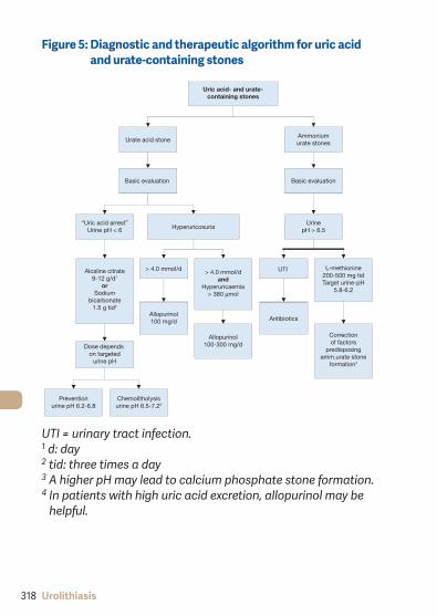
Figure 5: Diagnostic and therapeutic algorithm for uric acid and urate-containing stones
UTI = urinary tract infection.1 d: day2 tid: three times a day3 A higher pH may lead to calcium phosphate stone formation.4 In patients with high uric acid excretion, allopurinol may be
helpful.
Uric acid- and urate-containing stones
Ammoniumurate stonesUrate acid stone
Basic evaluation Basic evaluation
“Uric acid arrest”Urine pH < 6
Alcaline citrate9-12 g/d1
orSodium
bicarbonate1.5 g tid2
UrinepH > 6.5Hyperuricosuria
Allopurinol100 mg/d Antibiotics
Correctionof factors
predisposingamm.urate stone
formation4
Allopurinol100-300 mg/dDose depends
on targetedurine pH
Chemolitholysisurine pH 6.5-7.23
Preventionurine pH 6.2-6.8
> 4.0 mmol/d > 4.0 mmol/dand
Hyperuricaemia> 380 µmol
UTI L-methionine200-500 mg tidTarget urine-pH
5.8-6.2

319Urolithiasis
Figure 6: Metabolic management of cystine stones
Cystine stones
Basic evaluation
Appropriate hydration with> 3.5 L/d in adults and
1.5 L/m2 body surface inchildren
andadjust urine pH
between 7.5 and 8.5with
alkaline citrates orsodium bicarbonate
Cystine excretion< 3 mmol/d
Cystine excretion> 3 mmol/d
Possible add. treatmentwith tiopronin
(depending on recurrence)
Additional treatment withtiopronin 800 mg/d up to
1,000 mg/d avg. dos.

320 Urolithiasis
Struvite/infection stones
Recommendations for therapeutic measures of infection stones
Strength rating
Surgically remove the stone material as completely as possible.
Strong
Prescribe antibiotics in case of persistent bacteriuria.
Strong
Prescribe ammonium chloride, 1 g, two or three times daily, to ensure urinary acidification.
Weak
Prescribe methionine, 200-500 mg, one to three times daily, as an alternative, to ensure urinary acidification.
Weak
2,8-Dihydroyadenine stones and xanthine stonesBoth stone types are rare. In principle, diagnosis and specific prevention is similar to that of uric acid stones.
Drug stonesDrug stones are induced by pharmacological treatment. Two types exist:• stones formed by crystallised compounds of the drug;• stones formed due to unfavourable changes in urine
composition under drug therapy.
Treatment includes general preventive measures and the avoidance of the respective drugs.

321Urolithiasis
Unknown stone composition
Investigation Rationale for investigationMedical history • Stone history (former stone events,
family history)• Dietary habits• Medication chart
Diagnostic imaging
• Ultrasound in the case of a suspectedstone
• Un-enhanced helical computedtomography
• Determination of Hounsfield unitsprovides information about the possible stone composition
Blood analysis • Creatinine• Calcium (ionised calcium or total
calcium + albumin)• Uric acid
Perform a urinalysis
• Dipstick test: leukocytes, erythrocytes, nitrites, protein, urine pH, specific weight
• Urine cultures• Microscopy of urinary sediment
(morning urine)• Cyanide nitroprusside test (cystine
exclusion). Further examinations depend on the results of the investigations listed above.
Further examinations depend on the results of the investigations listed above.
This short booklet text is based on the more comprehensive EAU Guidelines (ISBN 978-94-92671-13-4) available to all members of the European Association of Urology at their website, http://www.uroweb.org/guidelines/.