Embed Size (px)
Citation preview
Urine and its Importance
(Urine analysis)
Javad Zavar Reza Ph.D in Clinical Biochemistry
Department of Biochemistry
1
2
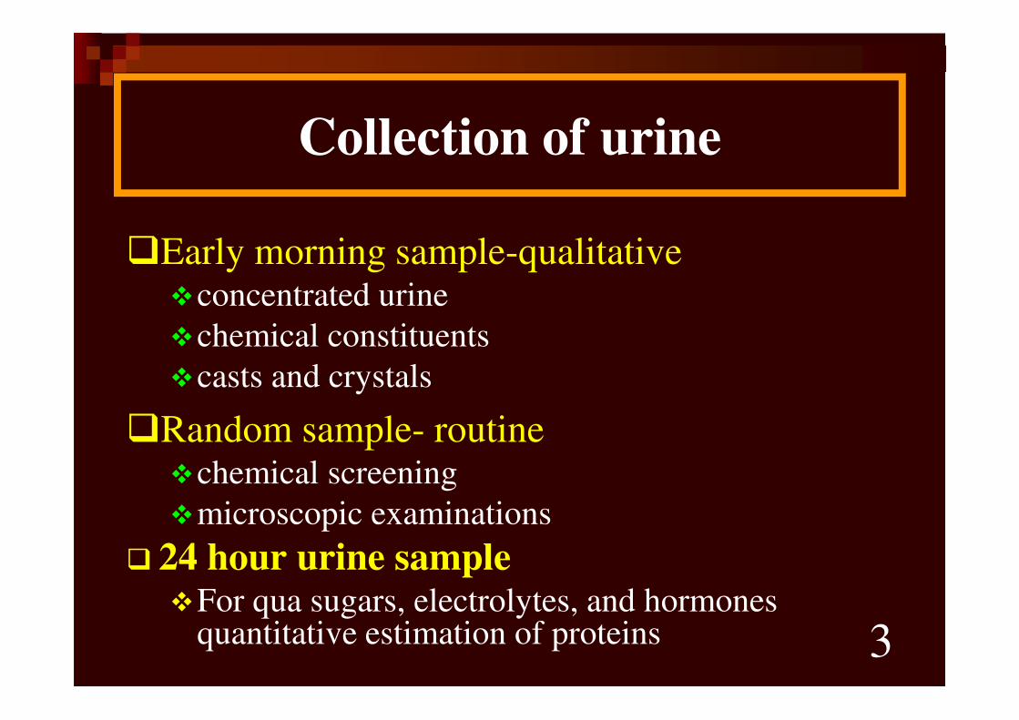
Collection of urine
�Early morning sample-qualitative�concentrated urine
�chemical constituents
�casts and crystals
�Random sample- routine�chemical screening
�microscopic examinations
� 24 hour urine sample�For qua sugars, electrolytes, and hormones
quantitative estimation of proteins 3
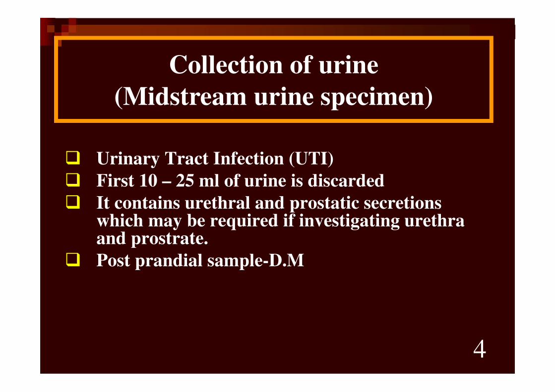
� Urinary Tract Infection (UTI)
� First 10 – 25 ml of urine is discarded
� It contains urethral and prostatic secretions which may be required if investigating urethra and prostrate.
� Post prandial sample-D.M
Collection of urine
(Midstream urine specimen)
4

� Macroscopic examination
� Chemical examination
� Microscopic
5
Urine examination
Macroscopic examination
�Volume:Normal = 600-1550ml
�Color
�Odour
�Reaction or urinary pH
�Specific gravity
6
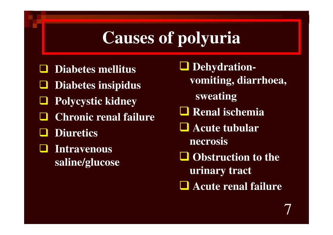
Causes of polyuria
7
� Diabetes mellitus
� Diabetes insipidus
� Polycystic kidney
� Chronic renal failure
� Diuretics
� Intravenous
saline/glucose
� Dehydration-
vomiting, diarrhoea,
sweating
� Renal ischemia
� Acute tubular
necrosis
� Obstruction to the
urinary tract
� Acute renal failure
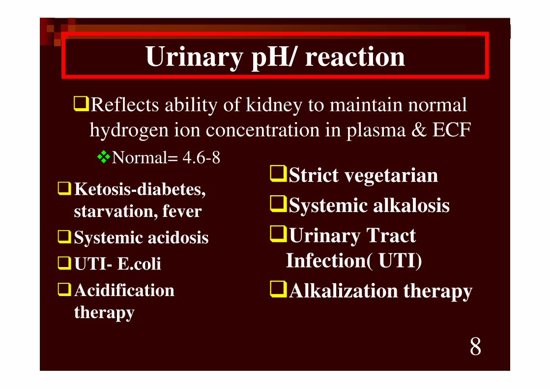
Urinary pH/ reaction
�Reflects ability of kidney to maintain normal
hydrogen ion concentration in plasma & ECF
�Normal= 4.6-8
8
�Ketosis-diabetes,
starvation, fever
�Systemic acidosis
�UTI- E.coli
�Acidification
therapy
�Strict vegetarian
�Systemic alkalosis
�Urinary Tract
Infection( UTI)
�Alkalization therapy
Biochemical examination
9
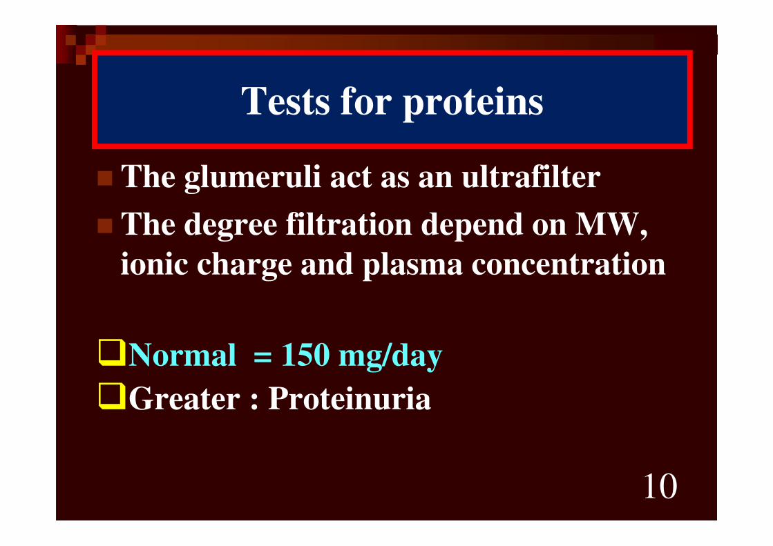
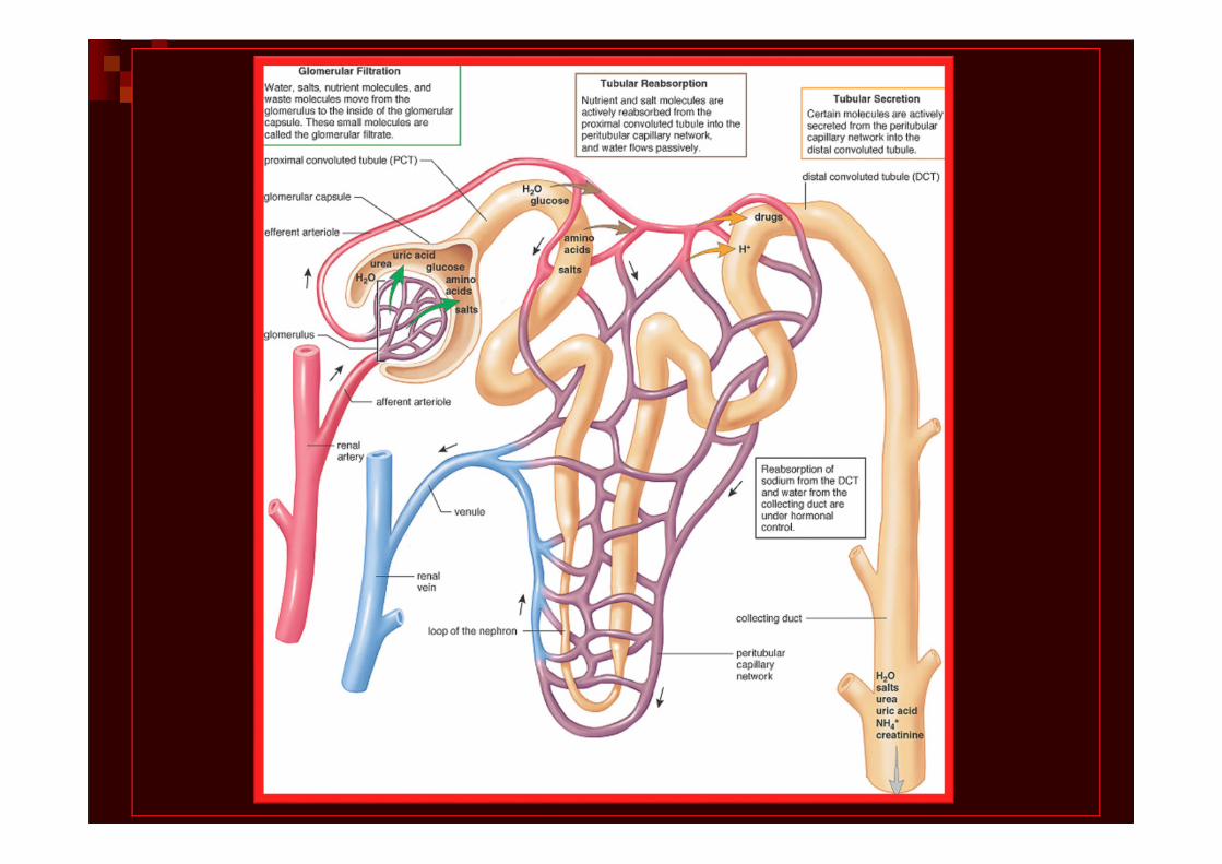
� The glumeruli act as an ultrafilter
� The degree filtration depend on MW,
ionic charge and plasma concentration
�Normal = 150 mg/day
�Greater : Proteinuria
10
Tests for proteins
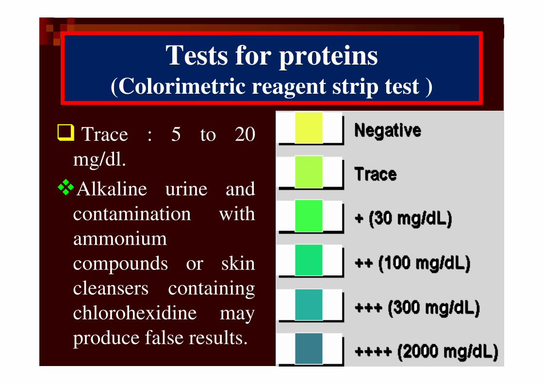
� Trace : 5 to 20
mg/dl.
�Alkaline urine and
contamination with
ammonium
compounds or skin
cleansers containing
chlorohexidine may
produce false results. 11
Tests for proteins(Colorimetric reagent strip test )
Microalbuminuria
�Alb is small and globular ,so large amounts
filtered into the glomerular urine , increased if
acidic groups are blocked.
�Mildly increased excretion(20- 300 mg/day)
called microalbuminurea
12
Qualitative Categories of Proteinuria
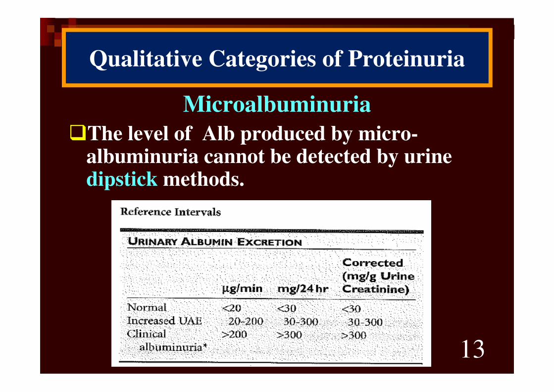
Microalbuminuria
�The level of Alb produced by micro-albuminuria cannot be detected by urine dipstick methods.
13
Qualitative Categories of Proteinuria
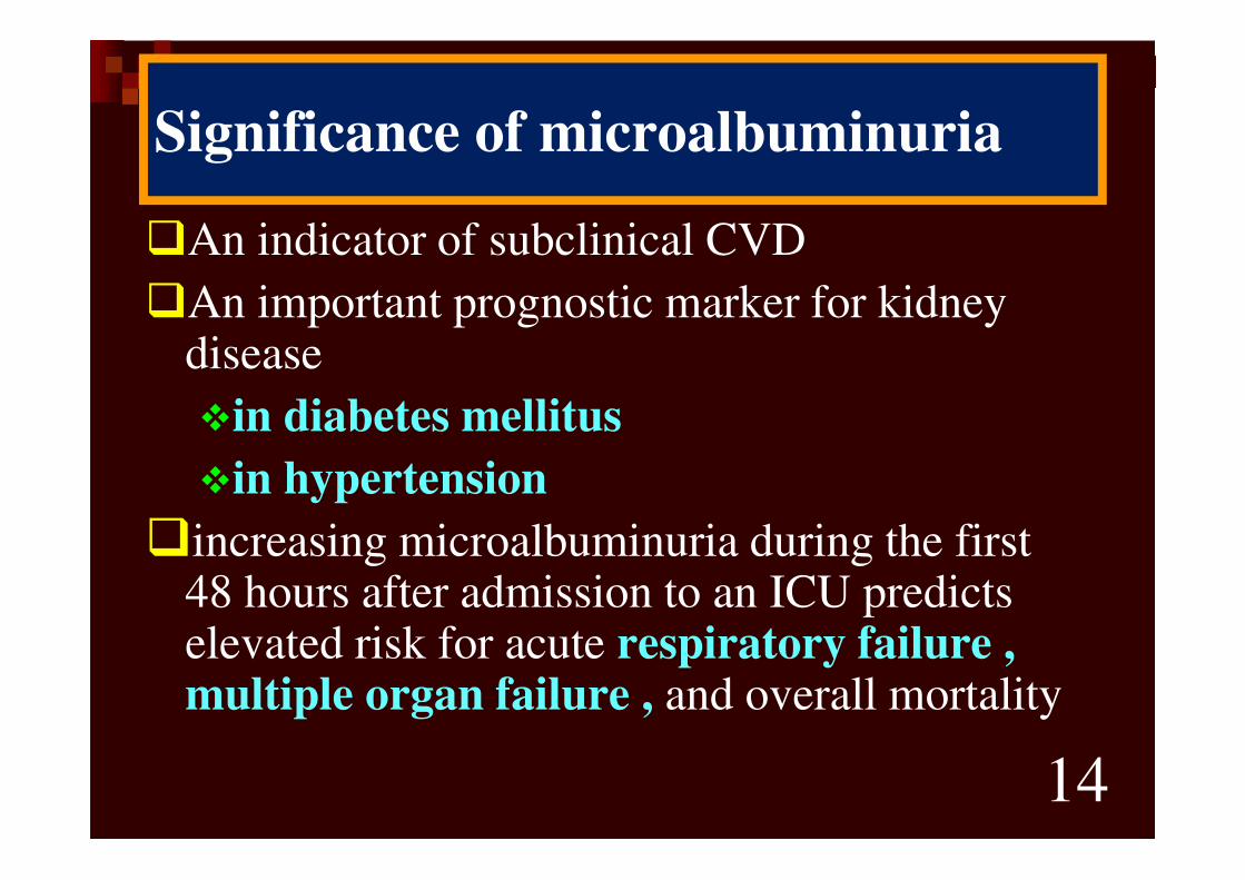
Significance of microalbuminuria
�An indicator of subclinical CVD
�An important prognostic marker for kidney disease
�in diabetes mellitus
�in hypertension
�increasing microalbuminuria during the first 48 hours after admission to an ICU predicts elevated risk for acute respiratory failure , multiple organ failure , and overall mortality
14
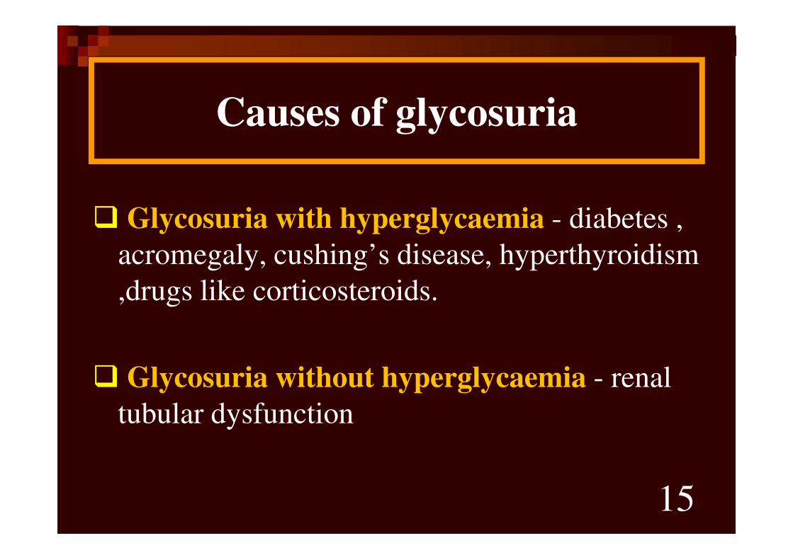
Causes of glycosuria
� Glycosuria with hyperglycaemia - diabetes ,
acromegaly, cushing’s disease, hyperthyroidism
,drugs like corticosteroids.
� Glycosuria without hyperglycaemia - renal
tubular dysfunction
15
16
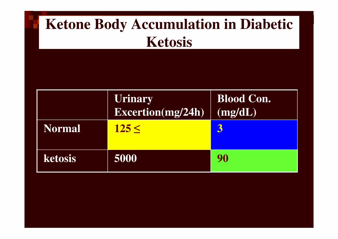
Ketone Body Accumulation in Diabetic
Ketosis
Urinary
Excertion(mg/24h)
Blood Con.
(mg/dL)
Normal 125 ≤ 3
ketosis 5000 90
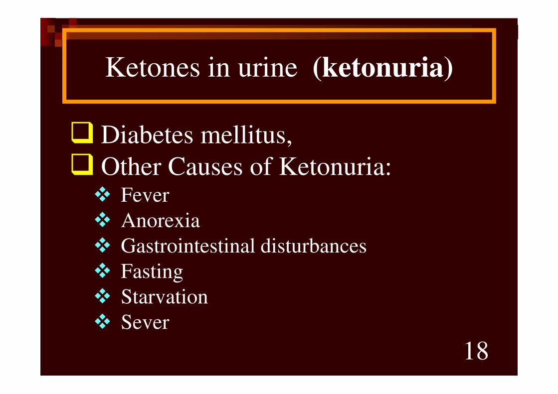
� Diabetes mellitus,
� Other Causes of Ketonuria:� Fever
� Anorexia
� Gastrointestinal disturbances
� Fasting
� Starvation
� Sever
18
Ketones in urine (ketonuria)
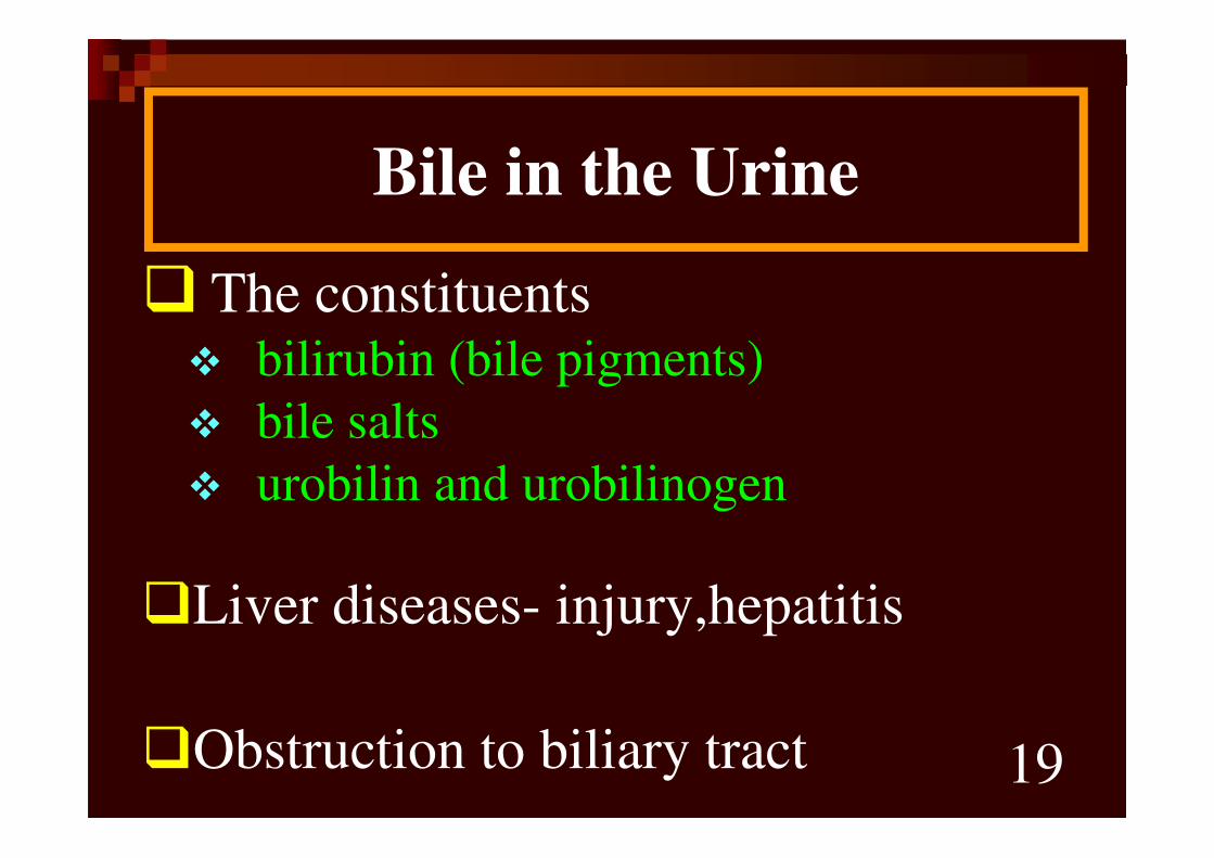
Bile in the Urine
� The constituents � bilirubin (bile pigments)
� bile salts
� urobilin and urobilinogen
�Liver diseases- injury,hepatitis
�Obstruction to biliary tract 19
Carbohydrate
&
Clinical Correlations
٢٠
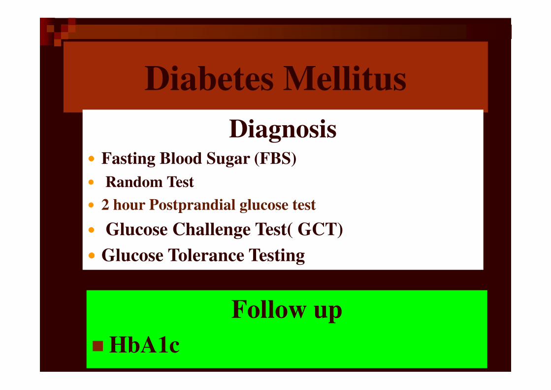
Diabetes Mellitus
Follow up
� HbA1c٢١
Diagnosis� Fasting Blood Sugar (FBS)
� Random Test
� 2 hour Postprandial glucose test
� Glucose Challenge Test( GCT)
� Glucose Tolerance Testing
٢٢
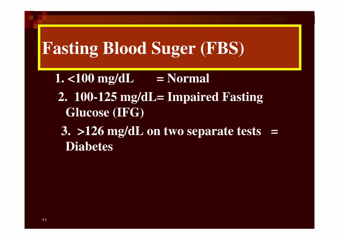
1. <100 mg/dL = Normal
2. 100-125 mg/dL= Impaired Fasting
Glucose (IFG)
3. >126 mg/dL on two separate tests =
Diabetes
Fasting Blood Suger (FBS)
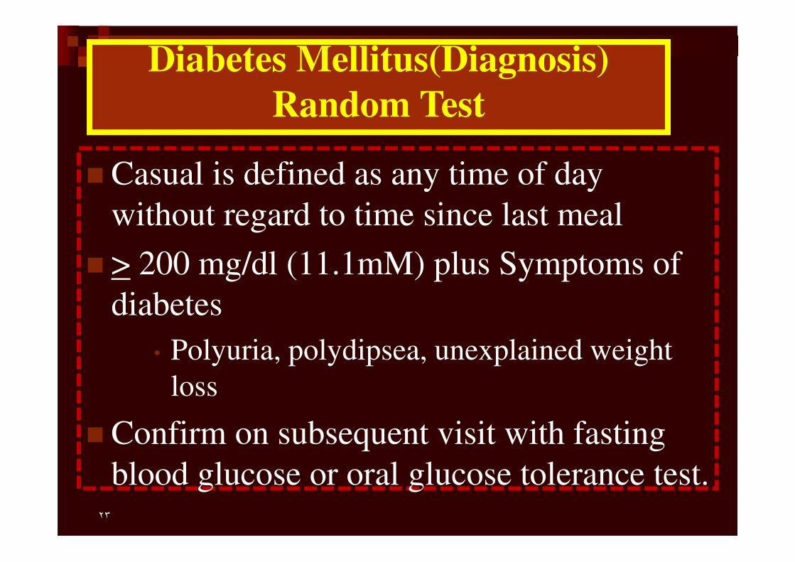
� Casual is defined as any time of day
without regard to time since last meal
� > 200 mg/dl (11.1mM) plus Symptoms of
diabetes
• Polyuria, polydipsea, unexplained weight
loss
� Confirm on subsequent visit with fasting
blood glucose or oral glucose tolerance test.
Diabetes Mellitus(Diagnosis)
Random Test
٢٣
٢٤
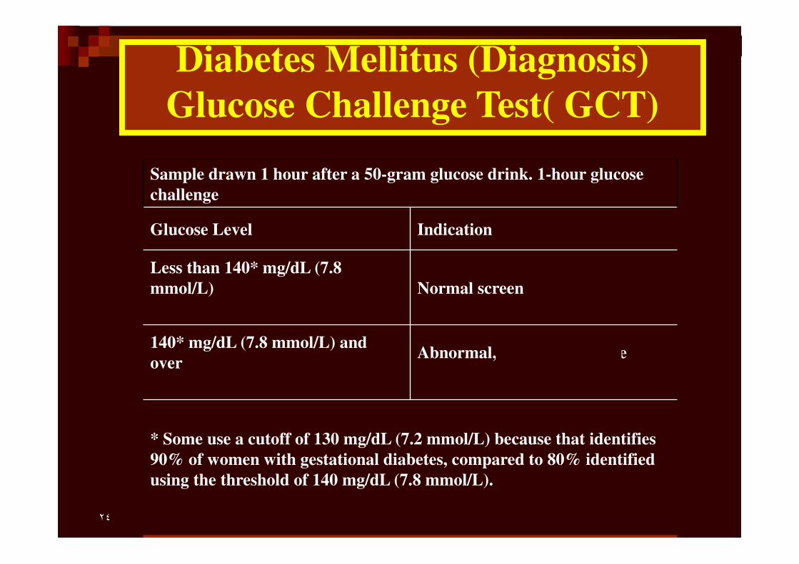
Diabetes Mellitus (Diagnosis)
Glucose Challenge Test( GCT)
Sample drawn 1 hour after a 50-gram glucose drink. 1-hour glucose
challenge
Glucose Level Indication
Less than 140* mg/dL (7.8
mmol/L) Normal screen
140* mg/dL (7.8 mmol/L) and
overAbnormal, needs OGTT (see
below)
* Some use a cutoff of 130 mg/dL (7.2 mmol/L) because that identifies
90% of women with gestational diabetes, compared to 80% identified
using the threshold of 140 mg/dL (7.8 mmol/L).
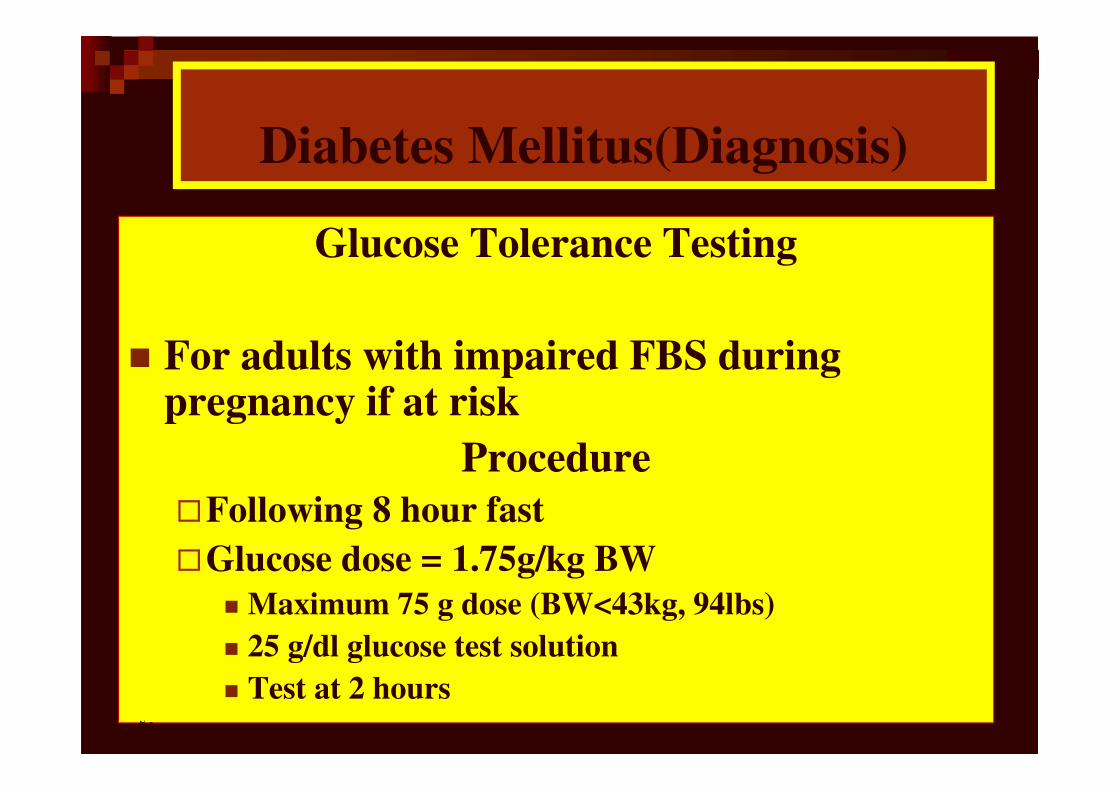
Glucose Tolerance Testing
� For adults with impaired FBS during pregnancy if at risk
Procedure
�Following 8 hour fast
�Glucose dose = 1.75g/kg BW
� Maximum 75 g dose (BW<43kg, 94lbs)
� 25 g/dl glucose test solution
� Test at 2 hours
Diabetes Mellitus(Diagnosis)
٢٥
٢٦
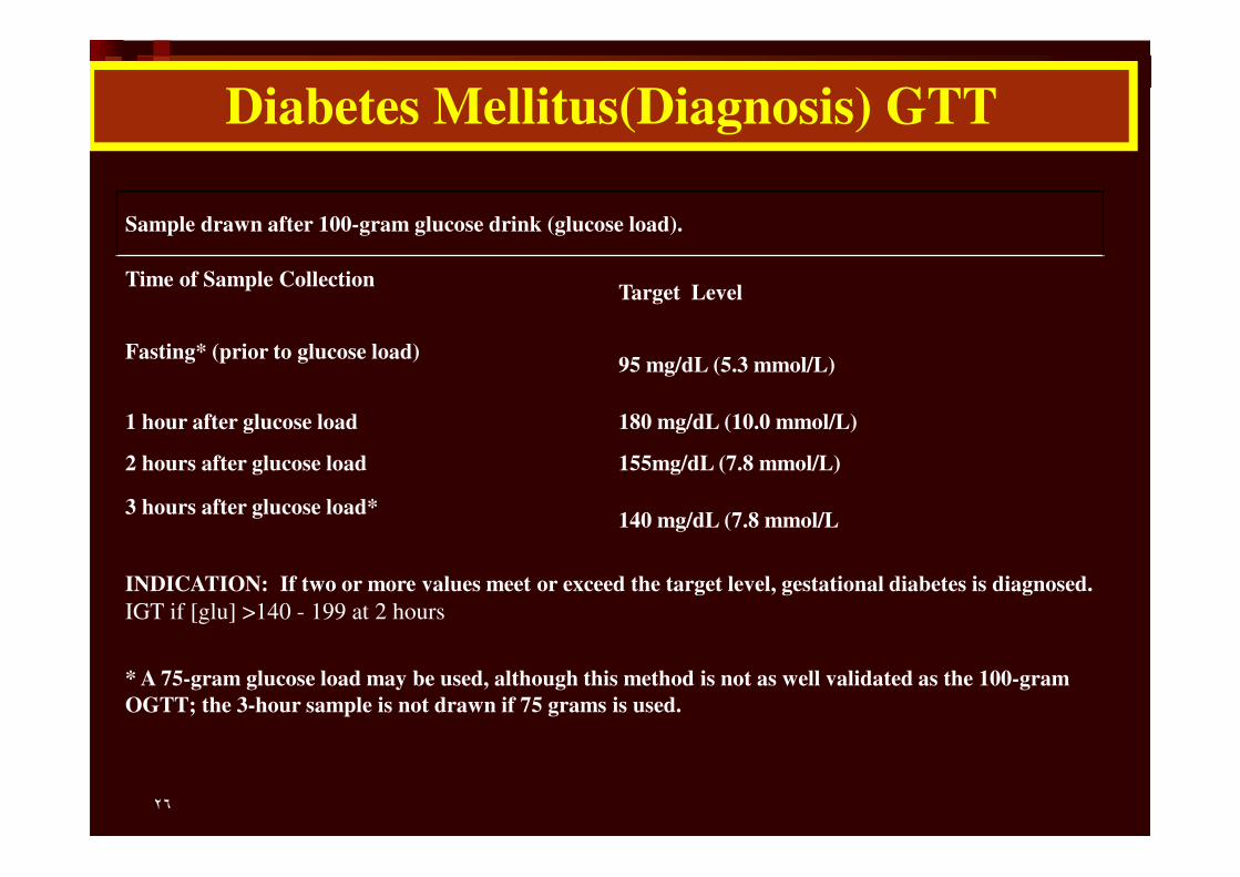
Sample drawn after 100-gram glucose drink (glucose load).
Time of Sample CollectionTarget Level
Fasting* (prior to glucose load)95 mg/dL (5.3 mmol/L)
1 hour after glucose load 180 mg/dL (10.0 mmol/L)
2 hours after glucose load 155mg/dL (7.8 mmol/L)
3 hours after glucose load*140 mg/dL (7.8 mmol/L
INDICATION: If two or more values meet or exceed the target level, gestational diabetes is diagnosed.
IGT if [glu] >140 - 199 at 2 hours
* A 75-gram glucose load may be used, although this method is not as well validated as the 100-gram
OGTT; the 3-hour sample is not drawn if 75 grams is used.
Diabetes Mellitus(Diagnosis) GTT
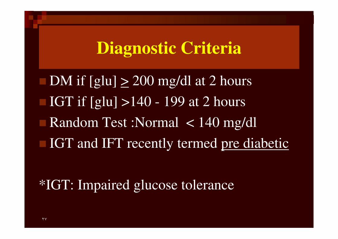
Diagnostic Criteria
� DM if [glu] > 200 mg/dl at 2 hours
� IGT if [glu] >140 - 199 at 2 hours
� Random Test :Normal < 140 mg/dl
� IGT and IFT recently termed pre diabetic
*IGT: Impaired glucose tolerance
٢٧
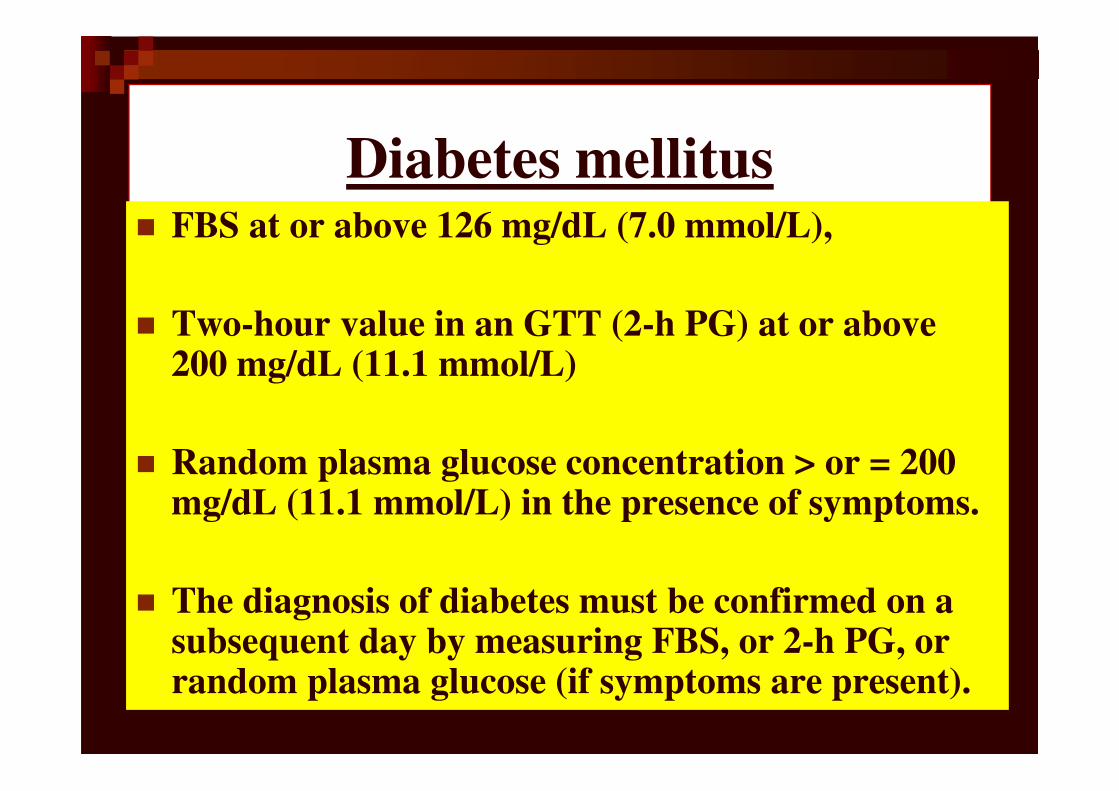
Diabetes mellitus� FBS at or above 126 mg/dL (7.0 mmol/L),
� Two-hour value in an GTT (2-h PG) at or above 200 mg/dL (11.1 mmol/L)
� Random plasma glucose concentration > or = 200 mg/dL (11.1 mmol/L) in the presence of symptoms.
� The diagnosis of diabetes must be confirmed on a subsequent day by measuring FBS, or 2-h PG, or random plasma glucose (if symptoms are present).
٢٨
٢٩
Follow up of Diabetes mellitus
HbA1c: the blood test with a
memory
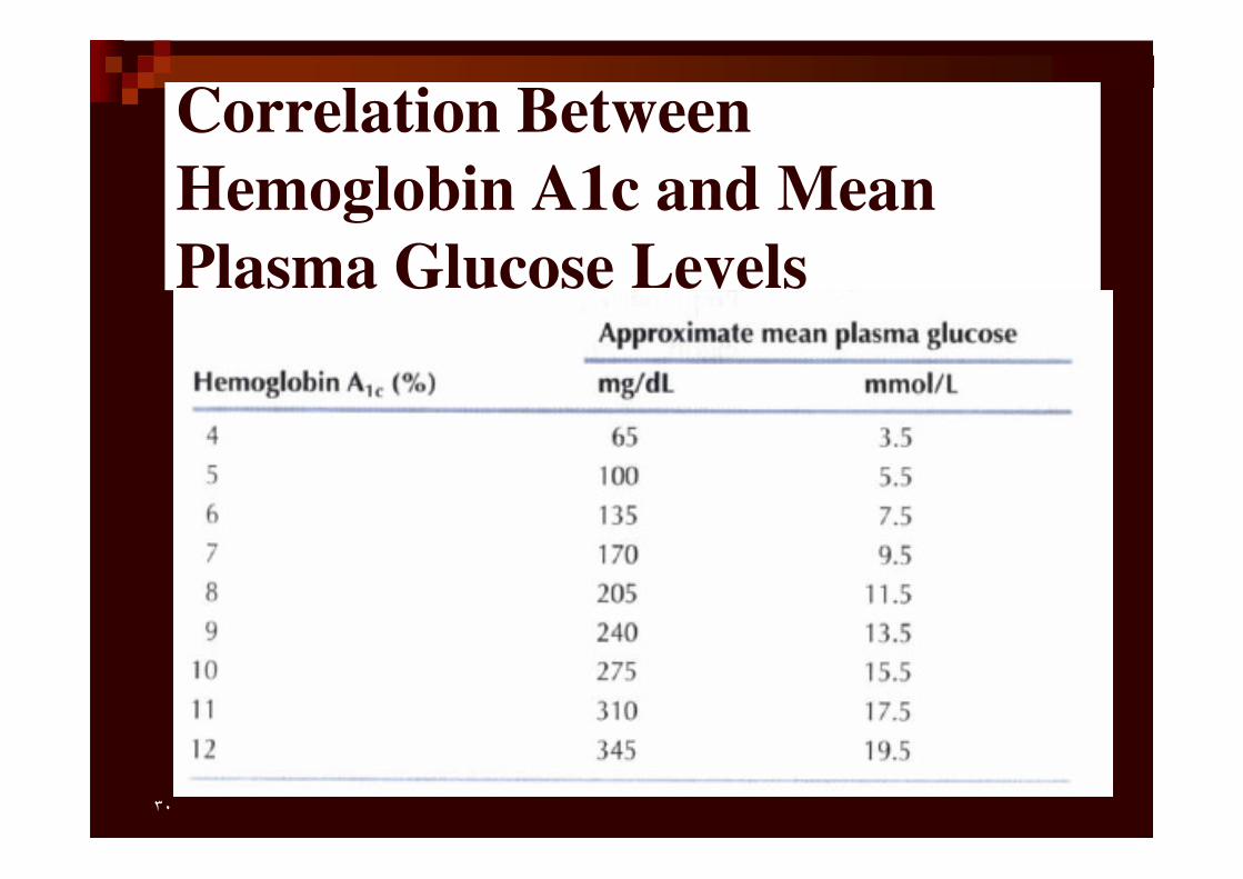
Correlation Between
Hemoglobin A1c and Mean
Plasma Glucose Levels
٣٠
Serum Protein
&
Clinical Correlations
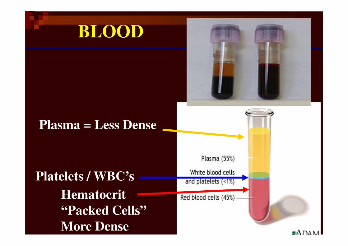
BLOOD
Plasma = Less Dense
Hematocrit
“Packed Cells”
More Dense
Platelets / WBC’s
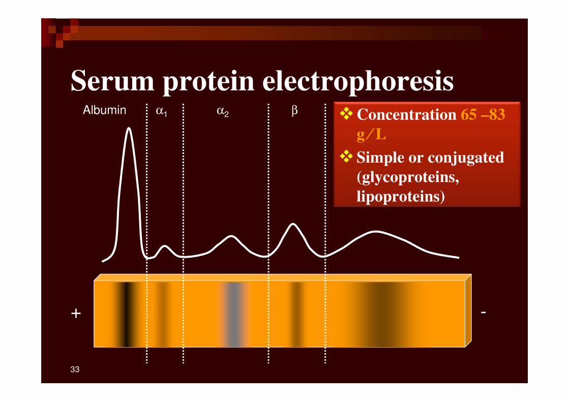
Serum protein electrophoresis
+ -
Albumin α1 α2 β γ
33
�Concentration 65 –83
g ⁄⁄⁄⁄ L
�Simple or conjugated
(glycoproteins,
lipoproteins)
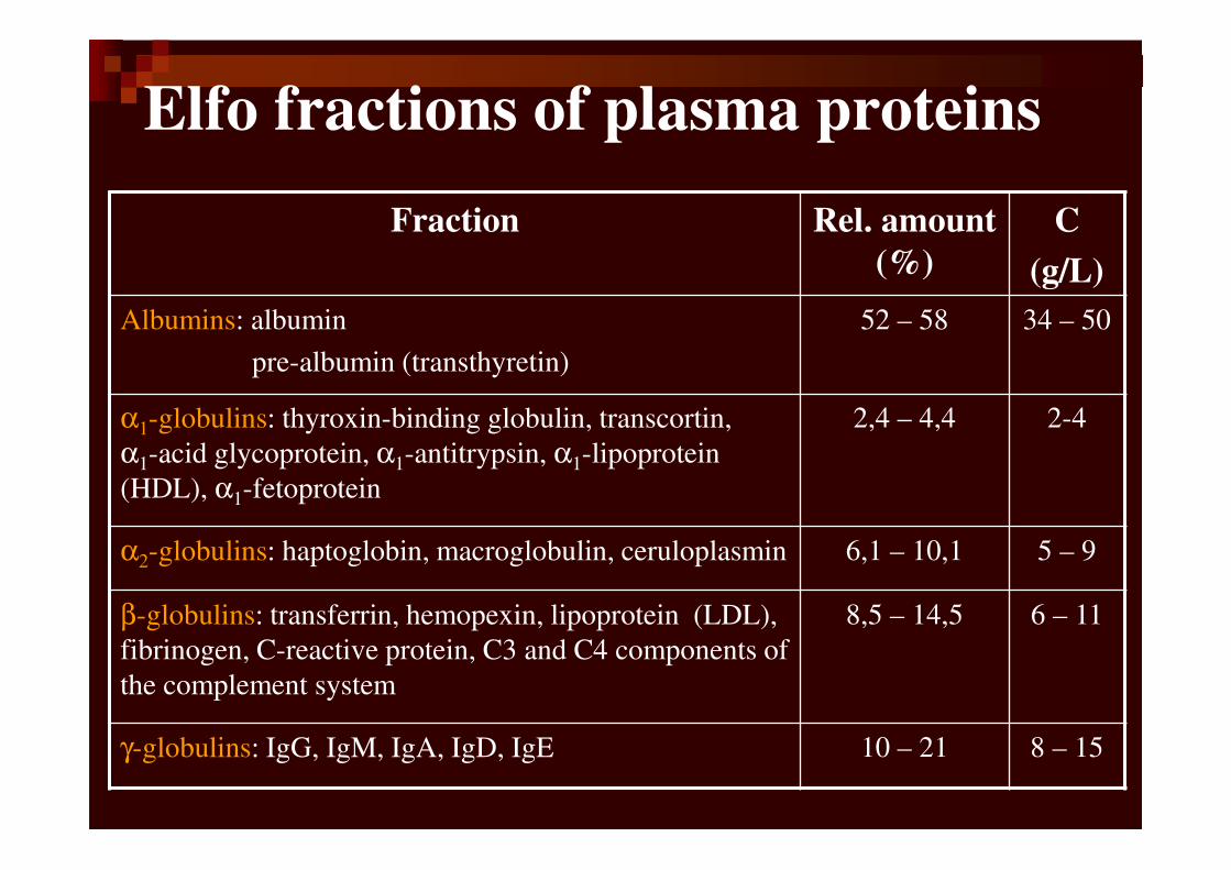
Elfo fractions of plasma proteins
Fraction Rel. amount
(%)
C
(g/L)
Albumins: albumin
pre-albumin (transthyretin)
52 – 58 34 – 50
α1-globulins: thyroxin-binding globulin, transcortin,
α1-acid glycoprotein, α1-antitrypsin, α1-lipoprotein
(HDL), α1-fetoprotein
2,4 – 4,4 2-4
α2-globulins: haptoglobin, macroglobulin, ceruloplasmin 6,1 – 10,1 5 – 9
β-globulins: transferrin, hemopexin, lipoprotein (LDL),
fibrinogen, C-reactive protein, C3 and C4 components of
the complement system
8,5 – 14,5 6 – 11
γ-globulins: IgG, IgM, IgA, IgD, IgE 10 – 21 8 – 15
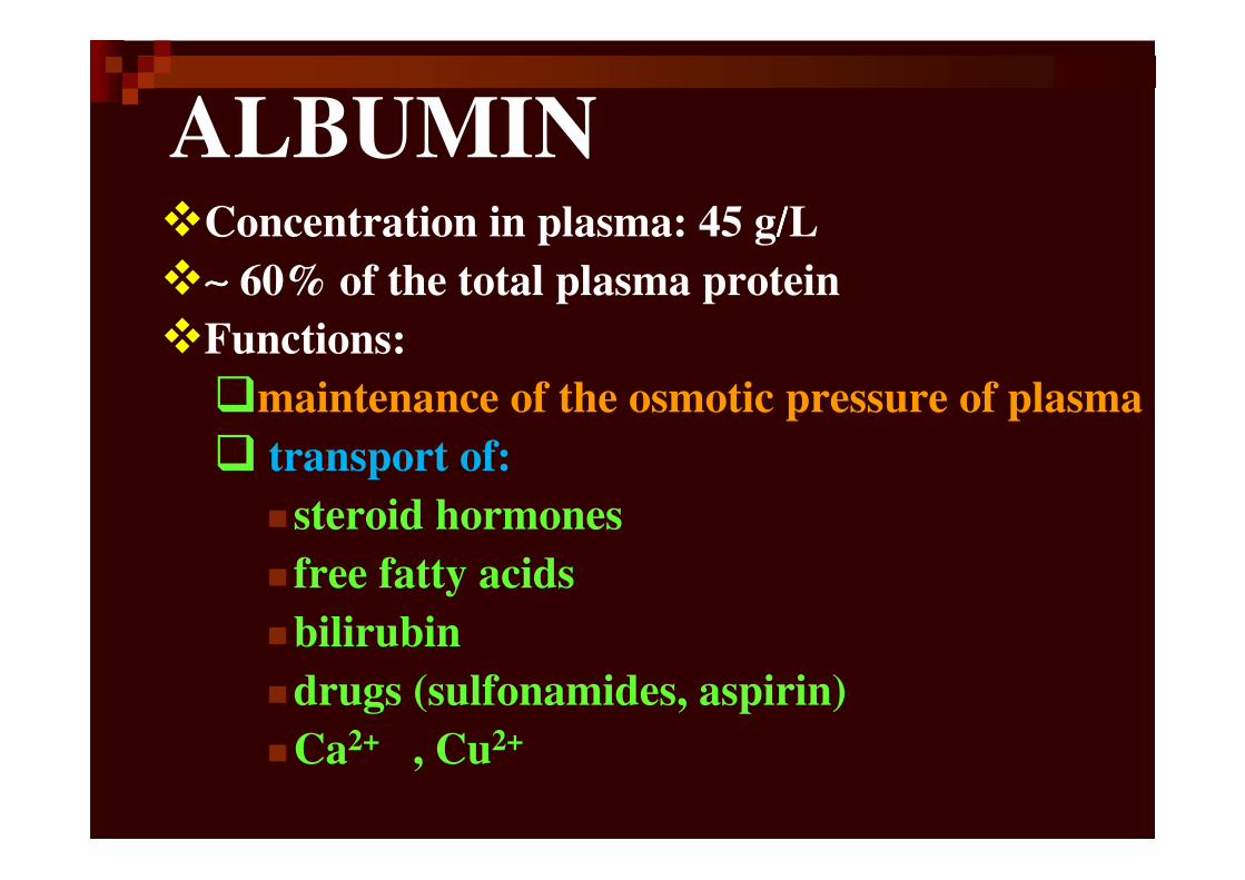
ALBUMIN�Concentration in plasma: 45 g////L�∼∼∼∼ 60% of the total plasma protein
�Functions:
�maintenance of the osmotic pressure of plasma
� transport of:
�steroid hormones
� free fatty acids
�bilirubin
�drugs (sulfonamides, aspirin)
�Ca2+ , Cu2+
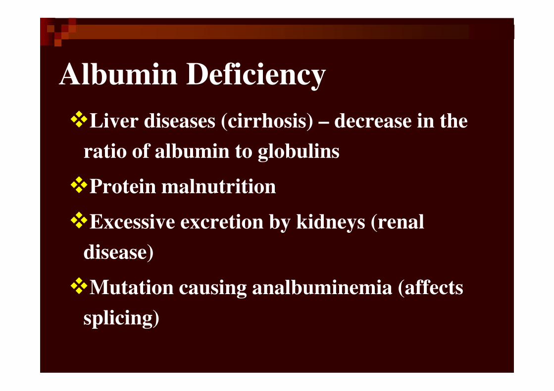
Albumin Deficiency
�Liver diseases (cirrhosis) – decrease in the
ratio of albumin to globulins
�Protein malnutrition
�Excessive excretion by kidneys (renal
disease)
�Mutation causing analbuminemia (affects
splicing)
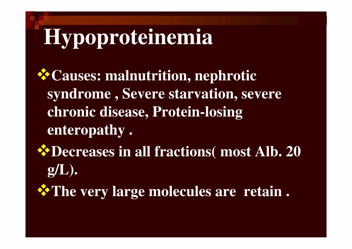
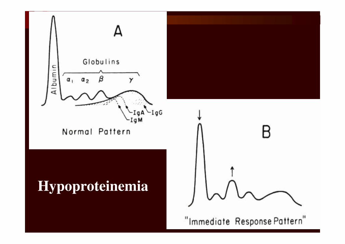
Hypoproteinemia
�Causes: malnutrition, nephrotic
syndrome , Severe starvation, severe
chronic disease, Protein-losing
enteropathy .
�Decreases in all fractions( most Alb. 20
g/L).
�The very large molecules are retain .
Hypoproteinemia
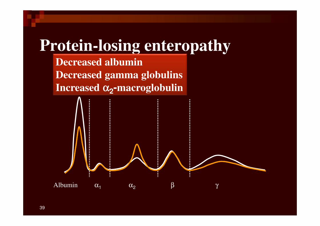
Protein-losing enteropathy
Albumin α1 α2 β γ
Decreased albumin
Decreased gamma globulins
Increased αααα2-macroglobulin
39
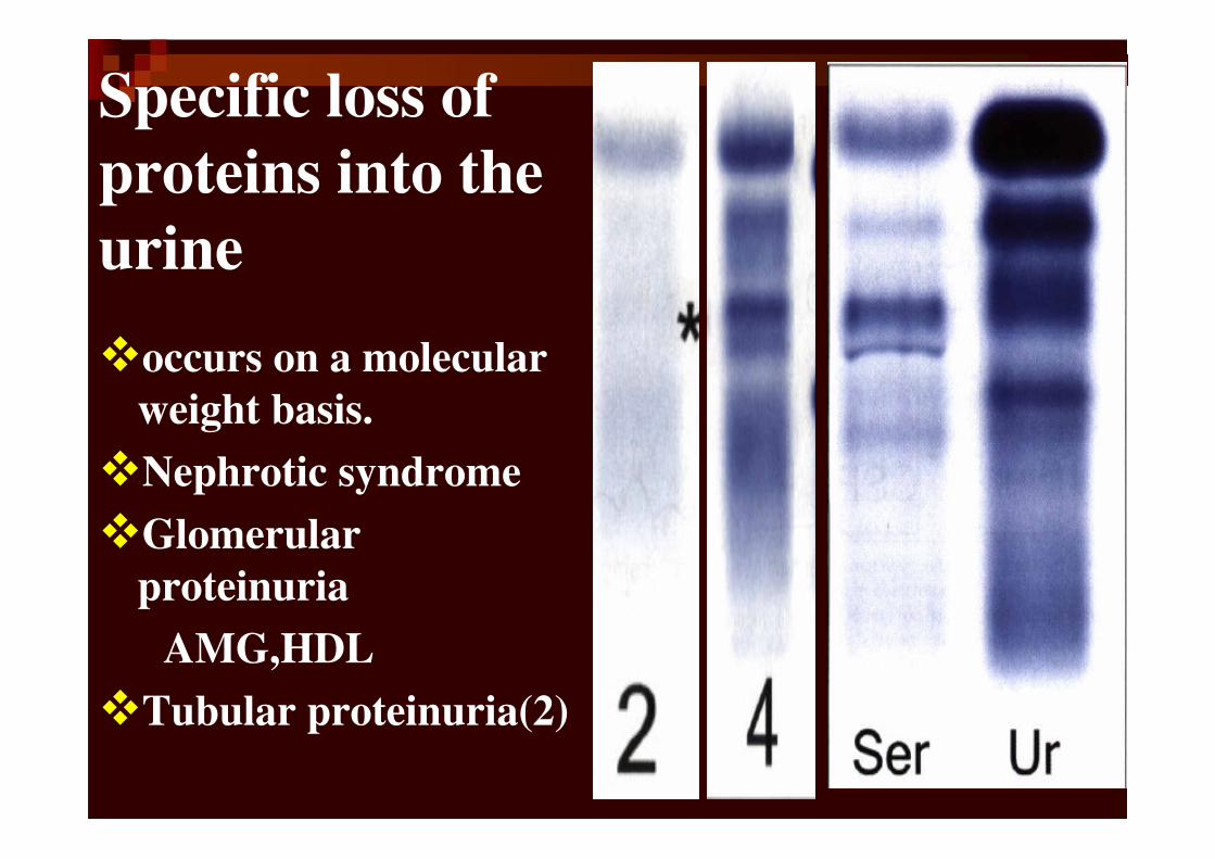
Specific loss of
proteins into the
urine
�occurs on a molecular
weight basis.
�Nephrotic syndrome
�Glomerular
proteinuria
AMG,HDL
�Tubular proteinuria(2)
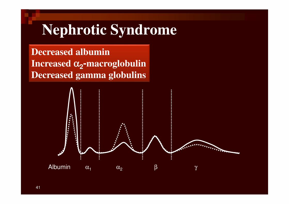
Nephrotic Syndrome
Albumin α1 α2 β γ
Decreased albumin
Increased αααα2-macroglobulin
Decreased gamma globulins
41
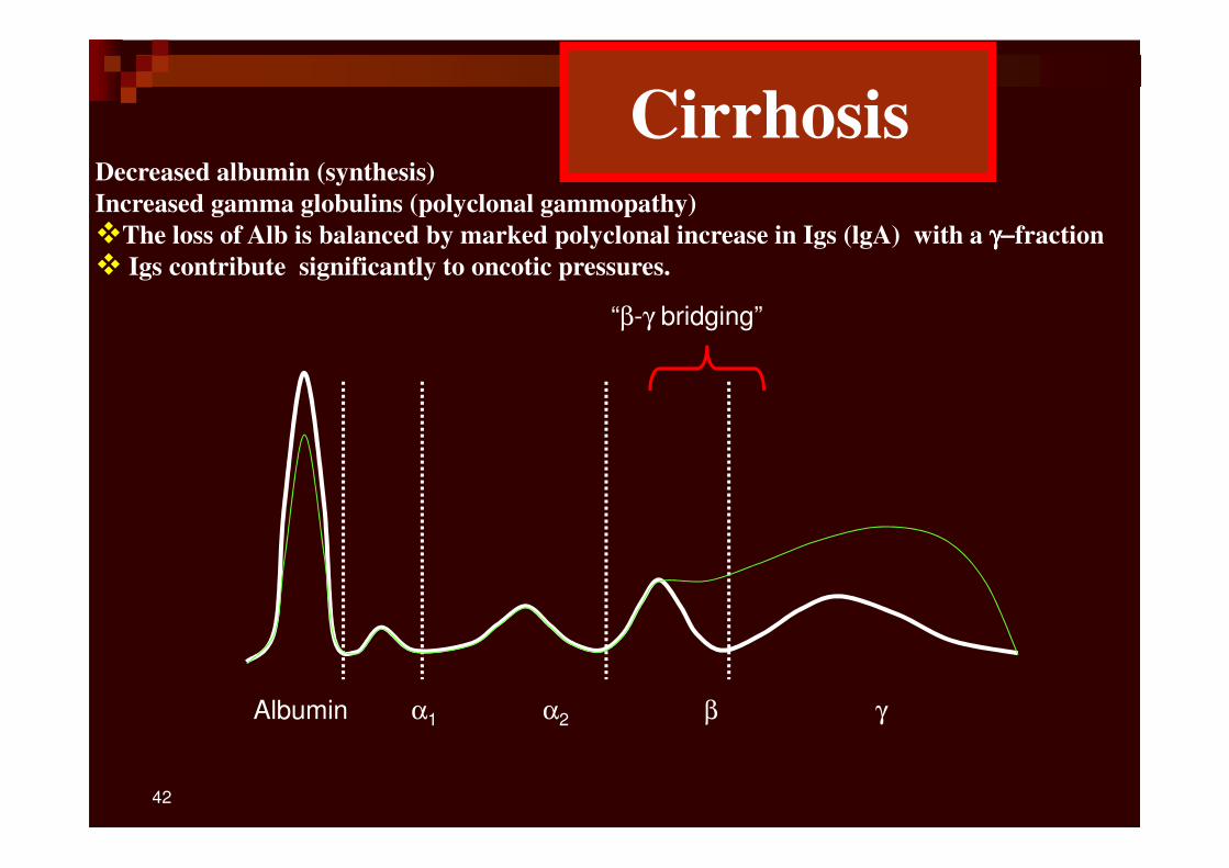
Albumin α1 α2 β γ
Decreased albumin (synthesis)
Increased gamma globulins (polyclonal gammopathy)
�The loss of Alb is balanced by marked polyclonal increase in Igs (lgA) with a γγγγ−−−−fraction
� Igs contribute significantly to oncotic pressures.
“β-γ bridging”
42
Cirrhosis
Tests for
Evaluation of Liver
Function
43
Liver Function tests
� May measure synthetic function
� May measure excretory function
� May indicate damage to cells:
determination
�of specific enzymes may be used to
show the
�location of liver damage
44
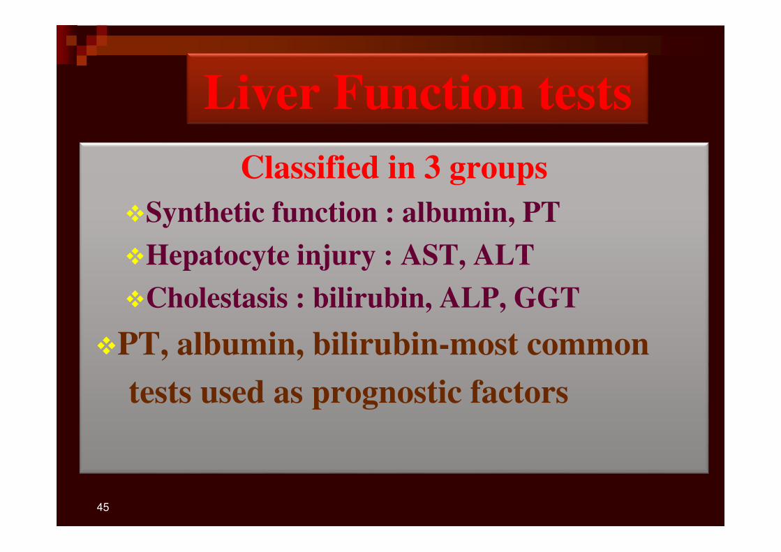
Classified in 3 groups
�Synthetic function : albumin, PT
�Hepatocyte injury : AST, ALT
�Cholestasis : bilirubin, ALP, GGT
�PT, albumin, bilirubin-most common
tests used as prognostic factors
Liver Function tests
45
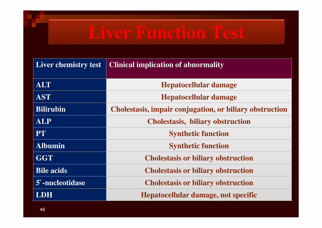
Liver Function Test
Liver chemistry test Clinical implication of abnormality
ALT Hepatocellular damage
AST Hepatocellular damage
Bilirubin Cholestasis, impair conjugation, or biliary obstruction
ALP Cholestasis, biliary obstruction
PT Synthetic function
Albumin Synthetic function
GGT Cholestasis or biliary obstruction
Bile acids Cholestasis or biliary obstruction
5`-nucleotidase Cholestasis or biliary obstruction
LDH Hepatocellular damage, not specific
46
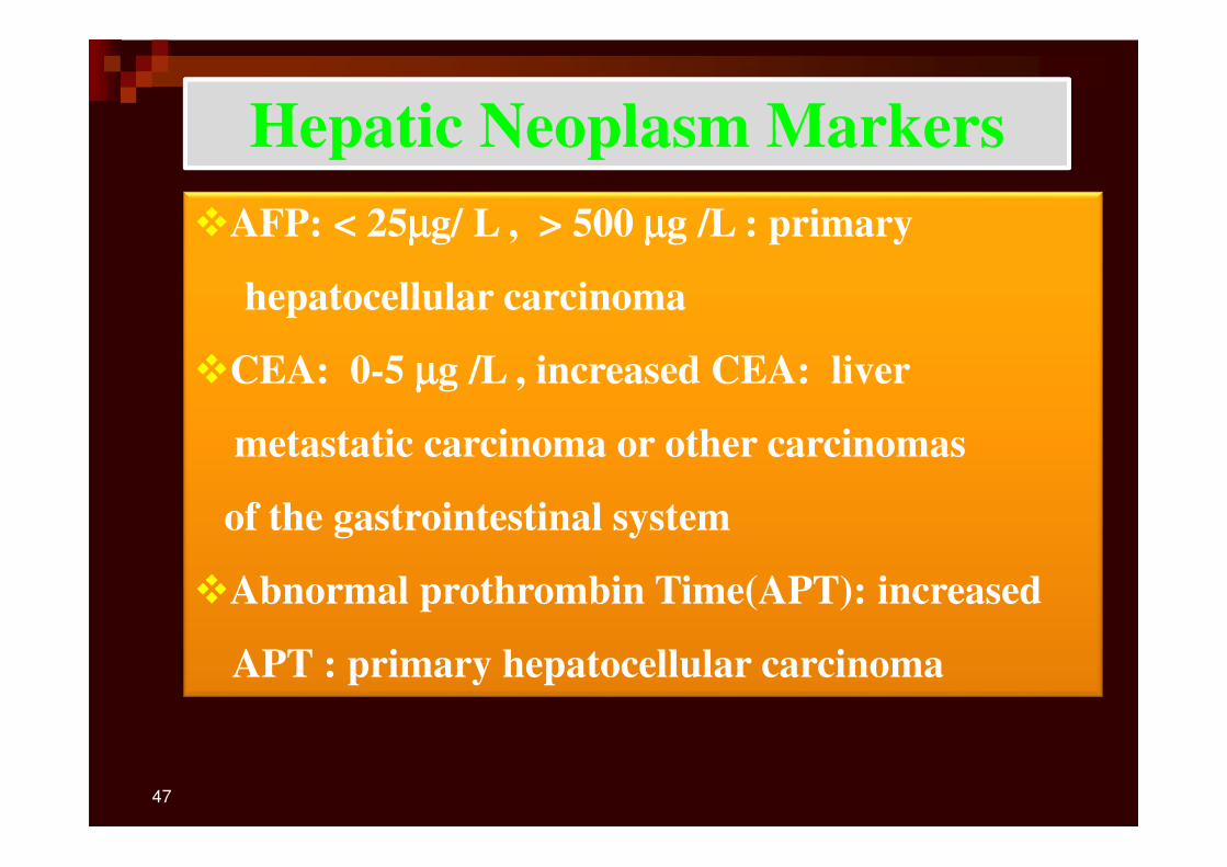
Hepatic Neoplasm Markers
�AFP: < 25µµµµg/ L , > 500 µµµµg /L : primary
hepatocellular carcinoma
�CEA: 0-5 µµµµg /L , increased CEA: liver
metastatic carcinoma or other carcinomas
of the gastrointestinal system
�Abnormal prothrombin Time(APT): increased
APT : primary hepatocellular carcinoma
47
Clinical utility of
Urea & Creatinine
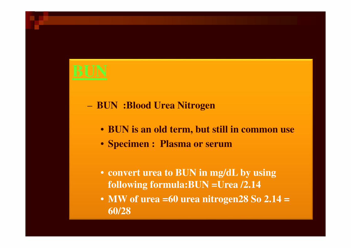
BUN
– BUN :Blood Urea Nitrogen
• BUN is an old term, but still in common use
• Specimen : Plasma or serum
• convert urea to BUN in mg/dL by using
following formula:BUN =Urea /2.14
• MW of urea =60 urea nitrogen28 So 2.14 =
60/28
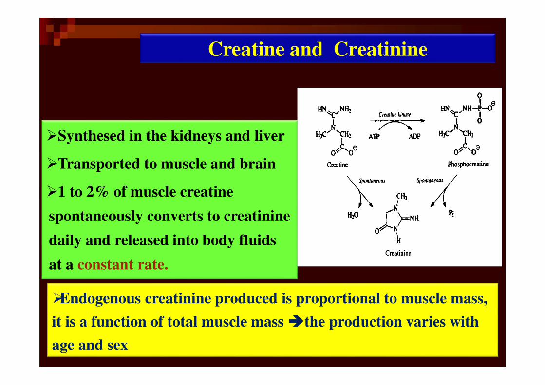
�Synthesed in the kidneys and liver
�Transported to muscle and brain
�1 to 2% of muscle creatine
spontaneously converts to creatinine
daily and released into body fluids
at a constant rate.
�Endogenous creatinine produced is proportional to muscle mass,
it is a function of total muscle mass ����the production varies with
age and sex
Creatine and Creatinine
�Released at a constant rate ���� its clearance is indicator of
GFR.
�Small quantity is reabsorbed by the tubules and other
quantities are actively secreted by the renal tubules ���� So its
clearance is approximately 7% greater than inulin clearance.
�The difference is not significant when GFR is normal but
when the GFR is low (less 10 ml/min), tubular secretion
makes the major contribution to Cra.
�So excretion the Cra. clearance significantly overestimates
the GFR.
Clinical utility of Creatinine
�Plasma Cra. is inversely related to the GFR
�But GFR can decrease by 50% before plasma Cra. rises
beyond the normal range ���� normal plasma Cra. does not
necessarily imply normal renal function.
�Raised Cra. usually indicates impaired renal function
�Changes in plasma Cra. can occur, independently of renal
function, due to changes in muscle mass.
�Cra is partially secreted by the proximal tubules via the
organic cation pathway, and is blocked by various drugs
including cimetidine, trimethoprim and salicylate.
Plasma creatinine
53
� Azotemia = Elevated plasma BUN� The key point: ↑↑↑↑ plasma urea without ↑↑↑↑ plasma creatinine.
� Prerenal ↑↑↑↑ BUN
� Low Blood Pressure ( CHF, Shock, hemorrhage,
dehydration )
� Decreased blood flow to kidney = No filtration
� Increased dietary protein or protein catabolism
� Prerenal ↓↓↓↓ BUN
� Decreased dietary protein
� Increased protein synthesis ( Pregnant women , children )
BUN disease correlations
54
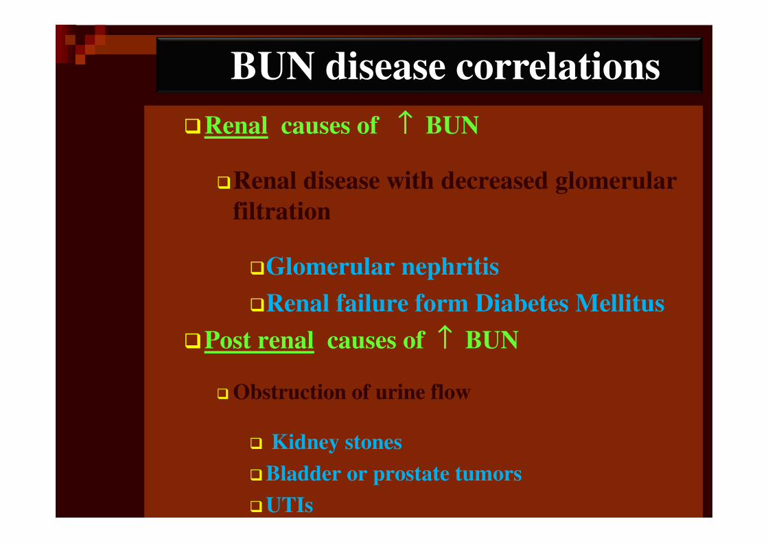
�Renal causes of ↑↑↑↑ BUN
�Renal disease with decreased glomerular
filtration
�Glomerular nephritis
�Renal failure form Diabetes Mellitus
�Post renal causes of ↑↑↑↑ BUN
� Obstruction of urine flow
� Kidney stones
�Bladder or prostate tumors
�UTIs
BUN disease correlations
55
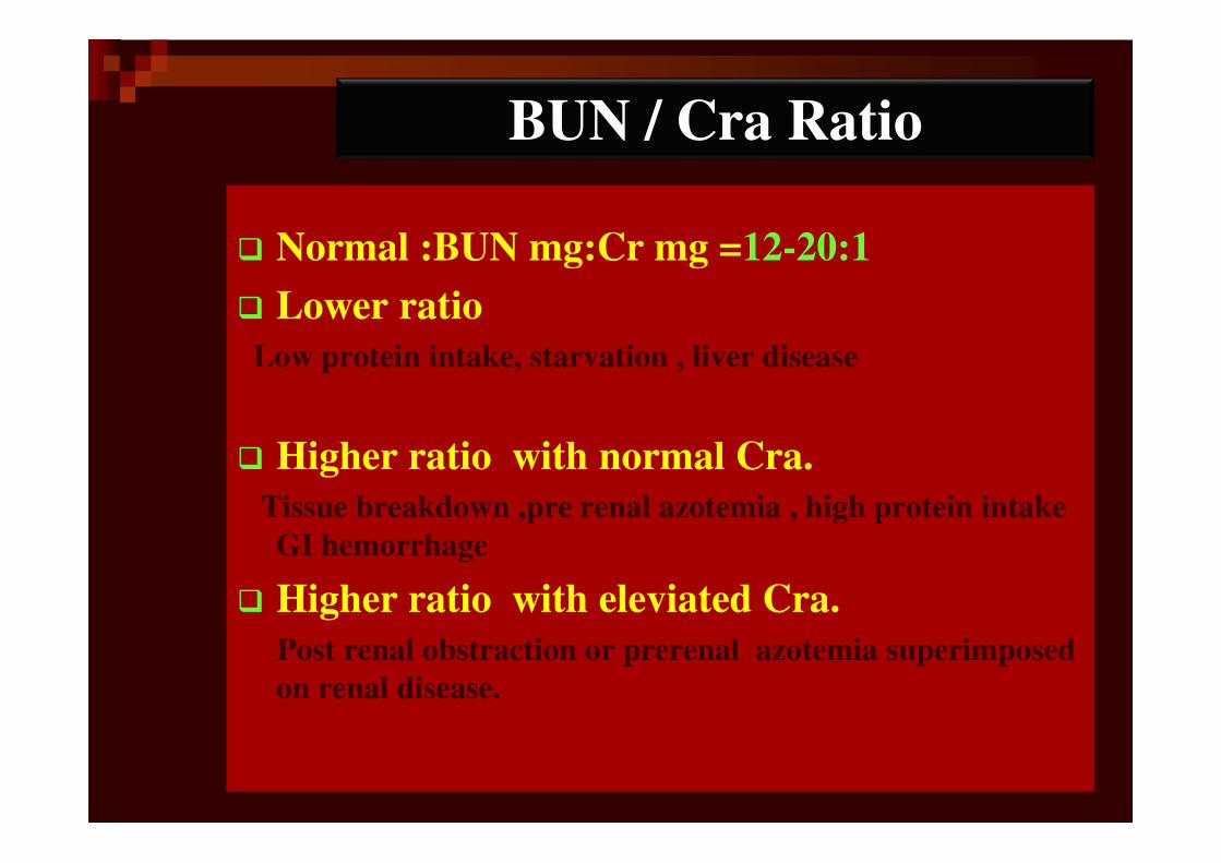
� Normal :BUN mg:Cr mg =12-20:1
� Lower ratio
Low protein intake, starvation , liver disease
� Higher ratio with normal Cra.
Tissue breakdown ,pre renal azotemia , high protein intake
GI hemorrhage
� Higher ratio with eleviated Cra.
Post renal obstraction or prerenal azotemia superimposed
on renal disease.
BUN / Cra Ratio
�Blood Tests to Detect
Inflammation
56
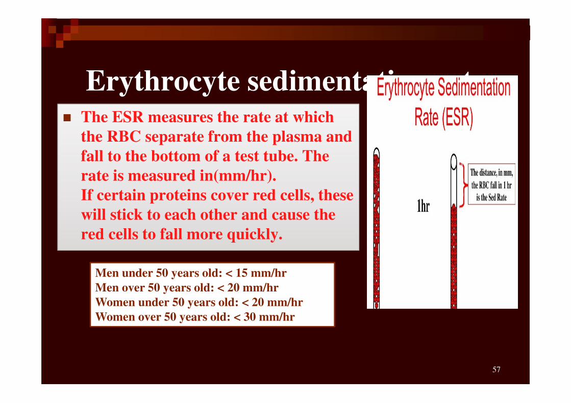
Erythrocyte sedimentation rate
57
� The ESR measures the rate at which
the RBC separate from the plasma and
fall to the bottom of a test tube. The
rate is measured in(mm/hr).
If certain proteins cover red cells, these
will stick to each other and cause the
red cells to fall more quickly.
Men under 50 years old: < 15 mm/hr
Men over 50 years old: < 20 mm/hr
Women under 50 years old: < 20 mm/hr
Women over 50 years old: < 30 mm/hr
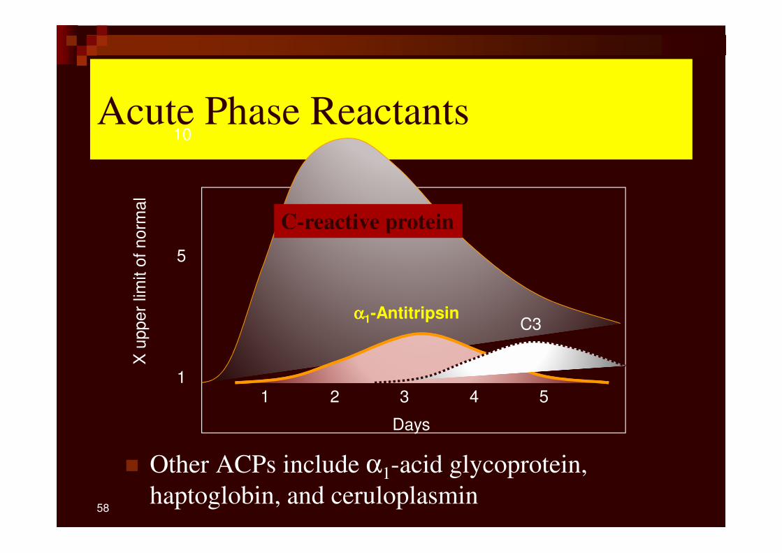
Acute Phase Reactants
� Other ACPs include α1-acid glycoprotein,
haptoglobin, and ceruloplasmin
X u
pper
limit o
f norm
al
1
5
10
Days
1 2 3 4 5
C-reactive protein
αααα1-AntitripsinC3
58
C-reactive protein
� The reference range for C-reactive protein is as
follows:
� CRP: 0-10mg/L
� High-sensitivity CRP (hs-CRP): < 1 mg/L
59
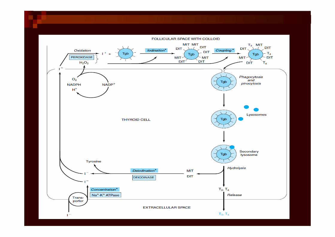
Thyroid hormone
60
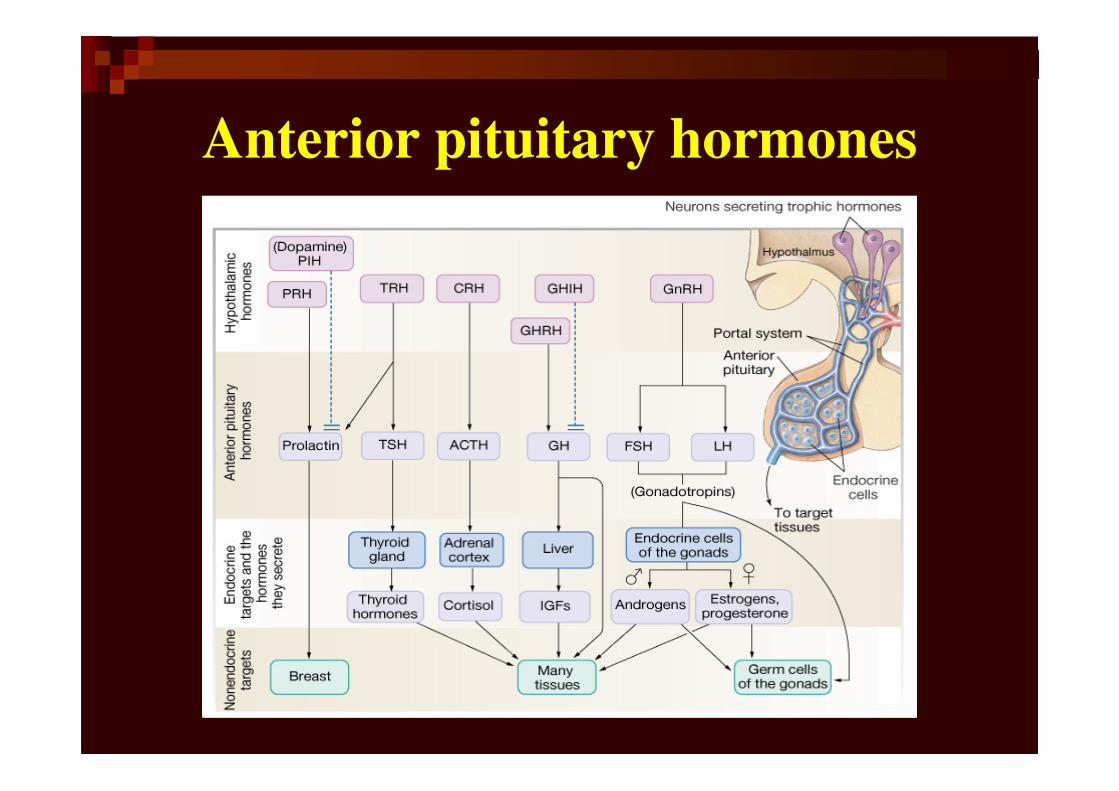
Anterior pituitary hormones
64
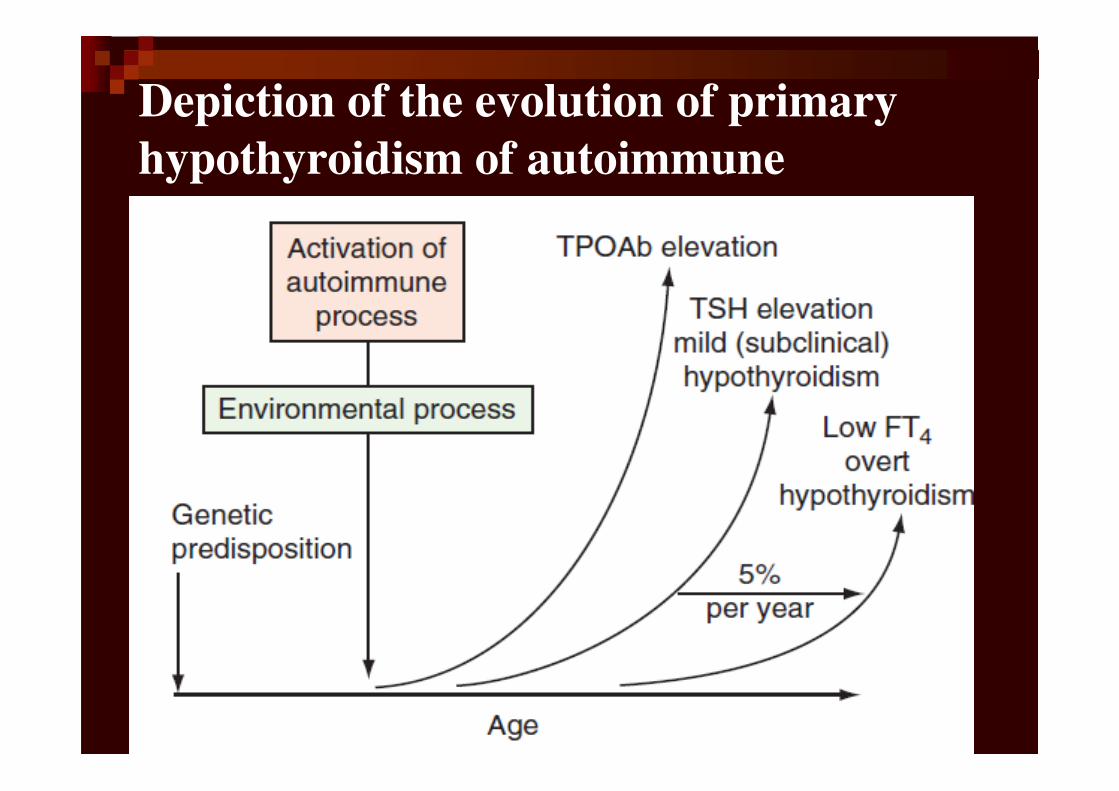
Depiction of the evolution of primary
hypothyroidism of autoimmune
origin