Embed Size (px)
Citation preview
Microscopic Examination of Urine
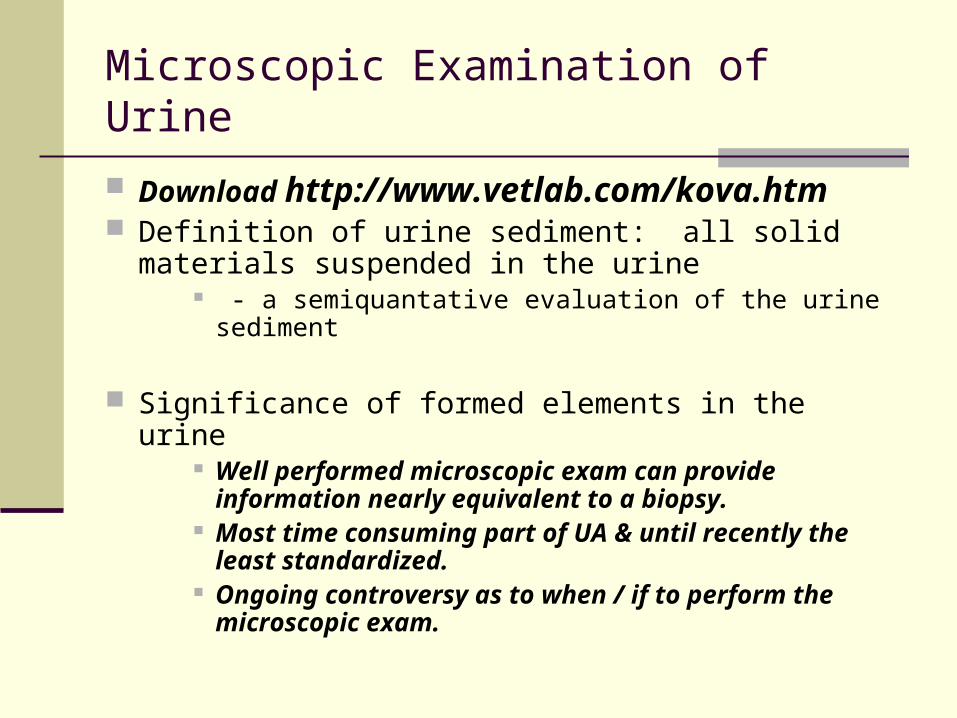
Download http://www.vetlab.com/kova.htm Definition of urine sediment: all solid materials
suspended in the urine - a semiquantative evaluation of the urine sediment
Significance of formed elements in the urine Well performed microscopic exam can provide
information nearly equivalent to a biopsy. Most time consuming part of UA & until recently
the least standardized. Ongoing controversy as to when / if to perform the
microscopic exam.
Microscopic Examination of Urine
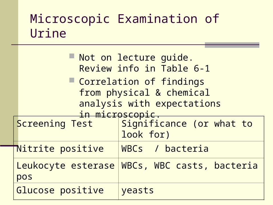
Not on lecture guide. Review info in Table 6-1
Correlation of findings from physical & chemical analysis with expectations in microscopic.
Screening Test Significance (or what to look for)
Nitrite positive WBCs / bacteria
Leukocyte esterase pos WBCs, WBC casts, bacteria
Glucose positive yeasts

Microscopic Examination of Urine
Specimen requirements Collection of specimen
Prefer the concentrated first morning specimen, collected = mid-stream, clean catch .
first morning most concentrated and will be able to demonstrate the most abnormalities. Mid stream, clean catch technique will eliminate fecal & vaginal contamination
Container must be clean and free of lint / debris usually disposable plastic, must be sure no soap
residue Fresh – tested within 2 hours of voiding, or
refrigeration needed.
Microscopic Examination of Urine
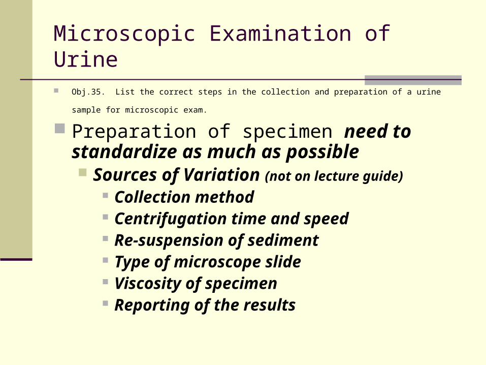
Obj.35. List the correct steps in the collection and preparation of a urine sample for microscopic exam.
Preparation of specimen need to standardize as much as possible Sources of Variation (not on lecture guide)
Collection method Centrifugation time and speed Re-suspension of sediment Type of microscope slide Viscosity of specimen Reporting of the results
Microscopic Examination of Urine
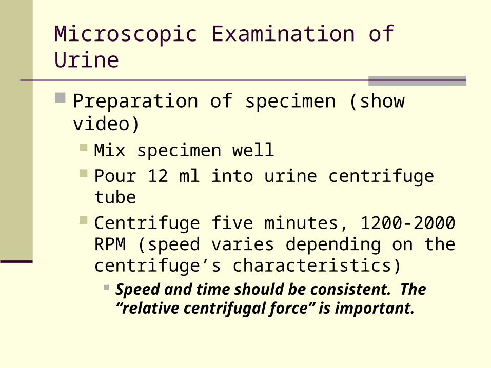
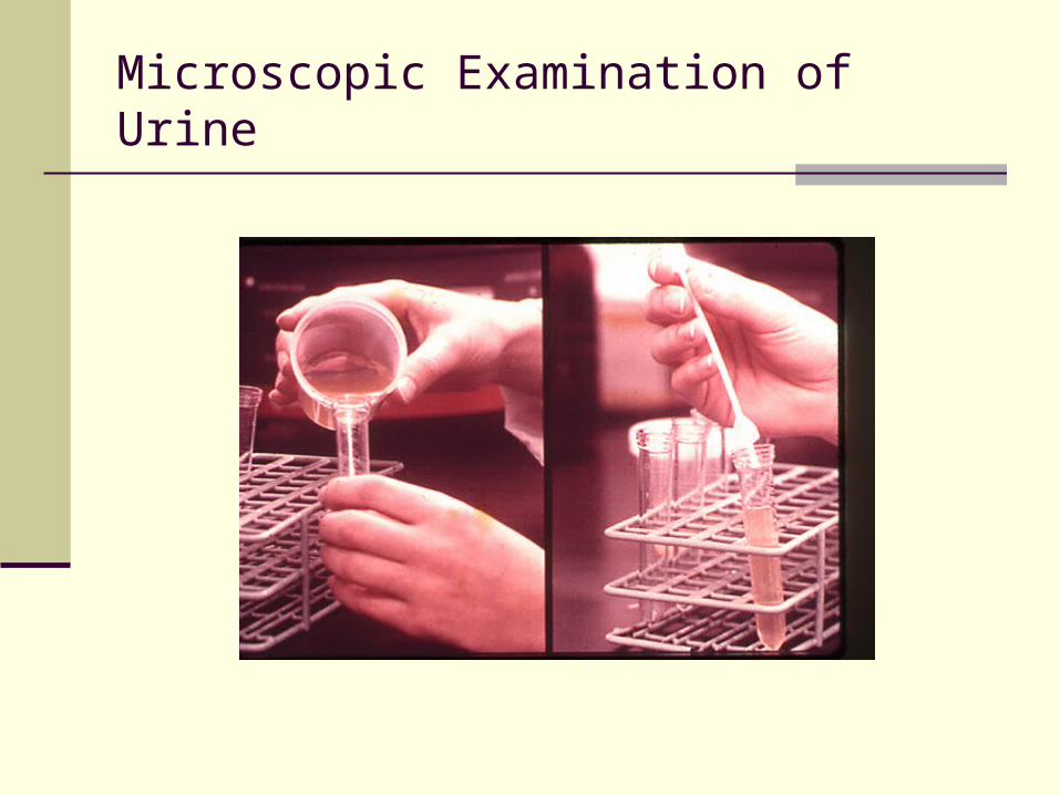
Preparation of specimen (show video) Mix specimen well Pour 12 ml into urine centrifuge tube Centrifuge five minutes, 1200-2000 RPM
(speed varies depending on the centrifuge’s characteristics)
Speed and time should be consistent. The “relative centrifugal force” is important.
Microscopic Examination of Urine
Microscopic Examination of Urine
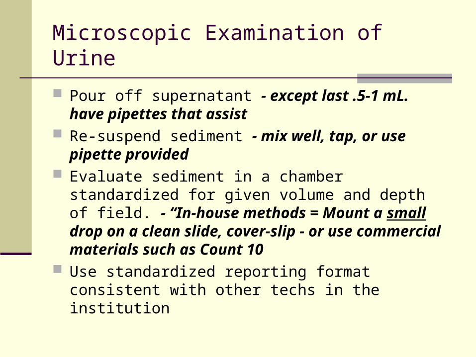
Pour off supernatant - except last .5-1 mL. have pipettes that assist
Re-suspend sediment - mix well, tap, or use pipette provided
Evaluate sediment in a chamber standardized for given volume and depth of field. - “In-house methods = Mount a small drop on a clean slide, cover-slip - or use commercial materials such as Count 10
Use standardized reporting format consistent with other techs in the institution
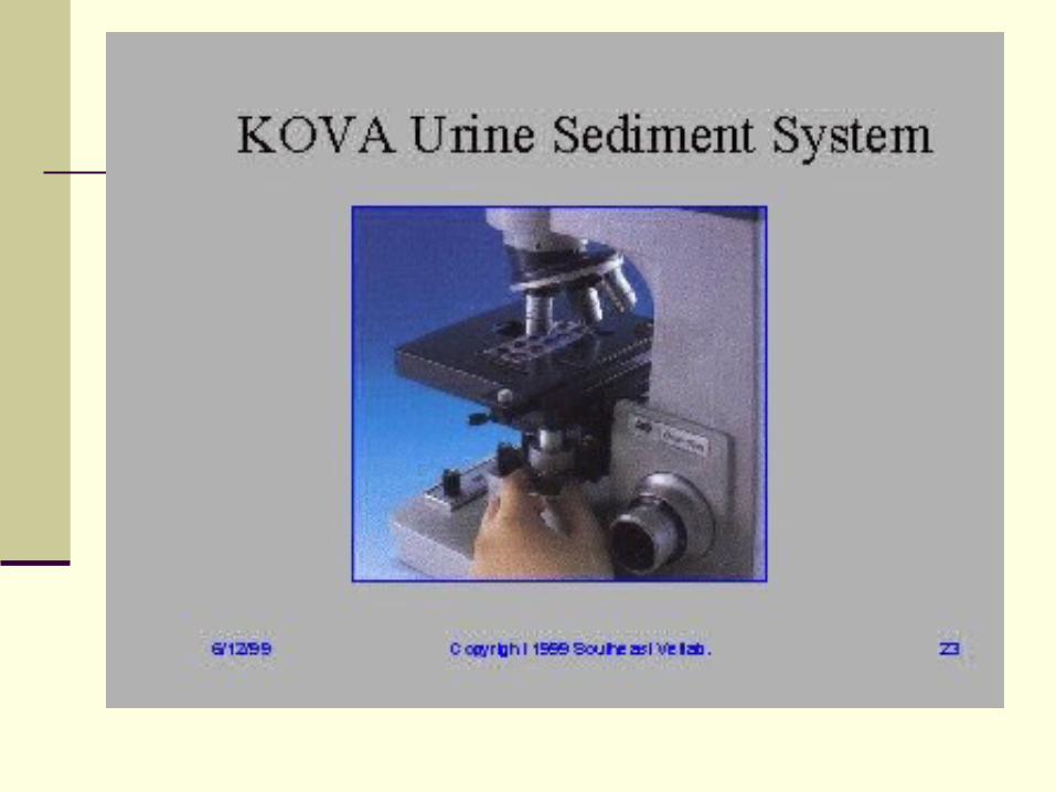
Microscopic Examination of Urine
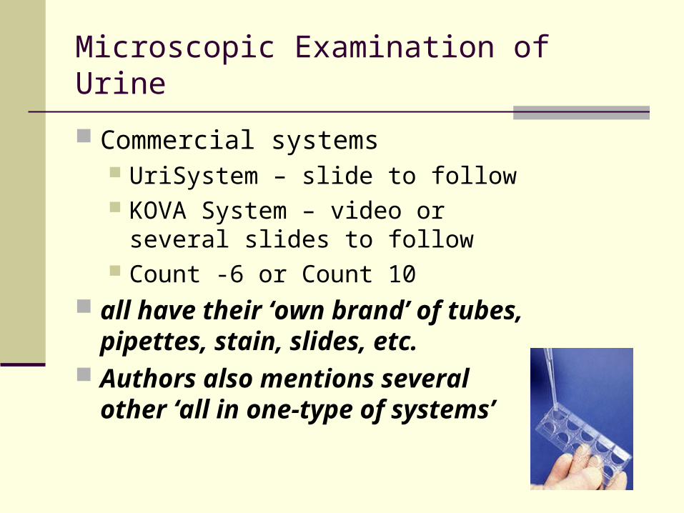
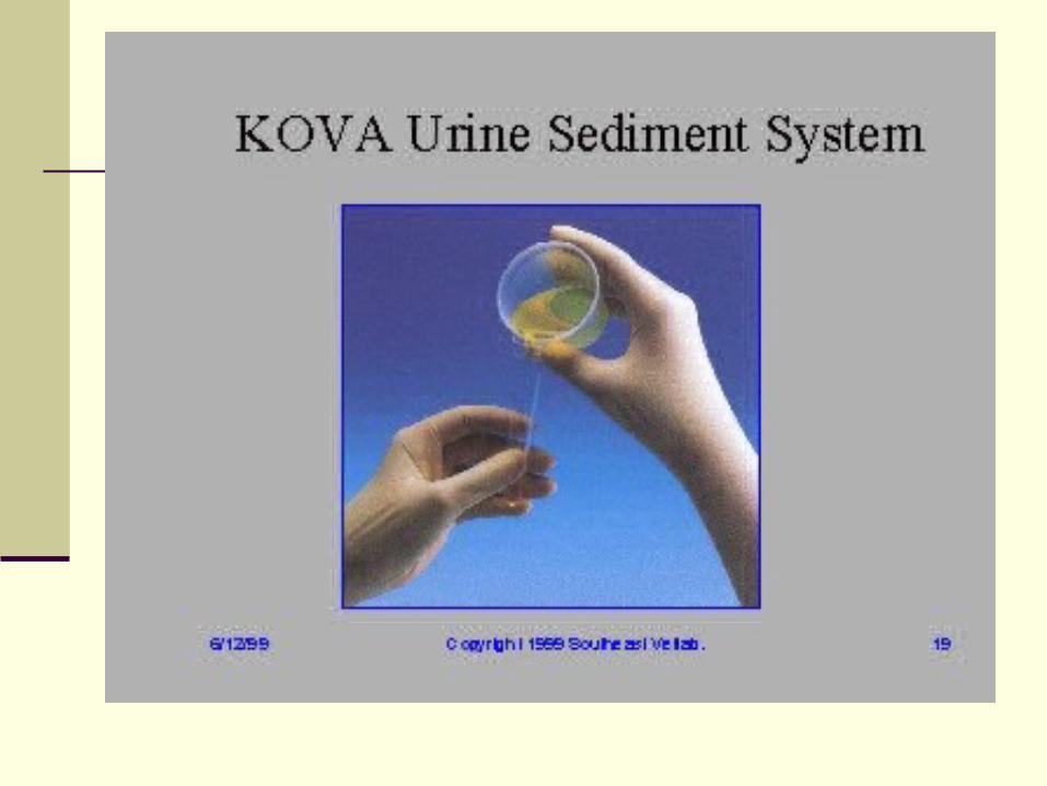
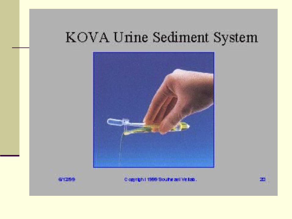
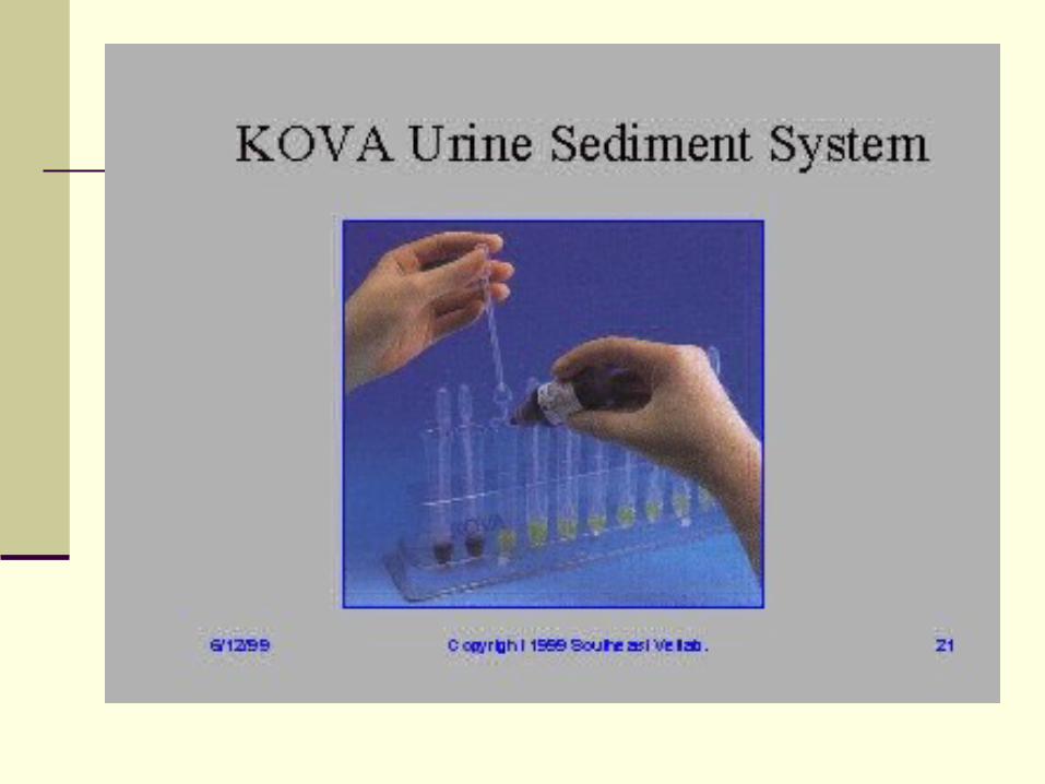
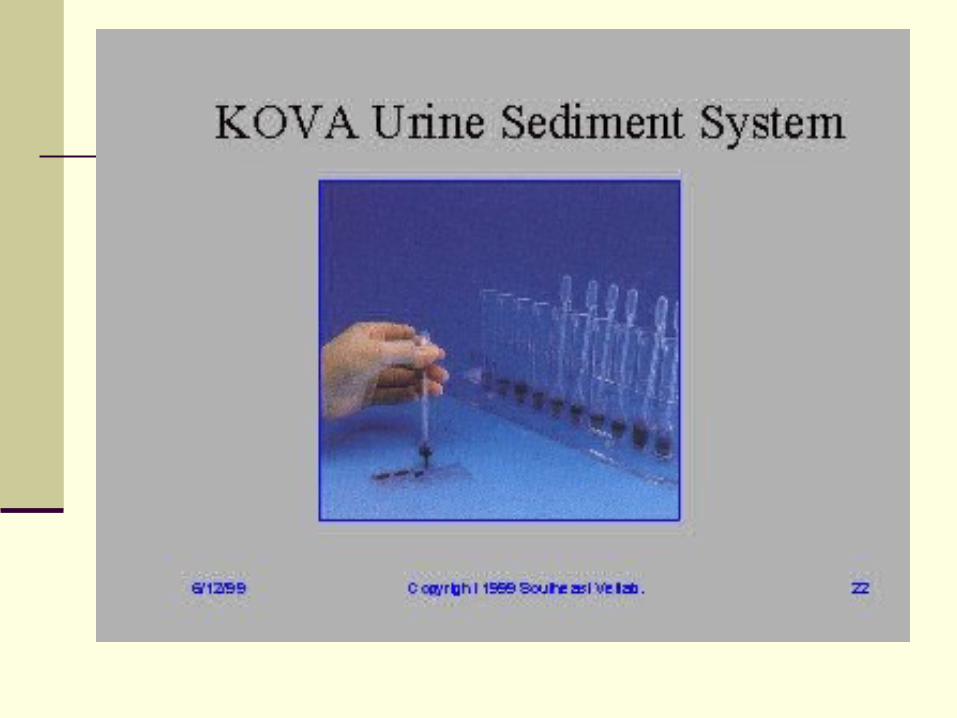
Commercial systems UriSystem – slide to follow KOVA System – video or several
slides to follow Count -6 or Count 10
all have their ‘own brand’ of tubes, pipettes, stain, slides, etc.
Authors also mentions several other ‘all in one-type of systems’
Microscopic Examination of Urine
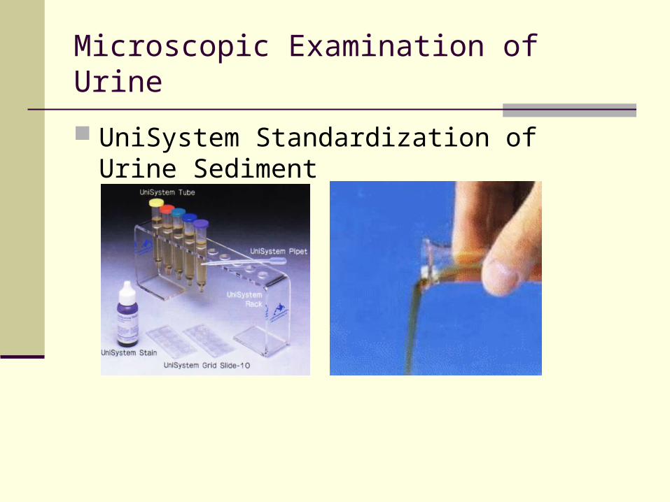
UniSystem Standardization of Urine Sediment
Microscopic Examination of Urine
Sedi-Stain (Sternheimer and Malbin) crystal violet, safranin-O Sedi-Stain & KOVA stain are commercial preparations
with addition of stabilizers to prevent precipitation. Supra-vital stain used to increase visibility of
structures. Assists greatly in differentiating renal tubular epithelial cells (which will take on an eosinophilic - oranges cytoplasm & dk purple nuclei) from transitional epithelial (which are more over-all blue)
Microscopic Examination of Urine
Not on lecture guide – Table 6-3 Sediment stain characteristics
Toluidine blue – nuclear structure Assists in differentiating WBC from renal epith.
2% acetic acid - removes interfering RBCs and enhances nuclei of WBC
Lipid stains - Oil Red O, Sudan III - stains triglycerides and neutral fats orange-red to ID lipid containing cells.
Microscopic Examination of Urine
Gram stain - to assist in ID of gram reaction of bacteria.
Hansel stain - methylene blue and eosin Y stains eosinophilic granules - ID eosinophils
Prussian blue reaction - makes iron granules blue in color (hemosiderin granules appear yellow until stained)
Microscopic Examination of Urine
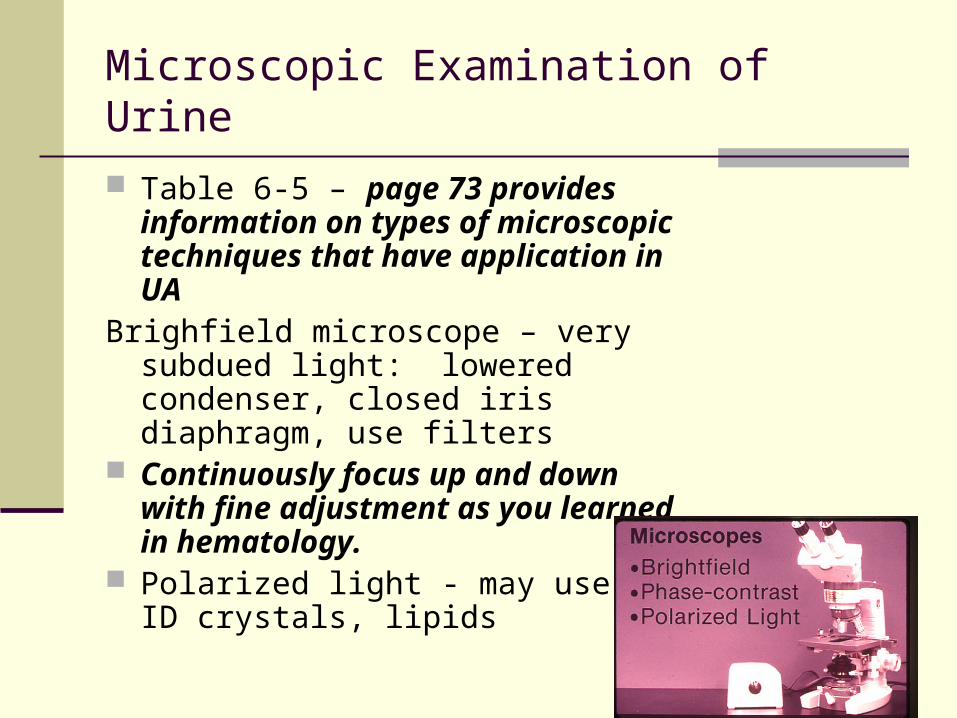
Table 6-5 – page 73 provides information on types of microscopic techniques that have application in UA
Brighfield microscope – very subdued light: lowered condenser, closed iris diaphragm, use filters
Continuously focus up and down with fine adjustment as you learned in hematology.
Polarized light - may use to ID crystals, lipids
Microscopic Examination of Urine
Types of Sediment As one author puts it:
Cells Casts Crystals Critters
Microscopic Examination of Urine
Types of Sediment Organized – biological part
RBC WBC Casts Epithelial cells Bacteria, parasites, yeast and fungi
Unorganized Crystals Amorphous crystalline matter.
Microscopic Examination of Urine
Examination - should correlate with physical and chemical
dipstick, may need to recheck
Scanning - – 10-15 fields using low power (10X). Look for casts, mucous, and squamous epithelial cells in general getting an overall feel
Report squamous epithelial cells, crystals, mucous, etc. using semi-quantitative terms such as rare, few, moderate, or many (or trace, 1+,2+,3+, & 4+) according to lab protocol.
Microscopic Examination of Urine
Enumeration - quantitate. Method may vary from lab to lab Average number of RBC/hpf Average number of WBC/hpf Average number of any renal tubular or
transitional epithelial cells /hpf.

Microscopic Examination of Urine
Average number (and type) of casts/__average # of casts /hpf______
authors have varied back and forth as whether low or high power should be reported... use low power to locate and enumerate the various types , but may need to switch to high power to identify the type...
Strasinger says report / lpf (use hpf to ID)
Unorganized sediment – few, moderate, many, packed; kinds seen
Note presence of bacteria, yeasts, crystals, epithelial cells (covered), etc.
quantitate these also
Microscopic Examination of Urine
.Changes in urine sediment when allowed to stand important to keep in mind the changes in microscopic
structures that can occur (don’t forget the other chemical changes ie bilirubin, pH, ketones)
RBC distorted – crenation, swelling, disintegration
WBC disintegrates in alkaline urine
Cast disintegrate in alkaline urine
Bacterial growth – increased alkalinity
Increased precipitation of crystals, especially amorphous
Microscopic Examination of Urine
Microscopic sediment Red Blood Cells White Blood Cells Epithelial Cells Casts Crystals Miscellaneous structures
Students go to end of area’s lecture guide. Continue to next slide.
Microscopic Examination of Urine
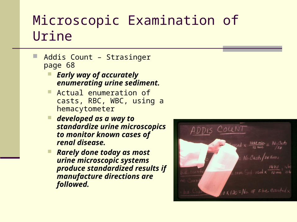
Addis Count – Strasinger page 68 Early way of accurately
enumerating urine sediment. Actual enumeration of casts,
RBC, WBC, using a hemacytometer
developed as a way to standardize urine microscopics to monitor known cases of renal disease.
Rarely done today as most urine microscopic systems produce standardized results if manufacture directions are followed.