Embed Size (px)
Citation preview

METABOLISM
= summary of all chemical (and physical) processes included in:
1. Production of energy from internal and external sources
2. Synthesis and degradation of structural and functional tissue
components
3. Excretion of waste products and toxins from body
METABOLISM
•Proteins
•Saccharides
•Lipids
METABOLIC DISORDERS
1. Inherited metabolic disorders
(enzymopathies)
2. Combined metabolic disorders (DM, gout,
degenerative disorder of joints and bones)
3. Metabolic disorders from external reasons
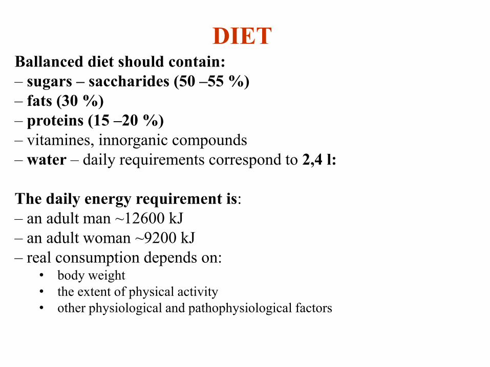
Ballanced diet should contain:
– sugars – saccharides (50 –55 %)
– fats (30 %)
– proteins (15 –20 %)
– vitamines, innorganic compounds
– water – daily requirements correspond to 2,4 l:
The daily energy requirement is:
– an adult man ~12600 kJ
– an adult woman ~9200 kJ
– real consumption depends on: • body weight
• the extent of physical activity
• other physiological and pathophysiological factors
DIET

Insulin versus glucagon

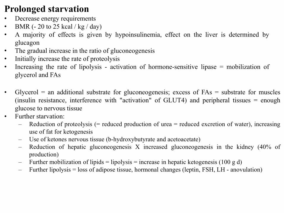
Prolonged starvation• Decrease energy requirements
• BMR (- 20 to 25 kcal / kg / day)
• A majority of effects is given by hypoinsulinemia, effect on the liver is determined by
glucagon
• The gradual increase in the ratio of gluconeogenesis
• Initially increase the rate of proteolysis
• Increasing the rate of lipolysis - activation of hormone-sensitive lipase = mobilization of
glycerol and FAs
• Glycerol = an additional substrate for gluconeogenesis; excess of FAs = substrate for muscles
(insulin resistance, interference with "activation" of GLUT4) and peripheral tissues = enough
glucose to nervous tissue
• Further starvation:
– Reduction of proteolysis (= reduced production of urea = reduced excretion of water), increasing
use of fat for ketogenesis
– Use of ketones nervous tissue (b-hydroxybutyrate and acetoacetate)
– Reduction of hepatic gluconeogenesis X increased gluconeogenesis in the kidney (40% of
production)
– Further mobilization of lipids = lipolysis = increase in hepatic ketogenesis (100 g d)
– Further lipolysis = loss of adipose tissue, hormonal changes (leptin, FSH, LH - anovulation)

Other changes as a result of starvation:
• Loss of K+ in the initial stage, a stable concentration of 3 mmol/L
• Mg2+ - unchanged or only slight hypomagnesemia
• Ca2+ - unchanged
• Phosphates – unchanged
• Uric acid – increase (protein catabolism)
• Next changes: Decreased heart rate (35 t/min, from 4. week slight increase)
Drop of blood pressure
ECG changes - flattening of the T wave, decrease of amplitude of QRS
In cases of extreme starvation - prolongation of the QT interval, T wave
inversion, ST segment depression
Why?
o The decrease of protein synthesis - myofibrils, myofilaments
o Changes in the composition of the ECT/ICT
o Losses of trace elements (Cu - ischemia)
o Sympathetic (catecholamines) - Arrhythmia


METABOLIC DISORDERS EXAMINATION
LABORATORY METHODS (biochemistry)
• Lack or absence of metabolite (blood, urine, tissue, cells)
• Overproduction of metabolite
• Pathological storing of metabolite in tissues (histochemistry)
• Pathological metabolite
FINDINGS OF CAUSE OF METABOLIC DISORDER
• Disorder in resorption or excretion (functional load tests)
• Measurement of activity of certain enzymes or enzyme systems
GENEALOGIC EXAMINATION
SCREENING TESTS (fenylketonuria, hyperlipoproteinemia,
aminoaciduria, thyroid gland hormones…)
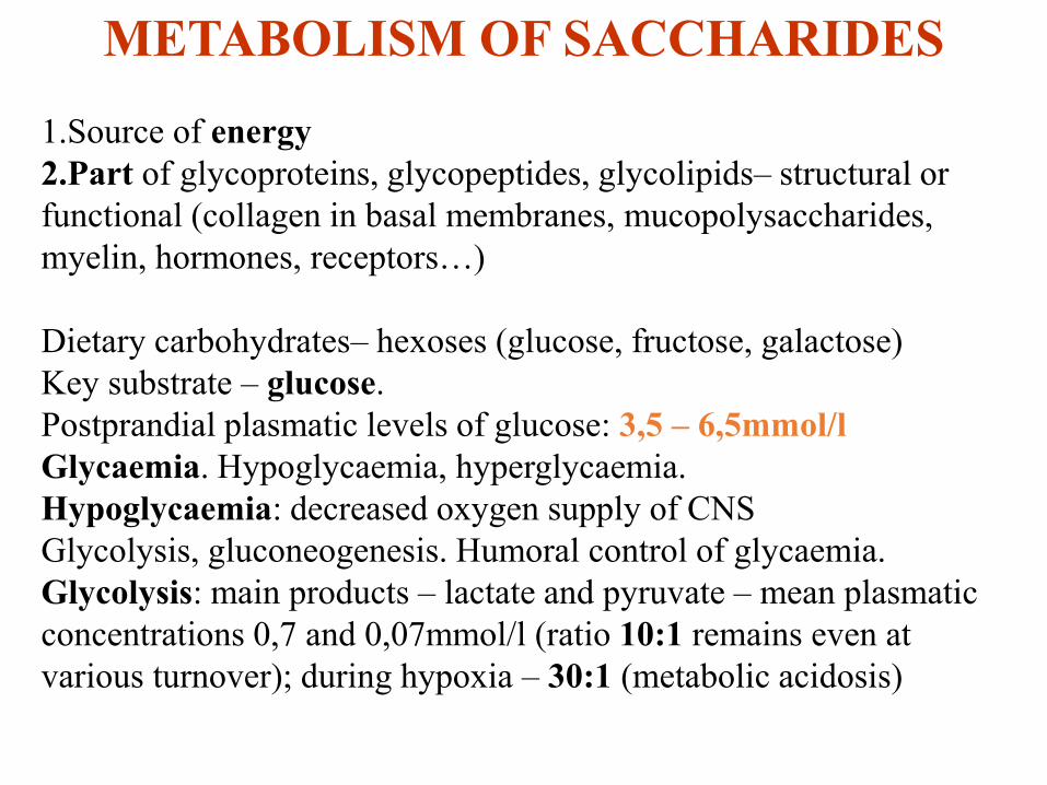
METABOLISM OF SACCHARIDES
1.Source of energy
2.Part of glycoproteins, glycopeptides, glycolipids– structural or
functional (collagen in basal membranes, mucopolysaccharides,
myelin, hormones, receptors…)
Dietary carbohydrates– hexoses (glucose, fructose, galactose)
Key substrate – glucose.
Postprandial plasmatic levels of glucose: 3,5 – 6,5mmol/l
Glycaemia. Hypoglycaemia, hyperglycaemia.
Hypoglycaemia: decreased oxygen supply of CNS
Glycolysis, gluconeogenesis. Humoral control of glycaemia.
Glycolysis: main products – lactate and pyruvate – mean plasmatic
concentrations 0,7 and 0,07mmol/l (ratio 10:1 remains even at
various turnover); during hypoxia – 30:1 (metabolic acidosis)

•Glucose turnover: 2mg/kg/min (11mmol/kg/min)~9g/hr~225g/day
•55% of glucose utilisation – terminal oxidation (CNS)
•20% - glycolysis, lactate back to liver, gluconeogenesis (Cori cycle)
•20% - absorption by liver and splanchnic tissues
•70% consumption of glucose at rest is insulin-independent
•Circulating glucose pool (pool) – only a little bigger than expenditure
by liver per 1 hour
•Brain oxidation is kept by pool only for approx. 3 hrs
•NECESSITY OF CONTINUOUS GLUCOSE PRODUCTION
FROM LIVER during starving
•80% - glycogenolysis, 20% - gluconeogenesis (more than 50% from
lactate trapped by liver for gluconeogenesis, rest – AA, esp. alanine;
lactate from glycolysis in muscles, ery, leu, etc.; AA – from
proteolysis of muscles)

•Morning glucose intake – 70% is needed by peripheral tissues
(muscles), 30% - splanchnic organs (liver)
•20-30% of consumed glucose – oxidised during 3-5 hrs to cover
needs of GIT, 70-80% stored as glycogen (muscle, liver)
•Muscle glycogen – later transported to liver (lactate from glycolysis
in muscles, re-uptake, gluconeogenesis in liver, glycogenolysis)
•During maximal absorption of exogenous glucose – release of
glucose from liver is suppressed (insulin and glucagon facilitate this
process)

LIVER GLUCOSTAT
- Maintaining the constant blood glucose
- Endocrine control:
• glycogenolysis (glucagon, adrenaline, noradrenaline = activation of glycogen
phosphorylase)
• why only liver and not muscles? (glucose-6-phosphatase in liver)
• gluconeogenesis (glucagon, adrenaline, noradrenaline, glucocorticoids, thyroid
hormones)
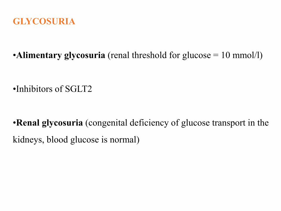
GLYCOSURIA
•Alimentary glycosuria (renal threshold for glucose = 10 mmol/l)
•Inhibitors of SGLT2
•Renal glycosuria (congenital deficiency of glucose transport in the
kidneys, blood glucose is normal)

METABOLISM OF LIPIDS
•Fat – approx. 50% of daily amount of substrates for oxidation (100gr,
900kcal)
•Main and most profitable form of energy store
•Daily intake: approx. 100gr (40% of daily diet)
•Main component of dietary sources and stores in body: triglycerides
•No strict dietary recommendation (part of FA synthetised in liver and
adipose tissue)
•BUT: 3-5% of FA are polyunsaturated!!! – ESSENTIAL FA
•Precursors of membrane phospholipids, glycolipids, prostaglandins
•Cholesterol – part of membranes, precursor of bile acids, steroid
hormones; daily intake – 300-600mg/day, synthesised too
•Lipoproteins: transport of lipids by blood plasma
•Apoproteins (from liver or intestine), catalytic function, receptors
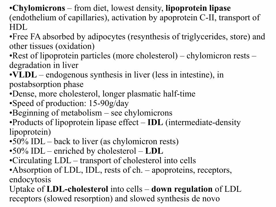
•Chylomicrons – from diet, lowest density, lipoprotein lipase(endothelium of capillaries), activation by apoprotein C-II, transport of HDL•Free FA absorbed by adipocytes (resynthesis of triglycerides, store) and other tissues (oxidation)•Rest of lipoprotein particles (more cholesterol) – chylomicron rests –degradation in liver•VLDL – endogenous synthesis in liver (less in intestine), in postabsorption phase•Dense, more cholesterol, longer plasmatic half-time•Speed of production: 15-90g/day•Beginning of metabolism – see chylomicrons•Products of lipoprotein lipase effect – IDL (intermediate-density lipoprotein)•50% IDL – back to liver (as chylomicron rests)•50% IDL – enriched by cholesterol – LDL•Circulating LDL – transport of cholesterol into cells•Absorption of LDL, IDL, rests of ch. – apoproteins, receptors, endocytosisUptake of LDL-cholesterol into cells – down regulation of LDL receptors (slowed resorption) and slowed synthesis de novo

•HDL – long plasmatic half-time, synthesis in liver and intestine
•Facilitation of other particles movement
•Exchange of key apoproteins
•They accept molecules of free cholesterol, estherify them (lecithin-
cholesterol-acetyltransferase) and incorporate back to particles
•Main effect: acceleration of clearance of triglycerides from plasma
and regulation of ration free:estherified cholesterol
•Free FA
•Average concentration: 400mM/l
•Bound to molecules of albumins
•Fast turnover (approx. 8g/hr): 50% - oxidation, 50% -
reestherification to triglycerides
•Total cholesterol: 185mg/l
•LDL cholesterol: 120mg/l
•HDL cholesterol
•Arteriosclerosis, genetic predisposition (LDL apo or receptor)

METABOLIC DISORDERS - SACCHARIDES
1. Diabetes mellitus
2. McArdle syndrom: glycogenesis from deficiency of
myophosphorylase
Accumulation of glycogen in muscles
Muscle stiffness, rigor during exercise, lower tolerance of load
3. Galactosemia (inherited deficiency of
phosphogalactosauridyltransferase; disorders of growths and
development)
1. HYPERLIPIDEMIA, HYPERLIPOPROTEINEMIA
2. INFREQUENT DISORDERS OF LIPID METABOLIS
METABOLIC DISORDERS - LIPIDS

Ad 1) 5% of population
Primary and secondary forms
Arteriosclerosis
•Hyperlipoproteinemia induced by lipids
•Familiar hypercholesterolemia (xantomatosis)
•Mixed hyperlipoproteinemia
•Familiar hypercholesterolemia with hyperlipemia
•Saccharides-induced triglyceridemia
•Secondary hyperlipoproteinemia (dependent; alimentary)
Ad 2)
•Lipidoses
•Abetalipoproteinemia (LDL, VLDL)
•Analfalipoproteinemia (HDL)
•Inherited defect acetyltranspherase LCAT (accumulation of lecithin)

BROWN ADIPOSE TISSUE
LIPIDS: structural, neutral and brown
Specific localisation
Sympathetic innervations of vessels and also
adipocytes
Several drops of fat in adipocyte
More mitochondria
Production of heat
Adaptation to cold
After meal – increased production of heat
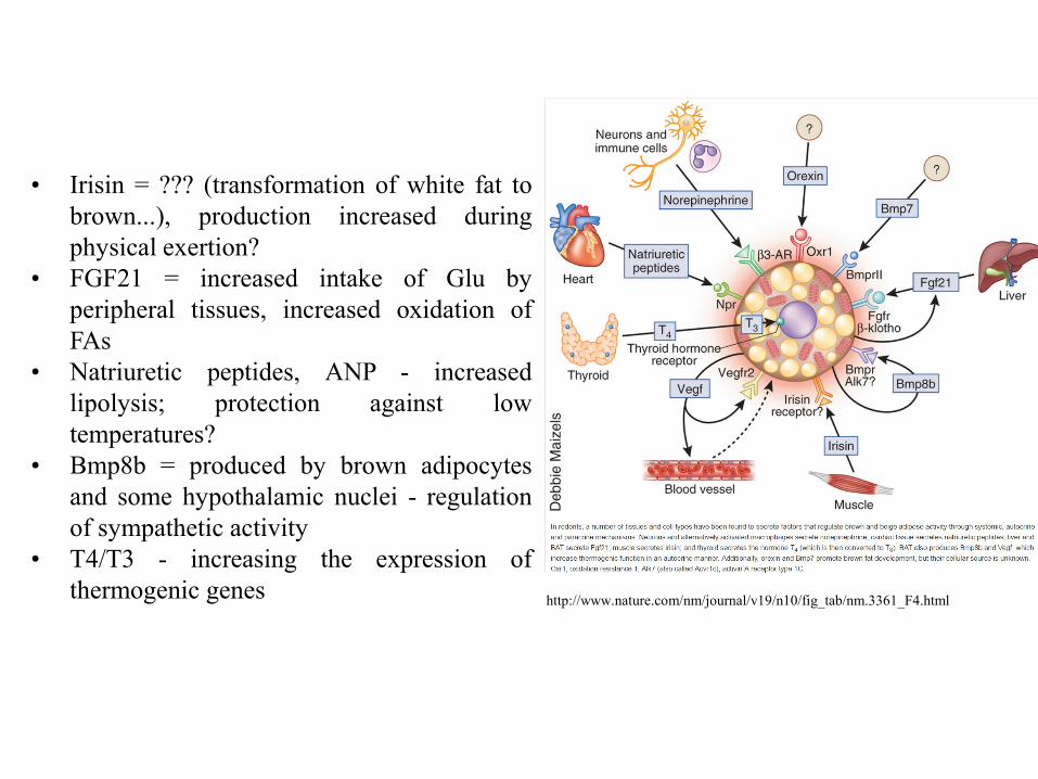
http://www.nature.com/nm/journal/v19/n10/fig_tab/nm.3361_F4.html
• Irisin = ??? (transformation of white fat to
brown...), production increased during
physical exertion?
• FGF21 = increased intake of Glu by
peripheral tissues, increased oxidation of
FAs
• Natriuretic peptides, ANP - increased
lipolysis; protection against low
temperatures?
• Bmp8b = produced by brown adipocytes
and some hypothalamic nuclei - regulation
of sympathetic activity
• T4/T3 - increasing the expression of
thermogenic genes

Exercise-induced adipose tissue
browning through PGC-1α and
irisin. Exercise increases the
expression levels of PGC-1α in the
muscle. This, in turn, upregulates
the expression of FNDC5, a type I
membrane protein, which is C-
terminally cleaved and secreted as
irisin into the circulation. Binding
of irisin to an unknown receptor on
the surface of adipocytes in WAT
changes their genetic profile. In
particular, irisin induces the
expression of PPAR-α, which is
thought to be an intermediate
downstream effector that increases
the expression of UCP1 (highly
expressed in BAT and a marker of
browning). The browning of WAT
is associated with augmented
mitochondrial density and oxygen
consumption. Browning is
accompanied by an increase in the
energy expenditure profile, leading
to favourable effects on
metabolism.
Peroxisome proliferator-activated receptor-gamma coactivator (PGC)-1a
Castillo-Quan JI: From white to brown fat through the PGC-1 alpha-dependent
myokine irisin: implications for diabetes and obesity. Disease Models & Mechanisms
2012, 5(3):293-295.

METABOLISM OF PROTEINS
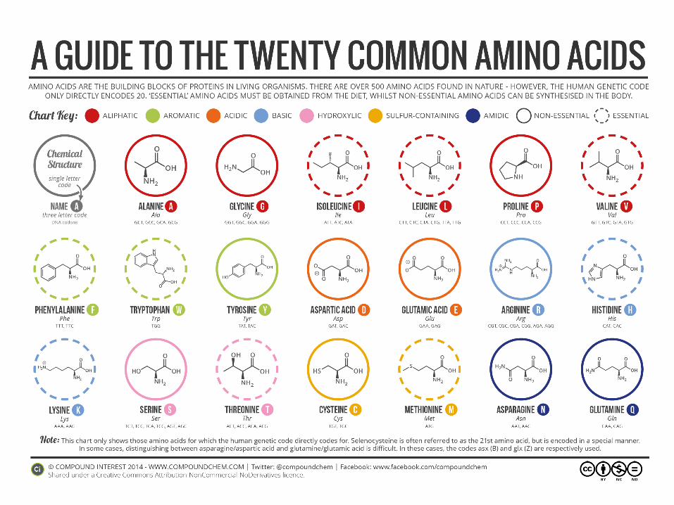
•Proteins = AA bound by peptide bonds (above 100 AA)
•Peptides (2-10 AA), polypeptides (10-100 AA)
•Primary, secondary, tertiary a quarterly structure of protein
Proteins, lipoproteins, glycoproteins

Total proteins in body: 10 kg
Metabolically active: 6 kg (e.g.60%)
Proteolysis of muscles: 50 g of proteins / day
Minimal daily intake: 50 g
Protein minimum: 0,5 g / kg of body mass
Protein optimum: 0,7 g / kg of body mass
Increased supply (growth, convalescence, pregnancy,
lactation): 1,5 – 2,0

AMINOACIDES
•Essential (not synthesised)
•Non-essential (from glucose metabolism – citrate cycle)
•Aminoacid pool
•Need of essential AA: 0,5 – 1,5 g / day
•Disorders of proteosynthesis
•Optimal source of E-AA:NE-AA milk, eggs
•During growth: 40% E-AA, in adults: 20%
•Precursors: purines, pyrimidines, polyamines, phospholipids,
creatin, carnitin, donors of methyl group, catecholamines, thyroid
gland hormones, neurotransmitters

Amino acids - the surplus in food
Degradation, used as an energy source
AMK as other substrates:
- Glucogenic AMK – synthesis of carbohydrates
- Ketogenic AMK – lipids and ketones

Isoelectric point = pI
Ionization states of amino acids as a function of pH:
Determination of pK1, pK2 and pI of alanine
pI =(pK1 + pK2) / 2 (isoelectric point, pI = 6)

Derivatives of AMK with physiological functions
-Aminomáselná kyselina
CH2
CH2
NH3+
OOC CH2
a
(GABA)
Histamin
CH2
CH2
NH3+
N
N
H
Dopamin
CH2
CH2
NH3+
OH
OH
Thyroxin
CH
CH2
NH3+
O
COO
I
I
OH
I
I
-

DEGRADATION OF PROTEINS
Binding to ubiquitin (74 AA).
Oxidation to CO2 and H2O after removing the amino-group
(deamination).
Gluconeogenesis (except of leucin), ketogenesis (5AA,
acetoacetate or CoA precursors), ureagenesis (all AA, ammonium
bound to glutamin or alanine, liver, Krebs-Henseleit cycle).
Regulated speed of degradation (muscle hypertrophy, atrophy of
denerved or non-stimulated muscle).

AMINOACIDS
AMMONIUMCO2 + ATP +
CARBAMOYLPHOSPHATE
CITRULIN
ASPARTATE
ARGININOSUKCINATE
FUMARATE
ARGININ
ORNITIN
UREA
URINE

Degradation of proteins
•lysozomes
• Extracellular proteins
• Membrane proteins
• Proteins with long half-time
• Process does not require ATP
•cytosol
• Metabolic proteins
• Proteins with short half-time
• Process requires ATP and ubiquitin

URIC ACID
Excreted in urine.
4mg/100ml of blood plasma
Kidney: filtration, resorption (98% filtration), tubular secretion
(80%)
Daily: approx. 1g excreted in urine
Disorder in uric acid metabolism – gout.
Hyperuricemia – primary (overproduction) or secondary (reduced
excretion, increased intake of purines in diet, blood disorders).
METABOLISM OF PURINES AND PYRIMIDINES
Purines and pyrimidines – physiological meaning of nucleosides
(reactants with ribose); from diet or synthesis de novo from AA in
liver; RNA is in balance with AA pool, DNA is stabile.
Recirculation or catabolism, eventually excretion in urine.
Pyrimidines – CO2 and NH3, purines – uric acid.

Synthesis of purines/pyrimidines
•de novo (new synthesis of purine/pyrimidine ring)
• „saving“ reactions (synthesis from nucleotides and bases)
is more energy saving than de novo synthesis
They decrease the synthesis de novo
substrates: a) bases (adenine, guanine, hypoxanthine)PRDP
b) ribonucleosidesATP
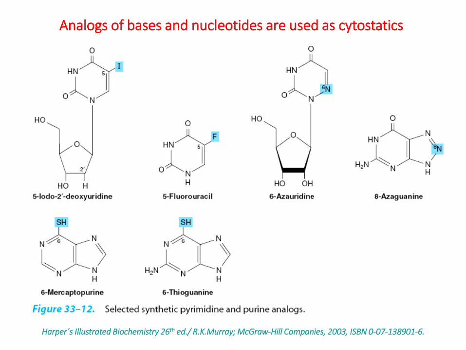
Harper´s Illustrated Biochemistry 26th ed./ R.K.Murray; McGraw-Hill Companies, 2003, ISBN 0-07-138901-6.
Analogs of bases and nucleotides are used as cytostatics

GOUT (arthritis urica)
•Primary and secondary gout
•Acute (gouty attack) and chronic (chalkstones, urolithiasis) form
•General metabolic disorder - disease of purine metabolism
•Local cumulating of uric acid salts (urate) in tissues, urine
(joints, kidneys), primary hyperuricemia
•Gouty attacks – repeated attacks of arthritis, typical localisation –
metatarsophalangeal joint (podagra; omagra, cheiragra…)
•Hurtfulness during attack – phagocytosis of urates grains
•Therapy: NSA, colchicin – inhibition of fagocytosis, allopurinol
– inhibition of xantinoxidase, phenylbutazon and probenecid –
inhibition of resorption

NITROGEN BALANCE
Necessity to keep AA pool. AA mixtures.
Amount of N in urine – indicator of intensity of irreversible
disintegration of proteins and AA.
Nitrogen balance: amount of N in urine = amount of N in dietary
proteins
•Negative nitrogen balance: loss exceeds intake (starvation,
immobilisation, catabolism, lack of E-AA!!!…)
•Positive nitrogen balance: intake exceeds loss (anabolic drugs,
growth, convalescence…)
Synthesis and degradation of body proteins: 3–4g/kg of body mass
(balanced diet)
From this amount: 5% - synthesis of albumins and proteins with
fast-exchange in liver
In deficient diet (energetically, amount of proteins or E-AA) –
proteosynthesis deceleration, compensatory –degradation
deceleration (BUT of lower extent loss of body proteins)
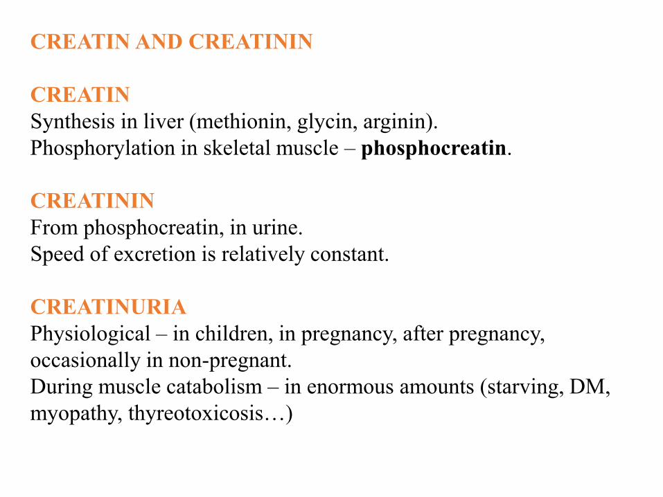
CREATIN AND CREATININ
CREATIN
Synthesis in liver (methionin, glycin, arginin).
Phosphorylation in skeletal muscle – phosphocreatin.
CREATININ
From phosphocreatin, in urine.
Speed of excretion is relatively constant.
CREATINURIA
Physiological – in children, in pregnancy, after pregnancy,
occasionally in non-pregnant.
During muscle catabolism – in enormous amounts (starving, DM,
myopathy, thyreotoxicosis…)

Wyss M, Kaddurah-Daouk R: Creatine and creatinine metabolism. Physiol
Rev 2000, 80(3):1107-1213.

METABOLIC DISORDERS – PROTEINS
QUANTITATIVE CHANGES
Proteinemia = plasmatic level of proteins.
Controlled:
1. Supply with full-value proteins and their use
2. Synthesis of proteins
3. Protein catabolism and loss from organism
Ad 1) nutrition disorders, special dietary trends
Ad 2) liver disorders, endocrine diseases
Ad 3) liver and muscles release E-AA when proteins are reduced in diet

METABOLIC DISORDERS – PROTEINS
QUALITATIVE CHANGES
1. Dysproteinemia = change in representation of particular
proteins (fractions shift) – nephrotic syndrome, cirrhosis,
acute inflammatory reactions, chronic inflammatory reactions,
tumours
2. Paraproteinemia = presence of pathological imunoglobulines
(with no antibodies specificity) – monoclonal immunopathy
3. Defect proteinemia = some components of plasma proteins are
missing or lowered (1/10 – 1/1000 normal values) –
syndromes of immunodeficiency, symptomatic hypo- and
dysgamaglobulinemia (familiar lack of IgA), polyclonal
hypergamaglobulinemia

METABOLIC DISORDERS –AMINOACIDES
1. Disorders of AA metabolism during hypovitaminoses and
avitaminoses – vit.C (colagen synthesis– proline hydroxylation;
metabolic osteopathy, haemorrhage, poor healing), vit.B6
(tryptophan metabolism – lack of nicotinic acid)
2. Disorders of AA metabolism during liver diseases – regulation
of plasmatic level of AA (transamination, oxidation,
decarboxylation, deamination, ammonia, urea, kidneys); badly
soluble AA (cystine, tyrosine) may form crystals in urine; liver
encephalopathy, liver coma, glutamine in coeliolymph

AMYLOIDOSIS
= infiltration of organs by amyloid (complex of protein with
polysaccharide)
Mechanism of disease is alteration of immune system.
Primary and secondary amyloidosis
Primary – idiopathic; infliction of heart, muscles, GIT; elderly
patients; no gender differences
Secondary – complication of chronic inflammatory diseases,
tumours; more frequent; infliction of kidney (most often), lien,
liver, adrenal glands