Embed Size (px)
Citation preview

Urinary system 5Congenital and cystic diseases of
the kidney, urinary calculi and urinary obstruction
Professor John Simpson

Congenital renal disease
• ~ 10% born with potentially significant malformation of urinary tract, but congenital renal disease much less common
• renal agenesis and hypoplasia cause ~ 20 % renal failure in kids
• ectopic kidneys and horseshoe kidney also potentially important

Cystic diseases of the kidney
• cysts may be– solitary or multiple– unilateral or bilateral– congenital (hereditary or not) or acquired
• many uncommon, but may be important if genetic screening & counselling available
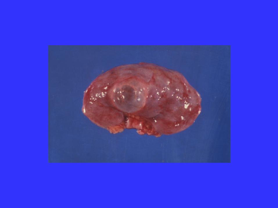
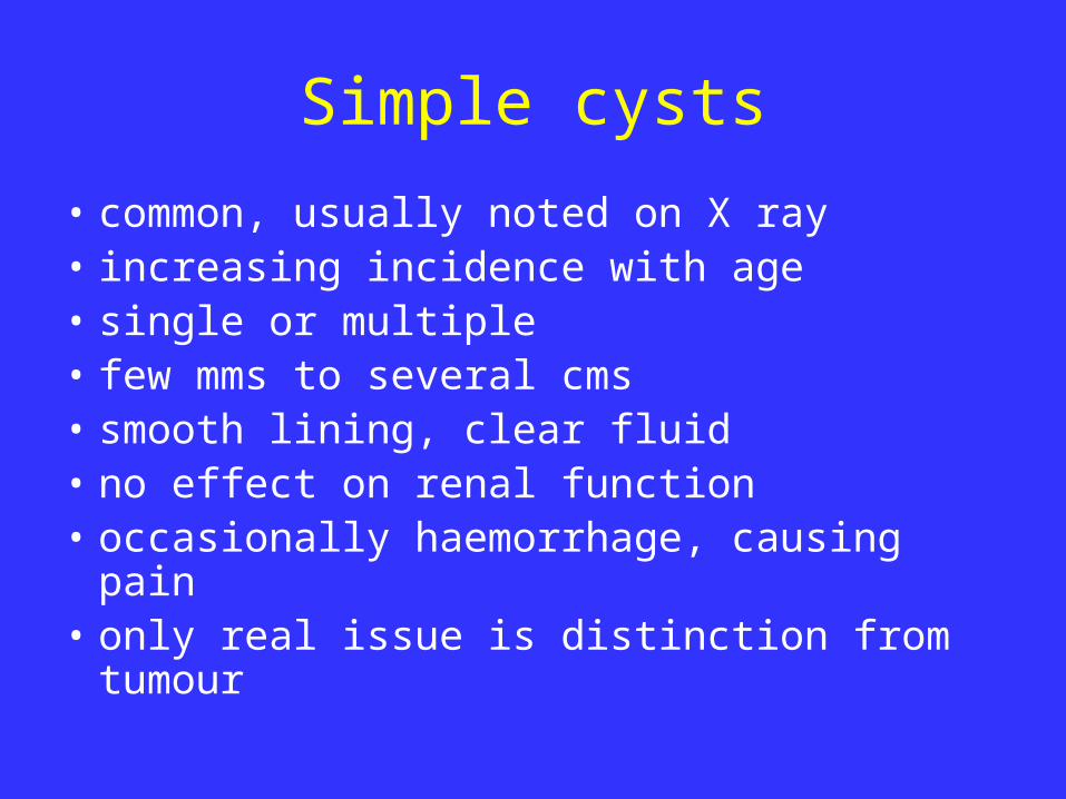
Simple cysts
• common, usually noted on X ray• increasing incidence with age• single or multiple• few mms to several cms• smooth lining, clear fluid• no effect on renal function• occasionally haemorrhage, causing pain• only real issue is distinction from tumour
AUTOSOMAL DOMINANT RENAL POLYCYSTIC DISEASE (ADPKD)
(there is also an ARPKD)
• relatively common in Europe (~ 1 in 500 births)• multiple bilateral cysts - enlarge & compress renal
parenchyma• congenital, but presents any time from late childhood on
(usually early/mid adulthood)• autosomal dominant inheritance with high penetrance –
so screening important if it’s available• genetic mutations for this condition now clear (one of 3
different genes - PKD 1-3)
ADPKD
• usually presents as one of -– chronic renal failure (up to 10% patients with CRF)– hypertension – or abdominal mass
• may also be– pain, haematuria, UTI, calculi
• occasionally cysts in other organs (clinically unimportant)
• and cerebral berry aneurysms cause death in up to 10%
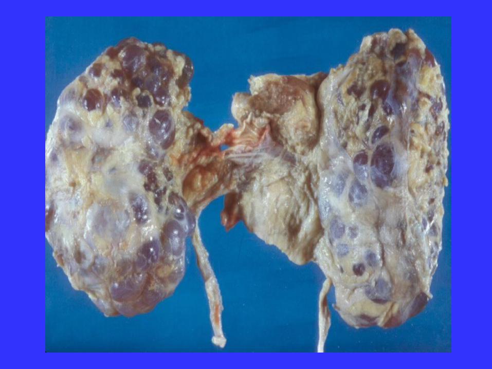
ADPKD
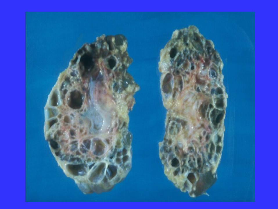
• kidneys enlarged – usually at least 1000G each• masses of cysts, up to 3 - 4 cm diameter -
usually no obvious intervening parenchyma.• cysts arise at all levels of the tubule• filled with clear serous fluid +/- evidence of
haemorrhage
Other cystic diseases
• dialysis-associated cystic disease - small cysts common in patients on long-term dialysis, prob due to tubular blockage in scarred kidneys: usually unimportant, but a few undergo malignant change - renal carcinoma
• uraemic medullary cystic disease (nephronophthisis) – cysts at CM junction– responsible for 20% CRF in children/adolescents
• medullary sponge kidney – cysts in papillae– no effect on renal function, but calculi can arise in cysts
• renal “dysplasia” – cysts all over– islands of undifferentiated tissue in kidney, usually cystic– only important if bilateral
Renal and urinary calculi – (nephrolithiasis & urolithiasis)
• maybe 1- 5% population at any one time in UK – much higher in Middle East (often bladder), less common in the tropics– ? protein (prothrombin fragment 1) in urine of black
races inhibits crystal formation• usually young/middle aged adults• overall, twice as common in men than women
– but calculi related to infection more common in women
• often recurrent - half will have another calculus within 10 yrs
Types of calculi
• calcium stones (Ca++ in complex with oxalate or phosphate or both) – most common stone
• triple (Mg NH4 PO4) or struvite stones – quite common
• uric acid stones – 5%• cystine or pure oxalate stones - inborn errors of
metabolism -1%• HIV – not really stones, but crystal precipation
due to antiviral agents

Pathogenesis of calculi
• most important factor is increased urinary concentration of stone’s constituents– once solubility exceeded, precipitation occurs
• enhanced by any reduction in urinary solubility – especially rise in pH or lack of inhibitors of crystal formation (citrate or pyrophosphate)
• urinary protein may act as “nidus” for stone formation
Calcium stones
• most patients have hypercalciuria– but only 10% have hypercalcaemia– e.g. due hyperparathyroidism, sarcoid, vit D
intoxication, prolonged immobilisation etc• so ? defect in tubular calcium reabsorption• (excretion of uric acid in urine also favours calcium stone
formation)
• (“nephrocalcinosis” is different pathology – refers not to stones/calculi but flecks of calcification in kidney - often due to hypercalcaemia and usually clinically unimportant)
Triple (struvite) stones
• almost always females with persistently alkaline urine due to UTI
• certain bacteria hydrolyse urea to form NH4 – especially P vulgaris/mirabilis
• bacteria also serve as nidi for stone formation
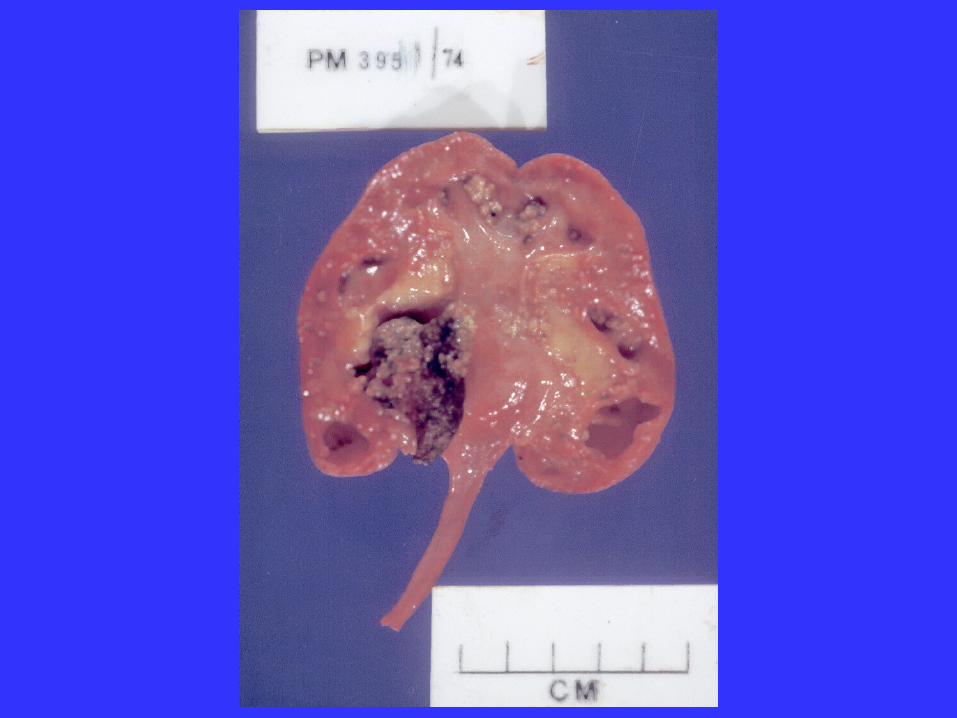
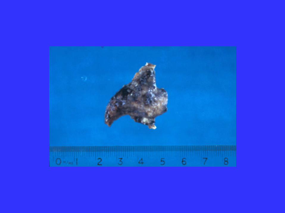
• (calculi often “staghorn” shaped – cast of part/all of pelvi-calyceal system)

Uric acid stones
• associated with gout (25% of patients with gout) or leukaemias (high cell turnover)
• hyperuricaemia causes hyperuricosuria
• but 50% of patients have neither
• (hyperuricosuria also favours calcium stone formation)
Renal and urinary calculi
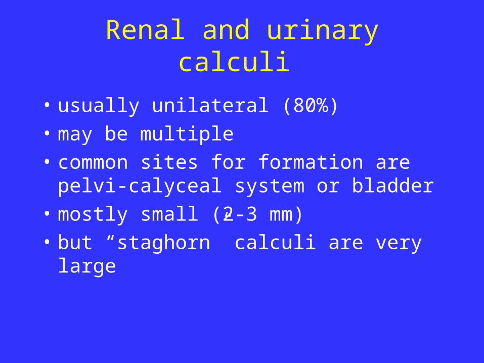
• usually unilateral (80%)
• may be multiple
• common sites for formation are pelvi-calyceal system or bladder
• mostly small (2-3 mm)
• but “staghorn” calculi are very large

Clinical effects
• depends on type, size and site of origin and/or arrest
• can be asymptomatic• migration into ureter producing “renal” colic • urinary obstruction• erosion of mucosa - haematuria• recurrent, intractable ascending urinary infection• renal damage (hydronephrosis and
pyelonephritis)• squamous metaplasia of urothelium, so slight
risk of squamous carcinoma
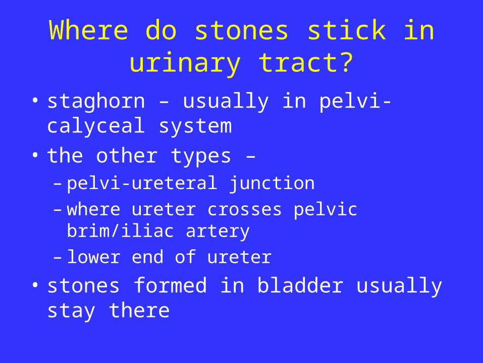
Where do stones stick in urinary tract?
• staghorn – usually in pelvi-calyceal system
• the other types –– pelvi-ureteral junction– where ureter crosses pelvic brim/iliac artery– lower end of ureter
• stones formed in bladder usually stay there
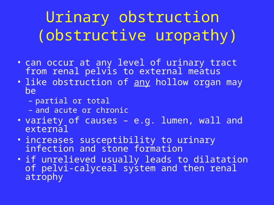
Urinary obstruction (obstructive uropathy)
• can occur at any level of urinary tract from renal pelvis to external meatus
• like obstruction of any hollow organ may be– partial or total– and acute or chronic
• variety of causes – e.g. lumen, wall and external• increases susceptibility to urinary infection and
stone formation• if unrelieved usually leads to dilatation of pelvi-
calyceal system and then renal atrophy
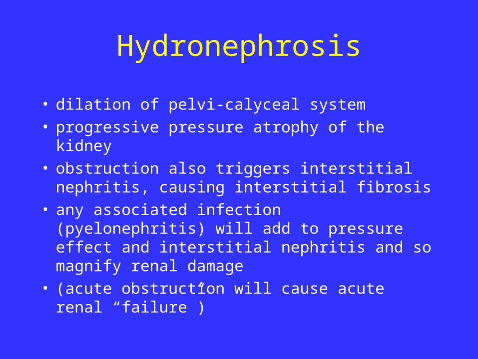
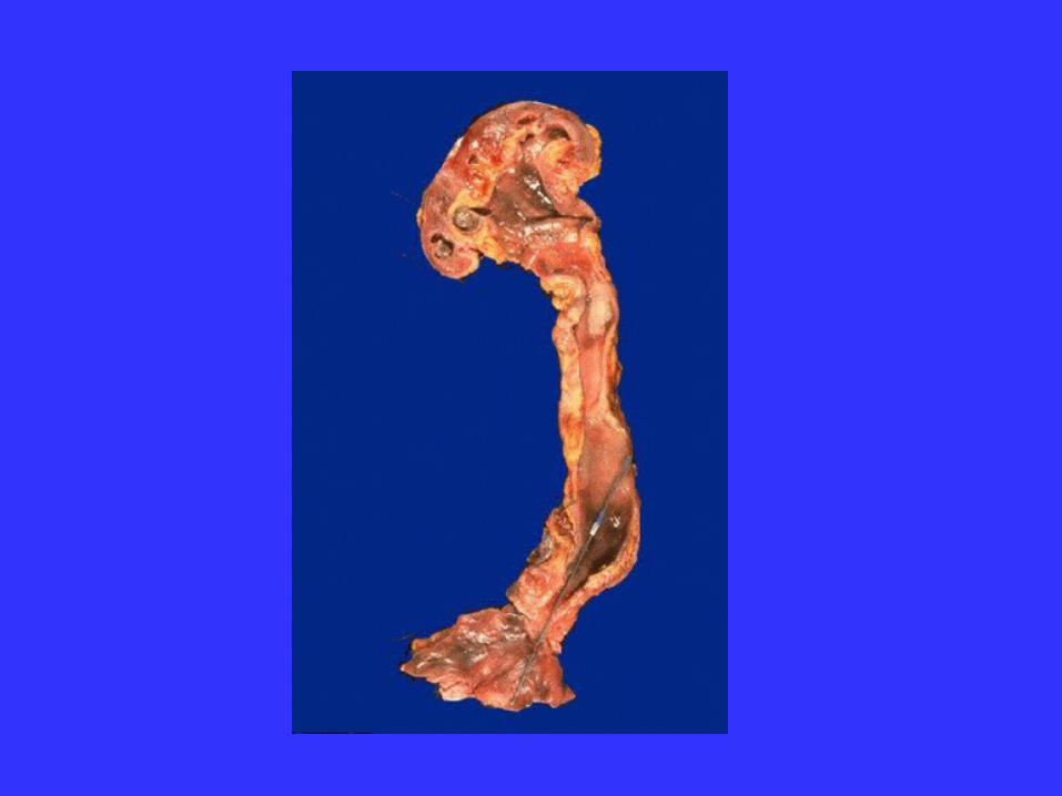
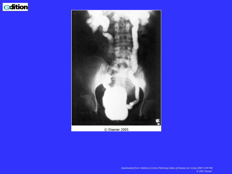
Hydronephrosis
• dilation of pelvi-calyceal system• progressive pressure atrophy of the kidney• obstruction also triggers interstitial nephritis,
causing interstitial fibrosis• any associated infection (pyelonephritis) will
add to pressure effect and interstitial nephritis and so magnify renal damage
• (acute obstruction will cause acute renal “failure”)
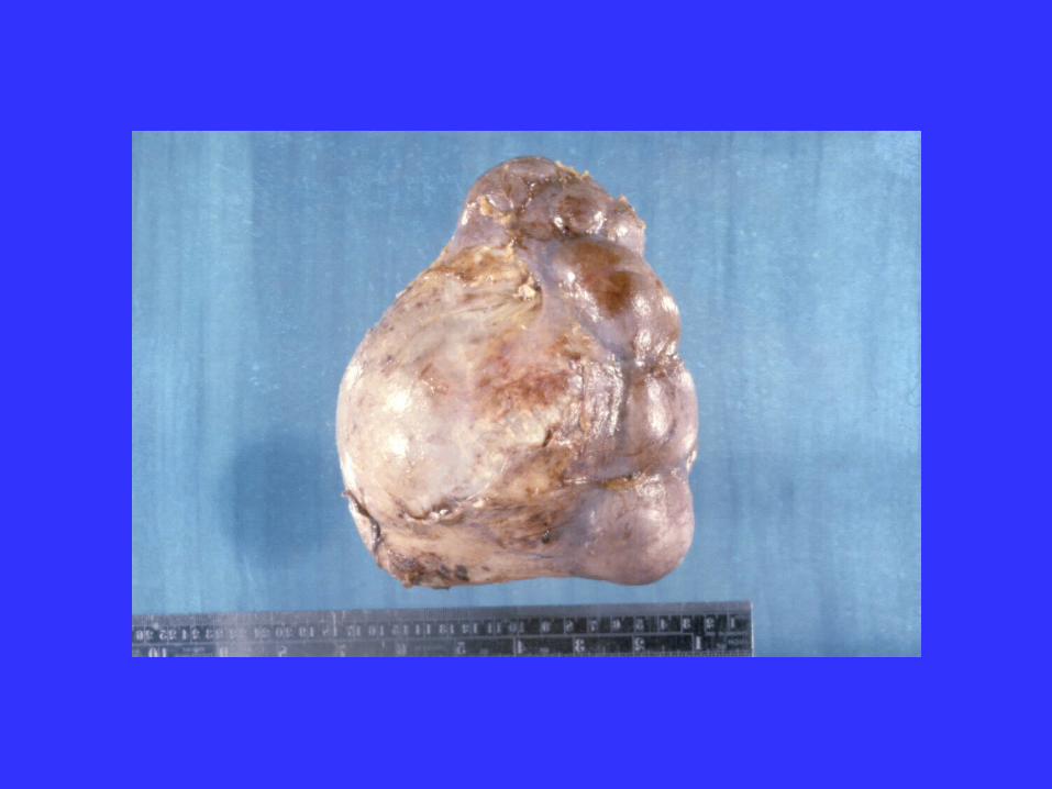
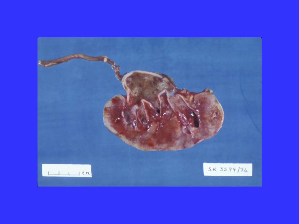
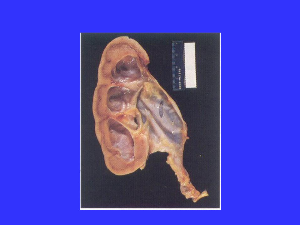
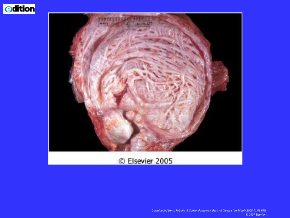
Hydronephrosis
• progressive damage
• gross thinning of cortex due to parenchymal atrophy
• eventually, kidney becomes thin-walled “cyst”
• if blockage is in ureter or lower, ureter(s) may also dilate (hydroureter)


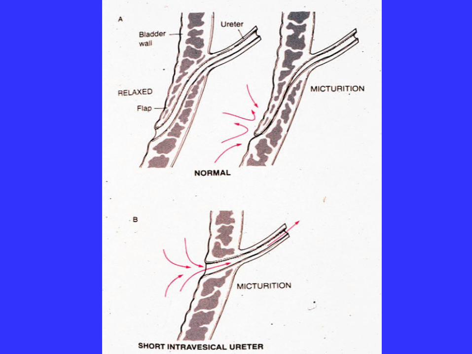
Unilateral disease
• block must be at level of vesico-ureteral (VU) valve or above
• may be silent for long time
• unilateral renal damage can cause secondary hypertension
- and so possible effects on other kidney
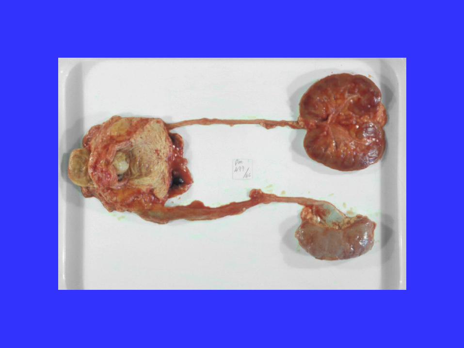
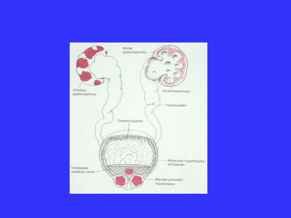
Bilateral disease
• blockage must be at level of internal bladder sphincter or beyond
• if obstruction incomplete– bladder muscle hypertrophies, causing
trabeculation– diverticula may form – bladder dilatation may make VU junctions
incompetent
• will always cause renal failure if unrelieved

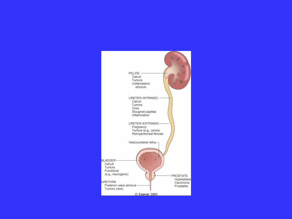
Causes of obstruction
• in the lumen– stones, blood clots, necrotic renal papillae
• in the wall– intrinsic tumours, strictures (post-
inflammatory*, congenital), neuropathic bladder, “pelviureteric dysfunction”
*schistosomiasis important cause

Causes of obstruction
• outside the wall– inflammation
• diverticulitis, salpingitis, prostatitis, retroperitoneal fibrosis
– tumours (prostate, cervix, uterus, colon, enlarged retroperitoneal nodes)
– others• benign prostatic hypertrophy, endometriosis, aortic
aneurysm, prolapsed uterus etc – even (temporarily) pregnancy
Downloaded from: Robbins & Cotran Pathologic Basis of Disease (on 14 July 2008 12:59 PM)
© 2007 Elsevier
Downloaded from: Robbins & Cotran Pathologic Basis of Disease (on 14 July 2008 01:09 PM)
© 2007 Elsevier

and finally -
Diseases of the urethra
Often present with urinary obstruction
• congenital valves
• traumatic rupture/stricture
• urethritis – e.g. due to gonococcus
• tumours – viral condyloma, transitional tumours


![CRYSTALLOGRAPHIC ANALYSIS OF URINARY CALCULI · 2020. 7. 6. · Urinary calculi have been analyzed and studied by differ - ent techniques with diverse merits and demerits [1] [2]](https://img.pdfslide.us/doc/110x75/60ba611187dd9d729d796019/crystallographic-analysis-of-urinary-calculi-2020-7-6-urinary-calculi-have.jpg)