Embed Size (px)
Citation preview
16
Modern Surgical Treatments of Urinary Tract Obstruction
Bannakij Lojanapiwat Faculty of Medicine, Chiang Mai University
Thailand
1. Introduction
Obstructive nephropathy is a term describing the damage to the renal parenchyma that
results from the obstruction to the flow of urine anywhere along the urinary system. Long
term obstruction causes chronic renal disease. Obstruction coexisting with infection and
impaired renal function, when complicated by elevated temperature and leukocytosis that
can lead to septic shock, are an absolute indication for urinary diversion such as
percutaneous nephrostomy. This particular patient needs emergency diversion. One of
the most common indications of nephrostomy placement is ureteric obstruction causing
uremia. It is therefore necessary to make the patients fit enough for the designated
surgery.
Percutaneous nephrostomy involving supravesicle drainage is one of the most common
procedures in urologic practice. Goodwin described a trocar nephrostomy technique in a
markedly dilated kidney in 1955. (Goodwin et al., 1955). Percutaneous nephrostomy is
performed for temporary or permanent supravesicle urinary diversion. The treatment goals
in patients with malignant ureteric obstruction are symptom relief and avoidance of any
complications from renal insufficiency. Permanent nephrostomy has been used in patients
with obstruction from uncorrectable causes such as inoperable tumors. (Table 1)
The indication of nephrostomy tube placement depends on whether the procedure is
elective or urgent. The purpose of nephrostomy tube placement in obstructive renal disease
is to preserve kidney function and drain infected urine. Establishing a safe and reliable
nephrostomy tract is key that range from simple urinary drainage to intrarenal surgical
operation. (Fig. 1-4)
Complications of obstruction as sepsis and pain.
Improve renal function.
Localized disease that additional therapy may prolong survival.
Improve quality of life.
Independent existence at home possible.
Table 1. Indication for palliative diversion.
www.intechopen.com
Chronic Kidney Disease
262
Careful discussion between patients, relatives and health care professionals about
nephrostomy tube placement must be undertaken before the intervention because patients
will require a drainage bag which reduces the quality of life.
Renal function in several patients recover following temporary percutaneous nephrostomy
tube placement. The definite treatment is need prior nephrostomy tube removal. Advance in
endourologic instrumentation and techniques, endourologic operations as the minimally
invasive surgery (percutaneous nephrolithotomy, endopyelotomy, infundibulotomy and
endoureterotomy) are the procedure of choice for these patients. The nonfunctioning
kidneys following the diversion usually require nephrectomy.
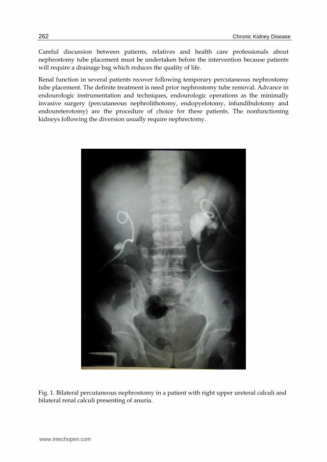
Fig. 1. Bilateral percutaneous nephrostomy in a patient with right upper ureteral calculi and bilateral renal calculi presenting of anuria.
www.intechopen.com
Modern Surgical Treatments of Urinary Tract Obstruction
263
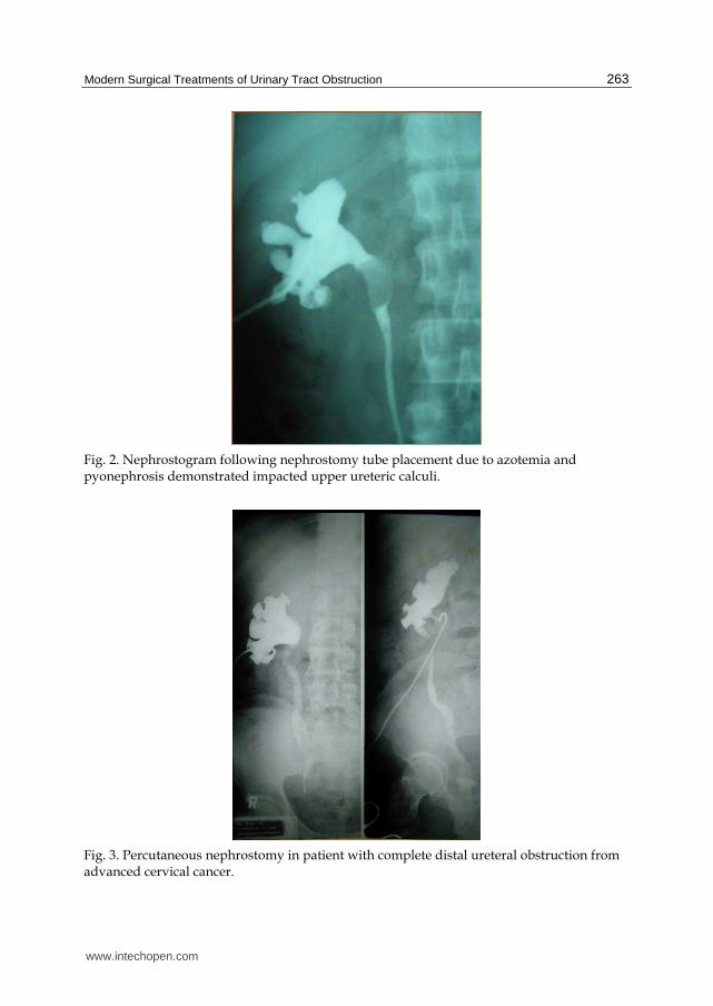
Fig. 2. Nephrostogram following nephrostomy tube placement due to azotemia and pyonephrosis demonstrated impacted upper ureteric calculi.
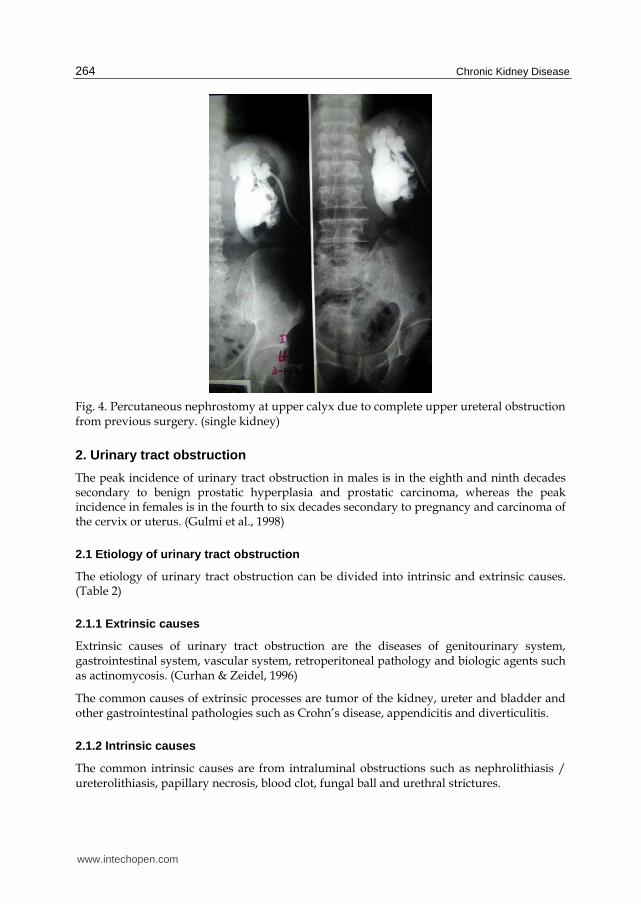
Fig. 3. Percutaneous nephrostomy in patient with complete distal ureteral obstruction from advanced cervical cancer.
www.intechopen.com
Chronic Kidney Disease
264
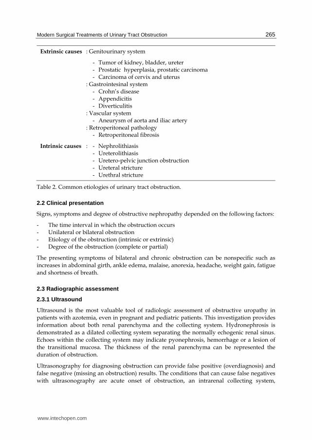
Fig. 4. Percutaneous nephrostomy at upper calyx due to complete upper ureteral obstruction from previous surgery. (single kidney)
2. Urinary tract obstruction
The peak incidence of urinary tract obstruction in males is in the eighth and ninth decades secondary to benign prostatic hyperplasia and prostatic carcinoma, whereas the peak incidence in females is in the fourth to six decades secondary to pregnancy and carcinoma of the cervix or uterus. (Gulmi et al., 1998)
2.1 Etiology of urinary tract obstruction
The etiology of urinary tract obstruction can be divided into intrinsic and extrinsic causes. (Table 2)
2.1.1 Extrinsic causes
Extrinsic causes of urinary tract obstruction are the diseases of genitourinary system, gastrointestinal system, vascular system, retroperitoneal pathology and biologic agents such as actinomycosis. (Curhan & Zeidel, 1996)
The common causes of extrinsic processes are tumor of the kidney, ureter and bladder and other gastrointestinal pathologies such as Crohn’s disease, appendicitis and diverticulitis.
2.1.2 Intrinsic causes
The common intrinsic causes are from intraluminal obstructions such as nephrolithiasis / ureterolithiasis, papillary necrosis, blood clot, fungal ball and urethral strictures.
www.intechopen.com
Modern Surgical Treatments of Urinary Tract Obstruction
265
Extrinsic causes : Genitourinary system
- Tumor of kidney, bladder, ureter - Prostatic hyperplasia, prostatic carcinoma - Carcinoma of cervix and uterus
: Gastrointesinal system - Crohn’s disease - Appendicitis - Diverticulitis
: Vascular system - Aneurysm of aorta and iliac artery
: Retroperitoneal pathology - Retroperitoneal fibrosis
Intrinsic causes : - Nephrolithiasis - Ureterolithiasis - Uretero-pelvic junction obstruction - Ureteral stricture - Urethral stricture
Table 2. Common etiologies of urinary tract obstruction.
2.2 Clinical presentation
Signs, symptoms and degree of obstructive nephropathy depended on the following factors:
- The time interval in which the obstruction occurs - Unilateral or bilateral obstruction - Etiology of the obstruction (intrinsic or extrinsic) - Degree of the obstruction (complete or partial)
The presenting symptoms of bilateral and chronic obstruction can be nonspecific such as
increases in abdominal girth, ankle edema, malaise, anorexia, headache, weight gain, fatigue
and shortness of breath.
2.3 Radiographic assessment
2.3.1 Ultrasound
Ultrasound is the most valuable tool of radiologic assessment of obstructive uropathy in
patients with azotemia, even in pregnant and pediatric patients. This investigation provides
information about both renal parenchyma and the collecting system. Hydronephrosis is
demonstrated as a dilated collecting system separating the normally echogenic renal sinus.
Echoes within the collecting system may indicate pyonephrosis, hemorrhage or a lesion of
the transitional mucosa. The thickness of the renal parenchyma can be represented the
duration of obstruction.
Ultrasonography for diagnosing obstruction can provide false positive (overdiagnosis) and
false negative (missing an obstruction) results. The conditions that can cause false negatives
with ultrasonography are acute onset of obstruction, an intrarenal collecting system,
www.intechopen.com
Chronic Kidney Disease
266
dehydration, and the misinterpretation of caliectasis for renal cortical cyst. (LeRoy., 1996) .
False positive imaging for the obstruction can be caused by parapelvic cyst, intrarenal
pelvis, high urine flow state and vesicoureteral reflux. (Stables et al., 1978)
2.3.2 Retrograde pyelography
Retrograde pyelography may be needed to demonstrate the cause of obstruction that is
either intrinsic and extrinsic. This assessment can evaluate the site, severity of obstruction
and degree of hydronephrosis especially in patients with poor kidney function.
2.3.3 Computer tomography (CT scan)
Computer tomography (CT scan) can demonstrate the information of obstruction and
hydronephrosis without contrast media. All kinds of urinary calculi and other
intraperitoneal / extraperitoneal pathology can be detected by this assessment.
3. Surgical approach
Nephrostomy can be performed either by open operation or by closed percutaneous methods.
With the development of endourologic and imaging techniques, percutaneous nephrostomy is
widely used. Recently, the percutaneous nephrostomy placement became the standard of care,
replacing surgical nephrostomy. (Banner et al., 1991 & Sherman et al., 1985)
Establishing safe and reliable nephrostomy tract is very important. The aim of the
nephrostomy tract ranges from simple urinary drainage to intrarenal surgical operation. For
percutaneous renal surgeries, some surgeons prefer a two stage surgery which can limit
bleeding, provide a clear field and let the nephrostomy tract mature.
A successful outcome without complications is the goal of this procedure, which requires
careful preoperative planning and proper techniques. The preoperative anatomy of the patient,
the nature of the urologic procedure planned and available equipment are very important.
3.1 Open nephrostomy technique
Explore the kidney and open the renal pelvis and choose the calix which is suitable for
nephrostomy. The catheter is introduced through thinned cortex into the renal pelvis.
3.2 Percutaneous nephrostomy techniques
3.2.1 Preoperative patient preparation
All patients need appropriate hemostasis evaluation and urine bacteriologic assessment.
Careful review and assessment of the degree of hydronephrosis, anatomic variance of the
pelvicaliceal system, and relative position of the kidney are key factors for success and will
reduce any potential complication of nephrostomy tube placement. This can be evaluated by
previous or currents plain Kidney-Urinary–Bladder (KUB) radiography, intravenous
pyelography, retrograde pyelography, computed tomogram and ultrasonographic studies.
These radiographic investigations demonstrate size, number and location of renal and
ureteral calculi as well as establishing baseline renal function and other pathology.
www.intechopen.com
Modern Surgical Treatments of Urinary Tract Obstruction
267
The evaluation of choice to detect urolithiasis and intraabdominal anatomy in patients with emergent or complex medical conditions is a computer tomography (CT scan) of the whole abdomen. Pre-nephrostomy placement with CT scan is recommended in selected patients with splenomegaly, colonic malposition and marked colonic distention. (LeRoy., 1996)
Patients who have urinary tract infection are treated with bacteriologically specific antibiotics and these patients need parenteral antibiotics for 36 to 48 hours before surgery to ensure adequate serum levels of effective antibiotics. The recommended regimen is ciprofloxacin 400 mg IV every 12 hr, ampicillin 1 gm IV every 6 hr with gentamicin 1 mg/kg every 8 hr or third generation cephalosporin.
Laboratory testing of any bleeding problem such as PT, PTT (Prothrombin time, Partial thromboplastin time) and platelet count should be done with appropriate adjustments especially in patients with a history of prolonged bleeding, liver disease, clinically easy brusisability or other conditions predisposing to a coagulopathy. A platelet count should be above 80,000 cells per ml prior to the procedure. Aspirin therapy should be discontinued 1 week prior to the procedure. Caumadin as an anticoagulants must be discontinued. Subcutaneous heparin can be administered for high risk patients with venous thrombosis.
3.2.2 Patient’s position
Nephrostomy tube placement can be preferred in both prone and supine positions with highly successful outcome. Most patients usually undergo the procedure in the prone position with abdominal support. Supine position is selected for patients with high surgical risks such as seriously ill patients, patients with endotracheal tubes with or without ventilation, patients with congestive heart failure, patients with complicated fractures and patients who have undergone a major surgical procedure.
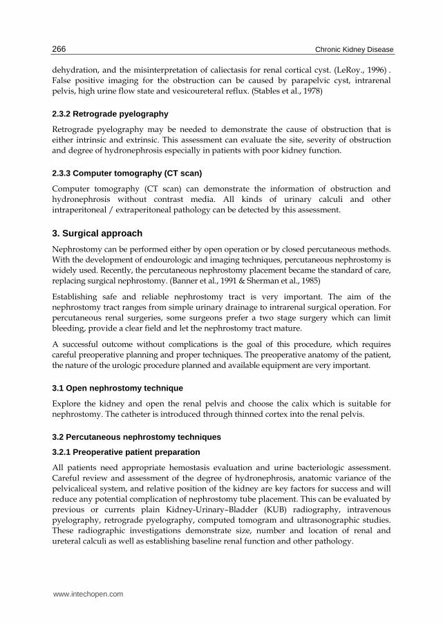
The advantages of prone or prone oblique with body side of targeted kidney slightly elevated are operator’s hands are outside the vertical x-ray beam. (Fig. 5) With supine position with the body side of targeted kidney elevated slightly off the tabletop, the renal access can be performed with ultrasound or CT guidance.
3.2.3 Anesthetic
Most patients need only local anesthesia, but some may need intravenous sedation or general anesthesia, the latter specifically for pediatric patients. The type of anesthesia administered depends on the individual patient and indication of nephrostomy tube placement. Simple percutaneous external drainage can be tolerated with local anesthesia or intravenous analgesia with sedation. General anesthesia is preferable in children with all indications of nephrostomy tube placement.
3.2.4 Imaging guidance
The imaging guidance equipment is very important in renal access. The guidance system for urinary tract interventions are fluoroscopic guidance, real-time ultrasonography and CT scan.
3.2.4.1 Ultrasound guidance
The puncture of the desired calix can be done in dilated systems. If only the renal pelvic can be identified, initial puncture can be done at renal pelvic following with antegrade
www.intechopen.com
Chronic Kidney Disease
268
Fig. 5. Patient in prone position for renal access.
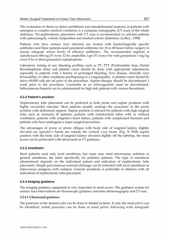
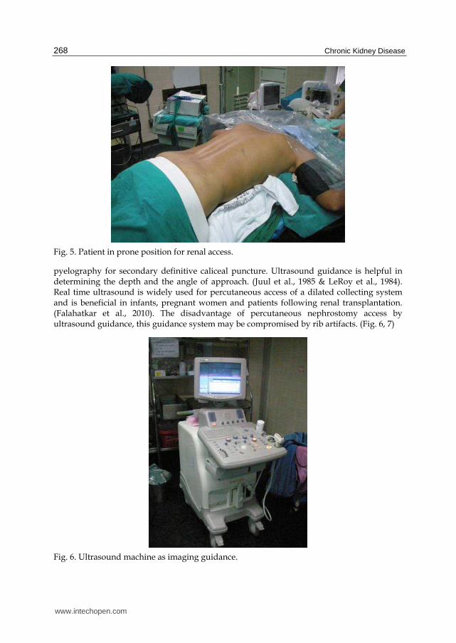
pyelography for secondary definitive caliceal puncture. Ultrasound guidance is helpful in determining the depth and the angle of approach. (Juul et al., 1985 & LeRoy et al., 1984). Real time ultrasound is widely used for percutaneous access of a dilated collecting system and is beneficial in infants, pregnant women and patients following renal transplantation. (Falahatkar et al., 2010). The disadvantage of percutaneous nephrostomy access by ultrasound guidance, this guidance system may be compromised by rib artifacts. (Fig. 6, 7)
Fig. 6. Ultrasound machine as imaging guidance.
www.intechopen.com
Modern Surgical Treatments of Urinary Tract Obstruction
269
Fig. 7. Ultrasound imaging demonstrated a guidewire in dilated pelvis.
3.2.4.2 Fluoroscopic guidance
Fluoroscopic guidance is essential for guidewire manipulation especially in patients with non or mild dilatation of renal pelvis. Collecting system can be opacified with contrast following cystoscopic retrograde ureteral catheter placement, injection of intravenous contrast material and direct percutaneous puncture with 22 gauge needle. Pyelotubular and pyelosinus backflow can be avoided by not overinjecting the collecting system.
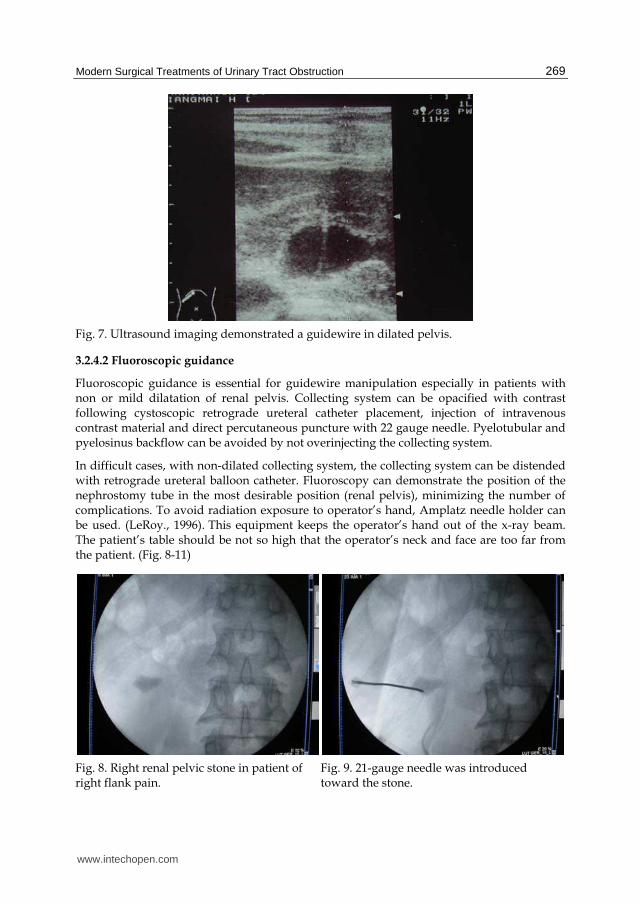
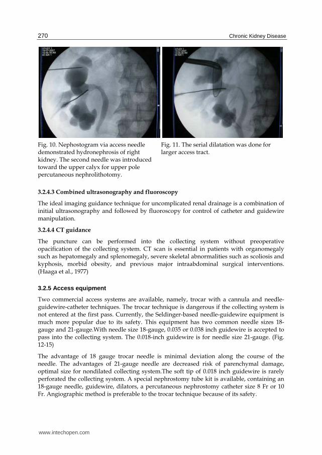
In difficult cases, with non-dilated collecting system, the collecting system can be distended with retrograde ureteral balloon catheter. Fluoroscopy can demonstrate the position of the nephrostomy tube in the most desirable position (renal pelvis), minimizing the number of complications. To avoid radiation exposure to operator’s hand, Amplatz needle holder can be used. (LeRoy., 1996). This equipment keeps the operator’s hand out of the x-ray beam. The patient’s table should be not so high that the operator’s neck and face are too far from the patient. (Fig. 8-11)
Fig. 8. Right renal pelvic stone in patient of right flank pain.
Fig. 9. 21-gauge needle was introduced toward the stone.
www.intechopen.com
Chronic Kidney Disease
270
Fig. 10. Nephostogram via access needle demonstrated hydronephrosis of right kidney. The second needle was introduced toward the upper calyx for upper pole percutaneous nephrolithotomy.
Fig. 11. The serial dilatation was done for larger access tract.
3.2.4.3 Combined ultrasonography and fluoroscopy
The ideal imaging guidance technique for uncomplicated renal drainage is a combination of initial ultrasonography and followed by fluoroscopy for control of catheter and guidewire manipulation.
3.2.4.4 CT guidance
The puncture can be performed into the collecting system without preoperative opacification of the collecting system. CT scan is essential in patients with organomegaly such as hepatomegaly and splenomegaly, severe skeletal abnormalities such as scoliosis and kyphosis, morbid obesity, and previous major intraabdominal surgical interventions. (Haaga et al., 1977)
3.2.5 Access equipment
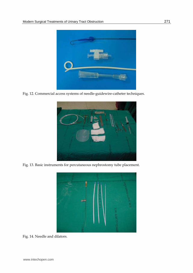
Two commercial access systems are available, namely, trocar with a cannula and needle-guidewire-catheter techniques. The trocar technique is dangerous if the collecting system is not entered at the first pass. Currently, the Seldinger-based needle-guidewire equipment is much more popular due to its safety. This equipment has two common needle sizes 18-gauge and 21-gauge.With needle size 18-gauge, 0.035 or 0.038 inch guidewire is accepted to pass into the collecting system. The 0.018-inch guidewire is for needle size 21-gauge. (Fig. 12-15)
The advantage of 18 gauge trocar needle is minimal deviation along the course of the needle. The advantages of 21-gauge needle are decreased risk of parenchymal damage, optimal size for nondilated collecting system.The soft tip of 0.018 inch guidewire is rarely perforated the collecting system. A special nephrostomy tube kit is available, containing an 18-gauge needle, guidewire, dilators, a percutaneous nephrostomy catheter size 8 Fr or 10 Fr. Angiographic method is preferable to the trocar technique because of its safety.
www.intechopen.com
Modern Surgical Treatments of Urinary Tract Obstruction
271
Fig. 12. Commercial access systems of needle-guidewire-catheter techniques.
Fig. 13. Basic instruments for percutaneous nephrostomy tube placement.
Fig. 14. Needle and dilators.
www.intechopen.com
Chronic Kidney Disease
272
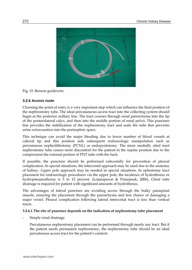
Fig. 15. Benson guidewire.
3.2.6 Access route
Choosing the point of entry is a very important step which can influence the final position of the nephrostomy tube. The ideal percutaneous access tract into the collecting system should begin at the posterior axillary line. The tract courses through renal parenchyma into the tip of the posterolateral calyx, and then into the middle portion of renal pelvis. This puncture line provides the stabilization of the nephrostomy tract and seals the tube that prevents urine extravasation into the perinephric space.
This technique can avoid the major bleeding due to fewer number of blood vessels at caliceal tip and this position aids subsequent endourologic manipulation such as percutanous nephrolithotomy (PCNL) or endopyelotomy. The more medially sited tract nephrostomy tube causes more discomfort for the patient in the supine position due to the compression the external portion of PNT tube with the back.
If possible, the puncture should be performed subcostally for prevention of pleural complication. In special situations, the intercostal approach may be used due to the anatomy of kidney. Upper pole approach may be needed in special situations. In ephostomy tract placement for endourologic procedures via the upper pole, the incidence of hydrothorax or hydropneumothorax is 5 to 12 percent. (Lojanapiwat & Prasopsuk, 2006). Chest tube drainage is required for patient with significant amounts of hydrothorax.
The advantages of lateral puncture are avoiding access through the bulky paraspinal muscle, ensuring the placement through the parenchyma and less chance of damaging a major vessel. Pleural complication following lateral intercostal tract is less than vertical tracts.
3.2.6.1 The site of puncture depends on the indication of nephrostomy tube placement
- Simple renal drainage
Percutaneous nephrostomy placement can be performed through nearly any tract. But if the patient needs permanent nephrostomy, the nephrostomy tube should be an ideal percutenous access tract for the patient’s comfort.
www.intechopen.com
Modern Surgical Treatments of Urinary Tract Obstruction
273
- Further endourologic procedures.
Percutaneous nephrolithotomy (PCNL) : Pelvic stone
- ideal tract is through any middle or lower calyx. : Calyceal stone
- ideal tract is directly through the stone-bearing calix peripheral to the stone.
: Staghorn stone - ideal tract is through upper pole calyx.
: Upper ureteral stone - ideal tract is through middle pole or lower upper pole.
: Diverticular stone - ideal tract is directly through diverticulum.
Endopyelotomy (EP) / endoureterotomy : Ureteropelvic junction obstruction (UPJO) and upper ureteral stricture
- ideal tract is through middle pole or upper pole calix (Lojanapiwat, 2006).
3.2.6.2 Upper pole access for renal access
The upper pole of the kidney aligned medially and posterior to the lower pole, making the upper pole a shorter and easier access route. The upper-pole approach provides a straight tract along the long axis of the kidney and ensures the ability to reach most of the collecting system while providing easier manipulation of rigid instrument. The operative techniques of upper pole access need coordination with the anesthetists for controlling breathing for prevention of intercostals vessel and pulmonary complication. (Lojanapiwat & Prasopsuk, 2006) (Table 3, 4)
- Ureteropelvic junction and proximal ureteral pathology - Buck of the upper pole calculi - Multiple lower pole caliceal calculi - Obesity or unusual body habitus - Staghorn calculi - Large upper ureteral calculi
Table 3. Indication for the upper pole access.
- Need coordination with the anesthetists for controlling breathing. - An intercostal puncture should be made in the lower half of the intercostal space to
avoid injuring the blood vessels. - During full expiration, the needle is passed through the retroperitoneum and
diaphragm to prevent the injury to the lung, while needle passage through the renal parenchyma to the collecting system is done during deep inspiration for downward displacement of the kidney.
- An Amplatz shealth is used during the percutaneous supracostal approach to maintain low-pressure irrigation.
Table 4. Technique of upper pole access.
www.intechopen.com
Chronic Kidney Disease
274
3.2.6.3 The causes of access failure
- Nondistended renal collecting systems
- Impacted large stone that prevent guide wire manipulation
- Obscuring the location of collecting system
- Small obstructed infundibular stone with minimal caliceal dilatation
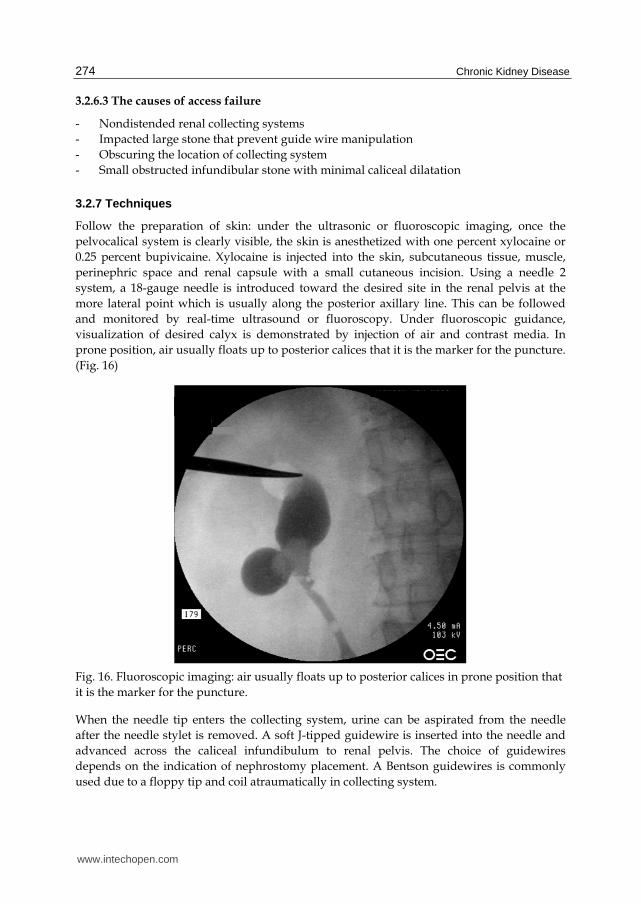
3.2.7 Techniques
Follow the preparation of skin: under the ultrasonic or fluoroscopic imaging, once the
pelvocalical system is clearly visible, the skin is anesthetized with one percent xylocaine or
0.25 percent bupivicaine. Xylocaine is injected into the skin, subcutaneous tissue, muscle,
perinephric space and renal capsule with a small cutaneous incision. Using a needle 2
system, a 18-gauge needle is introduced toward the desired site in the renal pelvis at the
more lateral point which is usually along the posterior axillary line. This can be followed
and monitored by real-time ultrasound or fluoroscopy. Under fluoroscopic guidance,
visualization of desired calyx is demonstrated by injection of air and contrast media. In
prone position, air usually floats up to posterior calices that it is the marker for the puncture.
(Fig. 16)
Fig. 16. Fluoroscopic imaging: air usually floats up to posterior calices in prone position that
it is the marker for the puncture.
When the needle tip enters the collecting system, urine can be aspirated from the needle
after the needle stylet is removed. A soft J-tipped guidewire is inserted into the needle and
advanced across the caliceal infundibulum to renal pelvis. The choice of guidewires
depends on the indication of nephrostomy placement. A Bentson guidewires is commonly
used due to a floppy tip and coil atraumatically in collecting system.
www.intechopen.com
Modern Surgical Treatments of Urinary Tract Obstruction
275
In special situations, such as an impacted stone in the collecting system, the manipulation
often requires small angled-tip catheters and hydrophilic coated wires. Then the needle is
removed, and progressively larger dilators are introduced over the guidewire to dilate the
access tract to facilitate the placement of soft nephrostomy tube. The size of tract dilatation
depends on the goal of percutaneous access. If the goal is to provide external urinary
drainage, serial dilators are inserted over the guidewire to dilate the tract to a sufficient size
for the nephrostomy tube. The 8 or 10 Fr nephrostomy tube is introduced over the
guidewire and optimal position is monitored by ultrasound or fluoroscopy.
The most reliable evidence for the proper placement of the nephrostomy tube can be
demonstrated by nephrostogram under fluoroscopic imaging. The guidewire is withdrawn
and the nephrostomy tube is secured with skin to prevent dislodging the catheter. The
catheter is connected to a urine bag for drainage. (Fig. 17-20) For permanant nephrostomy
tube placement, the tract can be further dilated and a regular Foley’s catheter can be used.
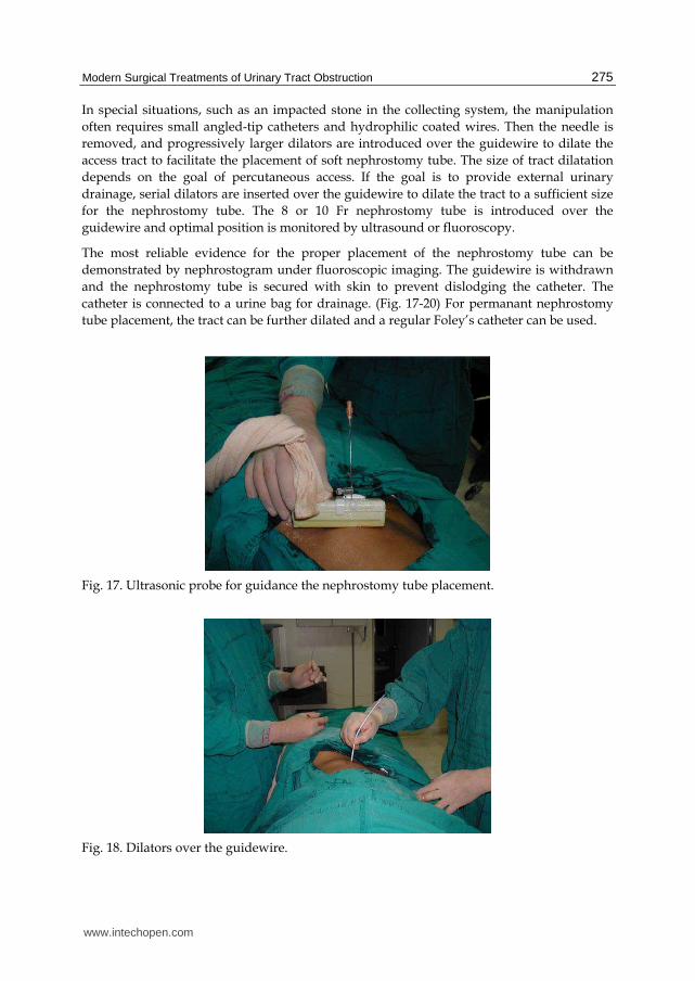
Fig. 17. Ultrasonic probe for guidance the nephrostomy tube placement.
Fig. 18. Dilators over the guidewire.
www.intechopen.com
Chronic Kidney Disease
276
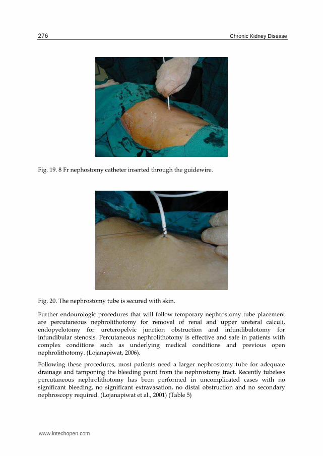
Fig. 19. 8 Fr nephostomy catheter inserted through the guidewire.
Fig. 20. The nephrostomy tube is secured with skin.
Further endourologic procedures that will follow temporary nephrostomy tube placement
are percutaneous nephrolithotomy for removal of renal and upper ureteral calculi,
endopyelotomy for ureteropelvic junction obstruction and infundibulotomy for
infundibular stenosis. Percutaneous nephrolithotomy is effective and safe in patients with
complex conditions such as underlying medical conditions and previous open
nephrolithotomy. (Lojanapiwat, 2006).
Following these procedures, most patients need a larger nephrostomy tube for adequate
drainage and tamponing the bleeding point from the nephrostomy tract. Recently tubeless
percutaneous nephrolithotomy has been performed in uncomplicated cases with no
significant bleeding, no significant extravasation, no distal obstruction and no secondary
nephroscopy required. (Lojanapiwat et al., 2001) (Table 5)
www.intechopen.com
Modern Surgical Treatments of Urinary Tract Obstruction
277
- single access - no obstructed renal unit - no significant bleeding - no significant extravasation - Secondary nephroscopy is not required. (stone free)
Table 5. Criteria for tubeless percutaneous nephrolithotomy.
4. Results
Overall success rate of uncomplicated nephrostomy tube placement is over 97% with less success in patients who required percutaneous tract for subsequent endourologic interventions. Factors which affect the success rate of nephrostomy tube placement during endourologic operation are stone burden, degree of hydronephrosis, history of previous open nephrolithotomy, and experience of surgeon. As same as other urologic procedure, a training simulator for ultrasound-guided percutaneous nephrostomy insertion is needed for a safe, non-threatening environment, without risk to patients. Commercial and a gelatin phantoms are available. Skill is required prior to undertaking procedures in patients. (Rock et al., 2010)
5. Complications
Complications following simple nephrostomy tube drainage are minor with a rate approaching 4%. (LeRoy, 1996). The common complications are hemorrhage, infection, improper catheter placement, nephrostomy tube dislodging after initial proper placement, nephrocutaneous fistula, stone formation and post-obstructive diuresis. Initial hematuria is common, but should be cleared in 24 – 48 hours post operatively.
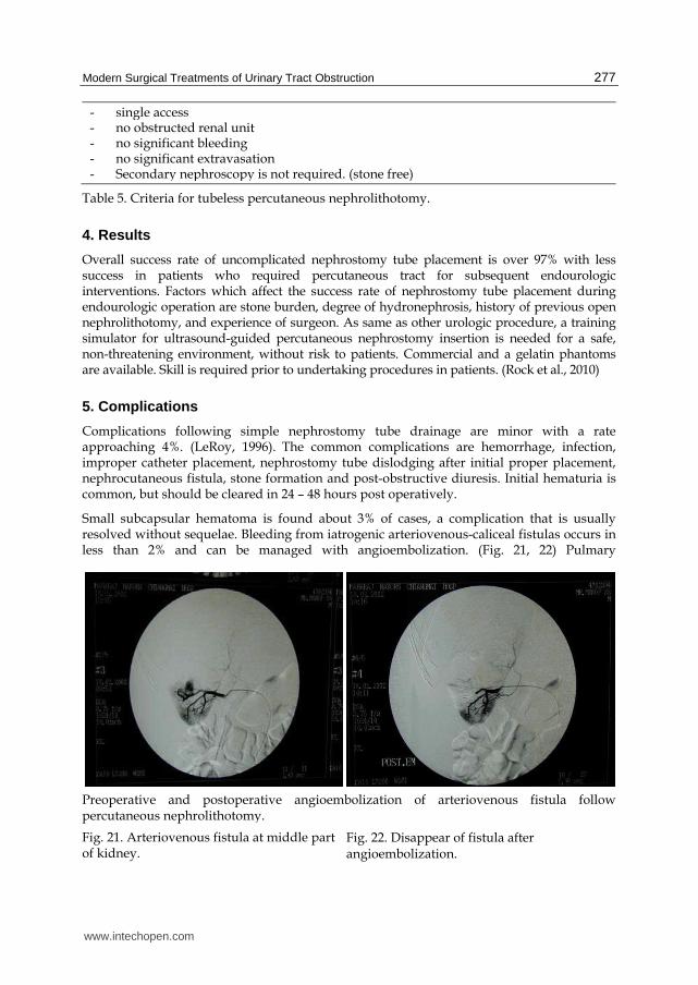
Small subcapsular hematoma is found about 3% of cases, a complication that is usually resolved without sequelae. Bleeding from iatrogenic arteriovenous-caliceal fistulas occurs in less than 2% and can be managed with angioembolization. (Fig. 21, 22) Pulmary
Preoperative and postoperative angioembolization of arteriovenous fistula followpercutaneous nephrolithotomy.
Fig. 21. Arteriovenous fistula at middle part of kidney.
Fig. 22. Disappear of fistula after angioembolization.
www.intechopen.com
Chronic Kidney Disease
278
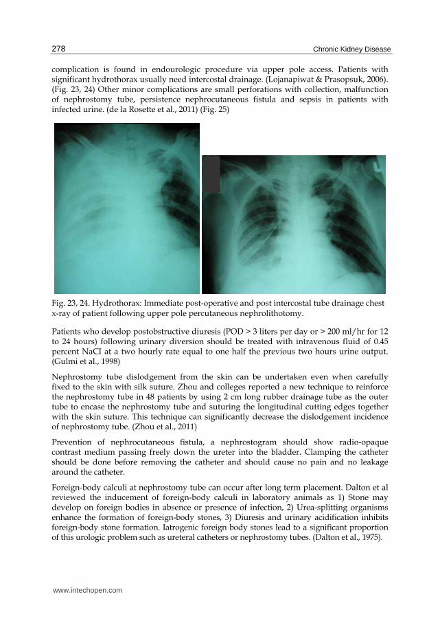
complication is found in endourologic procedure via upper pole access. Patients with significant hydrothorax usually need intercostal drainage. (Lojanapiwat & Prasopsuk, 2006). (Fig. 23, 24) Other minor complications are small perforations with collection, malfunction of nephrostomy tube, persistence nephrocutaneous fistula and sepsis in patients with infected urine. (de la Rosette et al., 2011) (Fig. 25)
Fig. 23, 24. Hydrothorax: Immediate post-operative and post intercostal tube drainage chest x-ray of patient following upper pole percutaneous nephrolithotomy.
Patients who develop postobstructive diuresis (POD > 3 liters per day or > 200 ml/hr for 12 to 24 hours) following urinary diversion should be treated with intravenous fluid of 0.45 percent NaCI at a two hourly rate equal to one half the previous two hours urine output. (Gulmi et al., 1998)
Nephrostomy tube dislodgement from the skin can be undertaken even when carefully fixed to the skin with silk suture. Zhou and colleges reported a new technique to reinforce the nephrostomy tube in 48 patients by using 2 cm long rubber drainage tube as the outer tube to encase the nephrostomy tube and suturing the longitudinal cutting edges together with the skin suture. This technique can significantly decrease the dislodgement incidence of nephrostomy tube. (Zhou et al., 2011)
Prevention of nephrocutaneous fistula, a nephrostogram should show radio-opaque contrast medium passing freely down the ureter into the bladder. Clamping the catheter should be done before removing the catheter and should cause no pain and no leakage around the catheter.
Foreign-body calculi at nephrostomy tube can occur after long term placement. Dalton et al reviewed the inducement of foreign-body calculi in laboratory animals as 1) Stone may develop on foreign bodies in absence or presence of infection, 2) Urea-splitting organisms enhance the formation of foreign-body stones, 3) Diuresis and urinary acidification inhibits foreign-body stone formation. Iatrogenic foreign body stones lead to a significant proportion of this urologic problem such as ureteral catheters or nephrostomy tubes. (Dalton et al., 1975).
www.intechopen.com
Modern Surgical Treatments of Urinary Tract Obstruction
279
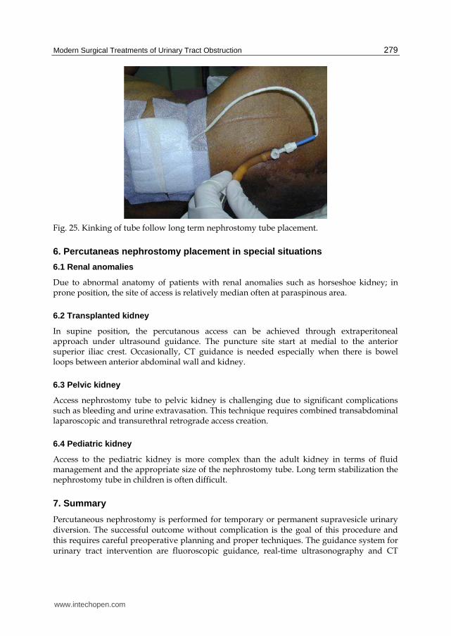
Fig. 25. Kinking of tube follow long term nephrostomy tube placement.
6. Percutaneas nephrostomy placement in special situations
6.1 Renal anomalies
Due to abnormal anatomy of patients with renal anomalies such as horseshoe kidney; in prone position, the site of access is relatively median often at paraspinous area.
6.2 Transplanted kidney
In supine position, the percutanous access can be achieved through extraperitoneal approach under ultrasound guidance. The puncture site start at medial to the anterior superior iliac crest. Occasionally, CT guidance is needed especially when there is bowel loops between anterior abdominal wall and kidney.
6.3 Pelvic kidney
Access nephrostomy tube to pelvic kidney is challenging due to significant complications such as bleeding and urine extravasation. This technique requires combined transabdominal laparoscopic and transurethral retrograde access creation.
6.4 Pediatric kidney
Access to the pediatric kidney is more complex than the adult kidney in terms of fluid management and the appropriate size of the nephrostomy tube. Long term stabilization the nephrostomy tube in children is often difficult.
7. Summary
Percutaneous nephrostomy is performed for temporary or permanent supravesicle urinary diversion. The successful outcome without complication is the goal of this procedure and this requires careful preoperative planning and proper techniques. The guidance system for urinary tract intervention are fluoroscopic guidance, real-time ultrasonography and CT
www.intechopen.com
Chronic Kidney Disease
280
scan. The ideal nephrostomy tract should course through renal parenchyma into the tip of posterolateral calix then into the middle portion of renal pelvis. The complication following simple nephrostomy tube drainage is minor.
8. References
Banner, MP.; Ramchandani, P. & Pollack, HM. (1991). Interventional procedures in the upper urinary tract. Cardiovasc Intervent Radiol, 14(5):267-84.
Curhan, CG. & Zeidel, ML. (1996). Urinary tract obstruction, In BM Brenner(ed): The kidney. 5th ed. Philadelphia, WB Saunders CO, Vol 2 Chap41, 1391.
Dalton, DL.; Hughes, J. & Glenn, JF. (1975). Foreign bodies and urinary stones Urology, 6: 1 de la Rosette, J.; Assimos, D.; Desai, M.; Gutierrez, J.; Lingeman, J.; Scarpa, R. & Tefekli, A.
(2011). CROES PCNL Study Group. The Clinical Research Office of the Endourological Society Percutaneous Nephrolithotomy Global Study: indications, complications, and outcomes in 5803 patients. J Endourol. 25(1):11-7.
Falahatkar, S.; Asgari, SA.; Nasseh, H.; Allahkhah, A.; Farshami, FJ.; Shakiba, M. & Esmaeili, S. (2010). Totally ultrasound versus fluoroscopically guided complete supine percutaneous nephrolithotripsy: a first report. J Endourol, 24(9):1421-6.
Goodwin, WE.; Casey, WC. & Woolf, W. (1955). Percutaneous trocar (needle) nephrostomy in hydronephrosis. J Am Med Assoc, 12; 157(11):891-4.
Gulmi, FA.; Felsen, D. & Vaughan, ED. (1998). Management of post-obstructive diuresis, AUA update series , Vol 17:178-83.
Haaga, JR.; Zelch, Mg.; Alfids, RJ.; Steward, BH. & Daugherty, JD. (1977). Interventional CT scanning. Radiol Clin North Am. 15(3):449-56.
Juul, N.; Nielsen, V. & Torp-Pederson, S. (1985). Percutaneous balloon catheter nephrostomy guided by ultrasound. Results of a new technique. Scand J Urol Nephrol, 19(4):291-4.
LeRoy, AT. (1996). Percutaneous access, In: Smith, AD. & Badlani, GH. & Bagley, DH. (eds): Smith‘s textbook of Endourology. 1st ed. St Louis, Missouri, Quality Medical Publishing, Inc, Chap 14, P 199-223.
LeRoy, AJ.; May, GR.; Bender, CE.; Williams, HJ Jr.; Mc Gough, PF.; Segura, JW. & Patterson, DF. (1984). Percutaneous nephrostomy for stone removal. Radiology, 151(3):607-12.
Lojanapiwat, B. & Prasopsuk, S. (2006). Upper-pole access for percutaneous nephrolithotomy: comparison of supracostal and infracostal approaches. J Endourol, 20(7):491-4.
Lojanapiwat, B. (2006). Previous open nephrolithotomy: does it affect percutaneous nephrolithotomy techniques and outcome? J Endourol, 20(1):17-20.
Lojanapiwat, B.; Soonthornpan, S. & Wudhikarn, S. (2001). Tubeless percutaneous nephrolithotomy in selected patients. J Endourol, 15(7):711-3.
Rock, BG.; Leonard, AP. & Freeman, SL. (2010). A training simul ator for ultrasound-guided percutaneous nephrostomy insertion. Br J Radiol, 83:612-4.
Stables, DP.; Ginsberg, NJ. & Johnson, ML. (1978). Percutaneous nephrostomy: a series and review of the literature. AJR Am J Roentgenol, 130(1):75-82.
Sherman, JL.; Hopper, KD.; Greene, AJ. & Johns, TT. (1985). The retrorenal colon on computed tomography: a normal variant. J Comput Assist Tomogr, 9(2):339-41.
Zhou, T.; Gao, X.; Yang, C.; Peng, Y.; Xiao, L.; Xu, C, et al. (2011). Reforcement for percutaneous nephrostomy tubes with a new technique. J Endourol, 25:41-4.
www.intechopen.com

Chronic Kidney DiseaseEdited by Prof. Monika Göőz
ISBN 978-953-51-0171-0Hard cover, 444 pagesPublisher InTechPublished online 16, March, 2012Published in print edition March, 2012
InTech EuropeUniversity Campus STeP Ri Slavka Krautzeka 83/A 51000 Rijeka, Croatia Phone: +385 (51) 770 447 Fax: +385 (51) 686 166www.intechopen.com
InTech ChinaUnit 405, Office Block, Hotel Equatorial Shanghai No.65, Yan An Road (West), Shanghai, 200040, China
Phone: +86-21-62489820 Fax: +86-21-62489821
Chronic kidney disease is an increasing health and economical problem in our world. Obesity and diabetesmellitus, the two most common cause of CKD, are becoming epidemic in our societies. Education on healthylifestyle and diet is becoming more and more important for reducing the number of type 2 diabetics andpatients with hypertension. Education of our patients is also crucial for successful maintenance therapy. Thereare, however, certain other factors leading to CKD, for instance the genetic predisposition in the case ofpolycystic kidney disease or type 1 diabetes, where education alone is not enough.
How to referenceIn order to correctly reference this scholarly work, feel free to copy and paste the following:
Bannakij Lojanapiwat (2012). Modern Surgical Treatments of Urinary Tract Obstruction, Chronic KidneyDisease, Prof. Monika Göőz (Ed.), ISBN: 978-953-51-0171-0, InTech, Available from:http://www.intechopen.com/books/chronic-kidney-disease/modern-surgical-treatments-of-urinary-tract-obstruction

© 2012 The Author(s). Licensee IntechOpen. This is an open access articledistributed under the terms of the Creative Commons Attribution 3.0License, which permits unrestricted use, distribution, and reproduction inany medium, provided the original work is properly cited.
![7 Catheter-associated Urinary Tract Infection (CAUTI) · UTI Urinary Tract Infection (Catheter-Associated Urinary Tract Infection [CAUTI] and Non-Catheter-Associated Urinary Tract](https://img.pdfslide.us/doc/110x75/5c40b88393f3c338af353b7f/7-catheter-associated-urinary-tract-infection-cauti-uti-urinary-tract-infection.jpg)