Embed Size (px)
Citation preview
University of Nigeria Research Publications
Aut
hor
KALU, Ochie.
REG NO: PG/PGD/97/20161
Title
Sonographic Evaluation of Splenetic Sizes in Normal
Adult Nigerians
Facu
lty
Medicine
Dep
artm
ent
Medical Radiography
Dat
e
2005
Sign
atur
e

DEGREE:
*ru
i revcrc (;od who deiennin
grateful to the I f e d of Ilepartn
cicpartmcnt mti this proprnrnmc o n 1
I n l w thank nll who pnvc me
day-to-day till thc last syilahlcs o f 1
usc this opport~mity to thank Mrs. ('
I,et me also thank my colleap
me. Agho. J.A, C'hie~wu I l,rncinlf
rcrnetnhcr n fallen col lcng~ir I 'a t r ick
the department of Medical Radl
1 Iniversity of Nip,cria, F3-wgtr Cnrnpt
Tcnchinyz J fospit~l , ( 1 INTI I ) I ~ ~ I I I ~ Z I I .
1;innlIy I thnnk (;or! for ~ t ~ r
fhtnilv.
Ikclirnfctf t o :
I)r. I..! Okoye. Prof'. H . ( ' Ijmerah
Prof. F.N h k . l l r . A.A Ofidilr
* nonr-rnl splenic s i m s for f l ~ c I ~ r n l i t v . ' I ' ~ c S ~ I C C ~ I was ITICASIII-CC~ i n the S I I ~ ~ I ~
position along the le f t mid sxillar-v lirw. The splenic volttme nntl weight wcre
c a l c ~ ~ l ~ t e d from existing fornmrrln. Age w n s corrclntetl with splenic Icrigtli.
weight anti volr~mc. 'T'hcre was significant difference hetlveen nwlc and
female sizes ( I ' < 0.05). No siani ficnnt difference ruistecl hrtwcrtl t h
splenic sizes of C'aucasians arid that o f the s t ~ d v grmtp ( P 0.05). h > r
correlations existed between age and splmic weight, vcrl~~rnc and Icr~pth. A
good correlations waq found hctwecn h d v weipht a n d ~p lcn i c weight, as
normdl sizcs will cnhancc CAI-lv dctc*c.t inn of spltriic. s i 7 ~ rl~nnnps it1 the
locality and this C O I I ~ I ~ he cr i t ical in evaluation of patholonies involving the
Tit le Page.. ........................
D~dir-3t ion
Approval Pape .....................
Acknowledgement.. ........ .. ... Ahtract
Tghlr nf Pnnfcnfc
CHAPTER ONF: RACKCRCI
1 1 Intrnrfrrctirrn
1 7 Statcm~nt nf Prnhlem
......... 1.3 Pirrpose of Study..
1.4 Significance of Study.. ....
1 .S Scope o f Study.. ...........
1.6 1.imitation.s of Study. ...... 1 7 1 itrrntrrrc Rcvir\rr
C1-I APTER TW(1: A N A T O M \
2.1 Imcation and Rclat ions of
. . 2.2 Stn~cture o f the Splecn..
.............. 2.3 Rlood Sitpply..
2.4 Functions o f the Spleen.. . .
2.5 Physiology of t h e Spleen..
2 . Absence o f the Spleen.. ...
Enlargement of the Splee n.. 15 .............................. 4*
f'arvz~c nf Snlenomenalv.. .................................. 16
MATERlALS AND
...... .3 Scanning Techniques..
4.4 Splenic Width and Thickhess ............................... ...... 4.5 Spelnic W e i ~ t , Area and Volume.. :. ............,...
f - Calculate splenic weight, splenic area and volwne of the different ages with
existing for111r11a and tilid their correlation with age.
- Establish any correlation between body weight arid splenic weight
- Establish rcIati011~11ip between meat1 splenic lengt h with subject Iieiglit.
- Compare tlie splenic length of Nigeria with Caucasians.
1.4 SIGNIFICANCE OF STUDY
- The result obtained from the study will show nonnal range of splenic sizes of
Nigeria11 adults to enable accurate evaluation of the spleen in pathologic
states.
- lt will show any similarity or dissimilarity between tlie splenic sizes of
Nigerians and Car~casinns,
- A population specifk riorrnopan of the splenic sizes will enable critical
clinical decisions to be made with more confidence.
- It will show the probable existence of tlie so-called "small spleen" in this
malaria endemic zme (Burns 1978, Grove 1978).
1.5 SCOPE OF STUDY
The study includes adults from the age of 20 - 75 years. The study was
carried out at the University of Nigeria Teaching Hospital @NTH) Enugu and
Bishop Slianalian Hospital, both in E r q y State of the southeast geopolitical
zone of Nigeria. i
concluded that abdominal palpation is a poor method for diagnosis of
splenornegaly
An ultraso~lograpliic splenic size of 130 x 80 x 30111111 WilS establislletl by
Bisset et a1 ( 1 990). He noted that the length and size of the spleen decreased in
middle and old age. Most studies are in agreement tl~at even tllorlgh the size of
the spleeri may vary, that generally the largest span is the lo~lgitudirlal
dimension, which sliould not be greater tllm 1301nm (Tatnayo et al 1993),
althongli Messhezy et a1 ( 1 997) repofled a inaxitnutn lengtli of I I G t m .
In their own study, Cllauhan et a1 (1996) evaluated the spleen of 90
- healthy adults and claimed that splenic recession rather then splenornepaly is
prevalent in adult patients wlio have sr~ffered fi-0111 falcipnn~t~~ malaria nr who
live in endemic falcipanm zones.
Lotus et al (1998) rtfter his study conclr~ded that racial differences in
splenic length could result iu incorrect interpretation of the splenic size. This
opinion also slrengthens the need for racial population specific splenic
nonnogalns.
Reviewed literature shows that some work has been done to evaluate the
effect of certain disease on the spleen. Hipenstein (1994) studied seven
neonates wlio were being treated with extracorporeal ~nei~~brane oxygeriation
(ECMO) and observed that rapid spleriic enlargement may be secondary to
sequestration of red blood cells, platelets and other Ile~natologic elements that m
I
have been datnnged. K a t h y (1998) also revealed that tioniial spleen may
become hyperactive mid prodr~ce a conditio~i kliowi as Iiypersplenisn~ it1 which
splenornegaly is a presentation.
In 45 heta-tlialnsearnia lreterozygotes and in 38 rioniial controls
Tassiopoulos et a1 ( I 995) determined by ~~ltrasoul~d the niaxim~in diameters and
volulnc of the spJee~~. 'They fou~id t l i ;~ t the volun~e of the splee~i is sigiificmt
m d bigger in I~cterozygotes as opposed to rior-rnals, il l 17.8% of heterozygotes a
palpable spleeri was found. This study lead to the hypothesis that in all
heterozygotes the final volrme of the spleen is it~creased, however, only in the
17.8% was a palpable spleeti found.
Dowriey (1992) assessed the predictability of the weight of resected
spleen From measr~re~nents obtained fiotii real titlie ultrasonograpliy scans. In a
. . . preliminary study of 12 spleens obtained at autopsy ruic'
ultrasonography, the product of the splenic length, width and
linearly related to splerlic weight (r = 0.92). Estirmtion of sl
therefore, possible by a simple calcr~latioti using these paratne
diseases (SCD) predisposes to splenic infection because of
hypospleriisn~, clefective phagocytic arid splenic fiinction. Splenic inL,.,.. ,,.
occr~r in patients who may be considered to have ahsent spleen (Cavenaugli et al,
Spler~ic Illass in patients will1 Iiae~iia~oblastosis wab revealed to be closely
related to the degree of Mastosis (Betimetter arid Abdul Kadyrov, 1993).
Accurate determination of change in splenic size thus appears to reveal a
wide variety of disorders especially linetnafologicnl disorders. If nonnogratns
are established for the population, sr~ch disorders, whicli affect splenic size, will
have enhanced detection.
Pozzato (1998) measured the altmsound splenic lengths of patients with
Iiver cirrhosis and portal liypertensioii pre and post transplant and found a
significant difference in splenic length after transplant, Studies done in the river
basin of Senegal sllowed that patients with intestinal scfiistoso~niasis had
splehomegaly compared with a co~itrol group (Rodriqrtes et al 1993).
Schistosominsis results in "pipe stern" type cirrhosis of the liver and thus can
lead to portal liypertensioti and eventr~ally spleaomegaly. Porcel (1998) deduced
that splenorrlegaly with small inul tiple liypoecl~oic lesions in AIDS patients
should raise the suspiciori of tuberculosis as the first diagnosis.
Grove (1978) and Bums (1978) note that Nigeria is in the malaria
endernic region of the world wllere there are titore likely occurrence of changes
in splenic sizes.
F~~rtliennore, M c ( h c e and Grove stated that sickle cell diseases tend to
occur in persons with origins in Amcan and equatorial cormtries. Nigeria is in
the zone where this disease is prevalent. Although, ultrasonopaohy is m
efficient, cheap and easy modality in the assessn~ent of spGnic size, no work is
known to have been done in the measurement of splenic sizes in the Nigerian
popdation. The researcher therefore, desires to determine a nonnograrn for the
Nigerian population, which is relevant in determining clmges in splenic sizes in
issr~es relating to splenic pathology.
. .
fonn lynpliatic follicles, wliich are sites of lynipliatic production, l'lie red pulp
is composed of erytlirocytes, lympl~ocytes, retic~~loelldotl~elial cells, wl~icll
sw-round nurnerous vascular sirir~soids tl~ror~gh wliicl~ the blood from the splenic
artery passes.
The splecn has a dinpliraguatic and visceral surface. On the rr~edial
visceral surface is the I~ilum tllrorrgli wliicl~ vessels arid nerves enter and leave
the spleen.
The visceral surf;ace faces dnwriwards arid lo the right towards tlie
abdominal cavity.
The visceral peritone~im or serious coat invests all surfaces gastric,
diaphragmatic nlid renal except the hilum. A m a l l area is in contact with the
splenic flexure. Its anterior harder is rlotchcd as a relic of the fusion of several
splenules from which the orgari arises ill the embryo.
2.3 BLOOD SUPPLY
,Yp/enic Arfeqr: This is the largest branch of the celiac tnrnk. It follows a tortuos
course along the superior border of tlie pancreas, posterior to the orner~tal bursa.
Between the layers of the splenorerlal ligament, tlie splenic artery divides into
five or lriore I~rariclies tlltlt enter withiti the liiliirn of the spleen.
Sp/cnic Vein: This is formed by several tributaries that emerge from the hilurn.
I t is joined by tlie inferiur tnesetlteric vein and n w posterior to the body and tail
I
The spleriic veil1 unites with the superior inesenteric vein posterior to the
neck of the pancreas to fonn tlie portal vein. The ~~orrnal calibre of the splenic
vein slior~ld rmt exceed 1 Omtn in AP diameter. I n portal hypertension the
diameter of the splenic vein is triore hatl 1Otn1n.
Lyrnplrntic Drni~rng~: The Iyi~pli drains inlo several nodes lyirig at the hilurn.
The splenic lymphatic vessels leave the lyrnpti nodes in the Iiilutn and pass
along tlie splenic vessel to the posterior sr~rface and superior border of the
pancreas.
Nerves: The nerves of the spleen derive from the celiac plexus and are
distributed mainly along branches of the spler~ic artery and they are vasomotor
in Function.

FIJNCTIONS OF TIIE SPLEEN I
Destrrrcfiorr of Red Dlood Cells: WIle~l the red cells iri the circ~~lation
hcconie old, they fragnierit and the reticdo rndolllelial cells of the spleen
take 1111 the fraginet~ts. The Iiaernoglobin from tile red cells is broken
down aiid the cot~stituents pass to the liver, where the iron is stored and
the pignerlts are excreted it1 the bile.
Forrtrntiorr of Red Blood Cells: From tlie 2nd to the 5th month of intra-
uterine lif'c, the spleen i s an imlmtant sife for pnduction o f red blood
cells (RRC).
Fornrnfiori of Ljwrplrocytes: The spleen fonns some of the lytnpliocytes
Forrttclfim of Antibodies nrrd Arttifo- in: The retic1110 erldotllelial cells of
tlic spleeri are together with other cells of the reticr~lo er~dothelial cells
system, respo~isihle for the production of antibodies a t i d arititoxins.
A Rescrrwir for /Ilood: The splccr~ car1 constrict mid push up tn 200mls of
blood illto the circulatiot~.
PHYSIOLOGY OF THE SPLEEN
Blood that circulates tlirougli the spleen comes frorri the spleriic artery,
wliich is a brrt~icli of the celiac tnlr~k, ~vllicli in fum is a brarich of tlie descending
aorta.
The portinti of arterial blood flint enters the spleen first ericow~ters the
white pulp, wliicli consists of r ~ i ~ s s e s of lyinplioid tissues containing
lyrnpllocytes and ~nacropllages.
The white pulp fonns clrunps aror~nd the splenic arterioles and is the main
site of immune and pl~;rgcxytic fr~rlctiori within the spleen. Here, the blood bonie
antigens encowiter lytnpliocytes, itiitiatilig the iml~lime response.
Some of the blood that enters the tenninal capillaries of the spleen
coiltinr~es through the ~nicrocirculatio~~ and enters lliglily distensible storage
areas called vellous sinuses. Most of the blood, however, oozes tlirough the
extremely permeable capillaty walls into the principal site of the splenic
filtration - the red pdp. Here the resident ~nacrophages of the mononuclear
pllagocyte system ( W S ) pliagocytose old damaged or dead blood cells (mainly
. erythrocyte), micro organisms and particle debris.
Haemoglol)in from plmgocytoserl erythrocytes is cntnbolised and the heme
(iron) is stored in the cytoplasm of the ~~lacropliages or
blood plasma, The macrophages also can remove certain particulate inclrisions
From the erythrocytes witliont l l a ~ ~ ~ i i ~ i g the Blood that filters
through the red pulp alrm fill& its way into the venous si~~rlses and hence into
;1 1011. the partial circul t '
! I
I
2.6 ABSENCE O F TIIE SPLEEN I
! Absence of the spleen from ally cause (ah-oplly, traurria, retrieval) leaves
I fhctional deficietlcies. For exarilple ler~kocytosis oftell occurs after .
I splcnectotny. This suggests that the spleen exerts some control over tlie rate of a
production of leukocytes, stem cells it1 the bone inarrow or their release into the
blood stream. Splenic al~setlce is also associated with decreased levels of iron in ' I
the circulation, reflecting splenic role i n the tlletabolisrn. Ilnmune function
decreases it1 the absence of spleen. Alltibody prodttctioti it1 response to sniall
doses of solrtble antigen diminishes.
The fhcfiorl of renloval of old and defective blood cells by the system
seems to be confirmed by the fact that the blood of asplenic individr~als contain
more ~norplrologically defective blood cells tlmi normal.
2.7 ENLARGEMENT OF THE SPLEEN (SPLENOMEGALY)
If the spleen enlarges, its long axes extetids downwards arid forwards
along the tenth rib in the direction of the utnbilicus and its anterior portio~i
approaclles the costal 111;1rgitl to the left of tlie greater crirvatttre of the stotnach.
The splccn must at least double its nortnal size before its anterior border
passes beyond the le'f? coastal tnargitl. Whatever the degree of enlargement the
spleen glides ill cotltact with the diaphragm and anterior abdolninal wall. No
colonic resonallce is formd on percr~ssion over the orgm.
The t~onl,:ll spleerl may he outlined by fat a d slc'l~ spleen is frequently
visible 011 the l ~ l a i ~ l radiographic film. Corlve~~tiotlal rad iopphy has difficulty
in outlining the spleen compared to ultrasonopaplry that shows the splenic
outline explicitly.
2.8 CAIJSES 01; SPI,ENOMEGAl,Y
Splenornegaly can be caused by circulatory disorder, infectio~~, connective tissue
disorders, lipoid storage disorders, hfiltrative disorders, turnours, trauma and
sarcoidosis.
Circulatory Disorder: The fillowiilg conditions are types of circrilatory
disorders that can cause spler~onlegaly.
Congestive cxcliac failwes
Portal hypertension
Portal or spler~ic vein tllrornbosis
Blood disorder
Hernolytic atlemia
Sickle cell disease
Splenornegaly can be caused by skeletal disorders like
Osteoporosis
Myelofibrosis
Infection: Tl~cse kinds of infectio~l can cause sple~~omegaly: typhoid,
tuberculosis, I~istoplns~nosis, bn~cellosis, nlalaria, scl~istoso~niasis, hydatid
disease.
- Con,nective ?'issue Disorders: Corirwtive tissue disorder wliich may result in
splenornegaly include:
- Systemic Ltipl~s Erytl~rexr~atosis (SLE)
- Polyarterititis riodosa
Lipoici Storage Discme: Gaucher's Disease
Inf i l t r:~tivc: Atnyloid
Tumours: Cysfs, ~nefaslasis, leukcrnia, ly~r~pl~otna
Trauma: In tmoina affecting the spleen, the spleer~ m y assutue increase in size
due to:
Conversely, the size OF spleen [nay becorne dinlinislied as a resolt of some
patlinlogies like:
- Sickle cell diseases
- Myelofibrosis
The pratlc of spleno~negnly is valllable in extrapolating likely cause of th *
cllange iri size.
I
I
CIJAPTER THREE
METHODOLOGY
3.1 DATA SELECTION
Duration:
The period of data collectio~~ was between Novetnber 200 1 and March
2003.
Population:
The poptilation consists of patients who were referred for abdominal or
pelvic ultrasou~ld to ' the University of Nigeria Teaching Hospital En~rgu and
Bishop Sliaiallan Hospilal, Nsukka, who met the exclusion criteria.
Sample Size:
A pilot study to estimate the feasibility o f the rr~airl study showed that
abo11t 20°4 of f i le patients met the exclusion criteria.
ple size of 245 was obtairled usi~lg the Ima
- Women who 113d recult abor-tio11.
- Subjects wlio Iiad recurring illness
- Patients who have had splenic resection
- Patients who have s~~ffered from any I~aemotologic disorder or disorders that
may compro~i~ise splcnic size including lytiplio~na, maeniia, etc.
- Patierit s with sytnpt oms of parasitic infectio~is like malaria or scl~isto~rliasis
- Patients who have sickle cell diseases.
Equipment
The equipne~it used for this study were SONOACE 3200 tdtrasotuid
eqdpment that has a frequency of 3.SMHz with sector probe and Concept niodel
of the dynamic imagi~lg ultrasound system with trmsducer freqwicy of 3.5MHz
and curvilinear probe. Both systerris are fitted with electronic calipers. Other
eqt~ipment used include ultrasound gel, weighing scale and 200m measuring rule
graduated on the wall.
The two t~ltrasor~ograpliic units were provided with printer fix purpose of
obtaining hard copy of t11e images.
3.3 SCANNING TECHNIQ1lE
On c7TTivid for the scan, tile lieiglit and weight of each patient were
obtained and recorded aIong with the age and sex. The patients were scariried in
the supine position and nieasurement of the spleen taketi in the same position.
Longitudinal ant1 transverses scans were carried out. For the Iotigitt~dinal scan
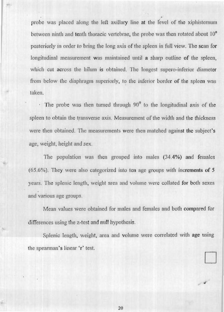
prohe was placed along the left axillmy line at the I'evel of the xipl~istenil~m
between ninth m d tenth tlioracic vertebrae, [lie probe was then rotated about 10'
posteriorly in order to bring the lorlg axis of the splee~i in f i l l1 view. The scan for
longitudinal measurement was rnnintgit~ed mtil a sharp outline of the spleen,
wliich cut across the I I ~ ~ I I I I I is vbtaiued. Tile longest supero-inferior diameter
from below the diaphragm superiorly, to l l~e inferior botder of the spleen was
taken.
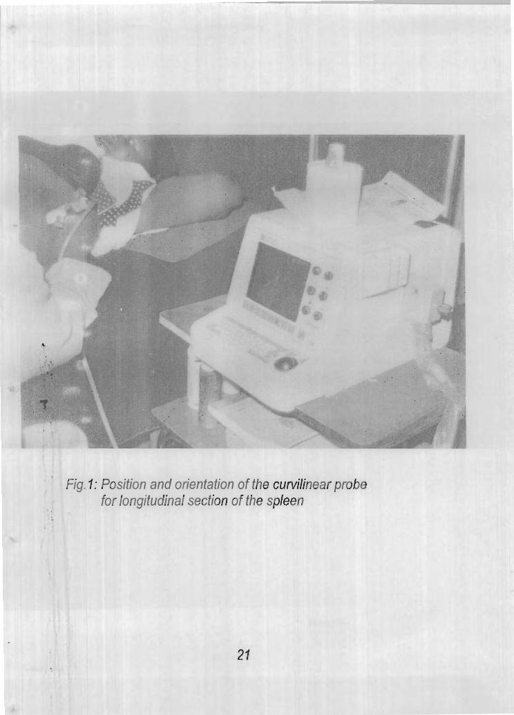
Tlie probe was hien tunled tllror~gli 90° to the lot~gitr~dinal axis of the
spleen to ohtairi the trmsverse axis. Measurement of the width arid the thickness
were then obtai~~ed. Tht: t~seasr~re~nent s were then matched agailist the subject's
age, weight, height arid sex.
The population was the11 grouped into ~riales (34.4%) and females
(65.6%). They were also categorized into ten age groups with itlcretrretits of 5
years. Tlie sple~iic le~igtli, weight area slid vol~me were collated for both sexes
and varlous age goups.
Mean values wen: obtained for males and females and both compared for
differences using the z-tcst arld null Iiypothesis.
Splenic length, %eight, area allti volume were correlated with age using
the speannari's linear 'r' test.
': Posifion and orientation of the curvilinear probe for longitudinal section of the spleen
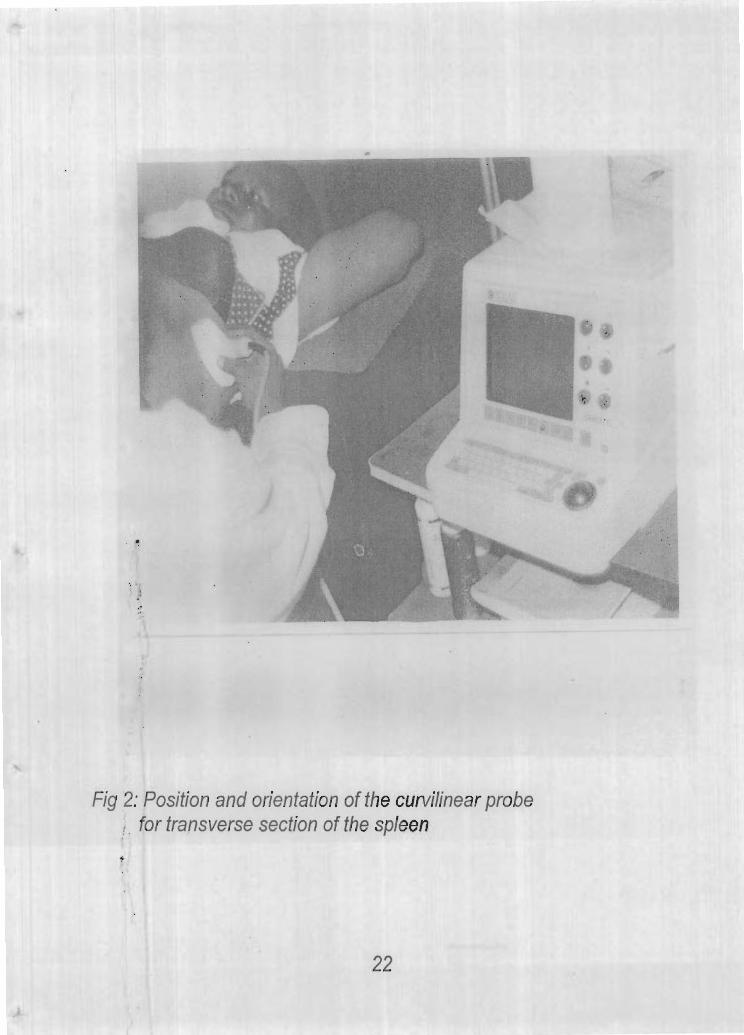
Fig 2: Position and orientation of the curvilinear probe : . . for transverse section of the spleen
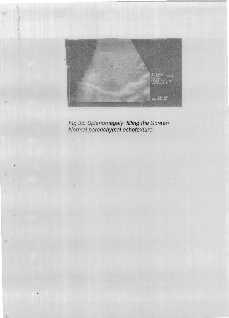
Fig 3c: Splenomegaly filling the Screen Normal parenchymal echotexfure
CHAPTER FOUR
DATA ANAI,YSIS AN11 PRESENTATION
4.1 TARI,ES AND GRAPHS
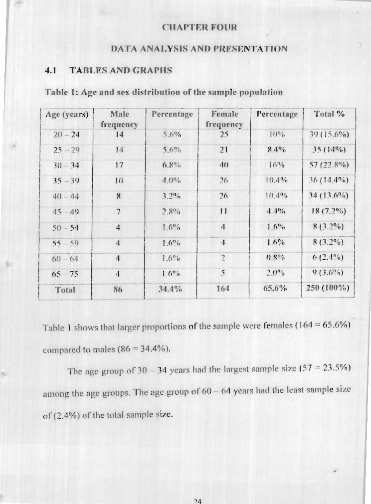
Tahlr 1: Age and sex rlistrihrrtion of the sample poprlnlion
- - - - . . .-
I Total I
. - . -
Malt frequency
-1'ablc I shows that larger proportions of the sample were females ( 1 64 = 65.6%)
compared to males (86 - 34.4%).
'l'hc ape gi-oup of 30 - 34 years had the largest sample size ( 7 - 23.5%)
among - the age groups. - 'I.l~e ape group of 60 - h4 years had the least sample size
o f (2.4%) o f the total sample size.
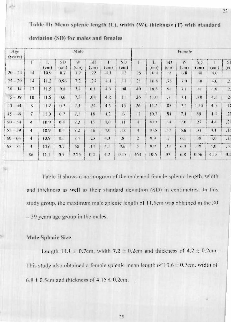
Tahlc 1 1 : M e a n splcnic Icngih ( I , ) , wirlfh (1%'). fhickness ('1') with st:lntlarrl
Table 11 sliows a ~wrn~ogr-atn of !lie male nn( l r m n l e splenic lenp~h. width
and thickness as wcll as thcir standard deviation (SI)) in ccntirnetrcs. In th is
study group. the ~naxirnrtm male splenic lengtli of' 1 1 . 5 3 1 1 ~ v a s o f - d a i r d i l l the 31)
- - 39 years age group in the males.
M R ~ Splenic Size
i x n ~ t h 1 1 . 1 k 0.7cm, width 7.2 + 0.2rm and thickness of 4.2 + 0.2~111.
Thi? s t i ~ d v alsn ih~airictl n lkrnnlc splcnic mcnrl length o f 10.0 + 0.7~111. width of
6 .8 4 O 5i.m and thickncsq of 4.15 4: 0.2ctn.
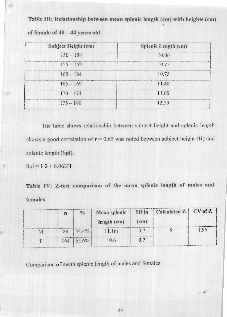
Tahle 111: Rel~tionship between mean splenic Icnqth (cm) with heights (cm) r
o f fcrnrlle of 40 - 44 yenrs old
I SubJcct Jfeight (cm) Splenic I , e n ~ t h (cml I
The table shows relationship
shows a good correlation of r = 0.65
splenic lenpth (Spl).
Spl = 1.2 + 0.063F-I
between subject heieht and splenic length
I was noted between sub~ject height (FI) and
Tshlc 1V: &test comparison of the mcan splcnic length nf males and
fcmalep
----- --.-- 7-,r--$c[ I Mcan splenic T SD in
Comparison of mean splenic length of males and females
i n : There i s n o significant difference between the male and female splenic
length
I : There is a significant difference between the ~ m l e and femzle splenic
length.
Significant level: a - -- 0.05
5.0 > 1.96 (p < 0.05). fhercforc Hr, i s rejcctcd and 1-1, is accepted
The z-test for comparison of mean splenic length showed that the null
hypothesis were rejected and t h ~ t there is significant difference be twem the
male and female splenic Imjqth.
Table V: Distribution of splenic length equal or l ~ s s than 7.8crn in length
among the age grauprs in both sexes
5 subjects had splenic length equal or les than 7.8cm. Two were f o ~ ~ n d in I
the 20 - 24 ycars ape group arnonp rcrnalc and onc cnch in the 31) - 44 venrs age
group for both sexes.
Table V1: Proportion of spleen less than or eqlral (r, 7.8cm in length
. .- -. -~ - .- - - - . . - - 1 - - - - -- . . -- -
4 I -- -- - - ---.- - -- 4 ----------
Total 5 1 2.0% --- - . - - . ~ I - o 1
Table V and table VI slmw that onlv 5 sljhjects (2%) m t of the 250
subjects have splenic length eqrral or less than 7.8cm. Among the 5 suhiects
there was one male (0.4%) and 4 fernales (1.6%) who had splenic length equal
o r lerq than 7 8cm
Tahle V11: Sex distrihlrtion of mean splenic weight
'T'his table shnwc diqtrihution of splenic weight with gee. A mean splenic ,
weight of 145 + I I em f'nr rmlc 2nd 1.70 + 1 Iprv fbr f'crnnlr was ohtair~ed. In
both sexes, t h e rnaxi~num splenic weight was fnrsnd in the 40 -- 44 years age
group, 15Xgm for male and 156gm for female. In the rnalec 30 39 years age
group, the spleen weighed 157gm. close to the ~naximrrm weight of the 40 - 44
ycar ape group.
Table VIII: Comparison between male nnd fcrnale splrnic weight
weights.
difference bet-ween male and female splenic
HI: There i s n significance difference hetween inale and female splenic
weights.
The c f n t i d i ~ n l frd I J ~ J i~ 7-feet
The significance level = 0.05
The calculated z value = 6.98 > 1.96 (P < 0.05): therefore Hh is reiected
and H I acccptcd.
Thcrc is significant diffcrcnce between males and females splenic weight I
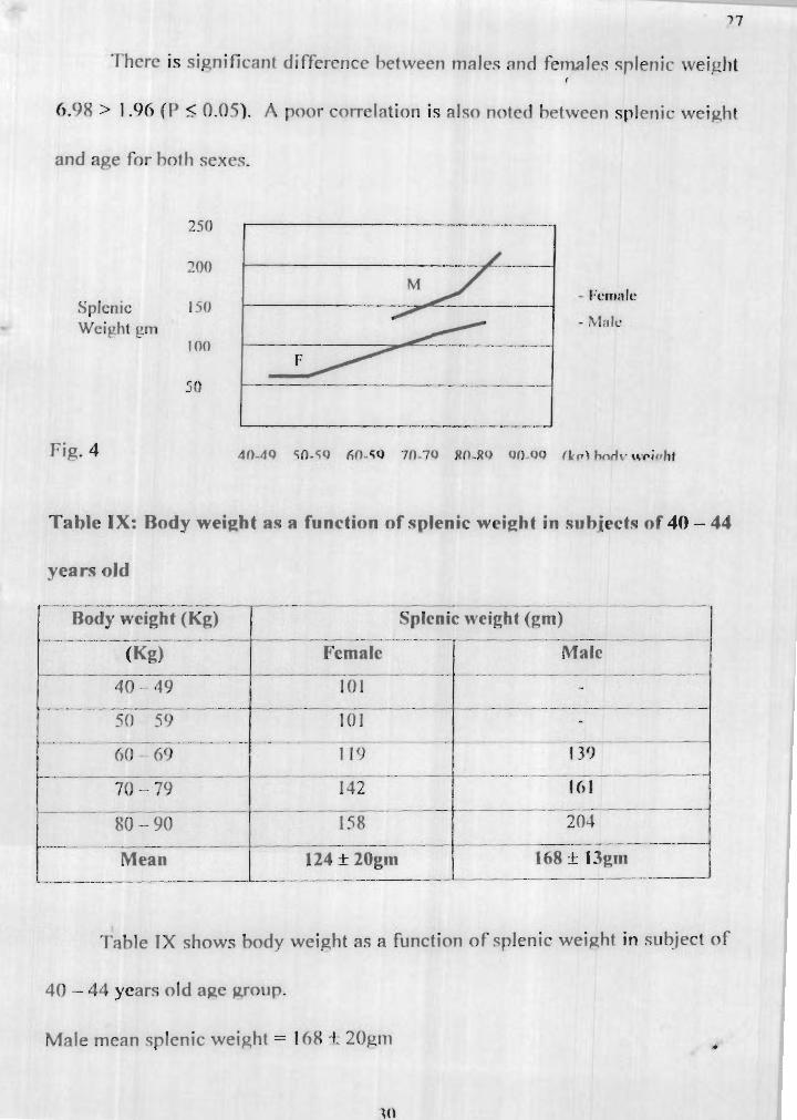
6.98 > 1.96 ( P < 0.05). A poor cm-relation is also noted hetween splenic wei~ht
and aRe for hoth sexes.
Table IX: Body weight a9 a function nf splenic w e i ~ h t in swh jecto o f 40 - 44
years old . ~ .- . .. . - .
/--Body wcieht (KET SpIcnic weight (gm) -7 I
t . - - --- - - I-- Mean 124 It 2Ogm 1 68 k 13gm __ _____l__l_ -I_----- -- - -- --I
Table IX shows body weight as a f ~ ~ n c t i o n of splenic weijzht in subject o f
40 - 44 ycars old age yrnnp.
Male mean splenic weiy,ht = It18 + 20pm a
Female mean splenic weight = 124 f 13gm X
Z vdue = 6.98 > 1.96
1 the male and female splenic
weight (P < -.-,,. ...,.-*- w,.w...w ..,.,,,, ,,,,,ed elf with M v weirrht (r =
0.6). Subjects whose body welght h 60kg and above (normal adult body ~eight)
showed better correlation of r = 0.75 thah with a w g e t e of all the body
weights. The female splenic weight showed poop codation with body weight
(r = 0.4). Using the spanman's linear 'r' moment.
splenic area of 64 f 3 .3cm2 for males and 58 f 5.8cm2 for females. Comparison I
Aa,e @earn) 20 - 24
25 - 29 .- I 30-34
35-39
wing z-test shows significant difference between the male and female .@c
1 Splenic Area (em3
Male 62.8 64.5
68.0
69.0
Ferdalc
56.6
60.4
61.5
62-5
Maximum normal splenic volume for male is 13.6rm' and 13.4rrn"m female.
area 15.7 > 1.96 (p < 0.051. 'l'he statistical test used i s z-tekt. A significance level f
o f 0.05 was used, the calculated z value = 15.7 (p < 0.05).
Table XI: Variation of rrplrnic volmne (ern3) with nee
-. - - --- -1
* -_--.l-__- _ - _ _ -
Agc (~cars ) Mnlc 1 k'crnalc -7
Table XI shows the distribution of splenic volt~rne with age. Male mean
splenic volume is 12.6 + 0.9cm' and female mean splenic volurne is 1 1.1 _+
1.5cm'. T h e ,graph shows non-linear nature.
A poor correlation is noted between age and splenic volrrme. The
maximum spleriic voltrrne for both sexes i s in the 40 - 44 vears age group.
Fig 4 Male m r t fernstle splenic volornt! with spt
Table XII: Body sr~rface area as a frrnctian of splenic vdrrrne in 40 - 44
years old apre group.
- - -- -
Body surfacc area (RSA)
___-____--.----- I . .
Mron V ~ r n g I c . R 9 A - 3 h2.
Mean male splenic vol[rrnc - 14.5crn ' 4 2.2
Mean female splenic vol~rmc - I 1.5cm' + 2.2
Male r = 0.9. female r = 0.8
S,V = 0.27 + h.hl3SA (crn')
'Thc male m t m horfv qlrrf;lcc nrea = 1 .%n2 wf~ilt- the female mean
body surface area (RSA) = 2.hm2. l'he study also showed that h e male mean
splenic volume = 14.45 + ?.5rm3 and the female mean qplmic volrme = 1 1.48 it.
2.2crn3. The coefficient of corrclatim of thc body wrfacc area aminct the
splenic voll~rne iq r = 0.7 fnr female, and r = 0.9 for male 11qinp the spearman's
linear 'r' mntnrnt
16
I 4
12
Splenic volt~rnr l fl 3 crn
8
T a hle X I 11: Comparison of splenic length of f hc ctrnd~ grnrrp nnd t h ~ t or the
HO: There i s no significant difference hetweer~ the splen i r lerwtli r ~ f the st~ldj.
proup and Carmqianq
4 : There is a s ienif icmt difference betweer1 fhe ql-rlrr~ic lerir~,th o f the s t~ldv
jzrtlup and Ca~~cas inns
0.67 < 1.96 ( P > 0.051. f l i ~ r e f o r e I l , , i q arcepterf
I lence the str~dy says that there i s no sigriificxrit diffcrencc hrtween the
splenic lenqlh o f the f'aircmian nnrl flint of thr s t r~dv ernllp (P > 0.05).
. - - - - - - -. .
Study group
4.2 I'llSCl ISClCINq
In thic; qtt~rfv, a sample ~ i 7 ~ o f 2 5 0 nrw I I ~ . 'l'here were 164 females
(65.6% o f the sample si7e) and 86 males (34.4%). The 30 - 34 veam w e group
had the l a r ~ c s t nlmhcr of partiripant9 wherca~: the leac;! wm the hO - 64 years
age grotlp for the hvth sexes.
4.3 SPJ ,ENIC I ,ENf;TIl
The splenic lennth for hnth sexes was the easiest dimension to measrtrc
t,he ~sses s rnen t n f thc splenic s i x . I n the male rnctan splenic lenqth was 1 I , 0
n
. - - 250
Splenic lerteifi
. - - .
11.5
0.7cm. Splenic lenath had a range nf 9.7cm to 12cm. though this valrre is in <
disa,qreernent with the work of Ijissct grid Khan, 1990, i t ;1qrees with Messinezy
(1997). The splenic lcngth ohtnincd in this sttdy s h v s n v stati~tical cliffcrcncc
(P > 0.05) between C'ar~ca~qian splenic len,pth, \vhic:h i s I I .6r1n, and 1 1.5cm fhr
the st~rdy wow.
The splenic length i n t h e male is ereatest in the 70 - 79 years age p,rnup
while the smallest splenic length in that sex is at 65 - 75 ymrs indicating
shrinkinp. Thc splenic length in the age Erorrp of 20 -- 39 vrars s I i t~ \v~d steady
increase with age and after 39 years it begins to shorten. Therefore, if the splenic
size is larger than the value in the nnrnnqratn for the aqc, pntholoay is str,~p,csted.
In the female, the ureatest splenic length i s in the 4n- 44 years w e group
as against 30 -- 39 ycarc in thc malc. The splenic Irneth of the fcrnale 20 - 44
years age proup also showed steady increase with ape and after 44 years when i t
started to dcclinc as a,qc incrtascc. While incrcasc in splenic length in the male
stops at about the age: o f 39 years. i t continues to increase in the female ti l l 5
years later.
1,ike in the male the smallest splenic length is fmmd at 65 years and
above. Therefore, the author deduced that splenic lenpth increasec with age and
at about middle a ~ c the length h c ~ i n s to decrease. Thiq is in agreement with
Dclnnd (1971)). In the rcrnalc. the splenic length start^ t o di~niniqh from 45 vears,
however. the m ~ t h n r did not investigate whethcr nnv relsltionship exist between -
splcnic size and rnenonarlse since 45 years i s a cornmon nerind of mennpauw 1
and the splccn also h c i n ~ ! a hacmotnlwic nrgan both in the innlc and thc female.
There i s a good correlation between subiect height and splenic lenath as also
reported by Deland, 1970. Female r = 0.65 (spl = 1.2440.fl67Il).
Spt = splenic length. f f = subiect height
It was ohqerved fhat 2% of the srhjects under qfr~riv h a w qp lmic lenqth
equal or less than 7.8cm as reported hv Cha~~han et 31 (1996). This i s aswciated
with natives of falciparum malaria endemic area. It was fi~rther nnted to be more
prevalent among women ( I .fiO/n of the stidy pop~llniion) and 0.4% fnr males.
The mean splenic width for males is 7.2 + 0.2crn and 6.8 + 0.5cm for
females. T h e splcnic width incrcnccs wi th aqe t i l l 3C) w a r s in the mnles hut
continues t i l l 49 years in the females. thereafter the width heains to decline with
c?Re in hoth mxle and fernale. When critical eval~rntinn o f fhe spleen iq required
in condition o f diminishing s~ len ic si7e before 49 years i t suggests pathology.
l Jnlike for splenic width, splenic thicknecs r e x h rnax im~~m dimension at
the same age group o f 40 - 44 years for hoth male and female and thereafter, the
splenic thickness stnrts to dirninish with age.
The splenic thickness obtained from the str~dv was 4.2 + O02c.m for males
I
4.5 SPI,ENIC* WFIGHT, A R E A AND VOI,IIMF
The splenic wei& was Iar~eqt in the 40 - 44 vears aRe arolrp for both -
sexes. The splenic mean weigl~t is 145 + 1 I Em and 130 + 1 ! em for males and
fcrnales respectively.
There was a statistically significant difference hetween mnle and female
splenic weipht (P < 0.05).
The range of snlenic weipht w a ~ fmnd to he between 120gm to 158gm
for both scxcs (P > 0.051, ;1 splcnic weight outside this range mav he si~rpected
to have a disorder in non-obese patients,
In the Caucasian. Dcland (1970) showed that mmirnrrrn splenic- weight
occurred at the ane of 20 - 29 years group m d thereafter ~rac-l~~allv declines.
While the maximum wriqllt obtained in this sturfv occr~rrt.d at 4 0 - 44 vcnrs age
group.
There was good correlation hetween cpl~nicr weight and aEe initiallv, as
ape increased splcnic weight decreased and this dccrcasc was not linear. A
similar characteriqtic wn5 notcd in the relationship of c;plenic area and ap,e. The
splenic area increased with age ti l l 39 years for male and 44 vears for fernfile.
Thc area of t h e spleen dccrcase in a non-lincar tnnnncr finer 39 years and this
accounted for the poor correlation between the age and the splenic area.
In thc annlvsi? of thc rtlniionqhip hetween hodv weight and splenic t
weiaht. a snecific ane p r o w (40-44) vears was choqen. al though the relgtinnship
between age and splenic weight was poor this amlvsis still exclwled the effect
of age. Therefore, the analvsis of the relationship between body weight and
splenic wciaht were independent of age. This 40 - 41) vegrs ap,e p,rollp has the
hiphest frequency in the sample pnprrlation and a l so i s the age wmrp where the
both qexes have the, largest splenic weight attictng the strrdv prorrps.
There is poor correlation between splenic weight and bodvwei~ht o f the
subiccts who arc t ~ n d c w c i ~ h t (59kg and less). Minirnv~m ntlnnal weip;ht for the
age range i s 60kg (Roper, 1975). Srrhjects whme w e i ~ h t are 6Okg and a h w e
showed correlation (r = 0.75). this suepests a strong: clinical relatintiship
between splenic weight and body weights of the s~~hjec t
The subiectq who weighed 60kq m d ahove hnd smne ct-raracterisfics with
Caucasians. When the r~ndenveight and t h e norrnnl s~rhier ts were combined. the
graph showed a poor correlation between splenic wcie;ht and hodv weight
showing that there was very poor cnrrelation between hody weight and splenic
weight of the underweight. 7'hereht-e. the splenic wei&t or size may he an
index to evaluate the well being of a person. This srrggests that work should be
carried out to show whether splenic size or weight can correlate with any levels
of cmaciafion. nody weight i s a useft11 factor in considering splenic weight.
In the s t ~ ~ d v o f Rody Surfgce Area (RSA) and qpl~nic vnflrme. the author
observed that males of 20 years and above have RSA of 1.5 to 2.5rn2 and the
females have 1.0 to 4.0m2. l h a t is. females have wider range of RSA than
males. Males hnve mean splenic voll~rne of 14crnJ while females hnve splenic
voltme o f l 0.?crn3. A ~ o o d corffirirnt of correlation w i s t i n e hptwrrn RSA and
splenic volumc was nhscrvcd. Fcm;llt r = 0.8. male r - 0.9, t~sine. the
Speannan's linear 'r' moment.
There was significant difference between the male ydenic vdrrrne and the
female splenic vol~~rne ( P < 0.05). C'or.rclatim h c t w ~ e n ope 2nd splenic vohrrne
was poor. Both male and female chow maximum splenir vol~rme at aEe groups
of 40 - 44 yenrs and therenfler the spleen begin? to shrink a.r; aRe increme. This
age group was used for comparison of body srjrface area (RSA) and splenic
vnlrrme. The graph of age and splenic volutnc: s h o w a non-tinearitv 3s age
lnrrrace
PFf APTFH FIVE
Medical ultrasonographv is a valuable tool for f he assecstnent nf splenic
size and pathological status of the spleen.
This studv has provided a nnclmal range of splenic size, which Ni,gerians
can use in the assessment of splenic size especially in matters relating to
p a t h o l o ~ y .
Splcnic length 9.5cm to 12.0crn
Width f,-.?rm tr, 7 4 r m
Small increase outside the rlormoRrarn may be a lead to unsuspected
disorder and cal Is for furlher invest i ~ a i ion.
The other splenic parameters, weight, vnlr~tne 3nt.l area are obtained from
these dimensions. For body weight o f 6Okg and above, (standard body weight),
the splenic dimensions have no difference with fhe Caucasian values. Significant
diffcrencc waq n o t d hptween m ~ l ~ nnd f ~ r n g l ~ dirn~ncinne
Subjects w h o are up to 30 years old and weigh 5 9 k ~ o r fess s h o w splenic
parameters that are less than their age group who weigh 6Okg and above. This
supgests splenic si7e as a strtlnp clinical marker. A normopram was obtained for
various paramctcrs of tlir spleen. Splenic' weieht may he val~~nhlr index in I '
assessing the well being of a patient.
Thc nonnojqam nbtaincd will hc 11sefi11 in assessinq manv disease
conditions in Nigeria, which manifest in spienornegaly nr diminution o f the ,
A small proportion of the study group shows 'small' splenic sizes, which
The correlation hetween age and splenic wei~ht , splenic length and
splenic volume are poor because the ~ r a p h was not linear. There are initial
steady increases with age t i l l middle age when the parameter starts to shrink
with aRe. The decrease as age increase arc irregular and not steady. hence
exhibit in^ poor correlations with age.
Splcnic length initially incrcmes and decline with ace. Splcnic weight
also increases with hody weight and hody surface area. while splenic volume
also increases with body surface area.
5.1 CONCI ,I ISION
This study stronglv recommends splenic length as the beqt predictive
parameter of splenic size hccat~sc:
- Splenic length i s easy to obtain
- Splenic length i s minimally ohstn~ctcd by Ras and rib shadows during scan
- Splenic length showed clear relationship .cvitli age, bodv weight and height
- Splenic length is an important factor in the determination of th r splenic
weight, volttme and area
- The splenic Icngth ohtaincd shows sensitivity in.dctcctin~ changes in splenic
- Rcscarch s t ~ ~ d i c s should he cnrricd r w f to find arly rrlntionship bctwccn
nutrition and splenic sizes.
- Studies should bc carried out to find how lonp i t tgkes fnr the spleen to be
restored to normal size after pathology is relieved. This will he nf cl inical
si~nificancc when there i s doubt about the cause of splenomej.yiIy. especially
after treatment has been given for a disorder.
- '1le researcher also su~nests stt~dies on he rates of' increase of splenic s i x
with time in various pathologic states affecting splenic size.
Alan Rums (1979) Histoflypf:N-igeria, 8th ed., J,ondon: George Allen Rc I Jnvin, p. 30
Arkes, I,.R ( 1986) A Palpable Spleen is not necess;rrilv En l a r~ed Patholopically, Medical-Jorrrnal of' Australia, July 7. 1 14(5): 15 -- 17
Armstrong Peter and Martin Wastie, ( 1 998) Diagnostic lrnaging 4th ed. Northampton: Rlackwell Science.
Risset R.A.L, Khan, A.N (1990) Dif'ferential Diagnosis . in - - Abdominal -
IJltrasound, I,ondon: Hilliere Tindall
Bessmel'tev, S.S, Abdulkadyrov, K.M ( 1993) The Determitlation o f Splenic Mass by Sonography Opredelew & Massy Selwenki Metodorn Smogram Lik-spr3ve . - May - June ( 5 - 6): 1 10 - 4
Carol, A. Krebbes, Vishan I,. Giyanani, Ronal L. Eisenheerg ( 1 993) Ul t raso~~nd Atl-as of r>r>iiease_Pr-o_~C~ss, Connecticut: Appleton and 1,ange. p. 101
Chauhan, R. Kapoor V, Vohra -J'.A, Jhala, P.J, IJpadhyaya, A.K, Pathak, K.J , ( 1 996) The Small Spleen in Malaria, Journal o f Association of Physicians, India: July 44 (7): 483 - 5
Cavenaugh, J.D, Joseph, A.E, Dillys, Hevan 13.H ( 1994) Splenic Sepsis in Sickle ell Discasc, Byit js~-J_ouqal.of F-jaqnat~l~ogy, London: 86 ( 1 )
Donald, R. Kirk ( 1 99 1 ) Bact-kal- Pediat~jclmaqing_ Diagnostic Radiology o f Infants -- -- and-Children. 2nd ed. Boston: 1,ittle and r o .
Downey, M.7' (1992) Estimation of Splenic Weight from Ultrasonographic Mcasurcmcnt, C-anadian Association of Radiology Journal, August 43 (4): 273 -- 7
Frank, 1-1. Deland ( I 970) Normal Splenic Size, Uadiologv. Dec. 97.589 - 592
Gerspacher I,ara, Pinto Silva R.A, Drurnmond S.C, Imnbertucci R.R. ( 1 998) Splenic Palpation for the Evaluation of Morbidity due to Shistosorniasis Mansoni. Memorial . Institute - . . - o f - - - Oswald-cru, - -- - - - Vol. 93 ( 1 ): 245 - 8
Green. J.H (1985) An Introd~uc~i~toP~I:u_rl!ann~sychology. 4th (S.1) ed. New York: Oxfbrd I Jniversity Press I
Grove, A.T ( 1 978). Africa. 3rd ed., London: Oxford University Press
Kathryn, I . , McCance, Huther S.B ( 19W) Pat_hophysiolo~:~~I~he~Bio~ogic~ Basis for Disease in Adults-and C'hildr_en, 3rd ed., Missouri: Mosby, p. 352
Kenneth J. W Taylor (1978) Atlas of ~ra~scaI~~UI_traso~nograpl~y, ... - - New York: 1,ongrnan Inc, p. 170
Klippenstein D.L. Zerin J.M. Hirschi R.B. Donn S.M (1994) Splenic Enlargement in Neonates During Extrncopreal Membrane Oxygenation (EMCO), Radiology, Fcb. 190 (2): 4 1 1 - 2
l,oflus, W.K and Metreweli, C. (1998) IJltrasound Assessment of Mild Splenomcgally: Spleen/Kidncy Ratio, Peadiatjc RadL@logy, Feh. 28 (2) : 98 - TOO
Lwanga, S.K and Clo-yaok Tyke (1986) Teaching Health Statistics: Twenty Ixssons and Seminar Outlines, WI JO Geneva, 157 - 159
Messinezy, M., MacDonalci L.M, Numan T.0, Westhood N.B, Chin S, Pearson, T.C' (1997) Splccn Sizing by llltrasound in Polycacthernia and Thrombo C'ythaemia, Comparison with spect British_Joumd-p_fl_Hea-matofogy-, - - JJry ( I ) : 163.-7
Nancy Roper ( 1975) Pocket Medical Dictionary 12th ed., English t .anpuage Rook Society Churchill 1,ivingstone. p. 574.
Nwabuokci, P . 0 ( 1986) F;up$atncntals of Statistics, E n u ~ u : Kon~na Rook.
Niederan, Sonnenberg, Miller, Erokenbtecht Scholten. Fri tislt ( 1983) Sonographic Measurement of the Normal I h e r , Spleen, Pancreas and Portal Vein Radiology. p. 149:537 - 540
Paul, M, Davics, (1976) Medical 'l'crrninoIogy it1 Hospita~Practi_c_q, 2nd Edition, I ,ondon: William Heinemmn Medical Rnoks I td.
Peter Armstrong and Martin I , . Wastie (1998) Dia_gnost.i_~_lm~aging 4th ed., I,onclon: Oxfbrd 131ac8kwell Scientific 1,td.. p. 2 1 1 +
Porcel-martin A, Kendon Vinceta P.J Baseumama-Quire1 A, Amaya Videal A, Rodriguez-Kamos C.1, Sorinde La Cruz M. Martin ~ je ra ra ( 1998) 1:ocal Splenic Lesion in Patients with A I D S Sonographic b'onndlings, Abdomen Imaging ( M a r - A t r ) 23 (2): 1 YYb -- 2000
PoZato, C., Marzano L. Botta, A.J, Anamia K.M. Ushlenghi CM.. (1998) Splenomegaly and Hypersplenisrn in Cirrhotic JJatients betbre and stter Orthotopic J,iver 'I'ransplantatim, Radiologic Medicine - 'l'ovino, April YS(4): 349 - 52
Keva Arncz Cuny and Betty U. 'l'empkin (1995) Ultrasogop,raphy: An 1ntrod"g-ion to Norn~al Structure and ~~unct ional 'Anat_o~my Philadelphia. WH Saunders Company, p. 139
Kodrigues-jr. A.J, Kodrigues, C.'i', Ciermano, M.A. Kasera Sirnior, I. Cerri, 6 .G (1995) Sonographic Assessment o f Normal Spleen Volume, Clinical . --
Anatomy, r((4): 252 - 5 -
Uoquet-P. Verlc, Kongs, A.J., 'I'alla I., Niang M., (1993) Hepatosplenic Alterations determined by Ultrasonographic in a IJopulation recently Infected with Schistosomiasis Mansoni in Richard-'l'oll, Senegal: 'l'rans- K-Society 0f~'1'ropica1-~Medicine and H y ~ i e n e , March - April, 87(2) : 190-3
I'amayo-S.G, Kicernan-L.S, Matthews W.C, Pullertion S.C, Uartok At;,, Warner Feigal D.W, Armstein G.D, Clcallandar N.S, 1,yche 1.C1.11, Examiner Dependence on Pl~ysical Diagnostic Test f'or the Detection o f Splenomcgaly, A Prospective Study with Splenomegaly. J-oumal of General Internal-Medic_ine, I YY3 J:eb. 8 (2): 65, - 75
'I'assiopot~los 'l'., Rombos Y., Konstapoulos K., Revanus K . , (1995) Spleen Size in Beta-thalassaemia Heterovgotes, Haernolo~ia, Hudapest: 26 (4): 205 - 9
Yakashi Kogi and Yukio Moriknwa ( 1 975) llltrasonic determination of the splemic size and its clinical usefi~lness in v ~ r i m s liver diseases Radiology Apr. 115;157-161.
APPENDIX - ,
Body surlace area (USA) i s acquired by the Lhhois and Dubois tbrmula
(Niedelran et a1 1983).
Weight x 1 kg x Hcight(crn1 x l x 6(rn2) 2.35 1.38
Splenic weight = 0.43 (length x width x thickness) grn
1992)
Splenic volurne = L x W x 'l'on' 27
Cross sectional area = 0.8 (I . , x W )
For a two sample test for mean:
'I'he correlations were analysed using the Spearman's linear 'r' test
A = y-bx
t3 = Sxx
Formula tor the sample size
P = Proportion trom previous work or pilot study (20Y0 or 0.2)
d - - 'l'olcrable error (0.05)
z = % value ot' specified level o f signi ticance ( 1.96)