Embed Size (px)
Citation preview

UNIVERSITÀ DEGLI STUDI DI CATANIA FACOLTÀ DI AGRARIA
DIPARTIMENTO DI SCIENZE E TECNOLOGIE FITOSANITARIE
SEZ. PATOLOGIA VEGETALE
DOTTORATO DI RICERCA IN TECNOLOGIE FITOSANITARIE XXII ciclo 2006-2010
MOUNIA DADEN
Further characterization studies on Citrus Tristeza Virus (CTV) isolates collection from Mediterranean countries and
their interaction with citrus viroids
Tesi di Dottorato
COORDINATORE TUTOR Prof. Gabriella Cirvilleri Prof. Antonino Catara 1

1
2
3
4
5
A vous chers parents, à mes sœurs et mon frère, vous avez 6
toujours vu en moi un certain potentiel, vous ne m’avez pas 7
laissé le choix, et je vous en remercie. 8
A mon grand père qui nous a quitté et que je n’ai pas eu 9
l’occasion de revoir, qu’ALLAH bénisse son âme. 10
11
12
13
14
15
16
17
18
19
20
21
22
" C'est celui qui s'égare qui découvre de nouveaux chemins " 23
Nils Kjær 24
" En cherchant à hâter les choses, on manque le but. 25
La poursuite de petits avantages fait avorter les grandes entreprises" 26
Confucius 27

ACKNOWLEDGMENTS 1
2
3
4
5

1
2
TABLE OF CONTENTS 1
2
page ACKNOWLEDGMENTS LIST OF TABLES LIST OF FIGURES ABSTRACT………………………………………………………………... 1 RIASSUNTO………………………………………………………………. 3 CHAPTER 1: BIBLIOGRAPHIC REVIEW Characterization of CTV family and virions
Characterization of the family: Closteroviridae


1

LIST OF TABLES 1
Table Page 1. CTV biogroups (Garnsey et al.,
2005)......................................................
2. Citrus viroids Family, genus species and disease caused......................................


1

LIST OF FIGURES 1 2
Figure Page
1. The three genera of the family Closteroviridae with its associated insect
vector..............................................................................................................
2. Genomic organization of citrus tristeza virus, showing the relative position of
the 12 ORFs and their expression products................................
3. Symptoms caused by citrus tristeza virus................................................. 4.Bark scaling symptoms on the trunk due to psorosis infection..................
5. The five domainsorganization in viroid structure......................................
6. The vein band disease symptoms on grapevine leaves induced by the mixed infection with
GYSVd and GFLV........................................................................................................
7. CTV sources under screenhouse conditions


Abstract:
The biological properties of a collection of citrus tristeza virus (CTV)
isolates from 13 Citrus-growing countries mostly Mediterranean, an area for a
long time free from severe CTV isolates, were investigated. The study confirmed
the presence of such isolates in this area, as a lot of biologically indexed isolates
were able to produce even stem pitting on Madam vinous Sweet Orange (SwO)
considered specific for the most dangerous strains.
The symptoms expressed by 27 CTV isolates on the standard set of CTV
indicator plants; Mexican lime (M.L), Duncan grapefruit (Gft), sour orange (SO)
seedlings, Madam vinous SwO seedlings and Madam vinous SwO grafted on sour
orange enable the establishment of the Biogroups that are present in the
Mediterranean Area ranging from the severe Biogroups 4 and 5, mostly in the
Middle East region, to the Mild Biogroup 1 in other regions.
Interestingly, all the isolates belonging to the Biogroups 4 and 5 gave a
positive reaction with the MCA13 MAbs, produced to detect severe CTV isolates
in Florida, but being sometimes ineffective to detect others elsewhere. Such rapid
tests are of important concern and are needed to identify isolates that have
potential economic impact in commercial citrus groves and to adopt a sound CTV
management strategy.
But discriminative analysis needed have to be, as rapid as reliable, and this was
another aim of this work; finding potential molecular markers that could be used
for sensitive and quick identification of virulent CTV isolates through the study of
a Mediterranean CTV collection.
For this purpose the symptom expression and biogroups obtained for the 27
studied isolates were compared with conventional Single Strand Conformation
Polymorphism (SSCP) and Capillary Electrophoresis CE-SSCP results, for four 3′
terminal genes (p18, p20, p27 and p23), and with genetic variation by sequencing.
The conventional SSCP is a relatively robust and easy to perform procedure, and
is a good primary molecular differentiation for CTV isolates, but it has as main

limitation the non quantification of profile differences in terms of the genetic
distance between the corresponding DNA fragments providing therefore only a
qualitative picture of the variation.
CE-SSCP, as alternative to gel slab techniques has become a promising alternative
to gel based SSCP methods, presents the advantages of high sensitivity, high
specificity and is easy to perform. The technique was able to distinguish between
a set of already known different sequences and the repeatability of the results
within and between different runs was also evaluated.
A great diversity of SSCP profiles was observed among isolates for the genes
studied for both methods and some isolates of the same biogroups displayed the
same haplotype, with both conventional and CE-SSCP analysis, mostly for the
p20 gene, even if they were from different geographical origin.
Other field CTV isolates have to be phenotyped and subjected to other
Conventional and Fluorescent SSCP tests so that the proposed models can be
verified, confirmed or modified.
Another part of the work, aiming to study the interaction between CTV and Citrus
Viroids, demonstrated the encapsidation of viroid particles by CTV coat protein.
This phenomenon was more likely to happen when the conditions were favorable
for CTV multiplication, and was confirmed for the available CTV strains in the
collection and for the three most economically important citrus viroids; citrus
cachexia viroid (CVd-II), citrus exocortis viroid (CEVd) and citrus dwarf viroid
(CDVd). This phenomenon can allow to viroid particles to be transmitted by
citrus aphids.
Riassunto:
Le proprietà biologiche di una collezione di isolati del virus della tristeza degli
agrumi (CTV) afferenti a 13 paesi produttori di agrumi, prevalentemente
Mediterranei, un area per molto tempo indenne da severi isolati di CTV, sono stati
indagati. Lo studio ha confermato la presenza di tali isolati nella zona, visto che
molti isolati saggiati biologicamente sono stati in grado di indurre anche la
butteratura del legno sull’Arancio dolce Madam vinous considerato sintomo
specifico indotto dai ceppi più pericolosi del virus.
I sintomi espressi da 27 isolati d CTV sul set standard delle piante indicatrice;
Limetta messicana, Pompelmo duncan, Arancio amaro da semenzale, l’Arancio

dolce Madam vinous e l’Arancio dolce innestato su Arancio amaro ha consentito
la definizione dei Biogruppi che sono presenti nel Mediterraneo che variano dai
severi Biogruppi 4 e 5, per lo più nella zona del Medio Oriente, al Biogruppo di
ceppi blandi nelle atre zone.
È interessante specificare che tutti gli isolati appartenenti al Biogruppo 4 e 5
hanno dato una reazione positiva con l’anticorpo monoclonale MCA13, prodotto
per individuare i ceppi severi del CTV in Florida, ma che si è rivelato a volte
inefficace per rilevarne altri altrove. Tali test diagnostici rapidi sono di grande
importanza e sono necessari per identificare gli isolati che hanno un potenziale
impatto economico in agrumeti commerciali e per adottare una strategia di lotta
adatta ai ceppi di CTV ritrovati in campo.
Ma le analisi discriminatorie devono essere quanto più rapide che affidabile, e
questo era un altro obiettivo di questo lavoro, trovare potenziali marcatori
molecolari che potrebbero essere utilizzati per un identificazione sensibile e
veloce di isolati virulenti di CTV attraverso lo studio di una collezione di CTV del
Mediterraneo.
A tal fine, l’espressione dei sintomi e i Biogruppi ottenuti per i 27 isolati studiati
sono stati confrontati con i risultati ottenuti con il metodo convenzionale del
Polimorfismo di Conformazione dei Singoli Filamenti (SSCP) e con
l’Elettroforesi Capillare del Polimorfismo di Conformazione dei Singoli Filamenti
(CE-SSCP) per i quattro geni (p18, p20, p23 e p23) e con le variazione genetiche
mediante il sequenziamento.
La SSCP convenzionale è una procedura relativamente robusta e facile da
eseguire ed è una buona differenziazione molecolare primaria tra gli isolati di
CTV, ma presenta come limitazione principale la non quantificazione delle
differenze di profili ottenuti in termini di distanza genetica con i frammenti
corrispondenti di DNA fornendo quindi solo una rappresentazione qualitativa
della variazione.
La CE-SSCP, in alternativa alla tecnica con il gel di polyacrylamide, è diventata
una promettente alternativa al SSCP convenzionale e presenta i vantaggi di alta

sensibilità, alta specificità ed è facile da eseguire. La tecnica è stata in grado di
distinguere tra una serie di sequenze già note come differenti e la ripetibilità dei
risultati è stata valutata sia per la singola corsa che tra le diverse corse
elettroforetiche.
Una grande diversità dei profili di SSCP è stata osservata fra gli isolati per i geni
studiati con entrambi i metodi ed alcuni isolati degli stessi Biogruppi hanno
esibito lo stesso aplotipo sia con la SSCP convenzionale che con la capillare,
principalmente per il gene p20, anche se provenivano da aree geografiche
differenti.
Altri isolati da CTV da campo devono essere sottoposti ad analisi del fenotipo e
altre prove di SSCP convenzionale e fluorescenti, in modo da poter verificare,
confermare o modificare i modelli proposti.
Un’altra parte del lavoro che aveva come scopo studiare l’interazione tra CTV e i
viroidi che colpiscono gli agrumi, ha dimostrato l’incapsidazione dei viroidi dalla
proteina di rivestimento del CTV. Questo fenomeno si è rivelato più probabile
quando le condizioni sono favorevoli per la moltiplicazione del CTV, ed è stato
confermato per i ceppi disponibili nella collezione di isolati e per i tre viroidi
economicamente più importanti degli agrumi; il viroide della cachessia (CVd-II),
il viroide dell’exocortite (CEVd) e il viroide del nanismo degli agrumi (CDVd).
Questo fenomeno può permettere alle particelle del viroide di essere trasmesse da
afidi vettori di CTV in agrumeto.
CHAPTER 1: BIBLIOGRAPHIC REVIEW
Citrus production is adversely affected by a number of diseases caused by
viruses and viroids that had major impacts on citrus production.
Citrus tristeza virus (CTV) alone has caused the loss of millions of trees
worldwide. The virus originated in Asia and has spread by man to most citrus
producing areas causing variable losses depending on the virus strains
predominant in each citrus region.
CTV isolates often differ in biological characteristics, such as symptom intensity
in different citrus species. The slow decline is very frequent on the Mediterranean
area, but more severe strains exist and can be found elsewhere.
Changing rootstocks is of common use by growers in response biotic and
abiotic stress that can limit citrus production.
To overcome the CTV ‘quick decline’ on sour orange and also the
Phytophthora root rot affecting trees grafted on sweet orange or rough lemon root
stocks, the trifoliate orange [Poncirus trifoliate (L.) Raf.] has been used as

rootstock, but some cultivars on this latter were dwarfed and unthrifty, with
obvious bark scaling near the graft union demonstrating to be sensitive to viroid
infection. Also known as "exocortis", these bark scaling symptoms signal the
presence of CEVd in the latently infected scion.
The propagation of viroid-infected trees has caused significal economic
losses in citrus industry and is a major threat in regions where susceptible
rootstocks are used.
Although viroids cause several different diseases in citrus [e.g., cachexia
and xyloporosis (HSVd) in addition to exocortis (CEVd)], it is important to note
that viroid infection can also be beneficial to citrus production. For example, both
CEVd and Citrus dwarfing viroid (formerly known as Citrus viroid III) have been
used to dwarf citrus, thereby allowing a predictable degree of tree size control and
the use of higher planting densities (Hutton et al., 2000).
Citrus as all woody perennial crops should have a long productive life
during which they can be challenged with other pathogens as CTV that is readily
aphid transmitted. While interaction of viroids with other plant pathogens (fungal,
viral) has been reported in some cultures as grapevine and potato, nothing is
known about what can induce this natural and non-pathogenic viroid variant in the
presence of another disease pressure.
Once trees are infected, the disease agents cannot be eliminated. This is of
particular economic importance, since citrus trees should have a long productive
life. Thus the best control measures would be to prevent the introduction of the
disease or propagate plants that are disease resistant.
Characterization of CTV family and virions
Characterization of the family: Closteroviridae
The plant virus family Closteroviridae is comprised of viruses with
flexuous rod-shaped virions of 1250 to 2200 nm in length (Alkowni et al., 2004).
These viruses all contain a positive sense single-stranded RNA genome that
approaches 20 kb (Alkowni et al., 2004). Initially, when established in 1998 the
family consisted of just two genera, Closterovirus and Crinivirus. The major

differentiating trait of the two genera was the possession of monopartite and
bipartite genome, respectively (Martelli et al., 2002). Karasev (2000), argued that
Closteroviruses should be classified by the type of insect vector rather than by the
number of genomic RNAs. He proposed a genus named Vinivirus, but the
International Committee on Taxonomy of Viruses (ICTV) study group on
Closteroviruses and Allied viruses changed it to Ampeloviruses (from ampelos,
Greek for grapevine) to prevent confusion with the genus Vitivirus (Martelli et
al., 2002). The revised version of the family was approved by the ICTV in July
2002. The family consists of 3 genera: Closterovirus, Crinivirus and Ampelovirus,
with the differentiating trait being the family of the insect vector (Fig. 1) (Martelli
et al., 2002).
Fig. 1 The three genera of the family Closteroviridae with its associated insect vector.
The ancestral virus in the evolution of the Closteroviruses was most likely
a monopartite virus. This suggestion is based on the phylogenetic clustering of all
the whitefly transmitted closteroviruses and the great uniformity in their genome
organization. This also indicates a recent origin of the bipartite genome that had
not yet had enough time to significantly evolve (Karasev, 2000).
Phylogenies inferred from the amino acid sequences of the closterovirus
HEL, RdRp and the HSP70 suggests that the closterovirus co-evolved with their
insect vectors. This co-evolution probably took place over a considerable period
of time for the great diversity to arise. Thus evolution within the family
Closteroviridae most probably followed the three families of insects: aphids,
mealybugs and whiteflies (Karasev, 2000).
CTV genome
As with other members of the family Closteroviridae, CTV virions are
bipolar and are coated with separate coat proteins 25 kDa (CP) and 27 kDa
(CPm), designated as major and minor CPs that encapsidate about 97 and 3% of
the virion length, respectively (Febres et al., 1996; Satyanarayana et al., 2004). Its
virions are flexuous filaments of about 2000 x 11 nm, with about 6% RNA
content, that are helically constructed with a basic pitch of about 3.7 nm (Bar-
Joseph et al., 1972).
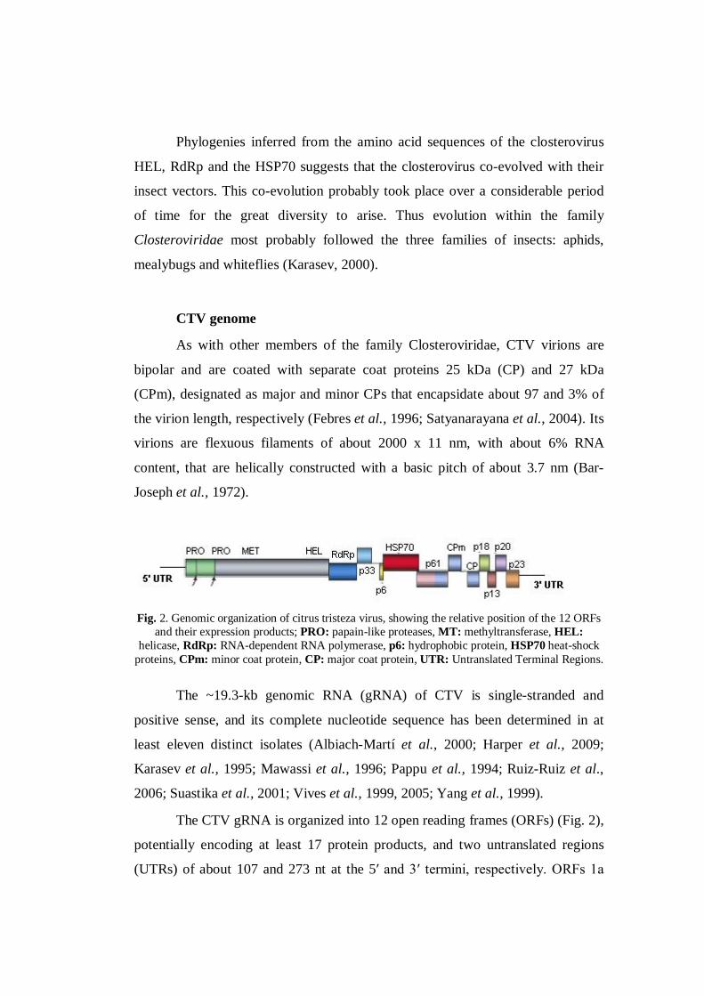
Fig. 2. Genomic organization of citrus tristeza virus, showing the relative position of the 12 ORFs
and their expression products; PRO: papain-like proteases, MT: methyltransferase, HEL: helicase, RdRp: RNA-dependent RNA polymerase, p6: hydrophobic protein, HSP70 heat-shock
proteins, CPm: minor coat protein, CP: major coat protein, UTR: Untranslated Terminal Regions. The ~19.3-kb genomic RNA (gRNA) of CTV is single-stranded and
positive sense, and its complete nucleotide sequence has been determined in at
least eleven distinct isolates (Albiach-Martí et al., 2000; Harper et al., 2009;
Karasev et al., 1995; Mawassi et al., 1996; Pappu et al., 1994; Ruiz-Ruiz et al.,
2006; Suastika et al., 2001; Vives et al., 1999, 2005; Yang et al., 1999).
The CTV gRNA is organized into 12 open reading frames (ORFs) (Fig. 2),
potentially encoding at least 17 protein products, and two untranslated regions
(UTRs) of about 107 and 273 nt at the 5′ and 3′ termini, respectively. ORFs 1a

and 1b, encoding proteins of the replicase complex, are directly translated from
the gRNA, and together with the 5′ and 3′ UTRs are the only regions required for
RNA replication. The remaining ORFs, expressed via 3’-coterminal subgenomic
RNAs, encode proteins required for virion assembly and movement (p6, p65, p61,
p27 and p25), asymmetrical accumulation of positive and negative strands during
RNA replication (p23), or suppression of post-transcriptional gene silencing (p25,
p20 and p23), with the role of proteins p33, p18 and p13 as yet unknown.
p23, p20 and p25 genes
The three proteins P23, P20 and P25 have been found to act as RNA
silencing suppressors in Nicotiana tabacum and Nicotiana benthamiana plants
(Lu et al., 2004).
The only CTV protein with no homologue in other closteroviruses is P23.
The molecular determinants of CTV induced symptoms are presently unknown,
although transgenic Mexican limes (Citrus aurantifolia) over-expressing the p23
protein display symptoms identical to those caused by CTV in this host, and
symptom appearance is associated with p23 accumulation (Ghorbel et al., 2001).
Therefore, the accumulation of p23 rather than its origin seems to determine the
intensity of the symptoms in transgenic limes.
In citrus protoplasts, its sgRNA is the most abundant at the beginning of
the infection and the second most prevalent in later stages, suggesting a role of
p23 in early steps of viral replication or transcription (Hilf et al., 1995; Navas-
Castillo et al., 1997). However, p23 accumulates at low levels in citrus-infected
plants (Pappu et al., 1997). In vitro, p23 has the ability to bind RNA in a non-
sequence-specific manner, and mutations affecting the cysteine and histidine
residues of a zinc finger domain conserved in different isolates, increase the
dissociation constant of the p23-RNA complex (López et al., 2000). Additionally,
p23 is involved in regulating the synthesis of plus and minus strands during RNA
replication, with the zinc finger domain and an adjacent basic region being
indispensable for asymmetrical accumulation of the plus strand (Satyanarayana et

al., 2002). Furthermore, transgenic Mexican lime plants constitutively expressing
p23 of the severe CTV strain T36 display alterations resembling the symptoms
induced by CTV in this host, with their intensity being associated with p23
accumulation (Ghorbel et al., 2001). This strongly suggests that this protein is an
important pathogenicity factor, a view supported by recent experiments showing
that p23 is a potent suppressor of local silencing in N. tabacum (Lu et al., 2004).
Sambade et al., (2003) discriminate between mild and severe CTV isolates
using a specific region in gene p23. In fact, they found that there were 43
polymorphic amino acid positions (approximately 20%); however, in three p23
regions, CTV isolates of the mild group had the same amino acid sequence that
differed from the sequence of other isolates. Those regions included positions 24–
29, 50–54 and 78–80. In the first, isolates of the mild group had Lys24, Glu26 and
Lys29, whereas isolates of the severe group had Glu24, Lys26 and Val29. These
changes result in a strong modification of the isoelectric point of this p23 region.
Amino acid changes in the second region left the isoelectric point essentially
unaffected: isolates of the mild group had Val50, Thr53 and Asn54, and isolates of
the severe group had Ile50, Asn53 (except for VT that also had Thr53) and Ser54.
Finally, in the third region, the isolates of the mild group had Ala78, Leu79 and
Lys80, whereas isolates of the severe group had Ala78, Ser79 and Arg80. The
isolates of the atypical group had Gly78, Leu79 and Lys80, except for T36 that had
Arg80. The two latter regions are located in the RNA binding domain of p23
identified previously, which includes several basic residues between positions 50
and 67, and a putative zinc finger motif (positions 68–86). Interestingly, the
residues involved in RNA-binding (the basic residues, and the Cys and His
coordinating the Zn ion), were conserved in all isolates with differences affecting
only certain positions in their close vicinity.
P20 accumulates in amorphous inclusion bodies within infected cells
(Gowda et al., 2000). p20 has been shown to be one of the three proteins involved
in suppression of RNA silencing (Lu et al., 2004). Suppressors of RNA silencing
have been shown to be required for the systemic infection of plants (Cronin et al.,

1995; Ding et al., 1995; Anandalakshmi et al., 1998; Brigneti et al., 1998;
Kasschau and Carrington, 2001; Bayne et al., 2005; Schwach et al., 2005).
It was also found that both CTV p20 and CP can interfere with the
systemic spread of silencing, while p23 can only suppress the local silencing (Lu
et al., 2004). Thus, CTV evolved a complex system of RNA silencing suppression
with three components targeting distinct facets of RNA silencing response.
Using the polymerase chain reaction (PCR) Mawassi et al., (1993) and
Pappu et al., (1993b) determined the nucleotide sequences of the p25, the coat
protein (CP) gene, of diverse CTV strains and found that the sequences were
conserved at 90% and that there was a relationship between the sequences and the
symptoms caused by the CTV strains. While Gillings et al., (1993) developed a
restriction fragment length polymorphism (RFLP) assay to differentiate CTV
strains using the Hin f1 restriction enzyme that digests PCR products of the CP
gene produced in seven different characteristics patterns that associated with
specific biological activities.
Cevik et al. (1996) analyzed in detail the CP gene sequences of many
biologically and geographically diverse strains of CTV. They grouped the strains
by known biological activity and found minor but consistent differences in the
nucleotide sequences for several groups of CTV strains.
The development of the MCA13 MAbs allowed the detection of severe
CTV isolates in Florida by ELISA test. The MCA 13 isolates reactivity is
conferred to the presence of the amino acid phenylalanine (F) at the position 124
of the coat protein amino acid sequence (Pappu et al., 1993a,b). This epitope is
conserved among severe CTV isolates that cause either decline, stem pitting or
seedling yellows.
CTV strains
Depending on virus strains and on the species or scion–rootstock
combinations, CTV may cause three distinct syndromes named tristeza, stem
pitting (SP) and seedling yellows (SY). Tristeza disease is a decline syndrome

caused by CTV infection of different citrus species [sweet oranges, mandarins,
grapefruits (Citrus paradisi Macf.), kumquats or limes (Citrus aurantifolia
(Christm.) Swing.)] propagated on rootstock species such as sour orange or lemon
[C. limon (L.) Burn. f.]. Its most dramatic expression is quick decline (Fig.3), a
syndrome in which a tree with normal appearance starts showing wilt symptoms
and completely collapses in a few weeks. Commonly, affected trees show dull
green or yellow thin foliage, leaf shedding and twig dieback, small chlorotic
leaves resembling the effects of nitrogen deficiency, and small pale-coloured
fruits that are unmarketable (Moreno et al., 2008). CTV induces obliteration,
collapse and necrosis of sieve tubes and companion cells close to the bud union,
producing an excessive amount of non-functional phloem (Schneider, 1959). This
causes progressive reduction of the root system with deficient supply of water and
minerals, which results in wilting, chlorosis and dieback symptoms. As this
specific interaction does not occur with many other citrus species, the tristeza
syndrome can be avoided using decline tolerant species as rootstocks.
SP disease (Fig.3) is probably initiated by interruption of meristematic
activity at limited areas of the cambium that results in irregular radial growth with
local depression at the inactivated points (Schneider, 1959). Extensive pitting may
limit radial growth and produce stunting, thin foliage with small yellow leaves,
low bearing and small fruits with low juice content that are unmarketable. Citrus
cultivars sensitive to SP are affected regardless of whether they are a seedling, or
used in a grafted combination as a rootstock or a scion. Acid limes show the
highest sensitivity, grapefruits and some sweet orange varieties intermediate
sensitivity, and mandarins the highest tolerance (Duran-Vila and Moreno, 2000;
Timmer et al., 2000). Contrasting with tristeza, the SP syndrome usually does not
cause tree death, but unthrifty growth and chronic yield reductions also cause high
cumulative economic losses. Moreover, areas invaded by SP isolates may suffer
permanent limitations to production by sensitive varieties.
The third CTV-induced syndrome, SY (Fig.3), is characterized by
stunting, production of small pale or yellow leaves, a reduced root system and
sometimes a complete cessation of growth of sour orange, grapefruit or lemon
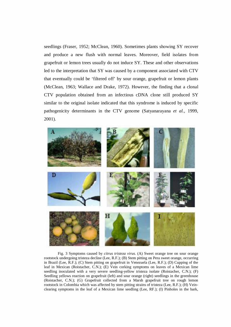
seedlings (Fraser, 1952; McClean, 1960). Sometimes plants showing SY recover
and produce a new flush with normal leaves. Moreover, field isolates from
grapefruit or lemon trees usually do not induce SY. These and other observations
led to the interpretation that SY was caused by a component associated with CTV
that eventually could be ‘filtered off’ by sour orange, grapefruit or lemon plants
(McClean, 1963; Wallace and Drake, 1972). However, the finding that a clonal
CTV population obtained from an infectious cDNA clone still produced SY
similar to the original isolate indicated that this syndrome is induced by specific
pathogenicity determinants in the CTV genome (Satyanarayana et al., 1999,
2001).
Fig. 3 Symptoms caused by citrus tristeza virus. (A) Sweet orange tree on sour orange
rootstock undergoing tristeza decline (Lee, R.F.); (B) Stem pitting on Pera sweet orange, occurring in Brazil (Lee, R.F.); (C) Stem pitting on grapefruit in Venezuela (Lee, R.F.); (D) Cupping of the leaf in Mexican (Roistacher, C.N.); (E) Vein corking symptoms on leaves of a Mexican lime seedling inoculated with a very severe seedling-yellow tristeza isolate (Roistacher, C.N.); (F) Seedling yellows reaction on grapefruit (left) and sour orange (right) seedlings in the greenhouse (Roistacher, C.N.); (G) Grapefruit collected from a Marsh grapefruit tree on rough lemon rootstock in Colombia which was affected by stem pitting strains of tristeza (Lee, R.F.); (H) Vein-clearing symptoms in the leaf of a Mexican lime seedling (Lee, RF.); (I) Pinholes in the bark,

caused by bristles in the wood, cause honeycombing on the back side of the bark patch over the sour orange rootstock (Lee, R.F.). Photographs presented in this figure were downloaded from www.ecoport.org. The author of the photograph is given in the parenthesis.
CTV diversity in the Mediterranean Region
The finding of Toxoptera citricida, the most efficient CTV vector, in
Portugal and Spain (Ilharco et al., 2005), let all mediterranean countries think
seriously about CTV threat, especially that the area is not particularly preserved
regarding destructive CTV strains as it was previously thought. Massive efforts
has been done to know more about the local strains.
The Mediterranean countries where CTV has caused extensive tree losses
include Israel and Spain. In the remaining countries CTV is confined to few
isolated foci (Djelouah and D’Onghia, 2001).
CTV-VT was originally isolated in 1970 from a declining sweet orange
(Citrus sinensis) cv. Valencia tree grafted on the sour orange rootstock in the
Hibatt Zion area, Israel (Bar-Joseph and Loebenstein, 1973).
Variation in natural CTV populations in eastern Spain was studied by
comparing the SSCP pattern of two gRNA regions, p20 and segment A located
within ORF1a, in randomly selected trees at various locations (D’Urso et al.,
2003).
CTV was detected on the Cyprus island in the 1980´s (Kyriakou et al.,
1992), a study conducted in 2007 by Papayiannis et al., on the Cypriot isolates
demonstrated high nucleotide diversity and highlighted the presence of isolates
inducing stem pitting on branches of grapefruit and sweet orange. The Cypriot
isolates clustered with a large universal isolates, including the severe isolates T36
and T3 from Florida, B246 from South Africa, B-CTV from India and the mild
isolate 28C from Portugal.
These strains were also found in Croatia (Cerni et al., 2005), 443-4
(AY791844) and 446-6 (AY791842) are the accession numbers of some
sequenced CPG of Croatian isolates.

CTV has been detected in Corsica in 1981, the isolate found B192 in the
Kumquat K123 (Bové et al., 2002) is from a major interest as it gives mild or no
symptoms on Mexican lime, no vein clearing or stem pitting even if it reacted
positively with MCA13, subsequent infections were found in 1994 and 1997 in
commercial orchards.
In Italy, CTV was reported at the beginning of 2001 in nurseries on
numerous trees imported illegally from abroad and also sporadically on isolated
trees in the field. During spring 2002 and 2003, surveys carried out in citrus
groves of the Ionian coast of Apulia (south-east Italy), established on sour orange,
disclosed the presence of large-sized foci of tristeza. The infected trees were
located in two commercial orchards of the Taranto province. In the first focus
(Castellaneta) infected Navelina orange trees, approximately 20 years old, were
symptomless or stunted and pitted, with heterogenous fruits in size and ripening.
In the second focus (Massafra), typical decline with necrosis at the bud union was
observed on Navelina orange and Clementine Mandarin trees, approximately 15
years old (Birisik, 2003; Birisik et al., 2004).
Surveys undertaken in some libyan orchards and nurseries revealed the
presence of CTV infection and local isolates clustered in the Mediterranean group
(Abukraa, 2008).CTV was officially reported in Syria in 2006 (Abou Kubaa et al.,
2009).
In Morocco, cv. ‘Meyer’ was introduced and grafted onto sour orange
during the 1930s (Cassin, 1963), this material was suspected to carry CTV, and
this was confirmed some years later by biological tests on Mexican lime (Chapot
and Delucchi, 1964). Other cases of CTV have been reported on ‘Meyer’ and
other citrus trees in Morocco and were eradicated on several occasions up to the
1990s (Nadori and Zebzami, 1992).
Lbida et al. studied the biological, serological and genomic diversity of
three CTV isolates from various geographical regions isolate P1 isolated from
lemon cv. ‘Meyer’ in a field near Marrakech in 1983, and isolates P2 and R1
detected in imported Spanish Clementine germplasm by the Moroccan NPPO in
1998 and 2000. P1 induced severe vein clearing on Mexican lime and grapefruit,

mild stem pitting on Mexican lime and moderate stem pitting on grapefruit and
reacted positively with the monoclonal antibody MCA-13. P2 and R1 only
induced mild vein clearing on Mexican lime. The coat protein amino-acid
sequence of P1 clones clusters close to severe strains CB3–104 and FL7,
respectively from Brazil and Florida (Group 5), whereas the sequences from P2
and R1 cluster close to typical strains 25–120 from Portugal and T30 from Florida
(Group M).
Diagnosis, characterization and differentiation of CTV isolates
Because CTV isolates induce such different disease phenotypes and
severities, efforts have been made to develop molecular techniques that rapidly
identify CTV isolates as well as molecular markers related to CTV-induced
symptoms.
Biological indexing was performed for years for the diagnosis of CTV
infection. Mexican lime (Citrus aurantifolia) seedlings is still a very powerful tool
that upon CTV infection show typical symptoms of vein clearing in young leaves,
leaf cupping, short internodes and stem pitting in greenhouse under relatively cool
conditions (Roistacher, 1991).
Apart from Mexican lime, Duncan grapefruit, Eureka lemon, Madam
vinous, and Sour orange can be used for detection and biological characterization
of CTV isolates (Chang-yong, 1996).
Comparison of CTV isolates to provide a pathogenicity profile has been
and still is done by indexing on this standard panel of citrus indicator species
(Garnsey et al., 1991; 2005) allowing the classification of CTV isolates into
several biogroups (table 1), but this biological characterization is a slow and
expensive procedure that cannot be used for routine identification.
Table.1. CTV biogroups (Garnsey et al., 2005)
Biogroups LR Dec SY GSP OSP
0 - - - - -

1 + - - - -
2 + + - - -
3 + + + - -
4 + + + + -
5 + + + + +
6 + + + - +
7 + + - + +
8 + - - + +
9 + + - + -
10 + - - + -
LR: foliar and stem pitting symptoms in Mexican lime; DEC: chlorosis and stunting on sweet orange/sour orange combination; SY: indicates a seedling yellows reaction in sour orange seedlings; GSP: indicates stem pitting in Duncan grapefruit seedlings; OSP: stem pitting in Madam Vinous sweet orange seedlings.
After CTV purification different antisera and monoclonal antibodies to the
coat protein having good specificity were obtained allowing CTV detection by
SDS-immunodiffusion tests (Garnsey et al., 1979), then an ELISA test was set up
(Bar-Joseph et al., 1979) improving the efficiency of mass CTV detection, its
availability was a critical tool to expand research in areas such as CTV
epidemiology, virus movement or isolate characterization, and to improve
efficiency of eradication programmes (Bar-Joseph et al., 1989; Gottwald et al.,
1996a,b, 1998, 2002; Kyriakou et al., 1996).
Finally, after the complete nucleotide sequence of the CTV gRNA was
available, a variety of diagnostic procedures based on specific detection of viral
RNA were developed, including molecular hybridization with cDNA or cRNA
probes (Barbarossa and Savino, 2006; Narváez et al., 2000; Rosner and Bar-
Joseph, 1984) and several RT-PCR amplification-based methods (Nolasco et al.,
1993; Olmos et al., 1999). Real-time RT-PCR protocols have greatly improved
sensitivity of detection and allowed quantification of genomic RNA copies in
infected citrus tissues or in viruliferous aphids (Bertolini et al., 2007; Ruiz-Ruiz et
al., 2007; Saponari et al., 2007).

Single-Strand Conformation Polymorphism (SSCP)
Firstly described by Orita and co-workers (1989 a, b), SSCP is a powerful
structural analysis in which DNA fragments of the same length can be separated
based on their sequence.
The theory behind SSCP is that any given single-stranded DNA (ssDNA)
fragment may form a sequence-specific tertiary structure. Double stranded DNA
(dsRNA) may be separated by denaturation to form two complementary ssDNA.
If ssDNA is allowed to re-nature without re-annealing to the complementary
strand, intra-strand base pairing within the single strands occurs, allowing tertiary
structures to form. The conformation of these structures is dependent on the
primary sequence of the DNA.
It has been demonstrated that small changes in the sequence may alter the
conformation of the ssDNA and therefore their electrophoretic profile. DNA
molecules with differences between their primary sequences will show
polymorphic banding patterns when electrophoresed in non denaturing flat
polyacrylamide gels.
When compared to nucleotide sequencing, it is much less expensive.
Because SSCP does not provide information about the exact number of base
changes, or the location of the changes, it cannot replace nucleotide sequencing or
be used for phylogenetic analyses. Its primary advantage is that it can be used to
process large numbers of samples rapidly; thus SSCP has potential for use as a
screening technique. Isolates that are identified as variants by SSCP can then be
selected for further study.
Compared with these other methods, SSCP is simpler to perform, may be
more sensitive, and is amenable to processing large numbers of samples. One of
the advantages of SSCP, when compared to the other molecular techniques is that
no further manipulation of the PCR product is needed; the DNA obtained from the
PCR reaction is subjected to SSCP without purification or enzymatic
manipulation. Because the conformations of the single strands cannot be

predicted, the conditions for SSCP must be determined empirically and may differ
based on the size of the DNA fragment being tested.
SSCP analysis can be limited by the formation of single and double
stranded fragment, and multiple conformations of the same fragment as the same
genetic sequence may fold to form multiple conformations each with a different
migration time.
Several works used SSCP for the CTV polymorphism detection, based on
the coat protein gene p25 (Cerni et al. 2005), ; Corazza-Nunes et al., 2006), the
minor coat protein gene p27 (Iglesias et al., 2008), the p20 gene (D’Urso et al.,
2003), the p23 gene (Iglesias et al., 2008).
Capillary Electrophoresis
Capillary Electrophoresis (CE) is a separation technique carried out in a
buffer-filled capillary tube that extends between two reservoirs containing
platinum electrodes. Separations depend on the rates at which charged analytes
migrate under an electric field. The migration rate of a species is determined by its
charge to size ratio. CE yields rapid, high resolution separations with very small
sample volume. CE has been applied to a variety of applications including
inorganic anions and cations, amino acids, drugs and explosives. Most notably,
CE was used for the human genome to aid in determining the complete sequence
for human DNA.
Capillary Electrophoresis Single-Strand Conformation Polymorphism
(CE-SSCP):
The introduction of automated multiple capillary electrophoresis
instruments allowed the throughput of SSCP to increase substantially (Munnely et
al., 1998). In fact, CE-SSCP is a powerful analysis technique that separates heat

denaturated DNA fragments of the same length according to their sequence. This
technique presents very high sensitivity, detecting differences up to one base pair.
The use of Capillary Electrophoresis (CE) as alternative to gel slab
techniques has become a promising alternative to gel based SSCP methods.
There are many optimization parameters that can be adjusted when
developing CE-SSCP. An increase of capillary length will increase the detection
power, but also will lengthen polymer fill time and analysis run time.
By labelling the PCR fragments with fluorescent dyes (either using dye-
labelled primers or post-PCR end labelling) it is possible to detect ssDNA
structure using Capillary Electrophoresis (CE-SSCP).
CE-SSCP is a qualitative assay and is dependent on a range of
compositions and conditions. First, assay temperature is an important parameter.
It is well known that the conformation of DNA is highly temperature dependent.
The question is then if two different conformations can be distinguished equally
well at any given temperature. The answer to that has obviously been no far a long
time, and most labs have therefore routinely performed SSCP assay at two or
more temperatures in order to obtain high sensitivity. Many studies show that
sensitivity of CE-SSCP is higher at ambient or sub-ambient temperatures; this
may often cause a problem as not all CE-instruments have cooling capacity.
However, this should not be a hindrance for performing SSCP at higher
temperatures.
The length of the PCR fragments is also an important parameter in SSCP
analysis. In gel based systems it was shown that the sensitivity of SSCP was
drastically reduced when fragments longer than 400bp. However, generally
mutation detection up to 400 bp can be performed, which is acceptable as most
exons that are analyzed rarely extend.
Polymer and buffer composition are very important determinants of good
CE-SSCP results. A number of different polymers have been suggested for the
assay. Commercial polymers supplied by the manufacturers are available, but are
not always the best choice.

The use of the POP™ conformation analysis polymer (CAP) on the
Applied Biosystems 3130/3130xl Genetic Analyzers is an efficient, convenient,
and cost-effective solution for performing SSCP (Applied Biosystem application
note, 2006).
The sensitivity of CE-SSCP has been found to be between 96% and 100%
(Andersen et al., 2003). Apart from the high sensitivity, the advantages of the
method are also a high specificity and the method is very easy to perform
reproducibly. The main disadvantages are the need for a PCR fragment labelled
with a fluorescent dye. In addition, the PCR fragments should not exceed 500 bp
.
Main citrus phytosanitary problems in the Mediterranean area
Among graft transmissible diseases that have been reported in the
Mediterranean countries, Citrus tristeza disease, Citrus psorosis disease and viroid
disease are of the most serious diseases and remain the most spread diseases
(Whiteside et al.,1988; Roistacher, 1991).
Psorosis disease is caused by citrus psorosis ophiovirus (Derrick et al.,
1988). In infected trees a scaly bark symptoms on the trunk (Fig. 4), staining of
interior wood of branch and gummy as well as shortened leaf internodes and
mottling patterns on leaves. Poor fruit quality and decreased yield were recorded
(Roistacher, 1991).
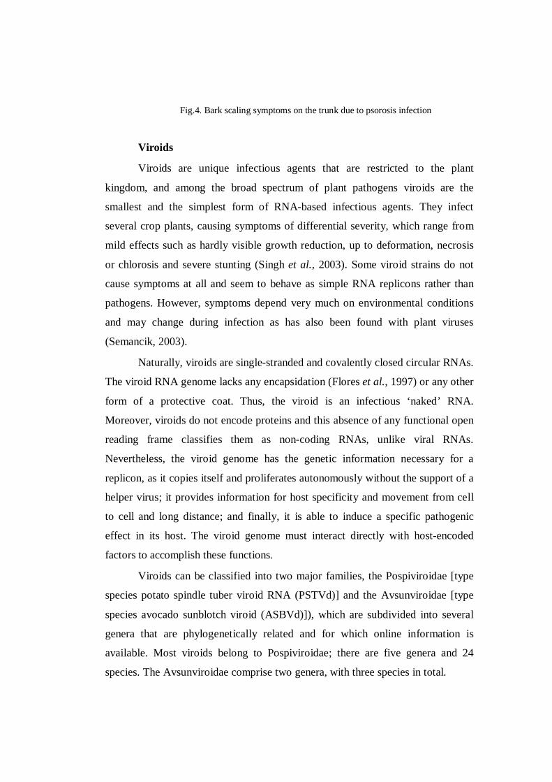
Fig.4. Bark scaling symptoms on the trunk due to psorosis infection
Viroids
Viroids are unique infectious agents that are restricted to the plant
kingdom, and among the broad spectrum of plant pathogens viroids are the
smallest and the simplest form of RNA-based infectious agents. They infect
several crop plants, causing symptoms of differential severity, which range from
mild effects such as hardly visible growth reduction, up to deformation, necrosis
or chlorosis and severe stunting (Singh et al., 2003). Some viroid strains do not
cause symptoms at all and seem to behave as simple RNA replicons rather than
pathogens. However, symptoms depend very much on environmental conditions
and may change during infection as has also been found with plant viruses
(Semancik, 2003).
Naturally, viroids are single-stranded and covalently closed circular RNAs.
The viroid RNA genome lacks any encapsidation (Flores et al., 1997) or any other
form of a protective coat. Thus, the viroid is an infectious ‘naked’ RNA.
Moreover, viroids do not encode proteins and this absence of any functional open
reading frame classifies them as non-coding RNAs, unlike viral RNAs.
Nevertheless, the viroid genome has the genetic information necessary for a
replicon, as it copies itself and proliferates autonomously without the support of a
helper virus; it provides information for host specificity and movement from cell
to cell and long distance; and finally, it is able to induce a specific pathogenic
effect in its host. The viroid genome must interact directly with host-encoded
factors to accomplish these functions.
Viroids can be classified into two major families, the Pospiviroidae [type
species potato spindle tuber viroid RNA (PSTVd)] and the Avsunviroidae [type
species avocado sunblotch viroid (ASBVd)]), which are subdivided into several
genera that are phylogenetically related and for which online information is
available. Most viroids belong to Pospiviroidae; there are five genera and 24
species. The Avsunviroidae comprise two genera, with three species in total.
Viroid localization is either nuclear (family Pospiviroidae) or chloroplastic
(family Avsunviroidae), where they replicate with the aid of host-encoded DNA-
dependent RNA polymerases. Viroids can therefore be considered as parasites of
the transcriptional machinery of the organelles (nucleus or chloroplast), in
contrast to most plant RNA viruses, which replicate in the cytoplasm and can be
therefore regarded as parasites of the translational machinery of the cell.
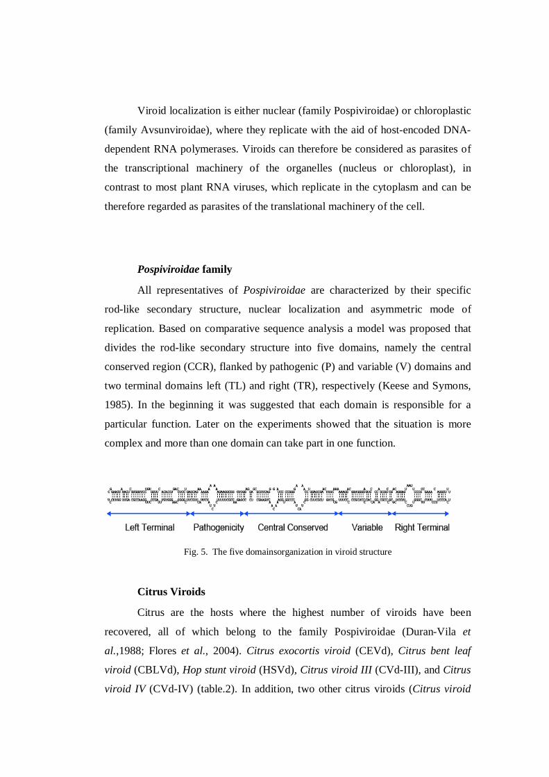
Pospiviroidae family
All representatives of Pospiviroidae are characterized by their specific
rod-like secondary structure, nuclear localization and asymmetric mode of
replication. Based on comparative sequence analysis a model was proposed that
divides the rod-like secondary structure into five domains, namely the central
conserved region (CCR), flanked by pathogenic (P) and variable (V) domains and
two terminal domains left (TL) and right (TR), respectively (Keese and Symons,
1985). In the beginning it was suggested that each domain is responsible for a
particular function. Later on the experiments showed that the situation is more
complex and more than one domain can take part in one function.
Fig. 5. The five domainsorganization in viroid structure
Citrus Viroids
Citrus are the hosts where the highest number of viroids have been
recovered, all of which belong to the family Pospiviroidae (Duran-Vila et
al.,1988; Flores et al., 2004). Citrus exocortis viroid (CEVd), Citrus bent leaf
viroid (CBLVd), Hop stunt viroid (HSVd), Citrus viroid III (CVd-III), and Citrus
viroid IV (CVd-IV) (table.2). In addition, two other citrus viroids (Citrus viroid

original source, CVd-OS reported in Japan with 68% homology with CVd-III,
and Citrus viroid V, CVd-V) have been proposed as tentative species of the genus
Apscaviroid (Ito et al., 2001; Serra et al., 2008a).
They vary in size from 275 to 375 nucleotides and were originally
classified in five different groups on the basis of: (i) electrophoretic mobility on
5% sequential polyacrylamide gels (sPAGE); (ii) sequence similarity determined
by molecular hybridization against specific DNA probes; (iii) host range; and (iv)
reaction on Etrog citron (Citrus medica L.) indicator (Duran-Vila et al., 1988). All
of them are now referred to five species of family Pospiviroidae.
Citrus viroid III (CVd-III), recently renamed Citrus dwarfing viroid
(CDVd) is a member of the genus Apscaviroid that induces, in Citrus medica L.,
stunting and a characteristic ‘leaf dropping pattern’ caused by the moderate
epinasty resulting from petiole and mid-vein necrosis (Rakowski et al., 1994;
Semancik et al., 1997).
Several CDVd variants were initially recognized by their distinct
mobilities in sequential polyacrylamide gel electrophoresis (sPAGE) (Duran-Vila
N et al., 1988), and these were later characterized as three distinct sequence
variants (CVd-IIIa, CVd-IIIb and CVd-IIIc) . These variants differ in size by as
much as 18 nucleotides located in the left and right regions flanking the CCR, but
limited information is available regarding whether or not these changes are
associated with distinct biological properties.
Citrus bent leaf viroid (CBLVd) initially described as Citrus viroid I
(CVd-I) induces moderate epinasty and point necrosis of the mid vein of Etrog
citron (Duran-Vila et al., 1986) and mild dwarfing in trees grafted on trifoliate
orange rootstock (Poncirus trifoliata (L.) Raf.) (Semancik et al., 1997). CBLVd is
an apscaviroid and appears to be a chimera containing parts of central domain (C)
of Apple scar skin viroid (ASSVd) and the pathogenicity (P) and terminal left
(TL) domains of CEVd (Ashulin et al., 1991). Two strains of CBLVd (namely
CVd-Ia and CVd-Ib) with distinct electrophoretic mobilities were identified in
citrus (Duran-Vila et al., 1988). Hataya et al., (1998) have suggested that CVd-Ia

arose by partial sequence duplications involving the right terminal region from
CVd-Ib.
The recently described Citrus viroid V (CVd-V) has a rod-like
conformation and induces, in Etrog citron, mild stunting and very small necrotic
lesions and cracks, sometimes filled with gum (Serra et al., 2008a, b). Moreover,
as Etrog citron plants co-infected with CDVd and CVd-V show synergistic
interactions manifested in enhanced leaf symptoms and very pronounceddwarfing
(Serra et al., 2008a), these host–viroid combinations provide a convenient model
to identify the pathogenicity determinants in members of the genus Apscaviroid.
Table2. Citrus viroids Family, genus species and disease caused.
Family Genus Species Citrus disease
Pospoviroidae
Central conserved
region (CCR)
Pospoviroid CEVd Exocortis
Apscaviroid CVd-I (CBLVd)
Hostuviroid CVD-II (HSVd) Cachexia
Apscaviroid CVd-III
Cocadviroid CVd-IV
At present, Koch’s postulates have only been fulfilled for two well-known
citrus diseases, exocortis caused by CEVd and cachexia of which the causal agent
is HSVd (variants CVd-IIb and CVd-IIc).
Other citrus viroids (CVd-I, CVd-II, CVd-III) have been identified as part
of the graft-transmissible dwarfing complex (GTDC) (Duran-Vila et al., 1988;
Gillings et al., 1991; Bar-Joseph, 1993).
Exocortis is characterized by bark scaling and splitting in sensitive species,
such as trifoliate orange [Poncirus trifoliata (L.) Raf.] and most of its hybrids,
Rangpur lime (Citrus limonia Osb.) and Palestine sweet lime (Citrus limettioides
Tan.). Most citrus species grown commercially, such as sweet orange, grapefruit,
and mandarin are tolerant to CEVd. Therefore, these species can act as
symptomless carriers and, when infected and propagated on sensitive rootstocks,
develop the stunting and bark scaling symptoms characteristic of the exocortis
disease.
Cachexia induces wood pitting and gumming on the trunk, above or below
the bud-union according to the position of the sensitive species [mandarin,
mandarin hybrids, kumquats, alemow (Citrus macrophylla Wester), rough lemon
(Citrus jambhiri Lush) and Rangpur lime], as well as stunting, chlorosis and tree
decline.
The Group III citrus viroids have been poorly described due to a host
range exclusive to citrus and the absence of a causal relationship to any citrus
diseases. CVd-III viroids dwarf citrus plants grafted on specific rootstocks but do
not cause any detrimental effects, apart from some reports of growth
abnormalities observed under some conditions. The CVd-IIIb variant is
distributed in all the citrus areas of the world and has been investigated as a graft
transmissible dwarfing agent in order to obtain high density plantings.
CVd-III was initially described as several independently transmissible
viroids, CVd-IIIa and CVd-IIIb, migrating as distinct bands in sPAGE analysis
(Duran-Vila et al., 1988b) with a relatively narrow size range of 280 to 292
nucleotides. Further analysis by hybridization with specific cDNA probes
demonstrates that they shared sequence homology (Semancik and Duran-Vila,
1991) and sequencing demonstrated that they are highly homologous variants of a
single viroid (Rakowsky et al., 1994). The sequence of the terminal regions of
CVd-III appears to be derived from the conserved regions of the Potato Spindle
Tuber Viroid (PSTVd) and the Apple Scar Skin Viroid (ASSVd) (Stasys et al.,
1995). Specific symptoms are induced by CVd-III in Citrus medica characterized
by leaf-drooping resulting from petiole bending (Duran-Vila et al., 1988b).
Types of interactions between micro organisms
The occurrence of more than one virus species in a single plant is not
uncommon especially in perennial crops, and when two or more viruses co-infect
a plant they may influence each other in several ways. They compete for host
resources but, however, there are few reports indicating that unrelated viruses
suffer a disadvantage during mixed infection (Poolpol and Inouye, 1986).
Often, one virus may assist a second, co-infecting virus, leading to
increased titres and more severe symptoms and this phenomenon is referred to as
viral synergism (Goodman and Ross, 1974; Vance et al., 1995; Pruss et al., 1997).
This occurrence, that virus accumulation differs for one or both viruses when in a
mixed infection relative to singly infected plants, appears to be a common result
of mixed virus infections.
In some cases, the two viruses may benefit from the co-infection (Scheets
1998; Fondong et al., 2000). Synergism has also been known to occur between
viruses and their satellite virus or RNA (Rodriguez-Alvarado et al., 1994; Sanger
et al., 1994; Scholthof, 1999), or even between viruses and viroids (Valkonen,
1992).
The mechanisms behind synergism may vary. In some cases the helper
virus may aid another virus in movement (Barker, 1989), thereby enabling it to
invade tissues it otherwise could not. In other cases, viral replication and
accumulation are enhanced (Savenkov and Valkonen, 2001).
Antagonistic interactions are sometimes observed, in which the unrelated
viruses suppress the infection of each other (Poolpol and Inouye, 1986). However,
this is different from cross-protection that takes place between closely related
viruses or virus strains (Fraser, 1998).
Viroids were also found to induce tolerance. In fact, Solel et al., (1995)
reported that Citrus medica and Rangpur lime seedlings both infected by CEVd
diplayed no severe defoliation when challenged with Phoma tracheiphila the
fungus responsible of mal secco disease comparing with viroid free seedling.
CEVd seems to reduce the systemic advance of the mycelium from the
leaves into the branches (Solel et al., 1995). The mechanism by which this occurs
is not known, but may involve the production of host pathogenesis-related (PR)
proteins, as in tomato in which CEVd induced the expression of two PR proteins
associated with hypersensitive responses and implicated in host defense against

fungal infections (Hadidi, 1988; Belle et al., 1989; Gracia Breijo et al., 1990;
Vera and Conejero, 1989).
Virus-Viroid interaction
Case of Grapevine Coinfection of plants with two or more several
unrelated viruses often results in a more severe disease than the sum effect of
infection with each of the viruses alone and is known as viral synergism.
The Vein-banding disease which can be devastating to grapevines with up
to 80 % fruit loss in sensitive varieties (Martelli and Savino, 1988) has been
hypothesized to be either a late season expression associated with fanleaf
degeneration caused by grapevine fanleaf virus (GFLV) (Martelli and Savino,
1988) or that these leaf symptoms associated with vein banding disease are the
response to grapevine yellow speckle viroids (GYSVd-1, GYSVd-2) intensified
by co-infection with fanleaf virus (Krake and Woodham, 1983).
Later in 1995, Szychowski et al., demonstrate that expression of the vein
banding disease is induced by an unique synergistic reaction between a viroid,
GYSVd-1 and a virus, GFLV.
Characteristic vein-banding symptoms became visible on the mature
leaves of vines which were dual infected with grapevine viroids and GFLV during
the third leafing season (Fig. 6).
Fig. 6. The vein band disease symptoms on grapevine leaves induced by the mixed
infection with GYSVd and GFLV.

Symptoms of yellow speckle are ephemeral, most evident at the end of the
summer and consist of a few to many chlorotic spots on leaves.
This unusual interaction between viroids and viruses extends the biological
potential of viroids.
Case of potato: Synergistic reaction occurs also between Potato Spindle
Tuber Viroid (PSTVd) and Potato virus Y (PVY), both were isolated from plants
of Kennebec cultivar with severe necrotic symptoms in the field (Singh and
Somerville, 1987). The same symptoms were reproduced in the greenhouse only
when potato plants were infected either simultaneously with PSTVd and PVY, or
with PSTVd prior to PVY infection. The study was extended on other cultivars,
and eight cultivars out of thirteen developed necrotic responses similar to cv.
Kennebec.
Infection of potato by PSTVd and PVY produces thus a synergistic
response resulting in severe necrosis not observed with either pathogen alone and
PVY concentration was found to be significantly higher in doubly infected plants
compared with those infected with PVY alone (Singh and Somerville, 1987).
Valkonen (1992), found that whereas PVY couldn’t be detected by ELISA
in plants of Solanum brevidens infected solely with this virus, its accumulation
was increased c. 1000-fold in plants doubly infected with PSTVd or tobacco
mosaic virus (TMV).
Syller and Marczewski (2001), reported that the reaction of potato plants
to the combined secondary infection with PSTVd and Potato Leafroll luteovirus
(PLRV) strikingly differed from those caused by either pathogen alone. The plants
were extremely stunted and dwarfed, their leaves being rugose and uprisen, and
showing greatly pronounced chlorosis. A synergistic reaction was found in all
doubly infected potato plants.
Furthermore, serious disturbances in sprout emergence were observed in
the double infection (Syller and Marczewski, 2001).

In contrast, when tomato plants cv. Rutgers were challenged with PSTVd
and PLRV, no combined effects could be observed (Syller and Marczewski,
2001).
Potato spindle tuber viroid (PSTVd) can decrease tuber yield and quality
in the potato (Solanum tuberosum L.) (Beemster and de Bokx, 1987). It also
reduces pollen viability in infected tomato (Lycopersicon esculentum Mill.)
(Hooker et al., 1978) and potato (Grasmick and Slack, 1986).
Salazar et al. (1995) reported that the aphid M. persicae readily transmitted
PSTVd to potato, Physalis floridana and Datura stramoniumplants from the source
plants doubly infected with the viroid and potato leafroll luteovirus (PLRV) but
not from plants infected with PSTVd alone.
Case of Citrus: Van Vuuren and Graça, 1996, challenged combination
Delta Valencia on Yuma Citrange rootstock with ultra-mild, mild, intermediate
and severe CTV isolates and a citrus viroid (CVd) isolate.
None of the CTV isolates caused stem pitting in sweet orange and they
were all free from the seedling yellows components. The CVd isolate gave a mild
reaction on Etrog citron, and according to sPAGE results, belongs to Group III of
citrus viroids.
Tree volume measurements were made annually, and all fruits were
harvested, sized and weighed. The presence of CVd isolate in combination with
CTV isolates reduced tree size in general by 26% comparing with those
challenged by CTV alone (Van Vuuren and Graça, 1996). Also, the production of
the CVd infected trees was equal to the uninfected trees, due to the 21% higher
production efficiency (kg/m3 canopy) of CVd infected trees.
CHAPTER 2. OBJECTIVES
The first objective of the thesis was to evaluate whether the Mediterranean
CTV collection was infected with other pathogens or not, in order to:
1- Achieve information on geographical distribution of virus diseases in the
Mediterranean basin,

2- Monitor the presence of other risky introduction of viruses/viroids in our country,
3- Ascertain if the reaction of bioindicators or the SSCP profiles are eventually
influenced by any mixed infection,
4- Build up a storage collection of different strains/isolates of citrus virus/viroids of
the Mediterranean.
The characterization of CTV isolates was performed with the biological
indexing on the universal indicators for the determination of the biogroups, and
the sequencing.
The second objective of the thesis was to evaluate the variability in four
regions of the CTV genome using the conventional SSCP and the Capillary
Electrophoresis-SSCP using the Genetic Analyzer. The purpose was:
1- to better evaluate the differences in the collection of CTV isolates,
2- to achieve a broad information on CTV isolates in the Mediterranean,
3- to study if mixed infections play a pressure on CTV genome.
Four regions of the genome; p18, p20, p23 and p25 are studied considering
their potential involvement in symptom production, and their predominant
sequence variants were compared in the RNA population of those isolates with the
conventional SSCP first then with the CE-SSCP.The profiles obtained will be
compared with the results of the biological indexing and the gene sequencing in
view of developing SSCP markers.
Another aim of this study was to investigate on CTV interaction with one
of the citrus viroids in a mixed infection.
CHAPTER 3. MATERIAL AND METHODS
The CTV collection
CTV sources were collected in citrus orchards during surveys conducted
by the Mediterranean Agronomic Institute of Bari (MAIB) in several countries,
mostly from the Mediterranean area. These sources grafted onto different

rootstocks (Sour orange, Citrange troyer or Rough lemon) were assigned with an
“IAMB-Q” number and maintained under insect-proof screenhouse (Fig. 7).
Fig. 7. CTV sources under screenhouse conditions.
Globally, these CTV sources included countries such Albania, Algeria,
China, Croatia, Cyprus, Egypt, Iran, Italy (Apulia and Sicily), Lebanon,
Montenegro, Morocco, Palestine, Syria and Trinidad.
Biological indexing
CTV strain discrimination was carried out by graft-inoculation
(Roistacher, 1991) of five specific woody indicators: Mexican lime, sour orange,
sweet orange, Duncan grapefruit and sweet orange/sour orange combination as
reported by Garnsey et al., (1991) for CTV biogroups establishment.
Three indicators, about 1 year old, were chip budded using 2 blind buds
from each selected CTV source. After sealing the graft with parafilm, the
inoculated plants were labelled with isolate code and all with the positives and
negatives controls were placed in an aphid proof greenhouse at cool temperatures
(22-24°C). Budding and pruning equipment was disinfected in dilute bleach
between treatments.
Observations were carried out after 2 weeks for graft success and after first
flushing for symptom development (Roistacher, 1991).Plants were fertilized as
well as sprayed when needed to control insect or mite infestations.The evaluation

of the presence of CTV-syndromes was performed taking into account that vein-
clearing and leaf-cupping can be induced by all CTV strains in Mexican lime
(universal indicator); CTV-“stem pitting” can provoke pittings in grapefruit and
rarely in sweet orange, whereas “yellows” reaction in sour orange and grapefruit
seedlings are induced by CTV-SY strain.
Symptom severity was quantified by the response in each host and rated as
mild or severe. CTV-SP symptom evaluation consists on the observation of pits in
grapefruit and sweet orange; in the latter only severe CTV isolates can induce
pitting and, eventually gumming. Moreover, the presence of vein corking in the
leaves of Mexican lime and even in the sweet orange is a marker of isolate
severity.
In order to verify the success of the graft inoculation into the specific
indicators, Direct Tissue Blot Immunoassay (DTBIA) (Djelouah and D’Onghia,
2001), was carried out for all the indexed plants. Five tender shoots from each
indicator were cut transversely with a sterile razor and the sections were pressed
carefully on the nitrocellulose membrane. After blocking with 1% bovine serum
albumin (BSA), the membrane was incubated with the Mabs 3DF1+3CA5
mixture conjugated with alkaline phosphatase (PlantPrint). Membranes were
developed by using the BCIP-NBT (Sigma fast tablets), then read under a light
microscope at 10x and 20x magnification. The positive reaction was revealed by
the presence of purple–violet blots in the region of phloem tissue cells.
Serological detection
The plant sources were preliminarily tested for CTV infection by DAS
ELISA as reported by Bar Joseph et al., (1979), using two commercial kits; i.e.
one polyclonal antiserum (PAbs) from Agritest (Italy) and one mixture of
Monoclonal antibodies 3DF1+3CA5 from Ingenasa (Spain) which is known for its
reaction with most CTV isolates and which is specific to the two highly preserved
epitopes of CTV coat protein (Garnsey et al., 1989).
Plates were coated with polyclonal antibodies diluted 1:250 in coating
buffer (Annex 1) and incubated for 2 h at 37°C. After washing three times the

plates with washing buffer (Annex 1), samples were grinded in extraction buffer
(Annex 1) at 1/10 concentration, using bark or petiole tissue. Two wells were
filled with 100µl of extract of each sample, and of positive and of negative
controls. Plates were then incubated overnight at 4°C.
After the same washing as described above, 100µl of alkaline phosphatase
linked antibodies diluted 1:250 in conjugate buffer (Annex 1), were added to each
well and the plates were incubated for 2 h at 37°C.
Plates were dried after the last wash, then 100µl of P-nitrophenyl
phosphate prepared with 1mg/1ml in substrate buffer (Annex 1), were added to
each well. The plates were incubated at room temperature and an absorbance
reading was done up to 2h in a conventional ELISA plate reader at 405nm.
CTV sources were considered positive if the OD405 values were more
than 2,5 times above the values of healthy extracts.
TNA extraction and RT-PCR reaction
Total RNA was extracted from finely trimmed leaf tissue (50 mg) using
TRIzol® reagent (Life Technologies), that contains phenol and guanidinium
isothyocyanate, further purified following the manufacturer’s instructions. Briefly,
after tissue homogenization in 1ml TRIzol®, an incubation of 5’ at RT for the
complete dissociation of nucleoproteic complex, addition of 1/5 V of Chloroform,
hand agitation for 15’’, incubation at RT for 3’, centrifugation at 12000g for 15’ at
4°C, 0,5 ml of isopropanol is added then to the surnatant and incubated for 10’ at
RT, then centrifuged at 12000g for 10’ at 4°C, the pellet is then washed with 1ml
of 75% ethanol, vortexed and centrifuged at 7500g for 5’ at 4°C, the pellet is then
dried and finally resuspended in sterile distilled water.
For cDNA synthesis, 2 µl of the RNA extract was heat denatured at 94°C
for 2 min and chilled on ice. Single step RT-PCR was performed in a 25 µl
reaction volume containing 20mM Tris-HCl (pH 8.4), 50mM KCl, 3mM MgCl2
0.4mM dNTPs, 1µM of each primer (Table 3), 4 units of RNaseOut¨, 20 units of
SuperScript II reverse transcriptase, and 2 units of Taq DNA polymerase (Life
Technologies).

Thermocycling conditions were: 1 cycle of 45 min at 42°C for reverse
transcription, 1 cycle of 2 min at 95°C for inactivation of reverse transcriptase, 35
cycles of 30 s at 94°C, 30 s at 55°C and 1 min at 72°C, and a final extension of 10
min at 72°C.
RT-PCR products were analysed in a 1% agarose gel stained with
ethidium bromide.
Tab.3 . Primers used for CTV amplification by the RT-PCR
CTV Genome part Primer sequence (5’ to 3’) Fluorescence Fragment size
p20 F: ACAATATGCGAGCTTACTTTA
R: AACCTACACGCAAGATGGA
6-FAM
NED 540bp
p23 F:GGTTGTATTAACTAACTTTAATTC
R:AACTTATTCCGTCCACTTCAATCA
6-FAM
VIC 594bp
p25 F: ATGGACGACGAAACAAAGAA
R: ATCAACGTGTGTTGAATTTCC
NED
VIC 415bp
p18 F: TTCTATCGGGATGGTGGAGT
R: GACGAGATTATTACAACGG
6-FAM
NED 425bp
Single strand conformation polymorphism analysis (SSCP)
The test was performed for the 4 CTV genes amplified by RT-PCR (p20,
p23, p25 and p18) aiming a primary molecular differentiation between isolates
prior to cloning and sequencing. It is performed directly on the RT-PCR products;
for each sample, 1.5 to 3µl of RT-PCR product were taken and the denaturing
buffer (95% formamide, 20mM EDTA, pH 8.0, 0.05% bromophenol blue and
0.05% xylene cyanol) was added until a final volume of 10 µl (the volume of each
sample varies with the concentration of the PCR products). Samples were then
heated at 95°C for 5 min, and quickly transferred to an ice box for 5 min.
Denatured products were separated by electrophoresis at 4°C in 8% non-
denaturating polyacrylamide gel using TBE buffer (Annex 2) and a constant
voltage of 200V for 3h. The gels were silver stained by fixation in acetic acid
solution (10% for at least 20 min), wash with distilled water 3 times for 1 min,
Incubation in 1% nitric acid for 3 min, wash again with distilled water 3 times for
1 min, incubation in silver nitrate solution (Annex ) for 30 min and wash with
distilled water for 20 sec.
Developing solution was then added and incubated until the appearance of
the bands; the reaction was stopped by quick gels immersion in 10% acetic acid
for 10 min.
Viroid detection
Biological indexing: the procedure was as for the biological indexing for
CTV, with the difference of using Citrus medica as indicator plant, and the
indexed plant were kept in the greenhouse at 35°C.
RNA extraction and fractionation
Leaf tissue of C. medica (5 g) were homogenized in an extraction buffer
containing 40 ml of water-saturated phenol and 10ml of Tris buffer (125mM Tris–
HCl, pH 8.9, 15mM EDTA, 0.8% (w=v) SDS, 0.8% (v=v) b-mercaptoethanol),
and the total nucleic acids were partitioned in 2M LiCl. The preparations were
further purified by non-ionic cellulose chromatography. Specifically, the
preparations were resuspended in 37ml STE buffer (50mM Tris–HCl, pH 7.2,
100mM NaCl, 1mM EDTA) containing 35% ethanol and mixed with 1.25 g of
non-ionic cellulose CF-11 (Whatman).
The cellulose was washed three times with STE containing 35% ethanol,
and the RNAs bound to the cellulose were eluted with STE and concentrated by
ethanol precipitation.

RNA analysis and purification of Viroid circular forms
Aliquots of the nucleic acid preparations were analyzed by two sequential
rounds of polyacrylamide gel electrophoresis (sPAGE), the first under non-
denaturing conditions and the second under denaturing conditions. For preparative
purposes, the denaturing gel was stained with ethidium bromide and the segment
containing circular viroid forms was frozen, crushed and mixed with phenol:
chlorophorm: isoamyl alcohol and elution buffer (Tris–HCl, pH 8.9, 1mM EDTA,
0.5% (w=v) SDS). The extracted RNAs were recovered by ethanol precipitation.
For analytical purposes, the denaturing gel was stained with silver.
RT-PCR amplification
Retrotranscription and PCR amplification was performed on purified
circular viroids (10 ng) using two specific oligonucleotides of opposite polarity as
reported in table. Primer CEVd-R1 was annealed in buffer (10mM Tris–HCl, pH
8.5, 20mM KCl) at 95°C for 2min. First-strand cDNA was synthesised with 25 U
of avian myeloblastosis virus reverse transcriptase (AMV-RT), using dNTPs
(1mM each) in RT buffer (50mM Tris–HCl, pH 8.5, 8mM MgCl2,
30mMKCl,1mM DTT). The reaction mixture (20 ml final volume) was incubated
at 42°C for 45 min. Second-strand cDNA synthesis and PCR amplification (50 ml
final volume) was performed using 4 ml of the first-strand cDNA reactionmixture,
2.5U Pfu DNA polymerase (Stratagene), respective primers (0.5 mg each), dNTPs
(1mM each), BSA 0.1mg=ml in PCR buffer (20mM Tris–HCl, pH 8.8, 10mM
KCl, 10mM (NH4)2SO4, 2mM MgSO4, 0.1% Triton X-100). PCR parameters
consisted of 30 cycles of 95°C for 30 s, 65°C for 30 s and 72°C for 1min, with a
final extension at 72°C.
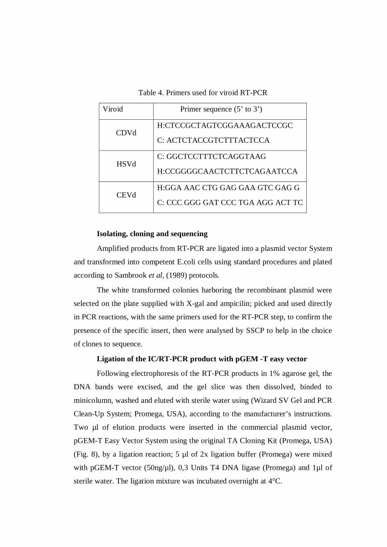
Table 4. Primers used for viroid RT-PCR
Viroid Primer sequence (5’ to 3’)
CDVd H:CTCCGCTAGTCGGAAAGACTCCGC
C: ACTCTACCGTCTTTACTCCA
HSVd C: GGCTCCTTTCTCAGGTAAG
H:CCGGGGCAACTCTTCTCAGAATCCA
CEVd H:GGA AAC CTG GAG GAA GTC GAG G
C: CCC GGG GAT CCC TGA AGG ACT TC
Isolating, cloning and sequencing
Amplified products from RT-PCR are ligated into a plasmid vector System
and transformed into competent E.coli cells using standard procedures and plated
according to Sambrook et al, (1989) protocols.
The white transformed colonies harboring the recombinant plasmid were
selected on the plate supplied with X-gal and ampicilin; picked and used directly
in PCR reactions, with the same primers used for the RT-PCR step, to confirm the
presence of the specific insert, then were analysed by SSCP to help in the choice
of clones to sequence.
Ligation of the IC/RT-PCR product with pGEM -T easy vector
Following electrophoresis of the RT-PCR products in 1% agarose gel, the
DNA bands were excised, and the gel slice was then dissolved, binded to
minicolumn, washed and eluted with sterile water using (Wizard SV Gel and PCR
Clean-Up System; Promega, USA), according to the manufacturer’s instructions.
Two µl of elution products were inserted in the commercial plasmid vector,
pGEM-T Easy Vector System using the original TA Cloning Kit (Promega, USA)
(Fig. 8), by a ligation reaction; 5 µl of 2x ligation buffer (Promega) were mixed
with pGEM-T vector (50ng/µl), 0,3 Units T4 DNA ligase (Promega) and 1µl of
sterile water. The ligation mixture was incubated overnight at 4°C.

Fig. 8 Site map of the pGEM-T cloning vector.
Preparation of competent cells
All the steps in this procedure were done aseptically. Escherichia coli
INVaF cells (Invitrogen, USA), were streaked across the surface of Luria Bertani
(LB) solid plate (Annex ) and incubated at 37°C for 16h.
A single colony was then incubated into 2 ml of LB liquid (Annex )
medium and incubated overnight at 37°C with shaking at 250 rpm. Additional
growth of bacteria cells was performed by inoculating 50 µl of bacterial culture in
10 ml of LB liquid and incubating them at 37°C for 3h with shaking at 250rpm.
The culture was chilled in ice bath for 10 min to stop growth and then, was
harvested by centrifugation at 6000 rpm for 10 min at 4°C. supernatant, the
bacterial pellet was gently resuspended in 0.6 ml cold 0.1 M CaCl2 then kept in
ice for at least 2h before transformation. Aliquots were prepared and stocked at -
80°C.
Transformation of competent cells
One hundred µl of competent cells suspension were added to the 10 µl
ligation mixture in a sterile microfuge tube and were kept in ice for 30 min.
The tubes were incubated in a water bath at 42°C for 2 min, and then
cooled immediately in an ice bath for another 2 min. The cells were mixed with
600 µl with LB liquid then incubated for 30 min at 37°C followed by another 30
min at 37°C with agitation at 150rpm.
Screening of the colonies
After centrifugation for 2 min and elimination of the major part of the
remaining liquid, the bacterial suspension was spread onto plated agarized LB
medium supplied with X-gal (5-bromo-4-chloro-3-indolyl-ß-D-galactoside)
(Sigma) and ampicilin (50mg/ L). Inverted plates were incubated overnight at
37°C.
Using sterile toothpicks, white colonies harboring the recombinant plasmid
were selected on the plate then re-inoculated and numbered in another plate.
Identification of the bacterial colonies carrying recombinant DNA
Plasmids were extracted from bacteria cells using the “boiling method”
(Sambrook et al., 1989). The transformed colonies were “picked” and inoculated
in 2ml of LB liquid containing ampicillin and incubated overnight at 37°C and
250 rpm centrifugation.
Bacterial pellets were poured to an microfuge tubes then collected by
centrifugation at 13000rpm for 1 min, resuspended in 350 µl of STET (Annex)
and 15 µl lysozyme (20 mg/ml), vortexed and then incubated in boiling water for
40 sec and immediately chilled in ice for 2 min. After a centrifugation at 14,000
rpm for 20 min, and elimination of the pellet, soluble
plasmid DNA were extracted with addition of 175 µl of phenol and 175 µl
of isomilic alcool (24:1) followed by vortexing and then centrifugation at 13,000
rpm for 10 min, 300 µl of surfactant were transferred then in fresh tubes
containing 200 µl of Ammonium acetate (NH4OAC) and 1ml of cold absolute
ethanol and centrifuged at 13,000 rpm for 10 min.
The pellet was washed with 500 µl of 70% cold ethanol, centrifuged at
13,000 rpm for 10 min and, after surfactant elimination and drying into Vacuum
pump for 1h, was resuspended in 50µl TE buffer (Annex) containing 1 µl of
RNase A (10 µg/µl), vortexed then conserved in ice.
Enzymatic digestion of plasmid
To verify if extracted plasmids contained the appropriate DNA insert, a
digestion with the restriction endonuclease EcoRI (Amersham) was carried out;
10-15 ng of plasmid DNA were incubated with 1 µl 10X buffer H (Roche), 2 U
EcoRI, (10 µl, final volume of digestion) at 37°C for 1h30.
Size of restricted DNAs was determined by electrophoresis in 1% TAE 1x
agarose gel. The gel was then stained by ethidium bromide (10 mg/ml). DNA
bands were observed under an UV transilluminator.
The tubes that contains plasmids carrying the CTV coat protein insert,
confirmed by enzymatic digestion, were transferred from ice to 37°C for 30min,
then 30 µl of PEG-NaCl (Annex) was added, put on ice for 1h, centrifuged at
14,000 rpm for 5 min, washed by 100 µl of 70% cold ethanol after liquid phase
elimination, centrifuged 14,000 rpm for 5 min, dried for 1h into vacuum pump
after liquid phase elimination and finally the pellet was dissolved in 30 µl of
sterile water.
PCR Amplification of recombinant DNA and CP gene Sequence
analysis
White colonies are picked and added to PCR mixture, for recombinant
DNA amplification using the same primers as in the RT-PCR before SSCP
analysis, in order to check on the genetic properties of selected CTV isolates and
to study the conformation complexity of the SSCP patterns obtained from each
isolate.
Clones that present different SSCP pattern originated from each isolate
were chosen and cDNA sequences of the selected recombinant plasmid were
obtained by automatic sequencing (Primm Company, Italy).
The sequences analysis and alignment were done using the software
packages BioEdit (Hall, 1999). The homology with other known CTV sequences
was determined with the BLAST program (Altschul et al., 1990).

The sequences were compared to previously published sequences
overlapping the CP gene retrieved from the GenBank entries: T30(AF26065), M2
(AY190048), SY568 (AF01623), ArgC269 (AY750771), Cal81 (AY995567), Isr
VT (U56902), Jordan (AY550252), Arg C257 (AY750757), Por 28C
(AF184118), Qaha (AY340974), T36 (AY170468), 19-121(AF184114).
Calculation of pair wise nucleotide distances between sequences and
clustering were done using the Molecular Evolutionary Genetic Analysis software
MEGA version 2.1 (Kumar et al., 2001) after excising the 20 terminal bases on
both sides of the amplified products corresponding to the primers used.
CE-SSCP
DNA amplification: the p20, p23, p25 and p18 gene were amplified using
internal fluorescent primers (Table 3).
Polymer synthesis: The POP conformation analysis polymer (CAP) was
prepared following the formulation for 5g of polymer (5% CAP, 10% glycerol);
2.78 g of 9% Conformational Analysis Polymer, 0.5g of 10X Genetic Analyzer
Buffer (running buffer), 0.5g 100% Glycerol, H2O up to 5.0g. .
Sample Preparation: 1µl of PCR products diluted serially were added to
the SSCP analysis master mixture (10µl of Hi-Di™ deionized formamide, 0.25µl
of GeneScan-500 ROX size standard [Applied Biosystems]) in each well of the
sample plate. The samples were subsequently denatured by heating at 95°C for 5
minutes at 95°C and were immediately cooled on ice for 5 minutes to avoid re-
annealing of the complementary strands prior to loading onto the Genetic
Analyzer. Run the sample plates using the newly created run modules, and then
analyze the results.
SSCP electrophoresis. SSCP electrophoresis was performed with the ABI
PRISM 3130 Genetic Analyzer (Applied Biosystems) (fig.9). The anode-cathode
buffer consisted of a TBE solution. Capillary of 35cm length were used, and the
electrophoresis conditions were set at a 10-s injection time, a 7-kV injection

voltage, a 13-kV electrophoresis voltage, a 120-s syringe pump time, a constant
temperature of 30°C, and a 24-min collection time. The retention times of the
phosphoramidite (HEX)- or 6-carboxyfluorescein (FAM)-labeled fragments were
determined by the electrophoretic mobility value relative to the electrophoretic
mobility for the ROX-labeled internal standard, shown as red peaks, obtained with
the ABI Prism 310 GeneScan Analysis Software (Applied Biosystems).
Fig.9. performed with the ABI PRISM 3130 Genetic Analyzer (Applied Biosystems)
Virus viroid interaction
Immunocapture RT-PCR (IC/RT-PCR)
Indexed plants that were found to be infected simultaneously by CTV and
citrus viroids were subjected to an ELISA test, as reported in the serological test
above, for capturing CTV particles in an ELISA plate. Then a PCR mixture was
prepared, as for RT-PCR detection for viroids. Each extracted sample was loaded
three times in the ELISA plate for the 3 different mixture with the appropriates
primers for each viroid. These was done for the three studied viroids CEVd, CVd-
II and CDVd. After washing, the wells were loaded with the different mixture and
then put on an incubator at 50°C for 1h. These mixture were then taken from the
wells and put on PCR tubes to complete the PCR in the thermocycler, with the
conditions reported above for viroid detection.

PCR products were then checked on 1% agarose gel, for the presence of
viroid amplicons indicationg that the viroids particles have been encapsidated by
the CTV coat protein, and some excised bands were cloned then sequenced.
CHAPTER 4. RESULTS AND DISCUSSION
Biological indexing:
Biological properties of the Mediterranean CTV isolates were investigated
by indexing onto a standard of indicator plants. Biological characterization
remains the most reliable procedure to accurately document the severity of CTV
isolates (Garnsey et al., 1995; 2005).
Twenty seven CTV isolates were compared for symptom expression on
five citrus indicator plants; Mexican lime, Duncan grapefruit, Madam Vinous
sweet orange grafted on sour orange, sour orange seedlings, and Madam Vinous
sweet orange seedlings. Reactions were relatively consistent among the three
replicate plants per host inoculated with each isolate, and some differences in
symptom severity between isolates were readily apparent.
Mexican lime reactions.
Inoculated plants were observed for vein clearing (Fig.10), leaf cupping
(Fig.11), stunting and stem pitting. Symptoms in Mexican lime showed a broad
range of severity (Table 5).
Some isolates expressed a pronounced vein clearing that could be seen
without having to put the leaf in backlighting (Fig.10 ), these include mostly
isolates from the Middle East region including Lebanon, Syria and Palestine.
These last were also strongly stunted (Fig.12 ) with pronounced stem pits in
Mexican lime (Fig.13 ), whereas the milder CTV isolates had minor stem pitting.
Sweet/sour reactions.
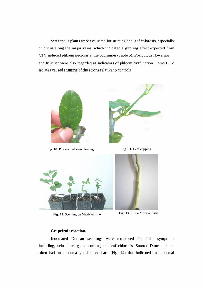
Sweet/sour plants were evaluated for stunting and leaf chlorosis, especially
chlorosis along the major veins, which indicated a girdling effect expected from
CTV induced phloem necrosis at the bud union (Table 5). Precocious flowering
and fruit set were also regarded as indicators of phloem dysfunction. Some CTV
isolates caused stunting of the scions relative to controls
Grapefruit reaction.
Inoculated Duncan seedlings were monitored for foliar symptoms
including, vein clearing and corking and leaf chlorosis. Stunted Duncan plants
often had an abnormally thickened bark (Fig. 14) that indicated an abnormal
Fig. 10: Pronounced vein clearing Fig. 11: Leaf cupping
Fig. 12: Stunting on Mexican lime Fig. 13: SP on Mexican lime

differentiation of phloem and xylem tissues. Stem pitting was determined by
peeling the main stem above the point of inoculation
Sweet orange reactions.
CTV isolates inducing stem pitting on sweet orange are considered as the
most dangerous as they occur in decline-tolerant scion–rootstock combinations.
Few mediterranean isolates induced discreat stem pitting (Fig. 15) on Madam
vinous (Table 5), these include mostly isolates from the middle East area.
Fig.14: SP on Duncan Grapefruit Fig. 15 : Discrete SP on Madam vinous SwO
Fig. 16: CPsV symptoms mature leaf of Madam vinous SwO induced by the isolate Q110
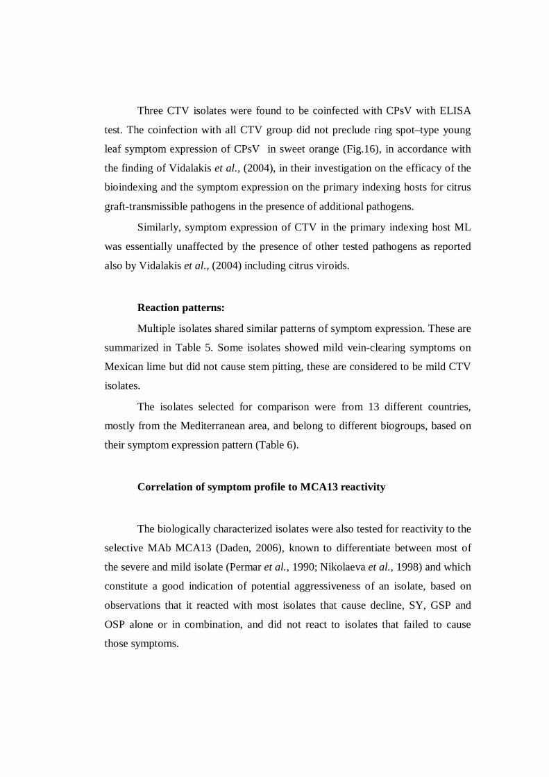
Three CTV isolates were found to be coinfected with CPsV with ELISA
test. The coinfection with all CTV group did not preclude ring spot–type young
leaf symptom expression of CPsV in sweet orange (Fig.16), in accordance with
the finding of Vidalakis et al., (2004), in their investigation on the efficacy of the
bioindexing and the symptom expression on the primary indexing hosts for citrus
graft-transmissible pathogens in the presence of additional pathogens.
Similarly, symptom expression of CTV in the primary indexing host ML
was essentially unaffected by the presence of other tested pathogens as reported
also by Vidalakis et al., (2004) including citrus viroids.
Reaction patterns:
Multiple isolates shared similar patterns of symptom expression. These are
summarized in Table 5. Some isolates showed mild vein-clearing symptoms on
Mexican lime but did not cause stem pitting, these are considered to be mild CTV
isolates.
The isolates selected for comparison were from 13 different countries,
mostly from the Mediterranean area, and belong to different biogroups, based on
their symptom expression pattern (Table 6).
Correlation of symptom profile to MCA13 reactivity
The biologically characterized isolates were also tested for reactivity to the
selective MAb MCA13 (Daden, 2006), known to differentiate between most of
the severe and mild isolate (Permar et al., 1990; Nikolaeva et al., 1998) and which
constitute a good indication of potential aggressiveness of an isolate, based on
observations that it reacted with most isolates that cause decline, SY, GSP and
OSP alone or in combination, and did not react to isolates that failed to cause
those symptoms.

A comparison of symptom patterns and MCA13 reactivity is summarized
in Table 6.
Interestingly, 17 CTV isolates out of 27 studied isolates that expressed
severe symptoms reacted to MCA13. This included all isolates that induce GSP
and SY and belongs to the following countries China, Croatia, Cyprus, Egypt,
Lebanon, Montenegro, Morocco, Palestine and Syria gave a clear cut positive
reaction with this MAb, even if the number of the tested CTV sources was not
always large to be significant.
While MCA 13 MAb failed to react only with the Algerian and all the
Apulian (Italian) CTV isolates which reacted only in Mexican lime (pattern 1).
Correlation of biological reaction to isolate genotype:
The CTV isolates belonging to the Middle East region including Lebanon,
Syria and Palestine presented a severe SP on Pompelmo grapefruit and a light SP
on Madam vinous Sweet orange on Volkameriana rootstock, the SP on Sweet
orange being an indicator of the extreme severity of a CTV isolate.
As shown in Table 6 symptom profile 1 were predominantly associated
with the T30 genotype while symptom profiles 4, 5, 6 and 8 were most frequently
associated with the T3 and VT genotype.
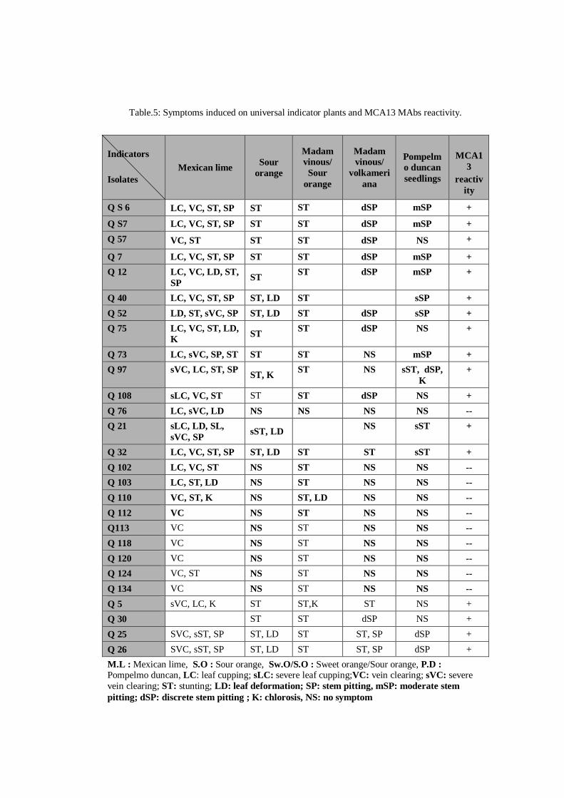
Table.5: Symptoms induced on universal indicator plants and MCA13 MAbs reactivity.
Indicators Isolates
Mexican lime Sour orange
Madam vinous/
Sour orange
Madam vinous/
volkameriana
Pompelmo duncan seedlings
MCA1
3 reactiv
ity
Q S 6 LC, VC, ST, SP ST ST dSP mSP +
Q S7 LC, VC, ST, SP ST ST dSP mSP + Q 57 VC, ST ST ST dSP NS +
Q 7 LC, VC, ST, SP ST ST dSP mSP + Q 12 LC, VC, LD, ST,
SP ST ST dSP mSP +
Q 40 LC, VC, ST, SP ST, LD ST sSP + Q 52 LD, ST, sVC, SP ST, LD ST dSP sSP + Q 75 LC, VC, ST, LD,
K ST ST dSP NS +
Q 73 LC, sVC, SP, ST ST ST NS mSP + Q 97 sVC, LC, ST, SP ST, K ST NS sST, dSP,
K +
Q 108 sLC, VC, ST ST ST dSP NS + Q 76 LC, sVC, LD NS NS NS NS -- Q 21 sLC, LD, SL,
sVC, SP sST, LD NS sST +
Q 32 LC, VC, ST, SP ST, LD ST ST sST + Q 102 LC, VC, ST NS ST NS NS -- Q 103 LC, ST, LD NS ST NS NS -- Q 110 VC, ST, K NS ST, LD NS NS -- Q 112 VC NS ST NS NS -- Q113 VC NS ST NS NS -- Q 118 VC NS ST NS NS -- Q 120 VC NS ST NS NS -- Q 124 VC, ST NS ST NS NS -- Q 134 VC NS ST NS NS -- Q 5 sVC, LC, K ST ST,K ST NS + Q 30 ST ST dSP NS + Q 25 SVC, sST, SP ST, LD ST ST, SP dSP + Q 26 SVC, sST, SP ST, LD ST ST, SP dSP + M.L : Mexican lime, S.O : Sour orange, Sw.O/S.O : Sweet orange/Sour orange, P.D : Pompelmo duncan, LC: leaf cupping; sLC: severe leaf cupping;VC: vein clearing; sVC: severe vein clearing; ST: stunting; LD: leaf deformation; SP: stem pitting, mSP: moderate stem pitting; dSP: discrete stem pitting ; K: chlorosis, NS: no symptom

Table6. CTV isolates biogroups and MCA13 reactivity.
The presence (+) or absence (–) of vein clearing (VC), stem pitting (SP) and decline (D) symptoms were observed in seedlings of Mexican lime (M. lime), Duncan grapefruit (D. Gft.), sweet orange (SwO), or in plants of sweet orange grafted on sour orange (SwO/SO). Seedling yellows (SY) was observed on sour orange.
Symptoms induced on various indicator plants
Isolates
Origin
M. lime
(VC, SP)
SwO/SO
(D)
SO
(SY)
D.Gft
(SP)
SwO
(SP)
Biogroup
MCA13 Reactivity
Q 5 Albania + + + -- -- 3 + Q76 Algeria + -- -- -- -- 1 -- Q25 China + + + + + 5 + Q26 China + + + + + 5 + Q73 Cyprus + + + + -- 4 + Q57 Egypt + + + + -- 4 + Q7 Lebanon + + + + + 5 + Q12 Lebanon + + + + + 5 + Q97 Iran + + + + -- 4 + Q21 Italy + + + + -- 4 + Q32 Italy + + + + -- 4 + Q 102 Italy + +/-- -- -- -- 1; 2 -- Q 103 Italy + +/-- -- -- -- 1; 2 -- Q 110 Italy + +/-- -- -- -- 1; 2 -- Q 112 Italy + +/-- -- -- -- 1; 2 -- Q113 Italy + +/-- -- -- -- 1; 2 -- Q 118 Italy + +/-- -- -- -- 1; 2 -- Q 120 Italy + +/-- -- -- -- 1; 2 -- Q 124 Italy + +/-- -- -- -- 1; 2 -- Q134 Italy + +/-- -- -- -- 1; 2 -- Q108 Montenegro + + + + -- 4 + Q75 Morocco + + + + -- 4 + Q40 Palestine + + + + + 5 + Q52 Palestine + + + + + 5 + QS6 Syria + + + + + 5 + QS7 Syria + + + + + 5 + Q30 Trinidad + + + + -- 4 +
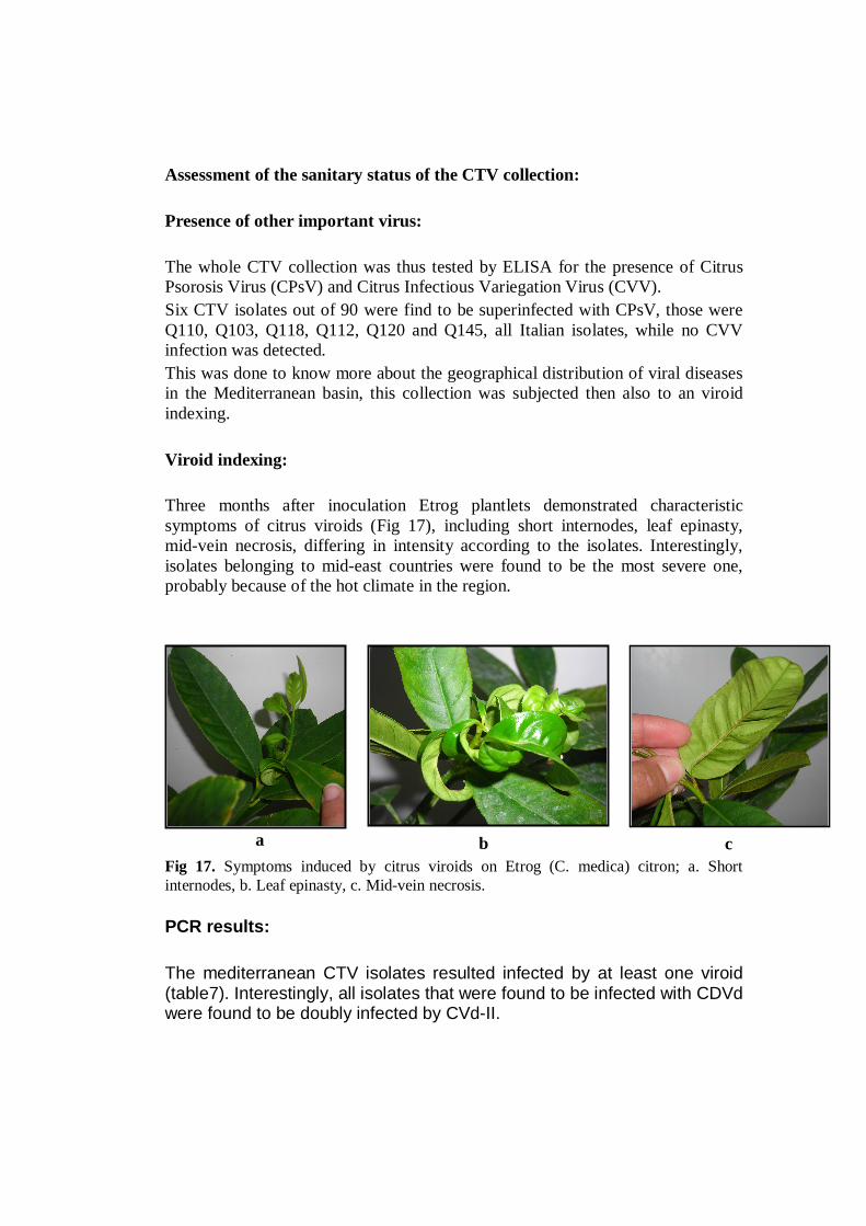
Assessment of the sanitary status of the CTV collection: Presence of other important virus: The whole CTV collection was thus tested by ELISA for the presence of Citrus Psorosis Virus (CPsV) and Citrus Infectious Variegation Virus (CVV). Six CTV isolates out of 90 were find to be superinfected with CPsV, those were Q110, Q103, Q118, Q112, Q120 and Q145, all Italian isolates, while no CVV infection was detected. This was done to know more about the geographical distribution of viral diseases in the Mediterranean basin, this collection was subjected then also to an viroid indexing. Viroid indexing: Three months after inoculation Etrog plantlets demonstrated characteristic symptoms of citrus viroids (Fig 17), including short internodes, leaf epinasty, mid-vein necrosis, differing in intensity according to the isolates. Interestingly, isolates belonging to mid-east countries were found to be the most severe one, probably because of the hot climate in the region.
Fig 17. Symptoms induced by citrus viroids on Etrog (C. medica) citron; a. Short internodes, b. Leaf epinasty, c. Mid-vein necrosis. PCR results: The mediterranean CTV isolates resulted infected by at least one viroid (table7). Interestingly, all isolates that were found to be infected with CDVd were found to be doubly infected by CVd-II.
a b c

Table.7: PCR results for viroid infection
CTV sources CDVd CVd-II CEVd
Q109 -- -- -- Q102 + + + Q113 + + -- Q7 + + + Q97 -- -- + Q30 + + + Q12 + + + Q75 -- -- + Q73 + + + Q40 + + + Qs7 + + + Q76 + + + Q52 + + + Q3 -- -- + Q110 + + + Qs6 + + -- Q57 + + + Q134 + + -- Q118 + + + Q120 + + -- Q103 + + + Q112 + + + Q25 + + + Q124 + + + Q21 + + + Q32 + + +
All the positive CTV isolates displayed at least symptoms on the primary CTV indicator M. lime even if they were found to be infected also by at least one of the most economically important citrus viroids, in accordance with the study conducted by Vidalakis et al., (2004), reporting that the coinfection of M. lime with CTV and the mixture of the viroids Citrus viroid-Ia (CVd-Ia), CVd-IIb, CVd-IIIb, and Citrus exocortis viroid (CEVd) did not preclude symptom expression of either the T30 or the VT CTV groups in the primary indicator M. lime.

Cloning and sequencing: Differences in the nucleotide sequence are reported in Fig 18.
Fig 18. Differences in CDVd DNA sequences
Conventional SSCP:
Four CTV genomic parts (p18, p20, p23 and CP) were amplified by the respective primers then analyzed by the conventional SSCP for a preliminary discrimination between CTV isolates. Tissue samples yielding different SSCP patterns in the same gel under any of the electrophoretic conditions were considered to contain a different population of sequence variants, hereafter called a haplotype. P20 profiles: Previously, it was observed that the SSCP profile of cDNA of the p20 gene faithfully reflected the composition of the RNA population, and that sequence variants accounting for at least 10% of the RNA population could be detected in the SSCP profile (Rubio et al., 2000). Therefore, SSCP analysis of cDNA is an easy procedure to compare the population composition of two CTV isolates for the gene analysed and to see if the major component of these populations is the same in both isolates. Great variability was observed in p20 haplotypes within the 44 CTV isolates belonging to different Mediterranean citrus growing countries that yielded 21 different haplotypes. The number of different haplotypes found in each region varied from one to five for p20. Overall, the highest diversity of haplotypes was found in

Italy, where for 7 isolates analyzed, 5 haplotypes were displayed (Fig c), accounting for 71% of variability within Italian isolate for the p20 gene, and the lowest diversity in Albania, where for 5 isolates analyzed, only 1 haplotype was displayed.
a. a. b.
c. d.
e. f.
Q7 Q126 Q6 Q127 Q4 Q12 Q75 Q74 Q76 Q79 1 2

Fig 20. SSCP profiles (a, b, c, d, e) and haplotype (f) detected for the p20 gene of the CTV genome using single-strand conformation polymorphism (SSCP) analysis of the cDNA obtained by RT-PCR using specific primers. The isolates belonging to the Middle East region (Lebanon, Palestine, Syria) and Egypt seems to display two haplotypes “1 and 2” for p20 gene (Fig 20. f). Interestingly, when some of these isolates their CP gene were cloned and sequenced (Daden, 2006) they differed by less than 1% at nucleotide level from the well characterized VT (Fig. 19) from Israel (Mawassi et al., 1993). These haplotypes “1 and 2” (Fig 20. f) represented 43% and 14% of all samples analysed, respectively, making from the haplotype “1” the predominant detected one for the analyzed isolates. Some of the SSCP profiles obtained consisted of just two DNA bands, as in the case of haplotype1 and 2, indicating that the viral population in those samples contained a predominant sequence variant (Kong et al., 2000), whereas others had more than two bands suggesting the presence of two or more sequence variants at various ratios, as it is for the two Moroccan isolates Q74 and Q75. Rubio et al. (2000) observed that sequence variants accounting for at least 10% of the total population in the infected tissue could be detected in the SSCP pattern. An association was observed between specific haplotypes and symptom expression, P18 profiles:
In contrast with p20 gene, SSCP of p18 gene displayed more conserved profiles (Fig. 21). In fact , some isolates that resulted to have different SSCP profiles using P20 gene displayed the same SSCP profile for the p18 gene, this is the case of the Lebanese isolates Q7, Q126, Q6, Q127, Q4, Q12, that displayed one haplotype, while with p20 gene they displayed two haplotypes “1 and 2”. Similar observations have been made by other workers when analyzing the p18 gene or the 5′ untranslated region of various CTV isolates (Ayllón et al., 2001). In such studies, it was found that mildly pathogenic CTV isolates contained one sequence type whereas severely pathogenic isolates contained two or three sequence types.
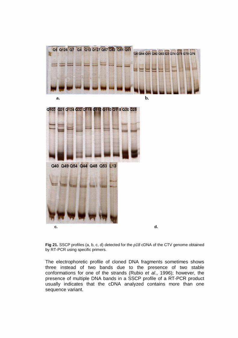
a. b.
c. d. Fig 21. SSCP profiles (a, b, c, d) detected for the p18 cDNA of the CTV genome obtained by RT-PCR using specific primers. The electrophoretic profile of cloned DNA fragments sometimes shows three instead of two bands due to the presence of two stable conformations for one of the strands (Rubio et al., 1996); however, the presence of multiple DNA bands in a SSCP profile of a RT-PCR product usually indicates that the cDNA analyzed contains more than one sequence variant.

P23 profiles: The CTV p23 is an RNA-binding protein involved in regulating the asymmetrical accumulation of viral RNA strands, when over-expressed in transgenic Mexican limes it displayed typical CTV symptoms, showing thus its involvement and importance in symptoms expression. In a study on CTV p23 polymorphism, Sambade et al., (2003) found that the nucleotide sequence in a region of this gene enables discrimination between mild and severe CTV isolates, that there was low nucleotide diversity (from 0 to 0.0035±0.0014) in groups of clones with the same SSCP pattern, and that SSCP analysis discriminated clones with a 0.0030 nucleotide distance confirming the reliability of this approach to differentiate sequence variants of gene p23. Considering this potential involvement of gene p23 in symptom expression, here we compared the predominant sequence variants of this gene from 27 CTV isolates of different geographic origin and biological characteristics with the conventional SSCP as primary molecular screening of CTV strains.
a. b.
Q7 Q126 Q6 Q127 Q4 Q12 Q75 Q74 Q76 Q79
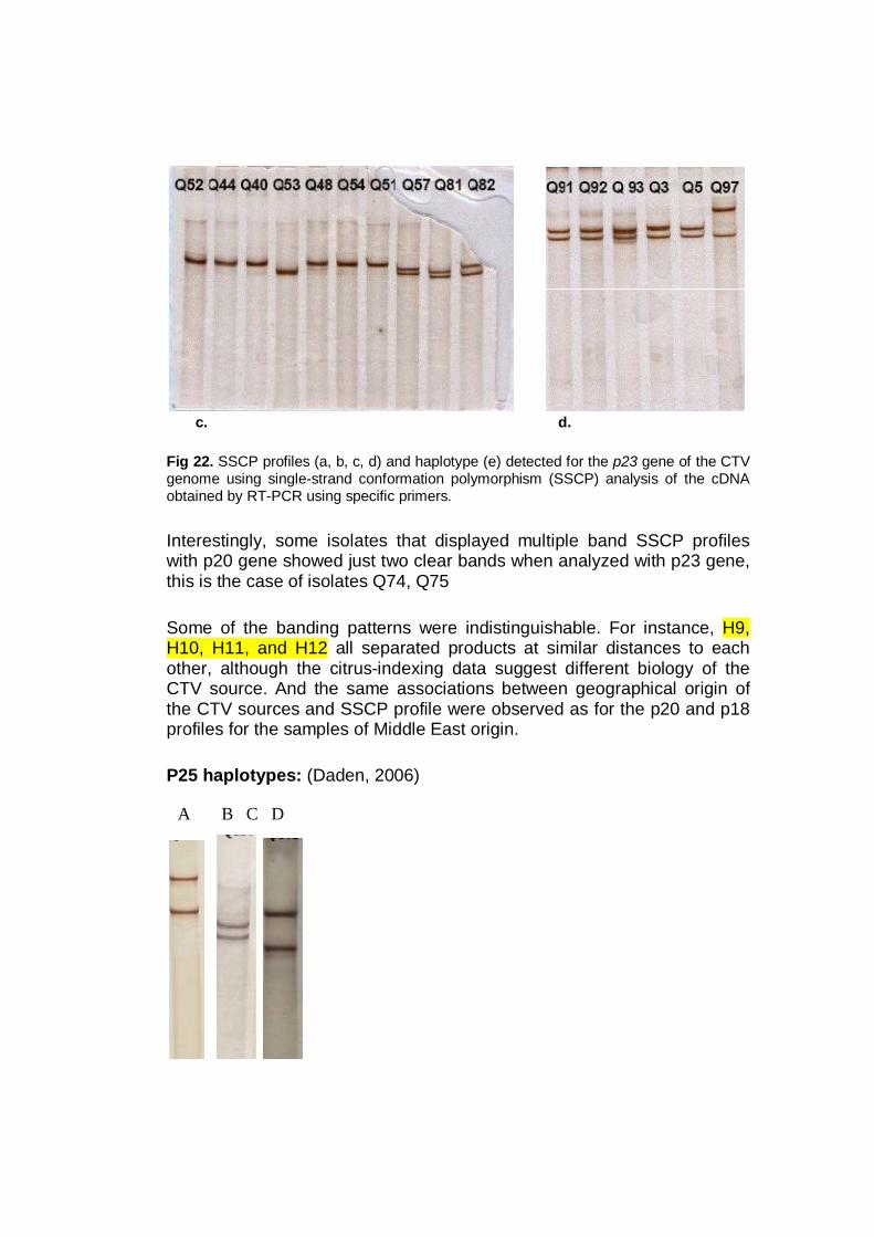
c. d. Fig 22. SSCP profiles (a, b, c, d) and haplotype (e) detected for the p23 gene of the CTV genome using single-strand conformation polymorphism (SSCP) analysis of the cDNA obtained by RT-PCR using specific primers. Interestingly, some isolates that displayed multiple band SSCP profiles with p20 gene showed just two clear bands when analyzed with p23 gene, this is the case of isolates Q74, Q75 Some of the banding patterns were indistinguishable. For instance, H9, H10, H11, and H12 all separated products at similar distances to each other, although the citrus-indexing data suggest different biology of the CTV source. And the same associations between geographical origin of the CTV sources and SSCP profile were observed as for the p20 and p18 profiles for the samples of Middle East origin. P25 haplotypes: (Daden, 2006)
A B C D

Developing SSCP markers by F-SSCP:
Conventional SSCP analysis is mostly used in primary CTV strain differentiation because it combines simplicity, low cost and the possibility of being used with many samples. Also, it has enough sensitivity to detect a single nucleotide difference in relatively large DNA fragments up to 700 nucleotides (Rubio et al., 1996) and differences between SSCP profiles are consistent if experimental conditions are carefully maintained. The main limitation of conventional SSCP analysis is that profile differences cannot be quantified in terms of the genetic distance between the corresponding DNA fragments (Rubio et al., 1996; Garcìa-Arenal et al., 2001), and therefore, it provides only a qualitative picture of the variation. To screen the genetic diversity of CTV isolates and be able to quickly classify them in an appropriate group that reflect their real severity, there is the need of developping new molecular markers. Fluorescent fragments amplified by PCR were added… The tecnique was able to detect a set of already known mutations by using a collection of 13 isolates previously sequenced The repeatability of the results within and between different runs was also evaluated Visual analysis of the electropherograms suggests that the SSCP Data Analysis. The peaks of the SSCP profiles were detected with the GeneMapperTM Software version 3.7 (Applied Biosystems), using Genescan-500HD ROX as a size standard, defining an apparent size for each peak. Thus, the location of peaks is arbitrarily expressed in base pairs (bp). For statistical comparison of SSCP profiles, the list of peaks detected by GeneMapperTM and their surfaces were analyzed. Because the data files did not follow a log-normal distribution, we used Spearman_s correlation coefficient (S) to compare the SSCP profiles. P20 F-SSCP profiles: So far 27 isolates have been analysed and 7 different haplotypes detected these (Fig.) are reported in details in the Annex Q52:

Q112
Q73
Q96
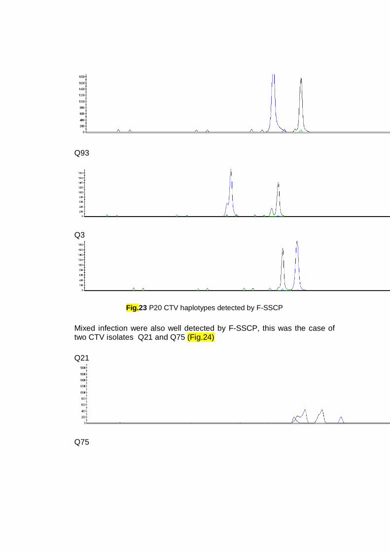
Q93
Q3
Fig.23 P20 CTV haplotypes detected by F-SSCP
Mixed infection were also well detected by F-SSCP, this was the case of two CTV isolates Q21 and Q75 (Fig.24) Q21
Q75

Fig.24 P20 CTV mixed infection haplotypes detected by F-SSCP
Interestigly, the same was noticed for the VT group that conserved the same haplotype (profile 1, Fig.) for p20 gene even in F-SSCP profiles, this was the case of the CTV isolates belonging to the Middle East region (Lebanon, Palestine, Syria) and Egypt. The haplotype 1 for p20 gene (Fig.) account for Most of the mild Italian isolates displayed the hapltotype 2, accounting for These last reacted negatively to MCA13 MAbs P18 profiles: Q93:
Q73 :
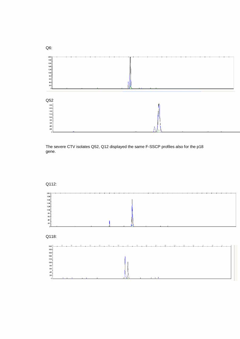
Q6:
Q52
The severe CTV isolates Q52, Q12 displayed the same F-SSCP profiles also for the p18 gene. Q112:
Q118:

P23 F-SSCP profiles Q110
Q12
Q52
Q5
The mild Italian CTV isolates Q110 (Fig. ), Q124, Q112, Q102, Q118 and the Algerian mild isolates Q76 and Q79 (Annex), displayed the same F-SSCP profile.

Similarly, the severe Lebanese CTV isolates Q12 (Fig.), Q6 and the Syrian QS6 isolate(Annex) displayed the same F-SSCP profile. The Palestinian isolates Q52, Q44, Q40 conserved the same F-SSCP profile. Q75
Q74
The Moroccan CTV isolates Q75 and Q74 displayed a complex profile indicating the presence of a mixed infection. P25 F-SSCP profiles: QS7
The Syrian CTV isolates QS7 (Fig.) and QS6, the Palestinian isolate Q52, Q40, Q44, and the Lebanese Q12, Q4 displayed the same F-SSCP profile for the p25 gene. Cloning and sequencing: Fifteen CTV isolates were chosen for multiple sequence alignment and genetic diversity analysis using the CP, p20 and p23 genes. Their PCR

products were cloned in the linearized and thymidylated pGem-T vector (Promega) using standard protocols. Of these, analysis with both conventional and CE-SSCP were performed of several clones, in order to confirm the occurrence of infections with a mixture of genomic variants. An haplotype is defined as an isolate showing a specific electrophoretic mobility of one or of the two strands of the PCR fragment. Both SSCP methods detected the different haplotype existing, with the The nucleotide sequence of selected haplotypes were determined in both directions using vector specific primers. by direct sequencing of the PCR products amplified with the same primers of the respective genomic region (table). of co-infections, suggested by the high intra-isolate variability, showed a variety of different patterns, suggesting the occurrence (Fig.). random selected minority SSCP haplotypes were sequenced. Phylogenetic trees were obtained using these sequences. Minority haplotypes of some isolates did not cluster with the corresponding predominant haplotype, indicating the presence of diverged sequence variants within individual isolates. The predominant sequence variant present in each CTV isolate was selected by SSCP analysis [33, 40]. For this purpose, p23 clones were PCR-amplified with the same primers and conditions described above, and the resulting DNA was SSCP analyzed in parallel with the RT-PCR product from which the clones were obtained. Those clones whose DNA strands comigrated with the most intense DNA bands of the starting RT-PCR product were sequenced. In total, six isolates contained mixed infections of 2 to 3 of the known CTV genotypes. Sequence analysis of different variants showed a CP gene with 669 nucleotides having greater than 90% nucleotide identity to most CTV CP gene sequences available in GenBank. A genomic variant (GenBank Accession No. AY764154) was closely related (98.5% nucleotide identity) to the T30 mild strain from Florida (GenBank Accession No. AF260651). However, other sequences obtained

showed only 93% nucleotide identity with this variant and were closely related to other CP gene sequences obtained from Croatian isolates. two isolates contained the T36 genotypes, six isolates contained the VT genotypes, five isolates contained T30 genotypes, ….T3 genotypes and…. B165 genotypes isolates.
3 5 6 8 9
Q 32
White colonies p20 Q 32

2 3 9
Q 110White colonies p20 Q 110 and Q 32
11 12 13 14 15 17 18 19 20 Initial PCR product Q32
Q32
1 3 5 6 7 8 9 10
White colonies p20 Q 21

White colonies p20 Q 21
12 13 14 15 16 17 18 Initial PCR product
Q 21
Fig. SSCP analysis of CTV isolate Q44 CP clones
To assess reliability of SSCP analysis to differentiate between sequence variant and select the predominant sequence variant of each CTV isolate, SSCP patterns of individual cDNA clones were compared with the pattern of the RT-PCR product from which they had been obtained. The SSCP pattern of some isolates had only two DNA bands, while others yielded a more complex pattern [34]. In the first case, most of the clones showed the same SSCP pattern as the corresponding RT-PCR product

and one of them was selected for sequence analysis. In the second case, a higher variability was observed between clones, but only those whose SSCP pattern coincided with the most intense bands in the pattern of the starting RT-PCR product were sequenced [33]. Alignments showed that the complete sequence of gene p23 was preserved in all cases, without insertions, gaps or unexpected stop codons, indicating that each selected clone corresponded to a translatable sequence. The Palestinian isolate Q44 showed the lowest number of SSCP profiles, whereas Q21 and Q32 exhibited the highest number of them. The percentage of predominant pattern was calculated for all isolate. The Palestinian Q44 isolate exhibited the highest value (93%), being the most homogeneous isolate for CP gene, followed by the Italian isolate Q110 with 86%. On the contrary, Q32 showed the lowest value (31%) for the major clone, being the most heterogeneous for the CP gene region among the analyzed isolates, (Daden, 2006) and also for p20 gene, followed by the Italian isolate Q21 with 40%.

Q134.1
Q134.4
Q76.5
Q76.10
T30
T385
Q32.7
Q76.4
Q113.13
Q113.4
Q113.7
Q113.3
Q32.5
Q124
T36
Qaha
SY568
T318
NuaGa
Q21.1
Q32.3
Q44.6
Q44.5
VT
Q52.6
Q52.4
Q52.1
Q52.11
Q75.17
Q75.1
Q21.13
Q21.14
BaraoB-4
Q25.4
Q32.35
Q25.1
Q75.12
Q32.9
Q75.8
Q32.1
C257-2
0.01

Citrus Viroids-CTV interaction: CTV virions are covered with two capsid proteins of 25 and 27 kDa, coating
about 95 and 5% of the virion length, respectively (Febres et al., 1996;
Satyanarayana et al., 2004), and are single-stranded, positive sense genomic RNA
(gRNA) approximately 20 kb in size, readily transmitted by citrus aphids species.
Viroids are, in the contrary, small naked RNA particles (359 nt) that completes
their infection cycle without generating either a capsid or other viroid-specific
proteins.
An essential role of the viral capsid protein in virus transmission by aphids has
been proved (Syller, 2000). The specific recognition of viral genomic RNA by
capsid protein CP is a critical event in the infection cycle of all single-stranded
RNA viruses. This recognition event plays a crucial role in specific encapsidation
of the viral genome as well as other steps in the virus life cycle.
Heterologous encapsidation between viruses in mixed infections may allow
transmission by aphids of normally non-aphid-transmissible viruses or change
virus-vector interactions, it is known also by transcapsidation, meaning the
coating of the nucleic acid of one virus or virus strain with the protein of another
in a double infections.
This phenomenon has been demonstrated even for the Nigerian cowpea virus
(CV) and the tobacco mosaic virus (TMV), which are distantly related
serologically, in conditions in which both viruses multiply readily (Kassanis and
Bastow, I971).
Samples were analyzed using ELISA with a CTV polyclonal antibodies to
ascertain for CTV infection. After plate development, wells of positive samples
were used for IC-RT-PCR analysis targeting the three citrus most economically

important citrus viroids; CVd-II, CEVd and CDVd, that have been already found
to infect the CTV isolate collection (table), using their respective pairs of primers
(table).
When analyzed using agarose gel electrophoresis, PCR products from the positive
samples consisted of a single amplicon of viroids expected size compared with a
positive control. That the CDVd RNA was contained in the coat protein of CTV
was indicated by the amplification of the RNA viroid with its specific primers
after the immunocapture of CTV particles in the wells of an ELISA plate with an
polyclonal antisera. This phenomenon occurs even at 25°C, the temperature at
which the M. lime plantlets inoculated with CTV isolates were kept in the
greenhouse.
When sap was extracted from C. medica used for biological indexing of citrus
viroids, plantlets that were kept in the greenhouse at 35°C, the phenomenon was
not verified, this can be explained by the low CTV title, found in ELISA test
value, and it’s reduced ability to multiply due to the high temperature. While
when other C. medica plantlets inoculated with CTV isolate were kept at 25°C,
the CDVd was amplified from ELISA plate, indicating that both CTV and CDVd
could multiply in C. medica and in ambient temperature.
In contrast, no amplified products were obtained when the sample was taken from
citrus plants with only one infection CTV or citrus viroids.
The expected bands were excised from agarose gel, eluted and then cloned in
(pGEM-T Easy Vector; Promega, Madison, WI) and sequenced.
This fact can allow to viroid particles to be transmitted by citrus aphids as it was
demonstrated for the Potato Tuber Spindle Viroids (PTSVd) that was found to be
vectored by aphids only when potato was doubly infected by Potato Leafroll
Luteovirus (PLRV) whose coat protein encapsidates the viroids.
The transmission of citrus viroids encapsidated in the viral capsid protein by
aphids is even more likely to happen in the open field if the most efficient CTV
aphid vector Toxoptera citricidus continue its spreading and get more diffused in
the area.

This phenomenon was more likely to happen when the conditions were favorable
for CTV multiplication, and was confirmed for the available CTV strains in the
collection and for the three most economically important citrus viroids.
This will change the idea about the non transmissibility of the citrus viroids by
insects (Laird et al., 1963), as the encapsidation of citrus viroids can lead to a
dependent transmission.
This finding strongly suggests that the dependent transmission is a phenomenon
that can occur easily in nature, and that apart from the risks of mutations that have
to be taken in consideration when inoculating a mild CTV isolate for cross
protection, as a strategy to contain damages in an area where the CTV is
established, there is also the risk that the encapsidation by CTV coat protein, of a
normally non-aphid-transmissible virus particle, occurs in a mixed infection
allowing thus its transmission by citrus aphids in the commercial orchards.
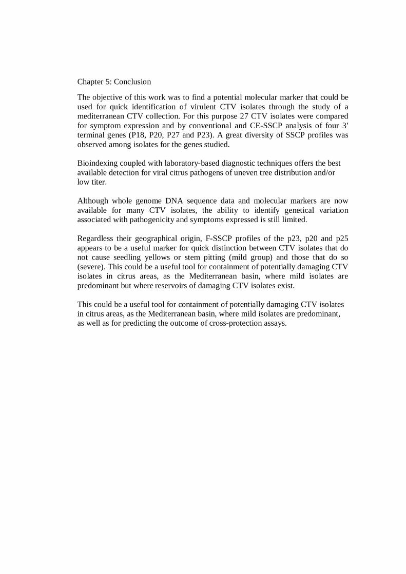
Chapter 5: Conclusion
The objective of this work was to find a potential molecular marker that could be used for quick identification of virulent CTV isolates through the study of a mediterranean CTV collection. For this purpose 27 CTV isolates were compared for symptom expression and by conventional and CE-SSCP analysis of four 3′ terminal genes (P18, P20, P27 and P23). A great diversity of SSCP profiles was observed among isolates for the genes studied. Bioindexing coupled with laboratory-based diagnostic techniques offers the best available detection for viral citrus pathogens of uneven tree distribution and/or low titer. Although whole genome DNA sequence data and molecular markers are now available for many CTV isolates, the ability to identify genetical variation associated with pathogenicity and symptoms expressed is still limited. Regardless their geographical origin, F-SSCP profiles of the p23, p20 and p25 appears to be a useful marker for quick distinction between CTV isolates that do not cause seedling yellows or stem pitting (mild group) and those that do so (severe). This could be a useful tool for containment of potentially damaging CTV isolates in citrus areas, as the Mediterranean basin, where mild isolates are predominant but where reservoirs of damaging CTV isolates exist. This could be a useful tool for containment of potentially damaging CTV isolates in citrus areas, as the Mediterranean basin, where mild isolates are predominant, as well as for predicting the outcome of cross-protection assays.

Litterature citted:
Alkowni R., Rowhani A., Daubert S., Golino D., 2004. Partial characterization of a new ampelovirus associated with grapevine leafroll disease. Journal of Plant Pathology 86: 123–133.
Anandalakshmi R., Pruss G.J., Ge X., Marathe R., Smith T.H., Vance V.B., 1998. A viral suppressor of gene silencing in plants. The Proceedings of the National Academy of Sciences U. S. A. 95: 13079–13084.
Andersen P.S., Jespersgaard C., Vuust J., Christiansen M., Larsen, L.A., 2003. High-throughput single strand conformation polymorphism mutation detection by automated capillary array electrophoresis: validation of the method. Human Mutation 21: 116-122.
Applied Biosystems, Application Note SSCP 2006. Single-Stranded Conformation Polymorphism (SSCP) on Applied Biosystems Capillary Electrophoresis Systems. www.appliedbiosystems.com
Ashulin L., Lachman O., Hadas R., Bar-Joseph M., 1991. Nucleotide sequence of a new viroid species, citrus bent leaf viroid (CBLVd) isolated from grapefruit in Israel. Nucleic Acids Resource 19: 4767.
Ayllón M.A., López C., Navas-Castillo J., Garnsey S.M., Guerri J., Flores R., Moreno P., 2001. Polymorphism of the 5′ terminal region of Citrus tristeza virus (CTV) RNA: Incidence of three sequence types in isolates of different origin and pathogenicity. Archives of Virology 146: 27-40.
Bar-Joseph, M., 1993. The use of viroids for dwarfing citrus trees. 9th International Congress of Virology 107.
Bar-Joseph M., 1996. A contribution to the natural history of viroids. Proceedings of the 13th International Organization of Citrus Virologists, IOCV, IOCV Riverside, CA, USA 226-229.
Bar-Joseph M., 2003. Natural history of viroids . Horticultural aspects. In: Hadidi A., Flores R., Randles J.W. Semancik J.S. (eds). Viroids. pp. 246-251. CSIRO Publishing, Collingwood, Victoria, Australia..
Barker H., 1989. Specificity of the effect of sap-transmissible viruses in increasing the accumulation of luteoviruses in co-infected plants. Annals of Applied Biology 115: 71-78.
Bayne E.H., Rakitina D.V., Morozov S.Y., Baulcombe D.C., 2005. Cell-to cell movement of Potato potexvirus X is dependent on suppression of RNA silencing. Plant Journal 44: 471–482.
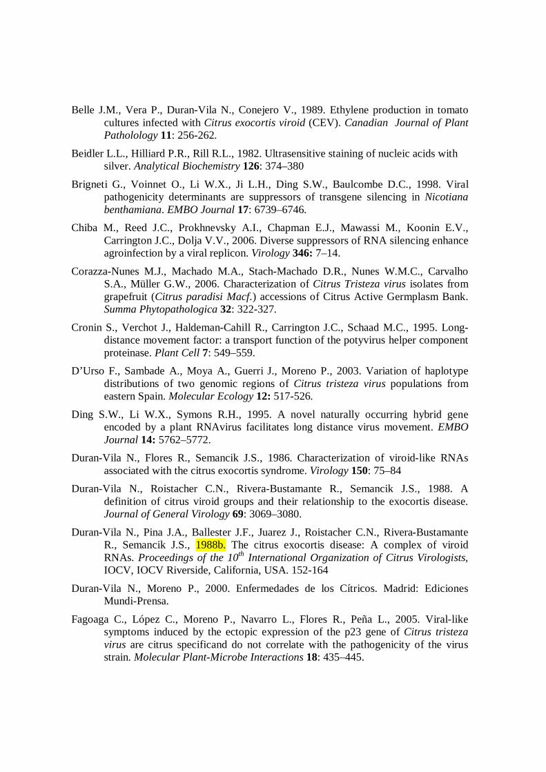
Belle J.M., Vera P., Duran-Vila N., Conejero V., 1989. Ethylene production in tomato cultures infected with Citrus exocortis viroid (CEV). Canadian Journal of Plant Patholology 11: 256-262.
Beidler L.L., Hilliard P.R., Rill R.L., 1982. Ultrasensitive staining of nucleic acids with silver. Analytical Biochemistry 126: 374–380
Brigneti G., Voinnet O., Li W.X., Ji L.H., Ding S.W., Baulcombe D.C., 1998. Viral pathogenicity determinants are suppressors of transgene silencing in Nicotiana benthamiana. EMBO Journal 17: 6739–6746.
Chiba M., Reed J.C., Prokhnevsky A.I., Chapman E.J., Mawassi M., Koonin E.V., Carrington J.C., Dolja V.V., 2006. Diverse suppressors of RNA silencing enhance agroinfection by a viral replicon. Virology 346: 7–14.
Corazza-Nunes M.J., Machado M.A., Stach-Machado D.R., Nunes W.M.C., Carvalho S.A., Müller G.W., 2006. Characterization of Citrus Tristeza virus isolates from grapefruit (Citrus paradisi Macf.) accessions of Citrus Active Germplasm Bank. Summa Phytopathologica 32: 322-327.
Cronin S., Verchot J., Haldeman-Cahill R., Carrington J.C., Schaad M.C., 1995. Long-distance movement factor: a transport function of the potyvirus helper component proteinase. Plant Cell 7: 549–559.
D’Urso F., Sambade A., Moya A., Guerri J., Moreno P., 2003. Variation of haplotype distributions of two genomic regions of Citrus tristeza virus populations from eastern Spain. Molecular Ecology 12: 517-526.
Ding S.W., Li W.X., Symons R.H., 1995. A novel naturally occurring hybrid gene encoded by a plant RNAvirus facilitates long distance virus movement. EMBO Journal 14: 5762–5772.
Duran-Vila N., Flores R., Semancik J.S., 1986. Characterization of viroid-like RNAs associated with the citrus exocortis syndrome. Virology 150: 75–84
Duran-Vila N., Roistacher C.N., Rivera-Bustamante R., Semancik J.S., 1988. A definition of citrus viroid groups and their relationship to the exocortis disease. Journal of General Virology 69: 3069–3080.
Duran-Vila N., Pina J.A., Ballester J.F., Juarez J., Roistacher C.N., Rivera-Bustamante R., Semancik J.S., 1988b. The citrus exocortis disease: A complex of viroid RNAs. Proceedings of the 10th International Organization of Citrus Virologists, IOCV, IOCV Riverside, California, USA. 152-164
Duran-Vila N., Moreno P., 2000. Enfermedades de los Cítricos. Madrid: Ediciones Mundi-Prensa.
Fagoaga C., López C., Moreno P., Navarro L., Flores R., Peña L., 2005. Viral-like symptoms induced by the ectopic expression of the p23 gene of Citrus tristeza virus are citrus specificand do not correlate with the pathogenicity of the virus strain. Molecular Plant-Microbe Interactions 18: 435–445.
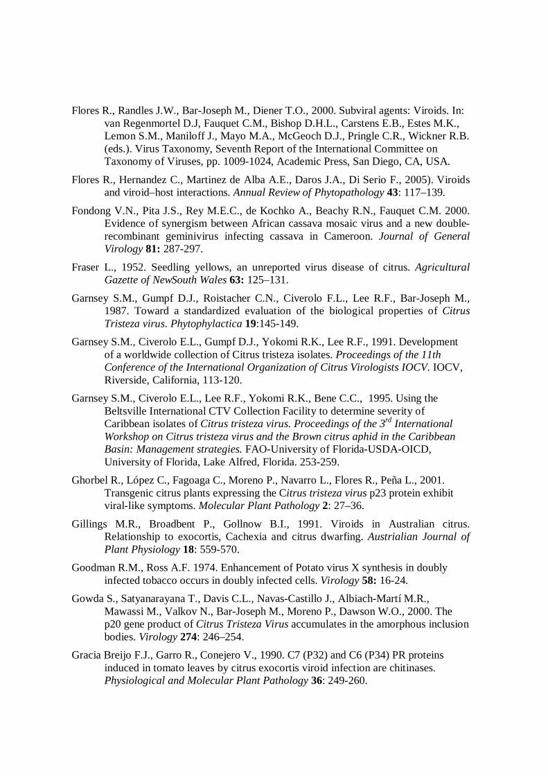
Flores R., Randles J.W., Bar-Joseph M., Diener T.O., 2000. Subviral agents: Viroids. In: van Regenmortel D.J, Fauquet C.M., Bishop D.H.L., Carstens E.B., Estes M.K., Lemon S.M., Maniloff J., Mayo M.A., McGeoch D.J., Pringle C.R., Wickner R.B. (eds.). Virus Taxonomy, Seventh Report of the International Committee on Taxonomy of Viruses, pp. 1009-1024, Academic Press, San Diego, CA, USA.
Flores R., Hernandez C., Martinez de Alba A.E., Daros J.A., Di Serio F., 2005). Viroids and viroid–host interactions. Annual Review of Phytopathology 43: 117–139.
Fondong V.N., Pita J.S., Rey M.E.C., de Kochko A., Beachy R.N., Fauquet C.M. 2000. Evidence of synergism between African cassava mosaic virus and a new double-recombinant geminivirus infecting cassava in Cameroon. Journal of General Virology 81: 287-297.
Fraser L., 1952. Seedling yellows, an unreported virus disease of citrus. Agricultural Gazette of NewSouth Wales 63: 125–131.
Garnsey S.M., Gumpf D.J., Roistacher C.N., Civerolo F.L., Lee R.F., Bar-Joseph M., 1987. Toward a standardized evaluation of the biological properties of Citrus Tristeza virus. Phytophylactica 19:145-149.
Garnsey S.M., Civerolo E.L., Gumpf D.J., Yokomi R.K., Lee R.F., 1991. Development of a worldwide collection of Citrus tristeza isolates. Proceedings of the 11th Conference of the International Organization of Citrus Virologists IOCV. IOCV, Riverside, California, 113-120.
Garnsey S.M., Civerolo E.L., Lee R.F., Yokomi R.K., Bene C.C., 1995. Using the Beltsville International CTV Collection Facility to determine severity of Caribbean isolates of Citrus tristeza virus. Proceedings of the 3rd International Workshop on Citrus tristeza virus and the Brown citrus aphid in the Caribbean Basin: Management strategies. FAO-University of Florida-USDA-OICD, University of Florida, Lake Alfred, Florida. 253-259.
Ghorbel R., López C., Fagoaga C., Moreno P., Navarro L., Flores R., Peña L., 2001. Transgenic citrus plants expressing the Citrus tristeza virus p23 protein exhibit viral-like symptoms. Molecular Plant Pathology 2: 27–36.
Gillings M.R., Broadbent P., Gollnow B.I., 1991. Viroids in Australian citrus. Relationship to exocortis, Cachexia and citrus dwarfing. Austrialian Journal of Plant Physiology 18: 559-570.
Goodman R.M., Ross A.F. 1974. Enhancement of Potato virus X synthesis in doubly infected tobacco occurs in doubly infected cells. Virology 58: 16-24.
Gowda S., Satyanarayana T., Davis C.L., Navas-Castillo J., Albiach-Martí M.R., Mawassi M., Valkov N., Bar-Joseph M., Moreno P., Dawson W.O., 2000. The p20 gene product of Citrus Tristeza Virus accumulates in the amorphous inclusion bodies. Virology 274: 246–254.
Gracia Breijo F.J., Garro R., Conejero V., 1990. C7 (P32) and C6 (P34) PR proteins induced in tomato leaves by citrus exocortis viroid infection are chitinases. Physiological and Molecular Plant Pathology 36: 249-260.

Hadidi A., 1988. Synthesis of disease-associated proteins in viroid-infected tomato leaves and binding of viroid to host proteins. Phytopathology 78: 575-578.
Hadidi A., Flores R., Randles J.W., Semancik J.S., 2003. Viroids. pp 370. CSIRO Publishing, Collingwood, Victoria, Australia.
Hataya T., Nakahara K., Ohara T., Ieki H., Kano T., 1998. Citrus viroid Ia is a derivative of citrus bent leaf viroid (CVd-Ib) by partial sequence duplications in the right terminal region. Archives of Virology 143: 971–980.
Iglesias N.G., Gago-Zachert S.P., Robledo G., Costa N., Plata M.I., Vera O., Grau O. Semorile L.C., 2008. Population structure of Citrus tristeza virus from field Argentinean isolates. Virus Genes 36:199–207.
Ito T., Ieki H., Ozaki K., Ito T., 2001. Characterization of a new citrus viroid species tentatively termed citrus viroid OS. Archives Virology 146: 975-982.
Karasev A.V., 2000. Genetic diversity and evolution of closteroviruses. Annual Review of Phytopathology 38: 293–324.
Kasschau K.D., Carrington J.C., 2001. Long-distance movement and replication maintenance functions correlate with silencing suppression activity of potyviral HC-Pro. Virology 285: 71–81.
Keese P., Symons R. H., 1985. Domains in viroids: evidence of intermolecular RNA rearrangements and their contribution to viroid evolution. Proceedings of National Academy Sciences U S A 82: 4582-4586.
Krake L.R., Woodham R.C., 1983. Grapevine yellow speckle agent implicated in the aetiology of vein banding disease. Vitis 22: 40-50.
Kumar S., Dudley J., Nei M. Tamura K., 2008. MEGA: A biologist-centric software for evolutionary analysis of DNA and protein sequences. Briefings in Bioinformatics 9: 299-306.
Laird E.F., Harjung M.K., Weathers L.G., 1963. Attempts to transmit citrus exocortis virus by insects. Plant Disease Reports 53: 850-851.
López C., Navas-Castillo J., Gowda S., Moreno P., Flores R., 2000. The 23-kDa protein coded by the 3-terminal gene of citrus tristeza virus is an RNA-binding protein. Virology 269: 462–470
Lu R., Folimonov A., Shintaku M., Li W.X., Falk B.W., Dawson W.O., Ding S.W., 2004. Three distinct suppressors of RNA silencing encoded by a 20-kb viral RNA genome. Proceedings of National Academy Sciences U.S A. 101: 15742–15747.
Martelli G. P., Savino V., 1988. Fanleaf degeneration. In: R. C. PEARSON and A. C. GOHEEN (eds) pp. 48-49.Compendium of Grape Diseases, APS Press, St . Paul, Minnesota.
Martelli G.P., Agranovsky A.A., Bar-Joseph M., Boscia D., Candresse T., Coutts R.H.A., Dolja V.V., Falk B.W., Gonsalves D., Jelkman W., Karasev A.V., Minafra A., Namba S., Vetten H.J., Wisler G.C., Yoshikawa N., 2002. The family Closteroviridae revised. Archives of Virology 147: 2039–2044.

McClean A.P.D., 1963. The tristeza complex: Its variability in field-grown citrus in South Africa. South Africa Journal of Agriculture Sciences. 6: 303–332.
McClean A.P.D. (1960) Seedling-yellows in South African citrus trees. South Africa Journal of Agriculture Sciences 3: 259–279.
Moreno P., Ambròs S., Albiach-Martí M.R., Guerri J., Pena M., 2008. Review; Plant diseases that changed the world, Citrus tristeza virus: a pathogen that changed the course of the citrus industry. Molecular Plant Pathology 9: 251–268.
Önelge N., Cinar A., Szychowski J.A., Vidalakis G., Semancik J.S., 2004. Citrus viroid II variants associated with ‘Gummy Bark’ disease. European Journal of Plant Pathology 110: 1047–1052.
Orita M., Suzuki Y., Sekiya T., Hayashi K., 1989a. Rapid and sensitive detection of point mutations and DNA polymorphisms using the polymerase chain reaction. Genomics 5: 874–879.
Orita M., Iwahana H., Kanazawa H., Hayashi K., Sekiya T., 1989b. Detection of polymorphisms of human DNA by gel electrophoresis as single-strand conformation polymorphisms. Proceedings of National Academy of Sciences USA 86: 2766–2770.
Palacio-Bielsa A., Romero-Durbán J., Duran-Vila N., 2004. Characterization of citrus HSVd isolates. Archives of Virology 149: 537–552.
Poolpol P., Inouye T., 1986. Enhancement of cucumber mosaic virus multiplication by zucchini yellow mosaic virus in doubly infected cucumber plants. Annals of the Phytopathological Society of Japan 52: 22-30.
Pruss G., Ge X., Shi M.X., Carrington J.C., Vance V.B., 1997. Plant viral synergism: The potyviral genome encodes a broad range pathogenicity enhancer that transactivates replication of the heterologous viruses. Plant Cell 9: 859-868.
Rakowski A.G., Szychowski J.A., Avena Z.S., Semancik J.S., 1994. Nucleotide sequence and structural features of the Group III citrus viroids. Journal of General Virology 75: 3581–3584.
Reanwarakorn K., Semancik J.S., 1998. Regulation of pathogenicity in hop stunt viroid-related group II citrus viroids. Journal of General Virology 79: 3163–3171.
Reanwarakorn K., Semancik J.S., 1999. Correlation of hop stunt viroid variants to cachexia and xyloporosis disease of citrus. Phytopathology 89: 568–574.
Rodriguez-Alvaradro G., Kurath G., Dodds J.A., 1994. Symptom modification by satellite tobacco mosaic virus in pepper types and cultivars infected with helper tobamaviruses. Phytopathology 84: 617-621.
Roistacher C.N., Bash J.A., Semancik J.S., 1993. Distinct disease symptoms in Poncirus trifoliata induced by three citrus viroids from three specific groups. Proceedings of the 12th Conference of the International Organization of Citrus Virologists IOCV, IOCV Riverside, 173–179.

Rubio L., Ayllón M.A., Guerri J., Pappu H.R., Niblett C.L., Moreno P., 1996. Differentiation of citrus tristeza closterovirus (CTV) isolates by single-strand conformation polymorphism analysis of the coat protein gene. Annals of Applied Biology 129: 479–489.
Sanger M., Passmore B., Falk B.W., Biening G., Ding B., Lucas W., 1994. Symptom severity of beet western yellows virus strain ST9 is conferred by the ST9-associated RNA and is not associated with virus release from the phloem. Virology 200: 48-55.
Satyanayanana T., Gowda S., Boyko V.P., Albiach-Martí M.R., Mawassi M., Navas-Castillo J., Karasev A.V., Dolja V., Hilf M.E., Lewandowsky D.J., Moreno P., Bar-Joseph M., Garnsey S.M., Dawson W.O., 1999. An engineered closterovirus RNA replicon and analysis of heterologous terminal sequences for replication. Proceeding of National Academy Sciences USA 96: 7433–7438.
Satyanayanana T., Bar-Joseph M., Mawassi M., Albiach-Martí M.R., Ayllón M.A., Gowda S., Hilf M.E., Moreno P., Garnsey S.M., Dawson W.O., 2001. Amplification of Citrus tristeza virus from a cDNA clone and infection of citrus trees. Virology 280: 87–96.
Satyanarayana T., Gowda S., Ayllón M.A., Albiach-Martí M.R., Rabindran S., Dawson W.O., 2002. The p23 protein of citrus tristeza virus controls asymmetrical RNA accumulation. Journal of Virology 76: 473–483.
Savenkov E.I., Valkonen J.P.T., 2001. Potyviral helper-component proteinase expressed in transgenic plants enhances titers of potato leaf roll virus but does not alleviate its phloem limitation. Virology 283: 285-293.
Scheets K., 1998. Maize chlorotic mottle machlomovirus and wheat streak mosaic rymovirus concentrations increase in the synergistic disease corn lethal necrosis. Virology 242: 28-38.
Schneider H., 1959. The anatomy of tristeza-virus-infected citrus. In: Citrus Virus Diseases (Wallace, J.M., ed.), Berkeley, CA: University of California Division of Agricultural Sciences 73–84.
Scholthof K.B., 1999. A synergism induced by satellite panicum mosaic virus. Molecular Plant-Microbe Interactions 12: 163-166.
Schwach F., Vaistij F.E., Jones L., Baulcombe D.C., 2005. An RNA-dependent RNA-polymerase prevents meristem invasion by potato virus X and is required for the activity but not the production of a systemic silencing signal. Plant Physiology 138: 1842–1852.
Semancik J.S., 2003. Viroids: pathogenesis. In Viroids. Hadidi, A., Flores, R., Randles, J.W., and Semancik, J.S. (eds). 61–66. Collinwood: CSIRO Publishing.
Semancik J.S., Roistacher C.N., Rivera-Bustamante R., Duran-Vila N., 1988. Citrus cachexia viroid, a new viroid of citrus: Relationship to viroids of the exocortis disease complex. Journal of General Virology 69: 3059–3068.
Semancik J.S., Duran-Vila N., 1991. The grouping of citrus viroids : additional physical and biological determinants and relationships with diseases of citrus. Proceedings of the 11th International Organization of Citrus Virologists, IOCV. IOCV Riverside, California, USA. 178 188.

Semancik J.S., Gumpf D.J., Bash J.A., 1992. Interference between viroids inducing exocortis and cachexia diseases of citrus. Annals of Applied Biology 121:577–583.
Semancik J.S., Rakowski A.G., Bash J.A., Gumpf D.J., 1997. Application of selected viroids for dwarfing and enhancement of production of “Valencia” orange. Journal of Horticultural Science 72: 563–570
Serra P., Barbosa C.J., Daròs J.A., Flores R., Duran-Vila N., 2008a. Citrus viroid V: Molecular characterization and synergistic interactions with other members of the genus Apscaviroid. Virology 307: 102-112.
Serra P., Eiras M., Bani-Hashemian S.M., Murcia N., Kitajima E.W., Daròs J.A., Flores R., Duran-Vila N., 2008b. Citrus viroid V: occurrence, host range, diagnosis, and identification of new variants. Phytopathology 98: 1199–1204.
Singh R.P., Somerville T.H., 1986. New disease symptoms observed on field-grown potato plants with potato spindle Tuber Viroid and Potato virus Y infections. Potato Research 30: 127-133
Singh R.P., Ready K.F.M., Nie X., 2003. Viroids: biology. In Viroids. Hadidi, A., Flores, R., Randles, J.W., and Semancik, J.S. (eds). 30–48Collinwood: CSIRO Publishing.
Solel Z., Mogilner N., Gafny R., Bar-Joseph M., 1995. Induced tolerance to mal secco disease in Etrog citron and Rangpur lime by infection with the Citrus Exocortis Viroid. Plant Disease 79: 60-62.
Stasys R.A., Dry I.B., Rezaian M.A., 1995. The termini of a new citrus viroid contain duplications of the central conserved regions from two viroid groups. Federation of the European Biological Society Letters 358: 182-184.
Syller J., Marczewski W., 2001. Potato Leafroll Virus-assisted aphid transmission of Potato Spindle Tuber Viroid to Potato Leafroll Virus-resistant potato. Phytopathology 149: 195-201.
Szychowski J.A., Mckenry M., Walker M.A., Wolpert J.A., Credi R., Semanck J.S., 1995. The vein-banding disease syndrome: A synergistic reaction between grapevine viroids and fanleaf virus. Vitis 34: 229-232.
Timmer L.W., Garnsey S.M., Graham J.H., 2000. Compendium of Citrus Diseases. St Paul, MN: APS Press.
Wallace J.M., Drake R.J., 1972. Studies on recovery of citrus plants from seedling yellows and the resulting protection against reinfection. Proceedings of the 5th Conference of the International Organization of Citrus Virologists, IOCV. Gainesville, FL: University of Florida Press. 127–136.
Valkonen J.P.T., 1992. Accumulation of potato virus Y is enhanced in Solanum brevidens also infected with tobacco mosaic virus or potato spindle tuber viroid. Annals of Applied Biology 121: 321-327.
Van Vuuren S.P., da Graça J.V., 1996. Effects of Citrus Tristeza Virus and a Citrus Viroid isolate on growth and prodaction of Delta Valencia on Yuma Citrange

rootstock. Proceeding of 13th International Organization of Citrus Virologists, IOCV, IOCV Riverside, California, USA. 158-162.
Vance V.B., Berger P.H., Carrington J.C., Hunt A.G., Shi X.M., 1995. 5 ́ Proximal potyvirus sequences mediate potato virus X/ potyviral synergistic disease in transgenic tobacco. Virology 206: 583-590.
Vera P., Conejero V., 1989. The induction and accumulation of the pathogenesis-related P69 proteinase in tomato during citrus exocortis viroid infection and in response to chemical treatments. Physiology of Molecular Plant Patholology 34: 323-334.
Vidalakis G., Davis J.Z, Semancik J.S., 2005. Intra-population diversity between citrus viroid II variants described as agents of cachexia disease. Annals of Applied Biology 146: 449–458
Yagushi S., Takahashi T., 1984. Response of cucumber cultivars and others cucurbitaceous species to infection by hop stunt viroid. Phythopathology 109: 21.