Embed Size (px)
Citation preview

UNIT IVDEPT of Pediatrics
&Pediatric Hemato-Oncology
Sri Ramachandra Medical Centre
9 years old girl presented to us with
High grade intermittent fever 5days
Red spots in the leg 3 days
Myalgia
No other associated symptoms
Not investigated outside
Treated with oral medications- details not known
Past medical history nil significant
August 2009

On examination
• Pallor+
• Petechiae +
• Normotensive
• No icterus/lymphadenopathy
• Abdomen – liver 3cm RCM, spleen 4cm along the axis
• Rest of the clinical examination- normal
• With fever,pallor,bleeding diasthesis and hepatosplenomegaly- acute infectious illness like Dengue/malaria and DD of malignancy was suspected initially

LAB. INVESTIGATIONS
Hb – 8 gm/dl
Total counts- 5920 cell/mm DC P34% L 63%E 2%M1%
Platelets- 33,000
MCV – 99%
Retic. count- 21% (repeated to double check)
SGPT -121
Sr.bilirubin – 1.65 Direct 0.6
ESR normal

Peripheral smear- F/o microangiopathic hemolytic
anaemia,
With high retics, anemia and smear showing
hemolysis and thrombocytopenia, auto immune
hemolytic anemia with Evan’s syndrome suspected

DCT negative
Repeat smear - atypical lymphocytes seen
Suggested to look for evidence of EBV infection by
pathologist
(Clinically no lymphadenopathy or pharngitis)

Viral serology – VCA IgG & IgM positive for EBV
• Sr.feritin -569
• Vit. B12/red cell folate normal
• G6PD level normal
Blood C/S – no growth
Widal – negative
MP-negative
Dengue serology – negative
Leptospirosis negative
ANA DSDNA negative
Urine Microscopy nil significant
Bone marrow increase in erythroid precursors & megakaryocytes,
some megaloblastic changes reactive marrow
LDH 454

Summary
9 year girl with
Pallor and Petechiae
Hepatosplenomegaly
DCT negative microangiopathic hemolytic anaemia
and thrombocytopenia
Reticulocytosis
Atypical lymphocytes
EBV serology: positive
Final diagnosis
•Viral mediated hemolytic anaemia with thrombocytopenia
•(etiology- EBV infection)

• MANAGEMENT
• Empirical antibiotics
• supportive therapy
• Clinical improvement with stable Hb and rise in
platelets. Reticulocytosis settled in 72 hrs

Reviewed in out patients fortnightly
Remained well, normal counts
Hepatosplenomegaly regressed
5 days after the last follow up
when she had normal counts

Nov 2009
Presented again with
H/o myalgia and lethargy for one day after an “URI”
Sudden onset of pallor and petechiae
Hepatosplenomegaly+
Normotensive
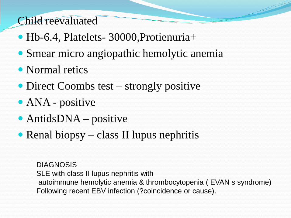
Child reevaluated
Hb-6.4, Platelets- 30000,Protienuria+
Smear micro angiopathic hemolytic anemia
Normal retics
Direct Coombs test – strongly positive
ANA - positive
AntidsDNA – positive
Renal biopsy – class II lupus nephritis
DIAGNOSIS
SLE with class II lupus nephritis with
autoimmune hemolytic anemia & thrombocytopenia ( EVAN s syndrome)
Following recent EBV infection (?coincidence or cause).

Multidisciplinary team – Pediatric Hematologist ,
Pediatric Nephrologist and Rheumatologist
Treated with steroids for 6 weeks and on tapering doses
of steroids Azathiopurine was added.
On Follow up
Regression of hepatosplenomegaly
proteinuria decreased
liver enzymes were normal
Blood counts – normal
Viral serology ( EBV ) – decreasing titres of VCA
IgM &IgG – still positive
Relationship between SLE and
EBV - Literature review
Co incidence or cause

CASE REPORTS
Few case reports for EBV induced SLE
Patients evaluated and diagnosed to have SLE, who had EBV infection (VCA positive) prior to this
R.Verdolini et al.(2002) Systemic lupus erythematosis induced by Epstein-Barr virus infection. Br J Dermatol 146:877-881
Ozgur Kasapcopur et al.(2006) Systemic lupus erythematosis due to Epstein Barr virus or Epstein Barr virus infectipn provocating acute exacerbation of systemic lupus erythematosus? Rheumatol int 26:765-767
Ren Fail. 2009;31(2):144-8.Kikuchi-Fujimoto disease and systemic lupus erythematosus: the EBV connection?

Mechanism of EBV evolving to SLE (molecular
mimicry)
Ability of antibodies against EBNA 2 antigen of EBV
to cross react with Sm D1 , that leads to an
autoimmune response.
Homologous between EBNA 1 antigen and
PPPGMRP peptide of Cterminal region of Sm antigen
Autoimmun Rev. 2009 Feb;8(4):337-42. Epub 2009 Jan 22.

Patients with SLE have abnormally high frequencies of EBV-infected cells in their blood, and this is associated with the occurrence of SLE disease flares.
Abnormal regulation of EBV infection in SLE patients reflects the sensitivity of the virus to perturbation of the immune system.
J Immunol. 2005 Jun 1;174(11):6599-607.

Exposure to EBVinfection may predict a disease phenotype of mild SLE disease with cutaneous and joint manifestations Ann N Y Acad Sci. 2009 Sep;1173:658-63.

Learning objectives EBV infection could work as a trigger in some cases of
SLE, particularly if the patient is genetically susceptible.
In contrary to the existing evidence in literature EBV associated SLE could be severe with Evan’s syndrome and lupus nephritis
SLE patients have increased risk of EBV infections and found to have chronic EBV infection

Thank you