Embed Size (px)
DESCRIPTION
Donahoo AL, Richards LJ (2009) Understanding the mechanisms of callosal development through the use of transgenic mouse models. Semin Pediatr Neurol 16(3):127-142 (REVIEW)
Citation preview
UotA
TctscocarlcftcmHm
F
S
L
A
1d
nderstanding the Mechanismsf Callosal Development Throughhe Use of Transgenic Mouse Modelsmber-Lee S. Donahoo, Assoc. Prof., and Linda J. Richards Bsc(Hons)
The cerebral cortex is the area of the brain where higher-order cognitive processing occurs.The 2 hemispheres of the cerebral cortex communicate through one of the largest fibertracts in the brain, the corpus callosum. Malformation of the corpus callosum in humanbeings occurs in 1 in 4000 live births, and those afflicted experience an extensive range ofneurologic disorders, from relatively mild to severe cognitive deficits. Understanding themolecular and cellular processes involved in these disorders would therefore assist in thedevelopment of prognostic tools and therapies. During the past 3 decades, mouse modelshave been used extensively to determine which molecules play a role in the complexregulation of corpus callosum development. This review provides an update on thesestudies, as well as highlights the value of using mouse models with the goal of developingtherapies for human acallosal syndromes.Semin Pediatr Neurol 16:127–142 © 2009 Elsevier Inc. All rights reserved.
pms
t“tsatdtlscldca(mdadfim
he corpus callosum is the largest fiber tract in the mam-malian brain. It connects neurons in the left and right
erebral hemispheres and is essential for the coordinatedransfer of information between them. For the corpus callo-um to form, several critical developmental events must oc-ur in sequence. These include the patterning and formationf the midline, (which later acts as a substrate for pioneeringallosal axons), the generation of callosal neurons and theirxons, and the targeting and growth of these axons (a processegulated by specific midline glial structures). Finally, cal-osal axons must locate and innervate their targets in theontralateral hemisphere (Fig 1). As soon as they are asormed, developing neurons are nourished by the vascula-ure of the brain, and thus, like all fiber tracts in the brain,allosal axons are susceptible to vascular insult or cell deathechanisms both during development, and in the adult.ere we discuss the current state of research, using mouseodels to investigate these developmental mechanisms, to
rom The Queensland Brain Institute and The School of Biomedical Sci-ences, The University of Queensland, Brisbane, Queensland, Australia.
upported by an Australian Postgraduate Award and a Queensland BrainInstitute supplementary Scholarship (to A.S.D.).
.J.R. is a National Health and Medical Research Council Senior ResearchFellow.
ddress reprint requests to Linda J. Richards, The Queensland Brain Insti-tute and The School of Biomedical Sciences, The University of Queens-
dland, Brisbane 4072, Australia. E-mail: [email protected]
071-9091/09/$-see front matter © 2009 Elsevier Inc. All rights reserved.oi:10.1016/j.spen.2009.07.003
rovide insight into how the corpus callosum forms in hu-an beings, and the mechanisms that could underlie agene-
is or dysgenesis of the corpus callosum.Malformation of the corpus callosum can result in either
he complete absence of the corpus callosum (defined asagenesis of the corpus callosum,” ACC) or partial absence orhinning of the corpus callosum (defined here as “dysgene-is”).1 Because of the wide diversity of developmental mech-nisms regulating the midline crossing of callosal axons,here is a spectrum of phenotypic variations of agenesis andysgenesis of the corpus callosum.2 When callosal fibers failo cross the midline, they often remain ipsilateral and formongitudinal axon fascicles (known as Probst bundles). De-cribed in 1901,3 Probst bundles are likely to be a product ofallosal axon misguidance caused by disruption of the mid-ine structures and/or their secreted molecules. Probst bun-les form longitudinally in both cerebral hemispheres (adja-ent to the midline), with recent analyses revealing that thexons project along the rostrocaudal axis of the forebrainand do not form “whorls,” as classically described) in bothice and human beings.4-8 The morphology of Probst bun-les has been demonstrated in both acallosal human casesnd mouse models using the three-dimensional technique ofiffusion tensor magnetic resonance imaging, which identi-es fiber tracts by the direction of isotropic water move-ent7,9 (reviewed in this issue, Wahl 2009). These studies
emonstrate that because the axons are able to arrive at the127

mfiap(f(atgltaodccedvmdhr
tmamiemsgcHdgBccgPuctt
msfdmcih
Fttesleabptocaaversion of figure is available online.)
128 A-L.S. Donahoo and L.J. Richards
idline, Probst bundles result from failure of the callosalbers to cross the midline and not defective growth of thexons per se. This phenotype is conserved between ACCatients and most of the � 65 different mouse models of ACCTable 1). A newer method of acquiring and processing dif-usion tensor magnetic resonance imaging informationcalled high angular resolution diffusion imaging (HARDI)nd q-ball imaging) has also revealed fascinating informa-ion about the projection of callosal axons in cases of dys-enesis of the corpus callosum (partial ACC) where a cal-osal remnant remains. Sherr et al86 (also see Wahl, 2009his issue) have found that the position of the remnantlong the rostrocaudal axis cannot be used to predict therigin of the fibers that cross the midline. Their resultsemonstrate that different patients, with similar anatomi-al magnetic resonance imaging scans, can have dramati-ally different axonal connectivity patterns. These differ-nces in callosal connectivity may underlie the behavioralifferences observed in these patients. Although clearlyisible by anatomical magnetic resonance imaging, ACCay therefore represent a more generalized brain-wiringefect than that observed by T1-weighted images alone,ighlighting the value of HARDI and q-ball imaging inevealing more detailed anatomy.
Mouse models provide a way to experimentally elucidatehe molecular mechanisms that cooperate to enable the for-ation and maintenance of the corpus callosum, particularly
s cortical organization is conserved in most mammals87 andany of the genes known to be involved in callosal formation
n the mouse display similar expression profiles within thembryonic human brain.9 Complications in the study ofouse models arise from the use of the wide variety of mouse
trains and also the different techniques used to generateenetic modifications. Such complications are similarly en-ountered in studies mapping human genetic disorders.owever, mice used for research are usually the products ofiligent inbreeding, a process that causes the retention ofenetic instability within many strains (such as 129SV andALB/c, Table 1) that can result in sporadic occurrence ofallosal agenesis.77,78 However, this instability can be over-ome by backcrossing the genetically manipulated heterozy-ous mouse onto a more robust strain (such as C57BL/6).82
rovided back crossing is corporated, mouse models can besed to identify a large cohort of single-gene mutations thatause corpus callosum malformations (Table 1). Many ofhese genes represent excellent candidates for human screenso identify the genetic basis of acallosal syndromes.
As outlined earlier in the text, understanding the develop-ent of the corpus callosum involves an appreciation of the
equence of developmental and molecular events requiredor callosal axon growth and targeting (Fig 1). In the remain-er of this review, we will summarize how various mouseodels have elucidated the different mechanisms controlling
allosal development, and highlight how these studies havelluminated our understanding of callosal malformations in
igure 1 Corpus callosum development in the mouse. (A) Callosal forma-ion firstly requires the establishment of a substrate, which is achievedhrough the fusion of the telencephalic midline. This involves either thelimination or exclusion of the leptomeninges found between the hemi-pheres. (B) The pioneers of the corpus callosum originate from the cingu-atecortex (medial-mostpartof thecortex)andreach themidlinebetweeenmbryonicday14(E14)andE15. (C)Axons fromtheneocortex thengrowlongthepathwaydefinedbythepioneers, expanding thecorpuscallosumy E17. (C) On arrival of callosal axons at the midline, midline cellularopulations(glialwedge, indusiumgriseum,andthesubcallosalsling),andhe extracellular cues that they secrete, assist in the turning and channelingf these axons across the midline. (D) Axon guidance of the caudal corpusallosum may also be facilitated by the hippocampal commissure. After thexons have entered the contralateral hemisphere, they traverse dorsolater-lly before innervating homotopic areas of the contralateral cortex. (Color
uman beings.

PFoTacpthfigmfsup(ta(mbi
msavhloOppctdr
TFaFhmptaiifdfia
tmpshs
cis0lpihmmpsmHipn
tcflammipstvfBoispt
ttrpplfu2evi
Callosal development 129
atterning of theorebrain and Formationf the Commissural Plate
he cerebral cortex is first identified early in development assingle vesicle known as the prosencephalon. The prosen-
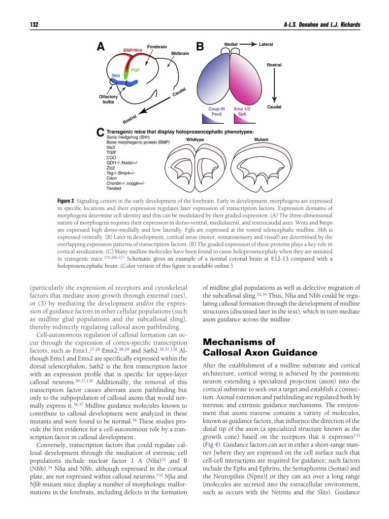
ephalon undergoes a process of rapid proliferation and ex-ansion, and over time, the dorsal wall invaginates to form 2elencephalic hemispheres that will become the cerebralemispheres in adults.88 The induction of tissue identity isrst established by the expression of proteins called morpho-ens. Morphogens are crucial in establishing signaling do-ains, called patterning centers, early in development (be-
ore prosencephalic invagination), which then dictate thepecialization of areas within the forebrain through the reg-lation of transcription factors89,90 (Fig 2). Forebrain mor-hogens include members of the bone morphogenic proteinBMP)91 and Wnt protein92 families, which are expressed byhe cortical hem, fibroblast growth factors (FGF), expressedt the midline and in the cortex,93 and Sonic hedgehogShh),94 which is expressed ventrally. These signaling do-ains interact to establish regional identity of the fore-
rain,89,95,96 as well as patterning of the forebrain midline,ncluding the formation of the commissural plate.
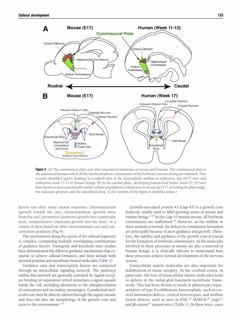
The commissural plate is defined as the anatomical do-ain through which all interhemispheric forebrain commis-
ures (the corpus callosum, the hippocampal commissure,nd the anterior commissure) cross the midline during de-elopment97 (Fig 3). Although the anatomy of this domainas been characterized during human development, the mo-
ecular and cellular composition that regulates the formationf the 3 forebrain commissures is yet to be fully determined.ne factor that is known to be a potent regulator of forebrainatterning is Fgf8, which is expressed by the commissurallate93 and regulates expression of other midline mole-ules.98 In mice deficient in Fgf8 (or its receptor FGFR1), theelencephalic midline and all 3 forebrain commissures areisrupted,74,75,99 which indicates that Fgf8 plays an integralole in this system.
elencephalic Midlineusion: Holoprosencephalynd Interhemispheric Cysts
or callosal axons to cross the midline into the contralateralemisphere, a substrate through which the axons can growust first be present. Thus, the inversion of the dorsalrosencephalic wall to form 2 telencephalic hemispheres andhe subsequent fusion of the telencephalic midline are imper-tive for the development of later axon tracts that facilitatenterhemispheric communication and information process-ng. Two main pathologies have been described in humanetuses where the midline substrate is either absent orisrupted, being replaced by a single ventricle or a fluid-lled space (or cyst): holoprosencephaly (which is due to
failure of telencephalic hemisphere formation), and in- ferhemispheric cyst formation (a build-up of fluid at theidline which disrupts the morphology of the telence-
halic midline structures). The formation of an interhemi-pheric midline cyst, or the disruption of midline fusion, isighly correlated with malformations of the corpus callo-um.100-103
Holoprosencephaly occurs when the prosencephalic vesi-le fails to invaginate to forms 2 telencephalic hemispheres;nstead, a singular hollow vesicle remains and many midlinetructures are lost. Holoprosencephaly, which occurs in.49-1.3 of 10,000 live-births104-107 with a higher incidence
evel of 1 in 250 at prenatal stages, encompasses a variety ofhenotypes, including alobar, semilobar, lobar, and middle
nterhemispheric holoprosencephaly.108-110 Holoprosencep-aly impedes callosal formation because of the loss of theidline substrate. In cases, such as lobar, semilobar, andiddle interhemispheric holoprosencephaly (all of which areartial holoprosencephalies), a remnant of the corpus callo-um remains intact in the areas that are unaffected at theidline.110-111 Analysis of callosal fiber connectivity usingARDI and q-ball imaging in these patients would provide
nteresting information about the ability of callosal axons toroject to their correct targets in this disorder and may revealovel insights into the basis of this disorder.Mouse models have proven very successful in unraveling
he molecular mechanisms that give rise to holoprosen-ephaly (Fig 2), most commonly demonstrating a crucial roleor patterning molecules in the development of the mid-ine. Shh was the first molecule identified in human beingss causing holoprosencephaly when mutated.112 In theouse forebrain, Shh is normally expressed at the ventralidline.94 Holoprosencephaly occurs when Shh is deficient
n transgenic mice113,114 and also when it is ectopically ex-ressed in the dorsal midline, thus disrupting cortical hemignaling centers.115 Holoprosencephaly also occurs whenhe receptor for Shh (Patched) is deficient.116 Zic2 is anotherentral patterning molecule, a deficiency of which has beenound to produce holoprosencephaly in the mouse.117,118
oth Shh and Zic2 have been identified from genetic screensf human holoprosencephalic patients.117 The midline fusesn a ventral to dorsal direction119 and in cases where theignaling centers are deficient, midline formation is impaired,ossibly leading to defects in the subsequent fusion of theelencephalic midline.
Interhemispheric cysts produce different phenotypes fromhose associated with holoprosencephaly. In particular, theelencephalic hemispheres have a definable (though dis-upted) midline in the presence of a cyst, whereas, in holo-rosencephaly the cortex is continuous, showing no mor-hologic differences at the midline as compared with the
ateral cortex.120 Therefore, these 2 midline anomalies riserom different mechanisms—holoprosencephaly from a fail-re of the dorsal prosencephalon to invaginate and form thehemispheres, whereas the cysts cause disturbances to the
stablished telencephalic midline, possibly by an increase ofentricular pressure.120 An alternate hypothesis is that somenterhemispheric midline cysts result from defects in midline
usion. Interhemispheric cysts, including those that associ-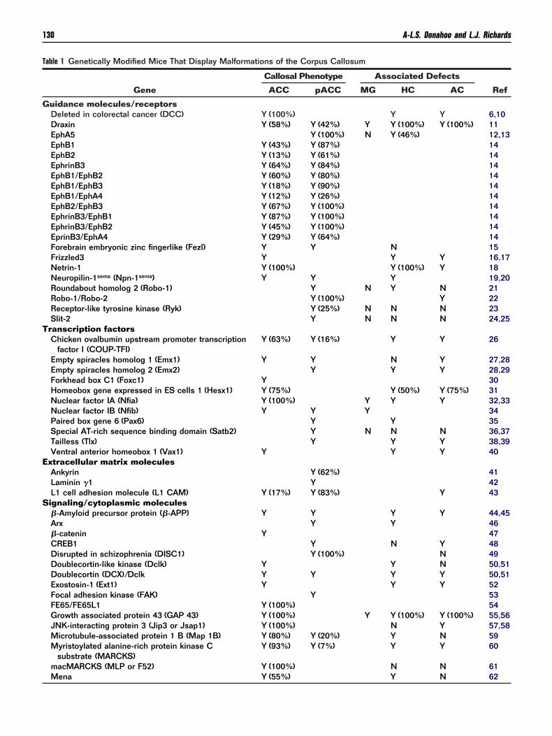
T
G
T
E
S
130 A-L.S. Donahoo and L.J. Richards
able 1 Genetically Modified Mice That Display Malformations of the Corpus Callosum
Gene
Callosal Phenotype Associated Defects
RefACC pACC MG HC AC
uidance molecules/receptorsDeleted in colorectal cancer (DCC) Y (100%) Y Y 6,10Draxin Y (58%) Y (42%) Y Y (100%) Y (100%) 11EphA5 Y (100%) N Y (46%) 12,13EphB1 Y (43%) Y (87%) 14EphB2 Y (13%) Y (61%) 14EphrinB3 Y (64%) Y (84%) 14EphB1/EphB2 Y (60%) Y (80%) 14EphB1/EphB3 Y (18%) Y (90%) 14EphB1/EphA4 Y (12%) Y (26%) 14EphB2/EphB3 Y (67%) Y (100%) 14EphrinB3/EphB1 Y (87%) Y (100%) 14EphrinB3/EphB2 Y (45%) Y (100%) 14EprinB3/EphA4 Y (29%) Y (64%) 14Forebrain embryonic zinc fingerlike (Fezl) Y Y N 15Frizzled3 Y Y Y 16,17Netrin-1 Y (100%) Y (100%) Y 18Neuropilin-1sema (Npn-1sema) Y Y Y 19,20Roundabout homolog 2 (Robo-1) Y N Y N 21Robo-1/Robo-2 Y (100%) Y 22Receptor-like tyrosine kinase (Ryk) Y (25%) N N N 23Slit-2 Y N N N 24,25
ranscription factorsChicken ovalbumin upstream promoter transcription
factor I (COUP-TFI)Y (63%) Y (16%) Y Y 26
Empty spiracles homolog 1 (Emx1) Y Y N Y 27,28Empty spiracles homolog 2 (Emx2) Y Y Y 28,29Forkhead box C1 (Foxc1) Y 30Homeobox gene expressed in ES cells 1 (Hesx1) Y (75%) Y (50%) Y (75%) 31Nuclear factor IA (Nfia) Y (100%) Y Y Y 32,33Nuclear factor IB (Nfib) Y Y Y 34Paired box gene 6 (Pax6) Y Y 35Special AT-rich sequence binding domain (Satb2) Y N N N 36,37Tailless (Tlx) Y Y Y 38,39Ventral anterior homeobox 1 (Vax1) Y Y Y 40
xtracellular matrix moleculesAnkyrin Y (62%) 41Laminin �1 Y 42L1 cell adhesion molecule (L1 CAM) Y (17%) Y (83%) Y 43ignaling/cytoplasmic molecules�-Amyloid precursor protein (�-APP) Y Y Y Y 44,45Arx Y Y 46�-catenin Y 47CREB1 Y N Y 48Disrupted in schizophrenia (DISC1) Y (100%) N 49Doublecortin-like kinase (Dclk) Y Y N 50,51Doublecortin (DCX)/Dclk Y Y Y Y 50,51Exostosin-1 (Ext1) Y Y Y 52Focal adhesion kinase (FAK) Y 53FE65/FE65L1 Y (100%) 54Growth associated protein 43 (GAP 43) Y (100%) Y Y (100%) Y (100%) 55,56JNK-interacting protein 3 (Jip3 or Jsap1) Y (100%) N Y 57,58Microtubule-associated protein 1 B (Map 1B) Y (80%) Y (20%) Y N 59Myristoylated alanine-rich protein kinase C
substrate (MARCKS)Y (93%) Y (7%) Y Y 60
macMARCKS (MLP or F52) Y (100%) N N 61
Mena Y (55%) Y N 62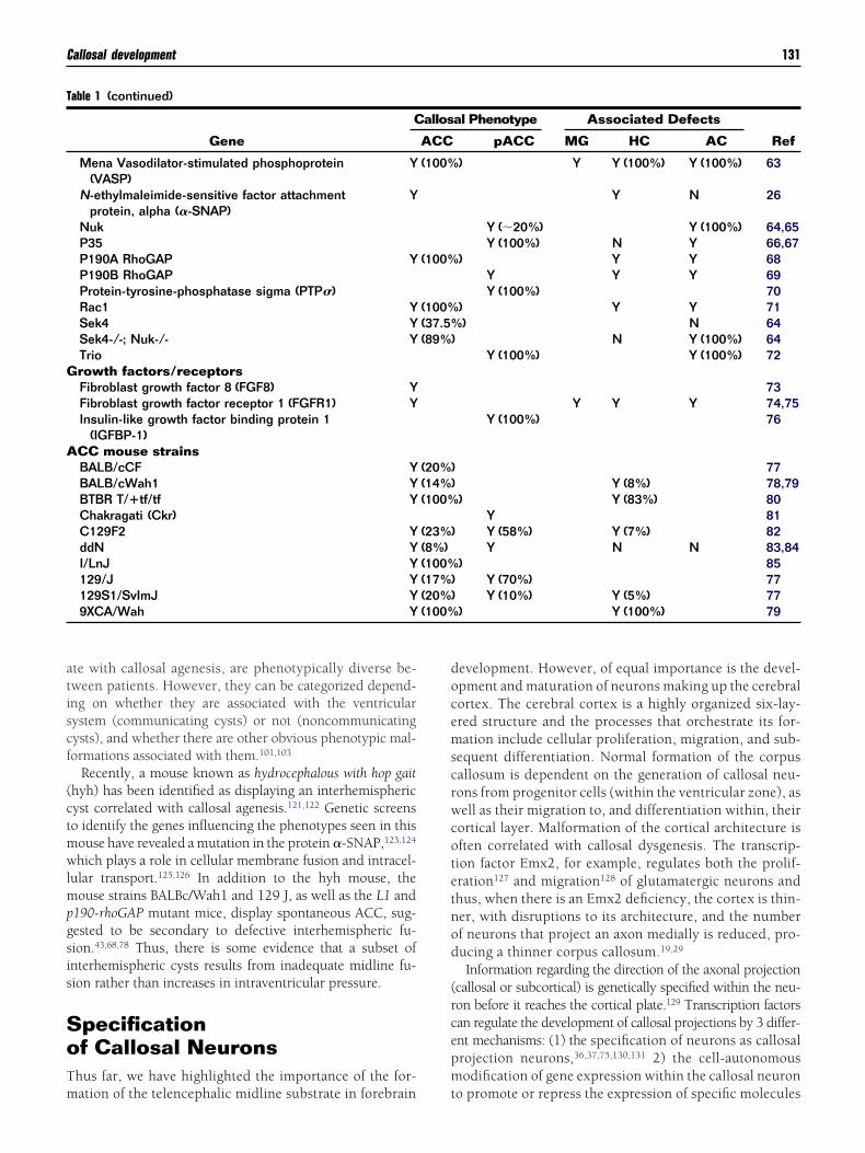
atiscf
(ctmwlmpgsis
SoTm
docemscrwcotetnod
(rcepm
T
G
A
(100%
Callosal development 131
te with callosal agenesis, are phenotypically diverse be-ween patients. However, they can be categorized depend-ng on whether they are associated with the ventricularystem (communicating cysts) or not (noncommunicatingysts), and whether there are other obvious phenotypic mal-ormations associated with them.101,103
Recently, a mouse known as hydrocephalous with hop gaithyh) has been identified as displaying an interhemisphericyst correlated with callosal agenesis.121,122 Genetic screenso identify the genes influencing the phenotypes seen in thisouse have revealed a mutation in the protein �-SNAP,123,124
hich plays a role in cellular membrane fusion and intracel-ular transport.125,126 In addition to the hyh mouse, the
ouse strains BALBc/Wah1 and 129 J, as well as the L1 and190-rhoGAP mutant mice, display spontaneous ACC, sug-ested to be secondary to defective interhemispheric fu-ion.43,68,78 Thus, there is some evidence that a subset ofnterhemispheric cysts results from inadequate midline fu-ion rather than increases in intraventricular pressure.
pecificationf Callosal Neurons
hus far, we have highlighted the importance of the for-
able 1 (continued)
Gene
C
Mena Vasodilator-stimulated phosphoprotein(VASP)
Y
N-ethylmaleimide-sensitive factor attachmentprotein, alpha (�-SNAP)
Y
NukP35P190A RhoGAP YP190B RhoGAPProtein-tyrosine-phosphatase sigma (PTP�)Rac1 YSek4 YSek4-/-; Nuk-/- YTriorowth factors/receptorsFibroblast growth factor 8 (FGF8) YFibroblast growth factor receptor 1 (FGFR1) YInsulin-like growth factor binding protein 1
(IGFBP-1)CC mouse strainsBALB/cCF YBALB/cWah1 YBTBR T/�tf/tf YChakragati (Ckr)C129F2 YddN YI/LnJ Y129/J Y129S1/SvImJ Y9XCA/Wah Y
ation of the telencephalic midline substrate in forebrain t
evelopment. However, of equal importance is the devel-pment and maturation of neurons making up the cerebralortex. The cerebral cortex is a highly organized six-lay-red structure and the processes that orchestrate its for-ation include cellular proliferation, migration, and sub-
equent differentiation. Normal formation of the corpusallosum is dependent on the generation of callosal neu-ons from progenitor cells (within the ventricular zone), asell as their migration to, and differentiation within, their
ortical layer. Malformation of the cortical architecture isften correlated with callosal dysgenesis. The transcrip-ion factor Emx2, for example, regulates both the prolif-ration127 and migration128 of glutamatergic neurons andhus, when there is an Emx2 deficiency, the cortex is thin-er, with disruptions to its architecture, and the numberf neurons that project an axon medially is reduced, pro-ucing a thinner corpus callosum.19,29
Information regarding the direction of the axonal projectioncallosal or subcortical) is genetically specified within the neu-on before it reaches the cortical plate.129 Transcription factorsan regulate the development of callosal projections by 3 differ-nt mechanisms: (1) the specification of neurons as callosalrojection neurons,36,37,75,130,131 2) the cell-autonomousodification of gene expression within the callosal neuron
al Phenotype Associated Defects
RefpACC MG HC AC
) Y Y (100%) Y (100%) 63
Y N 26
Y (�20%) Y (100%) 64,65Y (100%) N Y 66,67
) Y Y 68Y Y Y 69Y (100%) 70
) Y Y 71) N 64
N Y (100%) 64Y (100%) Y (100%) 72
73Y Y Y 74,75
Y (100%) 76
77Y (8%) 78,79
) Y (83%) 80Y 81Y (58%) Y (7%) 82Y N N 83,84
) 85Y (70%) 77Y (10%) Y (5%) 77
) Y (100%) 79
allos
ACC
(100%
(100%
(100%(37.5%(89%)
(20%)(14%)(100%
(23%)(8%)(100%(17%)(20%)
o promote or repress the expression of specific molecules
(fosat
cftdwctomcmvs
lp(pNm
otlsa
MCAanctimkdg(ncit(
s avail
132 A-L.S. Donahoo and L.J. Richards
particularly the expression of receptors and cytoskeletalactors that mediate axon growth through external cues),r (3) by mediating the development and/or the expres-ion of guidance factors in other cellular populations (suchs midline glial populations and the subcallosal sling),hereby indirectly regulating callosal axon pathfinding.
Cell-autonomous regulation of callosal formation can oc-ur through the expression of cortex-specific transcriptionactors, such as Emx1,27,28 Emx2,28,29 and Satb2.36,37,130 Al-hough Emx1 and Emx2 are specifically expressed within theorsal telencephalon, Satb2 is the first transcription factorith an expression profile that is specific for upper-layer
allosal neurons.36,37,130 Additionally, the removal of thisranscription factor causes aberrant axon pathfinding butnly to the subpopulation of callosal axons that would nor-ally express it.36,37 Midline guidance molecules known to
ontribute to callosal development were analyzed in theseutants and were found to be normal.36 These studies pro-
ide the first evidence for a cell-autonomous role by a tran-cription factor in callosal development.
Conversely, transcription factors that could regulate cal-osal development through the mediation of extrinsic cellopulations include nuclear factor 1 A (Nfia)32 and BNfib).34 Nfia and Nfib, although expressed in the corticallate, are not expressed within callosal neurons.132 Nfia andfib mutant mice display a number of morphologic malfor-
Figure 2 Signaling centers in the early development of thin specific locations and their expression regulates latemorphogens determine cell identity and this can be modnature of morphogens requires their expression in dorsoare expressed high dorso-medially and low laterally. Fexpressed ventrally. (B) Later in development, cortical aoverlapping expression patterns of transcription factors.cortical arealization. (C) Many midline molecules have bin transgenic mice.115,206-217 Schematic gives an examholoprosencephalic brain. (Color version of this figure i
ations in the forebrain, including defects in the formation s
f midline glial populations as well as defective migration ofhe subcallosal sling.32,34 Thus, Nfia and Nfib could be regu-ating callosal formation through the development of midlinetructures (discussed later in the text), which in turn mediatexon guidance across the midline.
echanisms ofallosal Axon Guidance
fter the establishment of a midline substrate and corticalrchitecture, cortical wiring is achieved by the postmitoticeuron extending a specialized projection (axon) into theortical substrate to seek out a target and establish a connec-ion. Axonal extension and pathfinding are regulated both byntrinsic and extrinsic guidance mechanisms. The environ-
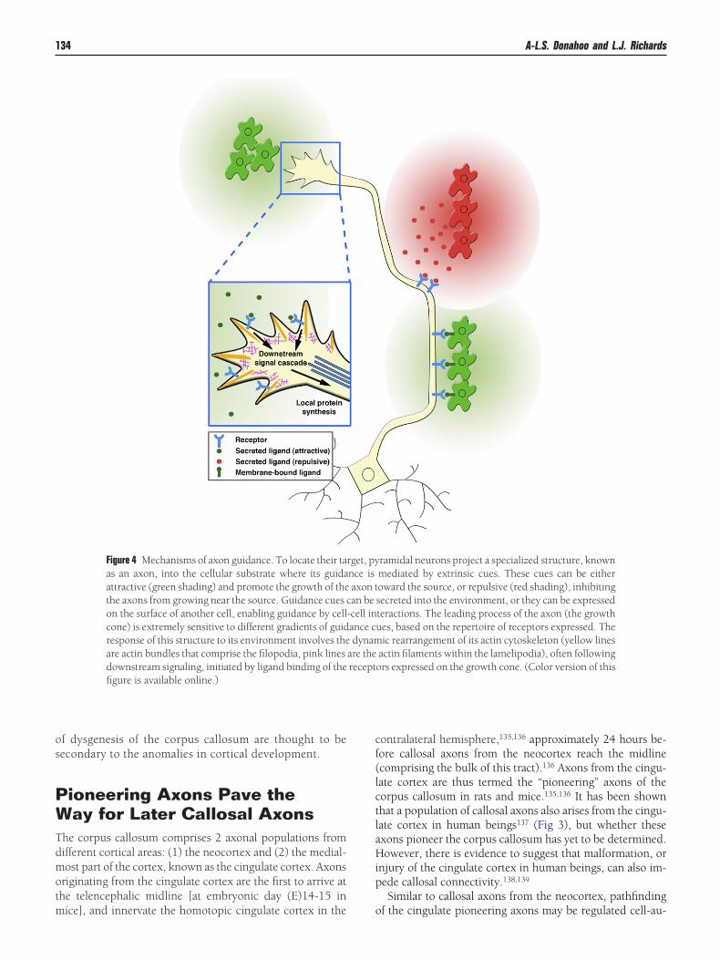
ent that axons traverse contains a variety of molecules,nown as guidance factors, that influence the direction of theistal tip of the axon (a specialized structure known as therowth cone) based on the receptors that it expresses133
Fig 4). Guidance factors can act in either a short-range man-er [where they are expressed on the cell surface such thatell-cell interactions are required for guidance; such factorsnclude the Ephs and Ephrins, the Semaphorins (Semas) andhe Neuropilins (Npns)] or they can act over a long rangemolecules are secreted into the extracellular environment,
rain. Early in development, morphogens are expressedession of transcription factors. Expression domains ofby their graded expression. (A) The three-dimensionall, mediolateral, and rostrocaudal axes. Wnts and Bmpsexpressed at the rostral telencephalic midline. Shh isotor, somatosensory and visual) are determined by thee graded expression of these proteins plays a key role innd to cause holoprosencephaly when they are mutateda normal coronal brain at E12-13 compared with a
able online.)
e forebr exprulated-ventragfs arereas (m(B) Theen fouple of
uch as occurs with the Netrins and the Slits). Guidance
f(favc
iohos
twtioeaa
mhctaffihts
sptwstf
versio
Callosal development 133
actors can elicit many axonal responses: chemoattractiongrowth toward the cue), chemorepulsion (growth awayrom the cue), permissive (promotes growth into a particularrea), nonpermissive (represses growth into the area), or aariety of these based on other environmental cues and con-entration gradients (Fig 4).
The environment along the extent of the callosal trajectorys complex, comprising multiple overlapping combinationsf guidance factors. Transgenic and knockout mice studiesave demonstrated the different guidance mechanisms that co-perate to achieve callosal formation, and these include bothecreted proteins and membrane-bound molecules (Table 1).
Guidance cues and transcription factors are connectedhrough an intracellular signaling network. The pathwaysithin this network are generally activated by ligand-recep-
or binding (or separation) which stimulates a signal cascadenside the cell, including alterations to the phosphorylationf coreceptors and secondary messengers. Cytoskeletal mol-cules can then be directly altered through the signal cascadend thus can alter the morphology of the growth cone and
Figure 3 (A) The commissural plate and other anatomicathe anatomical domain which all the interhemispheric cois easily identified (green shading) in a sagittal view atembryonic week 11-12 in human beings. (B) In the corbeen shown to have anatomically similar cellular populatthe indusium griseum, and the subcallosal sling. (Color
xon to the environment.134 a
Growth-associated protein 43 (Gap-43) is a growth coneolecule widely used to label growing axons in mouse anduman beings.9,55 In the Gap-43 mutant mouse, all forebrainommissures are malformed.55 However, as the midline inhese animals is normal, the defects in commissure formationre principally because of axon guidance and growth. There-ore, the stability and guidance of the growth cone is crucialor the formation of forebrain commissures. As the moleculesnvolved in these processes in mouse are also conserved inuman beings, it is clinically relevant to understand howhese processes achieve normal development of the nervousystem.
Extracellular matrix molecules are also important fortabilization of tissue integrity. In the cerebral cortex, inarticular, the loss of extracellular matrix molecules leadso defects in the radial-glial basement-membrane frame-ork. This has been shown to result in phenotypes repre-
entative of type II cobblestone lissencephaly, such as cor-ical lamination defects, cortical heterotopias, and midlineusion defects, such as seen in FAK,53 MARCK,60 Jsap157
arities in mouse and humans. The commissural plate isures of the forebrain traverse during development. This
lencephalic midline in embryonic day (E17) mice andlane, developing human fetal brains (week 17; D) havethose seen in mouse (at E17), including the glial wedge,n of this figure is available online.)
l similmmissthe teonal pions to
nd �-catenin47 mutant mice (Table 1). In these mice, cases
os
PWTdmotm
cf(lctlaHip
134 A-L.S. Donahoo and L.J. Richards
f dysgenesis of the corpus callosum are thought to beecondary to the anomalies in cortical development.
ioneering Axons Pave theay for Later Callosal Axons
he corpus callosum comprises 2 axonal populations fromifferent cortical areas: (1) the neocortex and (2) the medial-ost part of the cortex, known as the cingulate cortex. Axons
riginating from the cingulate cortex are the first to arrive athe telencephalic midline [at embryonic day (E)14-15 in
Figure 4 Mechanisms of axon guidance. To locate their taas an axon, into the cellular substrate where its guidaattractive (green shading) and promote the growth of thethe axons from growing near the source. Guidance cues con the surface of another cell, enabling guidance by cellcone) is extremely sensitive to different gradients of guidresponse of this structure to its environment involves theare actin bundles that comprise the filopodia, pink linesdownstream signaling, initiated by ligand binding of thefigure is available online.)
ice], and innervate the homotopic cingulate cortex in the o
ontralateral hemisphere,135,136 approximately 24 hours be-ore callosal axons from the neocortex reach the midlinecomprising the bulk of this tract).136 Axons from the cingu-ate cortex are thus termed the “pioneering” axons of theorpus callosum in rats and mice.135,136 It has been shownhat a population of callosal axons also arises from the cingu-ate cortex in human beings137 (Fig 3), but whether thesexons pioneer the corpus callosum has yet to be determined.owever, there is evidence to suggest that malformation, or
njury of the cingulate cortex in human beings, can also im-ede callosal connectivity.138,139
Similar to callosal axons from the neocortex, pathfinding
ramidal neurons project a specialized structure, knownmediated by extrinsic cues. These cues can be eitheroward the source, or repulsive (red shading), inhibitingsecreted into the environment, or they can be expressedteractions. The leading process of the axon (the growthues, based on the repertoire of receptors expressed. The
ic rearrangement of its actin cytoskeleton (yellow linesactin filaments within the lamelipodia), often followingors expressed on the growth cone. (Color version of this
rget, pynce isaxon tan be
-cell inance cdynam
are therecept
f the cingulate pioneering axons may be regulated cell-au-

teeotapapltbgSfmigm
sCNbflNteciccTatmeiTmtl
MPFCtMsmgha
ic
lfer
t(aiimrR
ssdsgsdsalt
ctmliflttcorwwad
tpt(iitsn
Callosal development 135
onomously by transcription factors, or determined by thextracellular guidance cues expressed at the midline. Thextracellular guidance molecules involved in callosal devel-pment that have been identified thus far at the midline areemporally expressed during both cingulate axon crossingnd neocortical crossing of the midline, suggesting that bothopulations of callosal axons are regulated by similar guid-nce cues. Possible differences in the guidance of these 2opulations may be dependent on the different expression
evels of transcription factors within the cingulate cortex andhe neocortex. Several transcription factors are expressed byoth the cingulate cortex and the neocortex, but occur in aradient across these areas (high medial to low lateral forp8,140 Emx1, and Emx2,141 and low medial to high lateralor Pax6142 and Coup-TFI143; Fig 2). Whether the graded
ediolateral expression pattern of these transcription factorss functionally relevant in the connectivity of either the cin-ulate or the neocortical callosal axons has yet to be deter-ined.Recently, the receptor neuropilin-1 (Npn-1) has been
hown to be involved in corpus callosum formation.19,20,144
ingulate pioneering axons of the corpus callosum expresspn-1 at a crucial temporal stage for callosal development inoth mouse and human beings,9 and the axons of explantsrom the cingulate cortex are attracted toward the Npn-1igand, Sema3C (which is expressed at the midline19). Aspn-1 is highly expressed on axons from the cingulate cor-
ex, it is likely to be a key regulator of cingulate axons. How-ver, Npn-1 is also expressed (albeit a lesser extent) in theortex,145 and a recent study using dominant negative Npn-1n cortical axons demonstrated a cell-autonomous affect onallosal axon guidance.144 Thus, Npn-1 can also play a role inell-autonomous callosal axon guidance from the neocortex.he pioneering axons are hypothesized to guide neocorticalxons by providing a structural framework for callosal axonso follow by direct axon-axon contact, and this is possiblyediated through Npn-1. Evidence to support this hypoth-
sis is that the cortical axons that were injected with a dom-nant-negative Npn-1 displayed defects in fasciculation.144
herefore, Npn-1 may play an important role in callosal for-ation, first by the guidance of cingulate pioneering axons to
he midline, and then through the cell-cell guidance of cal-osal axons from the neocortex.
idline Cellularopulations Regulate theormation of the Corpus Callosumallosal axons are guided medially to the midline where
hey are channeled into the contralateral hemisphere.idline cellular populations that have been shown to as-
ist in the formation of the corpus callosum include theidline zipper glia, the glial wedge, the indusium griseum
lia, and the subcallosal sling.119,146,147 These populationsave been identified in the rostral forebrain of both mouse
nd human beings9 (Fig 3), suggesting that their function an formation of the human corpus callosum may also beonserved.
Midline zipper glia are positioned at the telencephalic mid-ine and are thought to assist in the ventral fusion of theorebrain on the basis of their anatomical location.119 How-ver, there is currently nothing known about the molecularegulation of forebrain midline fusion.
The glial wedge is a specialized wedge-shaped glial struc-ure positioned at the medial aspect of the lateral ventricle148
Figs 1 and 3). The glial wedge assists the guidance of callosalxons across the midline by preventing their ventral growthnto the septum. This boundary is thought to be due to bothts anatomical position and also its expression of the guidance
olecule Slit2, which has been shown in vitro to act as aepellent for callosal axons expressing the Slit2 receptor,obo.21,22,24
The indusium griseum is an area of neurons and glia po-itioned dorsal to the corpus callosum (Figs 1 and 3). Indu-ium griseum glia express Slit2 and are thought to act as aorsal repulsive barrier for the axons of the corpus callo-um.24 FGFR1 is crucial for the positioning of the indusiumriseum glia.74,75 In FGFR1 mutant mice, the indusium gri-eum glia do not form properly, causing callosal pathfindingefects despite the maintenance of guidance cue expres-ion.74 Thus, the indusium griseum glia and the glial wedgere important for the correct development of the corpus cal-osum, due to both their position and the expression of func-ional guidance molecules.24,74,75
The subcallosal sling (originally termed the “glial sling”149)omprises a transient population of neurons that arise fromhe medial wall of the lateral ventricle and migrate to theidline, parallel with, and immediately ventral to, the cal-
osal axons.150 The subcallosal sling has also been describedn human beings9,151 (Fig 3). The specific ‘U’ shape of thisormation and the position of these cells relative to the cal-osal axons are suggested to be requisites for the formation ofhe corpus callosum.152 However, an alternate hypothesis ishat the subcallosal sling cells require the axons of the corpusallosum to direct their migration to the midline (as previ-usly described for a postnatal glial population of cells inat153). As the formation of the sling occurs simultaneouslyith the pathfinding of the callosal axons, determininghether callosal axons direct sling cell migration or callosal
xons project across the midline guided by sling cues, will beifficult.ACC in mouse models is often correlated with a disruption
o the subcallosal sling. Interestingly, this correlation isrominent when Probst bundles have formed; in this situa-ion the sling cells either migrate aberrantly into the septumas in Nfia32) or accumulate lateral to these structures (as seenn JSAP homozygous mutants57 and when callosal agenesis isnduced by gamma-irradiation in Swiss mice154). In contrast,he sling is not malformed in mouse mutants that have aubpopulation of callosal axons crossing the midline and doot display Probst bundles (such as Satb236 and BALB/cCF
callosal mice149).
H(ATcsghcaltdtsvHtpft
AFtmittdfEfnpnwiesuaf
iltmficlncip
rraasawc
VCFcvarpdsditcsttadibddflotl
mtbmpoii
nrtgvSlhn
136 A-L.S. Donahoo and L.J. Richards
ippocampal CommissurePossible Differenceslong the Rostrocaudal Axis?)
he environment that callosal axons project through is notonsistent along the rostrocaudal axis of the telencephalon,uggesting that different mechanisms regulate different re-ions of callosal pathfinding. In the caudal telencephalon, theippocampal commissure lies directly ventral to the corpusallosum and is thought to provide a guidance substrate forxons of the splenium (the caudal portion of the corpus cal-osum).155-158 In many mouse strains and transgenic mutantshat have malformation of the corpus callosum there is also aisruption of the hippocampal commissure (which crosseshe midline approximately 24 hours before the corpus callo-um159 (Table 1). Thus, the developmental mechanisms in-olved in the formation of these commissures may overlap.umans also have a hippocampal commissure (Fig 3)160 but
his projection is less developed than in rodents. The hip-ocampal commissure may have a structural role in callosalormation although this hypothesis has yet to be fully inves-igated.
ctivity-Dependent Target Findingollowing crossing of the midline, callosal axons project intohe contralateral hemisphere and innervate targets in a ho-otopic manner. Information transfer between cortical areas
s dependent upon their connectivity, and as the homotopicargeting of callosal neurons is highly specific, understandinghe mechanisms involved in this event is imperative to un-erstanding function. The caudal corpus callosum axonsorm the splenium and originate within the visual cortex.ye enucleation studies in cats have demonstrated a role
or visual experience in callosal formation and mainte-ance.161,162 A phenomenon known as exuberance andruning during development establishes the axonal orga-ization of the visual cortex, a process that has beenidely investigated in cats.163-165 This process involves the
nitial overproduction of axons that invade the contralat-ral hemisphere in search of a target, followed by the sub-equent refinement of the connections by the removal of anynnecessary axons. Neural activity plays a crucial role inxon exuberance and pruning, but this process has yet to beully investigated in mice.
A series of elegant studies in mice using neural activitynhibitors coupled with in vivo electroporation has high-ighted that activity plays a central role in the contralateralargeting of callosal axons in both the occipital and the so-atosensory cortices.166,167 In both of these areas, the callosal
bers mis-targeted when activity was inhibited, and in someases resulted in the aberrant innervation of other corticalayers instead. Interestingly, the dependence of axonal con-ectivity on activity may not be consistent across the rostro-audal axis. In the visual cortex, reduced excitability resultedn aberrant layer positioning of axonal arbors.166 The same
rocedure in somatosensory cortex also demonstrated aber- vant layer positioning of axonal arbors; however, a loss ofegion-specific targeting was also found.167 This suggests thatctivity could have different roles in different cortical areas,nd this may be modulated through the expression of region-pecific “positional cues” (undetermined thus far). Whetherctivity provides instructive cues for growing axons orhether it regulates molecular cues within the contralateral
ortex has yet to be determined.
asculature Insult andorpus Callosum Maintenance
ollowing development of the corpus callosum, the cerebralortex requires continual metabolic nourishment from theascular system to ensure the maintenance of this axon tractnd its function. Many clinical studies in human beings haveevealed a correlation between reduced cerebral blood sup-ly and atrophy of the corpus callosum.168,169 Symptoms thatevelop from reduced blood flow include a reduction in theize of the corpus callosum (likely through demyelination),isruptions to callosal connectivity, and lesions to neighbor-
ng cortical areas (in particular the cingulate gyrus). Reduc-ion in cerebral blood flow is also correlated with deficits inognitive function.138,139 However, a caveat to some of thesetudies is that they were conducted using elderly patients,hus it is difficult to determine whether the observed cogni-ive decline stemmed from reduced blood flow or fromge.170,171 Lesions of the white matter are most commonlyescribed in stroke patients, although there is increasing ev-
dence for this occurrence in Alzheimer’s disease and possi-ly other neurodegenerative disorders. In patients with hy-rocephalus (which is commonly associated with callosalysgenesis), there is often a reduction in cerebral bloodow.172 Thus, the preservation of the cortical vasculature, notnly during development but also throughout life, is requiredo maintain the homeostasis and function of the corpus cal-osum.
Stroke has been widely investigated, primarily using ratodels173; however, the potential for genetic modification in
his model is limited. Recently, several research groups haveegun to generate mouse models of stroke enabling the re-oval of relevant genes implicated in cerebrovascular re-air.174,175 The removal of either matrix metalloproteinase-2r 9, for example, reduces the severity of cerebrovascularnjury (including lesions of white matter tracts) following thenduction of stroke in these mice.176,177
There is increasing evidence that many molecular mecha-isms involved in nervous system development also play aole in the development of the vascular system; in particular,here is strong overlap between molecules implicated in axonuidance and those that play a role in angiogenesis and bloodessel pathfinding178 (Table 2). As discussed earlier, theema-Npn ligand-receptor pair are crucial regulators of cal-osal axon guidance.19,144 In addition, Npn-1 mutant miceave deficiencies in blood vessel formation.179,180 Npn-1 isot only a receptor for the class III Semaphorins but also for
ascular endothelial growth factor (VEGF), both of which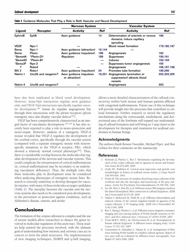
hHoltt
gcnmt(sstuc(itwsl(viA
CTovigco
anwwlmsidd
ATG
R
T
E
VSS***
SN
N
Callosal development 137
ave also been implicated in blood vessel development.owever, Sema-Npn interactions regulate axon guidancenly, and VEGF-Npn interactions specifically regulate vascu-ar development.181 Semas do regulate angiogenesis buthrough their interactions with the plexin receptors (plexinransgenic mice also display vascular defects182).
VEGF has been comprehensively characterized as an inte-ral factor of vasculature development.183,184 It has also re-ently been reported to play a role in neuro-protection andeuro-repair. However, analysis of a transgenic VEGF-Aouse revealed that VEGF-A regulates the development of
he cerebral cortex, specifically through the vascular systemcompared with a separate transgenic mouse with neuron-pecific mutations to the VEGF-A receptor, Flk1, whichhowed a relatively normal cortical phenotype185). Thus,here is considerable overlap between the molecules that reg-late development of the nervous and vascular systems. Thisould complicate the interpretation of cortical malformationsie, cortical malformations may be secondary to an underly-ng vasculature deficiency). Thus, the multiple roles thathese molecules play in development must be consideredhen analyzing phenotypes of transgenic mouse lines. Re-
earch is currently underway to develop therapies for vascu-ar injuries, with many of these molecules as target candidatesTable 2). The interplay between the vascular and the ner-ous systems also needs to be considered post-development,n the prevention or protection against conditions, such aslzheimer’s disease, tumors, and stroke.
onclusionshe formation of the corpus callosum is complex and the usef mouse models allow researchers to dissect the genes in-olved in molecular regulation of this axon tract. These stud-es help unravel the processes involved, with the ultimateoal of understanding how intrinsic and extrinsic cues act inoncert to form the adult structures. The implementation
able 2 Guidance Molecules That Play a Role in Both Vascul
Ligand Receptor
Nervous Syst
Activity
phrinB EphB Axon guidance
EGF Npn-1 —ema Npn-1 Axon guidance (attractiema Plexin Axon guidance (repulsioSema4A *Plexin-D1 RepulsionSema4D *Plexin-B1Sema3F Npn-2
? Robo4 —lit 2 Robo1 Axon guidance (repulsioetrin-1 Unc5b and neogenin? Axon guidance (repulsio
or attraction)
etrin-4 Unc5b and neogenin? —
f new imaging techniques, HARDI and q-ball imaging,
llows a more detailed characterization of the callosal con-ectivity within both mouse and human patients afflictedith congenital malformations. Future use of this techniqueill provide insight into the processes that contribute to cal-
osal formation. Further research to unveil the regulatoryechanisms along the rostrocaudal, mediolateral, and dor-
oventral axes of the forebrain will expand our understand-ng of callosal formation and will bring us 1 step closer to theevelopment for therapies and treatments for acallosal syn-romes in human beings.
cknowledgmentshe authors thank Rowan Tweedale, Michael Piper, and Ilanobius for their comments on the manuscript.
eferences1. Richards LJ, Plachez C, Ren T: Mechanisms regulating the develop-
ment of the corpus callosum and its agenesis in mouse and human.Clin Genet 66:276-289, 2004
2. Ozaki HS, Wahlsten D: Cortical axon trajectories and growth conemorphologies in fetuses of acallosal mouse strains. J Comp Neurol336:595-604, 1993
3. Probst M: The structure of complete dissolving corpus callosum of thecerebrum and also the microgyry and heterotropy of the grey sub-stance. Archiv Fur Psychiatrie Nervenkrankheiten 34:709-786, 1901
4. Lee SK, Mori S, Kim DJ, et al: Diffusion tensor MR imaging visualizesthe altered hemispheric fiber connection in callosal dysgenesis. AJNRAm J Neuroradiol 25:25-28, 2004
5. Nakata Y, Barkovich AJ, Wahl M, et al: Diffusion abnormalities andreduced volume of the ventral cingulum bundle in agenesis of thecorpus callosum: A 3T imaging study. AJNR Am J Neuroradiol 30:1142-1148, 2009
6. Ren T, Zhang J, Plachez C, et al: Diffusion tensor magnetic resonanceimaging and tract-tracing analysis of Probst bundle structure in Ne-trin1- and DCC-deficient mice. J Neurosci 27:10345-10349, 2007
7. Tovar-Moll F, Moll J, de Oliveira-Souza R, et al: Neuroplasticity inhuman callosal dysgenesis: A diffusion tensor imaging study. CerebCortex 17:531-541, 2007
8. Utsunomiya H, Yamashita S, Takano K, et al: Arrangement of fibertracts forming Probst bundle in complete callosal agenesis: Report oftwo cases with an evaluation by diffusion tensor tractography. Acta
Neural Development
Vascular System
Ref Activity Ref
4 Determination of arteriole or venousdomains; induce capillarysprouting
186
Blood vessel formation 179,180,1879,144 —88 Angiogenesis 18289 Suppresses 190,191— Induces 192-194— Suppresses tumor angiogenesis 195
Angiogenesis 196,197,1984 Promotes tube formation 199,2008,201 Angiogenesis (promotion or
suppression) attracts bloodvessels
202,204,205
Angiogenesis 203
ar and
em
1
on) 1n) 1
1
n) 2n 1
Radiol 47:1063-1066, 2006
138 A-L.S. Donahoo and L.J. Richards
9. Ren T, Anderson A, Shen W, et al: Imaging, anatomical, and molecularanalysis of callosal formation in the developing human fetal brain.Anat Rec A Discov Mol Cell Evol Biol 288:191-204, 2006
10. Fazeli A, Dickinson SL, Hermiston ML, et al: Phenotype of mice lack-ing functional deleted in colorectal cancer (Dcc) gene. Nature 386:796-804, 1997
11. Islam SM, Shinmyo Y, Okafuji T, et al: Draxin, a repulsive guidanceprotein for spinal cord and forebrain commissures. Science 323:388-393, 2009
12. Hu Z, Yue X, Shi G, et al: Corpus callosum deficiency in transgenicmice expressing a truncated ephrin-A receptor. J Neurosci 23:10963-10970, 2003
13. Yue Y, Chen ZY, Gale NW, et al: Mistargeting hippocampal axons byexpression of a truncated Eph receptor. Proc Natl Acad Sci U S A99:10777-10782, 2002
14. Mendes SW, Henkemeyer M, Liebl DJ: Multiple Eph receptors andB-class ephrins regulate midline crossing of corpus callosum fibers inthe developing mouse forebrain. J Neurosci 26:882-892, 2006
15. Molyneaux BJ, Arlotta P, Hirata T, et al: Fezl is required for the birthand specification of corticospinal motor neurons. Neuron 47:817-831, 2005
16. Wang Y, Thekdi N, Smallwood PM, et al: Frizzled-3 is required for thedevelopment of major fiber tracts in the rostral CNS. J Neurosci 22:8563-8573, 2002
17. Wang Y, Zhang J, Mori S, et al: Axonal growth and guidance defects inFrizzled3 knock-out mice: A comparison of diffusion tensor magneticresonance imaging, neurofilament staining, and genetically directedcell labeling. J Neurosci 26:355-364, 2006
18. Serafini T, Colamarino SA, Leonardo ED, et al: Netrin-1 is required forcommissural axon guidance in the developing vertebrate nervous sys-tem. Cell 87:1001-1014, 1996
19. Piper M, Plachez C, Zalucki O, et al: Neuropilin 1-Sema signalingregulates crossing of cingulate pioneering axons during developmentof the corpus callosum. Cereb Cortex 19 (Suppl 1):i11-i21, 2009
20. Gu C, Rodriguez ER, Reimert DV, et al: Neuropilin-1 conveys sema-phoring and VEGF signaling during neural and cardiovascular devel-opment. Dev Cell 5:45-57, 2003
21. Andrews W, Liapi A, Plachez C, et al: Robo1 regulates the develop-ment of major axon tracts and interneuron migration in the forebrain.Development 133:2243-2252, 2006
22. Lopez-Bendito G, Flames N, Ma L, et al: Robo1 and Robo2 cooperateto control the guidance of major axonal tracts in the mammalianforebrain. J Neurosci 27:3395-3407, 2007
23. Keeble TR, Halford MM, Seaman C, et al: The Wnt receptor Ryk isrequired for Wnt5a-mediated axon guidance on the contralateral sideof the corpus callosum. J Neurosci 26:5840-5848, 2006
24. Shu T, Sundaresan V, McCarthy MM, et al: Slit2 guides both precross-ing and postcrossing callosal axons at the midline in vivo. J Neurosci23:8176-8184, 2003
25. Bagri A, Marin O, Plump AS, et al: Slit proteins prevent midlinecrossing and determine the dorsoventral position of major axonalpathways in the mammalian forebrain. Neuron 33:233-248, 2002
26. Armentano M, Filosa A, Andolfi G, et al: COUP-TFI is required for theformation of commissural projections in the forebrain by regulatingaxonal growth. Development 133:4151-4162, 2006
27. Qiu M, Anderson S, Chen S, et al: Mutation of the Emx-1 homeoboxgene disrupts the corpus callosum. Dev Biol 178:174-178, 1996
28. Yoshida M, Suda Y, Matsuo I, et al: Emx1 and Emx2 functions indevelopment of dorsal telencephalon. Development 124:101-111,1997
29. Pellegrini M, Mansouri A, Simeone A, et al: Dentate gyrus formationrequires Emx2. Development 122:3893-3898, 1996
30. Zarbalis K, Siegenthaler JA, Choe Y, et al: Cortical dysplasia and skulldefects in mice with a Foxc1 allele reveal the role of meningeal differ-entiation in regulating cortical development. Proc Natl Acad Sci U S A104:14002-14007, 2007
31. Dattani MT, Martinez-Barbera JP, Thomas PQ, et al: Mutations in thehomeobox gene HESX1/Hesx1 associated with septo-optic dysplasia
in human and mouse. Nat Genet 19:125-133, 199832. Shu T, Butz KG, Plachez C, et al: Abnormal development of forebrainmidline glia and commissural projections in Nfia knock-out mice.J Neurosci 23:203-212, 2003
33. das Neves L, Duchala CS, Tolentino-Silva F, et al: Disruption of themurine nuclear factor I-A gene (Nfia) results in perinatal lethality,hydrocephalus, and agenesis of the corpus callosum. Proc Natl AcadSci U S A 96:11946-11951, 1999
34. Steele-Perkins G, Plachez C, Butz KG, et al: The transcription factorgene Nfib is essential for both lung maturation and brain develop-ment. Mol Cell Biol 25:685-698, 2005
35. Jones L, Lopez-Bendito G, Gruss P, et al: Pax6 is required for thenormal development of the forebrain axonal connections. Develop-ment 129:5041-5052, 2002
36. Alcamo EA, Chirivella L, Dautzenberg M, et al: Satb2 regulates callosalprojection neuron identity in the developing cerebral cortex. Neuron57:364-377, 2008
37. Britanova O, de Juan Romero C, Cheung A, et al: Satb2 is a postmitoticdeterminant for upper-layer neuron specification in the neocortex.Neuron 57:378-392, 2008
38. Land PW, Monaghan AP: Expression of the transcription factor, tail-less, is required for formation of superficial cortical layers. CerebCortex 13:921-931, 2003
39. Monaghan AP, Bock D, Gass P, et al: Defective limbic system in micelacking the tailless gene. Nature 390:515-517, 1997
40. Bertuzzi S, Hindges R, Mui SH, et al: The homeodomain protein vax1is required for axon guidance and major tract formation in the devel-oping forebrain. Genes Dev 13:3092-3105, 1999
41. Scotland P, Zhou D, Benveniste H, et al: Nervous system defects ofAnkyrinB (�/�) mice suggest functional overlap between the celladhesion molecule L1 and 440-kD AnkyrinB in premyelinated axons.J Cell Biol 143:1305-1315, 1998
42. Chen ZL, Haegeli V, Yu H, et al: Cortical deficiency of laminin gam-ma1 impairs the AKT/GSK-3beta signaling pathway and leads to de-fects in neurite outgrowth and neuronal migration. Dev Biol 327:158-168, 2009
43. Demyanenko GP, Tsai AY, Maness PF: Abnormalities in neuronalprocess extension, hippocampal development, and the ventricularsystem of L1 knockout mice. J Neurosci 19:4907-4920, 1999
44. Muller U, Cristina N, Li ZW, et al: Mice homozygous for a modifiedbeta-amyloid precursor protein (beta App) gene show impaired be-havior and high incidence of agenesis of the corpus callosum. AnnN Y Acad Sci 777:65-73, 1996
45. Magara F, Muller U, Li ZB, et al: Genetic background changes thepattern of forebrain commissure defects in transgenic mice underex-pressing the beta-amyloid-precursor protein. Proc Natl Acad Sci U S A96:4656-4661, 1999
46. Kitamura K, Yanazawa M, Sugiyama N, et al: Mutation of ARX causesabnormal development of forebrain and testes in mice and X-linkedlissencephaly with abnormal genitalia in humans. Nat Genet 32:359-369, 2002
47. Machon O, van den Bout CJ, Backman M, et al: Role of beta-catenin inthe developing cortical and hippocampal neuroepithelium. Neuro-science 122:129-143, 2003
48. Rudolph D, Tafuri A, Gass P, et al: Impaired fetal T cell develop-ment and perinatal lethality in mice lacking the cAMP responseelement binding protein. Proc Natl Acad Sci U S A 95:4481-4486,1998
49. Shen S, Lang B, Nakamoto C, et al: Schizophrenia-related neural andbehavioral phenotypes in transgenic mice expressing truncated Disc1.J Neurosci 28:10893-10904, 2008
50. Koizumi H, Tanaka T, Gleeson JG: Doublecortin-like kinase functionswith doublecortin to mediate fiber tract decussation and neuronalmigration. Neuron 49:55-66, 2006
51. Deuel TA, Liu JS, Corbo JC, et al: Genetic interactions between dou-blecortin and doublecortin-like kinase in neuronal migration andaxon outgrowth. Neuron 49:41-53, 2006
52. Inatani M, Irie F, Plump AS, et al: Mammalian brain morphogenesisand midline axon guidance require heparan sulfate. Science 302:
1044-1046, 2003
Callosal development 139
53. Beggs HE, Schahin-Reed D, Zang K, et al: FAK deficiency in cellscontributing to the basal lamina results in cortical abnormalities re-sembling congenital muscular dystrophies. Neuron 40:501-514,2003
54. Guenette S, Chang Y, Hiesberger T, et al: Essential roles for the Fe 65amyloid precursor protein-interacting proteins in brain development.EMBO J 25:420-431, 2006
55. Shen Y, Mani S, Donovan SL, et al: Growth-associated protein-43 isrequired for commissural axon guidance in the developing vertebratenervous system. J Neurosci 22:239-247, 2002
56. Shen Y, Mani S, Meiri KF: Failure to express GAP-43 leads to disrup-tion of a multipotent precursor and inhibits astrocyte differentiation.Mol Cell Neurosci 26:390-405, 2004
57. Ha HY, Cho HI, Lee KW, et al: The axon guidance defect of thetelencephalic commissures of the JSAP1-deficient brain was partiallyrescued by the transgenic expression of JIP1. Dev Biol 277:184-199,2005
58. Kelkar N, Delmotte MH, Weston CR, et al: Morphogenesis of thetelencephalic commissure requires scaffold protein JNK-interactingprotein 3 (JIP3). Proc Natl Acad Sci U S A 100:9843-9848, 2003
59. Meixner A, Haverkamp S, Wässle H, et al: map 1B is required for axonguidance and is involved in the development of the central and pe-ripheral nervous system. J Cell Biol 151:1169-1178, 2000
60. Stumpo DJ, Bock CB, Tuttle JS, et al: MARCKS deficiency in miceleads to abnormal brain development and perinatal death. Proc NatlAcad Sci U S A 92:944-948, 1995
61. Wu M, Chen DF, Sasaoka T, et al: Neural tube defects and abnormalbrain development in F52-deficient mice. Proc Natl Acad Sci U S A93:2110-2115, 1996
62. Lanier LM, Gates MA, Witke W, et al: Mean is required for neurulationand commissure formation. Neuron 22:313-325, 1999
63. Menzies AS, Aszodi A, Williams SE, et al: Mena and vasodilator-stimulated phosphoprotein are required for multiple actin-dependentprocesses that shape the vertebrate nervous system. J Neurosci 24:8029-8038, 2004
64. Orioli D, Henkemeyer M, Lemke G, et al: Sek4 and Nuk receptorscooperate in guidance of commissural axons and in palate formation.EMBO J 15:6035-6049, 1996
65. Henkemeyer M, Orioli D, Henderson JT, et al: Nuk controls pathfind-ing of commissural axons in the mammalian central nervous system.Cell 86:35-46, 1996
66. Chae T, Kwon YT, Bronson R, et al: Mice lacking p35, a neuronalspecific activator of Cdk5, display cortical lamination defects, sei-zures, and adult lethality. Neuron 18:29-42, 1997
67. Kwon YT, Tsai LH, Crandall JE: Callosal axon guidance defects inp35(�/�) mice. J Comp Neurol 415:218-229, 1999
68. Brouns MR, Matheson SF, Hu KQ, et al: The adhesion signaling mol-ecule p190 rhoGAP is required for morphogenetic processes in neuraldevelopment. Development 127:4891-4903, 2000
69. Matheson SF, Hu KQ, Brouns MR, et al: Distinct but overlappingfunctions for the closely related p190 RhoGAPs in neural develop-ment. Dev Neurosci 28:538-550, 2006
70. Meathrel K, Adamek T, Batt J, et al: Protein tyrosine phosphataseSigma-deficient mice show aberrant cytoarchitecture and structuralabnormalities in the central nervous system. J Neurosci Res 70:24-35,2002
71. Kassai H, Terashima T, Fukaya M, et al: Rac1 in cortical projectionneurons is selectively required for midline crossing of commissuralaxonal formation. Eur J Neurosci 28:257-267, 2008
72. Briancon-Marjollet A, Ghogha A, Nawabi H, et al: Trio mediates ne-trin-1-induced Rac1 activation in axon outgrowth and guidance. MolCell Biol 28:2314-2323, 2008
73. Huffman KJ, Garel S, Rubenstein JL: Fgf8 regulates the developmentof intra-neocortical projections. J Neurosci 24:8917-8923, 2004
74. Smith KM, Ohkubo Y, Maragnoli ME, et al: Midline radial glia trans-location and corpus callosum formation require FGF signaling. Nat
Neurosci 9:787-797, 200675. Tole S, Gutin G, Bhatnagar L, et al: Development of midline cell typesand commissural axon tracts requires Fgfr1 in the cerebrum. Dev Biol289:141-151, 2006
76. Doublier S, Duyckaerts C, Seurin D, et al: Impaired brain develop-ment and hydrocephalus in a line of transgenic mice with liver-spe-cific expression of human insulin-like growth factor binding pro-tein-1. Growth Horm IGF Res 10:267-274, 2000
77. Wahlsten D: Deficiency of corpus callosum varies with strain andsupplier of the mice. Brain Res 239:329-347, 1982
78. Wahlsten D, Bishop KM, Ozaki HS: Recombinant inbreeding in micereveals thresholds in embryonic corpus callosum development. GenesBrain Behav 5:170-188, 2006
79. Schimanski LA, Wahlsten D, Nguyen PV: Selective modification ofshort-term hippocampal synaptic plasticity and impaired memoryextinction in mice with a congenitally reduced hippocampal commis-sure. J Neurosci 22:8277-8286, 2002
80. Wahlsten D, Metten P, Crabbe JC: Survey of 21 inbred mouse strainsin two laboratories reveals that BTBR T/� tf/tf has severely reducedhippocampal commissure and absent corpus callosum. Brain Res 971:47-54, 2003
81. Torres G, Hallas BH, Vernace VA, et al: A neurobehavioral screening ofthe ckr mouse mutant: Implications for an animal model of schizo-phrenia. Brain Res Bull 62:315-326, 2004
82. Wahlsten D, Schalomon PM: A new hybrid mouse model for agenesisof the corpus callosum. Behav Brain Res 64:111-117, 1994
83. Ozaki HS, Murakami TH, Toyoshima T, et al: Agenesis of the corpuscallosum in ddN strain mouse associated with unusual facial appear-ance (flat-face). Neurosci Res 1:81-87, 1984
84. Ozaki HS, Murakami TH, Toyoshima T, et al: The fibers which leavethe Probst’s longitudinal bundle seen in the brain of an acallosalmouse: A study with the horseradish peroxidase technique. Brain Res400:239-246, 1987
85. Gruber D, Waanders R, Collins RL, et al: Weak or missing paw later-alization in a mouse strain (I/LnJ) with congenital absence of thecorpus callosum. Behav Brain Res 46:9-16, 1991
86. Wahl M, Strominger Z, Jeremy RJ, et al: Variability of homotopic andheterotopic callosal connectivity in partial agenesis of the corpus cal-losum: A 3T diffusion tensor imaging and Q-ball tractography study.AJNR Am J Neuroradiol 30:282-289, 2009
87. Krubitzer L, Kaas J: The evolution of the neocortex in mammals: Howis phenotypic diversity generated? Curr Opin Neurobiol 15:444-453,2005
88. Bystron I, Blakemore C, Rakic P: Development of the human cerebralcortex: Boulder Committee revisited. Nat Rev Neurosci 9:110-122,2008
89. O’Leary DD, Chou SJ, Sahara S: Area patterning of the mammaliancortex. Neuron 56:252-269, 2007
90. Takahashi H, Liu FC: Genetic patterning of the mammalian telen-cephalon by morphogenetic molecules and transcription factors. BirthDefects Res C Embryo Today 78:256-266, 2006
91. Furuta Y, Piston DW, Hogan BL: Bone morphogenetic proteins(BMPs) as regulators of dorsal forebrain development. Development124:2203-2212, 1997
92. Parr BA, Shea MJ, Vassileva G, et al: Mouse Wnt genes exhibit discretedomains of expression in the early embryonic CNS and limb buds.Development 119:247-261, 1993
93. Crossley PH, Martin GR: The mouse Fgf8 gene encodes a family ofpolypeptides and is expressed in regions that direct outgrowth andpatterning in the developing embryo. Development 121:439-451,1995
94. Echelard Y, Epstein DJ, St-Jacques B, et al: Sonic hedgehog, a memberof a family of putative signaling molecules, is implicated in the regu-lation of CNS polarity. Cell 75:1417-1430, 1993
95. Shimogori T, Banuchi V, Ng HY, et al: Embryonic signaling centersexpressing BMP, WNT and FGF proteins interact to pattern the cere-bral cortex. Development 131:5639-5647, 2004
96. O’Leary DD, Sahara S: Genetic regulation of arealization of the neo-
cortex. Curr Opin Neurobiol 18:90-100, 2008
1
1
1
1
1
1
1
1
1
1
1
1
1
1
1
1
1
1
1
1
1
1
1
1
1
1
1
1
1
1
1
1
1
1
1
1
1
1
1
1
1
1
1
1
1
140 A-L.S. Donahoo and L.J. Richards
97. Rakic P, Yakovlev PI: Development of the corpus callosum and cavumsepti in man. J Comp Neurol 132:45-72, 1968
98. Okada T, Okumura Y, Motoyama J, et al: FGF8 signaling patterns thetelencephalic midline by regulating putative key factors of midlinedevelopment. Dev Biol 320:92-101, 2008
99. Storm EE, Garel S, Borello U, et al: Dose-dependent functions of Fgf8in regulating telencephalic patterning centers. Development 133:1831-1844, 2006
00. Barkovich AJ: Apparent atypical callosal dysgenesis: Analysis of MRfindings in six cases and their relationship to holoprosencephaly.AJNR Am J Neuroradiol 11:333-339, 1990
01. Barkovich AJ, Simon EM, Walsh CA: Callosal agenesis with cyst: A betterunderstanding and new classification. Neurology 56:220-227, 2001
02. Oba H, Barkovich AJ: Holoprosencephaly: An analysis of callosal for-mation and its relation to development of the interhemispheric fis-sure. AJNR Am J Neuroradiol 16:453-460, 1995
03. Probst FP: Congenital defects of the corpus callosum. Morphology andencephalographic appearances. Acta Radiol Suppl 331:1-152, 1973
04. Bullen PJ, Rankin JM, Robson SC: Investigation of the epidemiologyand prenatal diagnosis of holoprosencephaly in the North of England.Am J Obstet Gynecol 184:1256-1262, 2001
05. Leoncini E, Baranello G, Orioli IM, et al: Frequency of holoprosen-cephaly in the international clearinghouse birth defects surveillancesystems: Searching for population variations. Birth Defects Res A ClinMol Teratol 82:585-591, 2008
06. Orioli IM, Castilla EE: Clinical epidemiologic study of holoprosen-cephaly in South America. Am J Med Genet A 143A:3088-3099, 2007
07. Rasmussen SA, Moore CA, Khoury MJ, et al: Descriptive epidemiologyof holoprosencephaly and arhinencephaly in metropolitan Atlanta,1968-92. Am J Med Genet 66:320-333, 1996
08. Barkovich AJ, Quint DJ: Middle interhemispheric fusion: An unusualvariant of holoprosencephaly. AJNR Am J Neuroradiol 14:431-440,1993
09. Demyer W, Zeman W, Palmer CG: The face predicts the brain: Diag-nostic significance of median facial anomalies for holoprosencephaly(arhinencephaly). Pediatrics 34:256-263, 1964
10. Hahn JS, Pinter JD: Holoprosencephaly: Genetic, neuroradiological,and clinical advances. Semin Pediatr Neurol 9:309-319, 2002
11. Simon EM, Hevner RF, Pinter JD, et al: The middle interhemisphericvariant of holoprosencephaly. AJNR Am J Neuroradiol 23:151-156,2002
12. Roessler E, Belloni E, Gaudenz K, et al: Mutations in the human Sonichedgehog gene cause holoprosencephaly. Nat Genet 14:357-360,1996
13. Chiang C, Litinqtunq Y, Lee E, et al: Cyclopia and defective axialpatterning in mice lacking Sonic hedgehog gene function. Nature383:407-413, 1996
14. Hayhurst M, Gore BB, Tessier-Lavigne M, et al: Ongoing Sonic hedge-hog signaling is required for dorsal midline formation in the develop-ing forebrain. Dev Neurobiol 68:83-100, 2008
15. Huang X, Litingtung Y, Chiang C: Ectopic Sonic hedgehog signalingimpairs telencephalic dorsal midline development: Implication forhuman holoprosencephaly. Hum Mol Genet 16:1454-1468, 2007
16. Ming JE, Kaupas ME, Roessler E, et al: Mutations in PATCHED-1, thereceptor for Sonic hedgehog, are associated with holoprosencephaly.Hum Genet 110:297-301, 2002
17. Brown SA, Warburton D, Brown LY, et al: Holoprosencephaly due tomutations in ZIC2, a homologue of drosophila odd-paired. Nat Genet20:180-183, 1998
18. Nagai T, Aruga J, Minowa O, et al: Zic2 regulates the kinetics ofneurulation. Proc Natl Acad Sci U S A 97:1618-1623, 2000
19. Silver J, Edwards MA, Levitt P: Immunocytochemical demonstrationof early appearing astroglial structures that form boundaries and path-ways along axon tracts in the fetal brain. J Comp Neurol 328:415-436,1993
20. Utsunomiya H, Yamashita S, Takano K, et al: Midline cystic malfor-mations of the brain: Imaging diagnosis and classification based on
embryologic analysis. Radiat Med 24:471-481, 200621. Bronson RT, Lane PW: Hydrocephalus with hop gait (hyh): A newmutation on chromosome 7 in the mouse. Brain Res Dev Brain Res54:131-136, 1990
22. Páez P, Bátiz LF, Roales-Buján R, et al: Patterned neuropathologicevents occurring in hyh congenital hydrocephalic mutant mice. J Neu-ropathol Exp Neurol 66:1082-1092, 2007
23. Chae TH, Kim S, Marz KE, et al: The hyh mutation uncovers roles foralpha snap in apical protein localization and control of neural cell fate.Nat Genet 36:264-270, 2004
24. Hong HK, Chakravarti A, Takahashi JS: The gene for soluble N-eth-ylmaleimide sensitive factor attachment protein alpha is mutated inhydrocephaly with hop gait (hyh) mice. Proc Natl Acad Sci U S A101:1748-1753, 2004
25. Clary DO, Griff IC, Rothman JE, et al: A family of NSF attachmentproteins involved in intracellular membrane fusion in animals andyeast. Cell 61:709-721, 1990
26. Stenbeck G: Soluble NSF-attachment proteins. Int J Biochem Cell Biol30:573-577, 1998
27. Heins N, Cremisi F, Malatesta P, et al: Emx2 promotes symmetric celldivisions and a multipotential fate in precursors from the cerebralcortex. Mol Cell Neurosci 18:485-502, 2001
28. Mallamaci A, Mercurio S, Muzio L, et al: The lack of Emx2 causesimpairment of reelin signaling and defects of neuronal migration inthe developing cerebral cortex. J Neurosci 20:1109-1118, 2000
29. Koester SE, O’Leary DD: Connectional distinction between callosaland subcortically projecting cortical neurons is determined prior toaxon extension. Dev Biol 160:1-14, 1993
30. Arlotta P, Molyneaux BJ, Chen J, et al: Neuronal subtype-specificgenes that control corticospinal motor neuron development in vivo.Neuron 45:207-221, 2005
31. Chen B, Wang SS, Hattox AM, et al: The Fezf2-Ctip2 genetic pathwayregulates the fate choice of subcortical projection neurons in the de-veloping cerebral cortex. Proc Natl Acad Sci U S A 105:11382-11387,2008
32. Plachez C, Lindwall C, Sunn N, et al: Nuclear factor I gene expressionin the developing forebrain. J Comp Neurol 508:385-401, 2008
33. Tessier-Lavigne M, Goodman CS: The molecular biology of axonguidance. Science 274:1123-1133, 1996
34. Dickson BJ, Senti KA: Axon guidance: Growth cones make an unex-pected turn. Curr Biol 12:R218-R220, 2002
35. Koester SE, O’Leary DD: Axons of early generated neurons in cingu-late cortex pioneer the corpus callosum. J Neurosci 14:6608-6620,1994
36. Rash BG, Richards LJ: A role for cingulate pioneering axons in thedevelopment of the corpus callosum. J Comp Neurol 434:147-157,2001
37. deAzevedo LC, Hedin-Pereira C, Lent R: Callosal neurons in the cin-gulate cortical plate and subplate of human fetuses. J Comp Neurol386:60-70, 1997
38. Buklina SB: The corpus callosum, interhemisphere interactions, andthe function of the right hemisphere of the brain. Neurosci BehavPhysiol 35:473-480, 2005
39. Buklina SB: Clinical-neuroendocrinological syndromes due to lesions ofthe cingulate gyrus in humans. Neurosci Behav Physiol 28:601-607, 1998
40. Sahara S, Kawakami Y, Izpisua Belmonte JC, et al: Sp8 exhibits recip-rocal induction with Fgf8 but has an opposing effect on anterior-posterior cortical area patterning. Neural Dev 2:10, 2007
41. Gulisano M, Broccoli V, Pardini C, et al: Emx1 and Emx2 show dif-ferent patterns of expression during proliferation and differentiationof the developing cerebral cortex in the mouse. Eur J Neurosci8:1037-1050, 1996
42. Stoykova A, Gruss P: Roles of Pax-genes in developing and adult brainas suggested by expression patterns. J Neurosci 14:1395-1412, 1994
43. Liu Q, Dwyer ND, O’Leary DD: Differential expression of COUP-TFI,CHL1, and two novel genes in developing neocortex identified bydifferential display PCR. J Neurosci 20:7682-7690, 2000
44. Hatanaka Y, Matsumoto T, Yanagawa Y, et al: Distinct roles of neuro-pilin 1 signaling for radial and tangential extension of callosal axons.
J Comp Neurol 514:215-225, 2009
1
1
1
1
1
1
1
1
1
1
1
1
1
1
1
1
1
1
1
1
1
1
1
1
1
1
1
1
1
1
1
1
1
1
1
1
1
1
1
1
1
1
1
1
1
1
Callosal development 141
45. Kawakami A, Kitsukawa T, Takagi S, et al: Developmentally regulatedexpression of a cell surface protein, neuropilin, in the mouse nervoussystem. J Neurobiol 29:1-17, 1996
46. Shu T, Puche AC, Richards LJ: Development of midline glial popula-tions at the corticoseptal boundary. J Neurobiol 57:81-94, 2003
47. Silver J: Glia-neuron interactions at the midline of the developingmammalian brain and spinal cord. Perspect Dev Neurobiol 1:227-236, 1993
48. Shu T, Richards LJ: Cortical axon guidance by the glial wedge duringthe development of the corpus callosum. J Neurosci 21:2749-2758,2001
49. Silver J, Lorenz SE, Wahlsten D, et al: Axonal guidance during devel-opment of the great cerebral commissures: Descriptive and experi-mental studies, in vivo, on the role of preformed glial pathways.J Comp Neurol 210:10-29, 1982
50. Shu T, Li Y, Keller A, et al: The glial sling is a migratory population ofdeveloping neurons. Development 130:2929-2937, 2003
51. Lent R, Uziel D, Baudrimont M, et al: Cellular and molecular tunnelssurrounding the forebrain commissures of human fetuses. J CompNeurol 483:375-382, 2005
52. Silver J, Ogawa MY: Postnatally induced formation of the corpuscallosum in acallosal mice on glia-coated cellulose bridges. Science220:1067-1069, 1983
53. Kakita A, Zerlin M, Takahashi H, et al: Some glial progenitors in theneonatal subventricular zone migrate through the corpus callosum tothe contralateral cerebral hemisphere. J Comp Neurol 458:381-388,2003
54. Abreu-Villaca YY, Schmidt SL: Effects of prenatal gamma irradiationon the development of the corpus callosum of Swiss mice. Int J DevNeurosci 17:693-704, 1999
55. Wahlsten D: Defects of the fetal forebrain in mice with hereditaryagenesis of the corpus callosum. J Comp Neurol 262:227-241, 1987
56. Wahlsten D, Smith G: Inheritance of retarded forebrain commissuredevelopment in fetal mice: Results from classical crosses and recom-binant inbred strains. J Hered 80:11-16, 1989
57. Rubinstein D, Youngman V, Hise JH, et al: Partial development of thecorpus callosum. AJNR Am J Neuroradiol 15:869-875, 1994
58. Kier EL, Truwit CL: The normal and abnormal genu of the corpuscallosum: An evolutionary, embryologic, anatomic, and MR analysis.AJNR Am J Neuroradiol 17:1631-1641, 1996
59. Wahlsten D: Prenatal schedule of appearance of mouse brain commis-sures. Brain Res 227:461-473, 1981
60. Gloor P, Salanova V, Olivier A, et al: The human dorsal hippocampalcommissure. An anatomically identifiable and functional pathway.Brain 116:1249-1273, 1993
61. Olavarria JF, Safaeian P: Development of callosal topography in visualcortex of normal and enucleated rats. J Comp Neurol 496:495-512,2006
62. Zufferey PD, Jin F, Nakamura H, et al: The role of pattern vision in thedevelopment of corticocortical connections. Eur J Neurosci 11:2669-2688, 1999
63. Innocenti GM, Price DJ: Exuberance in the development of corticalnetworks. Nat Rev Neurosci 6:955-965, 2005
64. Koppel H, Innocenti GM: Is there a genuine exuberancy of callosalprojections in development? A quantitative electron microscopicstudy in the cat. Neurosci Lett 41:33-40, 1983
65. Innocenti GM, Clarke S: Multiple sets of visual cortical neurons pro-jecting transitorily through the corpus callosum. Neurosci Lett 41:27-32, 1983
66. Mizuno H, Hirano T, Tagawa Y: Evidence for activity-dependent cor-tical wiring: Formation of interhemispheric connections in neonatalmouse visual cortex requires projection neuron activity. J Neurosci27:6760-6770, 2007
67. Wang CL, Zhang L, Zhou Y, et al: Activity-dependent development ofcallosal projections in the somatosensory cortex. J Neurosci 27:11334-11342, 2007
68. Gupta RK, Saksena S, Hasan KM, et al: Focal Wallerian degenerationof the corpus callosum in large middle cerebral artery stroke: Serial
diffusion tensor imaging. J Magn Reson Imaging 24:549-555, 200669. Meguro K, Constans JM, Courtheoux P, et al: Atrophy of the corpuscallosum correlates with white matter lesions in patients with cerebralischaemia. Neuroradiology 42:413-419, 2000
70. Dufouil C, Godin O, Chalmers J, et al: Severe cerebral White matterhyperintensities predict severe cognitive decline in patients with ce-rebrovascular disease history. Stroke 40:2219-2221, 2009
71. Rabinstein AA, Romano JG, Forteza AM, et al: Rapidly progressivedementia due to bilateral internal carotid artery occlusion with infarc-tion of the total length of the corpus callosum. J Neuroimaging 14:176-179, 2004
72. Del Bigio MR: Neuropathological changes caused by hydrocephalus.Acta Neuropathol 85:573-585, 1993
73. Sarti C, Pantoni L, Bartolini L, et al: Cognitive impairment and chroniccerebral hypoperfusion: What can be learned from experimentalmodels. J Neurol Sci 203-204:263-266, 2002
74. Shibata M, Ohtani R, Ihara M, et al: White matter lesions and glialactivation in a novel mouse model of chronic cerebral hypoperfusion.Stroke 35:2598-2603, 2004
75. Yoshizaki K, Adachi K, Kataoka S, et al: Chronic cerebral hypoperfu-sion induced by right unilateral common carotid artery occlusioncauses delayed white matter lesions and cognitive impairment in adultmice. Exp Neurol 210:585-591, 2008
76. Nakaji K, Ihara M, Takahashi C, et al: Matrix metalloproteinase-2plays a critical role in the pathogenesis of white matter lesions afterchronic cerebral hypoperfusion in rodents. Stroke 37:2816-2823,2006
77. Svedin P, Hagberg H, Savman K, et al: Matrix metalloproteinase-9gene knock-out protects the immature brain after cerebral hypoxia-ischemia. J Neurosci 27:1511-1518, 2007
78. Carmeliet P, Tessier-Lavigne M: Common mechanisms of nerve andblood vessel wiring. Nature 436:193-200, 2005
79. Kawasaki T, Kitsukawa T, Bekku Y, et al: A requirement for neuropi-lin-1 in embryonic vessel formation. Development 126:4895-4902,1999
80. Gerhardt H, Ruhrberg C, Abramsson A, et al: Neuropilin-1 is requiredfor endothelial tip cell guidance in the developing central nervoussystem. Dev Dyn 231:503-509, 2004
81. Vieira JM, Schwarz Q, Ruhrberg C: Selective requirements for NRP1ligands during neurovascular patterning. Development 134:1833-1843, 2007
82. Zhang Y, Singh MK, Degenhardt KR, et al: Tie2Cre-mediated inacti-vation of plexinD1 results in congenital heart, vascular and skeletaldefects. Dev Biol 325:82-93, 2009
83. Carmeliet P, Ferreira V, Breier G, et al: Abnormal blood vessel devel-opment and lethality in embryos lacking a single VEGF allele. Nature380:435-439, 1996
84. Ferrara N, Carver-Moore K, Chen H, et al: Heterozygous embryoniclethality induced by targeted inactivation of the VEGF gene. Nature380:439-442, 1996
85. Haigh JJ, Morelli PI, Gerhardt H, et al: Cortical and retinal defectscaused by dosage-dependent reductions in VEGF-A paracrine signal-ing. Dev Biol 262:225-241, 2003
86. Adams RH, Wilkinson GA, Weiss C, et al: Roles of ephrinB ligandsand EphB receptors in cardiovascular development: Demarcation ofarterial/venous domains, vascular morphogenesis, and sprouting an-giogenesis. Genes Dev 13:295-306, 1999
87. Vieira JM, Schwarz Q, Ruhrberg C: Role of the neuropilin ligandsVEGF164 and SEMA3A in neuronal and vascular patterning in themouse. Novartis Found Symp 283:230-235, 2007; discussion:235-241
88. Tamagnone L, Artigiani S, Chen H, et al: Plexins are a large family ofreceptors for transmembrane, secreted, and GPI-anchored semaphor-ins in vertebrates. Cell 99:71-80, 1999
89. Yukawa K, Tanaka T, Bai T, et al: Semaphoring 4A induces growthcone collapse of hippocampal neurons in a Rho/Rho-kinase-depen-dent manner. Int J Mol Med 16:115-118, 2005
90. Gitler AD, Lu MM, Epstein JA: PlexinD1 and semaphoring signalingare required in endothelial cells for cardiovascular development. Dev
Cell 7:107-116, 2004
1
1
1
1
1
1
1
1
1
2
2
2
2
2
2
2
2
2
2
2
2
2
2
2
2
2
2
142 A-L.S. Donahoo and L.J. Richards
91. Toyofuku T, Yabuki M, Kamei J, et al: Semaphoring-4A, an activatorfor T-cell-mediated immunity, suppresses angiogenesis via plexin-D1. EMBO J 26:1373-1384, 2007
92. Basile JR, Barac A, Zhu T, et al: Semaphorins promote angiogenesis bystimulating Rho-initiated pathways through plexin-B. Cancer Res 64:5212-5224, 2004
93. Basile JR, Castilho RM, Williams VP, et al: 4D provides a link betweenaxon guidance processes and tumor-induced angiogenesis. Proc NatlAcad Sci U S A 103:9017-9022, 2006
94. Conrotto P, Valdembri D, Corso S, et al: Sema4D induces angiogen-esis through Met recruitment by plexin B1. Blood 105:4321-4329,2005
95. Futamura M, Kamino H, Miyamoto Y, et al: Possible role of semaphoring3F, a candidate tumor suppressor gene at 3p21.3, in p53-regulated tu-mor angiogenesis suppression. Cancer Res 67:1451-1460, 2007
96. Huminiecki L, Gorn M, Suchting S, et al: Magic roundabout is a newmember of the roundabout receptor family that is endothelial specificand expressed at sites of active angiogenesis. Genomics 79:547-552,2002
97. Jones CA, London NR, Chen H, et al: Robo4 stabilizes the vascularnetwork by inhibiting pathologic angiogenesis and endothelial hyper-permeability. Nat Med 14:448-453, 2008
98. Suchting S, Heal P, Tahtis K, et al: Soluble Robo4 receptor inhibits invivo angiogenesis and endothelial cell migration. FASEB J 19:121-123, 2005
99. Wang B, Xiao Y, Ding B, et al: Induction of tumor angiogenesis byslit-robo signaling and inhibition of cancer growth by blocking roboactivity. Cancer Cells 4:19-29, 2003
00. Wang LJ, Zhao Y, Han B, et al: Targeting slit-roundabout signalinginhibits tumor angiogenesis in chemical-induced squamous cell car-cinogenesis. Cancer Sci 99:510-517, 2008
01. Colamarino SA, Tessier-Lavigne M: The axonal chemoattractant ne-trin-1 is also a chemorepellent for trochlear motor axons. Cell 81:621-629, 1995
02. Bouvree K, Larrivee B, Lv X, et al: Netrin-1 inhibits sprouting angio-genesis in developing avian embryos. Dev Biol 318:172-183, 2008
03. Lejmi E, Leconte L, Pédron-Mazoyer S, et al: Netrin-4 inhibits angio-genesis via binding to neogenin and recruitment of Unc5B. Proc Natl
Acad Sci U S A 105:12491-12496, 200804. Park KW, Crouse D, Lee M, et al: The axonal attractant Netrin-1 is anangiogenic factor. Proc Natl Acad Sci U S A 101:16210-16215, 2004
05. Yang Y, Zou L, Wang Y, et al: Axon guidance cue Netrin-1 has dualfunction in angiogenesis. Cancer Biol Ther 6:743-748, 2007
06. Maity T, Fuse N, Beachy PA: Molecular mechanisms of Sonic hedge-hog mutant effects in holoprosencephaly. Proc Natl Acad Sci U S A102:17026-17031, 2005
07. Traiffort E, Dubourg C, Faure H, et al: Functional characterization ofsonic hedgehog mutations associated with holoprosencephaly. J BiolChem 279:42889-42897, 2004
08. Fernandes M, Gutin G, Alcorn H, et al: Mutations in the BMP pathwayin mice support the existence of two molecular classes of holoprosen-cephaly Development 134:3789-3794, 2007
09. Geng X, Speirs C, Lagutin O, et al: Haploinsufficiency of Six3 fails toactivate Sonic hedgehog expression in the ventral forebrain and causesholoprosencephaly. Dev Cell 15:236-247, 2008
10. Kuang C, Xiao Y, Yang L, et al: Intragenic deletion of Tgif causesdefectsin brain development. Hum Mol Genet 15:3508-3519, 2006
11. Zhang W, Kang JS, Cole F, et al: Cdo functions at multiple points inthe Sonic Hedgehog pathway, and Cdo-deficient mice accuratelymodel human holoprosencephaly. Dev Cell 10:657-665, 2006
12. Andersson O, Reissmann E, Jornvall H, et al: Synergistic interactionbetween Gdf1 and Nodal during anterior axis development. Dev Biol293:370-381, 2006
13. Brown L, Paraso M, Arkell R, et al: In vitro analysis of partial loss-of-function ZIC2 mutations in holoprosencephaly: alanine tract expan-sion modulates DNA binding and transactivation. Hum Mol Genet14:411-420, 2005
14. Zakin L, De Robertis EM: Inactivation of mouse Twisted gastrulationreveals its role in promoting Bmp4 activity during forebrain develop-ment. Development 131:413-424, 2004
15. Cole F, Krauss RS: Microform holoprosencephaly in mice that lack theIg superfamily member Cdon. Curr Biol 13:411-415, 2003
16. Anderson RM, Lawrence AR, Stottmann RW, et al: Chordin and nog-gin promote organizing centers of forebrain development in themouse. Development 129:4975-4987, 2002
17. Petryk A, Anderson RM, Jarcho MP, et al: The mammalian twistedgastrulation gene functions in foregut and craniofacial development.
Dev Biol 267:374-386, 2004![Understanding the Mechanism of Action of …1 Understanding the Mechanism of Action of Pyrrolo[3,2-b]quinoxaline-derivatives as Kinase Inhibitors Andrea Unzue,1 Claudia Jessen-Trefzer,](https://img.pdfslide.us/doc/110x75/5f4f919d19504476da3a788a/understanding-the-mechanism-of-action-of-1-understanding-the-mechanism-of-action.jpg)