Embed Size (px)
Citation preview
Triage EKG—Unit 6, Case 21
What is your interpretation of the EKG?
History/Clinical Picture—Elderly male with cardiac risk factors presenting with syncope and hemodynamic instability
Rate—140s
Rhythm—Wide complex regular tachycardia.
Axis—Left axis
P Waves—P waves marked after the 8th, 11th, and 14th QRS complexes
Q, R, S Waves—? Q waves in aVL. RBBB morphology with QRS > 140ms
T Waves—Unable to assess given rate and QRS aberrancy
U Waves—Unable to assess given rate and QRS aberrancy
PR Interval—No consistent PR interval
QRS Width—Wide
ST Segment—Unable to assess given rate and QRS aberrancy
QT Interval—Unable to assess given rate and QRS aberrancy
Diagnosis—Likely Monomorphic Ventricular Tachycardia given the following:
AV dissociation (see P waves marked after the 8th, 11th, and 14th QRS complexes)
QRS > 140ms
Positive concordance (all precordial QRS complexes positive—although rarely this is seen in WPW).
Resource Links: Life in the Fast Lane Dr. Steve Smith’s Blog
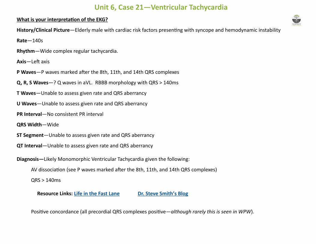
Unit 6, Case 21—Ventricular Tachycardia
Management of Wide Complex Tachycardia
Assess Hemodynamic Stability
• Hypotensive?
• Altered Mental Status?
• Chest Pain?
• Acute CHF Exacerbation?
• History of Long QT?
• Signs of Shock?
• Rate > 150?
• Polymorphic?
1. IMMEDIATE SYNCHRONIZED CARDIOVERSION
2. Proceed down interventions pathway
Interventions
1. Defibrillator pads. Synchronized
cardioversion if patient becomes
unstable
2. Consider Lidocaine (1mg/kg)
3. Procainamide (50mg/min) or
Amiodarone (150mg over 10 min)
4. Consider reversible causes
5. Replete Magnesium, Potassium,
and Calcium as needed
Hypoglycemia
Hypo/Hyperkalemia
Hypoxia
Hypothermia
Hypovolemia
Acidosis
Toxins
Tamponade
Tension Pneumo
Trauma
MI
PE
Created by William Burns, MD Edited by Nick Hartman, MD & Kristen Grabow Moore, MD, MEd
Unit 6, Case 21—Ventricular Tachycardia
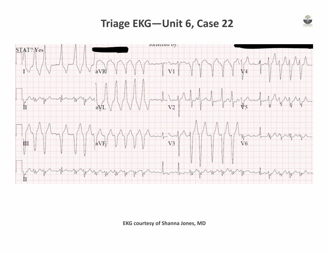
Triage EKG—Unit 6, Case 22
EKG courtesy of Shanna Jones, MD
What is your interpretation of the EKG?
History/Clinical Picture—young man with symptomatic irregular, wide complex tachycardia & borderline unstable vitals Rate— ~150 Rhythm— atrial fibrillation Axis— left axis deviation P Waves— absent Q, R, S Waves— inferior q waves, poor R-wave progression T Waves— lateral TWI (I and aVL) U Waves— not present PR Interval— not applicable QRS Width— wide, around 140 ms ST Segment— depression laterally (V5, V6, I, aVL) QT Interval— prolonged ~530 Diagnosis: Atrial Fibrillation with RVR in the setting of an accessory pathway (i.e. Wolff Parkinson White)
Discussion: It is critical that you recognize this rhythm even if the patient does not tell you that they have WPW. An irregular-ly irregular rhythm, changing QRS morphologies, and a very rapid rate are the hallmarks. Delta waves may also be seen. The ventricular rate at times is in the 250-300 range. This is faster than the AV node can conduct, and it implies the presence of an accessory pathway.
What are your management options for this situation?
Appropriate management options include synchronized cardioversion or chemical cardioversion with procainamide.
What medications are contraindicated in this situation?
Beta-blockers, calcium channel blockers, adenosine, and amiodarone are all incorrect choices as AV-nodal blockade can lead to preferential conduction down the accessory pathway with subsequent hemodynamic collapse, often from ventricular fi-brillation.
Created by Duncan Wilson, MD Edited by Nick Hartman, MD & Kristen Grabow Moore, MD, MEd
Resource Links: Life in the Fast Lane — great overview Dr. Steve Smith’s Blog – good case
Unit 6, Case 22—Afib with RVR & WPW
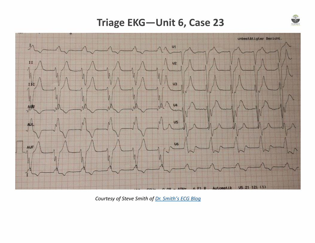
Courtesy of Steve Smith of Dr. Smith’s ECG Blog
Triage EKG—Unit 6, Case 23
What is your interpretation of the EKG?
History/Clinical Picture— A patient with myasthenia gravis with a bizarre wide complex rhythm
Rate— 60-70
Rhythm— lack of p-waves suggest the origin of this rhythm is either junctional or ventricular, or a sinus rhythm deranged by
electrolyte imbalance (which is the case here)
Axis— left axis deviation
P Waves— not present
Q, R, S Waves— bizarre wide R and S waves, some with notching, throughout
T Waves— very peaked, most notable in V2 and V3
U Waves— not seen
PR Interval— not applicable
QRS Width— very wide and bizarre, just under 200ms
ST Segment— discordant STD in V5/6
QT Interval— appears grossly normal
Diagnosis: Hyperkalemia
Discussion: This patient has acute hyperkalemia after induction with succinylcholine in the setting of neuromuscular junction
disease, in this case myasthenia gravis. EKG findings suggestive of hyperkalemia that are seen in this EKG include: peaked T-
waves, prolonged QRS duration with bizarre morphology, and disappearance of p-waves. The potassium level returned at 6.9
and the EKG changes resolved with treatment for hyper K. Treatment should include: stabilization of cardiac membrane
potential with calcium gluconate or calcium chloride, shifting potassium intracellularly with some combination of insulin/
dextrose, albuterol, sodium bicarb, and enhanced clearance with kayexelate, loop diuretics, or renal replacement therapy.
Resource Links: Life in the Fast Lane — great overview Dr. Steve Smith’s Blog – good case
Created by Duncan Wilson, MD Edited by Nick Hartman, MD & Kristen Grabow Moore, MD, MEd
Unit 6, Case 23—Hyperkalemia
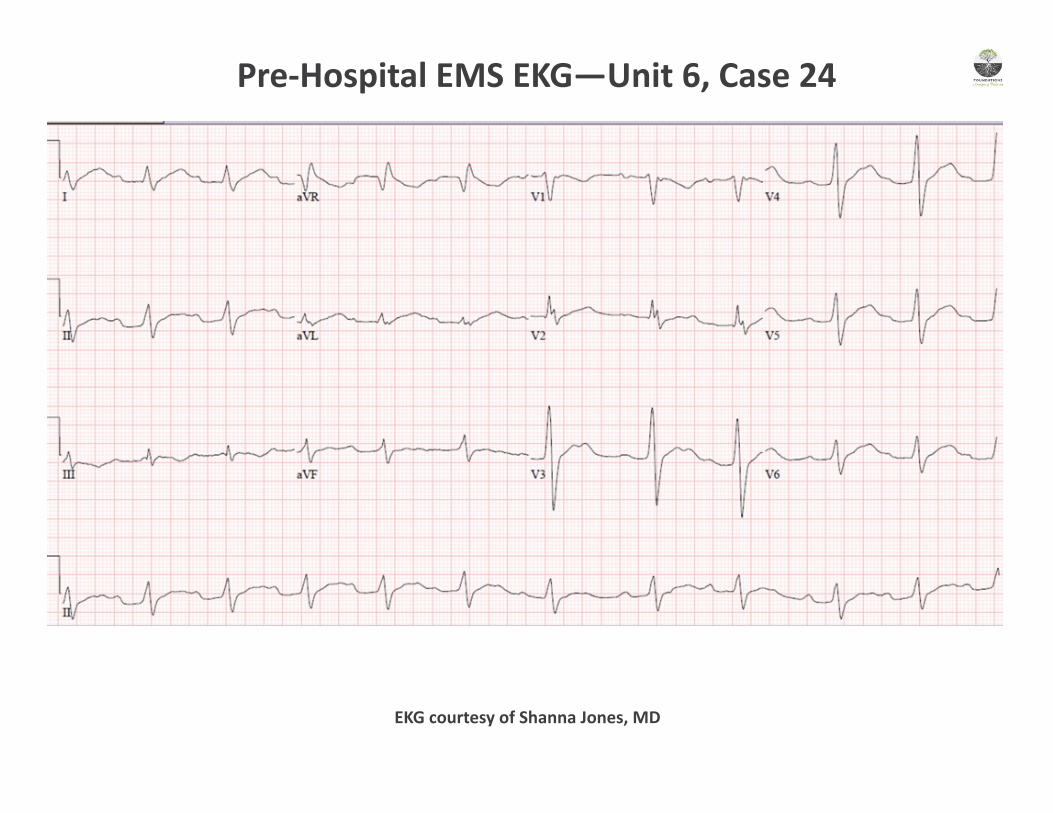
Pre-Hospital EMS EKG—Unit 6, Case 24
EKG courtesy of Shanna Jones, MD
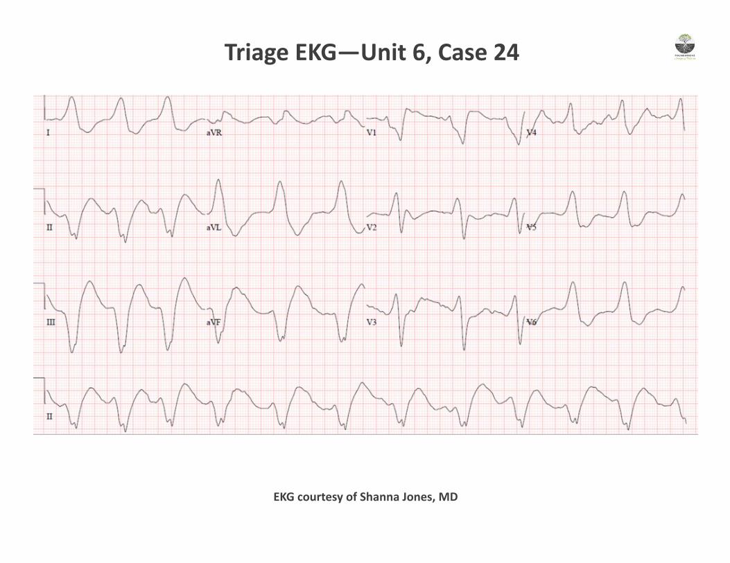
Triage EKG—Unit 6, Case 24
EKG courtesy of Shanna Jones, MD
What is your interpretation of the Pre-Hospital EMS EKG? History/Clinical Picture—young, potentially suicidal woman with hypotension, altered mental status
Rate— 66
Rhythm— sinus rhythm
Axis—normal axis
P Waves—present
Q, R, S Waves—Q wave in aVR, tall R wave in aVR, S waves in the inferior (II, II, aVF), anterior (V2, V3, V4), and lateral (V5, V6, I,) leads
T Waves—abnormal morphology - enlarged in the anterolateral leads with a biphasic component likely incorporating a U wave, flattening/inversion in III, flattening in aVF
U Waves—present, biphasic component of the T wave most obvious in V3 and V4, inverted T in III likely a terminal U wave
PR Interval—prolonged ~268
QRS Width—wide ~168ms
ST Segment— somewhat difficult to assess but no obvious elevation or depression
QT Interval— prolonged ~519
Diagnosis—Sinus Rhythm with conduction delay, prolonged QRS and QT concerning for TCA overdose
Physiology: Sodium channel blockade leading to interventricular conduction delay ECG Characteristics of Na Channel or TCA Overdose QRS > 100 in Lead II, Terminal R > 3mm in aVR, R/S Ratio > 0.7 in aVR
Unit 6, Case 24—Sodium Channel Blockade
P P P P P P P P P P
P
What is your interpretation of the Triage EKG? History/Clinical Picture—young, potentially suicidal woman with hypotension, altered mental status
Rate— 70
Rhythm— wide complex, irregular
Axis—left axis
P Waves— unclear if present, possibly buried in the T waves Q, R, S Waves—q waves in II, II, and aVF, improved R wave progression in the precordial leads, tall R wave in aVR
T Waves—Abnormal morphology given increased size diffusely and particularly slurred appearance in aVR and V1
U Waves—None apparent but very limited assessment given abnormal T waves
PR Interval—not applicable
QRS Width—wide ~182ms
ST Segment—very difficult to assess given abnormal T wave morphology
QT Interval—prolonged ~632
Diagnosis—Severe Interventricular Conduction Delay consistent with Sodium Channel Blockade consistent with TCA Overdose
ECG Characteristics of Na Channel or TCA Overdose QRS > 100 in Lead II, Terminal R > 3mm in aVR, R/S Ratio > 0.7 in aVR Resource Links: Life in the Fast Lane Dr. Steve Smith’s Blog
Unit 6, Case 24—Sodium Channel Blockade
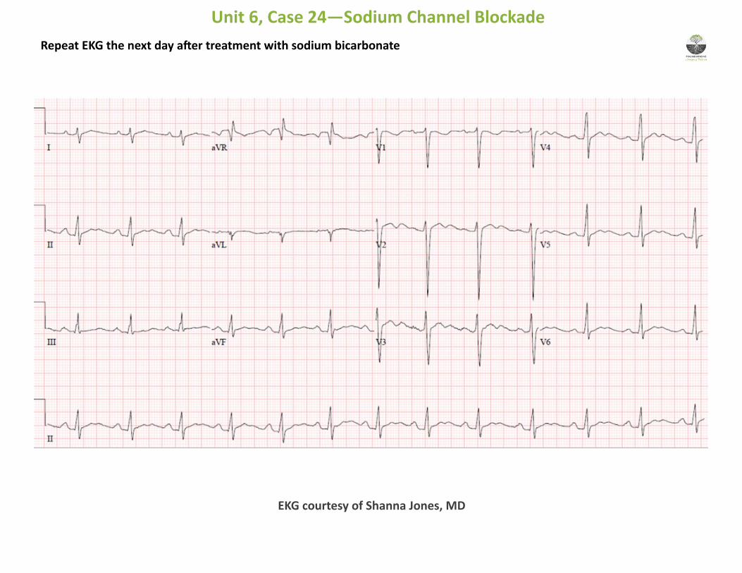
Repeat EKG the next day after treatment with sodium bicarbonate
Unit 6, Case 24—Sodium Channel Blockade
EKG courtesy of Shanna Jones, MD
What aspects of the EKG changed significantly when it was repeated?
1. The QRS widened dramatically
What are possible causes of this dysrhythmia?
Tricyclic Antidepressants (Elavil, Doxepin, __triptylines, __ipramine)
Antiarrythmics (Procainamide, Flecainide, Encainamide, Amiodarone)
Local Anesthetics (Bupivacaine, Ropivacaine)
Antimalarials (Chloroquine, Hydroxychloroquine)
Propanolol
Carbamazepine
What complications should you watch for?
QRS > 100 predictive of seizures
QRS > 160 predictive of VT/VF
What treatment options do you have for this patient?
Sodium bicarb (1-2 mg/kg) q3-5 minutes until BP improves and QRS begins to narrow
Support BP with boluses and pressors as needed
Created by William Burns, MD Edited by Nick Hartman, MD & Kristen Grabow Moore, MD, MEd
Unit 6, Case 24—Sodium Channel Blockade