Embed Size (px)
Citation preview
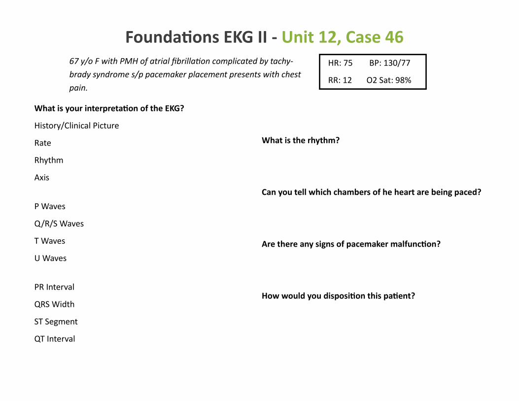
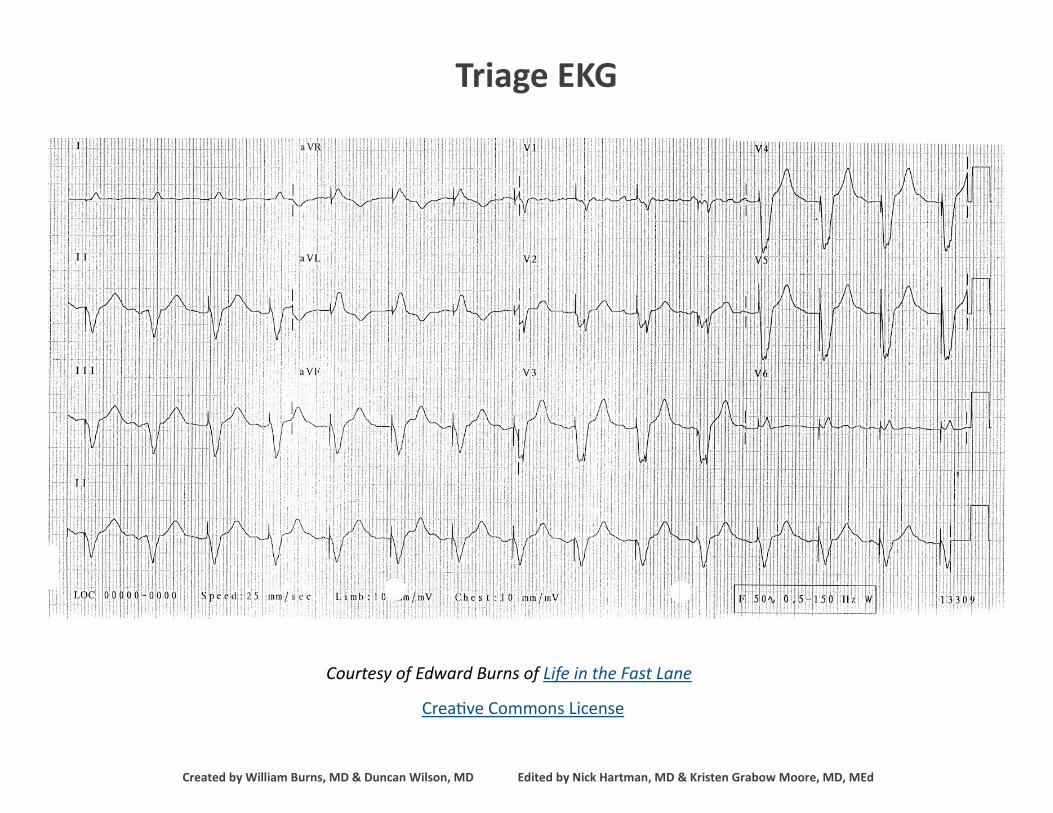
67 y/o F with PMH of atrial fibrillation complicated by tachy-
brady syndrome s/p pacemaker placement presents with chest
pain.
HR: 75 BP: 130/77
RR: 12 O2 Sat: 98%
What is your interpretation of the EKG?
History/Clinical Picture
Rate
Rhythm
Axis
P Waves
Q/R/S Waves
T Waves
U Waves
PR Interval
QRS Width
ST Segment
QT Interval
What is the rhythm?
Can you tell which chambers of he heart are being paced?
Are there any signs of pacemaker malfunction?
How would you disposition this patient?
Foundations EKG II - Unit 12, Case 46
Courtesy of Edward Burns of Life in the Fast Lane
Creative Commons License
Triage EKG
Created by William Burns, MD & Duncan Wilson, MD Edited by Nick Hartman, MD & Kristen Grabow Moore, MD, MEd