Embed Size (px)
Citation preview
FISHERIES RESEARCH BOARD OF CANADA
Translation Series No. 1496
Structure of the glandular - stomach of the opisthobranchiate molluscs (Gastropoda, Opisthobranchia)
By Yu. S. Minichev
Original title: Stroenié 'zhelezistogo zheludka u zadnezhabernykh mollyuskov:(Gastropôda,
'Opïsthobranchia). '
From: Zoologicheskii Zhurnal. (Zoological Journal), 68 (12): 1780-1787, 1969.
Translated by the Translation Bureau(HPF) Foreign Languages Division ,
Department of the Secretary of State of Canada
Fisheries Research Board of Canada Biological Station
Nanaimo, B.C.
1970
20 pages typescript
DEPARTMENT OF THE SECRETARY OF STATE TRANSLATION BUREAU
FOREIGN LANGUAGES DIVISION
01) fee PI-
SECRÉTARIAT D'É .TAT BUREAU DES TRADUCTIONS
DIVISION DES LANGUES ÉTRANGÈRES CANADA
INTO - EN TRANSLATED FROM - TRADUCTION DE
RUSSIAN ENGLISH
DATE OF PUBLICATION DATE DE PUBLICATION
VOLUME ISSUE NO. NUMÉRO
YEAR ANNàE
1969
PUBLISH ER - 'EDITEUR
Academy of Sciences of the U.S.S.R. Publishing House n Nauka "
PLACE OF PUBLICATION LIEU DE PUBLICATION
Moscow
PAGE NUMBERS IN ORIGINAL NUMÉROS DES PAGES DANS
• L'ORIGINAL
17go NUMBER OF TYPED PAGES
NOMBRE IDE PAGES DACTYLOGRAPHIÉES
20
Dr. D.B. Quayle, Biol.Stn. - Nanaimo, B.C.
DATE COMPLETED ACHEVe LE
PERSON REQUESTING DEMANDE FAR
JUL 2 2 1970
AUTHOR,- AUTEUR
Yu. S • MINIGHEI
TITLE IN ENGLISH - TITRE ANGLAIS
The structure of the glandular stomach in the opisthobranchiate molluscs Title in foreign language (transliterate foreign characters)
Stroyeniye Zhelezistovo zheludka u zadnezhabernykh molluskov (autrejeral p Olkletkobpanchla )
REF5RENCE IN FOREIGN Ir ANGUA,9, E (NAME OF BOOK OR PUBLICATION) IN FULL. TRANSLITERATE FOREIGN CHAZtACTERS. REFERENCE EN LANGUE ETRANGERE (NOM DU LIVRE OU PUBLICATION), AU COMPLET. TRANSCRIRE EN CARACTERES PHONÉTIQUES.
Zoologicheskii Zhurnal
REFERENCE IN ENGLISH - RF .E' RENCE EN ANGLAIS
Zoological Journal
REQUESTING DEPARTMENT Fisheries & Forestry TRANSLATION BUREAU' NO. MIN 1ST ERE-CLIENT NOTRE DOSSIER N C'
0259
• BRANCH OR DIVISION DIRECTION OU DIVISION
Fisheries Research Board TRANSLATOR (INITIALS) H.P.F. TRADUCTEUR (INITIALESI
YOUR NUMBER
VOTRE DOSSIER NO 769-18-14 UNEDITED DRAFT TRANSLATION
Only for information
DATE OF REQUEST 1.4.70 . TRADUCTION NON REVISÉE DATE DE LA DEMANDE
, Informsation seulement
SOS-200- 10-o (REV. 2/68) .
•
DEPARTMENT OF THE SECRETARY OF STATE
TRANSLATION BUREAU
FOREIGN LANGUAGES DIVISION
CANADA
ryc m-76 SECRÉTARIAT D'ÉTAT
BUREAU DES TRADUCTIONS
DIVISION DES LANGUES ÉTRANGÈRES
CLIENTS NO. DEPARTMENT DIVISI ON/BRANCH . CITY
N° DU CLIENT MINISTERE I DIVISION/DIRECTION VILLE
769-18-14 Fisheries & Forestry Fisheries Research Bd. Nanaimo e B
BUREAU NO. LANGUAGE TRANSLATOR (I NITI ALS) DATE N° DU BUREAU LANGUE TRADUCTEUR (INITIALES)
0259 Russian H.P.F. JUL 22 1970
uNLDiTED DRAFT TRANSLATION ' Only for information
TRADUCTION NON REVISÉE Information seulement
Yu. S.. MINIGHa Stroyentye'zhelezietovo zheludka u zadnezhabernykh mollyuskov ( gmtronocla p Oqiehobraneig ) Zoologicheskii Zhurnal e Academy of Sciences of the U.S.S.R., Publishing Rouse ° Naukev° , Vol. XLVIII (12) : 1780-1787 1969 •
.1
ifte The Structure of the Glandular Stomach irgopisthobranchiate
Molluscs (Gastropoda, Opisthobranchia). UNEDITED DRAFT TRANSLATION Only for information
BY TRADUCTION NON REVISÉE • Information seulement
Yu. S.MINICHEV
Biological Research Institute of the Leningrad State University
SUMMARY
In this article the author studied morphology of
the stomachs in the Opisthobranchia. In different orders of
the Opisthobranchia the gastric adnexa are not homologous.
The author showed the importance of the alimentary system
in the solution of a number of problems relating to the
systematics and phylogeny of the Opisthobranchia.
In recent times great importance has been ascribed
to the peculiar features of the stomach structure for the
clarification of different questions pertaining to the
systematics and phylogeny of the Gastropoda (Graham, 1939,
1949; Johansson, 1941; Morton, 19521953, 1955; Fretter &
Graham, 1962; et alii). However, most of the works were
carried out on the pulmonate and prosobranchiate molluscs.
There are some works devoted to the study of digestive organs
the of single forms of/Opisthobranchia (Howells, 1936, 1942;
Millot, 1937,1938; Fretter,, 1939, 1940; Forrest, 1953;
Hurst,.1965; et alii),.but so far no light has been throWn
on the evolution of the stomach in different phylogenetic
ramifications.
We'give below some conclusions arrived at as the
result of studies concerned with the morphology of the
digestive organs in the opisthobranciate molluscs. The
collections made by the expedition ship "VITIAZ" in the
Pacific ocean,and personal . coilections Made by the author in
the White Sea and in the Sea of Japan provided the material
for this article.
Peculiar Features in the Structure of the Stomach
in Different Groups of the Opisthobranchiate Molluscs.
The majority of the representatives of the Cephalaspidea -
the earliest and:most primitive order of the opisthobranchiate
molluscs - are benthyc animals that feed on deposits, mainly
of plant origin, arriving from the surface. In this respect
they retained an early mode of life .peCuliar to the ancestors
of the Gastropoda (Graham, 1955)- With the type of nutrition
based on food of plant origin (phytophagous or herbivorous
nutrition) is associated an entirely singular structural plan
of the digestive system, and of the stomach in the first place.
On summing up the basic data pertaining to the structure of
the stomach in primitive molluscs (Yonge, 1932; Graham, 1939,
1949; Morton, 1952, 1953; et alii) one may distinguish several
most characteristic features. A greater portion of the
stomach wall is lined with the ciliary epithelium; in
specific areas there develops a solid cuticular lining,
forming a singular gastric shield. A gastric diverticulum,
which serves to increase the sorting area, arises not in-
frequently in various phylogenetic stems; the connective tissue
surrounding the diverticulum is a site where the phagocytes,
participating in the process of digestion, aggregate. The
presence of the sac of the protostyle, which is adjacent to
• the groove running along the anterior portion of the gut, is
a feature already found as a specific characteristic in the
ancestors of the Gastropoda (Owen, 1956; Beklemishev, 1964).
Through the differentiation of the sac of the protostyle from
the gut, and, as the result of enhancement of the role played
by various hydrolytic enzymes, the protostyle is transformed
into the crystalline style (Johansson, 1941; Morton, 1952).
The latter is present mainly in the phytophagous forms.
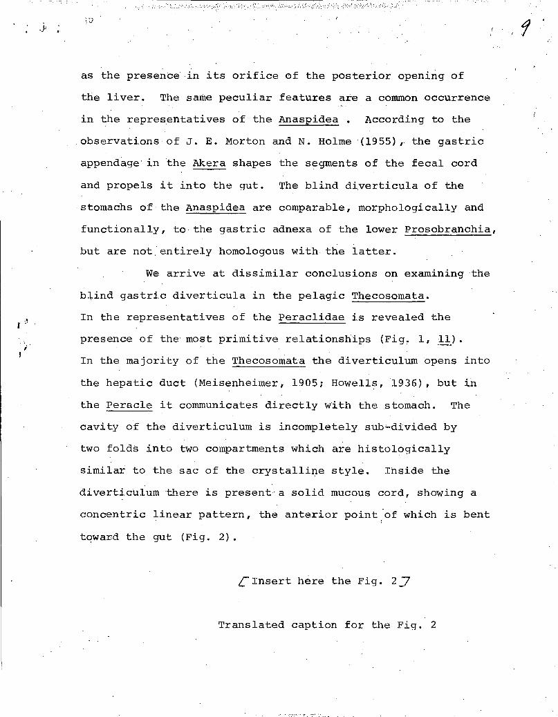
Insert here the Fig. 17
Translated caption for the fig. 1
Fig. 1. Schematic drawings of the structure of stomachs in
different Gastropoda. Arrows indicate the direction of the
process of morphological transformation.
1. - Lymnaea, 2. - Ophicardelus, 3. - Acteon, 4. - Bullacta,
/2\
2 eio(,(
„ „
• •
CNOMIA ilT0e111151 "A“.".1 ■'0 Gastrc)pc,da. Crt /C•:1: ■ 3NIII y2a3ali1 i1anpn13-
upeoCip33ùB:dii
1— Lymnriea, 2 Ophicar tic1:1::, 3 — A.:teon, 4 — 131111a,la, 5 --Aplv.ia, —Tro,:liidno,
—11::Inince,i, 9 — II 12—BatIty3oik; a — Ku:tn.:a, g. co—rp ,..:;,.
h — IICffli. Oc — i0a0;l. tp s p — (1,
2, G — sio .\lurto:%, r.i55; 3-- Fre11cr anti Graham, ( 951)
5. - Aplysia, 6. - Trochidae, 7. - Ringiculoides,
8. - Haminoea, 9. - Akera, 10. - Anopsia, 11. - t'oracle,
12. - Bathydoris; d-gut, g. co - Gastric diverticulum,
h - liver, hl - Hepatic duct, oe - oesophagus, tp - typhlosole,
sp - sac of the protostyle (1,2,6 - according to Morton, 1955;
3 - according to Fretter & Graham, 1954).
In Fig. 1 are depicted schematic drawings of the
structure of the stomach in different Gastropoda. The
Trochidae (Fig. 1, 6) occupy the central position, possessing,
as they do, a complex of primitive traits in their stomach
structure. From the Trochidae type of the stomach have
developed (not in the sense of phylogenesis but in the sense
of direction of the process of morphological transformation)
the stomachs of the Pulmonata (Fig. 1, 1, 2) and the
Opisthobranchia
Primitive features in the structure of the stomach
may be found in different opisthobranchiate elluscs, and
in the first place - in the representatives of the
Cephalaspidea. In particular, the stomach of the Ringiculoides
kurilensis Minichev is divided both morphologically and
functionally into two chambers (Minichev, 1967). The posterior
chamber (Fig.1,7) is lined with a cuticular layer, and exteriorly
it is surrounded by broad muscular bands; the anterior chamber,
with thinner walls, is formed by the ciliary epithelium. A
similar structure of the stomach is also characteristic in
the other members of the Ringiculidae and, possibly, the
Acteonidae (Fig. 1, 3). The digestive diverticula
(the liver) open out into the anterior chamber by two
independent ducts. The orifice of the left liver is situated
closer to the oesophagus, and the orifice of the right liver
is located in the lower portion of the gut. The ridges formed
by tall ciliary epithelium, and situated between the orifices
of the liver and the gut, represent the major and minor
typhlosoles. In the anterior portion of the gut, the typhlosoles
border the little furrow of the gut which is a characteristic
feature of the sac of the protostyle of the prosobranchiate
molluscs. A series of transverse ciliary groovlets is
associated with the major typhlosole. This area of the stomach,
differentiated morphologically into a small chamber,
corresponds to the ciliary sorting zone of the Archaeogastropoda.
The anterior portion of the gut is widened and possesses all the
features peculiar to the sac of the protostyle (the disposition of
the typhlosoles, the presence of a sulcus in the gut, tall ciliary
eplthelium ,etc.). The semi-liquid mass filling the anterior portion
of the gut and projecting into the lumen of the stomach rep-
resents the protostyle of the most primitive type. In the
Ringiculoides there is present, in the connective tissue
surrounding the anterior chamber of the stomach, a great number
of minute (with a diameter of about 7 microns) amoebocytes.
Experimental data indicate that the region of the protostyle is
the site where the most energetic phagocytosis occurs
robservations on the nutrition of Diaphana globosa (Loven)2
Some primitive features may also be found in the
stomachs of , other Cephalaspidea. Thus,.J.,E. Morton (1955),
referring to the data obtained by V. Fretter. (1939), noted
a great similarity in the structure of the stomach of the
Haminoea to that of the primitive Pulmonata. In the stomach
of some species of the Refusa are retained the major and
minor typhlosoles, associated as they àÉe", with a pair of
the hepatic ducts. In the Acteon the posterior portion of
the stomach is lined with the cuticula and yet retains the
longitudinal sorting ciliary striplets (Fretter, 1939). The
cuticular area of the 'stomach in the Ringiculidae, and
particularly in the Acteonidae, resembles the gastric
diverticulum in the primitive Prosobranchia. However, it may
be said that this area rather corresponds to the gastric shield
in the prosobranchiate molluscs.
A transition from the phytophagous mode of life to
the predatory one occured in the Cephalaspidea fairly early
• in the course of their evolution. Apparently, it is only .
the Atyidae that retained in pure form the pribary mode of
feeding. Nevertheless, even in the latter one may observe a
considerable change in, and simplification of, the stomach.
This transformation is doubtless associated wi-th the formation
of the composite "masticatory" stomachs ("Gizzard"). The
stomach of Bullacta eXarata (Philippi) (Fig. 1, 4) underwent a
particularly conspicuous change. In this form the oesophagus
merges into the widened portion of the gut from which the former
is separated by an annular fold with a séries of thread like
appendages. In the Bullacta occurred the morphological merger
of the glandular stomach and the gut. Along the ventral
surface of the stomach runs a high ridge, bent at its anterior
portion, the typhlosole. The anterior end of the typhlosole,
together with the two large dorsal projections, form the limits
of a narrow chamber into which open the hepatic ducts. The
left side of the stomach bears a series of obliquely placed
folds which are furnished, in their turn, with transverse
ridges. To the right of the typhlosole runs a deep ciliary
sulcus associated in the region of the hepatic orifice with
small transverse folds. The interpretation of all these
structures is somewhat difficult; the primitive relationships
are preserved only in so far as the presence of a pair of the
hepatic ducts is concerned, and also in the presence of the
major typhlosole. It is interesting to note that the connective
tissue with the blood bearing lacunae - which is character-
istic for the primitive forms - is absent in the Bullacta.
The majority of the Cephalaspidea are predators
which as yet did not succeed, in the process of evolution, to
acquire the crystalline style but have already lost the early
features of the stomach of the phytophagous forms. K. Kubomura
(1957) found the crystalline style in the Philinidae. The
author's investigations in the case of Philine argenttta
Gould and Philine scalpta Adams confirmed the data furnished
by Kubomura; however, we consider that these species possess
a sac of the protostyle. The presence of the carbohydrases
in the mucous mass filling the initial portion of the gut has
not been proved. Moreover, this mass does not show the
concentric series of layers, a characteristic of the ,
crystalline styles in other molluscs. The stomach of the
Philinidae is indeed a rudimentary organ the main function
of which is a temporary accumulation of food particles prior
to their entrance into the diverticula of the liver.
In many Opïsthobranchia the stomach is furnished
with special blind diverticula the homology and iunction of
which has not been completely cleared up. In the Anaspidea
a blind diverticulum, lined with the ciliary epithelium and
furnished with a longitudinal epithelial fold, leads out
from the posterior portion of the stomach (Fig. 1; 5, 9) (Howells,
1942; Morton & Holme, 1955). In the Stylochelus the
. diverticulum is bent or twisted spirally and contains fecal
masses shaped like a mucous cord. A rudiment of a similar
diverticùluM is also present in the Anopsiidae .(Fig. 1, 10)
a family which is connected phylogenetically with the
Anaspidea. These formations may be likened to either the sac
of the crystalline style or the gastric diverticulum of the
pulmonate and the prosobrançhiate mollusc
diverticula of the Anaspidea, and especially. of the Aplysildae,
resemble greatly those in the Ellobiidae, the Turbinidae and
others (Morton, 1955, 1955a ). A dharacteristic feature of the
gastric diverticulum_in the PulMonata and the Prosobranchia
is the projection into it of the posterior portion of the major
typhlosole with an area of the sorting ciliary z one, as well
S. The gastric
as the presence in its orifice of the posterior opening of
the liver. The same peculiar features are a common occurrence
in the representatives of the Anaspidea . According to the
observations of J. E. Morton and N. Holme (1955), the gastric
appendage in the Akera shapes the segments of the fecal cord•
and propels it into the gut. The blind diverticula of the
stomachs of the Anaspidea are comparable, morphologically and
functionally, to the gastric adnexa of the lower Prosobranchia,
but are not entirely homologous with the latter.
We arrive at dissimilar conclusions on examining the
blind gastric diverticula in the pelagic Thecosomata.
In the representatives of the Peraclidae is revealed the
presence of the most primitive relationships (Fig. 1, 11).
In the majority of the Thecosomata the diverticulum opens into
the hepatic duct (Meisenheimer, 1905; Howells, 1936), but in
the Peracle it communicates directly with the stomach. The
cavity of the diverticulum is incompletely sub-divided by
two folds into two compartments which are histologically
similar to the sac of the crystalline style. Inside the
diverticulum there is present'a solid mucous cord, showing a
concentric linear pattern, the anterior point -of which is bent
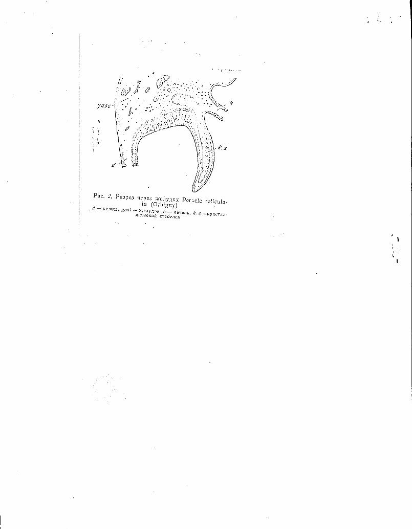
toward the gut (Fig. 2).
(*Insert here the Fig. 2 j
Translated caption for the Fig.
•. fî
! "(.,,D 'iS' • • • • ". ; .1 •• ..
Pile. 2. 13 a3pe3 ilepe3 Peracle ta (OE- )iguy) (1— iimuua, gas! — n ) ,-,OK. h — r eidEr, k. s—tzpacra.1-
cre5o:ieh:
Fig. 2. Section through the stomach of the Peracle reticulata
(Orbigny) d-gut, gast - stomach, h - liver, k.s - crystalline
style
C. Yonge (1926, 1932), while discussing the function of the
stomach, put forward a suggestion that blind diverticula in
the Thecosomata are the neoplastic formations which arose as
the result of the molluscs effecting a transition to the
phytophagous mode of nutrition. The alimentary system of the•
Thecosomata went through a singular evolutionary process. In
particular, the essential food distributing mechanism in _these
molluscs is associated with their fins (Yonge, 1926). Yonge
assumed that the "masticatory" stomach ("gizzard") in the
Thecosomata was preserved as a feature common to their
carnivorous ancestors and is now a functionally rudimentary
organ. We consider that the ancestors of the Thecosomata were
phytophagous forms; the author's observations and the data
given by H. Howells (1936) indicate that the gizzard of the
Thecosomata is a highly efficient organ ensuring the
trituration of different food animals with the hard outer skeleton
(radiolaria, foraminifora, diatoms, etc.). Aeparently, the
Thecosomata and the Cephalaspidea share a common origin from
the early prosobranchiate molluscs which possess a primitive
protostyle. The latter is retained in some of the Cephalaspidea
and is transformed into the crystalline style in the Thecosomata.
It is interesting to note thàt the blind stomach
diverticula are present in some nudibranchiate molluscs -
including the carnivorous forms - the most highly organized
group among the Opisthobranchia. In the Anthobranchia
[formerly the Doridacea which the author classified as an
independent order (Minichev, 1968) ,7, the blind diverticulum
is usually located at the base of the gut and is lined with
the tall epithelium without the cuticle and the cilia. In a
number of cases this organ has a glandular character and is
histologically similar to the liver. As is known, in the
doridid larvae two lobes of the liver are laid down, but one
of them disappears in the process of metamorphosis (Thompson,
1958), or is retained in the form of the socalled"gall
bladder". Doubtless, the blind diverticulum is - at least in
some species of the Anthobranchia - the homologue of the right
lobe of the liver in the lower Opisthobranchia. Of interest
is the supposition of N. Millot (1938) that gastric adnexa
in the Anthobranchia and the Anaspidea are functionally.similar.
If this supposition be true, we have here a good example of
the substitution of functions, i.e., the transformation of
the secretory organ (the liver) into the excretory and
distributing organ (an analogue of the gastrib diverticulum).
Traces of the primitive structural plan may be
revealed, in the main, only in the stomachs of •the lower
Opisthobranchia. Already in the Çephalaspidea one may observe
the reduction (anatomical) of the structure associated with
the preliminary treatment and distribution of food. The
.tomach becomes a site where the food accumulates for a short
period of time, whereas the basic role in the process of
digestion is acquired by the oesophagus. Indeed, it is in
the oesophagus that food undergoes mechanical treatment and
is subjected to the action of enzymes. Into the gizzard
(which is formed ontogenetically from the lower portion of
the oesophagus) are secreted the enzymes from the liver, the
enzymes that hydrolyse starch, glycogen, proteins, etc.
(Fretter, 1939). The oesophagus acquires the ability to
perform peristaltic movements; and in it the food particles
may remain for a long time. The dissolved substances and
small food particles enter the liver where their absorption
and phagocytosis take place. Larger particles from the
oesophagus and the undigested material from the liver are
ejected into the gut.
A further simplification of the stomach occurs in
many evolutionary forms. In some instances one may observe
a complete substitution of the stomach,by the liver. This
process begins already in the higher Cephalaspidea. Thus, in
the Gastropteron rubrum (Raf.) the two primary hepatic ducts
are sub-divided into 8 - 10 secondary ducts; and, in this
species, a greater portion of the stomach wall assumes
glandular character. In the Enotepteron flavum Minichev only
insignificant areas of the stomach remain, those furnished with.
the ciliary epithelium, located among the numerous hepatic ducts.
Substitution of the stomach by the liver is very
characteristic for the representatives of the Gymnosomata
order. In particular, in the Anopsia the oesophagus opens
into a chamber which is almost completely formed by the
hepatic epithelium (Figi 1, 102; in other forms the stomach
becomes completely reduced(anatomically). Doubtless, the
reduction of the stomach in the 2Emmarmatil is cOnditioned
by the mode of nutrition: they suck out the soft tissues
of the shelled Pteropoda molluscs. Substitution of the
stomach by the liver in the Gymnosomata fully reseffibles a
somewhat SiMilar phenomenon observable in the parasitic
• Prosobranchia (Ivanov, 1945). Reduction of the stomach and
its replacement by the liver in the Gastropoda molluscs is
doubtless •associated wità feeding on food of high calory
value, needing no preliminary mechanical treatment (trituration)
or a special method of distribution along the alimentary tract.
DISCUSSION
The primitive representatives of different phylogen-
etical branches of the Opisthobranchia retained basic features
of the structure of the alimentary tract of the ancestral
forms. The presence of intracellular digestion, of the ciliary
sorting mechanisms in the stomach, the primitive form of the
protostyle, participation of the blood elements in the pro-
cesses of digestion, etc., all are early peculiar features
common in some lower opisthobranchiate molluscs. The change
in the functions of the anterior portion of the digestive
tube, an increased rôle played by the digestion occurring
in the alimentary tract cavities, a decreasing role of the
phagocytosis, and substitution of the stomach by the liver,
all are the principal paths followed by evolution of the
alimentary tract in the Opisthobranchia.
The lower Cephalaspidea possess, in one or another
combination, the following distinctive features of the
stomach: the ciliary sorting zone with growelets and
ridges converging upon the intestinal sulcus; a zone with the
cuticular lining; a sac of the protostyle adjacent to the
intestinal sulcus; and paired hepatic ducts. On comparing the
digestive system of the Opisthobranchia with that of the
Prosobranchia, one may note a very great resemblance of the
stomach structure in the Cephalaspidea and the
Archaeogastropoda (Graham, 1949; Motton, 1953, 1955a; et alii).
The other sections of the digestive system of the Cephalaspidea
likewise indicate that this group is closely related to the
early primitive Prosobranchia. In particular, the Acteon has
a broad non-specialized radula (Fretter, 1939; Gabe et
.Prenant, 1952; Fretter & Graham, 1954; Hurst, 1965) which has
the following characteristic features: a very large number
of small radular plates (teeth?)of a single type, but not
differentiated into the lateral and central plates; the
weakly differentiated odontoblasts; and the odontophore
occupying the central and lateral walls of the pharynx. These
specific features do, not allow us to trace the descent of the
Actèonidae from a type of the present day Prosobranchia. Even
in the extremely primitive forms of the prosobranchiate
molluscs there are present several groups of radular plates
differing in form, size and their location. It is possible
that the Acteon retained its pharyngeal armament, which
originated at the primary sources of the formation of the
class of Gastropoda.
Thus,in the structure of the alimentary system,
we bring to light one more proof of the origin of the
Cephalaspidea - and therefor, of all the Opisthobranchia -
from the very early prosobranchiate molluscs.
First stages in the evolution of the stomach were
doubtless similar in the Opisthobranchia and the Pulmonata.
Yet, as time went on, a sharp divergence became discernible:
in the opisthobranchiate molluscs there occurred a gradual
morphological and functional simplification of the stomach;
whereas the pulmonate molluscs, while retaining many
primitive features, developed along the path of enhancing the
role of musculature in the function of the stomach. The
presence of muscular stomach (gizzard) is a characteristic
trait of the digestive tract in the Basommatophora and
several other groups of the Pulmonata. Many Opisthobranchia
possess the so-called "gizzard", a muscular chamber, often
furnished with the chitinoid teeth, located in front of the
glandular stomach. This chamber is of the ectodermal origin
and is not homologous to the muscular stomach of the pulmonate
molluscs. It must be noted here that in the Ringiculidae,
the Ellobiidae and the Trochidae (the "primitive" families of
the three sub-classes of the Gastropoda) the glandular stomach
t •
has a muscular envelope. In the Opisthobranchia this
musculature becomes reduced and is functionally replaced by the
muscles of the oesophagus; whereas it is retained in the
pulmonate molluscs and its role is progressively enhanced.
The "masticatory' stomach of the Cephalaspidea takes
shape fairly early in the course of phylogenesis, but, already
in the representatives of higher families (which have become
actively predatory), it disappears. In the phytophagous
Anaspidea it is retained in a modified form. It is
interesting to note that the chitinoid plates of the
"masticatory" stomach may contain cellulase
alii, 1951; a revue of digestive enzymes in molluscs vide
Stone & Morton, 1958), which fact is doubtless connected with
the progressively increasing role of the body cavity food
digestion in this portion of the alimentary tract.
In ào far as its function is concerned the evolution
of the digestive system followed a similar pattern in the
pulmonate and the opisthobranchiate molluscs. In particular,
in the primitive forms the phagocytic function of the
amoebocytes of the blood - the cells that seize and digest
food particles from the stomach - is displayed very prom-
inently. In the higher forms - for example, in the
nudibranchiate molluscs - the role of Wandering phagocytes in
the process of digestion becomes diminished, but there
appears the so-called process of "fragmentation of the phagocytes"
from the epithelium of the glands of the alimentary tract
(Millot, 1937; Forrest, 1953). Such a change in the phagocytic
(fiaàkilitéito et
1(i` I
mechanics doubtless occurs fairly, frequently in different
phylogenetic stems of the Opisthobranchia and the Pulmonata.
Distinctive structural features of the stomach may
acquire great importance in the solution of a certain number
of problems in systematics (taxonomy?). Thus, the
structural singularity of the stomach in the Bullacta
(morphological unification of the stomach and the gut,
development of the secondary sorting structures, etc.) bears
witness to profound functional changes of the entire
alimentary tract in this genus. The causes of these changes
are not dlear,but they are mot conditioned by the type of
feeding, for the Bullacta, as well as the typical Alvidae,
are the unspecialized phytophagous forms. Tchangsi (1934),
who studied in detail the organization of Bullacta, placed
this form in the Scaphandridae, basing his decision on the
similar structure of their radulas. Yet, the distinctive
features of the "masticatory" stomach, of the nervous system,
and of their shells indicate a close relationship between
the Bullacta and the Atyidae. J. Thiele (1926) placed the
genus in the Atyidae, as a sub-family. The original
structure of the stomach and of the ctenidium, and of the sex
organs enable one to separate •the Bullacta, as an independent
family, under the name the Bullactidae Thiele, 1926.
LITERATURE
r2,Johansson, J., 1941: Ein Beitrag zur Kentniss der
Kristallstielsackes der Mollusken und der Flimmerbewegung in
„ /8
demselben (A contribution to the knowledge of the
crystalline style sac, in molluscs and the ciliary movement
in this organ). Arch. Zool., 33, 3:1-8.
Pi. Meisenheimer, J., 1905: Pteropoda, Wiss. Ergebn. Deutsch.
• Tifsee Exp. "Valdivia” (Pteropoda. Scientific results of Germain
deep-sea expedition "Valdivia").
Thiele,'J., 1926: Gastropoda, Handbuch der Zoologie,
5 (Gastropoda. Handbook of zoology, 5).
1. Beklemishev, V.N.,1964.2771ncip1es of Comparative Anatomy of /(Osnovy sravnitel'noy anatomii bespozvonochnykh)/ Invertebrates, 2, "Nauka" Publishing House, Moscow.
2. Ivanov, A.V., 1945. Morphologica1 Adaptation of the Digestive
System of Parasitic GastroPoda, Uéhenye zapiski, Lenin-
grad University, Series: Biology, 15: 112-119;. Morpholo-
gicheskié adaptatsii pishchevaritellnoy sistemy u para-
ziticheskikh Gastropoda. •
3. Minichev, Yu.S., 1967. Issledovaniya po morfologii.nizshikh
Opisthobranchia, Tr. Zool. inst. AN sssa (Research on
the Morphology of Lower Opisthobranchia). Trudy, Zool.
Institute, AS USSR, 44:109-182.- 1968. 0 proiskhozhdenii
i sisteme golozhabernykh molluskov. Molluski i ikh roi' v
ekosistemakh, "Nauka" Publ. House: 11 - 13, Leningrad.
(The Origin and the System of Nudibranchia. Mcilllusks and
Their Role in Eco-Systems).
'4. - • f
1
F.,exae M 1 ut e n 13. H., 1984. Ocuoubt epanturreabitort • anaromnis iecnoanonoitubrx, 2, 113:1-no «Ilaytia», M.
11 nation A. B., 1945. MorultomornItectine raw-mum! nuntenapwre:tatort ClICTCM14 . y na- pn3writtieenitx Gastropod a, y‘t. ann. TIonnurp. yit-rn, cep. 6noit, 15: 112-119.
• u n e a 10. C. 1967. 14cne;tonannst no moptp:tornit iijuitx Opisthobranchia, Tp. CCCP, •14: 109-4182.— 19t38. O npottexoeitut n ClieTeMe romma-
6cptibtx maaanicitott. 4\101.110C1■ 11 n ux pom, u 3Koclicremax, 1-11,1-rio (1-1ayNa»: 11— 13, JI.
e-/' • •F or r e st J. E., 1953. On the feeding, habits and morphology of alimentary canal in lit-toral Dorids, Proc. Linnenn Soc , London, 161, 2: 225-235.
S. Frett er V., 1939, The structure and function of the alitnerkary canal of some tecti-branch molluscs, with a note on excretion, Trans. Roy, Sm.i.Edinburgh, 59, 2: 590-6l6.-10•l0. On the structure of the gut of the ascoglossai nudibranchs, Proc. Zool.
• . Soc. London, 110: 185-198. e•P r et t er V. and G Tabora A. , . 1951. Observations on he primitive opisthobranch •
. mollusc Actaeon tornatilis (L.), J. marine biol. Assoc. U. K., 28, 2: 493-32.— 1962. British prosobranch molluscs, London.
7-C a b e M. et Pr en an t M. • 1952. Recherches sur la grine radulaire des mollusques. L'appareil radulaire d'Adeon tornatilis, Arch. Zool. •ptl. et en. Notes et Revue,
• 69: 15-25, deC 'r a h a in A., 1939, On the structure of the alimentary canal of tityle•bearing prom).
• branchs, Proc, Zool, Soc, London, le. 75-112.—ion. The molluscan stomach, Trans. Poy, Soc. Edinburgh, 61, 3: 737-778.-- 195F. Molluscan diets, Proc, malacol.
• Soc. London, 31: l44-159. 9,11 a sh im o t o Y., Ma t su mot o S. and Hib iya T., 1951. Comparative studies on
. the stomachal plates and crystalline style.. 1 On Oe enzymes of the stomachal plates in an opisthobranch, Dolabella scapula, Bull. Ipan Soc. Scient Fish., 17: 41-46.
/1P:if ow ells H., 1936. The anatomy and histology of the gut of Cymbulia peronii (Bla- inv.), Proc. malacol..Soc. London, 22, 2: 62-7.— 1912. The structure and function of the alimentary canal of Aplysia punctata, Çliart. J. microscop. Sci., 83: 357-397.
11.1-iurst A., 1985, Studies on the structure and fuattion of the feeding apparatus of Phi-
: 1786 •
a inh)t.t) et one and ;.10CT110-
'11 >CTCM1)1 3paaom. )1turap- ' .aiontnx >1:aGep-apennit ;nu cl)a •
'orresl, Ilie1111J,
lchi; n
ic npu inacia 1 1 1111)1X iii ai
lIntubt N NaK rt • Talc yttliB• lannu yAKa, 'idae, CTBa• 10.10-
(CT130
o
no-
Tp. )>iza•
line aperta, with a comparative considerlition of some other opisthobranchs, Mala-cologia, 2, 3: 281-137.
oh a ns son .1., 1911. Ein Beitrag zur Kenntnis des Kristallstielsackes der Molhisken und clot Flinimerhewegung in demselberi, Ark. Zool., 33, 3: , .
/3 . 1“t b o ni u r a K., 1957. Sonie Japanese gastropods with the crystalline style, Sc!. ' Rep. Saitania Univ., 2B: 269-277,
,M ei sen h ei in er J., 1905. Pteropoda, Wiss. Ergebn. Deutsch...Tiefsee Exp. <Valdivia», • 9, 1: 1-31.1.
.M o t N., 1937. On the structure and function of the wandering cells in the wall of . the alimentary canal of nudibranchiate Mollusca, J. exptl. biol., 14, 4: 405-412.-
. 1938. On the morphology of the alimentary canal, process of • feeding and physiolo- . gy 'of digestion of the nuilibranch mollusc Jorunna iomentosa, Philos. Trans. Roy.
Soc. London, B, 228, 551: 173-217. 14 • .M orton J. E., 1952. The role . of the crystalline style, Proc, malocol. ,Soc. London, 29;
:2: 85-92.- 1953. The function of the gastropod stonlach, .Proc. Linnean Soc. Lon- ' don, 164, 2: 210-2.16.- 1955. The evolution of the Elloblidae With >a discussion of
the origin of the Pulmonata, Proc. Zool. Soc. London, 125, 1: 127-168.- 1955a. The structure and function of the stomach and sorting caecum in Lunella smaragda (Marlyn) (Turbinidae), Proc. malacol. Soc. London, 31, 3: 123-137.
or t on J. E. and Holme N., 1955. .The occurence at Plymouth of the opisthobranch Akera bullata, with notes on its habits and relationships, J. marine biol. Assoc. U. K.. 31, I: 101-1.12.
w e n G., 1956. Observation on the stomach and digestive diverticula of the Lamelli-. bronchia. 2 Nuculidae, Quart. J. inicroscop. Sci., 97: 541-568.
'S tone B. A. and Morton J. E., 1958. The distribution of cellulases. and' related enzymes in Mollusca, Proc. malacol. Soc. London, 33, 3: 127-141.
'T chang-S i, 1934. Contribution a l'étude des opisthobranches de la côté .de •Tsingiao, Contr. Inst. Zool. Nat. Acad. Peipinp,-, 2, 2: 1-1.18..
'T hiele J., 1926. Gastropoda, Handbuch der Zoologie, 5. .2.2. 'Thom p so n T. E., 1958. The natural history, embryology,' larval biology and post-
. larval development of Malaria proximo (Alder et Hancock) (Gastropoda, Opistho- bronchia), Philos. Trans. Roy. Soc. London B, 242; 1-58.
"Y onge C., 1926. Ciliary feeding, mechanism in the thecosomatous pleiopods, 3. Lin-nean Soc. London. (loot.), 36: 417-.129.-1932. Notes on feeding and digestion in
• Pteroceras and Vermetus, with a discussion on the occurence of the , crystalline sty-le in the Gastropoda, Sei. Reps, Great .Barrier Reef Exp. 1928-4929, I, 10.
THE STRUCTURE OF TUE STOMACH OF TIIE OPISTHOBRANCHIATE MOLLUSCS (GASTROPODA, OPISTHOBRANCHIA)
Y. S. MINICHEV .
Biological Research Institute, Stale Unittersily, of Leningrad
Summary
i z.cti- The morphology of stomachs in Opisthobranchia has been tudied. Castro' outgronths
)9- I 'of stomachs are not homologous to each other in different orders. The importance of ool. , ',digestive system for the solution of some questions of systematics and phylogeny in
,opisthobranchiate molluscs has been shown. • . , )62. . .
.so-ch, :ol.
on tes
la. on
)7.
i •
•
( • ••
i•