Embed Size (px)
Citation preview

Personal non-commercial use only. PUJ copyright © 2021. All rights reserved DOI: 10.21608/puj.2021.63371.1108
95
INTRODUCTION
Chrysomya albiceps is a species belonging to the blowfly family, Calliphoridae[1,2]. It is of great medical and sanitary importance as the adults feed on decaying organic matter, human and animal excreta. Its medical importance is associated with cutaneous myiasis produced in both man and animals[3,4]. It results in economic damage to cattle breeding by causing primary myiasis[1,5]. This blowfly species is incriminated in spreading of pathogenic micro-organisms[6]. In South Africa, it is involved in spreading carbuncles, caused by Bacillus anthracis[7].
The wide use of conservative insecticides has led to many problems such as development of resistant strains, environmental drawbacks, and harmful effects on non-target insects as natural enemies, and pollinators[8]. Accordingly, it is urgent to search for effective and safe alternative insecticides. Insect growth regulators (IGRs) are insecticides that disturb the activity of the endocrine system regulating molting and metamorphosis processes. Some IGRs inhibit chitin synthesis and deposition in insects, interfering with formation of exoskeletons. These CSIs compounds are
used as alternatives to traditional insecticides. They are safely biodegradable compounds[9] with minimum effect on natural enemies and their effect is specific to target pests[10]. This may explain their classification as biorational insecticides[11]. Usage of CSIs deteriorates reproduction and development of adult insects[12,13]. Benzoylphenylureas (BPUs), a group of insecticides that belong to the chitin synthesis inhibitors, act mostly on larvae through inhibition of chitin biosynthesis causing abortion of molting[14-17]. The possible target of BPUs is the sulfonylurea receptor in integument epidermal cells that facilitates chitin vesicles transportation[18,19].
Lufenuron (Match 10%), besides being a CSI insecticide, additionally acts as a juvenile hormone as well as ecdysteroid agonist. Moreover, it causes the formation of abnormal new cuticles and death of the insects[13,20]. Chlorfluazuron (5%), also an insect growth regulator of the BPUs group, is used to control chewing pests and affects various insect pests, especially lepidopteran larvae[11]. The present study evaluates the toxic effect of LF and CF on C. albiceps L3 mortality, larval deformations, pupation percent, and
Keywords: chitinase, chlorfluazuron, Chrysomya albiceps, integument, lufenuron, myiasis, ultrastructure.
Received: 17 March, 2021, Accepted: 9 April, 2021.
Corresponding Author: Karima S Khater, Tel.: +20 1093426278, E-mail: [email protected]
Print ISSN: 1687-7942, Online ISSN: 2090-2646, Vol. 14, No. 1, April, 2021.
Original Article
Toxicological and ultrastructural effects of chitin synthesis inhibitors (lufenuron and chlorfluazuron) on third larval instars integument of Chrysomya albiceps (Diptera: Calliphoridae)
Karima S Khater
Zoology Department, Faculty of Science, Zagazig University, Zagazig-44519, Sharkia, Egypt
ABSTRACTBackground: Chrysomya albiceps is of medical and veterinary importance as larvae cause cutaneous myiasis in both man and animals. Chitin synthesis inhibitors (CSI) are commonly used in control of dipterous flies causing myiasis. Several compounds are utilized to interfere with chitin deposition and molting processes during development.Objectives: In comparison to chlorfluazuron (CF), the current study aims to investigate the toxicological and ultrastructural effects of the CSI, lufenuron (LF) on the biological parameters of the third larval instar (L3) of C. albiceps.Material and Methods: Early L3 of C. albiceps were fed on diets mixed with four concentrations of LF and CF. Average larval mortality rates were subjected to probit model analysis for calculating LC25, LC50, and LC90. Larval deformation and mortality, percent pupation, adult emergence and chitinase enzyme activity were recorded. Using transmission electron microscope (TEM), ultrastructural study was carried out on non-treated and treated L3 to evaluate LF effects on the integument and muscle layer. Results: By recording LC50 values for LF and CF (146 and 194 ppm, respectively), LF showed more toxic effects on L3 than CF, and at a lower concentration. Reduction in pupation percentage, complete cessation of adult emergence from pupae and decrease in chitinase activity were observed after treatment with all concentrations of both compounds. Ultrastructural changes after treatment with LC50 of LF indicated tegumental, nuclear and mitochondrial toxicological effects, and muscle fibers disorganization.Conclusion: LF proved to be a successful CSI in controlling myiasis causing C. albiceps L3.

PARASITOLOGISTS UNITED JOURNAL
96
adult emergence. The titre of chitinase enzyme activity following treatment with LC50 of the investigated CSIs was measured. The ultrastructural changes in cuticle, epidermis and muscle fibres following larval LF treatment were also studied.
MATERIAL AND METHODS
In this case control study, all experiments were carried out during the year 2018 at the Entomology laboratory, Zoology Department, Faculty of Sciences, Zagazig University.
Insect colony: A colony of C. albiceps was maintained under laboratory conditions (25±2oC and 54-73 relative humidity). The larvae were collected from rabbit carcasses and were reared in plastic jars (10.5 x 7 cm) containing approximately 50 gm fresh beef meat. About 50 larvae were kept in each jar to avoid competition. The following transformed pupae were moved to new clean plastic jars containing dry wheat bran. The jars were covered by muslin secured with a rubber band. The pupae were sieved from the wheat bran and transferred to rearing cages (30 x 30 x 30 cm) for adult emergence. Emerging adults were supplied with sucrose granules, 10% sucrose solution and offered fresh beef meat (as source of protein and egg deposition medium)[21].
The chitin synthesis inhibitors: The two CSIs were kindly obtained from the Laboratory of Physiology, Plant Protection Research Institute, Dokki, Giza. The LF (Match 10% EC-CAS No. CG A-184699) formula is composed of: N-[2,5-dichlor-o-4-(1,1,2,3,3 hexa-fluoro-propoxyl) phenyl) amino 2,6 diflubenzamid][22]. The CF or Atabron (5% EC) tested in the present study has the IUPAC name: N-[[3,5-dichloro-4-[3-chloro5-(trifluoromethyl) pyridin-2-yl] oxyphenyl] carbamoyl]-2,6 difluorobenzamide[11].
Bioassay and morphological studies: Early L3 of C. albiceps were exposed to four different concentrations; 120, 140, 160, and 180 ppm for both LF and CF. The procedures were replicated five times for each concentration. Twenty larvae were used for each replicate. Two ml of each concentration were added to 20 gm of meat in the glass jar (6 cm x 9 cm). Water was added to the meat as control experiment. Larvae were transferred to jars containing treated meat. Experiments were carried out under laboratory conditions at 27±20C, 65±5% relative humidity and 16:8 light:dark cycle[23]. Larval mortality was recorded 24 h after treatments; dead L3 were discarded, and live ones were transferred to other jars containing meat for feeding till pupation. The pupae were counted and moved to jars containing autoclaved wheat bran, then placed in cages till adult emergence. Number of emerged adults were counted. Morphological aberrations of treated L3 were determined. Photos of
normal and deformed L3 were taken by Canon-Power Shot–G12 camera fixed to a Stereo- microscope (Optika, Italy) after 24 h of feeding on treated diet.
Determination of chitinase activity: The activity of chitinase enzyme was determined according to method described by Ishaaya and Casida[14]. Briefly, 3,5- dinitrosalicylic acid was used for analysis of glucose from chitin digestion. The remaining undigested chitin was centrifuged (Model: PLC-012E, Taiwan) for 15 min at 6.000 rpm. The supernatant was removed for determination of N-acetyl-β-D-glucosamine (NAGA) produced by chitin digestion. The enzyme activity was expressed as µg NAGA x1000/min/gm.
Ultrastructural techniques: Being the most effective CSI, the histopathological effects of LC50 of LF on ultrastructure of the integument of normal and treated larvae was examined using TEM in comparison with untreated L3. Five L3 specimens were dissected, cuticle with underlying muscles were cleaned from any fat bodies and divided into small portions using a sharp blade. The samples were immersed in 3% glutaraldehyde for 24 h, phosphate buffer (pH 7.3) for 1 h, and 1% osmium tetraoxide for 1-2 h. They were dehydrated in series of ethanol to propylene oxide. Semi-thin sections were cut and stained with 0.25% toludine blue[24], then examined by light microscopy. Subsequently, thin sections (80 nm) were cut, stained with uranyl acetate and lead citrate, and photographed in a JEOL JEM-2100 TEM (Japan) at 80 Kv in the Electron Microscope unit, Faculty of Agriculture, Al Mansoura University.
Statistical analysis: Data were recorded in Costat statistical software (Cohort software, Berkeley). Lethal concentration was calculated[25]. Data were analyzed using Chi-square test, least significance differences test (LSD), and one-way analysis of variance (ANOVA) at 95% of the upper confidence limit (UCL) and lower confidence limit (LCL). Statistical significance was considered when P<0.05.
Ethical consideration: This study was performed using commercially available chitin synthesis inhibitors (CSIs). No human specimens were examined. No in vivo experiments were conducted. All experimental issues were approved by the Zoology Department, Faculty of Sciences, Zagazig University.
RESULTS
Toxicological and morphological studies: The sensitivity of C. albiceps L3 to the two CSIs under investigation was demonstrated by LC50 values of 146 and 194 ppm for LF and CF, respectively. The insecticidal efficacy increased with increased concentrations. It was observed that LF is 1.3 times more toxic against C. albiceps L3 than CF (Table 1).
Chitin synthesis inhibitors in myiasis Khater
97
Table 1. Toxicity of LF and CF on L3 of C. albiceps after 24 h of treatment.
Lethal Concentrations LF (ppm) CF (ppm)
LC25 (LCL-UCL)LC50 (LCL-UCL)LC90 (LCL-UCL)
130 (105-138)146 (129-167)184 (182-250)
162 (153- 169)194 (184- 210)302 (263- 387)
Statistical analysisSlope ± SEChi square (df=4)Relative potency
12.9 ± 1.019.031.328
8.5 ± 0.92.12
0.752LF: Lufenuron; CF: Chlorfluazuron; ppm: Part per million; LCL: Lower confidence level; UCL: Upper confidence limit; SE: Standard error; df: Degree of freedom.
From table (2), it is apparent that increased concentrations led to increase in larval mortality and deformities, and reduced pupation percentage. Following treatment with 120, 140, 160, and 180 ppm LF, a significant increase (P=0.000) in the recorded mean number of larval mortalities were 2.00±0.70, 8.40±0.54, 11.20±1.64, and 19.20±0.83, respectively. While significant elevation (P=0.000) in the mean number of larval mortalities were 1.00±0.70, 1.80±1.64, 4.20±0.44, and 8.60±2.19 after treatment with the same concentrations of CF (Table 2). Significant larval deformations were recorded following application of LF, and CF compared with control. The concentration of 160 ppm induced 3.40±0.81 and 1.4±0.89 larval deformities after treatment by LF and CF, respectively. Whereas 120 ppm scored 0.40±0.54 and 1.0±0.00 after treatment with both CSIs, respectively. Significant reduction in pupation percent was recorded after application of both compounds. It was observed that 180 ppm LF has the most pronounced effect by scoring reduction percentage of 96.9% followed by 72.70% after application with 160 ppm of the same compound when compared with control. Significant reduction in pupation percentages (50.50 and 27.30%) were induced by 180 and 160 ppm of CF, respectively (Table 2). Following treatment with all concentrations of the two CSI, complete block of adult emergence was observed.
Treatment of L3 with the two CSIs showed similar larval morphological abnormalities when compared
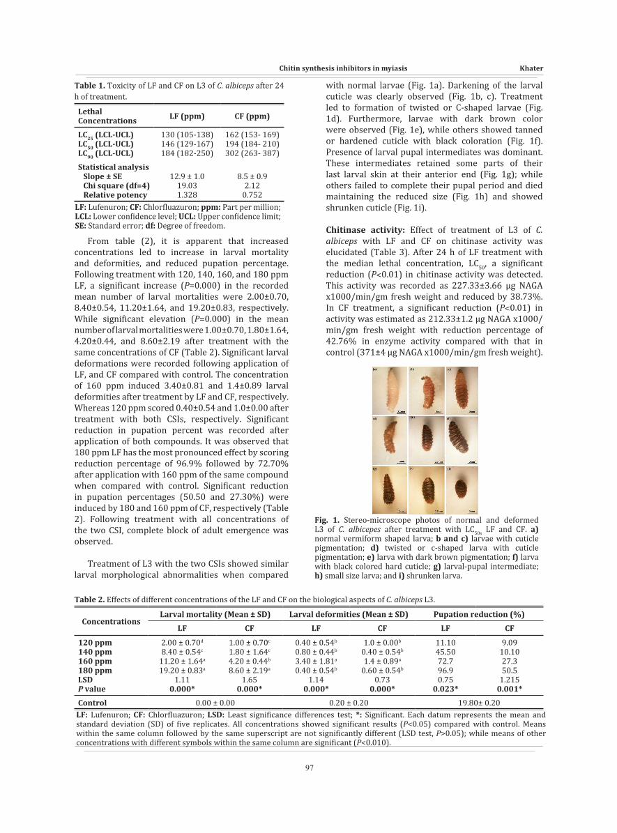
with normal larvae (Fig. 1a). Darkening of the larval cuticle was clearly observed (Fig. 1b, c). Treatment led to formation of twisted or C-shaped larvae (Fig. 1d). Furthermore, larvae with dark brown color were observed (Fig. 1e), while others showed tanned or hardened cuticle with black coloration (Fig. 1f). Presence of larval pupal intermediates was dominant. These intermediates retained some parts of their last larval skin at their anterior end (Fig. 1g); while others failed to complete their pupal period and died maintaining the reduced size (Fig. 1h) and showed shrunken cuticle (Fig. 1i).
Chitinase activity: Effect of treatment of L3 of C. albiceps with LF and CF on chitinase activity was elucidated (Table 3). After 24 h of LF treatment with the median lethal concentration, LC50, a significant reduction (P˂0.01) in chitinase activity was detected. This activity was recorded as 227.33±3.66 µg NAGA x1000/min/gm fresh weight and reduced by 38.73%. In CF treatment, a significant reduction (P˂0.01) in activity was estimated as 212.33±1.2 µg NAGA x1000/min/gm fresh weight with reduction percentage of 42.76% in enzyme activity compared with that in control (371±4 µg NAGA x1000/min/gm fresh weight).
Fig. 1. Stereo-microscope photos of normal and deformed L3 of C. albicepes after treatment with LC50s LF and CF. a) normal vermiform shaped larva; b and c) larvae with cuticle pigmentation; d) twisted or c-shaped larva with cuticle pigmentation; e) larva with dark brown pigmentation; f) larva with black colored hard cuticle; g) larval-pupal intermediate; h) small size larva; and i) shrunken larva.
Table 2. Effects of different concentrations of the LF and CF on the biological aspects of C. albiceps L3.
ConcentrationsLarval mortality (Mean ± SD) Larval deformities (Mean ± SD) Pupation reduction (%)
LF CF LF CF LF CF
120 ppm140 ppm160 ppm180 ppmLSDP value
2.00 ± 0.70d
8.40 ± 0.54c
11.20 ± 1.64a
19.20 ± 0.83a
1.110.000*
1.00 ± 0.70c
1.80 ± 1.64c
4.20 ± 0.44b
8.60 ± 2.19a
1.650.000*
0.40 ± 0.54b
0.80 ± 0.44b
3.40 ± 1.81a
0.40 ± 0.54b
1.140.000*
1.0 ± 0.00b
0.40 ± 0.54b
1.4 ± 0.89a
0.60 ± 0.54b
0.730.000*
11.1045.5072.796.90.75
0.023*
9.0910.1027.350.5
1.2150.001*
Control 0.00 ± 0.00 0.20 ± 0.20 19.80± 0.20LF: Lufenuron; CF: Chlorfluazuron; LSD: Least significance differences test; *: Significant. Each datum represents the mean and standard deviation (SD) of five replicates. All concentrations showed significant results (P<0.05) compared with control. Means within the same column followed by the same superscript are not significantly different (LSD test, P>0.05); while means of other concentrations with different symbols within the same column are significant (P<0.010).

PARASITOLOGISTS UNITED JOURNAL
98
Table 3. Effect of treatments with LC50 of LF and CF on chitinase activity in C. albiceps L3.
TreatmentsChitinase activity Statistical analysis
Mean ± SD Inhibition% P valueWater (control)LF (LC50)CF (LC50)
371.00 ± 6.92227.33 ± 6.35212.33 ± 2.08
038.7342.76
LF vs. control: P ˂ 0.01*CF vs. control: P ˂ 0.01*
LF vs. CF: P ˂ 0.05*LF: Lufenuron; CF: Chlorfluazuron; *: Significant.
Ultrastructural studies: The structure of the normal integument of C. albiceps larva consists of inner basement membrane, epidermis composed of a single epithelial cell layer, outer cuticle differentiated into an outer epicuticle, exocuticle and an inner endocuticle. Beneath the integument is a clear muscle layer (Fig. 2a). The endocuticle consists of characteristic successive sheets or lamellae (Fig. 2b-d); and cuticle lamellae that consist of microfibrils of chitin arranged horizontally and oriented at a definitive angle to each other (Fig. 3a, b). Such a structure arrangement is referred to as helicoidal architectures. The endocuticle lamellae are interrupted by numerous vertical columns of oriented microfibrils in characteristic structures called pore canal (PC) fibers (Fig. 1c). The PC fibers are chitinous in structure and extend from a structure called the apical membrane protrusion (AMP) (Figs. 2c and 3a). A special pattern of pigmentation was observed in larval endocuticle (Fig. 3c).
The normal epidermis consists of a single layer of columnar epithelial cells with large nuclei (Figs. 2a and
4a) that have obvious free nerve endings (Fig. 3d). The epidermal cells have a distinct supply of numerous tracheal structures (Fig. 4b). The nucleus of each cell is extremely large with well-defined condensed chromatin (Fig. 4a-d). Numerous round shaped mitochondria are located adjacent to the nucleus (Fig. 4c). The cytoplasm contains a distinct rough endoplasmic reticulum (RER), and the lysosomal structures are scattered throughout the cytoplasm (Figs. 4b-d). Lucent vesicles or areas of rarefied cytoplasm is diagnostic for this species (Figs. 4d and 5a). Microtubules (Fig. 5a) which provide a device for intracellular transport in the cell were obviously observed. These participate in many cellular functions such as the motion of secretory granules and macromolecules inside epidermal cells. Presence of Golgi cristae indicate the secretory function of the epidermal cells for chitin deposition (Figs. 4d, and 5a).
The ultrastructural alterations of cuticle following treatment with median lethal concentration of LF presented as vacuolization (Fig. 5b-d) especially at the exocuticle and between cuticular lamellae at the
Fig. 2. a) Semi-thin section of integument of normal C. albiceps L3 showed layers of cuticle with pigments, epidermal cells, and muscle. b-d) TEM photograph of cuticle of normal C. albiceps L3 showed b) layers of cuticle, epicuticle, exocuticle and endocuticle, vertical chitin fibres between cuticle lamellae (pore canals) and pigment granules which is characteristic of larvae of this species; c) pore canals between cuticular lamellae and apical plasma membrane protrusions; d) lamellar structure of endocuticle.APMP: Apical plasma membrane protrusions; CL: Chitin lamellae; EP: Epicuticle; EX: Exocuticle; EN: Endocuticle; EPI: Epidermis; M: Muscle; N: Nucleus; PC: Pore canal; PG: Pigment.
Fig. 3. a-c) TEM photograph of endocuticle of normal C. albiceps L3 showed a) apical plasma membrane protrusions and pore canals; b) paralleled chitin lamellae; c) numerous pigment granules; d) L3 with epidermal cells underlying the cuticle, free nerve endings, and pigment granules. APMP: Apical plasma membrane protrusions; CL: Chitin lamellae; EPC: Epidermal cell; PG: Pigment; NE: Nerve ending.

Chitin synthesis inhibitors in myiasis Khater
99
endocuticle (Fig. 5c, d). The PC fibres were moderately distorted after larval treatment (Fig. 5d). Borders between cuticular lamellae disappeared from some regions (Fig. 6a). Vacuolization in the cytoplasm of epidermal cells was clearly obvious (Fig. 6b). Autophagic vacuoles and cytoplasm condensation were noticed as signs of apoptosis (Fig. 6b, d). Nuclear changes presented as coagulation in chromatin (Fig. 6c). Strong blebbing of nuclear membrane (Fig. 6d) was a diagnostic cytopathological sign of apoptosis; the chromatin became peripherally located, marginal to the nuclear membrane (Fig. 7c, d), and most cell organelles disappeared (Fig. 7 b,c). Treatments also induced other cytological deformations of the nucleus which appeared as irregular shape and strong clumping of chromatin material (Fig. 7b). Vacuolation in the cytoplasm, swallowing endoplasmic reticulum (Fig. 7a-d), and fragmentation in RER were observed (Figs. 6b and 7b). Presence of numerous lysosomes portrayed signs of
detoxification due to lysis by foreign substances (Fig. 7b, d). This was observed as elongation of mitochondria (Fig. 7d). Damaged organelles and un-degraded debris in autolysosomes were observed (Fig. 6d). Additionally, another sign of apoptosis detected after treatment with LF, was the appearance of phagolysosome (Fig. 6b) and autophagic vacuoles (Fig. 7d).
A clear muscle layer is located beneath the epidermal cell layer in normal larvae. This muscle layer has a typical cytological striated structure and clearly distinguished Z lines (Fig. 8a). It helps in contraction and relaxation during larval molting. After treatment with LF there was vacuolation, disorganization and destruction of muscle fibres. Furthermore, abnormal morphology of mitochondria of muscular tissue seen as destruction in inner mitochondrial membrane and mitochondrial cristae were clear (Fig. 8b).
Fig. 4. a) TEM photograph of epidermal cell of normal L3 of C. albiceps showed large irregular nucleus with nuclear membrane, obvious chromatin, rough endoplasmic reticulum and mitochondria. b) TEM photograph of cuticle and epidermal cell of normal L3 of C. albiceps showing numerous tracheae; c) numerous mitochondria near the nuclear membrane and rough endoplasmic reticulum; d) rough endoplasmic reticulum, numerous lysosomes, mitochondria, secretory granules or dense vesicles, Golgi cristae and lucent vesicle.CH: Chromatin; CL: Chitin lamellae; GC: Golgi cristae; GA: Golgi apparatus; MT: Mitochondria; N: Nucleus; LV: Lucent vesicle; LY: Lysosome; RER: Rough endoplasmic reticulum; SG: Secretory granule.
Fig. 5. a) TEM photograph of epidermal cell of normal L3 of C. albiceps showed microtubules, secretory granules or dense vesicles, Golgi cristae and lucent vesicle. b-d) TEM photograph of cuticle in L3 of C. albiceps treated with LC50 LF showed b) vacuolation at endocuticle and between cuticular lamellae; c) exocuticle with vacuoles; d) vacuolation between lamellae of the endocuticle and in pore canal region. CL: Chitin lamellae; EP: Epicuticle; EX: Exocuticle; EN: Endocuticle; LV: Lucent vesicle; PG: Pigments; PC: Pore canal; SG: Secretory granule; TR: Trachea; V: Vacuole.
Fig. 6. a) TEM photograph of cuticle of C. albiceps L3 after treatment with LC50 of LF showed disappearance of borders between lamellae of endocuticle; b-d) TEM photograph of epidermal cell of C. albiceps L3 after treatment with LC50 of LF showed b) fragmentation of rough endoplasmic reticulum, numerous lysosomes, secretory granules or dense vesicles, phagolysosome and vacuole; c) disappearance of chromatin, splitting of nucleolus and disappearance of most cell organelles; d) disappearance of chromatin and strong blebbing of nuclear membrane and autolysosomes. AL: Autolysosomes; D: Desmosome; N: Nucleus; Nu: Nucleolus; LY: Lysosome; PL: Phagolysosome; PG: Pigment; RER: Rough endoplasmic reticulum; V: Vacuole.

PARASITOLOGISTS UNITED JOURNAL
100
Fig. 7. a-c) TEM photograph of epidermal cell of C. albiceps L3 after treatment with LC50 of LF showed a) swallowing in rough endoplasmic reticulum; b) clumping of condensed chromatin that marginates the nucleus, blebbing of nuclear membrane, swallowed mitochondria, and vacuoles in cytoplasm; c) swallowed rough endoplasmic reticulum and condensed cytoplasm; d) fragmented and swallowed endoplasmic reticulum, deformed mitochondria, and autophagic vacuoles. AV: Autophagic vacuole; CC: Condensed cytoplasm; CH: Chromatin; LY: Lysosome; MT: Mitochondria; N: Nucleus; NE: Nuclear envelop; PG: Pigment; RER: Rough endoplasmic reticulum; V: Vacuole.
Fig. 8. a) TEM photograph of muscles of normal C. albiceps L3 showing striated muscle fibres and Z lines. b) TEM photograph of muscles of C. albiceps L3 after treatment with LC50 of LF showed vacuolation, disorganization, destruction in muscle fibres and deformed mitochondria.DMT: Deformed mitochondria; MF: Muscle fibres; V: Vacuole; ZL: Z Line.
DISCUSSION
Nowadays, IGRs are considered effective in the field of insect control by inhibiting the life cycle. They do not persist for long due to their quick biodegradation with low mammalian toxicity. Among them, CSIs inhibit molting process and lead to production of faulty cuticle[26]. The present investigation reported that the larval mortality basically resulted from molting failure with incomplete life cycle and death within the cuticle. Similar results were recorded with Tribolium castaneum[27], Spodoptera littoralis[28,29], and Musca domestica[22,30]. In addition, LF proved to be more toxic at all concentration levels than CF inducing larval mortality, larval deformation, and reduction of pupation percent. Treatment of L3 of C. albiceps with the two CSIs showed high significant mortality after treatment with 180, 160 and 140 ppm of LF and with 180 ppm CF. Furthermore, extremely high significant reduction in pupation percentage was recorded after treatment with 180 and 160 ppm of LF. The results clearly proved that after treatment with the two CSIs, all pupae were dead, and the adult emergence completely ceased.
Previous studies reported the toxic effect of LF and CF against different insects. Percentage of pupal mortality in T. castaneum increased at higher concentrations of CF[31]. Pupal and adult mortalities were reported after treatment of M. domestica with LF while the larval stages were not affected[32]. Lufenuron was highly active against Lobesia botrana eggs[33]. High significant mortality in larvae, pupae, and adults of T. castaneum was recorded at different concentrations of CF. This result could be due to the anti-feeding effect of CF[27]. Lufenuron inhibited adult emergence of S.
littoralis after larval treatment[34], and was more potent than CF and chromafenzoide when applied on one day old eggs of Pectinophora gossypiella[35]. Lufenuron proved to be effective against yellow sugarcane borer Diatraea flavipennella[36]; and was more toxic against second instar larvae of S. littoralis than flufenoxuron[37]. Chlorfluarzuron affected the larval development of Bradysia odoriphaga[38]. In contrast, LF was the least toxic compared with flufenoxuron and hexaflumuron when used against M. domestica[22]. In fact, the majority of insecticides induced variable effects against different insects, because evaluation of the insecticide toxicity is species specific[39].
The two CSIs under investigation in the present study induced L3 abnormalities. The highest percentage of larval deformations was caused by LF. The survived larvae showed two forms of abnormalities, larvae with darkened cuticle and twisted or c-shaped larvae with inability to complete molting and development. Furthermore, small sized and shrunken larvae were clearly observed. Similar deformations were recorded on larvae of M. domestica[22,31,33], T. castaneum[27], L. botrana[34], S. littoralis[34,37], and on B. odoriphaga[38]. Furthermore, the two tested CSIs treatments led to formation of larval-pupal intermediates. These intermediates retained some parts of the last larval skin at their anterior end. They were often completely sclerotized, while others failed to complete their pupal period and died. Similar results were induced after treatment of Ephestia figulilella last instar larvae with hexaflumuron and LF causing deformity in both pupal and adult stages and in some cases produced larval-pupal intermediates. Distorted pupae failed to form the pupal skin, and larval pupal intermediates were

Chitin synthesis inhibitors in myiasis Khater
101
clearly observed[39]. Some treated larvae acquired dark pigmentation at the posterior end of the abdomen[40]. Larval deformation after treatment of M. domestica L3 by LF presented as irregular and elongated shapes[30]. Otherwise, LF and flufenoxuron treatments induced full darkened color and curved shaped larvae[22].
Both LF and CF impaired moulting process through significant reduction of chitinase enzyme that is essential in apolysis. This enzyme helps to digest the main constituents of the old endocuticle with the aid of protease enzyme[41], so that the normal development and transformation into adult stage of C. albiceps is blocked. This clarifies why the treated larvae fail to complete their life cycle and acquire the adult stage. This result is in accordance with Bayoumi et al.,[42] who observed 77.7% and 14.2% reduction in the cuticle chitinase activity after treatment of S. littoralis larvae with flufenoxuron and chlorfluazuron, respectively[42]. Reduction in activity of chitinase in Pectinophora gossypiella larvae was recorded one-day post treatment of eggs with LF, CF and chromafenzoide[35]. The researchers concluded that the reduction may be due to blocking and inhibition of the enzyme active site.
The insect cuticle is a mechanical barrier that maintains homeostasis and protects the insect against infection and desiccation. The ultrastructural changes of the cuticle revealed structural alterations of the lamellae of endocuticle that caused misplacement of its archetypal structure. Blebbing of nuclear membrane and disappearance of most cell organelles was observed. Another sign of apoptosis is the appearance of phagolysosomes. The use of chitin synthesis inhibitors against insects clearly induces deformities of the cuticle[43,44]. Insects become more vulnerable to contagions by viruses[45,46] and intestinal bacteria[47].
The larval cuticle of T. castaneum unveiled main structural alterations and loss of multilayered array of the procuticle by benzoylureas[48]. Ultrastructural investigations elicited disruption in deposition of procuticle after diflubenzuron application on Oxya japonica[49]. Both polyoxin D and diflubenzuron elicited a similar effect on Lucilia cuprina larvae as the procuticle lacked the normal lamellar arrangement[50]. Analogous results were recorded with the house fly after treatment with chlorfluazuron[51]. The ultrastructural deteriorations of cuticle and epidermis of the sixth instar pharate of Choristoneura fumiferana induced by ecdysone agonist, RH-5992 were revealed. After 24 h the cuticle lacked the usual lamellar arrangement, and the epidermal cells showed alterations in their organelles. Golgi apparatus showed hypertrophied circular vesicle revealing the secretory activity due to secretion of detoxification substances[52]. Aedes aegypti larvae treated with novaluron showed abortive discontinuous cuticle that detached from the epidermis with the latter’s degeneration in some cases[53]. Pyriproxyfen treatment causes vacuolization in cuticle
and destruction of cuticular lamellae in addition to destruction of nuclear envelop and increase of lysosomes in Culex pipiens larvae[54]. Diflubenzuron and LF applications lead to defects in chitin synthesis and organization of Drosophila melanogaster[55]. Presence of vacuoles and numerous mitochondria were recorded in 7 days old nymph of Schistocerca gregaria following treatment with teflubenzuron. Vacuoles were observed in the epidermal cells and an increase in the number of mitochondria. Mitochondria supply energy important for almost all chemical reactions and hydrogen ion transport mechanisms in the cell[56]. The appearance of vacuoles and autophagic vacuoles following application by CSIs in the current investigation were probably induced as a result of toxification process[57]. Also, vacuoles and phagic vacuoles that appeared in epidermal cells were thought to be an immune response in larvae to exposure to pyriproxyfen[58]. A spectacular increase in RER was detected in Chrysodeixis chalcites larvae after treatment with tebufenozide. Furthermore, clear increase of nucleus volume was observed, and presence of numerous mitochondria were demonstrated[59]. Treatment of Aubeonymus mariaefranciscae adults with hexaflumuron resulted in obvious disorganization in ultrastructural features of the integument of embryos. The lamellar arrangement of endocuticle was absent. This may be due to defective deposition of chitin leading to death[60]. Treatment of L3 of house fly with LF led to reduction in endocuticle and the epithelial cell layer became disorganized. Moreover, it became difficult to differentiate between exo and endocuticle[20]. After treatment of S. gregaria with LF, vacuolation, disorganization and destruction of muscle fibres were observed[61].
In conclusion the ultrastructural changes induced by the chitin synthesis inhibitor LF on the cuticle, epidermis, and the underlying muscles of L3 of C. albiceps, clarify why insects after application become unable to complete their life cycle. Thus, these compounds seem to be valuable in control of this myiasis producing insect and can be used in integrated pest management programs.
Conflict of interest: There is no conflict of interest.Funding statement: This research did not receive any specific grant from funding agencies in the public, commercial, or not-for-profit sectors.
REFERENCES
1. Queiroz MMC. Temperature requirements of Chrysomya albiceps (Wiedemann, 1819) (Diptera, Calliphoridae) under laboratory conditions. Mem Inst Oswaldo Cruz 1996; 91(6):1-5.
2. Povolony D. Chrysomya albiceps (Wiedemann, 1819): The first forensic case in central Europe involving this blowfly (Diptera: Calliphoridae). Acta Univ Agric Silivic Menedel Brun 2002; 50(3):105-112.

PARASITOLOGISTS UNITED JOURNAL
102
3. Hall MJR, Farkas R. Traumatic myiasis of humans and animals. In: Papp L, Darvas B (Editors): Manual of Palaearctic Diptera. Volume 1. General and Applied Dipterology. Science Herald Budapest 2000; 751-768.
4. Verves YG. Records of Chrysomya albiceps in the Ukraine. Medi Vet Entomol 2004; 18:308-310.
5. Zumpt F. Myiasis in man and animals in the old world. Butterworths London 1965; 267 pp.
6. Greenberg B. Flies and disease: I. Ecology, classification, and biotic associations. Princeton University Press Princeton New Jersey 1971; 856 pp.
7. Braack LEO, Retief PE. Dispersal density and habitat preference of the blowflies Chrysomya albiceps (Wd.) and Chrysomya marginalis (Wd.) (Diptera, Calliphoridae). Onderst J Vet Res 1986; 53:13-18.
8. Vattikonda SR, Sangam SR. Effect of forskolin on the growth and differentiation of the ovary of Papilio demoleus L. (Lepidoptera: Papilionidae). Int Res J Environ Sci 2017; 6:13–17.
9. Kostyukovsky M, Chen B, Atsmi, S, Shaaya, E. Biological activity of two juvenoids and two ecdysteroids against three stored product insects. Insect Biochem Mole Biol 2000; 30:891-897.
10. Sontakke BK, Mohapatra LN, Swain LK. Comparative bioefficacy of Buprofezin 25 EC against sucking pests of cotton and its safety to natural enemies. Ind J Entomol 2013; 75(4):325-329.
11. Shaurub EH, Zohdy NZ, Abdel-Aal AE, Emara SA. Effect of chlorfluazuron and flufenoxuron on development and reproductive performance of the black cutworm, Agrotis ipsilon (Hufnagel) (Lepidoptera: Noctuidae). Inv Rep Dev 2018; 62(1):27–34.
12. Arthur FH, Hartzer KL. Susceptibility of selected stored product insects to a combination treatment of pyriproxyfen and novaluron. J Pest Sci 2018; 91:699-705.
13. Muhammad Y, Muhammad S, Saqi KA, Mansoor UH, Saeed A, Muhammed I. Bioactivity of Lufenuron against Tribolium castaneum (Herbst) (Coleoptera: Tenebrionidae) Sains Malaysiana. 2019; 48(1):75–80.
14. Ishaaya I, Casida JE. Dietary TH6040 alters composition and enzyme activity of house fly larval cuticle. Pestic Biochem Physiol 1974; 4:484-490.
15. Post LC, de Jong BJ, Vincent WR. 1-(2,6-disubstituted benzoyl)-3-phenylurea insecticides: inhibitors of chitin synthesis. Pestic Biochem Physiol 1974; 4:473–483.
16. Uchida M, Asai T, Sugimoto T. Inhibition of cuticle deposition and chitin biosynthesis by a new insect growth regulator, buprofezin in Nilaparvata lugens Stal Agric Biol Chem 1985; 49:1233–1234.
17. De Cock A, Degheele D. Effects of buprofezin on the ultrastructure of the third instar cuticle of Trialeurodes vaporariorum. Tissue Cell 1991; 23(5):755–762.
18. Abo-Elghar GE, Fujiyoshi P, Matsumura F. Significance of the sulfonylurea receptor (SUR) as the target of diflubenzuron in chitin synthesis inhibition in Drosophila melanogaster and Blatella germanica. Insect Biochem Mol Biol 2004; 34:743–752.
19. Matsumura F. Studies on the action mechanism of benzoylurea insecticides to inhibit the process of chitin synthesis in insects: A review on the status of research
activities in the past, the present and the future prospects. Pestic Biochem Physiol 2010; 97:133–139.
20. Ahmed SS, Abd elhamid AE, Mo'men SAA. Lufenuron–induced ultrastructural changes in the larvae of Musca domestica (Diptera: Muscidae). Egypt Acad J Biol Sci 2019; 11(1):89-99.
21. Khater KS. Ultrastructural effects of chlorpyrifos and phenthoate on the midgut of Chrysomya albiceps larvae (Diptera: Calliphoridae). Egypt Acad J Biol Sci 2018; 10(1):49-61.
22. Nasr EE, Selim S, Tanani MA. Toxicological and biological indices of the house fly, Musca domestica after CSI'S treatments: Lufenuron, flufenoxuron and hexaflumuron. J Egypt Soc Parasitol 2020; 50(2):322–332.
23. Smith KE, Wall R, Howard JJ. In vitro insecticidal effect of fipronil and beta-cyfluthrin on larvae of the blowfly, Lucilia sericata. Vet Parasitol 2000; 88:261-268.
24. Davis D. Histopathologic Technic and Practical Histochemistry. Mc Graw-Hill, New York 1971; 3rd ed., pp. 112.
25. Reddy PJ, Krishna D, Murthy US, Jamil K. A microcomputer FORTRAN program for rapid determination of lethal concentration of biocides in mosquito control. CABIOS 1992; 8:209-213.
26. Hammock CD, Quistad GB. Metabolism and mode of action of juvenile hormone, juvenoids and other insect growth regulators. Progress Pestic Biochem 1981; 1:1-83.
27. Bakr RF, El-Moniary OM, El- Bakry NM, El-Shourbagy NM. Toxicological and behavioral effects of chlorfluazuron on pheromone production and perception of Tribolium castaneum (Coleoptera: Tenebrionidae). Egypt Acad J Biol Sci 2010; 2(2):61-72.
28. Abdel Rahman SM, Hegazi EM, Elwey AE. Direct and latent effect of two chitin inhibitors to Spodoptera littoralis larvae (Boisd). Amer Eurasian J Agric Enviro Sci 2007; 2(4):457-464.
29. Gelbic I, Adel MM, Hussein HM. Effects of nonsteroidal ecdysone agonist RH-5992 and chitin biosynthesis inhibitor lufenuron on Spodoptera littoralis (Boisduval, 1833). Cent Eur J Biol 2011; 6(5):861-869.
30. Khalil MS, Assar AA, Abo El-Mahasen MM, Mahmoud SH. Morphological effects of some insect growth regulators on Musca domestica, (Diptera, Muscidae). Egypt Acad J Biol Sci 2010;2(2):29-36.
31. Riddiford LM, Truman JW. Biochemistry of insect hormones and insect growth regulators: In Rockstein MR (Editor), Biochemistry of Insects, Academic Press, New York 1978; pp. 307-357.
32. Ghoneim KS, Amer MS, Bream AS, Al-Dali AG, Hamadah KS. Developmental and morphogenic responses of the house fly Musca domestica to the CSIs: Lufenuron and Diofenolan. Al-Azhar Bull Sci 2004;2(2):25-42.
33. Saenz- de-Cabezon FJ, Perez- Moreno I, Zalon FG, Marco V. Effects of lufenuron on Lobesia botrana (Lepidoptera: Tortricidae) egg, larval and adult stages. J Econ Entomol 2006; 99(2):427-431.

Chitin synthesis inhibitors in myiasis Khater
103
34. Adel MM. Lufenuron impair the chitin synthesis and development of Spodoptera littoralis (Bosid) (Lepidoptera: Noctuidae). J App Sci Res 2012; 8(5): 2766-2775.
35. Kandil MA, Ahmed AF, Moustafa HZ. Toxicological and biochemical studies of lufenuron, chlorfluazuron and chromafenzoide against Pectinophora gossypiella (Saunders). Egypt Acad J Biol Sci 2012; 4(1):37-47.
36. Fonseca AP, Marques EJ, Torres JB , Silva LM, Siqueira, HA� . Lethal and sublethal effects of lufenuron on sugarcane borer Diatraea flavipennella and its parasitoid Cotesia flavipes. Ecotoxicol 2015; 24(9):1869-1879.
37. El-Sayed E K, Massoud M A Z, Attia M A. Biochemical and biological influences of sub-lethal concentrations of emamectin benzoate and certain IGR insecticides against Spodoptera littoralis (Lepidoptera: Noctuidae). Alex Sci Exch J 2017; 38(2):212-219.
38. Zhang P, Zhao YH, Wang QH, Mu W, Liu F. Lethal and sublethal effects of the chitin synthesis inhibitor chlorfluazuron on Bradysia odoriphaga Yang and Zhang (Diptera: Sciaridae). Pestic Biochem Physiol 2017;136: 80–88.
39. Srivastava KP. A textbook of Applied Entomology: Methods of Insect Pest Control. 1988; New Delhi, Kalyani Publishers, India.
40. Khajepour S , Izadi H, Asari MJ . Evaluation of two formulated chitin synthesis inhibitors, hexaflumuron and lufenuron against the Raisin Moth, Ephestia figulilella. Insect Sci 2012; 12:102.
41. Reynolds SE, Samuels RI. Physiology and biochemistry of insect moulting fluid. Adv Insect Physiol 1996;26:157-232.
42. Bayoumi AE, Balana-Fouce R, Sobeiha AK, Hussein EMK. The biochemical influence of some chitin synthesis inhibitors against the cotton leafworm Spodoptera littoralis (Boisd.). Bol San Veg Plagas 1997; 23:583-593.
43. Van Eck WH. Mode of action of two benzylphenyl urea as inhibitors of chitin synthesis in insects. Insect Biochem 1979; 9(3):295-300.
44. Merzendorfer H, Kim, HS, Chaudhari SS, Kumari M, Specht CA, Butcher S. et al. Genomic and proteomic studies on the effects of the insect growth regulator diflubenzuron in the model beetle species Tribolium castaneum. Insect Biochem Mol Biol 2012; 42:264–276.
45. Wang P, Granados RR. Calcofluor disrupts the midgut defense system in insects. Insect Biochem Mol Biol 2000; 30:135–143.
46. Arakawa T, Furuta Y, Miyazawa M, Kato M. Flufenoxuron, an insect growth regulator, promotes peroral infection by nucleopolyhedrovirus (BmNPV) budded particles in the silkworm, Bombyx mori L. J Virol Methods 2002;100: 141–147.
47. Kuraishi T, Binggeli O, Opota O, Buchon N Lemaitre B. Genetic evidence for a protective role of the peritrophic matrix against intestinal bacterial infection in Drosophila melanogaster. Proc Natl Acad Sci USA 2011; 108(38):15966-15971.
48. Merzendorfer H. Chitin synthesis inhibitors: Old molecules and new developments. Insect Sci 2013; 20:121–138.
49. Lim SJ, Lee SS. The toxicity of diflubenzuron on Oxya japonica (Willemse) and its effect on moulting. Pestic Sci 1982; 13(5):537–544.
50. Binnington KC, Retnakaran A, Stone S, Skelly P. Studies on cyromazine and diflubenzuron in the sheep blowfly, Luiclia cuprina: Inhibition of vertebrate and bacterial dihydrofolate reductase by cyromazine. Pestic Biochem Physiol 1987; 27:201-210.
51. Bakr RF, Hussein MH. Morphogenic and histopathological studies on the effect of chitin synthesis inhibitor, chlorfluazuron against Musca domestica (L.) by using methacrylate embedding technique. J Egypt Soc Parasitol 1988;18(2):635-646.
52. Retnakaran A, Macdonald A, Tomkins WL, Davis CN, Brownwright AJ, Palli SR. Ultrastructural effects of a non- steroidal ecdysone agonist, RH- 5992, on the sixth instar larva of the spruce budworm, Choristoneura fumiferana. J Insect Physiol 1997; 43(1):55–68.
53. Farnesi LC, Brito JM, Linss JG, Pelajo-Marchado M, Valle D, Rezende GL. Physiological and morphological aspects of Aedes aegypyi developing larvae: Effects of the chitin synthesis inhibitor; novaluron. PLOS One 2012; 7(1), e30363.
54. El- Shazly MM, Refaie BM. Larvicidal effect of the juvenile hormone mimic pyriproxyfen on Culex pipiens. J Amer Mosqu Control Assoc 2002; 18(4):321-328.
55. Gangishetti U, Breitenbach S, Zander M, Saheb SK, Muller U, Schwarz H et al. Effects of benzoylphenyl urea on chitin synthesis and orientation in the cuticle of the Drosophila larva. Eur J Cell Biol 2009; 88:167–180.
56. Al-Mokhlef AA, Mariy FM, Emam AK, Ali GM. Effect of teflubenzuron on ultrastructure and components of the integument in Schistocerca gregaria (Forskal) 5th instar nymphs. Ann Agric Sci 2012; 57(1):1–6.
57. Ker RF. The effects of diflubenzuron on the growth of insect cuticle. Pestic Sci 1978; 9:259–265.
58. Locke M. Epidermis. In: Harrison FW, Locke M (Editors). Microscopic anatomy of invertebrates. New York: John Wiley & Sons. 1998; 11A:75-138.
59. Smagghe G, Vinuela E, Budia F, Degheele D. Effects of thenon-steroidal ecdysteroid mimic tebufenozide on the tomato looper Chrysodeixis chalcites (Lepidoptera: Nocutidae): An ultrastructural analysis. Arch Insect Biochem Physiol 1997; 35:179–190.
60. Farinos GP, Smagghe G, Marco V, Tirry L, Castanera P. Effects of topical application of hexaflumuron on adult sugar beet weevil, Aubeonymus mariaefranciscae, on embryonic development: Pharmacokinetics in adults and embryos. Pestic Biochem Physiol 1998; 61:169–182.
61. Al-Zeeb FAM, Ibrahim EH, Bakr RFA. Histopathological changes in the muscle of the desert locust, Schistocerca gregaria (Orthoptera: Acrididae) treated with insect growth regulator (IGR), Lufenuron. Egypt Acad J Biol Sci 2018; 10(1):13-25.