Embed Size (px)
Citation preview
MULTI-AUTHOR REVIEW
The use of metabolomics to dissect plant responses to abioticstresses
Toshihiro Obata • Alisdair R. Fernie
Received: 7 July 2012 / Revised: 9 July 2012 / Accepted: 9 July 2012 / Published online: 12 August 2012
� The Author(s) 2012. This article is published with open access at Springerlink.com
Abstract Plant metabolism is perturbed by various
abiotic stresses. As such the metabolic network of plants
must be reconfigured under stress conditions in order to
allow both the maintenance of metabolic homeostasis and
the production of compounds that ameliorate the stress.
The recent development and adoption of metabolomics and
systems biology approaches enable us not only to gain a
comprehensive overview, but also a detailed analysis of
crucial components of the plant metabolic response to
abiotic stresses. In this review we introduce the analytical
methods used for plant metabolomics and describe their
use in studies related to the metabolic response to water,
temperature, light, nutrient limitation, ion and oxidative
stresses. Both similarity and specificity of the metabolic
responses against diverse abiotic stress are evaluated using
data available in the literature. Classically discussed stress
compounds such as proline, c-amino butyrate and poly-
amines are reviewed, and the widespread importance of
branched chain amino acid metabolism under stress con-
dition is discussed. Finally, where possible, mechanistic
insights into metabolic regulatory processes are discussed.
Keywords Metabolomics � Plants � Abiotic stress �Metabolic response � Branched chain amino acid �Enzyme complex
Introduction
When plants face unfavourable growth conditions, abiotic
stress retards plant growth and productivity. Under most
abiotic stress conditions, plant metabolism is perturbed
either because of inhibition of metabolic enzymes, shortage
of substrate, excess demand for specific compounds or a
combination of these factors and many other reasons.
Therefore, the metabolic network must be reconfigured to
maintain essential metabolism and to acclimate by adopt-
ing a new steady state in light of the prevailing stress
conditions. This metabolic reprogramming is also neces-
sary to meet the demand for anti-stress agents including
compatible solutes, antioxidants and stress-responsive
proteins. The accumulation of reactive oxygen species
(ROS) is another problem causing oxidation and dysfunc-
tion of cellular components and in the worst case cell
death. The optimisation of metabolic flux via the organellar
electron transport chains is, moreover, crucial in order to
dampen ROS production. Maintenance of the redox state in
the cell is thus another important task to provide the
reducing power required for ROS scavenging. Despite such
important roles of metabolic regulation under stress con-
ditions, our current understanding of this process is
fragmented and far from complete.
Metabolomics is a powerful tool by which to gain a
comprehensive perspective of how metabolic networks are
regulated and has indeed been applied by many researches
in recent years. It can, additionally, be used to elucidate the
functions of genes as a tool in functional genomics and
systems biology approaches. The term ‘‘metabolomics’’ is
defined as comprehensive and quantitative analysis of all
small molecules in a biological system [1]. The plant
kingdom may contain between 200,000 and 1,000,000
metabolites, while for a single species the number may
Electronic supplementary material The online version of thisarticle (doi:10.1007/s00018-012-1091-5) contains supplementarymaterial, which is available to authorized users.
T. Obata � A. R. Fernie (&)
Max Planck Institute of Molecular Plant Physiology,
Am Muhlenberg 1, 14476 Potsdam-Golm, Germany
e-mail: [email protected]
Cell. Mol. Life Sci. (2012) 69:3225–3243
DOI 10.1007/s00018-012-1091-5 Cellular and Molecular Life Sciences
123
approach a few thousand (the estimate for Arabidopsis
is ca. 5,000) [2–5]. Indeed the KNApSAck database (http://
kanaya.naist.jp/KNApSAcK/) contains around 50,000
metabolite entries in plants described so far in the literature
[6]. Due to the large variety of chemical structures and
properties of small molecules, there is so far no single
technique to identify and quantify all of them. Even the most
comprehensive methods detect only between 1,000 and
2,000 molecular features [7, 8]. Several techniques includ-
ing gas chromatography-mass spectrometry (GC-MS),
liquid chromatography (LC)-MS, capillary electrophoresis
(CE)-MS and nuclear magnetic resonance spectroscopy
(NMR) are commonly used in plant metabolomics research.
They are used sometimes in combination since they are
largely complimentary with each independent method
having preferential coverage of diverse types of metabolite.
Here we briefly introduce the advantages and limitations of
each method. Thereafter studies employing the metabolo-
mic approach to dissect plant response to abiotic stresses
will be discussed. Finally, using data already collected,
we attempt to elucidate the general and stress-specific
responses.
Techniques used for plant metabolomic research
Gas chromatography-mass spectrometry (GC-MS)
Gas chromatography-mass spectrometry is the most widely
used technique for plant metabolomics research to date.
Polar metabolites are derivatised to render them volatile
and then separated by GC. Electron impact (EI) allows
robust interfacing of GC with MS resulting in highly
reproducible fragmentation patterns. For detection, time-
of-flight (TOF)-MS has become the method of choice
because of advantages including fast scan times, which
give rise to either improved deconvolution or reduced run
times for complex mixtures, and high mass accuracy. The
crucial advantage of this technology is that it has long been
used for metabolite profiling, and thus there are stable
protocols for machine setup and maintenance, and chro-
matogram evaluation and interpretation [9–11]. The
robustness of the protocol means that libraries of retention
time and mass spectra data for standard compounds can be
shared among laboratories [12]. There are several metab-
olite databases available including the NIST [13], FiehnLib
[14] and Golm metabolic databases (GMD, [15]), which
are useful tools for peak annotation. Additionally, the short
running time and relatively low running cost are also strong
advantages of GC-MS. However the use of GC-MS is
limited for thermally stable volatile compounds, making
the analysis of high molecular weight compounds (larger
than 1 kDa) difficult. Due to these characteristics, GC-MS
facilitates the identification and robust quantification of a
few hundred metabolites in plant samples including sugars,
sugar alcohols, amino acids, organic acids and polyamines,
resulting in fairly comprehensive coverage of the central
pathways of primary metabolism.
Liquid chromatography (LC)-MS
While GC has a limitation due to volatilisation of com-
pounds, LC does not require prior sample treatment and
separates the components in a liquid phase. The choice of
columns, including reversed phase, ion exchange and
hydrophobic interaction columns, provides the separation
of metabolites based on different chemical properties.
Therefore, LC has the potential to analyse a wide variety of
metabolites in plants. The recent development of ultra-
performance liquid chromatography (UPLC) makes the
technique more powerful because of its higher resolution,
sensitivity and throughput than conventional high-perfor-
mance liquid chromatography (HPLC) [16]. Electrospray
ionisation (ESI) is widely used for ionisation to connect LC
and MS. Many types of MS including quadrupole (Q),
TOF, qTOF, triple quadrupole (QqQ), ion trap (IT), linear
trap quadrupole (LTQ)-Orbitrap and Fourier transform ion
cyclotron resonance (FT-ICR)-MS are used depending on
the sensitivity, mass-resolution and dynamic range required
(see [17, 18] for the detail). The combination of these
techniques allows us to identify and quantify a large variety
of metabolites even if they have high molecular mass, great
polarity and low thermostability. On the other hand the
flexibility of the method also causes difficulty in estab-
lishing large mass spectral libraries for peak identification
because of the instrument-type dependent retention time
and mass spectra [19], and forces each research group to
create its own ‘‘in-house’’ LC-MS reference library. That
said, there are a number of websites that aid in mass-
spectral analyses [20], and recent recommendations for
metabolite reporting [7] should improve the transparency
of the methods used by researchers. Furthermore, isotope
labelling as a means of confirming the identity of peaks has
recently been proposed and demonstrated to allow the
identification of circa 1,000 metabolites using the FT-ICR-
MS approach [8, 21]. To date, LC-MS is mainly used with
a reverse phase column to analyse secondary metabolites
because of its ability to separate compounds with similar
structure and to detect a wide range of metabolites. How-
ever, it is worth noting that specialised protocols for
determining phosphorylated intermediates, which are not
readily detected by GC-MS, have also begun to be devel-
oped using this technology [22], as have methods for the
comprehensive analysis of phytohormones [23].
3226 T. Obata, A.R. Fernie
123
Capillary electrophoresis (CE)-MS
Capillary electrophoresis separates polar and charged
compounds on the basis of their charge-to-mass ratio. CE is
able to separate a diverse range of chemical compounds
and is a more powerful technique than LC with respect to
separation efficiency [24, 25]. ESI is commonly used for
ionisation as in LC-MS, with TOF-MS being the most
commonly used detector in CE-MS-based metabolomics
studies. This combination provides high mass accuracy and
high resolution. The high scan speed of TOF-MS makes
this instrument very suitable for full scan analyses in
metabolomics. One of the unique properties of CE-MS is
the small amount of sample required for analysis; only
nanolitres of sample are introduced into the capillary.
Together with high electric fields and short separation
lengths, it can produce analysis within seconds. It also
allows the metabolic analysis in volume-restricted samples.
On the other hand, this leads to low concentration sensi-
tivity requiring enrichment of metabolites within the
samples [26]. Another drawback of CE is the poor migra-
tion time reproducibility and lack of reference libraries,
which may only be partially overcome by the prediction of
migration time [27]. Since CE and LC can both separate a
large variety of metabolites via fundamentally different
mechanisms, they are often used in combination to provide
a wider coverage of metabolites [28–30]. That said, the use
of CE-MS in plant studies remains relatively rare.
Nuclear magnetic resonance (NMR) spectroscopy
Nuclear magnetic resonance spectroscopy offers an
entirely different analytical technique to that afforded by
MS-based techniques being based on atomic interaction. In
a strong magnetic field, atoms with non-zero magnetic
moment including 1H, 13C, 14N, 15N and 31P absorb and
re-emit electromagnetic radiation. The radiation is char-
acterised by its frequency (chemical shift), intensity, fine
structure and magnetic relaxation properties, all of which
reflect the precise environment of the detected nucleus.
Therefore, atoms in a molecule give a specific spectrum of
radiation that can be used for identification and quantifi-
cation of metabolites within a complex biological sample.
The sensitivity of this method is much lower than that of
MS-based techniques but the structural information con-
tent, reproducibility and quantitative aspects can be
superior to them, and some journals require NMR spectra
as the final proof of chemical structure [31, 32]. Further-
more the preparation of the sample is simple and even non-
destructive measurement is possible. In vivo NMR can
further generate kinetic measurements and examine meta-
bolic responses on the same plant rather than on a set of
similar plants [33]. The different subcellular pHs of the
vacuole from the rest of the cell cause distinctive signals
from an identical metabolite and thus allows quantification
at the subcellular level [34, 35]. Thus analysing the
metabolite composition of a tissue extract, determining the
structure of a novel metabolite, demonstrating the exis-
tence of a particular metabolic pathway in vivo, isotope
labelling experiment and localising the distribution of a
metabolite in a tissue are all possible by NMR. For isotope
labelling, NMR has the advantage of providing facile
access to atomic level labelling, which is highly laborious
in the case of MS methods yet can be essential in flux
estimation [35]. However, the number of compounds that
can be detected in a single analysis is limited to one to
several dozen [36, 37]. These properties of NMR make it
the ideal tool for broad-range profiling of abundant
metabolites whilst studying changes in non-annotated
profiles is highly useful for metabolite fingerprinting of
extensive sample collections [38, 39].
Metabolomic studies of plant stress responses
Metabolomics is becoming increasingly common in plant
physiology and biochemistry, and to date has been applied
to a staggering number of conditions. Here we will attempt
a synthesis of the most prominent studies dealing with
plant stress; however, the reader is also referred to two
previous reviews on this topic [40, 41]. In this section we
independently review water stress, temperature stress, light
stress, ionic stress, nutrient limitation and oxidative stress
before discussing stress combinations. We describe the
nature and symptoms of each stress, and then introduce
several metabolomic studies with the main metabolic
changes observed in each study and the conclusion drawn
from the results. Following this survey we discuss com-
monalities and differences between the various stress
responses.
Water stress
Water limitation is one of the major threats in crop pro-
duction and this condition is projected to get considerably
worse in coming decades [42]. For this reason considerable
research effort has been expended to understand the
response to this crucial and common stress. These studies
have revealed an important role for metabolic regulation
including regulation of photosynthesis and accumulation of
osmolytes in the drought stress response [43, 44]. Urano
et al. [28] reported metabolomic changes in Arabidopsis
leaves under drought condition. The accumulation of many
metabolites was observed, including amino acids such as
proline, raffinose family oligosaccharides, c-amino buty-
rate (GABA) and tricarboxylic acid (TCA) cycle
Metabolomics of plant stress 3227
123
metabolites, which are known to respond to drought stress
in plants. The authors also investigated the nc3-2 mutant,
which lacks the NCED3 gene involved in the dehydration-
inducible biosynthesis of abscisic acid (ABA), in order to
assess the effect of ABA in the metabolic response to
drought stress. By combination with transcriptome analysis
they clearly demonstrated that the ABA-dependent tran-
scriptional regulation is responsible to the activation of
metabolic pathways including branched chain amino acid,
polyamine and proline biosynthesis, GABA shunt and
saccharopin metabolism, but is not involved in the regu-
lation of the raffinose biosynthetic pathway during
dehydration.
In Arabidopsis research, drought tolerance is assessed
predominantly under lethal conditions. However, in tem-
perate climates, limited water availability rarely causes
plant death but does restrict biomass and seed yield.
Results of a recent elegant study experimentally demon-
strated that the survival rate under lethal conditions does
not predict superior growth performance and biomass yield
gain under moderate drought [45], making this mild stress
condition more important. Skirycz et al. [46] conducted
metabolite profiling of Arabidopsis leaves that develop
under mild osmotic stress. They revealed that the stress
response measured in growing and mature leaves was
largely distinct. Typical drought responses, namely accu-
mulation of proline, erythritol and putrecine, were
observed only in mature leaves, while many metabolites
were decreased in expanding leaves, sharing the same
tendency with transcriptional response. When we com-
pared the data from the studies [28, 46], 24 metabolites
were detected in both. The decrease of aspartate and
increase of proline are the only two responses shared
between mildly and severely desiccated leaves. Pro-
nouncedly, amino acid metabolism responds in opposite
ways; most amino acids were accumulated in severely
desiccated leaves but decreased in mildly desiccated plants.
These results highlight the variable response of plant
metabolism in different developmental stages and degrees
of desiccation. Metabolite profiling has additionally been
carried out in crop species exposed to water stress condi-
tions. Intriguingly, common changes in the levels of
metabolites including branched chain amino acids were
observed in wheat, barley and tomato [47–49].
Too much water, as occurs in situations such as flooding
or water-logging of the rhizosphere, also causes problems
because of the reduced oxygen availability (hypoxia/
anoxia). Under anoxic conditions, ATP has to be produced
by fermentation, resulting in cytosolic acidification and the
accumulation of toxic products. van Dongen et al. [50]
analysed metabolic responses in Arabidopsis roots under
anoxic conditions. The accumulation of amino acids, ala-
nine, proline and GABA, and the phosphoesters, glucose-6-
phosphate and glycerol-3-phosphate, were observed as well
as changes in the levels of minor sugars and various
organic acids. When oxygen is decreased to 4 %, there is a
general tendency for an increase in the levels of the
intermediates both of sucrose degradation and the TCA
cycle, and in the levels of most amino acids, whereas they
are decreased when the oxygen further decreased to 1 %,
indicating the inhibition and reactivation of metabolic
activities. Together with the transcriptomic data showing a
general downregulation of energy-consuming processes,
the results demonstrated a large-scale reprogramming of
metabolism under oxygen-limited conditions. Rocha et al.
[51] examined the accumulation of alanine under anoxic
conditions in Lotus japonicus, which is highly tolerant to
water logging. In the roots of L. japonicus, succinate,
alanine and the direct co-substrates for alanine synthesis,
glutamate and GABA, were highly accumulated during
water logging, whereas the majority of amino acids that are
derived from TCA cycle intermediate decreased. The
results are in agreement with the metabolic equilibriums
that are expected to drive the metabolic flux from glycol-
ysis, via alanine synthesis and oxoglutarate to succinate,
which prevents the accumulation of pyruvate activating
fermentation and leading to ATP production by succinyl-
CoA ligase.
Temperature stress
Exposure to freezing environments leads to serious damage
of the plant cell by ice formation and dysfunction of cel-
lular membranes [52]. Many plant species increase freezing
tolerance during exposure to non-freezing low temperature
by a process known as ‘‘cold acclimation’’. The molecular
basis of this process has been extensively studied, and the
contribution of particular metabolites including compatible
solutes [53] and the transcriptional regulatory network has
been elucidated [54, 55]. The first metabolomic studies of
cold acclimation were performed by two groups in 2004.
Cook et al. [56] compared metabolomic changes during
cold acclimation in two ecotypes of Arabidopsis thaliana,
Wassilewskija-2 (Ws-2) and Cape verde islands-1 (Cvi-1),
which are relatively freezing tolerant and sensitive,
respectively. The metabolome of Ws-2 plants was exten-
sively altered in response to low temperature. Seventy-five
percent of metabolites monitored were found to increase in
cold-acclimated plants including metabolites known to
increase in Arabidopsis plants upon exposure to low tem-
perature, such as the amino acid proline and the sugars
glucose, fructose, inositol, galactinol, raffinose and
sucrose. They also found novel changes—namely the
increase of trehalose, ascorbate, putrescine, citrulline and
some TCA cycle intermediates. There was considerable
overlap in the metabolite changes that occurred in the two
3228 T. Obata, A.R. Fernie
123
ecotypes in response to low temperature; however, quan-
titative differences were evident. Kaplan et al. [57]
conducted metabolome analysis of Arabidopsis over the
time course following the shift to cold and heat conditions.
Surprisingly the majority of heat shock responses were
shared with cold shock including the increase of pool sizes
of amino acids derived from pyruvate and oxaloacetate,
polyamine precursors and compatible solutes. The results
of this study were analysed together with following tran-
script profiling data by the same group [58], and revealed
that the regulation of GABA shunt and proline accumula-
tion under cold conditions are achieved by transcriptional
and post-transcriptional manners, respectively. Gray and
Heath [59] examined the effects of cold acclimation on the
Arabidopsis metabolome using a non-targeted metabolic
fingerprinting approach. It revealed a global reprogram-
ming of metabolism as well as differential responses
between the leaves that shifted to and those that developed
in the cold. Hannah et al. [60] took advantage of the natural
genetic variation of Arabidopsis to elucidate the function of
metabolism in cold acclimation. Although there is no clear
relationship between global metabolite changes and dif-
ferences in acclimation capacity or differences between the
accessions in acclimated freezing tolerance, the probable
importance of central carbohydrate metabolism is indicated
by the identification of glucose, fructose and sucrose
among metabolites positively correlating to freezing tol-
erance. Espinoza et al. [61] analysed the effect of diurnal
gene/metabolite regulation during cold acclimation by
means of metabolomics and transcriptomics. Approxi-
mately 30 % of all analysed metabolites showed circadian
oscillations in their pool size and low temperature affected
the cyclic pattern of metabolite abundance. These results
indicated that the interactions observed between circadian
and cold regulation are likely highly relevant components
of cold acclimation.
Metabolomics was also used to reveal the functions of
specific genes in cold acclimation. In the study described
above, Cook et al. [56] also investigated plants over-
expressing CBF3, which is one of the C-repeat/dehydration
responsive element-binding factor (CBF) transcriptional
activators induced rapidly under low temperature condi-
tions [62]. The metabolite profiles of non-acclimated CBF3
overexpressing lines were quite similar to those of the cold-
acclimated Ws-2 ecotype, suggesting a prominent role for
the CBF cold response pathway in configuring the low-
temperature metabolome of Arabidopsis. Maruyama et al.
[63] explored metabolic and transcript changes in Arabid-
opsis plants overexpressing CBF3/dehydration-responsive
element binding protein (DREB)1A and another DREB
protein DREB2A. They observed similar changes of
metabolites in CBF3-overexpressing plants like Cook et al.
[56] but DREB2A overexpression showed only a minor
effect. The esk1 mutant is isolated as freezing tolerant
without previous acclimation but the function of this gene
had been unknown. Lugan et al. [64] tried to elucidate the
basis of the freezing tolerance of esk1 by performing
metabolomic analysis under various environmental condi-
tions, namely cold, salinity and dehydration. Then the most
specific metabolic responses to cold acclimation were not
phenocopied by esk1 mutation. However, esk1 accumu-
lated lower amount of Na? in leaves than the wild type and
its metabolic profile, and osmotic potential were only
slightly impacted under dehydration stress. These results
suggested that ESK1 could rather be involved in water
homeostasis and as such highlighted the importance of
cellular water status in stress tolerance.
Light stress
Light is a highly energetic substrate driving photosynthesis
that can induce secondary destructive processes at the same
time. Therefore, too high light irradiance represents an
abiotic stress factor for plants. Wulff-Zottele et al. [65]
conducted metabolite profiling of Arabidopsis leaves for
6 days after transition to high light. Generally, most of the
metabolites of the glycolysis, TCA cycle and oxidative
pentose phosphate pathway were altered in their content,
indicating that plants exposed to high light undergo a
metabolic shift and enhance the Calvin-Benson cycle to fix
more carbon. In addition, elevation of glycine indicated the
activation of photorespiratory pathways. Caldana et al. [66]
investigated the early metabolic response against high light
as a part of a more comprehensive study. The accumulation
of the photorespiratory intermediates, glycine and glyco-
late, were observed in the early phase (5–60 min after
transition). Interestingly the response during the mid phase
(80–360 min) shares similar properties with low tempera-
ture treatment, which includes the accumulation of
shikimate, phenylalanine and fructose, and the decrease of
succinate; however, the physiological meaning of this
overlap is currently unknown.
Not only the quantity but also the quality of light affects
plant metabolism. In dense plant stands, such as crop fields
or forests, individuals shade each other and create com-
petition for light absorption [67]. Selective light absorption
by the upper leaf layers leads to an enrichment of far-red
wavelength [68], which induces excitation imbalances
between photosystem II and I disturbing both the redox
chemistry in the transport chain and its coordination with
the Calvin-Benson cycle [69, 70]. For this reason, Brauti-
gam et al. [71] grew Arabidopsis plants under light, which
preferentially excited either photosystem I (PSI light) or II
(PSII light) and then transferred it to the other light con-
dition to analyse how plants acclimate to the light quality
shift. After long-term acclimation of 48 h, plants exhibited
Metabolomics of plant stress 3229
123

two distinct metabolic states. A PSI–II shift resulted in a
decrease in primary products of photosynthesis, such as
sugars, but an increase in important intermediates of sub-
sequent metabolic pathways. By contrast, a PSII-I shift has
no effect on the sugar pools but leads to general down-
regulation of many subsequent metabolites, including
amino acids and organic acids. Each of the metabolites
exhibited a different accumulation profile for establishing
the final pool size, indicating high complexity by which the
two metabolic states were achieved. Comprehensive anal-
yses of these data alongside transcript profiles and other
physiological data suggested that photosynthesis and
metabolism were under the control of a binary combination
of inputs from the thioredoxin and plastoquinone systems.
The dependency of plants upon sunlight also inevitably
leads them into exposure to ultraviolet (UV) light,
including in the wavelength range of 280–320 nm (UV-B).
This wavelength potentially damages DNA, RNA and
proteins, and additionally increases the production of free
radicals [72, 73]. Kusano et al. [74] treated Arabidopsis
plants with UV light and analysed the metabolic effect of
UV light stress. Arabidopsis exhibits an apparent biphasic
response to UV-B stress, characterised by major changes in
the levels of primary metabolites, including ascorbate
derivatives. By contrast, mid- to late-term responses were
observed in the classically defined UV-B protectants, such
as flavonoids and phenolics. The results suggested that in
early stages of exposure to UV-B, the plant cell is ‘primed’
at the level of primary metabolism by a mechanism that
involves reprogramming of the metabolism to efficiently
divert carbon towards the aromatic amino acid precursors
of the phenylpropanoid pathway. It also suggested the
importance of ascorbate in the short-term response to
UV-B. Further studies are, however, required to determine
which of these metabolic changes are end responses to
adapt to the enhanced exposure to UV-B and which are part
of the perception-signalling relay, which alerts the plant
cell that it needs to respond to the stress [75].
Ion stress
High levels of salinity in the soil hinder the growth and
development of crops and cause serious problems for world
food production [76]. High concentrations of NaCl may
cause both hyperionic and hyperosmotic stress effects,
which lead to a decline of turgor, disordered metabolism
and the inhibition of uptake of essential ions, as well as
other problems in plant cells [77, 78]. Gong et al. [79]
conducted metabolite profiling of salt-treated Arabidopsis
thaliana and its relative Thellungiella halophila (salt
cress), which shows ‘extremophile’ characteristics mani-
fested by extreme tolerance to a variety of abiotic stresses,
among them low humidity, freezing and high salinity.
Proline increased dramatically in both species as did
inositols, hexoses and complex sugars. The concentrations
of metabolites were often several-fold higher in Thel-
lungiella and stress exacerbated the differences in some
metabolites. Transcript analyses supported the metabolic
results by suggesting that a Thellungiella is primed to
anticipate such stresses. The difference in metabolites
between Arabidopsis and Thellungiella under salt and
osmotic stresses was more recently assessed for a broader
range of metabolites [80]. Analysis of global physico-
chemical properties of metabolites revealed a shift from
nonpolar to polar metabolites in both species but that this
was much more pronounced in Thellungiella. Such a shift
may contribute to keep the water potential during dehy-
dration. Kim et al. [77] investigated the cellular level
metabolic response using Arabidopsis T87 cultured cells.
The results suggested that the methylation cycle for the
supply of methyl groups, the phenylpropanoid pathway for
lignin production and glycine betaine biosynthesis are
synergetically induced as a short-term response against
salt-stress treatment. The results also suggest the
co-induction of glycolysis and sucrose metabolism as well
as co-reduction of the methylation cycle as long-term
responses to salt stress.
Due to the importance of salinity stress in agriculture,
there are many metabolomic studies to assess the meta-
bolic effect of salinity in a variety of crop and related
plant species including tomato [40, 81], grapevine [82],
poplar [83], sea lavender (Limonium latifolium, [84]) and
rice [85]. Since these studies have been extensively
reviewed in [40, 86], we focus here on three recent studies
on legume species [87–89]. These recent studies took a
functional genomic approach that integrated ionomic,
transcriptomic and metabolomic analyses of the glycopyte
model legume Lotus japonicus and other Lotus species
subjected to long-term regimes of non-lethal levels of
salinity. In Lotus japonicus the metabolic changes were
characterised by a general increase in the steady-state
levels of many amino acids, sugars and polyols, with a
concurrent decrease in most organic acids [87]. The
responses to salinity stress were compared between
extremophile (L. creticus) and glycophytic (L. cornicula-
tus and L. tenuis), but the metabolic responses were
globally similar to each other [88]. These results suggest
that, in contrast to Thellungiella, the metabolic pre-
adaptation to salinity is not the major trait of L. creticus
contributing to the extramophile phenotype. However, by
comparing six species displaying different salt tolerances,
they observed several genotype-specific features. One of
them is the increase of asparagine levels in the more
tolerant genotypes, suggesting that the roles of asparagine
metabolism in supporting core nitrogen metabolism may
play a role in tolerance [89].
3230 T. Obata, A.R. Fernie
123
Heavy metals such as cadmium (Cd), cesium (Cs), lead
(Pb), zinc (Zn), nickel (Ni) and chromium (Cr) are major
pollutants of the soil causing stress on plants. Even the
essential nutrients including copper (Cu), iron (Fe) and
manganese (Mn) can cause heavy metal stresses with
inappropriate concentration. Generally heavy metals
induce enzyme inhibition, cellular oxidation and metabolic
perturbation, resulting in growth retardation and in extreme
instances in plant death [90]. Jahangir et al. [91] analysed
the effects of Cu, Fe and Mn on the metabolite levels of
Brassica rapa, which is a known metal accumulator.
Glucosinolates and hydroxycinnamic acids conjugated with
malates as well as primary metabolites such as carbohy-
drates and amino acids were found to be the discriminating
metabolites. Arabidopsis plants treated with Cd displayed
increased levels of alanine, b-alanine, proline, serine,
putrescine, sucrose and other metabolites with compatible
solute-like properties, notably GABA, raffinose and tre-
halose [92]. This study also indicated that concentrations of
antioxidants (a-tocopherol, campesterol, ß-sitosterol and
isoflavone) also increased significantly. When taken toge-
ther these data indicate an important role of antioxidant
defences in the mechanisms of resistance to cadmium
stress. Dubey et al. [93] conducted transcriptomic and
metabolomic analysis of rice roots treated with Cr. Under
these conditions proline accumulated to levels three-fold
those of the control as did ornithine, which can be used in
its synthesis. The content of several other metabolites
including lactate, fructose, uracil and alanine increased
following exposure to Cr stress; these were taken to sug-
gest the modulation of the sucrose degradation pathway
involving the three main fermentation pathways operating
as a rescue mechanism when respiration is arrested. Further
studies are however most likely warranted to gain a better
understanding of the mechanisms underlying these
changes.
Nutrient limitation
Nutrient starvation also dramatically affects plant growth
and metabolism. Especially limitation of macronutrients,
namely carbon (C), nitrogen (N), phosphorus (P) and sul-
phur (S), has direct effects on metabolism since most
organic molecules comprise a combination of these ele-
ments. Changing environmental conditions continually
alter the balance between C assimilation and utilisation.
Even short periods of C starvation lead to an inhibition of
growth, which is not immediately reversed when C
becomes available again [94, 95]. Osuna et al. [96]
investigated the metabolite profile of Arabidopsis seedlings
in liquid culture under C starvation. In C-starved seedlings,
as could be anticipated, carbohydrates, organic acids and
other C-containing metabolites, including myo-inositol,
raffinose, glycerate and fatty acids, decreased. Central
amino acids (glutamine, glutamate, aspartate and alanine)
and methionine, an S-containing amino acid, also
decreased, indicating the inhibition of N and S assimila-
tion, respectively. The increase of most other amino acids
indicates that proteolysis has commenced. Most of these
changes reverted rapidly after re-addition of sucrose into
the media. Usadel et al. [97] took advantage of extended
dark treatment to induce C starvation under more natural
conditions in the Arabidopsis rosettes. Intriguingly, how-
ever, the changes in metabolite levels were mostly
comparable to those observed in liquid culture seedlings
[96]. The marked decrease of carbohydrates within the first
4 h of extended night indicates that the treatment induced
C starvation very efficiently and that carbohydrates are
starting to acutely limit metabolism. On the other hand,
organic acids and other C-containing metabolites displayed
a rather gradual decrease. The prolonged dark treatment
induced severe C starvation and leaf senescence by the end
of the experiment. The metabolite profile of Arabidopsis
leaves subjected to prolonged darkness has been analysed
in a series of studies to elucidate the metabolic bases of
dark-induced senescence and the function of the mito-
chondrial alternative electron transport pathway during
dark treatment [98–100]. Although a similar metabolic
phenotype as the two studies described above [96, 97] was
observed during the first few days of dark treatment, a
subset of metabolites exhibits biphasic behaviour during
prolonged exposure to darkness. This was particularly
notable for some TCA cycle intermediates including
fumarate, isocitrate, malate and succinate, which accumu-
lated after 7 days of dark treatment despite decreasing
during the first 3 days of treatment. Additionally accumu-
lation of most amino acids including GABA became much
more prominent. Metabolite profiles were also analysed in
a range of mutants deficient in the genes involved in
mitochondrial alternative electron transport mediated
by the electron-transfer flavoprotein/electron-transfer
flavoprotein:ubiquinone oxidoreductase (ETF/ETFQO)
complex, namely ETFQO [98] and ETFb [99] as well as
enzymes involved in the provision of its substrates, namely
IVDH, D2HGDH [101] and PSHX [100]. Although indi-
vidual genotypes showed similar responses during the first
3 days of dark treatment, there are subtle differences in
their metabolic complements at the end of the experiment,
indicating an essential role of this alternative electron
transport machinery during dark-induced starvation [99].
Further detailed analysis revealed that the ETF/ETFQO
complex is involved in both the branched chain amino
acids and the lysine catabolism pathways, and acts as an
electron donor to the mitochondrial ubiquinol pool [100,
101]. These studies suggest that more integrative analysis
of the role of all aspects of protein degradation and
Metabolomics of plant stress 3231
123
consequent remobilisation should be performed within the
context of understanding metabolic responses to stress.
Nitrogen is required for the synthesis of nucleotides and
amino acids, which are the building blocks of nucleic acids
and proteins, and for the synthesis of phospholipids and
many secondary metabolites that have diverse roles in
signalling, structure and adaptation. The effect of N defi-
ciency on the metabolite levels in tomato leaves were
investigated by Urbanczyk-Wochniak and Fernie [102]. As
would perhaps be expected, amino acid levels generally
decreased under nitrogen deficiency. The level of 2-oxo-
glutarate, a key regulator of carbon and nitrogen
interactions [103], decreased under N starvation as well as
other TCA cycle intermediates including citrate, isocitrate,
succinate, fumarate and malate. Tschoep et al. [104] ana-
lysed the effect of mild but sustained N limitation in
Arabidopsis. Malate and fumarate levels were strongly
decreased in low N conditions like in tomato leaves [102].
However, their rosette protein content was unaltered and
total, and many individual amino acid levels increased
compared with N-replete plants. The results revealed that
Arabidopsis responds adaptively to low N condition. P is
an essential component of intermediates in central and
energy metabolism, signalling molecules and structural
macromolecules like nucleic acids and phospholipids.
Morcuende et al. [105] analysed the metabolite profile of
Arabidopsis seedlings grown in liquid culture under P
starvation. The levels of sugar phosphates were very low
but metabolites further down in glycolysis, glycerate-3-
phosphate, glycerate-2-phosphate and phosphoenolpyr-
uvate, increased in P-deficient seedlings. Pi-deficient
seedlings showed a marked accumulation of starch, sucrose
and reducing sugars as well as a general increase of organic
acids including citrate, fumarate, malate and oxoglutarate.
The levels of most major amino acids did not alter or
increased slightly, whereas those of several minor amino
acids including the aromatic amino acids and histidine,
arginine and threonine. Together with transcriptomic data,
analysis of metabolites revealed that P deprivation leads to
a shift towards the accumulation of carbohydrates, organic
acids and amino acids. The effect of P starvation has also
been studied on crop plants such as common bean and
barley. Hernandez et al. used metabolite profiling to assess
the effect of P deficiency in the roots [106] and nodules
[107] of the common bean. Most of the amino acids were
increased in P-stressed roots. The accumulation of several
sugars suggests that sugars may be partitioned preferen-
tially to P-stressed roots to support the expression of P
stress-induced genes. The reduced amounts of organic
acids likely reflect exudation of these metabolites from the
roots into the rhizosphere [106]. The metabolic response of
P-starved nodules is in contrast to that of roots. Amino
acids and other N-containing metabolites were decreased
as well as sugars, while organic acids were accumulated in
P-deficient nodules. Such a contrasting response may be
due to the N deficiency in P-starved nodules in which the
sole N supply from fixed N2 could be suppressed under
environmental limitations such as P starvation [107].
Huang et al. [108] profiled metabolites from both shoots and
roots of P-deficient barley. Severe P deficiency increased
the levels of phosphorylated intermediates (glucose-6-P,
fructose-6-P, inositol-1-P and glycerol-3-P) and organic
acids (2-oxoglutarate, succinate, fumarate and malate). The
results revealed that P-deficient plants modify carbohydrate
metabolism initially to reduce P consumption and salvage P
from small P-containing metabolites, which consequently
reduce the levels of organic acid in the TCA cycle [108].
Sulphur is another macronutrient essential for the syn-
thesis of the S-containing amino acids cysteine and
methionine as well as a wide range of S-containing metab-
olites including glutathione. There are some metabolomic
studies on the response to S starvation in Arabidopsis [109–
112], and they are nicely summarised in Hoefgen et al.
[113]. At the time course of S-stress response, two metabolic
states can be distinguished. The short-term metabolic
responses include the decrease of organic S-containing
compounds on the S assimilation such as cysteine and glu-
tathione, which leads to the accumulation of their precursor
O-acetyl-serine (OAS) as well as serine, and to the sub-
sequent re-channeling of the metabolic flow to glycine and
tryptophan. Glucosinolate catabolism is activated to salvage
S from it. As a long-term response the lipid contents and a
S-containing molecule, S-adenosyl-methionine, decreased.
Insufficient S supply leads to its disbalance with N and
further to the alterations in C1 metabolism that link photo-
respiration, S assimilation and dumping of N [113]. Results
of a very recent study on the Arabidopsis plants with
modified OAS levels suggest the importance of this
metabolite since OAS plays a signalling role for a specific
part of the sulphate response as well as for the regulation of
the transcript levels of a specific gene set irrespective of the
sulphur status of the plants [114].
Potassium (K) is not a component of organic molecules
but plays essential roles as a major cation in plants and as a
cofactor of enzymes [115]. Armengaud et al. [116] used
metabolite profiling to identify metabolic targets of K
stress. Metabolite profiles of low-K Arabidopsis plants
were characterised by a strong increase in the concentra-
tions of soluble sugars (sucrose, fructose and glucose) and
a slight net increase of total protein content and the overall
amino acid level. Several basic or neutral amino acids
accumulated during K deficiency, while acidic amino acids
decreased. In addition a strong decrease of pyruvate and
organic acids was recorded only in the roots but not in the
shoots. They also measured enzyme activities and con-
cluded that the primary effect of K deficiency induces an
3232 T. Obata, A.R. Fernie
123

inhibition of glycolysis by the direct inhibition of enzymes
[116].
Oxidative stress
Oxidative stress is a key underlying component of most
abiotic stresses and a major limiting factor of plant growth
in the field [117]. It occurs on the overproduction of
reactive oxygen species (ROS) in plant cells when plant
metabolism is perturbed by various stresses. This conse-
quently leads to oxidative damages of cellular components
such as DNA, proteins and lipids [118]. To cope with
oxidative stress, the metabolic network of plant cells must
be reconfigured either to bypass damaged enzymes or to
support adaptive responses. In the study by Baxter et al.
[119], heterotrophic Arabidopsis cells were treated with
menadione, which enhances the ROS production via elec-
tron transport chains and changes in metabolite abundance,
and 13C-labelling kinetics were quantified. The accumula-
tion of sugar phosphates related to glycolysis and oxidative
pentose phosphate pathways (OPPP) suggested the rerout-
ing of glycolytic carbon flow into the OPPP possibly to
provide NADPH for antioxidative effort. In addition the
decrease of ascorbate and accumulation of its degradation
product, threonate, indicated the activation of antioxidative
pathways in menadione-treated cells. The reduced glyco-
lytic activity probably leads to the decrease of levels of
amino acids derived from glycolytic intermediates. The
decrease of amino acids linked to TCA cycle intermediates
and decrease of malate indicated a perturbation of TCA
cycle. These observations in metabolite levels were
emphasised by 13C-redistribution analysis, which indicated
increased carbon flux into OPPP intermediates and inhi-
bition of metabolic flux into all TCA cycle intermediates
detected [119]. Lehmann et al. [120, 121] also conducted
both metabolite profiling and 13C-redistribution analysis of
menadione-treated Arabidopsis roots and found that the
metabolic response of roots is distinct from that of het-
erotrophic cells in culture [120]. The redirection of
glycolytic carbon flow and inhibition of the TCA cycle
were suggested also in the roots. Especially the inhibition
of the TCA cycle is more evident in roots as a perturbation
of metabolite levels. In addition, roots showed pronounced
accumulation of some metabolites including GABA, OAS,
pyruvate, many amino acids and glucosinolates. It seems
likely that cellular oxidation inhibited S assimilation and
caused OAS accumulation. A general increase of amino
acid levels is thought to be the result of enhanced protein
degradation. This is supported by 13C-labelling analysis in
which the 13C-redistribution was not affected in most
amino acids, indicating that the carbon in the increased
amino acids was not from synthetic pathways [121]. They
also followed the metabolic recovery process after the
removal of menadione from the culture media [121]. After
menadione removal many of the stress-related changes
reverted back to basal levels. However, each metabolic
pathway recovered in a differential time period, for
instance, glycolytic carbon flow reverted to control level
18 h after menadione removal, although the TCA cycle and
some amino acids such as aspartate and glutamate took
longer to recover. It suggests the involvement of pathway-
specific regulatory processes for the oxidative stress
response. These metabolic responses to menadione-
induced oxidative stress mentioned above seem to be
conserved among plant species and organs because quite
similar responses were observed both in Arabidopsis
seedlings in liquid culture [122] and rice suspension cells
[123]. They are additionally at least partially similar to
those observed when oxidative stress is mimicked by the
removal of enzymes involved in ameliorating against it,
such as manganese superoxide dismutase [124]. In the
Arabidopsis plants with suppressed expression of mito-
chondrial manganese superoxide dismutase revealed a
decrease of TCA cycle intermediates, probably because of
the inhibition of aconitase and isocitrate dehydrogenase
[124].
Stress combination
Whilst convenient both for experiments and discussion at
the single stress level, plants are actually subjected to a
combination of abiotic stress conditions in their natural
habitat. Even some abiotic stresses are already combina-
tions of stresses. For example high salt concentration
causes osmotic and ion stresses, and flooding results in
hypoxic and shading stresses. Although the metabolic
responses of plants under a single abiotic stress have been
analysed extensively as shown above, there are only few
studies regarding to the effect of stress combinations on
plant metabolism. Rizhsky et al. [125] applied a combi-
nation of drought and heat stress to Arabidopsis plants and
analysed the metabolic profile. The metabolite profile of
plants subjected to a combination of drought and heat stress
was more similar to that of plants subjected to drought than
to that of control plants or plants subjected to heat stress.
However, the plants subjected to combined stresses accu-
mulated high levels of sucrose and other sugars instead of
proline, which is highly accumulated to a very high level in
plants subjected to drought but not under stress combina-
tion. They concluded that sucrose replaces proline as the
major osmoprotectant in plants subjected to combined
stress because the toxic effect of high level of proline is
enhanced under heat stress, as they showed experimentally
[125]. Wulff-Zottele et al. [65] analysed the effect of
the combination of high light irradiance and S depletion,
which can occur in the field simultaneously [126].
Metabolomics of plant stress 3233
123
The combination of high light and S depletion gives rise to
similar metabolic pool modifications such as in high light.
Proline accumulated in a differential time course under
high light and stress combination. Other metabolites such
as raffinose and putrescine seem to replace proline during
the delay of proline accumulation in the plants subjected to
high light and S depletion. This replacement of proline
with sugars is similar to that observed under the combi-
nation of drought and heat stress [125]. Recently Caldana
et al. [66] reported a systematic study on the metabolomic
and transcriptomic response of Arabidopsis to eight envi-
ronmental conditions including the combinations of
changing light (darkness, high light) and/or temperature
(cold and heat). The analysis has demonstrated that dark-
ness and high temperature have a synergistic effect, thus
presenting a more extreme condition. The reconstructed
metabolic networks for this condition also revealed an
exclusive correlation between several amino acids,
including GABA with intermediates of the TCA cycle,
notably succinate. These results suggested that in the
absence of photosynthesis protein degradation occurs rap-
idly and subsequent amino acid catabolism serves as the
main cellular energy supply [66].
Common and stress-specific metabolic responses
against diverse abiotic stress
As described above, plants show a variety of metabolic
responses against diverse abiotic stresses. The question is
whether there are any common metabolic responses to all
abiotic stresses or the responses are always specific to the
stress factors. To evaluate the accumulation of these
compounds under stress conditions and to search for the
novel metabolic fingerprints related to the stress responses,
we analysed the published metabolite profiling data avail-
able in the above-mentioned literature. Studies dealing
with Arabidopsis leaves were chosen (dehydration [28],
salt [79], heat and cold [57], high light and sulphur limi-
tation [65], UV [74], light quality change [71], low
nitrogen [104] and potassium limitation [104]) to afford
greater comparability. Forty-five metabolites detected in
more than half of the studies were analysed and each datum
was converted into the fold change values against control
growth conditions and presented using the log2 scale
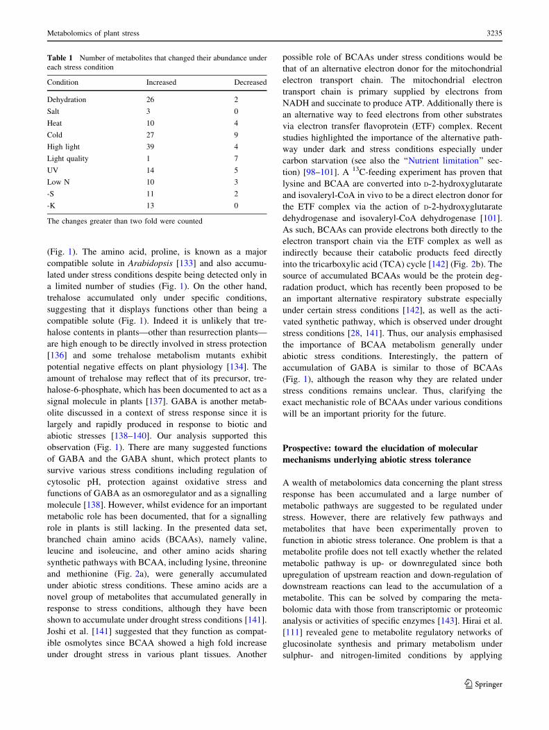
(Supplementary data, Table S1). Table 1 shows the number
of metabolites accumulated or decreased under each stress
condition. This reveals that abiotic stresses generally
induce accumulation of metabolites with only the light
quality change as an exception. The tendency of accumu-
lation is probably related to a cessation of the growth-
reducing consumption of metabolites. When subjected to
abiotic stresses, plants actively re-program their growth by
modulating both cell division and cell expansion. Growth
decreases rapidly upon stress onset, but it recovers and
adapts once stress conditions become stable [127]. Accu-
mulated metabolites might be used as building blocks to
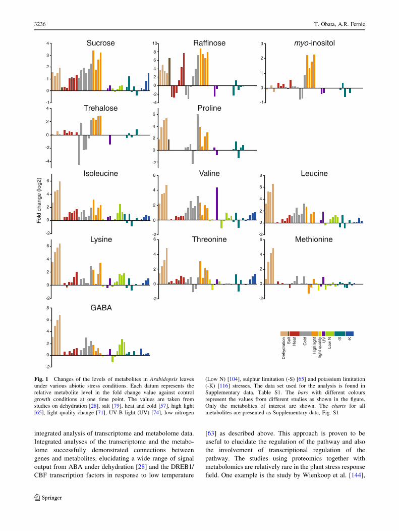
support a recovery of growth. Figure 1 provides an over-
view of the changes in the amount of selected metabolites.
Charts for all metabolites are found in the Supplementary
data, Fig. S1. In general, changes in the amounts of
metabolites were stress-specific in contrast to the general
responses observed in bacteria [128]. A stress-specific
change in the metabolite level would be a result of an
inhibition/activation of a specific metabolic pathway
especially in the short term. It should mainly be related to
the properties of enzymes such as sensitivity to tempera-
ture, oxidation and ion concentration. In addition,
rearrangement of the metabolic network should also result
in changes of metabolites, which are related to the regu-
lated pathways. Therefore, such a metabolite must be a
good candidate for an analysis to elucidate the specific
effects of an abiotic stress and the adaptive processes
against it. On the other hand, metabolites responding to
various stresses can be related to fundamental stress
responses. In the present analysis, some metabolites can be
seen to accumulate in most abiotic stress conditions
although the time and extent of accumulation varied among
conditions. Levels of sucrose were increased in most stress
conditions in at least one time point (Fig. 1). Sucrose is a
major transport sugar in most plant species and is known to
accumulate under stress conditions [129]. Compounds
defined as ‘‘compatible solutes’’ also accumulate under
various abiotic stress conditions. They are very soluble in
water and are non-toxic at high concentrations and function
to sustain the ordered vicinal water around proteins by
decreasing protein-solvent interactions at low water activ-
ities [52, 130]. This group of compounds includes betaines
and related compounds; polyols and sugars, such as man-
nitol, sorbitol and trehalose; and amino acids, such as
proline [131, 132]. Recent studies have revealed that they
function to protect plants not only from osmotic stress but
also from various stress factors [130, 131, 133, 134].
Therefore, the synthetic pathways of those metabolites
have been of interest for metabolic engineering and some
interventions have indeed increased the tolerance of some
crop plants to abiotic stress [130, 131]. Raffinose is a sugar
synthesised from sucrose and known to protect plant cells
as an osmoprotectant; it also accumulates under most stress
conditions especially at the later stages of stress treatment
(Fig. 1). Raffinose is also shown to function to protect
plants from oxidative damage [135], making the observa-
tion reasonable since oxidative damage likely underlies
most stress conditions. Interestingly, myo-inositol, which is
closely related to raffinose biosynthesis, did not show
prominent changes other than under high light conditions
3234 T. Obata, A.R. Fernie
123
(Fig. 1). The amino acid, proline, is known as a major
compatible solute in Arabidopsis [133] and also accumu-
lated under stress conditions despite being detected only in
a limited number of studies (Fig. 1). On the other hand,
trehalose accumulated only under specific conditions,
suggesting that it displays functions other than being a
compatible solute (Fig. 1). Indeed it is unlikely that tre-
halose contents in plants—other than resurrection plants—
are high enough to be directly involved in stress protection
[136] and some trehalose metabolism mutants exhibit
potential negative effects on plant physiology [134]. The
amount of trehalose may reflect that of its precursor, tre-
halose-6-phosphate, which has been documented to act as a
signal molecule in plants [137]. GABA is another metab-
olite discussed in a context of stress response since it is
largely and rapidly produced in response to biotic and
abiotic stresses [138–140]. Our analysis supported this
observation (Fig. 1). There are many suggested functions
of GABA and the GABA shunt, which protect plants to
survive various stress conditions including regulation of
cytosolic pH, protection against oxidative stress and
functions of GABA as an osmoregulator and as a signalling
molecule [138]. However, whilst evidence for an important
metabolic role has been documented, that for a signalling
role in plants is still lacking. In the presented data set,
branched chain amino acids (BCAAs), namely valine,
leucine and isoleucine, and other amino acids sharing
synthetic pathways with BCAA, including lysine, threonine
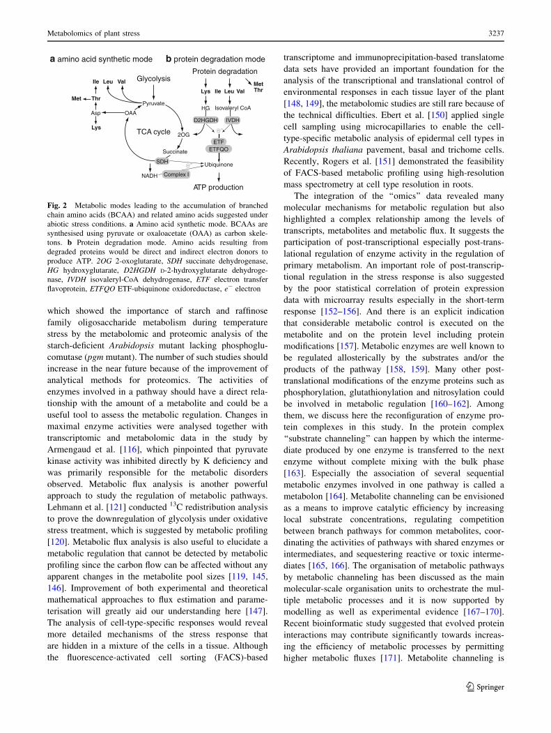
and methionine (Fig. 2a), were generally accumulated
under abiotic stress conditions. These amino acids are a
novel group of metabolites that accumulated generally in
response to stress conditions, although they have been
shown to accumulate under drought stress conditions [141].
Joshi et al. [141] suggested that they function as compat-
ible osmolytes since BCAA showed a high fold increase
under drought stress in various plant tissues. Another
possible role of BCAAs under stress conditions would be
that of an alternative electron donor for the mitochondrial
electron transport chain. The mitochondrial electron
transport chain is primary supplied by electrons from
NADH and succinate to produce ATP. Additionally there is
an alternative way to feed electrons from other substrates
via electron transfer flavoprotein (ETF) complex. Recent
studies highlighted the importance of the alternative path-
way under dark and stress conditions especially under
carbon starvation (see also the ‘‘Nutrient limitation’’ sec-
tion) [98–101]. A 13C-feeding experiment has proven that
lysine and BCAA are converted into D-2-hydroxyglutarate
and isovaleryl-CoA in vivo to be a direct electron donor for
the ETF complex via the action of D-2-hydroxyglutarate
dehydrogenase and isovaleryl-CoA dehydrogenase [101].
As such, BCAAs can provide electrons both directly to the
electron transport chain via the ETF complex as well as
indirectly because their catabolic products feed directly
into the tricarboxylic acid (TCA) cycle [142] (Fig. 2b). The
source of accumulated BCAAs would be the protein deg-
radation product, which has recently been proposed to be
an important alternative respiratory substrate especially
under certain stress conditions [142], as well as the acti-
vated synthetic pathway, which is observed under drought
stress conditions [28, 141]. Thus, our analysis emphasised
the importance of BCAA metabolism generally under
abiotic stress conditions. Interestingly, the pattern of
accumulation of GABA is similar to those of BCAAs
(Fig. 1), although the reason why they are related under
stress conditions remains unclear. Thus, clarifying the
exact mechanistic role of BCAAs under various conditions
will be an important priority for the future.
Prospective: toward the elucidation of molecular
mechanisms underlying abiotic stress tolerance
A wealth of metabolomics data concerning the plant stress
response has been accumulated and a large number of
metabolic pathways are suggested to be regulated under
stress. However, there are relatively few pathways and
metabolites that have been experimentally proven to
function in abiotic stress tolerance. One problem is that a
metabolite profile does not tell exactly whether the related
metabolic pathway is up- or downregulated since both
upregulation of upstream reaction and down-regulation of
downstream reactions can lead to the accumulation of a
metabolite. This can be solved by comparing the meta-
bolomic data with those from transcriptomic or proteomic
analysis or activities of specific enzymes [143]. Hirai et al.
[111] revealed gene to metabolite regulatory networks of
glucosinolate synthesis and primary metabolism under
sulphur- and nitrogen-limited conditions by applying
Table 1 Number of metabolites that changed their abundance under
each stress condition
Condition Increased Decreased
Dehydration 26 2
Salt 3 0
Heat 10 4
Cold 27 9
High light 39 4
Light quality 1 7
UV 14 5
Low N 10 3
-S 11 2
-K 13 0
The changes greater than two fold were counted
Metabolomics of plant stress 3235
123
integrated analysis of transcriptome and metabolome data.
Integrated analyses of the transcriptome and the metabo-
lome successfully demonstrated connections between
genes and metabolites, elucidating a wide range of signal
output from ABA under dehydration [28] and the DREB1/
CBF transcription factors in response to low temperature
[63] as described above. This approach is proven to be
useful to elucidate the regulation of the pathway and also
the involvement of transcriptional regulation of the
pathway. The studies using proteomics together with
metabolomics are relatively rare in the plant stress response
field. One example is the study by Wienkoop et al. [144],
Sucrose
0
1
2
3
4
-1
Raffinose myo-inositol
0
2
4
6
8
-2
10
-4
0
1
2
3
-1
enilorPesolaherT
0
2
4
6
-2
0
2
4
-2
-4
enicueLenilaVenicuelosI
Threonine
0
2
4
6
-2
0
2
4
6
-2Methionine
0
2
4
6
-2
0
2
4
6
-2
0
2
4
6
-2
8
GABA
0
2
4
6
-2
8
Fol
d ch
ange
(lo
g2)
Deh
ydra
tion
Sal
tH
eat
Col
d
Hig
h lig
htlig
ht q
ualit
yU
VLo
w N -S -K
0
2
4
6
-2
Lysine
Fig. 1 Changes of the levels of metabolites in Arabidopsis leaves
under various abiotic stress conditions. Each datum represents the
relative metabolite level in the fold change value against control
growth conditions at one time point. The values are taken from
studies on dehydration [28], salt [79], heat and cold [57], high light
[65], light quality change [71], UV-B light (UV) [74], low nitrogen
(Low N) [104], sulphur limitation (-S) [65] and potassium limitation
(-K) [116] stresses. The data set used for the analysis is found in
Supplementary data, Table S1. The bars with different colours
represent the values from different studies as shown in the figure.
Only the metabolites of interest are shown. The charts for all
metabolites are presented as Supplementary data, Fig. S1
3236 T. Obata, A.R. Fernie
123
which showed the importance of starch and raffinose
family oligosaccharide metabolism during temperature
stress by the metabolomic and proteomic analysis of the
starch-deficient Arabidopsis mutant lacking phosphoglu-
comutase (pgm mutant). The number of such studies should
increase in the near future because of the improvement of
analytical methods for proteomics. The activities of
enzymes involved in a pathway should have a direct rela-
tionship with the amount of a metabolite and could be a
useful tool to assess the metabolic regulation. Changes in
maximal enzyme activities were analysed together with
transcriptomic and metabolomic data in the study by
Armengaud et al. [116], which pinpointed that pyruvate
kinase activity was inhibited directly by K deficiency and
was primarily responsible for the metabolic disorders
observed. Metabolic flux analysis is another powerful
approach to study the regulation of metabolic pathways.
Lehmann et al. [121] conducted 13C redistribution analysis
to prove the downregulation of glycolysis under oxidative
stress treatment, which is suggested by metabolic profiling
[120]. Metabolic flux analysis is also useful to elucidate a
metabolic regulation that cannot be detected by metabolic
profiling since the carbon flow can be affected without any
apparent changes in the metabolite pool sizes [119, 145,
146]. Improvement of both experimental and theoretical
mathematical approaches to flux estimation and parame-
terisation will greatly aid our understanding here [147].
The analysis of cell-type-specific responses would reveal
more detailed mechanisms of the stress response that
are hidden in a mixture of the cells in a tissue. Although
the fluorescence-activated cell sorting (FACS)-based
transcriptome and immunoprecipitation-based translatome
data sets have provided an important foundation for the
analysis of the transcriptional and translational control of
environmental responses in each tissue layer of the plant
[148, 149], the metabolomic studies are still rare because of
the technical difficulties. Ebert et al. [150] applied single
cell sampling using microcapillaries to enable the cell-
type-specific metabolic analysis of epidermal cell types in
Arabidopsis thaliana pavement, basal and trichome cells.
Recently, Rogers et al. [151] demonstrated the feasibility
of FACS-based metabolic profiling using high-resolution
mass spectrometry at cell type resolution in roots.
The integration of the ‘‘omics’’ data revealed many
molecular mechanisms for metabolic regulation but also
highlighted a complex relationship among the levels of
transcripts, metabolites and metabolic flux. It suggests the
participation of post-transcriptional especially post-trans-
lational regulation of enzyme activity in the regulation of
primary metabolism. An important role of post-transcrip-
tional regulation in the stress response is also suggested
by the poor statistical correlation of protein expression
data with microarray results especially in the short-term
response [152–156]. And there is an explicit indication
that considerable metabolic control is executed on the
metabolite and on the protein level including protein
modifications [157]. Metabolic enzymes are well known to
be regulated allosterically by the substrates and/or the
products of the pathway [158, 159]. Many other post-
translational modifications of the enzyme proteins such as
phosphorylation, glutathionylation and nitrosylation could
be involved in metabolic regulation [160–162]. Among
them, we discuss here the reconfiguration of enzyme pro-
tein complexes in this study. In the protein complex
‘‘substrate channeling’’ can happen by which the interme-
diate produced by one enzyme is transferred to the next
enzyme without complete mixing with the bulk phase
[163]. Especially the association of several sequential
metabolic enzymes involved in one pathway is called a
metabolon [164]. Metabolite channeling can be envisioned
as a means to improve catalytic efficiency by increasing
local substrate concentrations, regulating competition
between branch pathways for common metabolites, coor-
dinating the activities of pathways with shared enzymes or
intermediates, and sequestering reactive or toxic interme-
diates [165, 166]. The organisation of metabolic pathways
by metabolic channeling has been discussed as the main
molecular-scale organisation units to orchestrate the mul-
tiple metabolic processes and it is now supported by
modelling as well as experimental evidence [167–170].
Recent bioinformatic study suggested that evolved protein
interactions may contribute significantly towards increas-
ing the efficiency of metabolic processes by permitting
higher metabolic fluxes [171]. Metabolite channeling is
a amino acid synthetic mode b protein degradation mode
Glycolysis
Pyruvate
2OG
OAAAsp
Lys
Met Thr
Ile Leu Val
Succinate
TCA cycle
Protein degradation
Lys Ile Leu ValMetThr
HG Isovaleryl CoA
Ubiquinone
ATP production
SDH
e-
e-
NADH Complex I
D2HGDH IVDH
ETFETFQO
Fig. 2 Metabolic modes leading to the accumulation of branched
chain amino acids (BCAA) and related amino acids suggested under
abiotic stress conditions. a Amino acid synthetic mode. BCAAs are
synthesised using pyruvate or oxaloacetate (OAA) as carbon skele-
tons. b Protein degradation mode. Amino acids resulting from
degraded proteins would be direct and indirect electron donors to
produce ATP. 2OG 2-oxoglutarate, SDH succinate dehydrogenase,
HG hydroxyglutarate, D2HGDH D-2-hydroxyglutarate dehydroge-
nase, IVDH isovaleryl-CoA dehydrogenase, ETF electron transfer
flavoprotein, ETFQO ETF-ubiquinone oxidoreductase, e- electron
Metabolomics of plant stress 3237
123
considered to be achieved not only when the enzyme
association is stable but also when the association is
dynamic. Transient complexes offer the possibility of fast
exchange of some of the polypeptide components upon
reassembly and thus can be a molecular basis for rapid fine
tuning or redirection of metabolism. The reassembly of
the metabolic enzyme complex therefore should have a
molecular basis underlying the metabolic regulation in
mitochondria under short-term oxidative stress. Metabolic
channeling including metabolon formation is reported in
many processes in plants such as glycolysis, cysteine
synthesis, the Calvin-Benson cycle, cyanogenic glucoside
biosynthesis, the phenylpropanoid pathway, the glycine
decarboxylase system and polyamine biosynthesis [165,
166]. There is however little evidence showing metabolic
channeling in plant mitochondrial metabolism although the
metabolon of TCA cycle enzymes and enzymes involved in
amino acid metabolism are well documented in bacteria,
yeast and mammals [164, 168]. More plant proteins
forming enzyme super complexes should exist and con-
tribute to metabolic regulation. Our recent study suggested
that some metabolic enzymes, including malic enzyme and
alanine amino transferase, altered their status in a protein
complex in relation to metabolic regulation under an oxi-
dative stress condition [122]. Further analysis is necessary
to prove the involvement of enzyme protein interactions in
metabolic regulation under abiotic stress conditions. Nev-
ertheless, the expected results would lead to the new
insight into plant metabolic regulation and to the full
understanding of the metabolic events under abiotic stress
conditions and further for the breeding of stress-tolerant
crops by elucidating the target metabolic pathways to be
modified.
To summarise, experiments to date have allowed us to
catalogue a vast array of metabolic changes in response to
stress. Without overgeneralising, since some of these are
very well understood at a mechanistic level, our under-
standing of the causes and effects of these changes remains
in some cases rather fragmentary. The metabolic changes in
stress responses are considered to be divided into three
phases, including a direct effect of environmental changes,
transient adaptation to stress conditions and the new steady
state established under prolonged stress conditions. It should
be noted that each phase adopts a different duration
depending on the type and the severity of the stress. A
detailed time course experiment, such as those conducted in
[66] or [128], is therefore necessary to distinguish to which
phase the metabolic changes are related. It is also highly
likely that integrating the results already obtained with those
from isotope feeding experiments, comprehensive phyto-
hormone measurements and further transcriptomic and
proteomic studies will further deepen our understanding
of these crucial survival processes. Once obtained such
information will provide an immense foundation for meta-
bolic engineering and synthetic biology approaches to
ensuring food security.
Open Access This article is distributed under the terms of the
Creative Commons Attribution License which permits any use, dis-
tribution, and reproduction in any medium, provided the original
author(s) and the source are credited.
References
1. Fiehn O (2001) Combining genomics, metabolome analysis, and
biochemical modelling to understand metabolic networks. Comp
Funct Genomics 2:155–168
2. Davies HV, Shepherd LV, Stewart D, Frank T, Rohlig RM,
Engel KH (2010) Metabolome variability in crop plant spe-
cies—when, where, how much and so what? Regul Toxicol
Pharmacol 58:S54–S61
3. De Luca V, St Pierre B (2000) The cell and developmental
biology of alkaloid biosynthesis. Trends Plant Sci 5:168–173
4. D’Auria JC, Gershenzon J (2005) The secondary metabolism of
Arabidopsis thaliana: growing like a weed. Curr Opin Plant Biol
8:308–316
5. Saito K, Matsuda F (2010) Metabolomics for functional
genomics, systems biology, and biotechnology. Annu Rev Plant
Biol 61:463–489
6. Afendi FM, Okada T, Yamazaki M, Hirai-Morita A, Nakamura
Y, Nakamura K, Ikeda S, Takahashi H, Altaf-Ul-Amin M,
Darusman LK, Saito K, Kanaya S (2012) KNApSAcK family
databases: integrated metabolite-plant species databases for
multifaceted plant research. Plant Cell Physiol 53:e1
7. Fernie AR, Aharoni A, Willmitzer L, Stitt M, Tohge T, Kopka J,
Carroll AJ, Saito K, Fraser PD, DeLuca V (2011) Recommen-
dations for reporting metabolite data. Plant Cell 23:2477–2482
8. Giavalisco P, Kohl K, Hummel J, Seiwert B, Willmitzer L (2009)13C isotope-labeled metabolomes allowing for improved
compound annotation and relative quantification in liquid chro-
matography–mass spectrometry-based metabolomic research.
Anal Chem 81:6546–6551
9. Fernie AR, Trethewey RN, Krotzky AJ, Willmitzer L (2004)
Metabolite profiling: from diagnostics to systems biology. Nat
Rev Mol Cell Bio 5:763–769
10. Lisec J, Schauer N, Kopka J, Willmitzer L, Fernie AR (2006)
Gas chromatography mass spectrometry-based metabolite pro-
filing in plants. Nat Protoc 1:387–396
11. Halket JM, Waterman D, Przyborowska AM, Patel RK, Fraser
PD, Bramley PM (2005) Chemical derivatization and mass
spectral libraries in metabolic profiling by GC/MS and LC/MS/
MS. J Exp Bot 56:219–243
12. Schauer N, Steinhauser D, Strelkov S, Schomburg D, Allison G,
Moritz T, Lundgren K, Roessner-Tunali U, Forbes MG,
Willmitzer L, Fernie AR, Kopka J (2005) GC–MS libraries for
the rapid identification of metabolites in complex biological
samples. FEBS Lett 579:1332–1337
13. Kumari S, Stevens D, Kind T, Denkert C, Fiehn O (2011)
Applying in silico retention index and mass spectra matching for
identification of unknown metabolites in accurate mass GC-TOF
mass spectrometry. Anal Chem 83:5895–5902
14. Kind T, Wohlgemuth G, Lee do Y, Lu Y, Palazoglu M, Shahbaz
S, Fiehn O (2009) FiehnLib: mass spectral and retention index
libraries for metabolomics based on quadrupole and time-of-
flight gas chromatography/mass spectrometry. Anal Chem 81:
10038–10048
3238 T. Obata, A.R. Fernie
123
15. Kopka J, Schauer N, Krueger S, Birkemeyer C, Usadel B,
Bergmuller E, Dormann P, Weckwerth W, Gibon Y, Stitt M,
Willmitzer L, Fernie AR, Steinhauser D (2005) GMD@
CSB.DB: the Golm Metabolome Database. Bioinformatics
21:1635–1638
16. Rogachev I, Aharoni A (2012) UPLC-MS-based metabolite
analysis in tomato. Methods Mol Biol 860:129–144
17. Allwood JW, Goodacre R (2010) An introduction to liquid
chromatography–mass spectrometry instrumentation applied in
plant metabolomic analyses. Phytochem Anal 21:33–47
18. Lei Z, Huhman DV, Sumner LW (2011) Mass spectrometry
strategies in metabolomics. J Biol Chem 286:25435–25442
19. Moco S, Bino RJ, Vorst O, Verhoeven HA, de Groot J, van Beek
TA, Vervoort J, de Vos CH (2006) A liquid chromatography–
mass spectrometry-based metabolome database for tomato.
Plant Physiol 141:1205–1218
20. Tohge T, Fernie AR (2009) Web-based resources for mass-
spectrometry-based metabolomics: a user’s guide. Phytochem-
istry 70:450–456
21. Feldberg L, Venger I, Malitsky S, Rogachev I, Aharoni A (2009)
Dual labeling of metabolites for metabolome analysis (DLEM-
MA): a new approach for the identification and relative
quantification of metabolites by means of dual isotope labeling
and liquid chromatography–mass spectrometry. Anal Chem 81:
9257–9266
22. Arrivault S, Guenther M, Ivakov A, Feil R, Vosloh D, van
Dongen JT, Sulpice R, Stitt M (2009) Use of reverse-phase
liquid chromatography, linked to tandem mass spectrometry, to
profile the Calvin cycle and other metabolic intermediates in
Arabidopsis rosettes at different carbon dioxide concentrations.
Plant J 59:826–839
23. Kanno Y, Jikumaru Y, Hanada A, Nambara E, Abrams SR,
Kamiya Y, Seo M (2010) Comprehensive hormone profiling in
developing Arabidopsis seeds: examination of the site of ABA
biosynthesis, ABA transport and hormone interactions. Plant
Cell Physiol 51:1988–2001
24. Ramautar R, Somsen GW, de Jong GJ (2009) CE-MS in met-
abolomics. Electrophoresis 30:276–291
25. Ramautar R, Mayboroda OA, Somsen GW, de Jong GJ (2011)
CE-MS for metabolomics: developments and applications in the
period 2008–2010. Electrophoresis 32:52–65
26. Monton MR, Soga T (2007) Metabolome analysis by capillary
electrophoresis–mass spectrometry. J Chromatogr A 1168:237–246
27. Sugimoto M, Hirayama A, Robert M, Abe S, Soga T, Tomita M
(2010) Prediction of metabolite identity from accurate mass,
migration time prediction and isotopic pattern information in
CE-TOFMS data. Electrophoresis 31:2311–2318
28. Urano K, Maruyama K, Ogata Y, Morishita Y, Takeda M,
Sakurai N, Suzuki H, Saito K, Shibata D, Kobayashi M, Yam-
aguchi-Shinozaki K, Shinozaki K (2009) Characterization of the
ABA-regulated global responses to dehydration in Arabidopsisby metabolomics. Plant J 57:1065–1078
29. Williams BJ, Cameron CJ, Workman R, Broeckling CD, Sum-
ner LW, Smith JT (2007) Amino acid profiling in plant cell
cultures: an inter-laboratory comparison of CE-MS and GC–MS.
Electrophoresis 28:1371–1379
30. Soga T, Imaizumi M (2001) Capillary electrophoresis method
for the analysis of inorganic anions, organic acids, amino acids,
nucleotides, carbohydrates and other anionic compounds. Elec-
trophoresis 22:3418–3425
31. Fernie AR (2007) The future of metabolic phytochemistry: larger
numbers of metabolites, higher resolution, greater understanding.
Phytochemistry 68:2861–2880
32. Schripsema J (2010) Application of NMR in plant metabolo-
mics: techniques, problems and prospects. Phytochem Anal 21:
14–21
33. Terskikh VV, Feurtado JA, Borchardt S, Giblin M, Abrams SR,
Kermode AR (2005) In vivo 13C NMR metabolite profiling:
potential for understanding and assessing conifer seed quality.
J Exp Bot 56:2253–2265
34. Ratcliffe RG, Shachar-Hill Y (2005) Revealing metabolic phe-
notypes in plants: inputs from NMR analysis. Biol Rev Camb
Philos Soc 80:27–43
35. Eisenreich W, Bacher A (2007) Advances of high-resolution
NMR techniques in the structural and metabolic analysis of
plant biochemistry. Phytochemistry 68:2799–2815
36. Krishnan P, Kruger NJ, Ratcliffe RG (2005) Metabolite finger-
printing and profiling in plants using NMR. J Exp Bot 56:
255–265
37. Martin FP, Dumas ME, Wang Y, Legido-Quigley C, Yap IK,
Tang H, Zirah S, Murphy GM, Cloarec O, Lindon JC, Sprenger
N, Fay LB, Kochhar S, van Bladeren P, Holmes E, Nicholson JK
(2007) A top-down systems biology view of microbiome-
mammalian metabolic interactions in a mouse model. Mol Syst
Biol 3:112
38. Lommen A, Weseman JM, Smith GO, Noteborn HPJM (1998)
On the detection of environmental effects on complex matrices
combining off-line liquid chromatography and 1H-NMR. Bio-
degradation 9:513–525
39. Dixon RA, Gang DR, Charlton AJ, Fiehn O, Kuiper HA, Rey-
nolds TL, Tjeerdema RS, Jeffery EH, German JB, Ridley WP,
Seiber JN (2006) Applications of metabolomics in agriculture.
J Agric Food Chem 54:8984–8994
40. Shulaev V, Cortes D, Miller G, Mittler R (2008) Metabolomics
for plant stress response. Physiol Plant 132:199–208
41. Bowne J, Bacic A, Tester MRU (2011) Abiotic stress and
metabolomics. Annu Plant Rev 43:61–85
42. Cominelli E, Galbiati M, Tonelli C, Bowler C (2009) Water: the
invisible problem. Access to fresh water is considered to be a
universal and free human right, but dwindling resources and a
burgeoning population are increasing its economic value.
EMBO Rep 10:671–676
43. Chaves MM, Oliveira MM (2004) Mechanisms underlying plant
resilience to water deficits: prospects for water-saving agricul-
ture. J Exp Bot 55:2365–2384
44. Verslues PE, Juenger TE (2011) Drought, metabolites, and
Arabidopsis natural variation: a promising combination for
understanding adaptation to water-limited environments. Curr
Opin Plant Biol 14:240–245
45. Skirycz A, Vandenbroucke K, Clauw P, Maleux K, De Meyer B,
Dhondt S, Pucci A, Gonzalez N, Hoeberichts F, Tognetti VB,
Galbiati M, Tonelli C, Van Breusegem F, Vuylsteke M, Inze D
(2011) Survival and growth of Arabidopsis plants given limited
water are not equal. Nat Biotechnol 29:212–214
46. Skirycz A, De Bodt S, Obata T, De Clercq I, Claeys H, De
Rycke R, Andriankaja M, Van Aken O, Van Breusegem F,
Fernie AR, Inze D (2010) Developmental stage specificity and
the role of mitochondrial metabolism in the response of Ara-bidopsis leaves to prolonged mild osmotic stress. Plant Physiol
152:226–244
47. Semel Y, Nissenbaum J, Menda N, Zinder M, Krieger U, Issman
N, Pleban T, Lippman Z, Gur A, Zamir D (2006) Overdominant
quantitative trait loci for yield and fitness in tomato. Proc Natl
Acad Sci USA 103:12981–12986
48. Witt S, Galicia L, Lisec J, Cairns J, Tiessen A, Araus JL, Pal-
acios-Rojas N, Fernie AR (2012) Metabolic and phenotypic
responses of greenhouse-grown maize hybrids to experimentally
controlled drought stress. Mol Plant 5:401–417
49. Bowne JB, Erwin TA, Juttner J, Schnurbusch T, Langridge P,
Bacic A, Roessner U (2012) Drought responses of leaf tissues
from wheat cultivars of differing drought tolerance at the
metabolite level. Mol Plant 5:418–429
Metabolomics of plant stress 3239
123
50. van Dongen JT, Frohlich A, Ramırez-Aguilar SJ, Schauer N,
Fernie AR, Erban A, Kopka J, Clark J, Langer A, Geigenberger
P (2009) Transcript and metabolite profiling of the adaptive
response to mild decreases in oxygen concentration in the roots
of Arabidopsis plants. Ann Bot 103:269–280
51. Rocha M, Licausi F, Araujo WL, Nunes-Nesi A, Sodek L,
Fernie AR, van Dongen JT (2010) Glycolysis and the tricar-
boxylic acid cycle are linked by alanine aminotransferase during
hypoxia induced by waterlogging of Lotus japonicus. Plant
Physiol 152:1501–1513
52. Guy CL (1990) Cold Acclimation and Freezing Stress Toler-
ance: Role of Protein Metabolism. Annu Rev Plant Biol Plant
Mol Biol 41:187–223
53. Wanner LA, Junttila O (1999) Cold-induced freezing tolerance
in Arabidopsis. Plant Physiol 120:391–400
54. Thomashow MF (2010) Molecular basis of plant cold acclima-
tion: insights gained from studying the CBF cold response
pathway. Plant Physiol 154:571–577
55. Medina J, Catala R, Salinas J (2011) The CBFs: three Arabid-opsis transcription factors to cold acclimate. Plant Sci 180:3–11
56. Cook D, Fowler S, Fiehn O, Thomashow MF (2004) A promi-
nent role for the CBF cold response pathway in configuring the
low-temperature metabolome of Arabidopsis. Proc Natl Acad
Sci USA 101:15243–15248
57. Kaplan F, Kopka J, Haskell DW, Zhao W, Schiller KC, Gatzke
N, Sung DY, Guy CL (2004) Exploring the temperature-stress
metabolome. Plant Physiol 136:4159–4168
58. Kaplan F, Kopka J, Sung DY, Zhao W, Popp M, Porat R, Guy
CL (2007) Transcript and metabolite profiling during cold
acclimation of Arabidopsis reveals an intricate relationship of
cold-regulated gene expression with modifications in metabolite
content. Plant J 50:967–981
59. Gray GR, Heath D (2005) A global reorganization of the
metabolome in Arabidopsis during cold acclimation is revealed
by metabolic fingerprinting. Physiol Plant 124:236–248
60. Hannah MA, Wiese D, Freund S, Fiehn O, Heyer AG, Hincha
DK (2006) Natural genetic variation of freezing tolerance in
Arabidopsis. Plant Physiol 142:98–112
61. Espinoza C, Degenkolbe T, Caldana C, Zuther E, Leisse A,
Willmitzer L, Hincha DK, Hannah MA (2010) Interaction with
diurnal and circadian regulation results in dynamic metabolic
and transcriptional changes during cold acclimation in Arabid-opsis. PLoS One 5:e14101
62. Liu Q, Kasuga M, Sakuma Y, Abe H, Miura S, Yamaguchi-
Shinozaki K, Shinozaki K (1998) Two transcription factors,
DREB1 and DREB2, with an EREBP/AP2 DNA binding
domain separate two cellular signal transduction pathways in
drought- and low-temperature-responsive gene expression,
respectively, in Arabidopsis. Plant Cell 10:1391–1406
63. Maruyama K, Takeda M, Kidokoro S, Yamada K, Sakuma Y,
Urano K, Fujita M, Yoshiwara K, Matsukura S, Morishita Y,
Sasaki R, Suzuki H, Saito K, Shibata D, Shinozaki K, Yamag-
uchi-Shinozaki K (2009) Metabolic pathways involved in cold
acclimation identified by integrated analysis of metabolites and
transcripts regulated by DREB1A and DREB2A. Plant Physiol
150:1972–1980
64. Lugan R, Niogret MF, Kervazo L, Larher FR, Kopka J, Bou-
chereau A (2009) Metabolome and water status phenotyping of
Arabidopsis under abiotic stress cues reveals new insight into
ESK1 function. Plant Cell Environ 32:95–108
65. Wulff-Zottele C, Gatzke N, Kopka J, Orellana A, Hoefgen R,
Fisahn J, Hesse H (2010) Photosynthesis and metabolism
interact during acclimation of Arabidopsis thaliana to high
irradiance and sulphur depletion. Plant Cell Environ 33:
1974–1988
66. Caldana C, Degenkolbe T, Cuadros-Inostroza A, Klie S, Sulpice
R, Leisse A, Steinhauser D, Fernie AR, Willmitzer L, Hannah
MA (2011) High-density kinetic analysis of the metabolomic
and transcriptomic response of Arabidopsis to eight environ-
mental conditions. Plant J 67:869–884
67. Terashima I, Araya T, Miyazawa S, Sone K, Yano S (2005)
Construction and maintenance of the optimal photosynthetic
systems of the leaf, herbaceous plant and tree: an eco-develop-
mental treatise. Ann Bot 95:507–519
68. Smith H (2000) Phytochromes and light signal perception by
plants—an emerging synthesis. Nature 407:585–591
69. Allen JF (2003) Cyclic, pseudocyclic and noncyclic photo-
phosphorylation: new links in the chain. Trends Plant Sci
8:15–19
70. Dietzel L, Brautigam K, Pfannschmidt T (2008) Photosynthetic
acclimation: state transitions and adjustment of photosystem
stoichiometry–functional relationships between short-term and
long-term light quality acclimation in plants. FEBS J 275:
1080–1088
71. Brautigam K, Dietzel L, Kleine T, Stroher E, Wormuth D, Dietz
KJ, Radke D, Wirtz M, Hell R, Dormann P, Nunes-Nesi A,
Schauer N, Fernie AR, Oliver SN, Geigenberger P, Leister D,
Pfannschmidt T (2009) Dynamic plastid redox signals integrate
gene expression and metabolism to induce distinct metabolic
states in photosynthetic acclimation in Arabidopsis. Plant Cell
21:2715–2732
72. Jin H, Cominelli E, Bailey P, Parr A, Mehrtens F, Jones J,
Tonelli C, Weisshaar B, Martin C (2000) Transcriptional
repression by AtMYB4 controls production of UV-protecting
sunscreens in Arabidopsis. EMBO J 19:6150–6161
73. Ries G, Heller W, Puchta H, Sandermann H, Seidlitz HK, Hohn
B (2000) Elevated UV-B radiation reduces genome stability in
plants. Nature 406:98–101
74. Kusano M, Tohge T, Fukushima A, Kobayashi M, Hayashi N,
Otsuki H, Kondou Y, Goto H, Kawashima M, Matsuda F, Niida
R, Matsui M, Saito K, Fernie AR (2011) Metabolomics reveals
comprehensive reprogramming involving two independent
metabolic responses of Arabidopsis to UV-B light. Plant J
67:354–369
75. Tohge T, Kusano M, Fukushima A, Saito K, Fernie AR (2011)
Transcriptional and metabolic programs following exposure of
plants to UV-B irradiation. Plant Signal Behav 6:1987–1992
76. Munns R (2005) Genes and salt tolerance: bringing them toge-
ther. New Phytol 167:645–663
77. Kim JK, Bamba T, Harada K, Fukusaki E, Kobayashi A (2007)
Time-course metabolic profiling in Arabidopsis thaliana cell
cultures after salt stress treatment. J Exp Bot 58:415–424
78. Tester M, Davenport R (2003) Na? tolerance and Na? transport
in higher plants. Ann Bot 91:503–527
79. Gong Q, Li P, Ma S, Indu Rupassara S, Bohnert HJ (2005)
Salinity stress adaptation competence in the extremophile
Thellungiella halophila in comparison with its relative Arabid-opsis thaliana. Plant J 44:826–839
80. Lugan R, Niogret MF, Leport L, Guegan JP, Larher FR, Savoure
A, Kopka J, Bouchereau A (2010) Metabolome and water
homeostasis analysis of Thellungiella salsuginea suggests that
dehydration tolerance is a key response to osmotic stress in this
halophyte. Plant J 64:215–229
81. Johnson HE, Broadhurst D, Goodacre R, Smith AR (2003)
Metabolic fingerprinting of salt-stressed tomatoes. Phytochem-
istry 62:919–928
82. Cramer GR, Ergul A, Grimplet J, Tillett RL, Tattersall EA,
Bohlman MC, Vincent D, Sonderegger J, Evans J, Osborne C,
Quilici D, Schlauch KA, Schooley DA, Cushman JC (2007)
Water and salinity stress in grapevines: early and late changes in
3240 T. Obata, A.R. Fernie
123

transcript and metabolite profiles. Funct Integr Genomics
7:111–134
83. Brosche M, Vinocur B, Alatalo ER, Lamminmaki A, Teichmann
T, Ottow EA, Djilianov D, Afif D, Bogeat-Triboulot MB, Alt-
man A, Polle A, Dreyer E, Rudd S, Paulin L, Auvinen P,
Kangasjarvi J (2005) Gene expression and metabolite profiling
of Populus euphratica growing in the Negev desert. Genome
Biol 6:R101
84. Gagneul D, Aınouche A, Duhaze C, Lugan R, Larher FR,
Bouchereau A (2007) A reassessment of the function of the
so-called compatible solutes in the halophytic plumbaginaceae
Limonium latifolium. Plant Physiol 144:1598–1611
85. Zuther E, Koehl K, Kopka J (2007) Comparative metabolome
analysis of the salt response in breeding cultivars of rice. In:
Jenks MA, Hasegawa PM, Jain SM (eds) Advances in molecular
breeding toward drought and salt tolerant crops. Springer,
Berlin, pp 285–315
86. Sanchez DH, Siahpoosh MR, Roessner U, Udvardi M, Kopka J
(2008) Plant metabolomics reveals conserved and divergent
metabolic responses to salinity. Physiol Plant 132:209–219
87. Sanchez DH, Lippold F, Redestig H, Hannah MA, Erban A,
Kramer U, Kopka J, Udvardi MK (2008) Integrative functional
genomics of salt acclimatization in the model legume Lotusjaponicus. Plant J 53:973–987
88. Sanchez DH, Pieckenstain FL, Escaray F, Erban A, Kraemer U,
Udvardi MK, Kopka J (2011) Comparative ionomics and met-
abolomics in extremophile and glycophytic Lotus species under
salt stress challenge the metabolic pre-adaptation hypothesis.
Plant Cell Environ 34:605–617
89. Sanchez DH, Pieckenstain FL, Szymanski J, Erban A, Bromke
M, Hannah MA, Kraemer U, Kopka J, Udvardi MK (2011)
Comparative functional genomics of salt stress in related model
and cultivated plants identifies and overcomes limitations to
translational genomics. PLoS One 6:e17094
90. Sharma SS, Dietz KJ (2009) The relationship between metal
toxicity and cellular redox imbalance. Trends Plant Sci 14:
43–50
91. Jahangir M, Abdel-Farid IB, Choi YH, Verpoorte R (2008)
Metal ion-inducing metabolite accumulation in Brassica rapa.
J Plant Physiol 165:1429–1437
92. Sun X, Zhang J, Zhang H, Ni Y, Zhang Q, Chen J, Guan Y
(2010) The responses of Arabidopsis thaliana to cadmium
exposure explored via metabolite profiling. Chemosphere 78:
840–845
93. Dubey S, Misra P, Dwivedi S, Chatterjee S, Bag SK, Mantri S,
Asif MH, Rai A, Kumar S, Shri M, Tripathi P, Tripathi RD,
Trivedi PK, Chakrabarty D, Tuli R (2010) Transcriptomic and
metabolomic shifts in rice roots in response to Cr(VI) stress.
BMC Genomics 11:648
94. Smith AM, Stitt M (2007) Coordination of carbon supply and
plant growth. Plant Cell Environ 30:1126–1149
95. Stitt M, Gibon Y, Lunn JE, Piques M (2007) Multilevel
genomics analysis of carbon signalling during low carbon
availability: coordinating the supply and utilisation of carbon in
a fluctuating environment. Funct Plant Biol 34:526
96. Osuna D, Usadel B, Morcuende R, Gibon Y, Blasing OE, Hohne
M, Gunter M, Kamlage B, Trethewey R, Scheible WR, Stitt M
(2007) Temporal responses of transcripts, enzyme activities and
metabolites after adding sucrose to carbon-deprived Arabidopsisseedlings. Plant J 49:463–491
97. Usadel B, Blasing OE, Gibon Y, Retzlaff K, Hohne M, Gunther M,
Stitt M (2008) Global transcript levels respond to small changes of
the carbon status during progressive exhaustion of carbohydrates in
Arabidopsis rosettes. Plant Physiol 146:1834–1861
98. Ishizaki K, Larson TR, Schauer N, Fernie AR, Graham IA,
Leaver CJ (2005) The critical role of Arabidopsis electron-
transfer flavoprotein:ubiquinone oxidoreductase during dark-
induced starvation. Plant Cell 17:2587–2600
99. Ishizaki K, Larson TR, Schauer N, Fernie AR, Graham IA,
Leaver CJ (2006) The mitochondrial electron transfer flavo-
protein complex is essential for survival of Arabidopsis in
extended darkness. Plant J 47:751–760
100. Araujo WL, Ishizaki K, Nunes-Nesi A, Tohge T, Larson TR,
Krahnert I, Balbo I, Witt S, Dormann P, Graham IA, Leaver CJ,
Fernie AR (2011) Analysis of a range of catabolic mutants
provides evidence that phytanoyl-coenzyme A does not act as a
substrate of the electron-transfer flavoprotein/electron-transfer
flavoprotein:ubiquinone oxidoreductase complex in Arabidopsisduring dark-induced sene. Plant Physiol 157:55–69
101. Araujo WL, Ishizaki K, Nunes-Nesi A, Larson TR, Tohge T,
Krahnert I, Witt S, Obata T, Schauer N, Graham IA, Leaver CJ,
Fernie AR (2010) Identification of the 2-hydroxyglutarate and
isovaleryl-CoA dehydrogenases as alternative electron donors
linking lysine catabolism to the electron transport chain of
Arabidopsis mitochondria. Plant Cell 22:1549–1563
102. Urbanczyk-Wochniak E, Fernie AR (2005) Metabolic profiling
reveals altered nitrogen nutrient regimes have diverse effects on
the metabolism of hydroponically-grown tomato (Solanum ly-copersicum) plants. J Exp Bot 56:309–321
103. Stitt M, Fernie AR (2003) From measurements of metabolites to
metabolomics: an ‘‘on the fly’’ perspective illustrated by recent
studies of carbon–nitrogen interactions. Curr Opin Biotechnol
14:136–144
104. Tschoep H, Gibon Y, Carillo P, Armengaud P, Szecowka M,
Nunes-Nesi A, Fernie AR, Koehl K, Stitt M (2009) Adjustment
of growth and central metabolism to a mild but sustained
nitrogen-limitation in Arabidopsis. Plant Cell Environ 32:300–
318
105. Morcuende R, Bari R, Gibon Y, Zheng W, Pant BD, Blasing O,
Usadel B, Czechowski T, Udvardi MK, Stitt M, Scheible WR
(2007) Genome-wide reprogramming of metabolism and regu-
latory networks of Arabidopsis in response to phosphorus. Plant
Cell Environ 30:85–112
106. Hernandez G, Ramırez M, Valdes-Lopez O, Tesfaye M, Graham
MA, Czechowski T, Schlereth A, Wandrey M, Erban A, Cheung
F, Wu HC, Lara M, Town CD, Kopka J, Udvardi MK, Vance CP
(2007) Phosphorus stress in common bean: root transcript and
metabolic responses. Plant Physiol 144:752–767
107. Hernandez G, Valdes-Lopez O, Ramırez M, Goffard N, Weiller
G, Aparicio-Fabre R, Fuentes SI, Erban A, Kopka J, Udvardi MK,
Vance CP (2009) Global changes in the transcript and metabolic
profiles during symbiotic nitrogen fixation in phosphorus-stressed
common bean plants. Plant Physiol 151:1221–1238
108. Huang CY, Roessner U, Eickmeier I, Genc Y, Callahan DL,
Shirley N, Langridge P, Bacic A (2008) Metabolite profiling
reveals distinct changes in carbon and nitrogen metabolism in
phosphate-deficient barley plants (Hordeum vulgare L.). Plant
Cell Physiol 49:691–703
109. Nikiforova VJ, Daub CO, Hesse H, Willmitzer L, Hoefgen R
(2005) Integrative gene-metabolite network with implemented
causality deciphers informational fluxes of sulphur stress
response. J Exp Bot 56:1887–1896
110. Nikiforova VJ, Kopka J, Tolstikov V, Fiehn O, Hopkins L,
Hawkesford MJ, Hesse H, Hoefgen R (2005) Systems rebal-
ancing of metabolism in response to sulfur deprivation, as
revealed by metabolome analysis of Arabidopsis plants. Plant
Physiol 138:304–318
111. Hirai MY, Yano M, Goodenowe DB, Kanaya S, Kimura T,
Awazuhara M, Arita M, Fujiwara T, Saito K (2004) Integration
of transcriptomics and metabolomics for understanding of global
responses to nutritional stresses in Arabidopsis thaliana. Proc
Natl Acad Sci USA 101:10205–10210
Metabolomics of plant stress 3241
123

112. Hirai MY, Klein M, Fujikawa Y, Yano M, Goodenowe DB,
Yamazaki Y, Kanaya S, Nakamura Y, Kitayama M, Suzuki H,
Sakurai N, Shibata D, Tokuhisa J, Reichelt M, Gershenzon J,
Papenbrock J, Saito K (2005) Elucidation of gene-to-gene and
metabolite-to-gene networks in Arabidopsis by integration of
metabolomics and transcriptomics. J Biol Chem 280:25590–
25595
113. Hoefgen R, Nikiforova VJ (2008) Metabolomics integrated with
transcriptomics: assessing systems response to sulfur-deficiency
stress. Physiol Plant 132:190–198
114. Hubberten HM, Klie S, Caldana C, Degenkolbe T, Willmitzer L,
Hoefgen R (2012) An additional role of O-acetylserine as a
sulphur status independent regulator during plant growth. Plant J
70:666–677
115. Marschner H (1995) Mineral nutrition of higher plants, 2nd edn.
Academic Press, London
116. Armengaud P, Sulpice R, Miller AJ, Stitt M, Amtmann A,
Gibon Y (2009) Multilevel analysis of primary metabolism
provides new insights into the role of potassium nutrition for
glycolysis and nitrogen assimilation in Arabidopsis roots. Plant
Physiol 150:772–785
117. Mittler R (2006) Abiotic stress, the field environment and stress
combination. Trends Plant Sci 11:15–19
118. Møller IM, Jensen PE, Hansson A (2007) Oxidative modifica-
tions to cellular components in plants. Annu Rev Plant Biol
58:459–481
119. Baxter CJ, Redestig H, Schauer N, Repsilber D, Patil KR,
Nielsen J, Selbig J, Liu J, Fernie AR, Sweetlove LJ (2007) The
metabolic response of heterotrophic Arabidopsis cells to
oxidative stress. Plant Physiol 143:312–325
120. Lehmann M, Schwarzlander M, Obata T, Sirikantaramas S,
Burow M, Olsen CE, Tohge T, Fricker MD, Møller BL, Fernie
AR, Sweetlove LJ, Laxa M (2009) The metabolic response of
Arabidopsis roots to oxidative stress is distinct from that of
heterotrophic cells in culture and highlights a complex rela-
tionship between the levels of transcripts, metabolites, and flux.
Mol Plant 2:390–406
121. Lehmann M, Laxa M, Sweetlove LJ, Fernie AR, Obata T (2011)
Metabolic recovery of Arabidopsis thaliana roots following
cessation of oxidative stress. Metabolomics 8:143–153
122. Obata T, Matthes A, Koszior S, Lehmann M, Araujo WL, Bock
R, Sweetlove LJ, Fernie AR (2011) Alteration of mitochondrial
protein complexes in relation to metabolic regulation under
short-term oxidative stress in Arabidopsis seedlings. Phyto-
chemistry 72:1081–1091
123. Ishikawa T, Takahara K, Hirabayashi T, Matsumura H, Fujisawa
S, Terauchi R, Uchimiya H, Kawai-Yamada M (2010) Metab-
olome analysis of response to oxidative stress in rice suspension
cells overexpressing cell death suppressor Bax inhibitor-1. Plant
Cell Physiol 51:9–20
124. Morgan MJ, Lehmann M, Schwarzlander M, Baxter CJ, Sien-
kiewicz-Porzucek A, Williams TC, Schauer N, Fernie AR,
Fricker MD, Ratcliffe RG, Sweetlove LJ, Finkemeier I (2008)
Decrease in manganese superoxide dismutase leads to reduced
root growth and affects tricarboxylic acid cycle flux and mito-
chondrial redox homeostasis. Plant Physiol 147:101–114
125. Rizhsky L, Liang H, Shuman J, Shulaev V, Davletova S, Mittler
R (2004) When defense pathways collide. The response of
Arabidopsis to a combination of drought and heat stress. Plant
Physiol 134:1683–1696
126. Buri M, Masunaga T, Wakatsuki T (2000) Sulfur and zinc levels
as limiting factors to rice production in West Africa lowlands.
Geoderma 94:23–42
127. Skirycz A, Inze D (2010) More from less: plant growth under
limited water. Curr Opin Biotechnol 21:197–203
128. Jozefczuk S, Klie S, Catchpole G, Szymanski J, Cuadros-Ino-
stroza A, Steinhauser D, Selbig J, Willmitzer L (2010)
Metabolomic and transcriptomic stress response of Escherichiacoli. Mol Syst Biol 6:364
129. Rolland F, Baena-Gonzalez E, Sheen J (2006) Sugar sensing and
signaling in plants: conserved and novel mechanisms. Annu Rev
Plant Biol 57:675–709
130. Chen THH, Murata N (2011) Glycinebetaine protects plants
against abiotic stress: mechanisms and biotechnological appli-
cations. Plant Cell Environ 34:1–20
131. Chen THH, Murata N (2002) Enhancement of tolerance of
abiotic stress by metabolic engineering of betaines and other
compatible solutes. Curr Opin Plant Biol 5:250–257
132. Diamant S, Eliahu N, Rosenthal D, Goloubinoff P (2001)
Chemical chaperones regulate molecular chaperones in vitro and
in cells under combined salt and heat stresses. J Biol Chem
276:39586–395891
133. Verbruggen N, Hermans C (2008) Proline accumulation in
plants: a review. Amino Acids 35:753–759
134. Fernandez O, Bethencourt L, Quero A, Sangwan RS, Clement C
(2010) Trehalose and plant stress responses: friend or foe?
Trends Plant Sci 15:409–417
135. Nishizawa A, Yabuta Y, Shigeoka S (2008) Galactinol and
raffinose constitute a novel function to protect plants from
oxidative damage. Plant Physiol 147:1251–1263
136. Wingler A (2002) The function of trehalose biosynthesis in
plants. Phytochemistry 60:437–440
137. Smeekens S, Ma J, Hanson J, Rolland F (2010) Sugar signals
and molecular networks controlling plant growth. Curr Opin
Plant Biol 13:274–279
138. Bouche N, Fromm H (2004) GABA in plants: just a metabolite?
Trends Plant Sci 9:110–115
139. Shelp BJ, Bown AW, Mclean MD (1999) Metabolism and
functions of gamma-aminobutyric acid. Trends Plant Sci 4:446–
452
140. Kinnersley A, Turano F (2000) Gamma aminobutyric acid
(GABA) and plant responses to stress. Crit Rev Plant Sci 19:
479–509
141. Joshi V, Joung JG, Fei Z, Jander G (2010) Interdependence of
threonine, methionine and isoleucine metabolism in plants:
accumulation and transcriptional regulation under abiotic stress.
Amino Acids 39:933–947
142. Araujo WL, Tohge T, Ishizaki K, Leaver CJ, Fernie AR (2011)
Protein degradation—an alternative respiratory substrate for
stressed plants. Trends Plant Sci 16:489–498
143. Cramer GR, Urano K, Delrot S, Pezzotti M, Shinozaki K (2011)
Effects of abiotic stress on plants: a systems biology perspective.
BMC Plant Biol 11:163
144. Wienkoop S, Morgenthal K, Wolschin F, Scholz M, Selbig J,
Weckwerth W (2008) Integration of metabolomic and proteomic
phenotypes: analysis of data covariance dissects starch and RFO
metabolism from low and high temperature compensation
response in Arabidopsis thaliana. Mol Cell Proteomics 7:
1725–1736
145. Fell DA (2005) Enzymes, metabolites and fluxes. J Exp Bot
56:267–272
146. Fernie AR, Geigenberger P, Stitt M (2005) Flux an important,
but neglected, component of functional genomics. Curr Opin
Plant Biol 8:174–182
147. Fernie AR, Stitt M (2012) On the discordance of metabolomics
with proteomics and transcriptomics: coping with increasing
complexity in logic, chemistry and network interactions. Plant
Physiol 158:1139–1145
148. Dinneny JR, Long TA, Wang JY, Jung JW, Mace D, Pointer S,
Barron C, Brady SM, Schiefelbein J, Benfey PN (2008) Cell
3242 T. Obata, A.R. Fernie
123
identity mediates the response of Arabidopsis roots to abiotic
stress. Science 320:942–945
149. Wee CW, Dinneny JR (2010) Tools for high-spatial and tem-
poral-resolution analysis of environmental responses in plants.
Biotechnol Lett 32:1361–1371
150. Ebert B, Zoller D, Erban A, Fehrle I, Hartmann J, Niehl A,
Kopka J, Fisahn J (2010) Metabolic profiling of Arabidopsisthaliana epidermal cells. J Exp Bot 61:1321–1335
151. Rogers ED, Jackson T, Moussaieff A, Aharoni A, Benfey PN
(2012) Cell type-specific transcriptional profiling: implications
for metabolite profiling. Plant J 70:5–17
152. Koussevitzky S, Suzuki N, Huntington S, Armijo L, Sha W,
Cortes D, Shulaev V, Mittler R (2008) Ascorbate peroxidase 1
plays a key role in the response of Arabidopsis thaliana to stress
combination. J Biol Chem 283:34197–34203
153. Bogeat-Triboulot MB, Brosche M, Renaut J, Jouve L, Le Thiec
D, Fayyaz P, Vinocur B, Witters E, Laukens K, Teichmann T,
Altman A, Hausman JF, Polle A, Kangasjarvi J, Dreyer E (2007)
Gradual soil water depletion results in reversible changes of
gene expression, protein profiles, ecophysiology, and growth
performance in Populus euphratica, a poplar growing in arid
regions. Plant Physiol 143:876–892
154. Grimplet J, Wheatley MD, Jouira HB, Deluc LG, Cramer GR,
Cushman JC (2009) Proteomic and selected metabolite analysis
of grape berry tissues under well-watered and water-deficit
stress conditions. Proteomics 9:2503–2528
155. Nie L, Wu G, Culley DE, Scholten JC, Zhang W (2007) Inte-
grative analysis of transcriptomic and proteomic data: challenges,
solutions and applications. Crit Rev Biotechnol 27:63–75
156. Bohmer M, Schroeder JI (2011) Quantitative transcriptomic
analysis of abscisic acid-induced and reactive oxygen species-
dependent expression changes and proteomic profiling in Ara-bidopsis suspension cells. Plant J 67:105–118
157. Morgenthal K, Wienkoop S, Wolschin F, Weckwerth W (2007)
Integrative profiling of metabolites and proteins: improving
pattern recognition and biomarker selection for systems level
approaches. Methods Mol Biol 358:57–75
158. Huang Z, Zhu L, Cao Y, Wu G, Liu X, Chen Y, Wang Q, Shi T,
Zhao Y, Wang Y, Li W, Li Y, Chen H, Chen G, Zhang J (2011)
ASD: a comprehensive database of allosteric proteins and
modulators. Nucleic Acids Res 39:D663–D669
159. Goodey NM, Benkovic SJ (2008) Allosteric regulation and
catalysis emerge via a common route. Nat Chem Biol 4:474–482
160. Dalle-Donne I, Rossi R, Giustarini D, Colombo R, Milzani A
(2007) S-glutathionylation in protein redox regulation. Free
Radic Biol Med 43:883–898
161. Lindermayr C, Durner J (2009) S-Nitrosylation in plants: pattern
and function. J Proteomics 73:1–9
162. Schulze WX (2010) Proteomics approaches to understand pro-
tein phosphorylation in pathway modulation. Curr Opin Plant
Biol 13:280–287
163. Spivey HO, Ovadi J (1999) Substrate channeling. Methods
19:306–321
164. Srere PA (1987) Complexes of sequential metabolic enzymes.
Annu Rev Biochem 56:89–124
165. Winkel BSJ (2004) Metabolic channeling in plants. Annu Rev
Plant Biol 55:85–107
166. Jørgensen K, Rasmussen AV, Morant M, Nielsen AH, Bjarnholt
N, Zagrobelny M, Bak S, Møller BL (2005) Metabolon forma-
tion and metabolic channeling in the biosynthesis of plant
natural products. Curr Opin Plant Biol 8:280–291
167. Graham JW, Williams TC, Morgan M, Fernie AR, Ratcliffe RG,
Sweetlove LJ (2007) Glycolytic enzymes associate dynamically
with mitochondria in response to respiratory demand and sup-
port substrate channeling. Plant Cell 19:3723–3738
168. Srere PA (2000) Macromolecular interactions: tracing the roots.
Trends Biochem Sci 25:150–153
169. Degenring D, Rohl M, Uhrmacher AM (2004) Discrete event,
multi-level simulation of metabolite channeling. Biosystems
75:29–41
170. Ro DK, Douglas CJ (2004) Reconstitution of the entry point of
plant phenylpropanoid metabolism in yeast (Saccharomycescerevisiae): implications for control of metabolic flux into the
phenylpropanoid pathway. J Biol Chem 279:2600–2607
171. Durek P, Walther D (2008) The integrated analysis of metabolic
and protein interaction networks reveals novel molecular orga-
nizing principles. BMC Syst Biol 2:100
Metabolomics of plant stress 3243
123


![Metabolomics Basics[1]](https://img.pdfslide.us/doc/110x75/553de2815503466f378b4864/metabolomics-basics1.jpg)