Embed Size (px)
Citation preview

MOLECULAR AND CELLULAR BIOLOGY, Sept. 2009, p. 5031–5045 Vol. 29, No. 180270-7306/09/$08.00�0 doi:10.1128/MCB.00144-09Copyright © 2009, American Society for Microbiology. All Rights Reserved.
The Tumor Suppressor Functions of p27kip1 Include Control of theMesenchymal/Amoeboid Transition�†
Stefania Berton,1 Barbara Belletti,1 Katarina Wolf,2 Vincenzo Canzonieri,3 Francesca Lovat,1Andrea Vecchione,4 Alfonso Colombatti,1,5 Peter Friedl,2 and Gustavo Baldassarre1*
Division of Experimental Oncology 2, Centro di Riferimento Oncologico, Istituto Nazionale Tumori, IRCCS Aviano 33081, Italy1;Microscopical Imaging of the Cell, Department of Cell Biology, NCMLS, Radboud University Nijmegen Medical Centre,
6500 HB Nijmegen, The Netherlands2; Division of Pathology, Centro di Riferimento Oncologico, Istituto Nazionale Tumori,IRCCS Aviano 33081, Italy3; Division of Pathology, II Faculty of Medicine, University La Sapienza,
Ospedale Sant’Andrea, Rome, Italy4; and Dipartimento di Scienze e Tecnologie Biomediche,and MATI Center of Excellence, University of Udine, 33100 Udine, Italy5
Received 1 February 2009/Returned for modification 9 March 2009/Accepted 2 July 2009
In many human cancers, p27 downregulation correlates with a worse prognosis, suggesting that p27 levelscould represent an important determinant in cell transformation and cancer development. Using a mousemodel system based on v-src-induced transformation, we show here that p27 absence is always linked to a moreaggressive phenotype. When cultured in three-dimensional contexts, v-src-transformed p27-null fibroblastsundergo a morphological switch from an elongated to a rounded cell shape, accompanied by amoeboid-likemorphology and motility. Importantly, the acquisition of the amoeboid motility is associated with a greaterability to move and colonize distant sites in vivo. The reintroduction of different p27 mutants in v-src-transformed p27-null cells demonstrates that the control of cell proliferation and motility represents twodistinct functions of p27, both necessary for it to fully act as a tumor suppressor. Thus, we highlight here a newp27 function in driving cell plasticity that is associated with its C-terminal portion and does not depend on thecontrol of cyclin-dependent kinase activity.
Dissemination of tumor cells is strictly linked to their abilityto attach to and move within the extracellular matrix (ECM) ina three-dimensional (3D) environment. The use of 3D exper-imental model systems revealed that a higher complexity in cellmigration and adaptation responses exists in the 3D modelthan in the classical 2D model (10, 16, 41, 49). A strikingexample is given by the fact that only in 3D could individuallymigrating cells use different mechanisms such as mesenchymaland amoeboid motility (16, 17). The relative slow mesenchymalmigration is characterized by a fibroblast-like spindle shapeand is dependent on integrin-mediated adhesion and on pro-tease function (16). The amoeboid motility can in some casesrepresent a less adhesive, integrin-independent type of move-ment. Cells use a propulsive mechanism and are highly de-formable, and rather than degrade the matrix, they are able tosqueeze through it (16). As a result, the cells that use theamoeboid motility can potentially move faster than cells thatuse a mesenchymal strategy. Mesenchymal and amoeboidmovements are also characterized by a different involvement ofsmall GTPases of the Rho family. A high RhoA activity isassociated mainly with the amoeboid motility, while the mes-enchymal migration needs a high Rac activity at the leadingedge to promote the extension of cellular protrusions (41, 48).Under certain circumstances, cancer cells can undergo conver-
sion from a mesenchymal toward an amoeboid motility, anevent referred as mesenchymal-amoeboid transition (MAT)(50). MAT represents a putative escape mechanism in tumorcell dissemination that could be induced by inhibition of peri-cellular proteolysis (50) or by increased membrane-associatedRhoA activity (18, 40).
Key mediators of cell motility through ECM substrates arethe members of the Src family kinases. The prototype of Srcfamily kinases, c-Src (14), is activated following cell-ECM ad-hesion and contributes to regulate the focal adhesion turnoverand the cytoskeletal modifications necessary for normal celladhesion and motility (52). The c-Src gene is the proto-onco-gene of the transforming gene v-src of Rous sarcoma virus, andits elevated protein level and activity have been found in manyhuman tumors (20, 28, 27, 34). Despite the accumulation ofinformation and new molecular understanding of how Src iscontrolled, there is still an incomplete picture about its role inthe generation of the malignant phenotype. v-Src shows higherlevels of the kinase activity and transforming ability than c-Src(14, 15, 52). It induces normal cells to acquire a variety oftransformed features, including alteration of morphology andincrease of invasion ability due to its role in focal adhesionremodeling (7, 9, 13).
Many data suggest that there is a close relationship betweencell-ECM interaction and the proliferation and movements inboth normal and tumor cells (5, 38, 43). Accordingly, Src ac-tivation may influence not only cell motility but also cell cycleprogression by targeting the cell cycle inhibitor p27kip1 to pro-teasomal degradation (22, 39). Recent evidences indicated thatp27kip1 (hereafter called p27) can also regulate cell migration,even though its role still remains controversial since it has been
* Corresponding author. Mailing address: Division of ExperimentalOncology 2, Centro di Riferimento Oncologico, Via Franco Gallini, 2,33081 Aviano, Italy. Phone: 39 0434 659 233. Fax: 39 0434 659 428.E-mail: [email protected].
† Supplemental material for this article may be found at http://mcb.asm.org/.
� Published ahead of print on 13 July 2009.
5031
Dow
nloa
ded
from
http
s://j
ourn
als.
asm
.org
/jour
nal/m
cb o
n 18
Oct
ober
202
1 by
222
.102
.14.
206.
reported to either block or stimulate cell movements (1, 4, 11,19, 21, 23, 29, 45).
Based on these notions, we tested the possible contributionof p27 to the growth and motility phenotypes induced by v-srctransformation, with special regard to those cellular invasivefeatures that can be observed in 3D environments. By studyingin vitro and in vivo the behavior of wild-type (WT) and p27-null fibroblasts transformed with v-src, we highlight a new rolefor p27 in the regulation of cellular plasticity that can ulti-mately drive tumor cell shape, motility, and invasion.
MATERIALS AND METHODS
Cell cultures and development of stable cell lines. 3T3-p27WT and p27-knock-out (p27KO) fibroblasts were described previously (1). All cell lines were grownin Dulbecco modified Eagle medium (DMEM) supplemented with 10% fetalbovine serum (FBS) (Sigma). To obtain stable cell clones, pM-vSrc (generouslyprovided by Renato Baserga, Thomas Jefferson University, Philadelphia, PA)was cotransfected with pTRE2pur (Clontech) or pMSCV-Hyg (Clontech) vector.All of the p27-rescued clones were obtained using retroviral transduction asreported previously (1). Briefly, retroviral transduction was performed by tran-siently transfecting the pMSCV vectors coding for p27T187A and p271-170 intoEcoPack 293 cells (Clontech). Three days later, conditioned medium from three100-mm dishes containing the retroviruses was used to transduce one 100-mmdish of exponentially growing p27WT or p27KO fibroblasts. Clones and masstransfections were selected and maintained in complete medium with puromycin(1.5 �g/ml) and/or hygromycin (0.4 mg/ml).
Preparation of cell lysates, immunoblotting, and immunoprecipitation. Celllysates were prepared using cold NP-40 lysis buffer (0.5% NP-40, 50 mM HEPES[pH 7], 250 mM NaCl, 5 mM EDTA, 0.5 mM EGTA [pH 8]) plus a proteaseinhibitor cocktail (Complete; Roche), 1 mM sodium orthovanadate, and 1 mMdithiothreitol as reported previously (2).
Immunoprecipitations were performed using 0.5 mg of total lysate in HNTGbuffer (20 mM HEPES, 150 mM NaCl, 10% glycerol, 0.1% Triton X-100) plusthe specific agarose-conjugated primary antibody, with gentle rocking overnightat 4°C. When primary antibodies were not agarose conjugated, protein A orprotein G Sepharose 4 Fast Flow (Amersham Biosciences) was added during thelast 2 h of incubation. Immunoprecipitates were washed six times in HNTGbuffer, and then nine parts were resuspended in 3� Laemmli sample buffer with50 mM dithiothreitol and one part was resuspended in kinase buffer. Kinaseassays were performed as previously described (2, 37) using 2 �g of histone H1as the substrate (Upstate Biotechnology) and [�-32P]ATP to reveal phosphory-lation levels.
For immunoblotting, 40 �g of proteins was separated by 4 to 20% sodiumdodecyl sulfate-polyacrylamide gel electrophoresis (Criterion precast gel; Bio-Rad) and transferred to nitrocellulose membranes (Hybond C; Amersham Inc.).Membranes were incubated with primary antibodies as indicated and then withhorseradish peroxidase-conjugated secondary antibodies (GE Healthcare) forECL detection (GE Healthcare) or Alexa-conjugated secondary antibodies (In-vitrogen) for Odyssey infrared detection (Licor). For immunoprecipitates, rabbitimmunoglobulin G and mouse immunoglobulin G True Blot (eBioscience) sec-ondary horseradish peroxidase-conjugated antibodies were used, following themanufacturer’s instructions.
Primary antibodies were from Transduction Laboratories (p27, cyclin-depen-dent kinase 1 [CDK1], CDK2, and cofilin), Cell Signaling (phosphorylated serine3 [pS3]-cofilin), Santa Cruz (sc-54 CDK1, sc-245 cyclin B1, sc-751 cyclin A,sc-7649 vinculin, and sc-19 c-Src,), and Sigma (OP18/stathmin and �-tubulin).
Growth curve, MTT assay, FACS analysis, and anchorage-independent cellgrowth. Cell proliferation was evaluated using growth curves, 3-(4,5-dimethyl-2-thiazolyl)-2,5-diphenyl-2H-tetrazolium bromide (MTT) assay, fluorescence-acti-vated cell sorting (FACS) analysis after propidium iodide incorporation, andsoft-agar assay, as previously described (1). For the MTT assay (Sigma), 1,000cells/well were seeded in 96-well plates. At the indicated times, MTT solution incomplete medium (0.28-mg/ml final concentration) was added and incubated at37°C for 4 h. The medium was discarded, and the formazan salts were dissolvedin dimethyl sulfoxide. The colorimetric substrate was measured and quantified at560 nm in an enzyme-linked immunosorbent assay plate reader.
For cell growth, 1 � 105 cells/well were seeded in six-well plates in completemedium. At the indicated times, cells were detached in trypsin-EDTA, resus-pended in trypan blue, and counted.
The cell cycle profile was analyzed by propidium iodide staining (50 �g/ml
propidium iodide and 100 �g/ml RNase A) followed by FACS analysis, asdescribed elsewhere (1, 2). The data were analyzed using WinMDI2.8 software.
To analyze the anchorage-independent cell growth, cells (1 � 103 and 5 � 103)were suspended in 2 ml top agar medium (DMEM-10% FBS, 0.4% low-melting-point agarose) and kept at 42°C until needed. The cell suspension was thenquickly overlaid on 2 ml of previously layered bottom agar medium (DMEM-10% FBS, 0.6% low-melting-point agarose) in six-well tissue culture plates intriplicate. DMEM-10% FBS with 1.5 �g/ml puromycin and/or 0.4 mg/ml hygro-mycin was added to the wells every 3 days as a feeder layer. On day 8, the numberof colonies was counted in 15 random fields at a magnification of �10.
Immunofluorescence of cells included in 3D matrices. Single cells included in3D Matrigel or collagen I were fixed in phosphate-buffered saline (PBS)–4%paraformaldehyde for 20 min at room temperature and then permeabilized inPBS with 0.2% Triton X-100 and blocked for at least 2 hours in PBS with 1%bovine serum albumin and 10% normal goat serum (Dako). Incubation withfluorescein isothiocyanate (FITC)-conjugated monoclonal anti-�-tubulin anti-body (Sigma) and phallotoxin-AlexaFluor546 (Molecular Probes, Invitrogen) foractin staining was performed for 2 hours at room temperature in PBS–1% bovineserum albumin and 1% normal goat serum. Incubation with anti-pSer3-cofilin(Cell Signaling) and anti-acetylated tubulin (Sigma) was performed overnight at4°C, followed by anti-rabbit–AlexaFluor488 and anti-mouse–AlexaFluor488staining, respectively. �1-Integrin expression was revealed using an anti-�1-integrin FITC-conjugated antibody (BD) by incubating the cells overnight at 4°C.For imaging of collagen fibers, laser light at a low density (the “reflection”parameter in confocal microscopy) was used. Immunofluorescence-labeled cellswere studied using a confocal laser-scanning microscope (Diaphot 200 Nikon;MRC-1024, Bio-Rad Laboratories) or a Leica epifluorescence microscope.Quantification of pS3-cofilin, acetylated tubulin, and phalloidin fluorescenceintensities was performed using the Leica LAS software and analyzing at least 40cells/clone.
Evasion assays. For evasion assays, cells (7.5 � 105cells/ml) were included inMatrigel (BD) (6-mg/ml final concentration) or collagen type I (BD or Purecol-Nutacon) (1.7-mg/ml final concentration) drops (10 �l of matrix volume perdrop). The drops, sufficiently spaced, were dispensed on the bottom of cellculture dishes and maintained for 1 h upside down to polymerize at 37°C.Matrigel was diluted in complete medium, whereas collagen I was diluted in asolution containing 10� DMEM (without phenol red), 7.5% sodium bicarbon-ate, and complete medium. After polymerization, the plates were turned on andthe drops were incubated for the indicated times in complete medium.
Cell motility was assessed by transmission microscopy using a Nikon TS100/Fmicroscope. Images were collected using a digital camera (Nikon Coolpix 995).Cells present outside each drop (five drops per cell line per experiment) werecounted to estimate their evasion ability using a 10� objective.
Animal experiments. Primary tumors were established by subcutaneous injec-tion of 1 � 106 transformed cells into the flanks of female athymic nude mice(Harlan, 8 weeks of age). At 15 days after injection, the animals were sacrificedand tumor analysis was performed. Spleen samples were also collected andrapidly frozen for RNA extraction.
A second experiment was performed with 1 � 106 cells injected into the tailveins. After 1 month or 20 days, as indicated, the animals were sacrificed andlung analysis was performed.
Tissue samples, RNA extraction, and reverse transcription-PCR (RT-PCR).Isolation of total RNA from spleen samples was performed using the RNeasyminikit (Qiagen). After retrotranscription with avian myeloblastosis virus reversetranscriptase and random primers according to the provider’s instruction(Promega), the obtained cDNAs were amplified by nested PCR in order toevaluate the presence of the injected cells in spleen samples. The followingprimers designed on vectors were used: pTREforward1, 5�-CAGCAGGCAGAAGTATGCAA-3�; pTREforward2, 5�-TGCAAAGCATGCATCTCAAT-3�; pTREreverse1, 5�-CGTGAGGAAGAGTTCTTGCAG-3�; pTREreverse2,5�-AGTTCTTGCAGCTCGGTGAC-3�; pMSCVforward, 5�-CCCTTGAACCTCCTCGTTCGACC-3�; pMSCVreverse, 5�-GAGACGTGCTACTTCCATTTGTC-3�; and p27-170reverse 5�-GGATCCCTCGAGTGTTCTGTTGGCTCTTTT-3�.
Statistics. Statistical analysis was done using the two-tailed unpaired Studentt test or the two-tailed unpaired Mann-Whitney U test. Differences were con-sidered significant at a P value of �0.05.
RESULTS
p27�/� transformed cells exhibit higher proliferation poten-tial than p27�/� cells. To gain new information on the role of
5032 BERTON ET AL. MOL. CELL. BIOL.
Dow
nloa
ded
from
http
s://j
ourn
als.
asm
.org
/jour
nal/m
cb o
n 18
Oct
ober
202
1 by
222
.102
.14.
206.

p27 during the process of cell transformation, the oncogenev-src was used to transform mouse fibroblasts derived fromp27�/� and p27�/� embryos. Independent cell clones stablyoverexpressing comparable levels of v-src for each genotypewere fully characterized (Fig. 1A). In agreement with the lit-erature data (22, 39), we observed a decrease in the amount ofendogenous p27 after v-src transformation, and the relativelower p27 levels remained constant and were confirmed aftereach experiment (Fig. 1A and data not shown).
Since p27 is a well-known tumor suppressor gene able toinhibit cell proliferation, the selected clones were initially an-alyzed for their proliferative behavior. FACS analysis of theDNA content in exponentially growing cells demonstrated thatp27�/� v-src-transformed fibroblasts (referred to below as v-srcfibroblasts) displayed a higher S-phase population than WTcells (Fig. 1B). Accordingly, both cell growth curves and MTTproliferation assays (Fig. 1C), demonstrated that the v-src-mediated cell proliferation rate could be further increasedwhen p27 expression was fully abrogated.
It is well accepted that p27 regulates cell cycle progressionby interacting with and inhibiting different cyclin-CDK com-plexes, in particular by impairing the activity of CDK2- andCDK1-containing complexes (reviewed in reference 2a).Hence, the activity of CDK2, and to a lesser extent that of
CDK1, was higher in p27KO than in p27WT v-src cells (CDK2,P 0.002; CDK1, P 0.07 [for WT versus KO using Student’st test]), although some clonal variability was detected (Fig.1D). Similar results were obtained by assaying the cyclin A-and B1-associated kinase activities (Fig. 1D) (cyclin A, P 0.01; cyclin B1, P 0.05 [for WT versus KO using Student’s ttest]), thus providing a molecular explanation for the differentproliferation rates observed.
To specifically analyze the contribution of p27 in the G1-Stransition in v-src-transformed cells, we synchronized the cellsin the G1 phase by serum starvation and then stimulated themto reenter the cell cycle by release in complete medium. Cellsnormally progressing through the cell cycle, such as p27�/�
and p27�/� 3T3 fibroblasts, were used as a control. Whilenormal cells arrest in G1 phase after serum starvation andenter the S phase about 15 h after serum stimulation (see Fig.S1A and S1B in the supplemental material), v-src-transformedfibroblasts were less sensitive to growth factor deprivation, asdemonstrated by both FACS analysis of DNA content (see Fig.S1C in the supplemental material) and Western blot analysis oftypical markers of the S (cyclin A) or G2-M (cyclin B1) phasesof cell cycle (see Fig. S1D in the supplemental material). Im-portantly, FACS analysis of transformed cells grown to conflu-ence demonstrated that p27 expression is necessary to induce
FIG. 1. The absence of p27 in v-src-transformed cells provides a proliferative advantage in vitro. (A) Western blot analysis of v-src and p27expression in p27�/� and p27�/� v-src-clones. �-Tubulin was used to normalize the amount of loaded proteins. (B) Cell cycle distribution ofexponentially growing p27�/� and p27�/� v-src cells evaluated by FACS analysis. The percentage of cells in each phase of the cell cycle is reportedin the graph and represents the mean ( standard deviation) from three independent experiments. (C) Growth curve analysis (upper graph) andMTT assay (lower graph) of p27�/� and p27�/� v-src cells. Data represent the means and standard deviations from three independent experimentsperformed in triplicate (growth curve) or six times (MTT assay) (P 0.001 for growth curve and P 0.0001 for MTT assay at day 4 using Student’st test). (D) Kinase activity associated with CDK2, CDK1, cyclin A, and cyclin B1, using histone H1 as the substrate. In the lower panels the amountof immunoprecipitated (IP) protein is shown. The normalized kinase activities (expressed in arbitrary units) reported under each lane representthe means ( standard deviations) from three independent experiments.
VOL. 29, 2009 ROLE OF p27kip1 IN v-src TRANSFORMATION 5033
Dow
nloa
ded
from
http
s://j
ourn
als.
asm
.org
/jour
nal/m
cb o
n 18
Oct
ober
202
1 by
222
.102
.14.
206.

G1 arrest after cell-cell contact, whereas it is dispensable innormal cells (see Fig. S1E and S1F in the supplemental mate-rial), as previously reported (32). Overall these data demon-strated that p27 plays an important role in the control of cellcycle progression also in v-src-transformed cells and that, asexpected, its major function is carried out during the G1-Stransition.
The tumorigenic potential of v-src fibroblasts was then testedin vitro by soft-agar assay. Both p27�/� and p27�/� v-src fi-broblasts were able to grow in an anchorage-independent wayin soft agar, although p27-null cells displayed a significantgrowth advantage both in the number and in the size of thecolonies formed (Fig. 2A and B).
Next, two different clones of p27�/� and p27�/� v-src fibro-blasts were subcutaneously injected in nude mice (n 21 forp27�/� cells and n 20 for p27�/� v-src cells). Xenograftswere monitored for 15 days, and then the mice were sacrificedand the tumor masses analyzed. The data showed that tumorsformed from p27�/� transformed cells were significantly largerthan those from the WT counterpart (Fig. 2C and D). Immu-nohistochemical analysis confirmed that tumors formed byp27�/� v-src cells express p27 protein both in the nucleus andin the cytoplasm (Fig. 2E). Further, the pathological analysisdemonstrated that tumors formed by p27-null cells displayedan increased number of mitotic figures (Fig. 2F and G) thatwere often classified as atypical mitosis (Fig. 2F, inset), indi-cating that p27�/� v-src cells formed hyperproliferative andless differentiated tumors. No significant variations were ob-served between the two WT and the two KO cell clones utilized(Fig. 2C and D), thus excluding bias due to clonal selection.
p27 expression discriminates between mesenchymal andamoeboid morphology and motility. Recently, the role of p27in cell motility has become a debated issue in the scientificliterature. We previously demonstrated a reduction in ECM-driven cell migration when p27 is expressed at the cytoplasmiclevel (1). We thus tested whether p27 could be involved in theregulation of cell morphology and motility during oncogenictransformation. p27�/� and p27�/� v-src fibroblasts were in-cluded in a 3D collagen I matrix, and their morphology wasfirst evaluated at different time points by bright-field transmis-sion microscopy to better discriminate the cell distributionalong the z axis (Fig. 3A, upper panels). v-src-transformedfibroblasts included in 3D matrices typically acquired twodifferent shapes: an elongated spindled shape with finger-like protrusion at the cellular edges or a rounded morphol-ogy characterized by numerous peripheral membrane ruf-fles. Counting the rounded versus the elongated cells, wecould verify that while p27�/� fibroblasts displayed similarpercentages of elongated and rounded cells (47.8% 7%elongated cells), p27�/� v-src fibroblasts acquired almost ex-clusively the rounded morphology (86% 3.3% rounded cells)(Fig. 3B). We next evaluated the organization of the cytoskel-etal components of transformed cells in 3D using immunoflu-orescence and confocal microscopy analysis. p27�/� andp27�/� v-src fibroblasts were cultured for 10 h within the col-lagen I matrix and then fixed and stained for �-tubulin andF-actin (Fig. 3A, bottom panels). This analysis confirmed thattransformed p27�/� cells retained a fibroblast-like elongatedmorphology with cellular protrusions that were positive forboth microtubules (MTs) and the actin cytoskeleton. Con-
versely, p27�/� v-src cells mainly displayed a rounded shape,lost the dendritic-like extensions, and showed a cortical distri-bution of actin and a perinuclear distribution of MTs (Fig. 3A).
The same morphological differences also emerged in time-lapse video microscopy of multicellular spheroids included inthe 3D collagen matrix. As shown by the photograms collectedafter 0 and 6 h (Fig. 3C) and by the movies obtained bycollecting one picture every 4 min (see Videos S1 and S2 in thesupplemental material), when p27�/� v-src cells detached fromthe cell cluster, they retained a rounded morphology andshowed a higher deformability. They were able to squeeze thecellular body through the collagen fibrils, moving in a propul-sive way with a reduced directional persistence. Conversely,p27�/� transformed fibroblasts moved from the cluster usinglong protrusions aimed to generate the traction force neededfor the cell body to advance (Fig. 3C; see Videos S1 and S2 inthe supplemental material). The features that emerged fromthese experiments were highly suggestive of the describedmodel for the amoeboid and the mesenchymal types of mor-phology and motility (16). Because the amoeboid mechanism isdefined as an extracellular protease-independent and Rho-dependent invasion while mesenchymal invasion is metallopro-teinase and integrin dependent and Rho independent (41, 50),we looked at the expression of �1-integrin and investigated therole of metalloproteinases and Rho activity in the motility ofp27�/� and p27�/� v-src cells. In p27�/� spindle-shaped cellsimmersed in 3D collagen, �1-integrin forms clusters that areabsent in p27�/� round cells, which show a nonclustered, lin-ear surface distribution of �1-integrin (see Fig. S2A in thesupplemental material), compatible with the differences be-tween mesenchymal and amoeboid cells in the different modelsystems (50).
To test the dependence of p27�/� and p27�/� v-src cells onthe activity of metalloproteinases, we blocked their activityusing the chemical inhibitor GM-6001. Under these condi-tions, WT cells tended to assume a round morphology both inspheroid assays and in single-cell assays (Fig. 3C and data notshown; see Video S3 in the supplemental material). Con-versely, in the absence of p27, GM-6001 did not alter theamoeboid motility of src-transformed cells (Fig. 3C and datanot shown; see Video S4 in the supplemental material). Wethen tested the involvement of the Rho pathway in the motilityof both cell lines using the ROCK inhibitor Y27632. Thiscompound did not modify the shape or motility mode of WTcells (Fig. 2C and data not shown; see Video S5 in the supple-mental material), while a striking conversion from amoeboid tomesenchymal motility was observed in p27-null cells both inspheroid assays and in single-cell assays (Fig. 3C and data notshown; see Video S6 in the supplemental material).
Overall, these data imply that p27 acts as a novel regulatorof the cellular shape and motility during the v-src transforma-tion process, since its absence favors the acquisition of anamoeboid motility.
The amoeboid motility of p27�/� v-src cells is associatedwith higher cell speed and invasive ability. To evaluatewhether the two morphologies and migration modes observedin p27�/� and p27�/� v-src fibroblasts resulted in a differencein the migration efficiency, we performed time-lapse videomicroscopy coupled with cell tracking analysis of individualcells included in the 3D collagen matrix (Fig. 4A and B; see
5034 BERTON ET AL. MOL. CELL. BIOL.
Dow
nloa
ded
from
http
s://j
ourn
als.
asm
.org
/jour
nal/m
cb o
n 18
Oct
ober
202
1 by
222
.102
.14.
206.

Videos S7 and S8 in the supplemental material). This analysisdemonstrated that p27�/� v-src cells had a twofold-higher cellspeed than the WT counterpart (speeds of 0.227 and 0.118�m/min for WT and KO cells, respectively). (Fig. 4A). Thedifference in speed is statistically highly significant, and it is
also well represented by the observation of the cell trajectories,showing how p27�/� transformed cells definitely cover longerpaths than WT cells in the same time frame (Fig. 4B). Similarresults emerged from other migration assays, such as cell eva-sion from drops of either collagen I or Matrigel matrices. In
FIG. 2. Absence of p27 in v-src-transformed cells provides a growth advantage in vivo. (A and B) Soft-agar assay of v-src-transformed cells. Inpanel A, representative pictures of colonies after 8 days of incubation in agar are shown. Bar, 50 �m. In panel B, the quantification representingthe means and standard deviations from three independent experiments each performed in triplicate is reported. For each cell line at least 15randomly selected fields (10� objective) were counted (P � 0.001 for p27�/� versus p27�/� by Student’s t test). (C) Typical images of tumors innude mice injected with 1 � 106 p27�/� (left) and p27�/� (right) v-src cells. (D) A representative picture of tumors explanted 15 days after injectionand quantification of tumor mass, representing the mean values and standard deviations for 11 mice for p27�/� v-src A4, 10 mice for p27�/� v-srcA6, and 10 mice for p27�/� v-src 3 and 11. (E) Representative image of p27 expression in tumors formed by p27�/� v-src cells. A nucleocytoplasmicexpression of p27 is clearly visible in the inset at higher magnification. (F) Representative images of hematoxylin-eosin staining performed onexplanted xenografts, as indicated. Black arrows indicate normal mitoses. Red arrows indicate atypical mitoses. The insets show higher magnifi-cation of a normal mitosis in p27�/� v-src tumors (upper panel) and of an atypical mitosis in p27�/� v-src tumors (lower panel). (G) Quantificationof mitotic figures obtained by analyzing 10 consecutive high-power fields in five tumors for each genotype. Error bars indicate standard deviations.
VOL. 29, 2009 ROLE OF p27kip1 IN v-src TRANSFORMATION 5035
Dow
nloa
ded
from
http
s://j
ourn
als.
asm
.org
/jour
nal/m
cb o
n 18
Oct
ober
202
1 by
222
.102
.14.
206.

agreement with the tracking data, p27�/� v-src cells displayeda greater ability than p27�/� cells to exit from drops, confirm-ing that p27-null cells have a higher capacity to move in 3D(Fig. 4C). Interestingly, in a 2D wound-healing assay, p27�/�
v-src cells did not display any motility advantage with respect top27�/� v-src cells (see Fig. S2B in the supplemental material),supporting the hypothesis that molecular requirements for 2Dmigration and 3D migration are distinct.
In order to determine if the motile behavior observed invitro could be linked to different metastatic potentials in vivo,we evaluated the abilities of p27�/� and p27�/� v-src cells toboth intravasate and extravasate. To evaluate the intravasa-tion, mice were injected subcutaneously with p27�/� andp27�/� v-src cells (n 9 per genotype) and analyzed, 2 weekslater, for the presence of transformed cells in the spleen byRT-PCR with primers designed on the transfected vector (Fig.5A and B). While none of the mice injected with p27�/� v-srccells (0/9) was positive for the presence of transformed cells inthe spleen, 4/9 mice injected with p27-null cells were positive.
The ability to extravasate, settle, and colonize distant siteswas also evaluated. In an experiment aimed to evaluate thesurvival of mice following metastasis formation, we injectedtransformed fibroblasts into the tail veins of nude mice (n 4per genotype). The mice were sacrificed when they displayedbreathing defects and fatigue (1 month later), and by simplemacroscopic observation of the lungs, a massive difference was
already observed, with more than a 10-fold difference in thenumber of tumor foci per lung (Fig. 5C).
All the in vitro and in vivo results suggested that the absenceof p27 in v-src-transformed fibroblasts induces an increasedproliferation coupled with a motility advantage that could beattributed to the switch from a mesenchymal to an amoeboidmotility mode.
p27 expression in p27�/� v-src cells reduces proliferationand tumor growth. To validate that the observed effects on theproliferative behavior were due to p27 itself, we stably reintro-duced p27 in p27�/� v-src cells. To avoid or reduce the v-src-induced degradation of p27 WT protein, we used the cDNAcoding for two less degradable forms, p27T187A and p271–170
(Fig. 6A). p27T187A carries a point mutation that impairs p27degradation via the ubiquitin-dependent proteasome pathway(31, 46). The deletion mutant p271–170 also lacks this residue,and we previously demonstrated that it retains the ability toblock cell proliferation while losing the migration-inhibitoryproperties of the WT protein (1). We selected several inde-pendent p27-rescued clones and pools expressing comparablelevels of v-src and p27 proteins (Fig. 6A).
In v-src-transformed fibroblasts, both p27 mutants were ableto reduce cell proliferation to the levels observed in the p27�/�
v-src cells, as shown by growth curves and MTT proliferationassays (Fig. 6C and data not shown). Western blot analysis ofcell cycle-regulating proteins demonstrated that all of the an-
FIG. 3. p27�/� v-src cells assume a rounded morphology and preferentially use amoeboid-like motility in a 3D environment. (A) Representativepictures of p27�/� and p27�/� transformed fibroblasts after 10 h of inclusion in 3D collagen I. In the upper panels, typical bright-field imagesacquired using a 10� objective are shown. In the lower panels, confocal images of the cells stained for F-actin with phalloidin-AlexaFluor546(pseudocolored in red) and for MTs with antitubulin-FITC (pseudocolored in green) are shown. In gray, the collagen fibers acquired using thereflection parameter are represented. Bar, 24 �m. (B) Quantification of the cellular shape assumed by p27�/� and p27�/� transformed fibroblastsincluded in 3D collagen I. Cells were classified as rounded or elongated. The results represent the means and standard deviations from threeindependent experiments in which five randomly selected fields were counted. (C) Cell spheroids were included in a 3D collagen matrix and treatedwith the metalloproteinase inhibitor GM6001 (10 �M) or with the ROCK inhibitor Y27632 (10 �M). Complete medium was used as a control.Spheroids were evaluated by bright-field microscopy, collecting one picture every 4 min for at least 12 h. The photograms collected after 0 and 6 hof incubation at 37°C in a typical experiment are reported. Bar, 50 �m.
5036 BERTON ET AL. MOL. CELL. BIOL.
Dow
nloa
ded
from
http
s://j
ourn
als.
asm
.org
/jour
nal/m
cb o
n 18
Oct
ober
202
1 by
222
.102
.14.
206.

alyzed cell clones expressed similar amounts of cyclin A, cyclinB1, CDK1, and CDK2 (Fig. 6B). However, kinase assays dem-onstrated that reintroduction of both p27T187A and p271–170 inp27�/� v-src cells strongly reduced the CDK activity associatedwith CDK2, cyclin A, cyclin B1, and, to a lesser extent, CDK1(Fig. 6D). Interestingly, as in the comparison between WT and
KO cells, also in the case of KO cells reexpressing p27T187A andp271–170, the higher inhibitory activity was observed for CDK2-containing complexes (CDK2, P 0.001; CDK1, P 0.06; cyclinA, P 0.006; cyclin B1, P 0.009 [for p27T187A versus KO usingStudent’s t test]; CDK2, P 0.0005; CDK1, P 0.06; cyclin A,P � 0.0001; cyclin B1, P 0.006 [for p271–170 versus KO using
FIG. 4. p27�/� v-src cells included in 3D matrices display increased motility. (A) Single-cell speed of p27�/� and p27�/� v-src fibroblastsincluded in 3D collagen I, calculated using time-lapse video microscopy coupled with semiautomatic cell tracking. Data represent the means fromthree independent experiments in which 40 cells for each cell line were tracked (P � 0,0001; Mann-Whitney U test). In the box plots the medianvalue (line within the box), the interquartile range representing 50% of the data (boundaries of the box), and the spread (vertical lines)representing the highest and lowest value (horizontal lines) are shown. (B) Orthotopically projected cell paths of p27�/� and p27�/� v-srcfibroblasts in 3D collagen I. The mean ( standard deviation) path length from three independent experiments is reported (P � 10�12 by Student’st test). (C) Evasion assay of p27�/� and p27�/� v-src-transformed fibroblasts in collagen I or Matrigel drops (as indicated). Pictures show cells thatexited from the matrix drops 24 h after the inclusion. The dashed line indicates the drop edge.
FIG. 5. p27�/� v-src cells display increased intravasation and extravasation abilities in vivo. (A and B) RT-PCR on RNA extracts from mouse spleens,using specific primers to identify transformed cells. Mice were injected subcutaneously with v-src cell clones (n 9 for genotype) and analyzed for thepresence of tumor cells in the spleen. The ribosomal 18S subunit was used as a control for RNA integrity and amount. The cDNA derived from oneprimary tumor was used as positive control (�). B and B1 indicate the negative control and the blank. (B) Numbers of positive and negative spleens.(C) Representative picture of lungs explanted from mice injected in the tail veins with v-src-transformed fibroblasts 1 month after the injection. The means( standard deviations) of the number of macroscopic lesions per lung in four animals per genotype are also reported.
VOL. 29, 2009 ROLE OF p27kip1 IN v-src TRANSFORMATION 5037
Dow
nloa
ded
from
http
s://j
ourn
als.
asm
.org
/jour
nal/m
cb o
n 18
Oct
ober
202
1 by
222
.102
.14.
206.

FIG. 6. Reexpression of p27 in p27�/� v-src cells reduces their in vitro and in vivo growth. (A) Western blot analysis of v-src and p27 expressionin the indicated cell clones. Vinculin was used to normalize the amount of proteins present in each lane. (B) Western blot analysis of cyclin A, cyclinB1, CDK1, CDK2, and vinculin expression in exponentially growing cells. (C) Growth curve analysis of p27�/� v-src p27T187A (upper graph) and
5038 BERTON ET AL. MOL. CELL. BIOL.
Dow
nloa
ded
from
http
s://j
ourn
als.
asm
.org
/jour
nal/m
cb o
n 18
Oct
ober
202
1 by
222
.102
.14.
206.
Student’s t test]). No significant differences in the kinase activitiesassociated with CDK1 and CDK2 were noted between KO cellsexpressing p27T187A or p271–170.
Moreover, both p27T187A and p271–170 were able to reducethe tumorigenic potential of p27�/� v-src cells in vitro and invivo, as demonstrated by soft-agar assay (Fig. 6E and F) and invivo analysis of tumor growth in nude mice after subcutaneousinjection (Fig. 6G) (n 6 for p27�/� and p27�/� v-src cells,n 8 for the p27T187A pool, and n 9 for p271–170 clones).
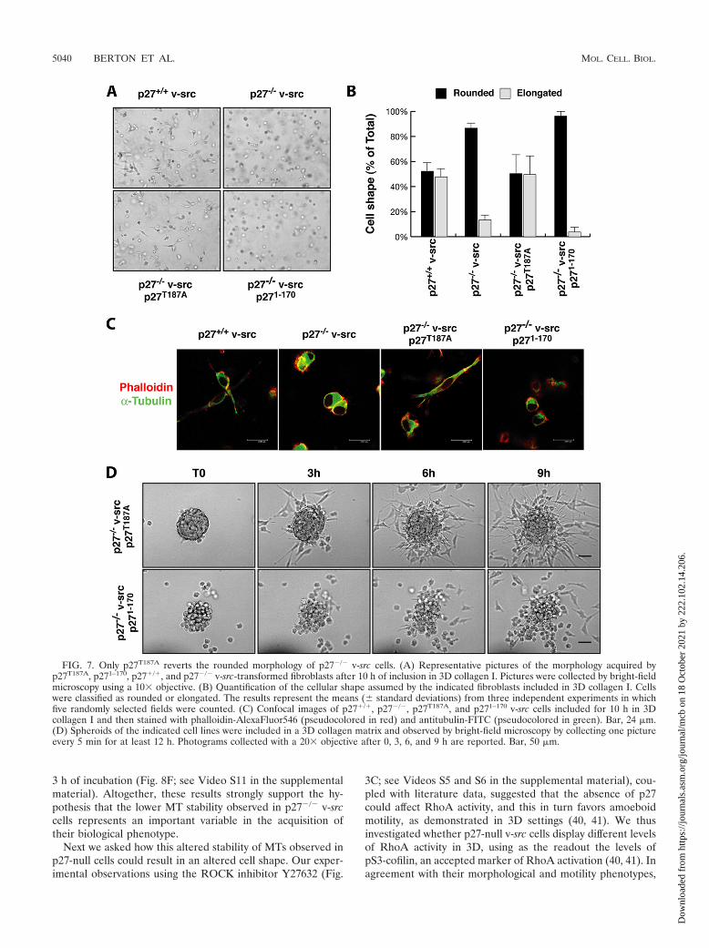
p27T187A but not p271–170 rescues the morphology and mo-tility phenotypes of p27�/� v-src cells. We next analyzed the3D cell shape and motile behavior of p27-rescued cells in orderto determine whether p27T187A and p271–170 could differentlyaffect these phenotypes. First, cells included in 3D collagen Imatrices were analyzed for their morphology after 10 h. Asdescribed above for p27�/� and p27�/� v-src cells, p27-rescuedcells also acquired two different morphologies in 3D, namely,the elongated and the rounded morphologies (Fig. 7A and B).Interestingly, many p27T187A v-src cells clearly appeared tohave the elongated and bipolar shape (49.6% 16% of elon-gated cells), similarly to p27�/� v-src cells (47.8% of elongatedcells). In contrast, almost all p271–170 cells (96% 2.9%) wererounded, as described for p27�/� v-src cells (86% 3.3%rounded) (Fig. 7A and B). The morphological differences wereevaluated by confocal microscopy analysis after staining of�-tubulin (green) and actin (red). p27T187A v-src cells appearedto have an elongated spindle shape based on both MTs andactin stress fiber content, while p271–170 expression was notable to rescue the morphological phenotype in p27-null v-srcfibroblasts and these cells still displayed a typical roundedamoeboid-like shape with cortical actin distribution and pe-rinuclear MT accumulation (Fig. 7C).
Interestingly, the behavior of cell clusters in 3D collagen Ialso supports the data obtained by analysis of individual cells(Fig. 7D; see Videos S9 and S10 in the supplemental material).In brief, p27T187A reverted the phenotype of p27�/� v-src fi-broblasts, and cells displayed long cellular protrusions (Fig.7D; see Video S9 in the supplemental material) that alloweddetachment from the spheroid using typical mesenchymalmechanisms. Conversely, p271–170-expressing cells detachedfrom the spheroid using an amoeboid-like motility, showing amarkedly spherical shape (Fig. 7D; see Video S10 in the sup-plemental material), highly dynamic membrane blebs, andsqueezing through the collagen lattices in a very flexible way,thus reproducing the motility behavior of p27-null cells. Thisdifferent motility was coupled with a very high ability of p271–170
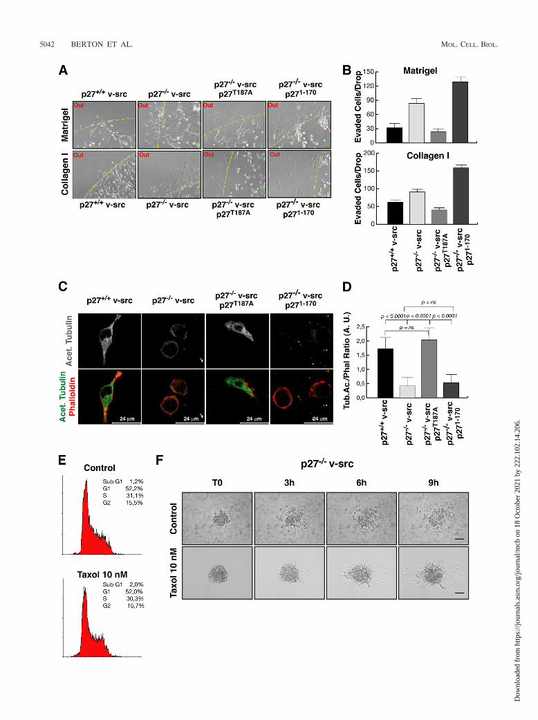
cells to evade 3D matrices, as demonstrated by evasion assays
of individual cells included in collagen I and Matrigel drops(Fig. 8A and B). At 24 h after inclusion, only a few p27�/� v-srcand p27T187A-rescued cells exited the matrix drop. Conversely,p271–170 expression was not able to reduce cell migration ofp27-null v-src fibroblasts, and many cells were present outsidethe drops in the same time frame (Fig. 8A). The count of allcells that evaded the matrix (five drops for each cell line)confirmed this observation, using both collagen I and Matrigelmatrices (Fig. 8B).
Decreased MT stability in p27�/� v-src fibroblasts influ-ences their biological behaviors. We previously demonstratedthat the C-terminal portion of p27 is necessary to mediate thestabilization of the microtubular network via the inhibition ofthe MT-destabilizing protein stathmin (1). The fact that rein-troduction of the p271–170 deletion mutant in p27�/� v-srcfibroblasts was not able to rescue the biological phenotype oftransformed cells suggested that the same mechanism couldalso operate in this context. To verify whether the v-src-trans-formed p27�/� cells immersed in 3D environments displayedaltered MT stability, we looked at the levels of stable acety-lated tubulin in cells included in 3D collagen I. This analysisclearly showed that cells expressing endogenous p27 orp27T187A displayed higher levels of acetylated tubulin, local-ized both in the cell body and in the cellular protrusions (Fig.8C). Conversely, p27-null and p271–170-expressing cells dis-played very low levels of acetylated tubulin, and, to a largerextent, localized perinuclearly. Moreover, in the latter cells,intact MTs were hardly distinguishable, while they could beeasily identified in cells expressing full-length p27 proteins(Fig. 8C). The quantification of the fluorescence intensities ofphalloidin and acetylated tubulin in the different cell linesincluded in 3D collagen revealed that p27WT and p27T187A
cells had a threefold-higher content of stable acetylated MTswith respect to both p27KO and p271–170 cells (Fig. 8D). Thisresult was consistent with previous reports showing a lower MTstability in cells devoid of p27 (1). We thus tested whether MTstability could influence the cell shape and motility of thesecells, using the MT-stabilizing drug taxol. p27�/� v-src cellswere treated with low doses of taxol (10 nM) and analyzed fortheir proliferative, morphological, and motility profiles. Prolif-eration was not affected by 10 nM taxol (Fig. 8E), but cellsimmersed in 3D collagen I significantly changed their shape, byassuming an elongated morphology coupled with mesenchymalmotility, as demonstrated by the 3D spheroid assay (Fig. 8F;see Video S11 in the supplemental material). Importantly, theability of p27-null cells to move in the presence of taxol wasgreatly reduced, starting to detach from the spheroid only after
p271–170 (lower graph) v-src fibroblasts. Data represent the means and standard deviations from three independent experiments performed induplicate, in which the cells were counted each day for 5 days (P 0.001 for p27�/� v-src versus p27T187A and p271–170 at day 5 using Student’s t test).(D) Cyclin A-, cyclin B1-, CDK1-, and CDK2-associated kinase activity, using histone H1 (32P-HH1) as a substrate (upper panels). In the lowerpanels the amount of immunoprecipitated (IP) protein is shown. The normalized kinase activities (expressed in arbitrary units) reported undereach lane represent the means ( standard deviations) from three independent experiments. (E and F) Soft-agar assay of the indicatedv-src-transformed cells. In panel E, representative pictures of colonies after 8 days of incubation in agar are shown. In panel F, the number ofcolonies per field is reported, representing the means and standard deviations from three independent experiments each performed in triplicate(*, P 0.007; §, P � 0.0001 [versus p27�/� v-src cells, using Student’s t test]). (G) Quantification of tumor masses explanted from micesubcutaneously injected with the indicated cells. Values represent the means and standard deviations for six mice injected with p27�/� v-src cells,six mice injected with p27�/� v-src cells, eight mice injected with p27T187A v-src cells, and nine mice injected with p271–170 v-src cells (*, P � 10�5
versus p27�/� v-src cells, using Student’s t test).
VOL. 29, 2009 ROLE OF p27kip1 IN v-src TRANSFORMATION 5039
Dow
nloa
ded
from
http
s://j
ourn
als.
asm
.org
/jour
nal/m
cb o
n 18
Oct
ober
202
1 by
222
.102
.14.
206.
3 h of incubation (Fig. 8F; see Video S11 in the supplementalmaterial). Altogether, these results strongly support the hy-pothesis that the lower MT stability observed in p27�/� v-srccells represents an important variable in the acquisition oftheir biological phenotype.
Next we asked how this altered stability of MTs observed inp27-null cells could result in an altered cell shape. Our exper-imental observations using the ROCK inhibitor Y27632 (Fig.
3C; see Videos S5 and S6 in the supplemental material), cou-pled with literature data, suggested that the absence of p27could affect RhoA activity, and this in turn favors amoeboidmotility, as demonstrated in 3D settings (40, 41). We thusinvestigated whether p27-null v-src cells display different levelsof RhoA activity in 3D, using as the readout the levels ofpS3-cofilin, an accepted marker of RhoA activation (40, 41). Inagreement with their morphological and motility phenotypes,
FIG. 7. Only p27T187A reverts the rounded morphology of p27�/� v-src cells. (A) Representative pictures of the morphology acquired byp27T187A, p271–170, p27�/�, and p27�/� v-src-transformed fibroblasts after 10 h of inclusion in 3D collagen I. Pictures were collected by bright-fieldmicroscopy using a 10� objective. (B) Quantification of the cellular shape assumed by the indicated fibroblasts included in 3D collagen I. Cellswere classified as rounded or elongated. The results represent the means ( standard deviations) from three independent experiments in whichfive randomly selected fields were counted. (C) Confocal images of p27�/�, p27�/�, p27T187A, and p271–170 v-src cells included for 10 h in 3Dcollagen I and then stained with phalloidin-AlexaFluor546 (pseudocolored in red) and antitubulin-FITC (pseudocolored in green). Bar, 24 �m.(D) Spheroids of the indicated cell lines were included in a 3D collagen matrix and observed by bright-field microscopy by collecting one pictureevery 5 min for at least 12 h. Photograms collected with a 20� objective after 0, 3, 6, and 9 h are reported. Bar, 50 �m.
5040 BERTON ET AL. MOL. CELL. BIOL.
Dow
nloa
ded
from
http
s://j
ourn
als.
asm
.org
/jour
nal/m
cb o
n 18
Oct
ober
202
1 by
222
.102
.14.
206.

p27-null cells included in 3D had higher levels of pS3-cofilin(Fig. 9A and B), which also resulted in high polarization inmigrating cells detached from the spheroid, as evaluated byimmunofluorescence analysis (data not shown). The higheradhesion-dependent pS3-cofilin phosphorylation was con-firmed by Western blot analysis of cells adhered to collagen Ifor 30 and 60 min (Fig. 9C and D), and, importantly, this isdependent on the presence of the last 28 C-terminal aminoacids in p27 (Fig. 9), in accord with the fact that p271-170 cellsretain the round shape and the amoeboid motility of p27-nullcells.
The invasive potential of p27�/� v-src cells is reduced byp27T187A expression. The importance of the C-terminal por-tion of p27 in controlling cell shape and motility in metastasisformation was then tested in nude mice by examining theability of p27T187A- and p271–170-rescued cells to intravasatefrom the primary site and to extravasate and form tumor fociinto the lungs when injected in the blood circulation.
RT-PCR analysis of the spleens, amplifying the exogenouscDNAs, showed that only 1/7 mice (14.2%) injected subcuta-neously with p27T187A-rescued cells displayed a positive spleen,whereas 3/5 mice (60%) injected with p271–170-rescued cellswere positive (Fig. 10A). Moreover, tail vein injection of fourmice with p27�/� and p27�/� v-src cells as controls, seven micewith two different p27T187A-rescued cell lines, and six mice withtwo different p271–170-rescued clones revealed that p27�/� v-src cells showed a higher ability to settle and form tumor fociin the lungs (10.5 foci per mouse), while p27�/� cells weremuch less aggressive (no macroscopically evident foci) (Fig.10B). Interestingly, mice injected with p27T187A-rescued cellsdisplayed on average 1.95 foci per animal, while the p271–170
counterpart showed 6.41 foci (Fig. 10B). Histopathologicalanalysis of lungs from the different mice confirmed the results,demonstrating an increased number of tumor foci per sectionin mice injected with p271–170 v-src cells with respect top27T187A-expressing cells (tumor foci per lung section: p27�/�
v-src, 0; p27�/� v-src, 6; p27T187A, 2.1; p271–170, 4.3). Moreimportantly, the immunohistochemical analysis of p27 expres-sion in lung foci formed by p27T187A and p271–170 v-src cellsdemonstrated that p27 expression was significantly retainedonly in those foci formed by the p271–170 v-src cells (Fig. 10C),further confirming the antimetastatic role of full-length p27 inthis model system.
DISCUSSION
Here we demonstrate that p27 protein plays an importantrole in the control of tumor cell growth and metastasis forma-tion. This protein, first identified as a universal CDK inhibitorand tumor suppressor gene product (12, 44), has in morerecent years been found to have several unexpected functionsthat are often not directly linked to its ability to block thecyclin-CDK complexes, such as the control of apoptotic celldeath (25, 26) or autophagy (24, 25), DNA transcription (30,33), and cell motility (1, 4, 11, 19, 21, 23, 29, 45). Thesedifferent p27 functions have often been linked to differentprotein domains and/or posttranslational modifications. Here,we add a new function for p27 in the control of cell plasticityin 3D models that eventually results in the control of cellmotility. Its C-terminal portion governs this new p27 function,
which is very well highlighted in 3D while less observable in 2Dsettings. Similar observations were obtained using primarymouse embryo fibroblasts or 3T3 fibroblasts of different geno-types (B. Belletti et al., submitted for publication), humanglioblastomas, and endothelial cells (42), demonstrating thatthe mechanism used by p27 to control cell motility is generaland is not dependent on v-src transformation.
The use of time-lapse video microscopy allowed us todemonstrate that the absence of p27 induces MAT in cellsimmersed in 3D matrices. This type of motility is accompa-nied by a profound alteration in the distribution of actin andMT cytoskeletons that allows the cells to assume a moreflexible architecture. Importantly, our data using inhibitorsof metalloproteinases and of the Rho pathway indicate notonly that the block of matrix degradation could induceMAT, as observed by others (50), but also that the block ofROCK activity may induce MAT, at least in our model.Thus, transformed cells may switch from one motility type toanother to escape from a block in cell motility imposedeither by protease or by Rho pathway inhibition, a findingthat should be taken in great account in the design of newantimetastatic therapies.
The observation that MT stability is a major regulator of cellplasticity in 3D (reference 3 and this study) raises the possi-bility that MT-stabilizing drugs such as taxol, at very low dosesthat do not affect cell proliferation, may be used as new anti-metastatic agents, at least in certain types of cancer.
The data presented here demonstrate that p27 controls cellmotility through its C-terminal portion. Both in vitro and invivo data support the hypothesis that the last 28 amino acidsare necessary to regulate pathways different from the cyclin/CDK activity and are important for cell shape and motilityregulation. Accordingly, our data demonstrate that the last 28amino acids of p27 are necessary to control Rho pathwayactivity, as demonstrated by higher cofilin phosphorylation inp27-null and p271-170 v-src cells and by the dependence of theirmotility on a functional ROCK pathway (Fig. 3C and data notshown). Interestingly, higher activity of small GTPases of theRho family has been previously linked to the MAT in severaldifferent model systems (18, 35, 40).
How does the C-terminal portion of p27 contribute to reg-ulation of Rho pathway? A direct binding of p27 and RhoAhas been observed when the two proteins are concomitantlyoverexpressed in 293 cells (4), although we were not able tohighlight this direct interaction in our model system (data notshown).
Similarly, hepatocyte growth factor-dependent hepatomacell scatter motility has been proposed to be regulated by p27via the modulation of Rac but not Rho activity (29). However,the domain necessary for p27 to regulate the scatter motilityhas been mapped in a different position of the protein (aminoacids 137 to 155) (29) and thus likely represents a mechanismdifferent from the one we observed here, which is dependenton the regulation of the Rho pathway and on amino acids 170to 198.
A third possibility is that p27 regulates adhesion-dependentRho activity indirectly through the regulation of MT stability.Increasing evidence underscores the existence of a tight rela-tionship between MT dynamics and small GTPase activity,since MT stability can regulate GTPase activity (51) and, in
VOL. 29, 2009 ROLE OF p27kip1 IN v-src TRANSFORMATION 5041
Dow
nloa
ded
from
http
s://j
ourn
als.
asm
.org
/jour
nal/m
cb o
n 18
Oct
ober
202
1 by
222
.102
.14.
206.
5042 BERTON ET AL. MOL. CELL. BIOL.
Dow
nloa
ded
from
http
s://j
ourn
als.
asm
.org
/jour
nal/m
cb o
n 18
Oct
ober
202
1 by
222
.102
.14.
206.

turn, small GTPases can affect adhesion-dependent MT stabi-lization (36), at least in part acting on stathmin (47). It is thuspossible that stathmin, an MT-destabilizing protein (8) thatindeed binds the C-terminal portion of p27 (1), could contrib-ute to the regulation of Rho activity by p27. Although this
hypothesis merits a formal experimental demonstration, it issupported by observations presented here (Fig. 7 to 9).
It could be speculated that the control of cell proliferationand motility by p27 could represent an energy-saving way forthe cells to more easily coordinate proliferation and movement
FIG. 8. Decreased MT stability accounts for the round shape and amoeboid motility of p27�/� v-src cells. (A) Matrix evasion assay of individualcells included in Matrigel (upper panels) or collagen I drops (lower panels). Typical images of matrix drops incubated for 24 h from inclusion at37°C are shown. (B) Quantification of the experiment shown in panel A. The number of cells that exited from matrix drops within 24 h is reported.p27T187A (P � 0.001) but not p271–170 (P not significant using the Student t test) reduces matrix cell evasion compared to that in p27�/� v-src cells.Data represent the means and standard deviations from three independent experiments performed five times. (C) Confocal images of p27�/�,p27�/�, p27T187A, and p271–170 v-src cells included for 5 h in 3D collagen I and stained with anti-acetylated tubulin (gray scale) (upper panels). Inthe lower panels the merging of phalloidin-AlexaFluor546 (red), and anti-acetylated tubulin-AlexaFluor488 (green) is shown. Bar, 24 �m.(D) Quantification of the fluorescence intensity of acetylated tubulin normalized with phalloidin for at least 40 cells per cell clone (expressed inarbitrary units). Error bars indicate standard deviations. (E) FACS analysis of p27�/� v-src cells treated or not with 10 nM taxol for 10 h andanalyzed for their cell cycle distribution. The percentage of cells in each phase of the cell cycle in a typical experiment is reported. (F) Spheroidsof p27�/� v-src cells, treated or not with 10 nM taxol as indicated, were included in a 3D collagen matrix and evaluated by bright-field microscopyby collecting one picture every 5 min for at least 12 h. Photograms collected after 0, 3, 6, and 9 h are shown. Bar, 50 �m.
FIG. 9. p27�/� v-src cells have higher RhoA activity. (A) p27�/� and p27�/� v-src cells included in 3D collagen I were stained for pS3-cofilin(AlexaFluor488, pseudocolored in green) and nuclei (with propidium iodide, red). In the upper panels the confocal 3D reconstruction ofpS3-cofilin staining is reported. In the middle, the merging of pS3-cofilin (AF488) and nuclei (red) in a 3D reconstruction is shown. In the lowerpanels a confocal section of the same field is reported. Bar, 24 �m. (B) Quantification of the fluorescence intensity of pS3-cofilin in at least 40 cellsper genotype (expressed in arbitrary units). Error bars indicate standard deviations. (C) Western blot analysis of pS3-cofilin and total cofilinexpression in p27�/�, p27�/�, p27T187A, and p271–170 v-src cells adhered to collagen I (20 �g/ml) for the indicated times. (D) Quantification ofpS3-cofilin, representing the ratio between pS3-cofilin and total cofilin expression in three independent experiments (*, P 0.01; P 0.005 [at60 min between p27�/� and p27�/� cells and p271–170 and p27�/� cells, respectively, using the Student t test; in the other cases the differences didnot reach statistical significance). Error bars indicate standard deviations.
VOL. 29, 2009 ROLE OF p27kip1 IN v-src TRANSFORMATION 5043
Dow
nloa
ded
from
http
s://j
ourn
als.
asm
.org
/jour
nal/m
cb o
n 18
Oct
ober
202
1 by
222
.102
.14.
206.

both under physiological and pathological conditions, using thesame protein(s) in different subcellular localization and/or itsdifferent protein domains. This in turn implies that that themechanism described here could be explored in the context ofthe design of new promising antimetastatic therapies.
ACKNOWLEDGMENTS
This work was supported by grants from AICR (Association forInternational Cancer Research) to G.B. and partially by grants fromAIRC (Associazione Italiana Ricerca sul Cancro) to A.V. StefaniaBerton is a recipient of a FIRC (Federazione Italiana Ricerca sulCancro) fellowship.
We thank Renato Baserga for the pM-v-src vector and SaraD’Andrea for excellent technical assistance.
We declare that we have no competing financial interests.
REFERENCES
1. Baldassarre, G., B. Belletti, M. S. Nicoloso, M. Schiappacassi, A. Vecchione,P. Spessotto, A. Morrione, V. Canzonieri, and A. Colombatti. 2005.p27(Kip1)-stathmin interaction influences sarcoma cell migration and inva-sion. Cancer Cell 7:51–63.
2. Baldassarre, G., B. Belletti, P. Bruni, A. Boccia, F. Trapasso, F. Pentimalli,M. V. Barone, G. Chiappetta, M. T. Vento, S. Spiezia, A. Fusco, and G.Viglietto. 1999. Overexpressed cyclin D3 contributes to retaining the growthinhibitor p27 in the cytoplasm of thyroid tumor cells. J. Clin. Investig.104:865–874.
2a.Belletti, B., M. S. Nicoloso, M. Schiappacassi, E. Chimienti, S. Berton, F.Lovat, A. Colombatti, and G. Baldassarre. 2005. p27(kip1) functional regu-lation in human cancer: a potential target for therapeutic designs. Curr. Med.Chem. 12:1589–1605.
3. Belletti, B., M. S. Nicoloso, M. Schiappacassi, S. Berton, F. Lovat, K. Wolf,V. Canzonieri, S. D’Andrea, A. Zucchetto, P. Friedl, A. Colombatti, and G.Baldassarre. 2008. Stathmin activity influences sarcoma cell shape, motility,and metastatic potential. Mol. Biol. Cell 19:2003–2013.
4. Besson, A., M. Gurian-West, A. Schmidt, A. Hall, and J. M. Roberts. 2004.p27Kip1 modulates cell migration through the regulation of RhoA activa-tion. Genes Dev. 18:862–876.
5. Bissell, M. J., and D. Radisky. 2001. Putting tumours in context. Nat. Rev.Cancer 1:46–54.
6. Reference deleted.7. Carragher, N. O., S. M. Walker, L. A. Scott Carragher, F. Harris, T. K.
Sawyer, V. G. Brunton, B. W. Ozanne, and M. C. Frame. 2006. Calpain 2 andSrc dependence distinguishes mesenchymal and amoeboid modes of tumourcell invasion: a link to integrin function. Oncogene 25:5726–5740.
8. Cassimeris, L. 2002. The oncoprotein 18/stathmin family of microtubuledestabilizers. Curr. Opin. Cell Biol. 14:18–24.
9. Cicchini, C., I. Laudadio, F. Citarella, M. Corazzari, C. Steindler, A.Conigliaro, A. Fantoni, L. Amicone, and M. Tripodi. 2008. TGFbeta-inducedEMT requires focal adhesion kinase (FAK) signaling. Exp. Cell Res. 314:143–152.
10. Cukierman, E., R. Pankov, D. R. Stevens, and K. M. Yamada. 2001. Takingcell-matrix adhesions to the third dimension. Science 294:1708–1712.
11. Daniel, C., J. Pippin, S. J. Shankland, and C. Hugo. 2004. The rapamycinderivative RAD inhibits mesangial cell migration through the CDK-inhibitorp27KIP1. Lab. Investig. 84:588–596.
12. Fero, M. L., E. Randel, K. E. Gurley, J. M. Roberts, and C. J. Kemp. 1998.The murine gene p27Kip1 is haplo-insufficient for tumour suppression. Na-ture 396:177–180.
13. Frame, M. C. 2004. Newest findings on the oldest oncogene; how activatedsrc does it. J. Cell Sci. 117:989–998.
14. Frame, M. C. 2002. Src in cancer: deregulation and consequences for cellbehaviour. Biochim. Biophys. Acta 1602:114–130.
15. Frame, M. C., V. J. Fincham, N. O. Carragher, and J. A. Wyke. 2002. v-Src’shold over actin and cell adhesions. Nat. Rev. Mol. Cell Biol. 3:233–245.
16. Friedl, P., and K. Wolf. 2003. Tumour-cell invasion and migration: diversityand escape mechanisms. Nat. Rev. Cancer 3:362–374.
17. Friedl, P. 2004. Prespecification and plasticity: shifting mechanisms of cellmigration. Curr. Opin. Cell Biol. 16:14–23.
18. Gadea, G., M. de Toledo, C. Anguille, and P. Roux. 2007. Loss of p53promotes RhoA-ROCK-dependent cell migration and invasion in 3D ma-trices. J. Cell Biol. 178:23–30.
19. Goukassian, D., A. Díez-Juan, T. Asahara, P. Schratzberger, M. Silver, T.
FIG. 10. p27T187A expression reduces the invasive potential of p27�/� v-src cells. (A) Nude mice subcutaneously injected with the indicated cellclones were analyzed for the presence of circulating cells by RT-PCR analysis of spleen samples. The results are shown as the percentage of positivespleens. (B) Representative pictures of lungs explanted from nude mice injected in the tail vein with the indicated v-src fibroblasts and sacrificed20 days after injection. Lungs were fixed in Bouin’s solution. The number of macroscopically detectable foci in the two lungs is indicated.(C) Immunohistochemistry analysis of p27 expression in lung sections from mice injected with p27T187A and p271–170 v-src fibroblasts. Theexpression of p27T187A (left panel) is lost in the lung tumor foci. In the same section the expression of endogenous mouse p27 retained in thenormal lung islet entrapped in the tumor metastasis is visible. Conversely, p271–170 expression (right panel) is retained both in the nucleus and inthe cytoplasm of the transformed cells (better seen in the inset; magnification, �40).
5044 BERTON ET AL. MOL. CELL. BIOL.
Dow
nloa
ded
from
http
s://j
ourn
als.
asm
.org
/jour
nal/m
cb o
n 18
Oct
ober
202
1 by
222
.102
.14.
206.

Murayama, J. M. Isner, and V. Andres. 2001. Overexpression of p27(Kip1)by doxycycline-regulated adenoviral vectors inhibits endothelial cell prolif-eration and migration and impairs angiogenesis. FASEB J. 15:1877–1885.
20. Irby, R. B., W. Mao, D. Coppola, J. Kang, J. M. Loubeau, W. Trudeau, R.Karl, D. J. Fujita, R. Jove, and T. J. Yeatman. 1999. Activating SRC muta-tion in a subset of advanced human colon cancers. Nat. Genet. 21:187–190.
21. Itoh, Y., N. Masuyama, K. Nakayama, K. I. Nakayama, and Y. Gotoh. 2007.The cyclin-dependent kinase inhibitors p57 and p27 regulate neuronal mi-gration in the developing mouse neocortex. J. Biol. Chem. 282:390–396.
22. Johnson, D., M. C. Frame, and J. A. Wyke. 1998. Expression of the v-Srconcoprotein in fibroblasts disrupts normal regulation of the CDK inhibitorp27 and inhibits quiescence. Oncogene 16:2017–2028.
23. Kawauchi, T., K. Chihama, Y. Nabeshima, and M. Hoshino. 2006. Cdk5phosphorylates and stabilizes p27kip1 contributing to actin organization andcortical neuronal migration. Nat. Cell Biol. 8:17–26.
24. Komata, T., T. Kanzawa, H. Takeuchi, I. M. Germano, M. Schreiber, Y.Kondo, and S. Kondo. 2003. Antitumour effect of cyclin-dependent kinaseinhibitors (p16(INK4A), p18(INK4C), p19(INK4D), p21(WAF1/CIP1) andp27(KIP1)) on malignant glioma cells. Br. J. Cancer 88:1277–1280.
25. Lee, S. H., and F. McCormick. 2005. Downregulation of Skp2 and p27/Kip1synergistically induces apoptosis in T98G glioblastoma cells. J. Mol. Med.83:296–307.
26. Liang, J., S. H. Shao, Z. X. Xu, B. Hennessy, Z. Ding, M. Larrea, S. Kondo,D. J. Dumont, J. U. Gutterman, C. L. Walker, J. M. Slingerland, and G. B.Mills. 2007. The energy sensing LKB1-AMPK pathway regulates p27(kip1)phosphorylation mediating the decision to enter autophagy or apoptosis.Nat. Cell Biol. 9:218–224.
27. Lutz, M. P., I. B. Esser, B. B. Flossmann-Kast, R. Vogelmann, H. Luhrs, H.Friess, M. W. Buchler, and G. Adler. 1998. Overexpression and activation ofthe tyrosine kinase Src in human pancreatic carcinoma. Biochem. Biophys.Res. Commun. 243:503–508.
28. Mazurenko, N. N., E. A. Kogan, I. B. Zborovskaya, F. L., and Kisseljov. 1992.Expression of pp60c-src in human small cell and non-small cell lung carci-nomas. Eur. J. Cancer 28:372–377.
29. McAllister, S. S., M. Becker-Hapak, G. Pintucci, M. Pagano, and S. F.Dowdy. 2003. Novel p27(kip1) C-terminal scatter domain mediates Rac-dependent cell migration independent of cell cycle arrest functions. Mol.Cell. Biol. 23:216–228.
30. Miskimins, R., R. Srinivasan, M. Marin-Husstege, W. K. Miskimins, and P.Casaccia-Bonnefil. 2002. p27(Kip1) enhances myelin basic protein gene pro-moter activity. J. Neurosci. Res. 67:100–105.
31. Montagnoli, A., F. Fiore, E. Eytan, A. C. Carrano, G. F. Draetta, A. Hershko,and M. Pagano. 1999. Ubiquitination of p27 is regulated by Cdk-dependentphosphorylation and trimeric complex formation. Genes Dev. 13:1181–1189.
32. Nakayama, K., N. Ishida, M. Shirane, A. Inomata, T. Inoue, N. Shishido, I.Horii, D. Y. Loh, and K. Nakayama. 1996. Mice lacking p27(Kip1) displayincreased body size, multiple organ hyperplasia, retinal dysplasia, and pitu-itary tumors. Cell 85:707–720.
33. Nguyen, L., A. Besson, J. I. Heng, C. Schuurmans, L. Teboul, C. Parras, A.Philpott, J. M. Roberts, and F. Guillemot. 2006. p27kip1 independentlypromotes neuronal differentiation and migration in the cerebral cortex.Genes Dev. 20:1511–1524.
34. Ottenhoff-Kalff, A. E., G. Rijksen, E. A. van Beurden, A. Hennipman, A. A.
Michels, and G. E. Staal. 1992. Characterization of protein tyrosine kinasesfrom human breast cancer: involvement of the c-src oncogene product.Cancer Res. 52:4773–4778.
35. Palamidessi, A., E. Frittoli, M. Garre, M. Faretta, M. Mione, I. Testa, A.Diaspro, L. Lanzetti, G. Scita, and P. P. Di Fiore. 2008. Endocytic traffickingof Rac is required for the spatial restriction of signaling in cell migration.Cell 134:135–147.
36. Palazzo, A. F., C. H. Eng, D. D. Schlaepfer, E. E. Marcantonio, and G. G.Gundersen. 2004. Localized stabilization of microtubules by integrin- andFAK-facilitated Rho signaling. Science 303:836–839.
37. Polyak, K., M. H. Lee, H. Erdjument-Bromage, A. Koff, J. M. Roberts, P.Tempst, and J. Massague. 1994. Cloning of p27Kip1, a cyclin-dependentkinase inhibitor and a potential mediator of extracellular antimitogenic sig-nals. Cell 78:59–66.
38. Radisky, D. C., and M. J. Bissell. 2004. Cancer: respect thy neighbor! Sci-ence 303:775–777.
39. Riley, D., N. O. Carragher, M. C. Frame, and J. A. Wyke. 2001. The mech-anism of cell cycle regulation by v-Src. Oncogene 20:5941–5950.
40. Sahai, E., R. Garcia-Medina, J. Pouyssegur, and E. Vial. 2007. Smurf1regulates tumor cell plasticity and motility through degradation of RhoAleading to localized inhibition of contractility. J. Cell Biol. 176:35–42.
41. Sahai, E., and C. J. Marshall. 2003. Differing modes of tumour cell invasionhave distinct requirements for Rho/ROCK signalling and extracellular pro-teolysis. Nat. Cell Biol. 5:711–719.
42. Schiappacassi, M., F. Lovat, V. Canzonieri, B. Belletti, S. Berton, D. DiStefano, A. Vecchione, A. Colombatti, and G. Baldassarre. 2008. p27Kip1expression inhibits glioblastoma growth, invasion, and tumor-induced neo-angiogenesis. Mol. Cancer Ther. 7:1164–1175.
43. Schwartz, M. A., and R. K. Assoian. 2001. Integrins and cell proliferation:regulation of cyclin-dependent kinases via cytoplasmic signaling pathways.J. Cell Sci. 114:2553–2560.
44. Sherr, C. J. 1994. G1 phase progression: cycling on cue. Cell 79:551–555.45. Sun, J., S. O. Marx, H. J. Chen, M. Poon, A. R. Marks, and L. E. Rabbani.
2001. Role for p27(Kip1) in vascular smooth muscle cell migration. Circu-lation 103:2967–2972.
46. Vlach, J., S. Hennecke, and B. Amati. 1997. Phosphorylation-dependentdegradation of the cyclin-dependent kinase inhibitor p27. EMBO J. 16:5334–5344.
47. Watabe-Uchida, M., K. A. John, J. A. Janas, S. E. Newey, and L. Van Aelst.2006. The Rac activator DOCK7 regulates neuronal polarity through localphosphorylation of stathmin/Op18. Neuron 51:727–739.
48. Webb, D. J., and A. F. Horwitz. 2003. New dimensions in cell migration. Nat.Cell Biol. 5:690–692.
49. Wolf, K., and P. Friedl. 2006. Molecular mechanisms of cancer cell invasionand plasticity. Br. J. Dermatol. 154:11–15.
50. Wolf, K., I. Mazo, H. Leung, K. Engelke, U. H. von Andrian, E. I. Deryugina,A. Y. Strongin, E. B. Brocker, and P. Friedl. 2003. Compensation mechanismin tumor cell migration: mesenchymal-amoeboid transition after blocking ofpericellular proteolysis. J. Cell Biol. 160:267–277.
51. Xu, J., F. Wang, A. Van Keymeulen, M. Rentel, and H. R. Bourne. 2005.Neutrophil microtubules suppress polarity and enhance directional migra-tion. Proc. Natl. Acad. Sci. USA 102:6884–6889.
52. Yeatman, T. J. 2004. A renaissance for SRC. Nat. Rev. Cancer 4:470–480.
VOL. 29, 2009 ROLE OF p27kip1 IN v-src TRANSFORMATION 5045
Dow
nloa
ded
from
http
s://j
ourn
als.
asm
.org
/jour
nal/m
cb o
n 18
Oct
ober
202
1 by
222
.102
.14.
206.