Embed Size (px)
Citation preview

A Unique Cytoskeleton Associated with Crawling in the Amoeboid Sperm of the Nematode, Ascaris suum
Sol Sepsenwol,* H a n s Ris,* and T h o m a s M. Roberts§
* Department of Biology, University of Wisconsin-Stevens Point, Stevens Point, Wisconsin 54481; * Department of Zoology and Madison Integrated Microscopy Resource, University of Wisconsin, Madison, Wisconsin 53706; and § Department of Biological Science, Florida State University, Tallahassee, Florida 32306
Abstract . Nematode sperm extend pseudopods and pull themselves over substrates. They lack an axoneme or the actin and myosins of other types of motile cells, but their pseudopods contain abundant major sperm protein (MSP), a family of 14-kD polypeptides found exclusively in male gametes. Using high voltage elec- tron microscopy, a unique cytoskeleton was discovered in the pseudopod of in vitro-activated, crawling sperm of the pig intestinal nematode Ascaris suum. It con- sists of 5-10-rim fuzzy fibers organized into 150-250- nm-thick fiber complexes, which connect to each of the moving pseudopodial membrane projections, vil- lipodia, which in turn make contact with the substrate. Individual fibers in a complex splay out radially from its axis in all directions. The centripetal ends interca- late with fibers from other complexes or terminate in a thickened layer just beneath the pseudopod membrane. Monoclonal antibodies directed against MSP heavily label the fiber complexes as well as individual pseu- dopodial filaments throughout their length. This rep-
resents the first evidence that MSP may be the major filament protein in the Ascaris sperm cytoskeleton. The large fiber complexes can be seen clearly in the pseudopods of live, crawling sperm by computer- enhanced video, differential-interference contrast mi- croscopy, forming with the villipodia at the leading edge of the sperm pseudopod. Even before the pseudo- pod attaches, the entire cytoskeleton and villipodia move continuously rearwards in unison toward the cell body. During crawling, complexes and villipodia in the pseudopod recede at the same speed as the spermato- zoon moves forward, both disappearing at the pseudo- pod-cell body junction. Sections at this region of high membrane turnover reveal a band of densely packed smooth vesicles with round and tubular profiles, some of which are associated with the pseudopod plasma membrane. The exceptional anatomy, biochemistry, and phenomenology of Ascaris sperm locomotion per- mit direct study of the involvement of the cytoskeleton in amoeboid motility.
EBOID motility, generally defined as the crawling of cells over a substrate by means of pseudopods, is ob- served throughout the animal kingdom. ~ Two types
of force-generating systems have been proposed to explain this kind of motility in diverse cells: a cytoskeleton-based system and a membrane flow-based system. In the former, cytoplasmic actin-myosin interactions reminiscent of the model for vertebrate skeletal muscle are required for loco- motion (see reviews by Clarke and Spudich, 1977; Pollard, 1981, 1986; Trinkhaus, 1984). In membrane-based models, the attachment and flow of membrane generate the necessary forces (see Ishihara et al., 1988). In this study we present a novel example, the crawling sperm of the nematode Ascaris suum, in which both cytoskeleton and membrane flow are in- volved in its locomotion. The cells are unusual because they
contain only traces of actin (<0. 5 %), tubulin (one centriole), and no myosin (Nelson and Ward, 1981; reviewed by Roberts et al., 1989), yet crawl by means of a pseudopod at >70 /~m/min on glass (Sepsenwol, S., and S. J. Taft, manuscript submitted for publication). The gametes most abundant pro- tein is the 14-kD major sperm protein (MSP) 2 (Klass and Hirsh, 1981; Nelson and Ward, 1981). In the activated sper- matozoon of the nematode Caenorhabditis elegans, MSP is localized in the pseudopod (Ward and Klass, 1982). When the pseudopods of nematode sperm are examined in thin sec- tion by conventional transmission electron microscopy, they appear to contain unorganized masses of filaments as thin as 2 nm in diameter (Roberts et al., 1989; Foor et al., 1971; Foot', 1974; Wright and Sommerville, 1984, 1985; Burg- hardt and Foor, 1978; Abbas and Cain, 1984). By examining
1. Ameboid movement is used here to describe any locomotion by which a cell crawls over a substrate. Some features of nematode sperm locomotion are quite different from those described in other crawling cells.
2. Abbreviations used in this paper: DIC, differential-interference contrast; HVEM, high voltage electron microscopy; MSP; major sperm protein.
© The Rockefeller University Press, 0021-9525/89/01/55/12 $2.00 The Journal of Cell Biology, Volume 108, January 1989 55-66 55
on December 21, 2018jcb.rupress.org Downloaded from http://doi.org/10.1083/jcb.108.1.55Published Online: 1 January, 1989 | Supp Info:
whole mounts and semithick sections by high voltage elec- tron microscopy (HVEM), however, we have discovered in the pseudopod of Ascaris sperm an elaborate, dynamic sys- tem of interconnected thin filaments which are organized into large units, the fiber complexes. The complexes can be visualized in living cells without staining or labeling, and their behavior in motile cells studied in real-time.
The structure and composition of filaments that form the complexes have been examined by immunolabeling at the light and electron microscopic levels. Entire fiber complexes as well as individual fibers fixed in situ label strongly with antibodies against MSP, indicating that this protein is a ma- jor constituent of the Ascaris sperm pseudopod cytoskeleton.
Materials and Methods
Unless otherwise specified, all biochemical reagents were purchased from Sigma Chemical Co. (St. Louis, MO).
Collection of Ascaris Male Ascaris worms were expressed from the small intestine of freshly killed pigs, rinsed, and transported in PBS (0.15 M NaCI, 7.5 mM MgCIz, 1.5 mM CaC12 in 0.1 M phosphate, pH 7.2-7.4) at 37-41°C. Worms were maintained in the laboratory in PBS gassed with 15% CO2 in N2.
Vas Deferens Extract An ultrafiltrate (0.2-#m pore size) of a 25,000 g supernatant of an homoge- nate of the glandular male vas deferens was prepared as described in Sepsen- wol et al. (1986).
Sperm Preparation for Electron Microscopy The contents of the seminal vesicle from one adult male (,~,107 spermatids) was emptied into 8-15 ml 0.15 M NaCI in 20 mM Hepes, pH 7.2 (Hepes-sa- line buffer), preheated to 40°C, and gassed with 15% CO2/N2.
Whole Mounts. 30 #1 of vas deferens extract were added to 6 ml sper- matid suspension in a 60-mm plastic dish containing acid-washed glass cov- erslips and 150-mesh gold grids which had been formvar-carbon-coated and made hydrophilic by glow discharge. The dish was transferred to a 40°C incubator and gassed with 15% CO2 in N2 throughout incubation and fixa- tion. After 15-20 rain, cells were fixed at room temperature for 30 min with one of three fixatives: 2.5% glutaraldehyde, 0.05% saponin, 0.2% tannic acid in 0.1 M Hepes buffer, pH 7.4 (Maupin and Pollard, 1983); or 2.5% glutaraldehyde, 0.1% Triton X-100 in Hepes buffer; or 2.5 % glutaraldehyde, 4 % paraformaldehyde in Hepes buffer. Grids and coverslips were rinsed and stored in Hepes buffer at 4°C. Washed grids were postfixed in 0.1% aqueous OsO4 for 10 rain and stained with 1% aqueous uranyl acetate for 10 rain. After dehydration in ethanol, grids and coverslips were critical point dried without intermediate solvent, using filter dried, liquid CO2 (Ris, 1985).
Semithick Sections. For plastic embedment spermatid suspensions were activated in polypropylene test tubes, fixed with glutaraldehyde-saponin- tannic acid fixative for 30 min, washed in buffer, pelleted at 1,000 g, postfixed in 0.1% OsO4 for 10 min, rinsed in distilled H20, and stained with 1% aqueous uranyl acetate. Fragments of pellets were dehydrated in graded ethanols and embedded in Epon-Araldite. Semithick sections (0.12-0.25 t~m) were mounted on Formvar-coated grids, stained with 7.5% aqueous uranyl magnesium acetate at 50°C for 2 h followed by 20 min in lead citrate, then lightly coated with carbon.
Scanning Electron Microscopy Critical point dried cells on coverslips were ion beam sputter coated with
a thin layer of platinum and examined with a low voltage scanning electron microscope (model S-900; Hitachi Ltd., Tokyo) operating at 2 kV.
High Voltage Electron Microscopy (HVEM) Electron micrographs of whole mounts and semithick sections were pre-
pared at I MV with the AEI EM7 HVEM at the Madison Integrated Micros- copy Resource.
Enhanced Video Differential-Interference Contrast (DIC) Microscopy Crawling cells activated as above were observed on a Universal microscope (Carl Zeiss, Inc., Thornwood, NY) with Nomarski DIC optics using a Zeiss 63× plan apochromat objective and a Sierra Scientific (Mountain View, CA) LSN-I Newvicon video camera. The video signal was processed directly with a (model 9200EX; Quantex, Sunnyvale, CA) real-time image- processing system. An out of focus image was stored and digitally subtracted from a frame-averaged image to reduce background. Image contrast was en- hanced and processed images were recorded on a Sony (model 5800) 3/4 inch videotape recorder, then played back through the Quantex system to freeze individual frames. Frames were photographed directly from the screen ofa Conrac high-resolution black and white monitor onto Panatomic- X black and white film (Eastman Kodak Co., Rochester, NY). (A prelimi- nary report on imaging techniques used to study Ascaris sperm appears in a presentation summary by Pawley et al., 1986.)
Crude MSP Immunogen Protein Preparation Spermatids from 25-50 males were frozen at -70"C, thawed, glass homogenized, and the crude debris was pelleted at 1,000 g. The supernate was centrifuged at 100,000 g 30 min at 4°C. The resultant supernate was dialyzed against 50 mM acetate buffer, pH 4.5, at 4°C overnight. Acid- precipitatable material was removed by centrifugation and the final super- hate, designated AS-SI00, was concentrated by vacuum dialysis against PKM buffer (20 mM Pipes, 25 mM KCI, 1 mM MgCI2, pH 7.6).
Monoclonal A n tibodies Antibodies against MSP were generated by standard hybridoma procedures using AS-S100 Ascaris sperm protein fraction (above) as immunogen in Balb/c mice. Antibody was purified from mouse ascites fluid by affinity chromatography against immobilized anti-mouse IgG (Cappel Laborato- ries, Cochranville, PA). Their antigen recognition patterns were tested by immunoblotting against AS-SI00, separated on a 10-20% gradient poly- acrylamide gel (Laemmli, 1970), and electrophoretically transferred to nitrocellulose (Towbin et al., 1979; as modified by Ward et al., 1986). Antibody-labeled bands were visualized using horseradish peroxidase-con- jugated anti-mouse IgG (Jackson Immunoresearch Laboratories, Inc., Avondale, PA), followed by 4-chloro-l-naphthol reagent.
Cell Labeling for Light Microscopy Activated sperm were pipetted onto glass coverslips, allowed to settle 5-10 min, and fixed 30 min in 2% glutaraldehyde, 0.5% Triton X-100 in PKM buffer. Cells were washed in buffer, treated three times for 10 min with 10 mM NaBI-h in PKM buffer, washed in PKM buffer, and incubated with anti-MSP antibody (20-30 t~g/ml) in PKM buffer for 2 h. After a 15-min wash in buffer, the cells were incubated in FITC-conjugated anti-mouse lgG (Cappel Laboratories), washed, and examined under a Zeiss standard mi- croscope with epifluorescent illumination.
Cells were stained with phalloidin as follows: sperm were fixed in glutar- aldehyde-Triton X-100 as above, blocked with borohydride, incubated 30 min with rhodamine-conjugated phalloidin (Molecular Probes, Inc., Junc- tion City, OR) at 4 t~g/ml in PKM buffer, washed in PKM buffer, and exam- ined as above. A primary culture of mouse peritoneal fibroblasts, grown for 6-7 d in RPMI medium on sterile coverslips, was fixed and labeled as above, and used as a positive F-actin control.
Cell Labeling for Electron Microscopy 5-nm colloidal gold particles prepared by the tannic acid-citrate method (Slot and Geuze, 1985) were conjugated to afffinity-purified anti-MSP anti- body as previously described (Roberts et al., 1986); gold-antibody was diluted to a light pink in PKM buffer. Sperm were prepared and labeled as above, incubating with the gold-antibody for 2 h. Labeled cells were washed in buffer, postfixed 30 min in 0.1% OsO4 in PKM buffer, washed in distilled H20, dehydrated in graded ethanols to propylene oxide, and embedded in Polybed 812 (polysciences, Inc., Warrington, PA). Thin sec- tions were mounted on Formvar-carbon-coated copper grids, stained with
The Journal of Cell Biology, Volume 108, 1989 56
uranyl acetate and lead citrate, and examined with a transmission electron microscope (model 100CX; JEOL USA, Peabody, MA) at 80 kV.
Results
General Morphology and Surface Features of the Pseudopod Fig. 1 is a scanning electron micrograph of an activated Ascaris spermatozoon attached to glass. It is the largest re- ported among the nematodes; in vitro-activated, motile sperm possess an hemispherical cell body 10-12/~m in di- ameter positioned above a pseudopod extending up to 15 #m beyond the cell body. Ascaris sperm are permanently and distinctly bipolar. When examined in sections of sperm fixed in situ, the cell body contains all of the cell's membrane- bound organeUes: mitochondria, condensed nucleus, a large refringent body, and numerous membranous organelles which fuse with the cell membrane during activation. The pseudopod contains filaments. The membrane over the cell body has no protrusions and contains numerous pits where the membranous organelles have fused with it. The pseudo- pod membrane, on the other hand, has no pits, but many finger-like projections, the villipodia, 3 which cover the pseudopod from tip to base. On the ventral surface, the vil- lipodia are flattened where they come into contact with the substrate. These membrane specializations can be observed in living cells with phase-contrast and DIC optics. They form continuously at the tip of the pseudopod, move rapidly rearwards over the surface and disappear at the junction of pseudopod and cell body. In Fig. 1, two viUipodia can be seen tucking under an infolding, or lip, where the cell body and pseudopod membranes meet. About 20 min after activa- tion in vitro, these moving villipodia form broad areas of at- tachment to the substrate and pull the cell body along as they move rearwards beneath the pseudopod. The cells crawl at a rate identical to the movement of the villipodia (25-75 #m/min on glass).
The Pseudopod Cytoskeleton: Fiber Complexes
Cells were activated on electron microscopy grids and fixed with glutaraldehyde in the presence of the nonionic deter- gent, Triton X-100. This fixative prevents rapid changes in overall cell morphology. The detergent removes the pseudo- pod membrane, revealing its fibrous cytoskeleton. The plasma membrane over the cell body does not solubilize un- der these conditions. The cells and exposed cytoskeleton were examined whole with HVEM. Fig. 2 is a low magnifi- cation stereo view showing the entire cell. 4 The thick cell body is electron opaque. The pseudopod, however, contains a striking array of 150-250-nm-wide branched assemblages, the fiber complexes, made up of much smaller fibers. The complexes extend from the leading edge through the length of the pseudopod and stop at the pseudopod-cell body junc- tion. Each branch of the complex terminates in finger-like
3. While the moving membrane specializations in Ascaris sperm resemble microvilli, they are generally stubbier and are not associated with actin fila- ments. The term "filipodia" used by others does not seem appropriate to de- scribe short stubby projections, hence, "villipodiaY
4. Stereo figures should be viewed with a standard 2× stereo print viewer.
Figure 1. Scanning electron micrograph of Ascaris spermatozoon 17 min after in vitro activation on glass. Glutaraldehyde-saponin fixation. Bar, 1 /zrn; Va~ce~, 2 kV.
projections of fibers congruent with the villipodia which arise at the leading edge and cover the surface of the pseu- dopod. In stereo, it can be seen that some complexes along the base of the pseudopod branch vertically and end in vil- lipodia on the dorsal surface. In sperm that are not attached to substrate, the pseudopod is more cylindrical with a blunted tip. Its complexes fill the entire volume, generating villipodia at the tip which move rearwards over the entire surface of the pseudopod membrane. When attached, the pseudopod is more flattened and complexes spread out mainly, but not ex- clusively, along the attached plane of the pseudopod. In crawling cells, complexes form a continuous mat of fibers along the base of the pseudopod (see Fig. 2), or send projec- tions 1 or 2 #m into the upper pseudopod membrane to form nonadherent villipodia.
In the stereo micrograph, Fig. 3, some complexes are shown at higher magnification in a more dispersed state. They consist of nonuniform or "fuzzy" fibers whose overall diameter in whole mounts ranges from 5 to 10 nm. The ar- rangement of the fibers in complexes varies; some of them are oriented parallel to and concentrate about a central core of the complex (Fig. 3, small arrow). Most fibers splay out radially from the core (Fig. 3, large arrowhead), at an angle of 10-90 ° to its axis and tipped toward the forward end of the cell. As also seen in Fig. 2, the radial fibers interweave with those from adjacent complexes with one end always an- chored in a complex. The fiber complexes are thus intercon- nected into one continuous unit filling the entire pseudopod. The tips of the complexes, normally much more dispersed when associated with intact membrane, are collapsed in whole-mounted, detergent-treated sperm (compare to Fig. 5). Fig. 4 is a semithick cross section through several fiber complexes and illustrates the amorphous dense 20-rim cores which identify individual complexes and the aster of fibers
Sepsenwol et al. Ascaris Sperm Fiber Complexes 57

Figure 2. Low power stereo electron micrograph of whole mount of Ascaris spermatozoon prepared with glutaralde- hyde-Triton X-100 detergent fixative. Note how numerous fiber complexes branch and terminate in villipodia, arising over the upper and lower sur- faces of the pseudopod. The spaces be- tween complexes are filled with splayed fibers. The region in which the com- plexes terminate is obscured by the electron-dense cell body, which con- tains the nucleus, mitochondria, mem- branous organelles, and refringent gran- ules. 15-20 complexes are in close ap- position to the cell body. Bar, 5 #m; Vaccel, 1 MV.
radiating from them. The cores do not appear to be separate structures but common attachment points of the ends of sev- eral individual fibers. The dense varicosities are cross sec- tions of fibers. In cells allowed to become aerobic, the termi- nal branches of all but a few of the complexes and their villipodia disappear. The remaining complexes become rigid and rodlike with densely packed fibers all oriented parallel to the long axis of the complexes (not shown). These rigid complexes form long spikes instead of villipodia and move side to side, but there is no evidence of rearward membrane flow. This and Fig. 3 suggest that the fibers in complexes of crawling sperm are able to splay out much like the ribs of an umbrella and, in similar fashion, tend to collapse to a po-
sition parallel to the axis of the complex. Fig. 5 a is a longitu- dinal semithick section of a fiber complex as it ends in a vil- lipodium. The fibers inside each villipodium are more highly dispersed than the rest of the complex, emerging in all direc- tions toward the villipodial membrane. In the villipodium, the radiating filaments appear to make numerous contacts with the electron-opaque region apposed to the inner layer of the plasma membrane (Fig. 5 b, arrow). The dense sub- membrane layer is a constant feature over both the pseudo- pod and cell body, as is the fibrous material on the outer sur- face. Where the membrane is cut perpendicularly, the dense lamina apposed to the inner face of the cell membrane ap- pears as a "carpet" of fibrillar material (Fig. 5 a, bracket).
Figure 3. Stereo electron micro- graphs of spermatozoon whole mount prepared with glutaralde- hyde-Triton X-100 fixative. A more highly dispersed preparation com- pared to Fig. 2; the individual com- plexes and their terminations are clearly distinguished. The configu- rations of fibers in relation to the complex are shown; fibers nor- mally are splayed out radially from the center of the complex (large ar- rowhead). In aging cells, fibers can collapse close to the core of the complex into parallel bundles (small arrow). Bar, 0.5 #m; V~ccel, 1 MV.
The Journal of Cell Biology, Volume 108, 1989 58

Figure 4. Electron micrograph of semithick cross section of fiber complexes in spermatozoon pseudopod prepared with glutaralde- hyde-tannic acid-saponin fixative. "Fuzzy" fibers radiate from cores of several complexes. The parallel fibers at the cores of the complexes are seen as punctate structures along the radial fila- ments. Bar, 1 ~m; Vatted, 1 MV.
Villipodia and Fiber Complex Translocation in Live Sperm
Many of the features of fiber complexes in the pseudopod, how they form and move, can be clearly observed in living fully activated Ascaris sperm using the higher resolution and contrast of computer-enhanced video DIC. Fig. 6 is a time- lapse sequence of enhanced video DIC images of a motile spermatozoon on glass. The cell in this sequence is activated and its pseudopod is flattened but not attached (i.e., not crawling) so that the relative movement of structures in the pseudopod can be easily followed at high magnification. The fiber complexes appear as large, refringent fibers 0.2-0.3/~m in diameter. They can be seen as early as 6 rain into activa- tion, before the pseudopod is fully extended and attached. The unusual behavior of the cytoskeletal complexes is shown in the low power sequence (Fig. 6, a-c). A group of five branched refringent fibers oriented roughly parallel to the cell's long axis is identified in Fig. 6 b and located '~10 s later in Fig. 6 c; the entire array moves as a unit toward the rear of the pseudopod (rate, '~25 #m/rain). At the same time, a group of villipodia can be seen above and to the left of the area (Fig. 6, asterisk) moving the same distance as the group of complexes. The overall length of the pseudopod remains constant. A sequence of images at higher magnification (Fig. 6, d-g) shows the continuous formation of individual villipo- dia at the leading edge in association with fiber complexes.
Figure 5. Semithick section ofa villipodium, associated fiber com- plex, and specialized plasma membrane in glutaraldehyde-tannic acid-saponin fixative. (a) Fiber complex shown in longitudinal sec- tion as it enters villipodium. Individual fibers have the same fuzzy appearance in section as in whole mount preparation shown in Fig. 3. Inside the villipodium, fibers are more loosely bundled than in the bottle-brush branches of the complex. The small fibers radiate in all directions toward a dense layer apposed to the inner lamina of plasma membrane. The layer is a feature of both pseudopod and cell body membranes. In some cross sections of the membrane, the layer resembles a carpet of closely packed fibrils (bracket). (b) En- largement of area in a; the specialized plasma membrane. Fibers from complexes make many contacts with the dense layer within the villipodium (small arrows). The irregular fibrous material on the outer layer of the plasma membrane does not appear to be con- tinuous with the submembrane material. Bars, 0.5 #m; V~cce~, 1 MV.
Sepsenwol et al. Ascaris Sperm Fiber Complexes 59

Figure 6. Time lapse, video-enhanced, DIC images of living, in vitro-activated Ascaris spermatozoa on glass; contrast-enhanced, background-subtracted, four-frame (1/15 sec) averaged image shown. (a-c) Low magnification sequence, showing overall bipolar mor- phology of a fully motile cell with its extended pseudopod and large refractile cell body. (a) One set of fiber complexes in b (*) is drawn for clarity in the first panel. (c) The same cell 10 s after b, showing the same set of complexes after it has migrated near the junctio~ of pseudopod and cell body. Their relative positions are unchanged. (d-g) Higher magnification sequence showing the association of tibet complexes with villipodia at feature 1, 2, and 3 as they progress rearward and a new villipodium formed at feature 4. d is a drawing oJ representative features found in e. Feature 1 can be seen in g disappearing at the cell body-pseudopod junction, f follows e by -vl0 s; g follows f by ,,,,3 s. Bars, 5 #m.

The complexes may branch as they are formed, migrate rear- ward toward the cell body, and disappear at the cell body-pseudopod junction. At Fig. 6 d, position 2, a villipo- dium forms at the leading edge from a previously assembled fiber complex and both migrate rearwards in succeeding frames. Some of the branches appear to join (see position 2 in Fig. 6 , fand g), but HVEM whole mounts show that vil- lipodia form on only one of the apposing complexes (Fig. 2). In all cases examined, the entire assembly of fibers moves rearward as a unit in concert with the villipodia. Once as- sembled at the leading edge, the interconnected fiber com- plexes form a semirigid framework which does not change shape. When a cell turns, new branched complexes are formed to the side of the turn, established complexes main- tain the same relative positions, and the cell body is pulled around in the new direction. The movement of fiber assem- blies occurs in activated cells whether or not they are at- tached to the substrate (i.e., crawling) and is always as- sociated with rapid retrograde membrane flow.
Both villipodia and fiber complexes disappear at the "lip" formed by the cell body where it joins the base of the pseudo- pod. Fig. 7 shows semithick sections of this region. In Fig. 7 a, the complexes are not tethered to any obvious structures within the cell body. There is, however, a dense aggregation of smooth membrane vesicles just inside the cell body, packed around the mitochondria and membranous or- ganelles at the junction (Fig. 7 a, dotted line). Farther into the cell body, the vesicles are more dispersed. At higher magnification (Fig. 7 b), most vesicle profiles within the band are round, but unusual tubular or flattened forms are also seen. Toward the center of the junction of pseudopod and cell body, the tubular vesicles are oriented with their long axes parallel to the plane of the junction. Those apposed and near to the plasma membrane are aligned parallel to it. Because of the submembrane dense layer, it is not possible to determine if there are vesicles continuous with the plasma membrane itself. In some sections, a few round vesicles can be seen near the lip in contact with the cell membrane (Fig. 7 a, arrow).
lmmunocytochemistry of the Fiber Complexes
Antibody Characterization. The labeling pattern obtained by probing nitrocellulose blots of the AS-S100 MSP- enriched fraction of sperm homogenate with anti-MSP anti- body AZ10 is shown in Fig. 8. The antibody reacts strongly and exclusively with the MSP band which runs at "~15 kD on SDS-polyacrylamide gels.
Labeling Patterns in Fixed, Permeabilized Sperm. Fig. 9 a shows that the fiber complexes are visible in unstained sperm fixed in glutaraldehyde-Triton. When incubated with anti- body AZI0, filament complexes label heavily by indirect im- munofluorescence (Fig. 9 b). Complexes in negative-control cells, incubated without first antibody, do not (Fig. 9, c and d). Some fluorescence found in the cell bodies of both nega- tive controls and anti-MSP-stained cells may be due to autofluorescence of residual glutaraldehyde. The much brighter fluorescence of antibody AZ10-stained cells indi- cates there is some MSP in the cell bodies of activated cells.
Phalloidin staining was used to determine if the small amount of actin known to exist in Ascaris sperm (<0.5 % total protein) forms F-actin assemblies in the pseudopod cyto-
Figure 7. Semithick sections of the pseudopod-cell body junction in glutaraldehyde-tannic acid-saponin fixative. (a) A sharp band of densely packed vesicles is seen just within the cell body, often in contact with fiber complexes. The lower margin of the band of vesi- cles is marked with a dotted line. Many round vesicles appear in contact with the membrane at the lip that marks the lower boundary of the cell body (arrow). Other vesicles seem to be dispersed among the filaments of complexes near the junction. (b) High magnifica- tion micrograph of vesicle region; the band is composed of smooth vesicles with round and tubular profiles. Cortically, the tubular vesicles orient along the pseudopod membrane; internally, they lie parallel to the plane of the junction. Bars, 0.5 t~m; V~¢~et, I MV.
Sepsenwol et al. Ascaris Sperm Fiber Complexes 61

Figure 8. Immunoblot pattern of anti- body AZ10 against Ascaris sperm pro- tein fraction AS-SI00. The left lane (COOM) shows the Coomassie Blue- staining pattern of AS-S100 sperm pro- teins on a 10-20% gradient polyacryl- amide gel. MSP is the most abundant protein. The right lane (AZ/0) is a nitro- cellulose immunoblot of Ascaris sperm proteins probed with antibody AZI0 fol- lowed by horseradish peroxidase-anti- mouse IgG.
skeleton. Fig. 9, e and f, show that pseudopods of permeabi- lized spermatozoa fail to label with phalloidin, whereas the mouse peritoneal fibroblast-positive controls show heavy labeling of their actin filament bundles (Fig. 9, g and h).
At the ultrastructural level (Fig. 10), colloidal gold-la- beled antibody AZ10 is shown to decorate the entire fiber complex, including the core as well as individual fibers. Both the preservation of the fibers and permeability to anti- body-gold conjugates varied considerably among cells. Fig. 10 a shows a thin section through a labeled cell in which the pseudopod remains packed with filaments. Filaments just beneath a disrupted portion of plasma membrane decorate heavily with gold particles, but those within the core of a nearby fiber complex do not. We suspect the nonuniform labeling reflects the failure of gold particles to penetrate areas where filaments are tightly packed, especially at the cores of complexes. In more thoroughly dispersed cells (Fig. 10 b), however, some filaments at the core of the fiber com- plexes were also labeled. Profiles of sperm probed with anti-mouse IgG-gold (Fig. 10 c) or BSA-gold (not shown) contained only a few bound gold particles.
Discussion
Summary of Anatomy and Behavior of Ascaris Sperm Cytoskeleton and Pseudopod Membrane Specializations
This work has demonstrated that a coherent, dynamic cy- toskeleton exists in the pseudopod of Ascaris sperm whose behavior suggests its direct participation in the cell's amoe- boid locomotion. The drawing in Fig. 11 summarizes the fea- tures of Ascaris sperm which have made it an excellent model for studying nematode sperm amoeboid motility: it is relatively large and bipolar with a permanent pseudopod. In sections of cells fixed in situ, all of the membrane-bound or- ganelles (nucleus, mitochondria, membranous organelles, and refractile body) are confined to the cell body; the pseu- dopod contains fibers. The fibers are organized into super- structures, the fiber complexes, which are visible in living, unstained, unlabeled cells. Radiating fibers from the corn-
Figure 9. Paired light and fluorescence micrographs of Ascaris sperm and mouse peritoneal fibroblasts. (a and b) Ascaris sperm incubated with antibody AZ10 followed by FITC-anti-mouse IgG. The filament complexes are heavily labeled. (c and d) Control sperm incubated with FITC-anti-mouse IgG only. (e and f ) Sperm labeled with rhodamine-phalloidin. (g and h) Mouse peritoneal fibroblast incubated with rhodamine-phaUoidin showing labeled actin filament bundles. Bars, 10/~m.
plexes, shown in the detail, insert into the dense layer of ma- terial lining the inside of the pseudopod membrane. The membrane puckers into viUipodia as it associates with fibers from the terminations of complexes at the leading edge. The villipodia, the pseudopod's membrane-cytoskeleton unit, appear to be adhesion sites, and spread out as they make con- tact with substrate or move along, unused, to the cell body where their components are recycled.
Because the fiber complexes can be seen inside the pseu- dopod of living cells with DIC-light microscopy, the be- havior of this system-membrane and cytoskeleton together- can be studied without the ambiguities associated with in-
The Journal of Cell Biology, Volume 108, 1989 62
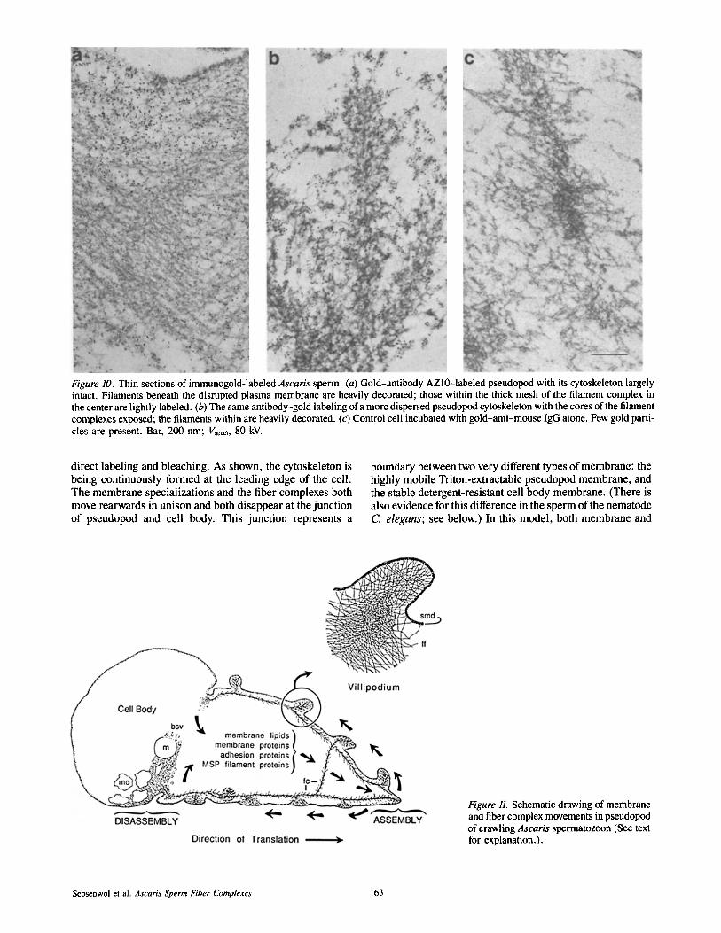
Figure 10. Thin sections of immunogold-labeled Ascaris sperm. (a) Gold-antibody AZl0-1abeled pseudopod with its cytoskeleton largely intact. Filaments beneath the disrupted plasma membrane are heavily decorated; those within the thick mesh of the filament complex in the center are lightly labeled. (b) The same antibody-gold labeling of a more dispersed pseudopod cytoskeleton with the cores of the filament complexes exposed; the filaments within are heavily decorated. (c) Control cell incubated with gold-anti-mouse IgG alone. Few gold parti- cles are present. Bar, 200 nm; l~ce~, 80 kV.
direct labeling and bleaching. As shown, the cytoskeleton is being continuously formed at the leading edge of the cell. The membrane specializations and the fiber complexes both move rearwards in unison and both disappear at the junction of pseudopod and cell body. This junction represents a
boundary between two very different types of membrane: the highly mobile Triton-extractable pseudopod membrane, and the stable detergent-resistant cell body membrane. (There is also evidence for this difference in the sperm of the nematode C. elegans; see below.) In this model, both membrane and
Figure 11. Schematic drawing of membrane and fiber complex movements in pseudopod of crawling Ascaris spermatozoon (See text for explanation.).
Sepsenwol et al. Ascaris Sperm Fiber Complexes 63

complexes are assumed to recycle, with the processes of as- sembly and disassembly being independent and proceeding at the same rate. To crawl, assembly and attachment of mem- brane and complexes at the tip is required to extend the pseu- dopod front. As they move rearwards, complexes and their associated villipodia may move either dorsally or ventrally. Disassembly of membrane and complexes at the base is re- quired to pull the cell body forward. This is derived from two observations: (a) the pseudopod length remains relatively constant during normal crawling; and (b) it can be shown in "aging" crawling cells that even when the assembly of new villipodia at the leading edge of the pseudopod ceases, the cell body continues to be pulled forward (Sepsenwol, S., and S. J. Taft, manuscript submitted for publication). As viUipo- dia reach the base of the pseudopod, it is proposed that pseu- dopodial membrane and membrane adhesion proteins are in- ternalized and filaments forming the complexes disaggregate (Fig. 11, curved arrows at base of pseudopod). The abundant vesicles at the base of the pseudopod and association of vesi- cles with the cell membrane is consistent with active endocy- tosis, but direct evidence is lacking. (See discussion of en- docytosis in C. elegans below.) Whether the filaments exert force (ie., push or pull) on the attached membrane is not known.
Identification of MSP in the Fiber Complexes of Ascaris Sperm
In the absence of actin, myosins, and/or tubulins in Ascaris sperm, the search for new types of cytoskeletal proteins has become central to understanding how this system of locomo- tion might work. MSP is an obvious candidate for a pseudo- pod cytoskeletal protein because it is present in the quantity needed to form the mass of filaments that pack the pseudo- pod. In sperm of both Ascaris and C. elegans, MSP com- prises >15% of the total protein. In C. elegans, anti-MSP antibodies had been shown to localize in the pseudopod of activated sperm (Ward and Klass, 1982). Localization at the ultrastructural level, however, has been frustrated by the lack of an identifiable filamentous superstructure in their pseudo- pods distinguishable from background granular material (Roberts, 1983). The ordered arrangement of filaments in Ascaris sperm allows an unequivocal localization of MSP an- tibodies to the fiber complexes and to the individual fila- ments that comprise them. This is the first evidence that MSP may be a filament protein in the sperm's pseudopod. This immunocytochemical identification does not preclude the involvement of other proteins in the assembly of fiber complexes. However, the abundance of MSP, its segregation into the pseudopods of motile sperm, and the anti-MSP labeling over all the parts of the complex suggests that it is the major species in this fibrous cytoskeleton. The absence of phalloidin staining indicates that the very low concentra- tion of actin is not in the form of F-actin aggregates in the pseudopod. This result does not exclude copolymerization of whatever actin might be present with other major compo- nents of the fibrous cytoskeleton. Indeed, the heterogeneity of fuzzy fibers in Ascaris sperm and their unique associa- tions with cell membranes and each other suggests that pro- teins in addition to MSP may be required to assemble func- tional fiber complexes.
An antibody generated against C. elegans MSP, TR20 (Roberts et al., 1986; Ward et al., 1986), shows the same
selectivity for Ascaris MSP on Western blots, and stains As- caris sperm fiber complexes in the same pattern as antibody AZ10 (see cover photo). This antibody cross-reactivity con- firms molecular studies showing strong similarity between Ascaris and C. elegans MSP. MSP cDNgs from both species have been cloned and sequenced (Burke and Ward, 1983; Bennett and Ward, 1986) and nucleotide homology is 72%; the predicted proteins both consist of 127 amino acids with 82% homology.
The C. Elegans Spermatozoon: Similarities and Differences
Previous work done on the sperm of the nematode C. elegans provides valuable insights into Ascaris sperm behavior. Their strong similarities give us reason to believe that they both crawl by the same mechanism. C. elegans sperm cells are rich in MSP (see above). Like Ascaris sperm, C. elegans spermatids can be activated to full motility in vitro (Nelson and Ward, 1980; Ward et al., 1983). The spermatids and ac- tivated sperm are three times smaller than Ascaris male ga- metes, but morphologically similar: they show the same per- manent compartmentation of organelles. The pseudopod of a crawling C elegans spermatozoon is covered with stubby projections, which form at the tip, move centripetally, and disappear at the pseudopod-cell body junction (see scanning electron micrograph in Nelson et al., 1982, Fig. 1). When attached, these make extensive contacts with the substrate and pull the cell along (Roberts and Streitmatter, 1984). C. elegans sperm possess a region of laminar membranes just inside the cell body at its border with the pseudopod, the site of the band of smooth vesicles in Ascaris sperm (see Ward et al., 1981; Fig. 4).
Roberts and Ward (1982a,b) showed that cationic latex beads, ferritin-labeled lectins, and fluorescent-labeled phos- pholipids all migrated away from the tip of the pseudopod and either accumulated on the surface (beads and lectins) or disappeared (phospholipid) at the pseudopod-cell body junction. Labels over the cell body were stationary in motile sperm. Pavalko and Roberts (1987) have shown that new membrane proteins insert at the tip of the pseudopod and move to the base. The pseudopod labels (colloidal gold and antibody) accumulate at the base. This is direct evidence for incorporation of new membrane proteins at the leading edge of the pseudopod; however, there is as yet no evidence that they are internalized. This lack of direct information about how or whether membrane components are recycled is an important caveat for the Ascaris sperm model.
There are differences between these two species of nema- tode sperm as well. Most importantly, there is no evidence of a higher order fiber structure in the pseudopod of C ele- gans sperm, even when examined in whole mount and semi- thick sections with HVEM (for review see Roberts et al., 1989; an exception is the nonmotilefer-3 sperm mutant, see Ward et al., 1981; Fig. 11, c and d). The pseudopod is filled entirely with a dense mass of granular material and relatively uniform interconnected 2-3 nm filaments, thought to be polymers of MSP (reviewed in Roberts et al., 1989). It is possible that any filamentous superstructure in C. elegans sperm pseudopod might be obscured by the high density of granular cytoplasm. The worms' metabolic systems are also quite different and affect the stability of the pseudopod: C. elegans spermatids form pseudopods and crawl under aero-
The Journal of Cell Biology, Volume t08, 1989 64
bic conditions; Ascaris sperm require anaerobiosis for acti- vation and locomotion, fully formed cytoskeletons become unstable when the cell is exposed to oxygen. It is not clear if this oxygen toxicity is a direct effect on the Ascaris sperm fiber complexes (by peroxide formation, for example) or the consequence of blocking its anaerobic mitochondrial ATP synthesis (for review see Kohler and Bachman, 1980).
Relation of Ascaris Sperm Motility to Other Models for Amoeboid Motility It would be a mistake to generalize the specialized amoeboid sperm of Ascaris into a universal model for all cells that crawl; there are even significant differences among families of nematodes. (The sperm of Nippostrongylus brasiliensis creep by means of transverse waves of constriction passing rearwards over their short pseudopods rather than individual villipodia [Wright and Sommerville, 1984]). As a counter example, however, Ascaris sperm are useful for testing the heuristic value of some models now proposed for amoeboid locomotion in actin-based cells. For example, two competing models for fibroblast motility are based on bulk membrane flow and discount a direct role for the cytoskeleton in mem- brane flow (see Ishihara et al., 1988). Bulk membrane flow theory supposes that the retrograde flow of membrane lipids assembled at the leading edge alone generates a motive force and sweeps membrane protein constituents, including adhe- sion proteins, rearwards (Bretscher, 1983). It is presumed that tethered transmembrane proteins are bound to fixed cy- toplasmic elements. After observing the comigration of vil- lipodia and fiber complexes, we have reservations about using bulk membrane flow models to describe Ascaris sperm motility. In such a system, in which both membrane and cy- toplasmic proteins are moving, it is not clear whether bulk membrane flow is the force or consequence of the continuous assembly and disassembly of the fiber complexes. Regarding the actomyosin-cytoskeleton model, recent reports have demonstrated that myosin I, the most abundant species in amoebae of the slime mold, Dictyosteleum, is not absolutely required for crawling (De Lozanne and Spudich, 1987; Knecht and Loomis, 1987). (Interestingly, these data have been cited as evidence for the irrelevance of the cytoskeleton to the engine for crawling [Bretscher, 1988].) While Ascaris sperm are neither amoebae nor fibroblasts and do not contain significant actin, their fiber complexes clearly represent a new type of cytoskeleton intimately involved in mem- brane/cell movement. These kinds of unexpected results should encourage the search for new mechanisms by which cytoskeletal elements such as actin or MSP might participate in amoeboid locomotion.
The authors would like to acknowledge the expert technical assistance of Judith Baumgartner of the University of Wisconsin at Stevens Point and James Pawley of the Madison Integrated Microscopy Resource, and the skillful artwork of Patricia Gasque.
This work was supported in part by National Science Foundation RUI grant DCB8610475, National Institutes of Health (NIH) AREA grant GM- 37435-01 to S. Sepsenwol, and NIH grant GM-29994 to T. M. Roberts. The Madison Integrated Microscopy Resource is supported by a grant from the Biomedical Research Technology Program, DRR-NIH P41RR00570.
Received for publication 31 May 1988, and in revised form 13 September 1988.
References
Abbas, M. K., and G. D. Cain. 1984. Amino acid and lipid composition of refringent granules from the ameboid sperm of Ascaris suum (Nematoda). Histochemistry. 81:59-65.
Bennett, K. L., and S. Ward. 1986. Neither a germ line-specific nor several somatically expressed genes are lost Or rearranged during embryonic chro- matin diminution in the nematode Ascaris lumbricoides var. suum. Dev. Biol. 118:141-147.
Bretscher, M. S. 1983. Distribution of receptors for transferrin and low density lipoprotein on the surface of giant HeLa cells. Proc. Natl. Acad. Sci. USA. 80:454-458.
Bretscher, M. S. 1988. Fibroblasts on the move [review]. J. Cell Biol. 106: 235-237.
Burghardt, R. C., and W. E. Foor. 1978. Membrane fusion during spermiogen- esis in Ascaris. J. Ultrastruct. Res. 62:190-202.
Burke, D. J., and S. Ward. 1983. Identification of a large multi-gene family encoding the major sperm protein of Caenorhabditis elegans. J. Mol. Biol. 171:1-29.
Clarke, M., and J. A. Spudich. 1977. Non-muscle contractile proteins: the role ofactin and myosin in cell motility and shape determination. Annu. Rev. Bio- chem. 46:797-822.
De Lozanne, A., and J. A. Spudich. 1987. Disruption of the Dictyostelium myo- sin heavy chain gene by homologous recombination. Science (Wash. DC). 236:1086-1091.
Foor, W. E. 1974. Morphological changes of spermatozoa in the uterus and glandular vas deferens of Brugia pahangi. J. Parasitol. 60:125-133.
Foor, W. E., M. H. Johnson, and P. C. Beaver. 1971. Morphological changes in the spermatozoa ofDipetalonema viteae in utero. J. Parasitol. 57:1163- 1169.
Ishihara, A., B. Holifield, and K. Jacobson. 1988. Analysis of lateral redistribu- tion of a monoclonal antibody complex plasma membrane glycoprotein which occurs during cell locomotion. J. Cell Biol. 106:329-343.
Klass, M., and D. Hirsb. 1981. Sperm isolation and biochemical analysis of the major sperm protein from Caenorhabditis elegans. Dev. Biol. 84:299- 312.
Knecht, D. A., and W. F. Loomis. 1987. Anti-sense RNA inactivation of myo- sin heavy chain gene expression in Dictyostelium discoideum. Science (Wash. DC). 236:1081-1086.
Kohler, P., and R. Bachmann. 1980. Mechanisms of respiration and phos- phorylation in Ascaris muscle mitochondria. Mol. Biochem. Parasitol. 1:75- 90.
Laemmli, U. K. 1970. Cleavage of structural proteins during the assembly of the head of bacteriophage T4. Nature (Lond.). 227:680-685.
Maupin, P., and T. D. Pollard. 1983. Improved preservation and staining of HeLa cell actin filaments, clathrin-coated membranes and other cytoplasmic structures by tannic acid-glutaraldehyde-saponin fixation. J. Cell Biol. 96: 51-62.
Nelson, G. A., and S. Ward. 1980. Vesicle fusion, pseudopod extension and amoeboid motility are induced in nematode spermatids by the ionophore monensin. Cell. 19:457-464.
Nelson, G. A., and S. Ward. 1981. Amoeboid motility and actin in Ascaris lum- bricoides sperm. Exp. Cell Res. 131:149-160.
Nelson, G. A., T. M. Roberts, and S. Ward. 1982. Caenorhabditis elegans spermatozoan locomotion: amoeboid movement with almost no actin. J. Cell Biol. 92:113-120.
Pavalko, F. M.. and T. M. Roberts. 1987. Caenorhabditis elegans spermatozoa assemble membrane proteins onto the cell surface at the tips of pseudopodial proiections. Cell Motil. Cytoskeleton. 7:169-177.
Pawley, J. B., S. Sepsenwol, and H. Ris. 1986. Four-dimensional microscopy of Ascaris sperm motility. Ann. NY Acad. Sci. 483:171 - 178.
Pollard, T. D. 1981. Cytoplasmic contractile proteins [review[. J. Cell Biol. 91(Pt. 2, Suppl.):156s-165s.
Pollard, T. D. 1986. Assembly and dynamics of the actin filament system of non-muscle cells. J. Cell Biochem. 31:87-95.
Ris, H. 1985. The cytoplasmic filament system in critical point-dried whole mounts and plastic-embedded sections. J. Cell Biol. 100:1474-1487.
Roberts, T. M. 1983. Crawling Caenorhabditis elegans spermatozoa contact the substrate only by their pseudopods and contain 2-nm filaments. Cell Motil. Cytoskeleton. 8:130-142.
Roberts, T. M., and G. Streitmatter. 1984. Membrane-suhstrate contact under the spermatozoon of Caenorhabditis elegans, a crawling cell that lacks filamentous actin. J. Cell Sci. 69:117-126.
Roberts, T. M., and S. Ward. 1982a. Centripetal flow of pseudopodial surface components could propel the amoeboid movement of Caenorhabditis elegans spermatozoa. J. Cell Biol. 92:132-138.
Roberts, T. M., and S. Ward. 1982b. Membrane flow during nematode spermi- ogenesis. J. Cell Biol. 92:113-120.
Roberts, T. M., F. M. Pavalko, and S. Ward. 1986. Membrane and cytoplas- mic proteins are transported in the same organelle complex during nematode spermatogenesis. J. Cell Biol. 102:1787-1796.
Roberts, T. M., S. Sepsenwol, and H. Ris. 1989. Sperm motility in nematodes: crawling movement without actin. In The Cell Biology of Fertilization. H. Schatten and G. Schatten, editors. Academic Press, Orlando, FL. 41-60.
Sepsenwol, S., T. Braun, and M. Nguyen. 1986. Adenylate cyclase activity is
Sepsenwol et al. Ascaris Sperm Fiber Complexes 65

absent in inactive and motile sperm in the nematode parasite, Ascaris suum. J. Parasitol. 72:962-964.
Slot, J. W., and H. J. Geuze. 1985. A new method of preparing gold probes for multiple labelling cytochemistry. Eur. J. Cell Biol. 38:87-93.
Towbin, H., T. Staehelin, and G. Gordon. 1979. Electrophoretic transfer of proteins from polyacrylamide gels to nitrocellulose sheets: procedure and some applications. Proc. Natl. Acad. Sci. USA. 76:1350-1354.
Trinkhaus, J. P. 1984. Cells into organs. In The Forces That Shape the Embryo. 2nd edition. Prentice-Hall, Englewood Cliffs, NJ. 541 pp.
Ward, S., and M. Klass. 1982. The localization of the major sperm protein in C. elegans sperm and spermatocytes. Dev. Biol. 92:203-208.
Ward, S., Y. Argon, and G. A. Nelson. 1981. Sperm morphology in wild-type and fertilization-defective mutants of Caenorhabditis elegans. J. Cell Biol.
91:26-44. Ward, S., E. Hogan, and G. A. Nelson. 1983. The initiation of Caenorhabditis
elegans spermiogenesis in vivo and in vitro. Dev. BioL 98:70-79. Ward, S., T. M. Roberts, S. Strome, F. M. Pavalko, and E. Hogan. 1986.
Monoclonal antibodies that recognize a polypeptide antigenic determinant shared by multiple Caenorhabditis elegans sperm-specific proteins. J. Cell Biol. 102:1778-1786.
Wright, E. J., and R. I. Sommerville. 1984. Postinsemination changes in the amoeboid sperm of a nematode, Nippostrongylus brasiliensis. Gamete Res. 10:397-413.
Wright, E. J., and R. I. Sommerville. 1985. Structure and development of the spermatozoon of the parasitic nematode, Nematospiroides dubius. Parasitol- ogy. 90:179-192.
The Journal of Cell Biology, Volume 108, 1989 66