Embed Size (px)
Citation preview
The Rosetta All-Atom Energy Function for Macromolecular Modelingand DesignRebecca F. Alford,† Andrew Leaver-Fay,‡ Jeliazko R. Jeliazkov,§ Matthew J. O’Meara,∥ Frank P. DiMaio,⊥
Hahnbeom Park,# Maxim V. Shapovalov,∇ P. Douglas Renfrew,○,◆ Vikram K. Mulligan,# Kalli Kappel,¶
Jason W. Labonte,† Michael S. Pacella,+ Richard Bonneau,○,◆ Philip Bradley,Δ
Roland L. Dunbrack, Jr.,∇ Rhiju Das,¶ David Baker,#,∞ Brian Kuhlman,‡ Tanja Kortemme,◇
and Jeffrey J. Gray*,†,§
†Department of Chemical and Biomolecular Engineering, Johns Hopkins University, 3400 North Charles Street, Baltimore, Maryland21218, United States‡Department of Biochemistry and Biophysics, University of North Carolina at Chapel Hill, 120 Mason Farm Road, Chapel Hill,North Carolina 27599, United States§Program in Molecular Biophysics, Johns Hopkins University, 3400 North Charles Street, Baltimore, Maryland 21218, United States∥Department of Pharmaceutical Chemistry, University of California at San Francisco, 1700 Fourth Street, San Francisco, California94158, United States⊥Department of Biochemistry, University of Washington, J-Wing, Health Sciences Building, Box 357350, Seattle, Washington 98195,United States#Department of Biochemistry, University of Washington, Molecular Engineering and Sciences, Box 351655, 3946 West Stevens WayNE, Seattle, Washington 98195, United States∇Institute for Cancer Research, Fox Chase Cancer Center, 333 Cottman Avenue, Philadelphia, Pennsylvania 19111, United States○Department of Biology, Center for Genomics and Systems Biology, New York University, 100 Washington Square East, New York,New York 10003, United States◆Center for Computational Biology, Flatiron Institute, Simons Foundation, 162 Fifth Avenue, New York, New York 10010, UnitedStates¶Biophysics Program, Stanford University, 450 Serra Mall, Stanford, California 94305, United States+Department of Biomedical Engineering, Johns Hopkins University, 3400 North Charles Street, Baltimore, Maryland 21218, UnitedStatesΔComputational Biology Program, Fred Hutchinson Cancer Research Center, 1100 Fairview Avenue North, Seattle, Washington98109, United States◇Department of Bioengineering and Therapeutic Sciences, University of California at San Francisco, San Francisco, California 94158,United States∞Howard Hughes Medical Institute, University of Washington, Seattle, Washington 98195, United States
*S Supporting Information
ABSTRACT: Over the past decade, the Rosetta biomolecular modeling suite hasinformed diverse biological questions and engineering challenges ranging frominterpretation of low-resolution structural data to design of nanomaterials, proteintherapeutics, and vaccines. Central to Rosetta’s success is the energy function: a modelparametrized from small-molecule and X-ray crystal structure data used to approximate theenergy associated with each biomolecule conformation. This paper describes themathematical models and physical concepts that underlie the latest Rosetta energyfunction, called the Rosetta Energy Function 2015 (REF15). Applying these concepts, weexplain how to use Rosetta energies to identify and analyze the features of biomolecularmodels. Finally, we discuss the latest advances in the energy function that extend itscapabilities from soluble proteins to also include membrane proteins, peptides containingnoncanonical amino acids, small molecules, carbohydrates, nucleic acids, and othermacromolecules.
Received: February 6, 2017Published: April 21, 2017
Article
pubs.acs.org/JCTC
© 2017 American Chemical Society 3031 DOI: 10.1021/acs.jctc.7b00125J. Chem. Theory Comput. 2017, 13, 3031−3048

■ INTRODUCTIONProteins adopt diverse three-dimensional conformations tocarry out the complex mechanisms of life. Their structures areconstrained by the underlying amino acid sequence1 andstabilized by a fine balance between enthalphic and entropiccontributions to non-covalent interactions.2 Energy functionsthat seek to approximate the energies of these interactions arefundamental to computational modeling of biomolecularstructures. The goal of this paper is to describe the energycalculations used by the Rosetta macromolecular modelingprogram:3 we explain the underlying physical concepts,mathematical models, latest advances, and application tobiomolecular simulations.Energy functions are based on Anfinsen’s hypothesis that
native-like protein conformations represent unique, low-energy,thermodynamically stable conformations.4 These folded statesreside in minima on the energy landscape, and they have a netfavorable change in Gibbs free energy, which is the sum ofcontributions from both enthalpy (ΔH) and entropy (TΔS),relative to the unfolded state. To follow these heuristics,macromolecular modeling programs require a mathematicalfunction that can discriminate between the unfolded, folded,and native-like conformations. Typically, these functions arelinear combinations of terms that compute energies as afunction of various degrees of freedom.The earliest macromolecular energy functions combined a
Lennard-Jones potential for van der Waals interactions5−7 withharmonic torsional potentials8 that were parametrized usingforce constants from vibrational spectra of small molecules.9−11
These formulations were first applied to investigations of thestructures of hemolysin,12 trypsin inhibitor,13 and hemoglobin14
and have now diversified into a large family of commonly usedenergy functions such as AMBER,15 DREIDING,16 OPLS,17
and CHARMM.18,19 Many of these energy functions also relyon new terms and parametrizations. For example, fastercomputers have enabled the derivation of parameters from abinitio quantum calculations.20 The maturation of X-raycrystallography and NMR protein structure determinationmethods has enabled the development of statistical potentialsderived from per-residue, inter-residue, secondary-structure,and whole-structure features.21−28 Additionally, there arealternate models of electrostatics and solvation, such as thegeneralized Born approximation of the Poisson−Boltzmannequation29 and polarizable electrostatic terms that accommo-date varying charge distributions.30
The first version of the Rosetta energy function wasdeveloped for proteins by Simons et al.31 Initially, it usedstatistical potentials describing individual residue environmentsand frequent residue-pair interactions derived from the ProteinData Bank (PDB).32 Later the authors added terms for packingof van der Waals spheres and hydrogen-bonding, secondary-structure, and van der Waals interactions to improve theperformance of ab initio structure prediction.33 These termswere for low-resolution modeling, meaning that the scores weredependent on only the coordinates of the backbone atoms andthat interactions between the side chains were treatedimplicitly.To enable higher-resolution modeling, in the early 2000s
Kuhlman and Baker34 implemented an all-atom energy functionthat emphasized atomic packing, hydrogen bonding, solvation,and protein torsion angles commonly found in folded proteins.This energy function first included a Lennard-Jones term,35 a
pairwise-additive implicit solvation model,36 a statisticallyderived electrostatics term, and a term for backbone-dependentrotamer preferences.37 Shortly thereafter, several terms wereadded, including an orientation-dependent hydrogen-bondingterm,38 in agreement with electronic structure calculations.39
This combination of traditional molecular mechanics energiesand statistical torsion potentials enabled Rosetta to reachseveral milestones in structure prediction and design, includingaccurate ab initio structure prediction,40 hot-spot predic-tion,41,42 protein−protein docking,43 small-molecule docking,44
and specificity redesign45 as well as the first de novo designedprotein backbone not found in nature46 and the firstcomputationally designed new protein−protein interface.47
The Rosetta energy function has changed dramatically sinceit was last described in complete detail by Rohl et al.48 in 2004.It has undergone significant advances ranging from improvedmodels of hydrogen bonding49 and solvation50 to updatedevaluation of backbone51 and rotamer conformations.52 Alongthe way, these developments have enabled Rosetta to addressnew biomolecular modeling problems, including the refinementof low-resolution X-ray structures and use of sparse data53,54
and the design of vaccines,55 biomineralization peptides,56 self-assembling materials,57 and enzymes that perform newfunctions.58,59 Instead of arbitrary units, the energy functionis now also fitted to estimate energies in kilocalories per mole.The details of the energy function advances are distributedacross code comments, methods development papers,application papers, and individual experts, making it challengingfor Rosetta developers and users in both academia and industryto learn the underlying concepts. Moreover, members of theRosetta community are actively working to generalize the all-atom energy function for use in different contexts60,61 and forall macromolecules, including RNA,62 DNA,63,64 small-molecule ligands,65,66 noncanonical amino acids and back-bones,67−69 and carbohydrates,70 further encouraging us toreexamine the underpinnings of the energy function. Thus,there is a need for an up-to-date description of the currentenergy function.In this paper, we describe the new default energy function,
called the Rosetta Energy Function 2015 (REF15). Ourdiscussion aims to expose the physical and mathematicaldetails of the energy function required for rigorous under-standing. In addition, we explain how to apply the computedenergies to analyze structural models produced by Rosettasimulations. We hope this paper will provide critically neededdocumentation of the energy methods as well as an educationalresource to help students and scientists interpret the results ofthese simulations.
■ COMPUTING THE TOTAL ROSETTA ENERGYThe Rosetta energy function approximates the energy of abiomolecule conformation. This quantity, called ΔEtotal, iscomputed as a linear combination of energy terms Ei that arecalculated as functions of geometric degrees of freedom (Θ)and chemical identities (aa) and scaled by a weight on eachterm (wi), as shown in eq 1:
∑Δ = ΘE wE ( , aa )i
i i i itotal(1)
Here we explain the Rosetta energy function term by term.First, we describe the energies of interactions betweennonbonded atom pairs, which are important for atomicpacking, electrostatics, and solvation. Second, we explain the
Journal of Chemical Theory and Computation Article
DOI: 10.1021/acs.jctc.7b00125J. Chem. Theory Comput. 2017, 13, 3031−3048
3032
empirical potentials used to model hydrogen and disulfidebonds. Next, we explain the statistical potentials used todescribe backbone and side-chain torsional preferences inproteins. Then we explain a set of terms that accommodatefeatures that are not explicitly captured yet are important fornative structural feature recapitulation. Finally, we discuss howthe energy terms are combined into a single function used toapproximate the energy of biomolecules. For reference, items inthe fixed-width font are names of energy terms in the Rosettacode. The energy terms are summarized in Table 1.Terms for Atom-Pair Interactions. Van der Waals
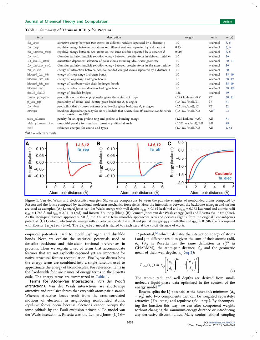
Interactions. Van der Waals interactions are short-rangeattractive and repulsive forces that vary with atom-pair distance.Whereas attractive forces result from the cross-correlatedmotions of electrons in neighboring nonbonded atoms,repulsive forces occur because electrons cannot occupy thesame orbitals by the Pauli exclusion principle. To model vander Waals interactions, Rosetta uses the Lennard-Jones (LJ) 6−
12 potential,5,6 which calculates the interaction energy of atomsi and j in different residues given the sum of their atomic radii,σi,j (σi,j in Rosetta has the same definition as ri,j
min inCHARMM), the atom-pair distance, di,j, and the geometricmean of their well depths, εi,j (eq 2):
εσ σ
= −⎡
⎣⎢⎢⎛⎝⎜⎜
⎞⎠⎟⎟
⎛⎝⎜⎜
⎞⎠⎟⎟
⎤
⎦⎥⎥E i j
d d( , ) 2i j
i j
i j
i j
i jvdw ,
,
,
12,
,
6
(2)
The atomic radii and well depths are derived from small-molecule liquid-phase data optimized in the context of theenergy model.50
Rosetta splits the LJ potential at the function’s minimum (di,j= σi,j) into two components that can be weighted separately:attractive (fa_atr) and repulsive (fa_rep). By decompos-ing the function this way, we can alter component weightswithout changing the minimum-energy distance or introducingany derivative discontinuities. Many conformational sampling
Table 1. Summary of Terms in REF15 for Proteins
term description weight units ref(s)
fa_atr attractive energy between two atoms on different residues separated by a distance d 1.0 kcal/mol 5, 6fa_rep repulsive energy between two atoms on different residues separated by a distance d 0.55 kcal/mol 5, 6fa_intra_rep repulsive energy between two atoms on the same residue separated by a distance d 0.005 kcal/mol 5, 6fa_sol Gaussian exclusion implicit solvation energy between protein atoms in different residues 1.0 kcal/mol 36lk_ball_wtd orientation-dependent solvation of polar atoms assuming ideal water geometry 1.0 kcal/mol 50, 71fa_intra_sol Gaussian exclusion implicit solvation energy between protein atoms in the same residue 1.0 kcal/mol 36fa_elec energy of interaction between two nonbonded charged atoms separated by a distance d 1.0 kcal/mol 50hbond_lr_bb energy of short-range hydrogen bonds 1.0 kcal/mol 38, 49hbond_sr_bb energy of long-range hydrogen bonds 1.0 kcal/mol 38, 49hbond_bb_sc energy of backbone−side-chain hydrogen bonds 1.0 kcal/mol 38, 49hbond_sc energy of side-chain−side-chain hydrogen bonds 1.0 kcal/mol 38, 49dslf_fa13 energy of disulfide bridges 1.25 kcal/mol 49rama_prepro probability of backbone ϕ, ψ angles given the amino acid type (0.45 kcal/mol)/kT kT 50, 51p_aa_pp probability of amino acid identity given backbone ϕ, ψ angles (0.4 kcal/mol)/kT kT 51fa_dun probability that a chosen rotamer is native-like given backbone ϕ, ψ angles (0.7 kcal/mol)/kT kT 52omega backbone-dependent penalty for cis ω dihedrals that deviate from 0° and trans ω dihedrals
that deviate from 180°(0.6 kcal/mol)/AU AUa 72
pro_close penalty for an open proline ring and proline ω bonding energy (1.25 kcal/mol)/AU AU 51yhh_planarity sinusoidal penalty for nonplanar tyrosine χ3 dihedral angle (0.625 kcal/mol)/AU AU 49ref reference energies for amino acid types (1.0 kcal/mol)/AU AU 1, 51
aAU = arbitrary units.
Figure 1. Van der Waals and electrostatics energies. Shown are comparisons between the pairwise energies of nonbonded atoms computed byRosetta and the forms computed by traditional molecular mechanics force fields. Here the interactions between the backbone nitrogen and carbonare used as examples. (A) Lennard-Jones van der Waals energy with well-depths εNbb = 0.162 kcal/mol and εCbb = 0.063 kcal/mol and atomic radiirNbb = 1.763 Å and rCbb = 2.011 Å (red) and Rosetta fa_rep (blue). (B) Lennard-Jones van der Waals energy (red) and Rosetta fa_atr (blue).As the atom-pair distance approaches 6.0 Å, the fa_atr term smoothly approaches zero and deviates slightly from the original Lennard-Jonespotential. (C) Coulomb electrostatic energy with a dielectric constant ϵ = 10 and partial charges qNbb = −0.604e and qCbb = 0.090e (red) comparedwith Rosetta fa_elec (blue). The fa_elec model is shifted to reach zero at the cutoff distance of 6.0 Å.
Journal of Chemical Theory and Computation Article
DOI: 10.1021/acs.jctc.7b00125J. Chem. Theory Comput. 2017, 13, 3031−3048
3033

protocols in Rosetta take advantage of this splitting by slowlyincreasing the weight of the repulsive component to traverserugged energy landscapes and to prevent structures fromunfolding during sampling.73
The repulsive van der Waals energy, fa_rep, varies as afunction of atom-pair distance. At short distances, atomicoverlap results in strong forces that lead to large changes in theenergy. The steep 1/di,j
12 term can cause poor performance inminimization routines and overall structure prediction anddesign calculations.74,75 To alleviate this problem, we weakenthe repulsive component by replacing the 1/di,j
12 term with asofter linear term for di,j ≤ 0.6σi,j. The term is computed usingthe atom-type-specific parameters mi,j and bi,j, which are fit toensure derivative continuity at di,j = 0.6σi,j. After the linearcomponent, the function transitions smoothly to the 6−12form until di,j = σi,j, where it reaches zero and remains zero (eq3 and Figure 1A):
∑
σ
εσ σ
σ σ
σ
=
+ ≤
− + < ≤
<
_
⎧
⎨
⎪⎪⎪
⎩
⎪⎪⎪
⎡
⎣⎢⎢⎛⎝⎜⎜
⎞⎠⎟⎟
⎛⎝⎜⎜
⎞⎠⎟⎟
⎤
⎦⎥⎥
E i j
w
m d b d
d dd
d
( , )
0.6
2 1 0.6
0
i ji j
i j i j i j i j i j
i ji j
i j
i j
i ji j i j i j
i j i j
fa rep
,,conn
, , , , ,
,,
,
12,
,
6
, , ,
, ,
(3)
Rosetta also includes an intraresidue version of the repulsivecomponent, fa_intra_rep, with the same functional formas the fa_rep term (eq 3). We include this term because theknowledge-based rotamer energy (fa_dun; see below)underestimates intraresidue collisions.The attractive van der Waals energy, fa_atr, has a value of
−εi,j for di,j ≤ σi,j and then transitions to the 6−12 potential asthe distance increases (eq 4 and Figure 1B):
∑
ε σ
εσ σ
σ
=
− ≤
− < ≤
< ≤
<
_
⎧
⎨
⎪⎪⎪⎪
⎩
⎪⎪⎪⎪
⎡
⎣⎢⎢⎛⎝⎜⎜
⎞⎠⎟⎟
⎛⎝⎜⎜
⎞⎠⎟⎟
⎤
⎦⎥⎥
E
w
d
d dd
f d d
d
2 4.5 Å
( ) 4.5 Å 6.0 Å
0 6.0 Å
i ji j
i j i j ij
i ji j
i j
i j
i ji j i j
i j i j
i j
fa atr
,,conn
, ,
,,
,
12,
,
6
, ,
poly , ,
,
(4)
For speed, we truncate the LJ term beyond di,j = 6.0 Å, wherethe van der Waals forces are small. To avoid derivativediscontinuities, we use a cubic polynomial function, f poly(di,j),for di,j > 4.5 Å to transition the standard Lennard-Jonesfunctional form smoothly to zero. These smooth derivatives arenecessary to ensure that bumps do not accumulate in thedistributions of structural features at inflection points in theenergy landscape during conformational sampling withgradient-based minimization (Sheffler, unpublished, 2006).Both terms are multiplied by a connectivity weight, wi,j
conn, toexclude the large repulsive energetic contributions that wouldotherwise be calculated for atoms separated by fewer than fourchemical bonds (eq 5).
=
≤
=
≥
⎧
⎨⎪⎪
⎩⎪⎪
w
n
n
n
0 3
0.2 4
1 5
i j
i j
i j
i j
,conn
,bonds
,bonds
,bonds
(5)
To ensure the connectivity rules include both atoms in a dipole,if an atom is part of a strong dipole, then we count bonds(ni,j
bonds) to include both atoms in the dipole. Such weights arecommon to molecular force fields that assume that covalentbonds are not formed or broken during a simulation. Rosettauses four chemical bonds as the “crossover” separation whenwi,jconn transitions from 0 to 1 (rather than three chemical bonds
as used by traditional force fields) to limit the effects of doublecounting due to knowledge-based torsional potentials.The comparison between eq 2 and the modified LJ potential
(eqs 3 and 4) is shown in Figure 1A,B.Electrostatics. Nonbonded electrostatic interactions arise
from forces between fully and partially charged atoms. Toevaluate these interactions, Rosetta uses Coulomb’s law withpartial charges originally taken from CHARMM and adjustedvia a group optimization scheme (Table S3 in the SupportingInformation).50 Coulomb’s law is a pairwise term commonlyexpressed in terms of di,j, the dielectric constant, ϵ, the partialatomic charges for each atom, qi and qj, and Coulomb’sconstant, C0 (=322 Å kcal/mol e−2, where e is the elementarycharge) (eq 6):
=ϵ
E i jC q q
d( , )
1i j
i jCoulomb
0
, (6)
To approximate electrostatic interactions in biomolecules, wemodify the potential to account for the difference in dielectricconstant between the protein core and the solvent-exposedsurface.76 Specifically, we substitute the constant ϵ in eq 6 witha sigmoidal function ϵ(di,j) that increases from ϵcore = 6 toϵsolvent = 80 when di,j is between 0 and 4 Å (eqs 7 and 8):
ϵ = ϵ + − ϵ⎛⎝⎜
⎞⎠⎟
⎡⎣⎢⎢
⎛⎝⎜
⎞⎠⎟⎤⎦⎥⎥d g
dg
d( )
41
4i ji j i j
,,
core,
solvent(7)
= + + −⎛⎝⎜
⎞⎠⎟g x x
xx( ) 1
2exp( )
2
(8)
As with the van der Waals term, we make several heuristicapproximations to adapt this calculation for simulations ofbiomolecules. To avoid strong repulsive forces at shortdistances, we replace the steep gradient with the constantEelec(dmin) for di,j < 1.45 Å. Next, since the distance-dependentdielectric assumption results in dampened long-range electro-statics, for speed we truncate the potential at dmax = 5.5 Å andmodify the Coulomb curve by subtracting 1/dmax
2 to shift thepotential to zero at di,j = dmax (eq 9).
=ϵ
− ≤
<
⎧⎨⎪
⎩⎪E i j d
C q q
dd d
d d
d d( , , )
( )
1 1
0i j
i j
i j
i ji j
i j
elec ,0
,
,2
max2 , max
max , (9)
We use the cubic polynomials f polyelec,low(di,j) and f poly
elec,high(di,j) tosmooth between the traditional form and our adjustments whileavoiding derivative discontinuities. The energy is also multi-plied by the connectivity weight, wi,j
conn (eq 5). The final
Journal of Chemical Theory and Computation Article
DOI: 10.1021/acs.jctc.7b00125J. Chem. Theory Comput. 2017, 13, 3031−3048
3034

modified electrostatic potential is given by eq 10 and comparedwith the standard form in Figure 1C.
∑=
<
≤ <
≤ <
≤ <
≤
_
⎧
⎨
⎪⎪⎪⎪
⎩
⎪⎪⎪⎪
E w
E i j d d
f d d
E i j d d
f d d
d
( , , ) 1.45 Å
( ) 1.45 Å 1.85 Å
( , , ) 1.85 Å 4.5 Å
( ) 4.5 Å 5.5 Å
0 5.5 Å
i ji j
i j
i j i j
i j i j
i j i j
i j
fa elec,
,conn
elec min ,
polyelec,low
, ,
elec , ,
polyelec,high
, ,
,
(10)
Solvation. Native-like protein conformations minimize theexposure of hydrophobic side chains to the surrounding polarsolvent. Unfortunately, explicitly modeling all of the inter-actions between solvent and protein atoms is computationallyexpensive. Instead, Rosetta represents the solvent as bulk waterbased upon the Lazaridis−Karplus (LK) implicit Gaussianexclusion model.36 Rosetta’s solvation model has twocomponents: an isotropic solvation energy, called fa_sol,which assumes that bulk water is uniformly distributed aroundthe atoms (Figure 2A), and an anisotropic solvation energy,called lk_ball_wtd, which accounts for specific waters nearpolar atoms that form the solvation shell (Figure 2B).The isotropic (LK) model36 is based on the function fdesolv
that describes the energy required to desolvate (removecontacting water) an atom i when it is approached by aneighboring atom j. In Rosetta, we exclude the LK ΔGref termbecause we implement our own reference energy (discussedlater). The energy of the atom-pair interaction varies with theseparation distance, di,j, the experimentally determined vapor-to-water transfer free energy, ΔGi
free, the sum of the atomicradii, σi,j, the correlation length, λi, and the atomic volume ofthe desolvating atom, Vj (eq 11):
π λ σ
σ
λ= −
Δ−
−⎡⎣⎢⎢
⎛⎝⎜
⎞⎠⎟
⎤⎦⎥⎥f V
G d
2expj
i
i i
i j i j
idesolv
free
3/2 2, ,
2
(11)
At short distances, fa_rep prevents atoms from overlapping;however, many protocols briefly downweight or disable thefa_rep term. To avoid scenarios where fdesolv encouragesatom-pair overlap in the absence of fa_rep, we smoothlyincrease the value of the function to a constant at shortdistances where the van der Waals spheres overlap (di,j = σi,j).At large distances, the function asymptotically approaches zero;therefore, we truncate the function at 6.0 Å for speed. We alsotransition between the constants at short and long distancesusing the distance-dependent cubic polynomials fpoly
solv,low andf polysolv,high with constants c0 = 0.3 Å and c1 = 0.2 Å that define awindow for smoothing. The overall desolvation function isgiven by eq 12:
σ σ
σ σ
σ=
≤ −
− < ≤ +
+ < ≤
< ≤
<
⎧
⎨
⎪⎪⎪⎪
⎩
⎪⎪⎪⎪
g
f i j d c
f i j d c d c
f i j d c d
f i j d d
d
( , , )
( , , )
( , , ) 4.5 Å
( , , ) 4.5 Å 6.0 Å
0 6.0 Å
i j i j i j
i j i j i j i j
i j i j i j
i j i j
i j
desolv
desolv , , , 0
polysolv,low
, , 0 , , 1
desolv , , 1 ,
polysolv,high
, ,
,
(12)
The total isotropic solvation energy, Efa_sol, is computed as asum of the energies for atom j desolvating atom i and vice versa,scaled by the previously defined connectivity weight (eq 13):
∑= +_E w g i j g j i[ ( , ) ( , )]i j
i jfa sol,
,conn
desolv desolv(13)
Figure 2. Two-component Lazaridis−Karplus solvation model. Rosetta uses two energy terms to evaluate the desolvation of protein side chains: anisotropic term (fa_sol) and an anisotropic term (lk_ball_wtd). (A) and (B) demonstrate the difference between isotropic and anisotropicsolvation of the NH2 group by CH3 on the asparagine side chain. The contours vary from low energy (blue) to high energy (yellow). The arrowsrepresent the approach vectors for the pair potentials shown in (C−E), where we compare the fa_sol, lk_ball, and lk_ball_wtd +fa_sol energies for the solvation of the NH2 group on asparagine for three different approach angles: (C) in line with the 1HD2 atom, (D) alongthe bisector of the angle between 1HD1 and 1HD2, and (E) vertically down from above the plane of the hydrogens (out of plane).
Journal of Chemical Theory and Computation Article
DOI: 10.1021/acs.jctc.7b00125J. Chem. Theory Comput. 2017, 13, 3031−3048
3035

Rosetta also includes an intraresidue version of the isotropicsolvation energy, fa_intra_sol, with the same functionalform as the fa_sol term (eq 13).A recent innovation (2016) is the addition of the energy
term lk_ball_wtd to model the orientation-dependentsolvation of polar atoms. This anisotropic model increases thedesolvation penalty for occluding polar atoms near sites wherewaters may form hydrogen-bonding interactions. For polaratoms, we subtract off part of the isotropic energy given by eq13 and then add the anisotropic energy to account for theposition of the desolvating atom relative to hypothesized waterpositions.To compute the anisotropic energy, we first calculate the set
of ideal water sites around atom i, ν ν= { , , ...}i i i1 2 . This setcontains one to three water sites, depending on the atom typeof atom i. Each site is 2.65 Å from atom i and has an optimalhydrogen-bonding geometry, and we consider the potentialoverlap of a desolvating atom j with each water. The overlap isconsidered negligible until the van der Waals sphere of thedesolvating atom j (with radius σj) touches the van der Waalssphere of the water (with radius σw) at site k, and then the termsmoothly increases over a zone of partial overlap ofapproximately 0.5 Å. Thus, for each water site k withcoordinates vi,k, we compute an occlusion measure dk
2 tocapture the gap between the hypothetical water and thedesolvating atom j, using the offset Ω = 3.7 Å2 to provide theramp-up buffer (eq 14):
ν σ σ= || − || − + + Ωd r ( )k j i k j2
,2
w2
(14)
Next, we find the soft minimum of dk2 over all water sites in i
by computing the logarithmic average:
∑= − −∈
⎡⎣⎢⎢
⎤⎦⎥⎥d i j d( , ) ln exp( )
kkmin
2 2
i (15)
Then dmin2 and Ω are used to compute a damping function,
f lkfrac, that varies from 0 when the desolvating atom is at least avan der Waals distance from any preferred water site to 1 whenthe desolvating atom overlaps a water site by more than ∼0.5 Å(eq 16):
=
<
−Ω
≤ < Ω
Ω ≤
⎧
⎨⎪⎪⎪
⎩⎪⎪⎪
⎡⎣⎢⎢
⎛⎝⎜
⎞⎠⎟⎤⎦⎥⎥f i j
d i j
d i jd i j
d i j
( , )
1 ( , ) 0
1( , )
0 ( , )
0 ( , )
lkfrac
min2
min2 2
min2
min2
(16)
We calculate the anisotropic energy for desolvating a polaratom, Elk_ball, by scaling the desolvation function gdesolv by thedamping function f lkfrac and an atom-type-specific weight, waniso,which is typically ∼0.7 (eq 17):
=_E i j w g i j f i j( , ) ( , ) ( , )ilk ball aniso, desolv lkfrac (17)
The amount of isotropic solvation energy subtracted is gdesolvmultiplied by wiso, where wiso is an atom-type-specific weight,typically ∼0.3 (eq 18):
Figure 3. Orientation-dependent hydrogen bonding model. (A) Degrees of freedom evaluated by the hydrogen-bonding term: the acceptor−donordistance, dHA; the angle between the base, acceptor, and hydrogen, θBAH; the angle between the acceptor, hydrogen, and donor, θAHD; and thedihedral angle corresponding to rotation around the base−acceptor bond, ϕB2BAH. (B) Lambert azimuthal projection of the Ehbond
B2BAH energy landscape
for an sp2-hybridized acceptor.49 (C) EhbondB2BAH energy landscape for an sp3-hybridized acceptor. (D−F) Example energies for hydrogen bonding of the
histidine imidazole ring acceptor with a protein backbone amide: (D) energy EhbondHA vs the acceptor−donor distance dHA; (E) energy EhbondBAH vs the
base−acceptor−hydrogen angle θBAH; (F) energy EhbondAHD vs the acceptor−hydrogen−donor angle θAHD.
Journal of Chemical Theory and Computation Article
DOI: 10.1021/acs.jctc.7b00125J. Chem. Theory Comput. 2017, 13, 3031−3048
3036

= −_ _E i j w g i j( , ) ( , )ilk ball iso iso, desolv (18)
The total weight on the isotropic contribution through both thefa_sol and lk_ball_wtd terms is thus ∼0.7. Theisotropic and anisotropic components are then summed toyield a new desolvation function, hdesolv (eq 19):
= +_ _ _h i j E i j E i j( , ) ( , ) ( , )desolv lk ball iso lk ball (19)
Like fa_sol, the energies for desolvation of atom i by atom jand desolvation of atom j by atom i are summed to yield theoverall lk_ball_wtd energy, but counting only thedesolvation of polar, hydrogen-bonding heavy atoms (O, N),defined as the set (eq 20):
∑ ∑= +_ _∈ ∈
E w h i j w h j i( , ) ( , )i
i jj
i jlk ball wtd ,conn
desolv ,conn
desolv
(20)
Figure 2 shows comparisons of fa_sol, lk_ball (eq 17),and the sum of fa_sol and lk_ball_wtd for the exampleof an asparagine NH2 desolvated from three different approachangles. As the approach angle varies, the sum oflk_ball_wtd and fa_sol creates a larger desolvationpenalty when water sites are occluded and a smaller penaltyotherwise, relative to fa_sol alone.Hydrogen Bonding. Hydrogen bonds are partially covalent
interactions that form when a nucleophilic heavy atom donateselectron density to a polar hydrogen.77 At short ranges (<2.5Å), they exhibit geometries that maximize orbital overlap.78 Theinteractions between hydrogen-bonding groups are alsopartially described by electrostatics. While this hybridcovalent−electrostatic character is complex, it is crucial forcapturing the structural specificity that underlies proteinfolding, function, and interactions.Rosetta calculates the energies of hydrogen bonds using
fa_elec and a hydrogen-bonding model that evaluatesenergies on the basis of orientation preferences of hydrogenbonds found in high-resolution crystal structures.38,49 To derivethis model, we curated intraprotein polar contacts from ∼8000high-resolution crystal structures (the Top8000 data set79) andidentified features using adaptive density estimation. We thenempirically fit the functional form of the energy such that theRosetta-generated polar contacts mimic the distributions fromTop8000. The resulting hydrogen-bonding energy is evaluatedfor all pairs of donor hydrogens, H, and acceptors, A, as afunction of four degrees of freedom (Figure 3A): (1) thedistance between the donor and acceptor, dHA; (2) the angleθAHD formed by the acceptor, the donor H, and the donorheavy atom, D; (3) the angle θBAH formed by the acceptor’sparent atom (“base”), B, the acceptor, and the donor; and (4)the torsion angle ϕB2BAH formed by the donor H, the acceptor,and two subsequent parent atoms B and B2. B, the parent atomof A, is the first atom on the shortest path to the root atom(e.g., Cα). The B2 atom of A is the parent atom of B (e.g., thesp2 plane is defined by B2, B, and A). For convenience, thehydrogen-bonding energy is subdivided into four separatet e rm s : l o n g - r a n g e b a c k bon e h yd r o g en bond s(hbond_lr_bb), short-range backbone hydrogen bonds(hbond_sr_bb), hydrogen bonds between backbone andside-chain atoms (hbond_bb_sc), and hydrogen bondsbetween side-chain atoms (hbond_sc).To avoid overcounting, side chain to backbone hydrogen
bonds are excluded if the backbone group is already involved in
a hydrogen bond to prevent formation of too many i to i-4 or ito i-3 hydrogen bonds for helical serines and threonines. Forspeed, the component terms have simple analytic functionalforms (Figure 3B−F and eqs S1−S7 in the SupportingInformation). The term is also multiplied by two atom-type-specific weights, wH and wA, that account for the varyingstrength of hydrogen bonds. The overall model is given by eq21, in which the Ehbond
B2BAH term depends on the orbitalhybridization of the acceptor, ρ, and the function f(x) (eq22) is used for smoothing to avoid derivative discontinuitiesand ensure that edge-case hydrogen bonds are considered:
∑ θ
θ ρ ϕ θ
= +
+ +
E w w f E d E
E E
( ( ) ( )
( ) ( , , ))
hbondH,A
H A hbondHA
HA hbondAHD
AHD
hbondBAH
BAH hbondB BAH
B BAH BAH2
2
(21)
=
< −
− + − − ≤ <
≤
⎧⎨⎪⎪
⎩⎪⎪
f x
x xx
x x
x
( )
0.1
0.0252
2.5 0.1 0.1
0 0.1
2
(22)
Disulfide Bonding. Disulfide bonds are covalent interactionsthat link sulfur atoms in cysteine residues. In Rosetta wetypically rely on a tree-based kinematic system3,80 to keep bondlengths and angles fixed so that we may sample theconformation space by changing only torsions. For this reason,we do not generally need terms that evaluate bond-length andbond-angle energetics. However, with disulfide bonds andproline (discussed below), the extra bonds cannot berepresented with a tree (since a tree graph is acyclic) andthus must be treated explicitly. Therefore, disulfide bonds are aspecial case of inter-residue covalent contacts that requires arepresentation with more degrees of freedom. To evaluatedisulfide-bonding interactions, Rosetta identifies pairs ofcysteines that have covalent bonds linking the Sγ atoms andcomputes the energies of these interactions using anorientation-dependent model called dslf_fa13.49 Themodel was derived by curating intraprotein disulfide bondsfrom Top8000 and identifying features using kernel densityestimates. For speed, the feature distributions are modeledusing skewed Gaussian functions and a mixture of one, two, andthree von Mises functions (eqs S8−S11).The overall disulfide energy is computed as a function of six
degrees of freedom (Figure 4) that map to four componentenergies. First, the component due to the sulfur−sulfurdistance, dSS, is evaluated as Edslf
SS (dSS). Second, the componentsdue to the angles formed by Cβ1 and Cβ2 with the S−S bond,θCβ1SS and θCβ2SS, respectively, are evaluated as Edslf
CSS(θCβSS).
Third, the components due to the dihedral angles formed byCα1Cβ1 and Cα2Cβ2 with the S−S bond, ϕCα1Cβ1SS and ϕCα2Cβ2SS,
respectively, are evaluated as EdslfCαCβSS(ϕCαCβSS). Finally, the
dihedral angle formed by Cβ1, Cβ2, and the S−S bond, ϕCβ1SSCβ2,
is evaluated as EdslfCβSSCβ(ϕCβ1SSCβ2
). The complete disulfide
bonding energy evaluated for all S−S pairs is given by eq 23:
Journal of Chemical Theory and Computation Article
DOI: 10.1021/acs.jctc.7b00125J. Chem. Theory Comput. 2017, 13, 3031−3048
3037

∑ θ θ
ϕ ϕ
ϕ
= + +
+ +
+
_ β β
α β
α β
α β
α β
β β
β β
E E d E E
E E
E
( ) ( ) ( )
( ) ( )
( )
dslf fa13S ,S
dslfSS
SS dslfCSS
C SS dslfCSS
C SS
dslfC C SS
C C SS dslfC C SS
C C SS
dslfC SSC
C SSC
1 2
1 2
1 1 2 2
1 2 (23)
Terms for Protein Backbone and Side-Chain Torsions.Rosetta evaluates backbone and side-chain conformations intorsion space to greatly reduce the search domain and increasethe computational efficiency. Traditional molecular mechanicsforce fields describe torsional energies in terms of sines andcosines, which have at times performed poorly at reproducingthe observed backbone dihedral distributions in unstructuredregions.81 Instead, Rosetta uses several knowledge-based termsfor torsion angles that are fast approximations of quantumeffects and more accurately model the preferred conformationsof protein backbones and side chains.
Ramachandran Maps. To evaluate the backbone ϕ and ψangles, we define an energy term called rama_prepro basedon Ramachandran maps for each amino acid using torsionsfrom 3985 protein chains with a resolution of ≤1.8 Å, R factorof ≤0.22 and sequence identity of ≤50%.82 Amino acids withlow electron density (in the bottom 25th percentile of eachresidue type) were removed from the data set. The resulting∼581 000 residues were used in adaptive kernel densityestimates52 of Ramachandran maps with a grid step of 10°for both ϕ and ψ. Residues preceding proline are also treatedseparately because they exhibit distinct ϕ, ψ preferences due tosteric interactions with the proline Cδ.
83 The energy, calledErama_prepro, is then computed by converting the probabilities toenergies at the grid points via the inverted Boltzmann relation84
(eq 24 and Figure 5):
∑ϕ ψ
ϕ ψ=
− | ‐ + ≠
− | + =
_
⎪⎪⎧⎨⎩
E
P i
P i
ln[ ( , aa )] C terminus, 1 Pro
ln[ ( , aa )] 1 Proi
i i i
i i i
rama prepro
reg
prepro
(24)
The energies are then evaluated using bicubic interpolation.The Supporting Information includes a detailed discussion ofwhy interpolation is performed on the backbone torsional
Figure 4. Orientation-dependent disulfide bonding model. (A)Degrees of freedom evaluated by the disulfide bonding energy: thesulfur−sulfur distance, dSS; the angle formed by Cβ and the two sulfuratoms, θCβSS; the dihedral angle corresponding to rotation about the
Cβ−sulfur bond, ϕCαCβSS; and the dihedral angle corresponding to
rotation about the S−S bond, ϕCβSSCβ. (B−E) Plots of the energy terms
(B) EdslfSS (dSS), (C) Edslf
CSS(θCβSS), (D) EdslfCβSSCβ(ϕCβSSCβ
), and (E)
EdslfCαCβSS(ϕCαCβSS).
Figure 5. Backbone torsion energies. (A) The angle ϕ is defined by the backbone atoms Ci−1−N−Cα−C, and the angle ψ is defined by N−Cα−C−Ni+1. (B, C) Backbone-dependent torsion energies (Erama_prepro) for the lysine residue (B) without a proline at i + 1 and (C) with a proline at i + 1.(D) Ep_aa_pp of lysine.
Journal of Chemical Theory and Computation Article
DOI: 10.1021/acs.jctc.7b00125J. Chem. Theory Comput. 2017, 13, 3031−3048
3038
energies rather than the probabilities (Figure S3 and eqs S12 andS13).Backbone Design Term. Rosetta also computes the
likelihood of placing a specific amino acid side chain given anexisting ϕ, ψ backbone conformation. This term, calledp_aa_pp, represents the propensity to observe an aminoacid relative to the other 19 canonical amino acids.85 Theknowledge-based propensity, P(aa|ϕ, ψ) (eq 25), was derivedusing the adaptive kernel density estimates for P(ϕ, ψ|aa) andBayes’ rule, and the equation for Ep_aa_pp is given in eq 26(Figure 5D):
ϕ ψ ϕ ψϕ ψ
| = |∑ | ′′
PP P
P(aa , )
( , aa) (aa)( , aa )aa (25)
∑ ϕ ψ= −
|_ _
⎡⎣⎢
⎤⎦⎥E
P
Pln
(aa , )
(aa )r
r r r
rp aa pp
(26)
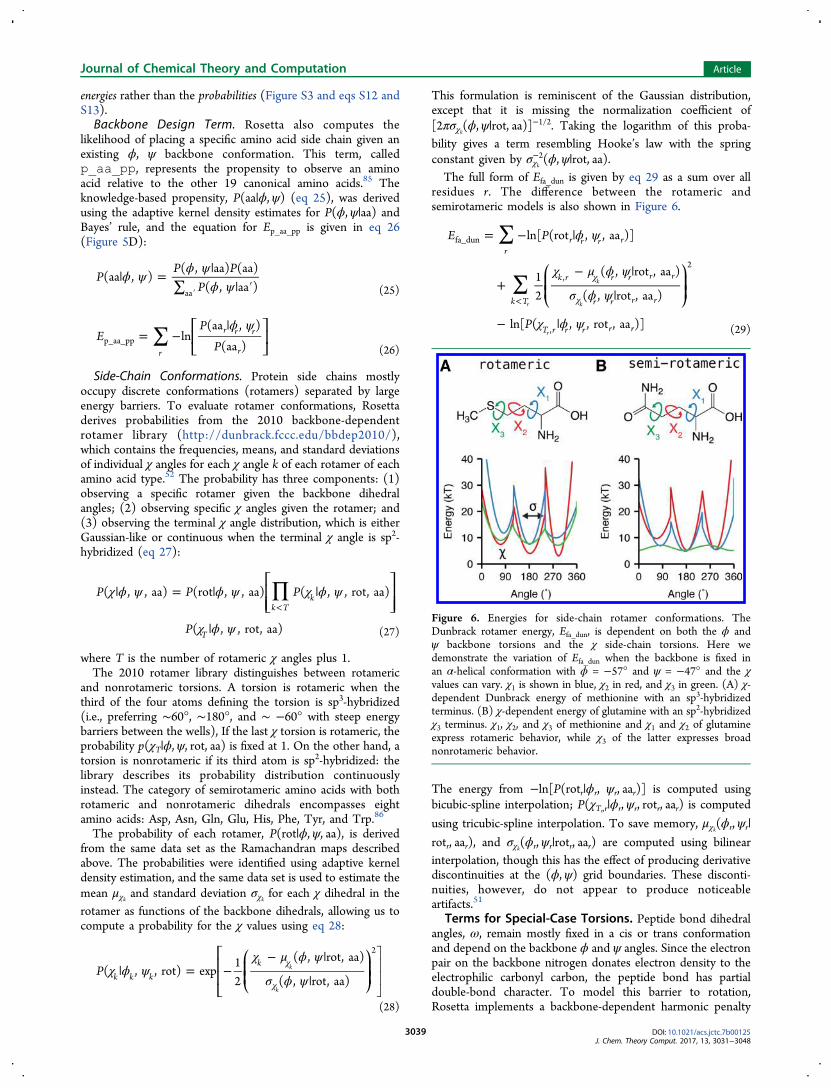
Side-Chain Conformations. Protein side chains mostlyoccupy discrete conformations (rotamers) separated by largeenergy barriers. To evaluate rotamer conformations, Rosettaderives probabilities from the 2010 backbone-dependentrotamer library (http://dunbrack.fccc.edu/bbdep2010/),which contains the frequencies, means, and standard deviationsof individual χ angles for each χ angle k of each rotamer of eachamino acid type.52 The probability has three components: (1)observing a specific rotamer given the backbone dihedralangles; (2) observing specific χ angles given the rotamer; and(3) observing the terminal χ angle distribution, which is eitherGaussian-like or continuous when the terminal χ angle is sp2-hybridized (eq 27):
∏χ ϕ ψ ϕ ψ χ ϕ ψ
χ ϕ ψ
| = | |
|
<
⎡⎣⎢⎢
⎤⎦⎥⎥P P P
P
( , , aa) (rot , , aa) ( , , rot, aa)
( , , rot, aa)
k Tk
T (27)
where T is the number of rotameric χ angles plus 1.The 2010 rotamer library distinguishes between rotameric
and nonrotameric torsions. A torsion is rotameric when thethird of the four atoms defining the torsion is sp3-hybridized(i.e., preferring ∼60°, ∼180°, and ∼ −60° with steep energybarriers between the wells), If the last χ torsion is rotameric, theprobability p(χT|ϕ, ψ, rot, aa) is fixed at 1. On the other hand, atorsion is nonrotameric if its third atom is sp2-hybridized: thelibrary describes its probability distribution continuouslyinstead. The category of semirotameric amino acids with bothrotameric and nonrotameric dihedrals encompasses eightamino acids: Asp, Asn, Gln, Glu, His, Phe, Tyr, and Trp.86
The probability of each rotamer, P(rot|ϕ, ψ, aa), is derivedfrom the same data set as the Ramachandran maps describedabove. The probabilities were identified using adaptive kerneldensity estimation, and the same data set is used to estimate themean μχk and standard deviation σχk for each χ dihedral in therotamer as functions of the backbone dihedrals, allowing us tocompute a probability for the χ values using eq 28:
χ ϕ ψχ μ ϕ ψ
σ ϕ ψ| = −
− |
|χ
χ
⎡
⎣⎢⎢
⎛⎝⎜⎜
⎞⎠⎟⎟
⎤
⎦⎥⎥P( , , rot) exp
12
( , rot, aa)
( , rot, aa)k k kk
2
k
k
(28)
This formulation is reminiscent of the Gaussian distribution,except that it is missing the normalization coefficient of[2πσχk(ϕ, ψ|rot, aa)]
−1/2. Taking the logarithm of this proba-bility gives a term resembling Hooke’s law with the springconstant given by σχk
−2(ϕ, ψ|rot, aa).The full form of Efa_dun is given by eq 29 as a sum over all
residues r. The difference between the rotameric andsemirotameric models is also shown in Figure 6.
∑
∑
ϕ ψ
χ μ ϕ ψ
σ ϕ ψ
χ ϕ ψ
= − |
+− |
|
− |
χ
χ
_
<
⎛⎝⎜⎜
⎞⎠⎟⎟
E P
P
ln[ (rot , , aa )]
12
( , rot , aa )
( , rot , aa )
ln[ ( , , rot , aa )]
rr r r r
k T
k r r r r r
r r r r
T r r r r r
fa dun
,2
,
r
k
k
r (29)
The energy from −ln[P(rotr|ϕr, ψr, aar)] is computed usingbicubic-spline interpolation; P(χTr,r|ϕr, ψr, rotr, aar) is computed
using tricubic-spline interpolation. To save memory, μχk(ϕr, ψr|rotr, aar), and σχk(ϕr, ψr|rotr, aar) are computed using bilinearinterpolation, though this has the effect of producing derivativediscontinuities at the (ϕ, ψ) grid boundaries. These disconti-nuities, however, do not appear to produce noticeableartifacts.51
Terms for Special-Case Torsions. Peptide bond dihedralangles, ω, remain mostly fixed in a cis or trans conformationand depend on the backbone ϕ and ψ angles. Since the electronpair on the backbone nitrogen donates electron density to theelectrophilic carbonyl carbon, the peptide bond has partialdouble-bond character. To model this barrier to rotation,Rosetta implements a backbone-dependent harmonic penalty
Figure 6. Energies for side-chain rotamer conformations. TheDunbrack rotamer energy, Efa_dun, is dependent on both the ϕ andψ backbone torsions and the χ side-chain torsions. Here wedemonstrate the variation of Efa_dun when the backbone is fixed inan α-helical conformation with ϕ = −57° and ψ = −47° and the χvalues can vary. χ1 is shown in blue, χ2 in red, and χ3 in green. (A) χ-dependent Dunbrack energy of methionine with an sp3-hybridizedterminus. (B) χ-dependent energy of glutamine with an sp2-hybridizedχ3 terminus. χ1, χ2, and χ3 of methionine and χ1 and χ2 of glutamineexpress rotameric behavior, while χ3 of the latter expresses broadnonrotameric behavior.
Journal of Chemical Theory and Computation Article
DOI: 10.1021/acs.jctc.7b00125J. Chem. Theory Comput. 2017, 13, 3031−3048
3039

centered near 0° for cis and 180° for trans (Figure 7A). Thisenergy, called omega, is evaluated on all peptide bonds in thebiomolecule (eq 30):
∑π σ ϕ ψ π
ω μ ϕ ψσ ϕ ψ
= −|
+− |
|
ω
ω
ω
⎛⎝⎜
⎞⎠⎟
⎛⎝⎜⎜
⎞⎠⎟⎟E ln
16 2
ln1
( , aa ) 2
[ ( , aa )]
2 ( , aa )
r r r r
r r r r
r r r
omega
2
2(30)
The means μω and standard derivations σω are backbone(ϕ, ψ)-dependent, as given by kernel regressions of ω on ϕ andψ.72
Most Rosetta protocols search over only simple torsionswithin chains and rigid-body degrees of freedom betweenchains. However, the side chain of proline requires specialtreatment because its ring cannot be represented by a kinematictree.87 Therefore, Rosetta implements a proline closure term,called pro_close (Figure 7B). There are two componentsto this energy, as shown in eq 31. First, there is a torsionalpotential that operates on the dihedral formed by Or−1−Cr−1−Nr−Cδ,r (called ωr′) given the observed mean μω′ and standarddeviation σω′, where r is the residue index. This term keeps theCδ atom in the peptide plane. Second, to ensure the correctgeometry for the two hydrogens bound to Cδ, we build a virtualnitrogen atom, Nv, off Cδ whose coordinate is controlled by χ3(Figure 7B). The pro_close term seeks to align the virtualatom Nv directly on top of the real backbone nitrogen. The N−Cδ−Cγ bond angle and the N−Cδ bond length are restrained totheir ideal values.
∑
ω μσ
σ
σ
=
′ −
+|| − ||
‐
|| − ||‐
ω
ω
_
∈
′
′
⎧
⎨
⎪⎪⎪⎪
⎩
⎪⎪⎪⎪
E
r
r
( )
N N
is not the N terminus
N Nis the N terminus
r
r
r r
r r
pro close
Pro
2
2
v,2
N,N2
v,2
N,N2
v
v
(31)
Tyrosine also requires special treatment for its χ3 angle becausethe hydroxyl hydrogen prefers to be in the plane of thearomatic ring.88 To enforce this preference, Rosetta implementsa sinusoidal penalty to model the barrier to a χ3 angle thatdeviates from planarity. This tyrosine hydroxyl penalty is calledyhh_planarity (eq 32 and Figure 7C):
∑ π χ= − +_E12
[cos( 2 ) 1]i
iyhh planarity 3,(32)
Terms for Modeling Nonideal Bond Lengths andAngles. Cartesian Bonding Energy. Recently, modelingCartesian degrees of freedom during gradient-based minimiza-tion has been shown to improve Rosetta’s ability to refine low-resolution structures determined by X-ray crystallography andcryogenic electron microscopy53 as well as its ability todiscriminate near-native conformations in the absence ofexperimental data.89 These data suggest that capturing nonidealbond lengths and angles can be important for accuratemodeling of minimum-energy protein conformations. Toaccommodate, Rosetta now allows these “nonideal” anglesand lengths to be included as additional degrees of freedom inrefinement and includes a Cartesian minimization mode inwhich the atom coordinates are explicit degrees of freedom inoptimization.To evaluate the energetics of nonideal bond lengths, angles,
and planar groups, an energy term called cart_bondedrepresents the deviation of these degrees of freedom from idealusing harmonic potentials (eqs 32−34):
∑= −_=
E k d d12
( )i
n
i i icart length1
length, ,02
(33)
∑ θ θ= −_=
E k12
( )i
m
i i icart angle1
angle, ,02
(34)
∑ ϕ ϕ πρ
= −_=
⎡⎣⎢⎢
⎛⎝⎜⎜
⎞⎠⎟⎟⎤⎦⎥⎥E k f
12
,2
i
l
i i ii
cart torsion1
torsion, wrap ,0
2
(35)
In these equations, di is a bonded-atom-pair distance with di,0 asits ideal distance, θi is a bond angle with θi,0 as its ideal angle,and ϕi is a bond torsion or improper torsion with ϕi,0 as its
Figure 7. Special case torsion energies. (A−C) Rosetta implements three additional energy terms to model torsional degrees of freedom with acutepreferences: (A) omega torsion, corresponding to rotation about C−N; (B) proline secondary omega torsion, corresponding to rotation about C−Nrelated to the Cδ in the ring; (C) tyrosine terminal χ torsion. (D) Omega energy. (E) Proline closure energy. (F) Tyrosine planarity energy.
Journal of Chemical Theory and Computation Article
DOI: 10.1021/acs.jctc.7b00125J. Chem. Theory Comput. 2017, 13, 3031−3048
3040

ideal value and ρi as its periodicity. The ideal bond lengths andangles90,91 were selected on the basis of their ability to rebuildside chains observed in crystal structures (Kevin Karplus andJames J. Havranek, unpublished); they were subsequentlymodified empirically.51 The spring constants for the angle andlength terms are from CHARMM32.19 Finally, all planar groupsand the Cβ “pseudotorsion” are constrained using empiricallyderived values and spring constants:The function fwrap(x, y) wraps x to the range [0, y). To avoid
double counting in the case of Ecart_torsion, the spring constantktorsion,i is zero when the torsion ϕi is being scored by either therama or fa_dun terms.Terms for Protein Design. Design Reference Energy. The
terms above are sufficient for comparing different proteinconformations with a fixed sequence. However, protein designsimulations compare the relative stability of different aminoacid sequences given a desired structure to identify models thatexhibit a large free energy gap between the folded and unfoldedstates. Explicit calculations of unfolded-state free energies arecomputationally expensive and error-prone. Rosetta thereforeapproximates the relative energies of the unfolded-stateensembles using an unfolded-state reference energy called ref.Rosetta calculates the reference energy as a sum of individual
constant unfolded-state reference energies, ΔGiref, for each
amino acid, aai (eq 36):1
∑= ΔE G (aa )i
i irefref
(36)
The ΔGiref values are empirically optimized by searching for
values that maximize native sequence recovery (discussedbelow) during design simulations on a large set of high-resolution crystal structures.50,51 During design, this energyterm helps normalize the observed frequencies of the differentamino acids. When design is turned off, the term contributes aconstant offset for a fixed sequence.Bringing the Energy Terms Together. The Rosetta
energy function combines all of the terms using a weightedlinear sum to approximate free energies (Table 1). Historically,we have adjusted the weights and parameters to balance theenergetic contributions from the various terms. This balance isimportant because the van der Waals, solvation, and electro-statics energies partially capture torsional preferences andoverlap can cause errors as a result of double counting ofatomic or residue-specific contributions.92 More recently, wehave fixed the physics-based terms with weights of 1.0 andperturbed the other weights and atomic-level parameters usinga Nelder−Mead scheme93 to optimize the agreement of Rosettacalculations with small-molecule thermodynamic data and high-resolution structural features.50 The energy function parametershave evolved over the years by optimization of the performanceof multiple scientific benchmarks (Table 2).50,51,94 Thesebenchmarks were chosen to test the recovery of native-likestructural features, ranging from individual hydrogen-bondgeometries to thermodynamic properties and interfaceconformations. In addition, and more recently, Song et al.,95
Conway and DiMaio,96 and O’Meara et al.49 have fit intratermparameters to recover features of the experimentallydetermined folded conformations. An in-depth review ofenergy function benchmarking can be found in Leaver-Fay etal.51 Table S3 lists the Rosetta database files containing thecurrent full set of physical parameters for each score term.Energy Function Units. Initially, Rosetta energies were
expressed in a generic unit called the Rosetta energy unit
(REU). This choice was made because some original Rosettaenergy terms were not calibrated with experimental data, andthe use of statistical potentials convoluted interpretation of theenergy. Over time the physical meaning of Rosetta energies hasbeen extensively debated within and outside the community,and several steps have been taken to clarify the interpretation.The most recent energy function (REF15) was parametrized onhigh-resolution protein structures and small-molecule thermo-dynamic parameters that were measured in kilocalories permole.50 The optimization data show a strong correlationbetween the experimental data and values predicted by Rosetta(ΔΔG upon mutation, R = 0.994; small-molecule ΔHvap, FigureS1). As a result, Rosetta energies are now a strongerapproximation of energies in units of kilocalories per mole.Therefore, as is standard practice for molecular force fields suchas OPLS, CHARMM, and AMBER, we now also expressenergies in kilocalories per mole.
■ ENERGIES IN ACTION: USING INDIVIDUAL ENERGYTERMS TO ANALYZE ROSETTA MODELS
Rosetta energy terms are mathematical models of the physicsthat governs protein structure, stability, and association.Therefore, the decomposed relative energies of a structure orensemble of structures can expose important details about thebiomolecular model. Now that we have presented the details ofeach energy term, we here demonstrate how these energies canbe applied to detailed interpretations of structural models. Inthis section, we discuss two common structure calculations: (1)estimating the free energy change (ΔΔG) of mutation97 and(2) modeling the structure of a protein−protein interface.101
ΔΔG of Mutation. The first example demonstrates howRosetta can be used to estimate and rationalize thermodynamicparameters. Here we present an example ΔΔG of mutationcalculation for the T193V mutation in the RT-RH-derivedpeptide bound to HIV-1 protease (PDB entry 1kjg; Figure8A).104 The details of this calculation are provided in theSupporting Information.Rosetta calculates the ΔΔG of the T193V mutation to be
−4.95 kcal/mol, and the experimentally measured value is−1.11 kcal/mol.104 Both the experiment and calculation revealthat T193V is stabilizing, yet these numbers alone do not revealwhich specific interactions are responsible for the stabilization.To investigate, we used various analysis tools accessible inPyRosetta105 to identify important energetic contributions to
Table 2. Common Energy Function Benchmarking Methods
test description ref(s)
sequence recovery percentage of the native sequence recoveredafter backbone redesign
1, 51
rotamer recovery percentage of native rotamers recoveredafter full repacking
51
ΔΔG prediction prediction of free energy changes uponmutation
97
loop modeling prediction of loop conformations 98
high-resolutionrefinement
discrimination of native-like decoys uponrefinement of ab initio protein models
99
docking prediction of protein−protein, protein−peptide, or protein−ligand interfaces
44, 100−102
homologymodeling
structure prediction incorporatinghomologous information from templates
103
thermodynamicproperties
recapitulation of thermodynamic propertiesof protein side-chain analogues
17
recapitulation ofcrystal structuregeometries
recapitulation of features (e.g., atom-pairdistance distribution) from high-resolutionprotein crystal structures
50
Journal of Chemical Theory and Computation Article
DOI: 10.1021/acs.jctc.7b00125J. Chem. Theory Comput. 2017, 13, 3031−3048
3041

the total ΔΔG. First, we decomposed the ΔΔG into individualenergy terms and observed the balance of terms, both favorableand unfavorable, that sum to the total (Figure 8B). Todecompose the most favorable term, Δfa_sol, we used theprint_residue_pair_energies function to identifyresidues that interact with the mutation site (in this case,residue 4) to produce a nonzero residue-pair solvation energy.With the resulting table, we found that a hydrophobic pocketaround the mutation site formed by residues V27, I45, G46,and I80 on HIV peptidase and residue F194 on the peptidemade a large (>0.05 kcal/mol) and favorable contribution tothe change in solvation energy (Figure 8C).We further investigated this result on the atomic level with
the function print_atom_pair_energy_table bygenerating atom-pair energy tables (see the SupportingInformation) for residues 5, 27, 45, 46, and 80 against boththreonine and valine at residue 193 (an example for residue 80is shown in Table 3). Here we find that the specific substitution
of the polar hydroxyl group on threonine with the nonpolaralkyl group on valine stabilizes the peptide in the hydrophobicprotease pocket. This result is consistent with chemicalintuition and demonstrates how breaking down the totalenergies can provide insight into characteristics of the mutatedstructures.Protein−Protein Docking. The second example shows
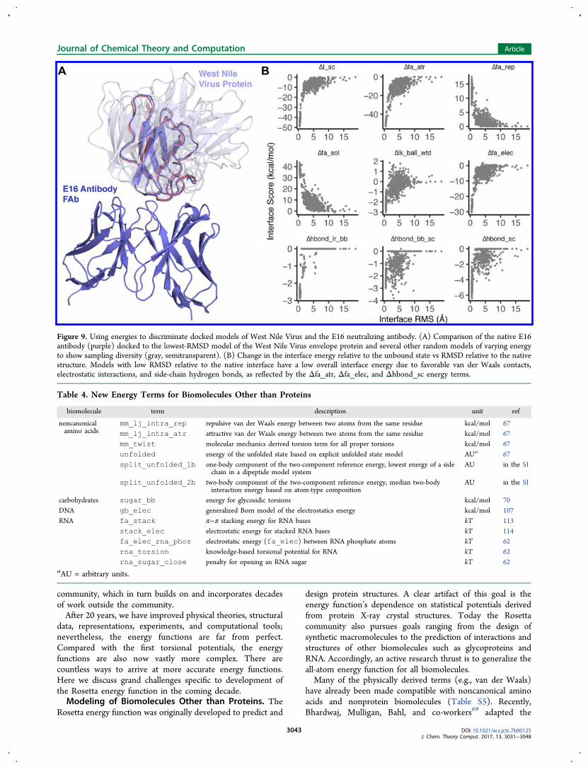
how the Rosetta energies of an ensemble of models can be usedto discriminate between models and investigate the character-istics of a protein−protein interface. Below we investigatedocked models of West Nile Virus envelope protein and a
neutralizing antibody (PDB entry 1ztx; Figure 9A).106
Calculation details can be found in the Supporting Information.To evaluate the docked models, we examine the variation of
the energies as a function of the root-mean-square deviation(RMSD) between the residues at the interface in each modeland the known structure. For our calculation, interface residuesare residues with a Cβ atom less than 8.0 Å away from the Cβ ofa residue in the other docking partner. The plot of energiesagainst RMSD values is called a funnel plot and is intended tomimic the funnel-like energy landscape of protein folding andbinding.As in the previous example, we decompose the energies to
yield information about the nature of interactions at theinterface. Here we observe significant changes in the followingenergy terms upon interface formation relative to the unboundstate: fa_atr, fa_rep, fa_sol, lk_ball_wtd,fa_elec , hbond_lr_bb , hbond_bb_sc , andhbond_sc (Figure 9B). The change in the Lennard-Jonesenergy upon interface formation is due to the introduction ofatom−atom contacts at the interface. As more atoms come intocontact near the native conformation (RMSD → 0), thefavorable attractive energy (fa_atr) decreases whereas theunfavorable repulsive energy (fa_rep) increases. The changein the isotropic solvation energy (Δfa_sol) is positive(unfavorable), indicating that polar residues are buried uponinterface formation. Balancing the desolvation penalty, thechanges in polar solvation energy (Δlk_ball_wtd) andelectrostatics (Δfa_elec) are negative because of theformation of polar contacts at the interface. Finally, thechanges in the three hydrogen-bonding energies(Δhbond_lr_bb, Δhbond_bb_sc, and Δhbond_sc)reflect the formation of backbone−backbone, backbone−side-chain, and side-chain−side-chain hydrogen bonds at theinterface.
■ DISCUSSIONThe Rosetta energy function represents our collaboration’songoing pursuit to model the rules in nature that governbiomolecular structure, stability, and association. This papersummarizes the latest version, which brings togetherfundamental physical theories, statistical-mechanical models,and observations of protein structures. This work representsalmost 20 years of interdisciplinary collaboration in the Rosetta
Figure 8. Structural model of the HIV-1 protease bound to the T193V mutant of RT-RH-derived peptide. (A) Structural model of the native HIV-1peptidase (teal and dark blue) bound to the native peptide (gray) superimposed onto the T193V mutant peptide (magenta). (B) Contributionsgreater than ±0.1 kcal/mol to the ΔΔG of mutation for T193V. The remaining contributions are dslf_fa13 = 0 kcal/mol, hbond_lr_bb =−0.09 kcal/mol, hbond_bb_sc = −0.05, hbond_sc = −0.0104, fa_intra_rep = 0.01, fa_intra_sol = −0.07, andyhh_planarity = 0. (C) Hydrophobic patch of residues surrounding position 193 on the RT-RH-derived peptide.
Table 3. Changes in Atom-Pair Energies (in kcal/mol)between I80 and T193 versus V193
I80 atoms
T193→V193 atoms CB CG1 CG2 CD1
N 0.000 0.000 0.000 0.000CA 0.000 0.000 0.000 0.004C 0.000 0.000 0.000 0.008O 0.000 0.000 0.000 −0.010CB 0.000 0.054 0.000 −0.002OG1 → CG1 0.008 −0.054 −0.316 −0.398CG2 → CG2′ 0.000 0.000 0.001 0.020
Journal of Chemical Theory and Computation Article
DOI: 10.1021/acs.jctc.7b00125J. Chem. Theory Comput. 2017, 13, 3031−3048
3042
community, which in turn builds on and incorporates decadesof work outside the community.After 20 years, we have improved physical theories, structural
data, representations, experiments, and computational tools;nevertheless, the energy functions are far from perfect.Compared with the first torsional potentials, the energyfunctions are also now vastly more complex. There arecountless ways to arrive at more accurate energy functions.Here we discuss grand challenges specific to development ofthe Rosetta energy function in the coming decade.Modeling of Biomolecules Other than Proteins. The
Rosetta energy function was originally developed to predict and
design protein structures. A clear artifact of this goal is theenergy function’s dependence on statistical potentials derivedfrom protein X-ray crystal structures. Today the Rosettacommunity also pursues goals ranging from the design ofsynthetic macromolecules to the prediction of interactions andstructures of other biomolecules such as glycoproteins andRNA. Accordingly, an active research thrust is to generalize theall-atom energy function for all biomolecules.Many of the physically derived terms (e.g., van der Waals)
have already been made compatible with noncanonical aminoacids and nonprotein biomolecules (Table S5). Recently,Bhardwaj, Mulligan, Bahl, and co-workers69 adapted the
Figure 9. Using energies to discriminate docked models of West Nile Virus and the E16 neutralizing antibody. (A) Comparison of the native E16antibody (purple) docked to the lowest-RMSD model of the West Nile Virus envelope protein and several other random models of varying energyto show sampling diversity (gray, semitransparent). (B) Change in the interface energy relative to the unbound state vs RMSD relative to the nativestructure. Models with low RMSD relative to the native interface have a low overall interface energy due to favorable van der Waals contacts,electrostatic interactions, and side-chain hydrogen bonds, as reflected by the Δfa_atr, Δfa_elec, and Δhbond_sc energy terms.
Table 4. New Energy Terms for Biomolecules Other than Proteins
biomolecule term description unit ref
noncanonicalamino acids
mm_lj_intra_rep repulsive van der Waals energy between two atoms from the same residue kcal/mol 67mm_lj_intra_atr attractive van der Waals energy between two atoms from the same residue kcal/mol 67mm_twist molecular mechanics derived torsion term for all proper torsions kcal/mol 67unfolded energy of the unfolded state based on explicit unfolded state model AUa 67split_unfolded_1b one-body component of the two-component reference energy, lowest energy of a side
chain in a dipeptide model systemAU in the SI
split_unfolded_2b two-body component of the two-component reference energy, median two-bodyinteraction energy based on atom-type composition
AU in the SI
carbohydrates sugar_bb energy for glycosidic torsions kcal/mol 70DNA gb_elec generalized Born model of the electrostatics energy kcal/mol 107RNA fa_stack π−π stacking energy for RNA bases kT 113
stack_elec electrostatic energy for stacked RNA bases kT 114fa_elec_rna_phos electrostatic energy (fa_elec) between RNA phosphate atoms kT 62rna_torsion knowledge-based torsional potential for RNA kT 62rna_sugar_close penalty for opening an RNA sugar kT 62
aAU = arbitrary units.
Journal of Chemical Theory and Computation Article
DOI: 10.1021/acs.jctc.7b00125J. Chem. Theory Comput. 2017, 13, 3031−3048
3043

rama_prepro, p_aa_pp, fa_dun, pro_close,omega, dslf_fa13, yhh_planarity, and ref termsto be compatible with mixed-chirality peptides. Several ofRosetta’s statistical potentials have been validated againstquantum-mechanical calculations to evaluate non-proteinmodels (Table 4). Early work by Meiler and Baker44 onRosetta Ligand introduced new atom and residue types fornon-protein residues. The first non-protein energy terms wereadded by Havranek et al.107 and Chen et al.,108 who modifiedthe hydrogen-bonding potential to capture planar hydrogenbonds between protein side chains and nucleic acid bases.Renfrew and co-workers67,109 added molecular mechanicstorsions and Lennard-Jones terms to model proteins withnoncanonical amino acids, oligosaccharides, β-peptides, andoligopeptoids.68 Labonte et al.70 implemented Woods’CarboHydrate-Intrinsic (CHI) function,110,111 which evaluatesglycan geometries given the axial−equatorial character of thebonds. Das and co-workers added a set of terms to modelWatson−Crick base pairing, π−π interactions in base stacking,and torsional potentials important for predicting and designingRNA structures.62,112−114 Bazzoli and Karanicolas115 recentlydeveloped a new polar solvation model that evaluates thepenalty associated with displacing waters in the first solvationshell. In addition, Combs116 tested a small-molecule force fieldbased on electron orbital models. Many of these terms arepresented in detail in the Supporting Information.Expanding Rosetta’s chemical library brings new challenges.
Currently there are separate energy functions for various typesof biomolecules. Typically, these functions mix physicallyderived terms from the protein energy function with molecule-specific statistical potentials, custom weights, and possiblycustom atomic parameters. If nature uses only one energyfunction, why do we need so many? Some discrepancies mayresult from features that we do not model explicitly, such asπ−π, n−π*, and cation−π interactions. Efforts to converge on asingle energy function will therefore pose interesting questionsabout the set of universal physical determinants of biomolecularstructure.Capturing the Intra- and Extracellular Environments.
Rosetta traditionally models the solvent surrounding theprotein using the Lazaridis−Karplus (LK) model, whichassumes a solvent environment made of pure water. In contrast,biology operates under various conditions influenced by pH,redox potential, temperature, solvent viscosity, chaotropes,kosmotropes, and polarizability. Therefore, modeling moredetails of the intra- and extracellular environments wouldenable Rosetta to identify structures that are important indifferent biological contexts.Rosetta currently includes two groups of energy terms to
model alternate environments (Table 5). Kilambi and Gray117
implemented a method to account for pH by including a termcalled e_pH that calculates the likelihood of a protein sidechain’s protonation state given a user-specified pH; it requiresthe inclusion of both protonated and deprotonated side chainsduring side-chain rotamer packing. This model can predict pKa
values with an RMS error under 1 unit,117 and it improvesprotein−protein docking, especially under acidic or basicconditions.60 The accuracy of this model is limited by thedistance-dependent Coulomb approximation and sensitivity tofine backbone rearrangements.In addition, Rosetta implements Lazaridis’ implicit mem-
brane model (IMM) for modeling proteins in a lipid bilayerenviornment.36,118,119 The IMM terms provide a fastapproximation of the nonpolar hydrocarbon core of the lipidbilayer and have been successfully applied to membrane proteinfolding,120 docking, and early design tasks.61 This continuummodel has a fixed thickness, omitting the detailed chemistry atthe membrane interface and any dynamic bilayer rearrange-ments.
The Origin of Energy Models: Top-Down versusBottom-Up Development. Traditionally, energy functionsare developed using a bottom-up approach: experimentalobservables serve as building blocks to parametrize physics-based formulas. The advent of powerful optimizationtechniques and artificial intelligence has recently empoweredthe top-down category, where numerical methods are used toderive models and/or parameters. Top-down approaches havebeen used to solve problems in various fields, includingstructural biology and bioinformatics. Recently, top-downdevelopment was also applied to optimize the Lennard-Jones,LK, and Coulomb parameters in the Rosetta energy function(Tables S4−S6).50,93Top-down approaches have enormous potential to improve
the accuracy of biomolecular modeling because moreparameters can vary and the objective function can beminimized with more benchmarks. These approaches alsointroduce new challenges. With any computer-derived modelthere is a risk of overfitting, as validation via structure-prediction data sets reflect observable states, whereassimulations are intended to predict features of states thatexperiments cannot yet observe. Computer-derived parametersalso introduce a unique kind of uncertainty. Consider thefollowing scenario: the performance of scientific benchmarksimproves as physical atomic parameters are perturbed awayfrom the measured experimental values. As there is less physicalbasis for the parameters, are the predictions and interpretationsstill meaningful?Top-down development will also provide power to develop
more complicated energy functions. Currently, the Rosettaenergy function advances by incrementally addressing weak-nesses: with each new paper, we modify analytic formulas, addcorrective terms, and adjust weights. As this paper demon-strates, the energy function is significantly more complicatedthan the initial theoretical forms. Given this complexityincrease, an interesting approach to leverage the power oftop-down development would be to simplify and subtract termsto evaluate their individual benefits.
A Highly Interdisciplinary Endeavor. The Rosettaenergy function has advanced rapidly because of the RosettaCommunity, a highly interdisciplinary collaboration among
Table 5. Energy Terms for Structure Prediction in Different Contexts
context term description unit ref(s)
membraneenvironment
fa_mpsolv solvation energy dependent on the protein orientation relative to the membrane kcal/mol 118, 121fa_mpenv one-body membrane environment energy dependent on the protein orientation relative to the
membranekcal/mol 118, 121
pH e_pH likelihood of side-chain protonation given a user-specified pH kcal/mol 117
Journal of Chemical Theory and Computation Article
DOI: 10.1021/acs.jctc.7b00125J. Chem. Theory Comput. 2017, 13, 3031−3048
3044
scientists with diverse backgrounds located in over 50laboratories around the world. The many facets of our teamenable us to probe different aspects of the energy function. Forexample, expert computer scientists and applied mathemati-cians have implemented algorithms to speed up calculations.Dedicated software engineers maintain the code and maintain aplatform for scientific benchmark testing. Physicists andchemists develop new energy terms that better model thephysical rules found in nature. Structural biologists maintain afocus on created biological features and functions. We lookforward to leveraging this powerful interdisciplinary scientificteam as we head into the next decade of energy functionadvances.
■ CONCLUSION: A LIVING ENERGY FUNCTIONFor the first time since 2004,48 we have documented all of themathematical and physical details of the Rosetta all-atomenergy function, highlighting the latest upgrades to both theunderlying science and the speed of calculations. In addition,we have illustrated how the energies can be used to analyzeoutput models from Rosetta simulations. These advances haveenabled Rosetta’s achievements in biomolecular structureprediction and design over the past 15 years. Still, the energyfunction is far from complete and will continue to evolve longafter this publication. Thus, we hope that this document willserve as an important resource for understanding thefoundational physical and mathematical concepts in the energyfunction. Furthermore, we hope to encourage both current andfuture Rosetta developers and users to understand the strengthsand shortcomings of the energy function as it applies to thescientific questions they are trying to answer.
■ ASSOCIATED CONTENT*S Supporting InformationThe Supporting Information is available free of charge on theACS Publications website at DOI: 10.1021/acs.jctc.7b00125.
Description of changes to the Rosetta energy functionsince 2000; data describing the calibration of Rosettaenergies in kcal/mol; additional details of energy termsand details on smoothing of statistical terms; energyterms for D-amino acids, noncanonical amino acids,carbohydrates, and nucleic acids; and methods describingexample energy calculations (PDF)Protocol capture within an interactive Python notebookdemonstrating the usage of the print_atom_-pair_energy_table function (ZIP)
■ AUTHOR INFORMATIONCorresponding Author*E-mail: [email protected] F. Alford: 0000-0003-0306-8476Hahnbeom Park: 0000-0002-7129-1912Michael S. Pacella: 0000-0001-8919-147XJeffrey J. Gray: 0000-0001-6380-2324Author ContributionsR.F.A., J.R.J., A.L.-F., T.K., B.K., and J.J.G. wrote themanuscript. R.F.A., J.R.J., M.S.P., and J.J.G. generated analysisscripts and examples. A.L.-F., M.J.O., F.P.D., H.P., M.V.S., P.B.,R.L.D., T.K., D.B., B.K., and J.J.G. wrote, verified, and/orcontributed figures on protein energy terms. P.D.R., K.K.,
V.K.M., J.W.L., R.B., and R.D. wrote, verified, and/orcontributed figures on non-protein energy terms.
FundingR.F.A. was funded by a Hertz Foundation Fellowship and anNSF Graduate Research Fellowship. J.R.J. and J.J.G. werefunded by NIH GM-078221. A.L.-F., J.J.G., and B.K. werefunded by NIH GM-73141. M.J.O. was funded by NSF GM-114961. P.D.R. and R.B. were funded by the SimonsFoundation. M.V.S. and R.L.D. were funded by NIH GM-084453 and NIH GM-111819. M.S.P. was funded by NSFBMAT 1507736. J.W.L. was funded by NIH F32-CA189246.K.K. was funded by an NSF Graduate Research Fellowship andan SGF Galiban Fellowship. D.B., H.P., and V.K.M. werefunded by NIH GM-092802. T.K. was funded by NIH GM-110089 and GM-117189.
NotesThe authors declare the following competing financialinterest(s): Drs. Gray, Baker, Bonneau, Kuhlman, Kortemme,and Bradley are unpaid members of the Executive Board of theRosetta Commons. Under institutional participation agree-ments between the University of Washington, acting on behalfof the Rosetta Commons, and each institution participating inthis article, each institution may be entitled to a portion ofrevenue received on licensing of software described here.
■ ACKNOWLEDGMENTS
Development of the Rosetta energy function would not bepossible without the entire Rosetta Commons collaboration: acommunity of scientists, engineers, and software developersthat have worked together for almost 20 years. We estimate thathundreds of scientists from the 50 institutions in the RosettaCommons have made minor and major contributions to theadvancement of the all-atom energy function. When writing thispaper, it was impossible to compile a complete list of energyfunction contributors. Instead, our author list reflects a smallsubset of the historical contributors who provided text, figures,and expertise needed to write a comprehensive, complete, andaccurate description of the current energy function formulation.We also want to give special recognition to the early pioneers ofthe Rosetta energy function who are not coauthors: Carol Rohl,Kim Simons, Charlie Strauss, Ingo Ruczinski, William Sheffler,Jens Meiler, Ora Schuler-Furman, James Havranek, and IanDavis. We also acknowledge individuals that contributed toassembling the manuscript. We thank Sergey Lyskov fordevelopment of the benchmark server that enables continuousand transparent energy function testing and Morgan Nance,Henry Lessen, and Rocco Moretti for helpful comments on themanuscript.
■ REFERENCES(1) Kuhlman, B.; Baker, D. Native Protein Sequences Are close toOptimal for Their Structures. Proc. Natl. Acad. Sci. U. S. A. 2000, 97(19), 10383−10388.(2) Richardson, J. S. The Anatomy and Taxonomy of ProteinStructure. Adv. Protein Chem. 1981, 34, 167−339.(3) Leaver-Fay, A.; Tyka, M.; Lewis, S. M.; Lange, O. F.; Thompson,J.; Jacak, R.; Kaufman, K. W.; Renfrew, P. D.; Smith, C. A.; Sheffler,W.; Davis, I. W.; Cooper, S.; Treuille, A.; Mandell, D. J.; Richter, F.;Ban, Y.-E. A.; Fleishman, S. J.; Corn, J. E.; Kim, D. E.; Lyskov, S.;Berrondo, M.; Mentzer, S.; Popovic, Z.; Havranek, J. J.; Karanicolas, J.;Das, R.; Meiler, J.; Kortemme, T.; Gray, J. J.; Kuhlman, B.; Baker, D.;Bradley, P. Rosetta3: An Object-Oriented Software Suite for the
Journal of Chemical Theory and Computation Article
DOI: 10.1021/acs.jctc.7b00125J. Chem. Theory Comput. 2017, 13, 3031−3048
3045

Simulation and Design of Macromolecules. Methods Enzymol. 2011,487, 545−574.(4) Anfinsen, C. B. Principles That Govern the Folding of ProteinChains. Science 1973, 181 (4096), 223−230.(5) Jones, J. E. On the Determination of Molecular Fields.I. Fromthe Variation of the Viscosity of a Gas with Temperature. Proc. R. Soc.London, Ser. A 1924, 106, 441−462.(6) Jones, J. E. On the Determination of Molecular Fields.II. Fromthe Equation of State of a Gas. Proc. R. Soc. London, Ser. A 1924, 106,463−477.(7) Levitt, M.; Lifson, S. Refinement of Protein Conformations Usinga Macromolecular Energy Minimization Procedure. J. Mol. Biol. 1969,46 (2), 269−279.(8) Urey, H. C.; Bradley, C. A. The Vibrations of PentatonicTetrahedral Molecules. Phys. Rev. 1931, 38 (11), 1969−1978.(9) Westheimer, F. Calculation of the Magnitude of Steric Effects. InSteric Effects in Organic Chemistry; Newman, M. S., Ed.; Wiley: NewYork, 1956; pp 523−555.(10) Lifson, S.; Warshel, A. Consistent Force Field for Calculationsof Conformations, Vibrational Spectra, and Enthalpies of Cycloalkaneand N-Alkane Molecules. J. Chem. Phys. 1968, 49 (11), 5116−5129.(11) Warshel, A.; Lifson, S. Consistent Force Field Calculations. II.Crystal Structures, Sublimation Energies, Molecular and LatticeVibrations, Molecular Conformations, and Enthalpies of Alkanes. J.Chem. Phys. 1970, 53 (2), 582−594.(12) Levitt, M. Energy Refinement of Hen Egg-White Lysozyme. J.Mol. Biol. 1974, 82 (3), 393−420.(13) Gelin, B. R.; Karplus, M. Sidechain Torsional Potentials andMotion of Amino Acids in Porteins: Bovine Pancreatic TrypsinInhibitor. Proc. Natl. Acad. Sci. U. S. A. 1975, 72 (6), 2002−2006.(14) Levinthal, C.; Wodak, S. J.; Kahn, P.; Dadivanian, A. K.Hemoglobin Interaction in Sickle Cell Fibers. I: TheoreticalApproaches to the Molecular Contacts. Proc. Natl. Acad. Sci. U. S. A.1975, 72 (4), 1330−1334.(15) Cornell, W. D.; Cieplak, P.; Bayly, C. I.; Gould, I. R.; Merz, K.M.; Ferguson, D. M.; Spellmeyer, D. C.; Fox, T.; Caldwell, J. W.;Kollman, P. A. A Second Generation Force Field for the Simulation ofProteins, Nucleic Acids, and Organic Molecules. J. Am. Chem. Soc.1996, 118 (9), 2309−2309.(16) Mayo, S. L.; Olafson, B. D.; Goddard, W. A. DREIDING: AGeneric Force Field for Molecular Simulations. J. Phys. Chem. 1990, 94(26), 8897−8909.(17) Jorgensen, W. L.; Tirado-Rives, J. The OPLS [OptimizedPotentials for Liquid Simulations] Potential Functions for Proteins,Energy Minimizations for Crystals of Cyclic Peptides and Crambin. J.Am. Chem. Soc. 1988, 110 (6), 1657−1666.(18) Brooks, B. R.; Bruccoleri, R. E.; Olafson, B. D.; States, D. J.;Swaminathan, S.; Karplus, M. CHARMM: A Program for Macro-molecular Energy, Minimization, and Dynamics Calculations. J.Comput. Chem. 1983, 4 (2), 187−217.(19) Brooks, B. R.; Brooks, C. L.; Mackerell, A. D.; Nilsson, L.;Petrella, R. J.; Roux, B.; Won, Y.; Archontis, G.; Bartels, C.; Boresch,S.; Caflisch, A.; Caves, L.; Cui, Q.; Dinner, A. R.; Feig, M.; Fischer, S.;Gao, J.; Hodoscek, M.; Im, W.; Kuczera, K.; Lazaridis, T.; Ma, J.;Ovchinnikov, V.; Paci, E.; Pastor, R. W.; Post, C. B.; Pu, J. Z.; Schaefer,M.; Tidor, B.; Venable, R. M.; Woodcock, H. L.; Wu, X.; Yang, W.;York, D. M.; Karplus, M. CHARMM: The Biomolecular SimulationProgram. J. Comput. Chem. 2009, 30 (10), 1545−1614.(20) Sun, H. COMPASS: An Ab Initio Force-Field Optimized forCondensed-Phase Applications Overview with Details on Alkane andBenzene Compounds. J. Phys. Chem. B 1998, 102 (38), 7338−7364.(21) Tanaka, S.; Scheraga, H. A. Model of Protein Folding: Inclusionof Short-, Medium-, and Long-Range Interactions. Proc. Natl. Acad. Sci.U. S. A. 1975, 72 (10), 3802−3806.(22) Tanaka, S.; Scheraga, H. A. Model of Protein Folding:Incorporation of a One-Dimensional Short-Range (Ising) Modelinto a Three-Dimensional Model. Proc. Natl. Acad. Sci. U. S. A. 1977,74 (4), 1320−1323.
(23) Miyazawa, S.; Jernigan, R. L. Residue-Residue Potentials with aFavorable Contact Pair Term and an Unfavorable High PackingDensity Term, for Simulation and Threading. J. Mol. Biol. 1996, 256(3), 623−644.(24) Wilmanns, M.; Eisenberg, D. Three-Dimensional Profiles fromResidue-Pair Preferences: Identification of Sequences with Beta/alpha-Barrel Fold. Proc. Natl. Acad. Sci. U. S. A. 1993, 90 (4), 1379−1383.(25) Jones, D. T.; Taylor, W. R.; Thornton, J. M. A New Approach toProtein Fold Recognition. Nature 1992, 358 (6381), 86−89.(26) Bowie, J. U.; Luthy, R.; Eisenberg, D. A Method to IdentifyProtein Sequences That Fold into a Known Three-DimensionalStructure. Science 1991, 253 (5016), 164−170.(27) Sippl, M. J. Calculation of Conformational Ensembles fromPotentials of Mean Force. An Approach to the Knowledge-BasedPrediction of Local Structures in Globular Proteins. J. Mol. Biol. 1990,213 (4), 859−883.(28) Skolnick, J.; Kolinski, A. Simulations of the Folding of aGlobular Protein. Science 1990, 250 (4984), 1121−1125.(29) Bashford, D.; Case, D. A. Generalized Born Models ofMacromolecular Solvation Effects. Annu. Rev. Phys. Chem. 2000, 51(1), 129−152.(30) Warshel, A.; Kato, M.; Pisliakov, A. Polarizable Force Fields:History, Test Cases, and Prospects. J. Chem. Theory Comput. 2007, 3(6), 2034−2045.(31) Simons, K. T.; Kooperberg, C.; Huang, E.; Baker, D. Assemblyof Protein Tertiary Structures from Fragments with Similar LocalSequences Using Simulated Annealing and Bayesian ScoringFunctions. J. Mol. Biol. 1997, 268 (1), 209−225.(32) Berman, H. M.; Westbrook, J.; Feng, Z.; Gilliland, G.; Bhat, T.N.; Weissig, H.; Shindyalov, I. N.; Bourne, P. E. The Protein DataBank. Nucleic Acids Res. 2000, 28 (1), 235−242.(33) Simons, K. T.; Ruczinski, I.; Kooperberg, C.; Fox, B. A.;Bystroff, C.; Baker, D. Improved Recognition of Native-like ProteinStructures Using a Combination of Sequence-Dependent andSequence-Independent Features of Proteins. Proteins: Struct., Funct.,Genet. 1999, 34 (1), 82−95.(34) Kuhlman, B.; Baker, D. Native Protein Sequences Are close toOptimal for Their Structures. Proc. Natl. Acad. Sci. U. S. A. 2000, 97(19), 10383−10388.(35) Neria, E.; Fischer, S.; Karplus, M. Simulation of Activation FreeEnergies in Molecular Systems. J. Chem. Phys. 1996, 105 (5), 1902−1921.(36) Lazaridis, T.; Karplus, M. Effective Energy Function for Proteinsin Solution. Proteins: Struct., Funct., Genet. 1999, 35 (2), 133−152.(37) Dunbrack, R. L., Jr.; Cohen, F. E. Bayesian Statistical Analysis ofProtein Side-Chain Rotamer Preferences. Protein Sci. 1997, 6 (8),1661−1681.(38) Kortemme, T.; Morozov, A. V.; Baker, D. An Orientation-Dependent Hydrogen Bonding Potential Improves Prediction ofSpecificity and Structure for Proteins and Protein-Protein Complexes.J. Mol. Biol. 2003, 326 (4), 1239−1259.(39) Morozov, A. V.; Kortemme, T.; Tsemekhman, K.; Baker, D.Close Agreement between the Orientation Dependence of HydrogenBonds Observed in Protein Structures and Quantum MechanicalCalculations. Proc. Natl. Acad. Sci. U. S. A. 2004, 101 (18), 6946−6951.(40) Bradley, P.; Misura, K. M. S.; Baker, D. Toward High-Resolutionde Novo Structure Prediction for Small Proteins. Science 2005, 309(5742), 1868−1871.(41) Kortemme, T.; Baker, D. A Simple Physical Model for BindingEnergy Hot Spots in Protein-Protein Complexes. Proc. Natl. Acad. Sci.U. S. A. 2002, 99 (22), 14116−14121.(42) Kortemme, T.; Kim, D. E.; Baker, D. Computational AlanineScanning of Protein−Protein Interfaces. Sci. STKE 2004, 2004 (219),pl2.(43) Gray, J. J.; Moughon, S.; Wang, C.; Schueler-Furman, O.;Kuhlman, B.; Rohl, C. A.; Baker, D. Protein-Protein Docking withSimultaneous Optimization of Rigid-Body Displacement and Side-Chain Conformations. J. Mol. Biol. 2003, 331 (1), 281−299.
Journal of Chemical Theory and Computation Article
DOI: 10.1021/acs.jctc.7b00125J. Chem. Theory Comput. 2017, 13, 3031−3048
3046
(44) Meiler, J.; Baker, D. ROSETTALIGAND: Protein-SmallMolecule Docking with Full Side-Chain Flexibility. Proteins: Struct.,Funct., Genet. 2006, 65 (3), 538−548.(45) Kortemme, T.; Joachimiak, L. A.; Bullock, A. N.; Schuler, A. D.;Stoddard, B. L.; Baker, D. Computational Redesign of Protein-ProteinInteraction Specificity. Nat. Struct. Mol. Biol. 2004, 11 (4), 371−379.(46) Kuhlman, B.; Dantas, G.; Ireton, G. C.; Varani, G.; Stoddard, B.L.; Baker, D. Design of a Novel Globular Protein Fold with Atomic-Level Accuracy. Science 2003, 302 (5649), 1364−1368.(47) Chevalier, B. S.; Kortemme, T.; Chadsey, M. S.; Baker, D.;Monnat, R. J.; Stoddard, B. L. Design, Activity, and Structure of aHighly Specific Artificial Endonuclease. Mol. Cell 2002, 10 (4), 895−905.(48) Rohl, C. A.; Strauss, C. E. M.; Misura, K. M. S.; Baker, D.Protein Structure Prediction Using Rosetta. Methods Enzymol. 2004,383, 66−93.(49) O’Meara, M. J.; Leaver-Fay, A.; Tyka, M. D.; Stein, A.;Houlihan, K.; DiMaio, F.; Bradley, P.; Kortemme, T.; Baker, D.;Snoeyink, J.; Kuhlman, B. Combined Covalent-Electrostatic Model ofHydrogen Bonding Improves Structure Prediction with Rosetta. J.Chem. Theory Comput. 2015, 11 (2), 609−622.(50) Park, H.; Bradley, P.; Greisen, P., Jr.; Liu, Y.; Mulligan, V. K.;Kim, D. E.; Baker, D.; DiMaio, F. Simultaneous Optimization ofBiomolecular Energy Function on Features from Small Molecules andMacromolecules. J. Chem. Theory Comput. 2016, 12 (12), 6201−6212.(51) Leaver-Fay, A.; O’Meara, M. J.; Tyka, M.; Jacak, R.; Song, Y.;Kellogg, E. H.; Thompson, J.; Davis, I. W.; Pache, R. A.; Lyskov, S.;Gray, J. J.; Kortemme, T.; Richardson, J. S.; Havranek, J. J.; Snoeyink,J.; Baker, D.; Kuhlman, B. Scientific Benchmarks for GuidingMacromolecular Energy Function Improvement. Methods Enzymol.2013, 523, 109−143.(52) Shapovalov, M. V.; Dunbrack, R. L. A Smoothed Backbone-Dependent Rotamer Library for Proteins Derived from AdaptiveKernel Density Estimates and Regressions. Structure 2011, 19 (6),844−858.(53) DiMaio, F.; Song, Y.; Li, X.; Brunner, M. J.; Xu, C.; Conticello,V.; Egelman, E.; Marlovits, T. C.; Cheng, Y.; Baker, D. Atomic-Accuracy Models from 4.5-Å Cryo-Electron Microscopy Data withDensity-Guided Iterative Local Refinement. Nat. Methods 2015, 12(4), 361−365.(54) Vortmeier, G.; DeLuca, S. H.; Els-Heindl, S.; Chollet, C.;Scheidt, H. A.; Beck-Sickinger, A. G.; Meiler, J.; Huster, D. IntegratingSolid-State NMR and Computational Modeling to Investigate theStructure and Dynamics of Membrane-Associated Ghrelin. PLoS One2015, 10 (3), e0122444.(55) Correia, B. E.; Bates, J. T.; Loomis, R. J.; Baneyx, G.; Carrico,C.; Jardine, J. G.; Rupert, P.; Correnti, C.; Kalyuzhniy, O.; Vittal, V.;Connell, M. J.; Stevens, E.; Schroeter, A.; Chen, M.; Macpherson, S.;Serra, A. M.; Adachi, Y.; Holmes, M. A.; Li, Y.; Klevit, R. E.; Graham,B. S.; Wyatt, R. T.; Baker, D.; Strong, R. K.; Crowe, J. E.; Johnson, P.R.; Schief, W. R. Proof of Principle for Epitope-Focused VaccineDesign. Nature 2014, 507 (7491), 201−206.(56) Masica, D. L.; Schrier, S. B.; Specht, E. A.; Gray, J. J. De NovoDesign of Peptide−Calcite Biomineralization Systems. J. Am. Chem.Soc. 2010, 132 (35), 12252−12262.(57) King, N. P.; Bale, J. B.; Sheffler, W.; McNamara, D. E.; Gonen,S.; Gonen, T.; Yeates, T. O.; Baker, D. Accurate Design of Co-Assembling Multi-Component Protein Nanomaterials. Nature 2014,510 (7503), 103−108.(58) Siegel, J. B.; Zanghellini, A.; Lovick, H. M.; Kiss, G.; Lambert, A.R.; St. Clair, J. L.; Gallaher, J. L.; Hilvert, D.; Gelb, M. H.; Stoddard, B.L.; Houk, K. N.; Michael, F. E.; Baker, D. Computational Design of anEnzyme Catalyst for a Stereoselective Bimolecular Diels−AlderReaction. Science 2010, 329 (5989), 309−313.(59) Wolf, C.; Siegel, J. B.; Tinberg, C.; Camarca, A.; Gianfrani, C.;Paski, S.; Guan, R.; Montelione, G.; Baker, D.; Pultz, I. S. Engineeringof Kuma030: A Gliadin Peptidase That Rapidly Degrades Immuno-genic Gliadin Peptides in Gastric Conditions. J. Am. Chem. Soc. 2015,137 (40), 13106−13113.
(60) Kilambi, K. P.; Reddy, K.; Gray, J. J. Protein-Protein Dockingwith Dynamic Residue Protonation States. PLoS Comput. Biol. 2014,10 (12), e1004018.(61) Alford, R. F.; Koehler Leman, J.; Weitzner, B. D.; Duran, A. M.;Tilley, D. C.; Elazar, A.; Gray, J. J. An Integrated FrameworkAdvancing Membrane Protein Modeling and Design. PLoS Comput.Biol. 2015, 11 (9), e1004398.(62) Das, R.; Karanicolas, J.; Baker, D. Atomic Accuracy in Predictingand Designing Noncanonical RNA Structure. Nat. Methods 2010, 7(4), 291−294.(63) Thyme, S. B.; Baker, D.; Bradley, P. Improved Modeling of Side-Chain–Base Interactions and Plasticity in Protein–DNA InterfaceDesign. J. Mol. Biol. 2012, 419 (3−4), 255−274.(64) Joyce, A. P.; Zhang, C.; Bradley, P.; Havranek, J. J. Structure-Based Modeling of Protein: DNA Specificity. Briefings Funct. Genomics2015, 14 (1), 39−49.(65) Lemmon, G.; Meiler, J. Rosetta Ligand Docking with FlexibleXML Protocols. Methods Mol. Biol. 2012, 819, 143−155.(66) Combs, S. A.; DeLuca, S. L.; DeLuca, S. H.; Lemmon, G. H.;Nannemann, D. P.; Nguyen, E. D.; Willis, J. R.; Sheehan, J. H.; Meiler,J. Small-Molecule Ligand Docking into Comparative Models withRosetta. Nat. Protoc. 2013, 8 (7), 1277−1298.(67) Renfrew, P. D.; Choi, E. J.; Bonneau, R.; Kuhlman, B.Incorporation of Noncanonical Amino Acids into Rosetta and Use inComputational Protein-Peptide Interface Design. PLoS One 2012, 7(3), e32637.(68) Drew, K.; Renfrew, P. D.; Craven, T. W.; Butterfoss, G. L.;Chou, F.-C.; Lyskov, S.; Bullock, B. N.; Watkins, A.; Labonte, J. W.;Pacella, M.; Kilambi, K. P.; Leaver-Fay, A.; Kuhlman, B.; Gray, J. J.;Bradley, P.; Kirshenbaum, K.; Arora, P. S.; Das, R.; Bonneau, R.Adding Diverse Noncanonical Backbones to Rosetta: EnablingPeptidomimetic Design. PLoS One 2013, 8 (7), e67051.(69) Bhardwaj, G.; Mulligan, V. K.; Bahl, C. D.; Gilmore, J. M.;Harvey, P. J.; Cheneval, O.; Buchko, G. W.; Pulavarti, S. V. S. R. K.;Kaas, Q.; Eletsky, A.; Huang, P.-S.; Johnsen, W. A.; Greisen, P. J.;Rocklin, G. J.; Song, Y.; Linsky, T. W.; Watkins, A.; Rettie, S. A.; Xu,X.; Carter, L. P.; Bonneau, R.; Olson, J. M.; Coutsias, E.; Correnti, C.E.; Szyperski, T.; Craik, D. J.; Baker, D. Accurate de Novo Design ofHyperstable Constrained Peptides. Nature 2016, 538 (7625), 329−335.(70) Labonte, J. W.; Adolf-Bryfogle, J.; Schief, W. R.; Gray, J. J.Residue-Centric Modeling and Design of Saccharide and Glycoconju-gate Structures. J. Comput. Chem. 2017, 38 (5), 276−287.(71) Yanover, C.; Bradley, P. Extensive Protein and DNA BackboneSampling Improves Structure-Based Specificity Prediction for C2H2Zinc Fingers. Nucleic Acids Res. 2011, 39 (11), 4564−4576.(72) Berkholz, D. S.; Driggers, C. M.; Shapovalov, M. V.; Dunbrack,R. L., Jr.; Karplus, P. A. Nonplanar Peptide Bonds in Proteins AreCommon and Conserved but Not Biased toward Active Sites. Proc.Natl. Acad. Sci. U. S. A. 2012, 109 (2), 449−453.(73) Khatib, F.; Cooper, S.; Tyka, M. D.; Xu, K.; Makedon, I.;Popovic, Z.; Baker, D.; Players, F. Algorithm Discovery by ProteinFolding Game Players. Proc. Natl. Acad. Sci. U. S. A. 2011, 108 (47),18949−18953.(74) Grigoryan, G.; Ochoa, A.; Keating, A. E. Computing van DerWaals Energies in the Context of the Rotamer Approximation.Proteins: Struct., Funct., Genet. 2007, 68 (4), 863−878.(75) Dahiyat, B. I.; Mayo, S. L. Probing the Role of PackingSpecificity in Protein Design. Proc. Natl. Acad. Sci. U. S. A. 1997, 94(19), 10172−10177.(76) Warshel, A.; Russell, S. T. Calculations of ElectrostaticInteractions in Biological Systems and in Solutions. Q. Rev. Biophys.1984, 17 (3), 283−422.(77) Hubbard, R. E.; Kamran Haider, M. Hydrogen Bonds inProteins: Role and Strength. In Encyclopedia of Life Sciences; JohnWiley & Sons: Chichester, U.K., 2010.(78) Li, X.-Z.; Walker, B.; Michaelides, A. Quantum Nature of theHydrogen Bond. Proc. Natl. Acad. Sci. U. S. A. 2011, 108 (16), 6369−6373.
Journal of Chemical Theory and Computation Article
DOI: 10.1021/acs.jctc.7b00125J. Chem. Theory Comput. 2017, 13, 3031−3048
3047

(79) Richardson, J. S.; Keedy, D. A.; Richardson, D. C. “The Plot”Thickens: More Data, More Dimensions, More Uses. In BiomolecularForms and Functions: A Celebration of 50 Years of the RamachandranMap; Bansal, M., Srinivasan, N., Eds.; World Scientific: Singapore,2013; pp 46−61.(80) Wang, C.; Bradley, P.; Baker, D. Protein−Protein Docking withBackbone Flexibility. J. Mol. Biol. 2007, 373 (2), 503−519.(81) Ho, B. K.; Thomas, A.; Brasseur, R. Revisiting theRamachandran Plot: Hard-Sphere Repulsion, Electrostatics, and H-Bonding in the Alpha-Helix. Protein Sci. 2003, 12 (11), 2508−2522.(82) Wang, G.; Dunbrack, R. L. PISCES: A Protein Sequence CullingServer. Bioinformatics 2003, 19 (12), 1589−1591.(83) Ting, D.; Wang, G.; Shapovalov, M.; Mitra, R.; Jordan, M. I.;Dunbrack, R. L. Neighbor-Dependent Ramachandran ProbabilityDistributions of Amino Acids Developed from a Hierarchical DirichletProcess Model. PLoS Comput. Biol. 2010, 6 (4), e1000763.(84) Finkelstein, A. V.; Badretdinov, A. Y.; Gutin, A. M. Why DoProtein Architectures Have Boltzmann-like Statistics? Proteins: Struct.,Funct., Genet. 1995, 23 (2), 142−150.(85) Shortle, D. Propensities, Probabilities, and the BoltzmannHypothesis. Protein Sci. 2003, 12 (6), 1298−1302.(86) Lovell, S. C.; Word, J. M.; Richardson, J. S.; Richardson, D. C.The Penultimate Rotamer Library. Proteins: Struct., Funct., Genet. 2000,40 (3), 389−408.(87) MacArthur, M. W.; Thornton, J. M. Influence of ProlineResidues on Protein Conformation. J. Mol. Biol. 1991, 218 (2), 397−412.(88) McDonald, I. K.; Thornton, J. M. Satisfying Hydrogen BondingPotential in Proteins. J. Mol. Biol. 1994, 238 (5), 777−793.(89) Conway, P.; Tyka, M. D.; DiMaio, F.; Konerding, D. E.; Baker,D. Relaxation of Backbone Bond Geometry Improves Protein EnergyLandscape Modeling. Protein Sci. 2014, 23 (1), 47−55.(90) Hall, M. B. Insight II, version 12000; Accelrys, Inc.: San Diego,CA, 2005.(91) Engh, R. A.; Huber, R. IUCr. Accurate Bond and AngleParameters for X-Ray Protein Structure Refinement. Acta Crystallogr.,Sect. A: Found. Crystallogr. 1991, 47 (4), 392−400.(92) Renfrew, P. D.; Butterfoss, G. L.; Kuhlman, B. Using QuantumMechanics to Improve Estimates of Amino Acid Side Chain RotamerEnergies. Proteins: Struct., Funct., Genet. 2008, 71 (4), 1637−1646.(93) Barton, R. R.; Ivey, J. S. Nelder−Mead Simplex Modificationsfor Simulation Optimization. Manage. Sci. 1996, 42 (7), 954−973.(94) O Conchuir, S.; Barlow, K. A.; Pache, R. A.; Ollikainen, N.;Kundert, K.; O’Meara, M. J.; Smith, C. A.; Kortemme, T. A. WebResource for Standardized Benchmark Datasets, Metrics, and RosettaProtocols for Macromolecular Modeling and Design. PLoS One 2015,10 (9), e0130433.(95) Song, Y.; Tyka, M.; Leaver-Fay, A.; Thompson, J.; Baker, D.Structure-Guided Forcefield Optimization. Proteins: Struct., Funct.,Genet. 2011, 79 (6), 1898−1909.(96) Conway, P.; DiMaio, F. Improving Hybrid Statistical andPhysical Forcefields through Local Structure Enumeration. Protein Sci.2016, 25 (8), 1525−1534.(97) Kellogg, E. H.; Leaver-Fay, A.; Baker, D. Role of Conforma-tional Sampling in Computing Mutation-Induced Changes in ProteinStructure and Stability. Proteins: Struct., Funct., Genet. 2011, 79 (3),830−838.(98) Mandell, D. J.; Coutsias, E. A.; Kortemme, T. Sub-AngstromAccuracy in Protein Loop Reconstruction by Robotics-InspiredConformational Sampling. Nat. Methods 2009, 6 (8), 551−552.(99) Tyka, M. D.; Keedy, D. A.; Andre, I.; DiMaio, F.; Song, Y.;Richardson, D. C.; Richardson, J. S.; Baker, D. Alternate States ofProteins Revealed by Detailed Energy Landscape Mapping. J. Mol. Biol.2011, 405 (2), 607−618.(100) Hwang, H.; Vreven, T.; Janin, J.; Weng, Z. Protein-ProteinDocking Benchmark Version 4.0. Proteins: Struct., Funct., Genet. 2010,78 (15), 3111−3114.
(101) Chaudhury, S.; Berrondo, M.; Weitzner, B. D.; Muthu, P.;Bergman, H.; Gray, J. J. Benchmarking and Analysis of ProteinDocking Performance in Rosetta v3.2. PLoS One 2011, 6 (8), e22477.(102) Raveh, B.; London, N.; Zimmerman, L.; Schueler-Furman, O.Rosetta FlexPepDock Ab-Initio: Simultaneous Folding, Docking andRefinement of Peptides onto Their Receptors. PLoS One 2011, 6 (4),e18934.(103) Song, Y.; DiMaio, F.; Wang, R. Y.-R.; Kim, D.; Miles, C.;Brunette, T.; Thompson, J.; Baker, D. High-Resolution ComparativeModeling with RosettaCM. Structure 2013, 21 (10), 1735−1742.(104) Altman, M. D.; Nalivaika, E. A.; Prabu-Jeyabalan, M.; Schiffer,C. A.; Tidor, B. Computational Design and Experimental Study ofTighter Binding Peptides to an Inactivated Mutant of HIV-1 Protease.Proteins: Struct., Funct., Genet. 2008, 70 (3), 678−694.(105) Chaudhury, S.; Lyskov, S.; Gray, J. J. PyRosetta: A Script-BasedInterface for Implementing Molecular Modeling Algorithms UsingRosetta. Bioinformatics 2010, 26 (5), 689−691.(106) Nybakken, G. E.; Oliphant, T.; Johnson, S.; Burke, S.;Diamond, M. S.; Fremont, D. H. Structural Basis of West Nile VirusNeutralization by a Therapeutic Antibody. Nature 2005, 437 (7059),764−769.(107) Havranek, J. J.; Duarte, C. M.; Baker, D. A Simple PhysicalModel for the Prediction and Design of Protein−DNA Interactions. J.Mol. Biol. 2004, 344 (1), 59−70.(108) Chen, Y.; Kortemme, T.; Robertson, T.; Baker, D.; Varani, G.A New Hydrogen-Bonding Potential for the Design of Protein-RNAInteractions Predicts Specific Contacts and Discriminates Decoys.Nucleic Acids Res. 2004, 32 (17), 5147−5162.(109) Renfrew, P. D.; Craven, T. W.; Butterfoss, G. L.; Kirshenbaum,K.; Bonneau, R. A Rotamer Library to Enable Modeling and Design ofPeptoid Foldamers. J. Am. Chem. Soc. 2014, 136 (24), 8772−8782.(110) Nivedha, A. K.; Thieker, D. F.; Makeneni, S.; Hu, H.; Woods,R. J. Vina-Carb: Improving Glycosidic Angles during CarbohydrateDocking. J. Chem. Theory Comput. 2016, 12 (2), 892−901.(111) Nivedha, A. K.; Makeneni, S.; Foley, B. L.; Tessier, M. B.;Woods, R. J. Importance of Ligand Conformational Energies inCarbohydrate Docking: Sorting the Wheat from the Chaff. J. Comput.Chem. 2014, 35 (7), 526−539.(112) Das, R.; Baker, D. Automated de Novo Prediction of Native-like RNA Tertiary Structures. Proc. Natl. Acad. Sci. U. S. A. 2007, 104(37), 14664−14669.(113) Sripakdeevong, P.; Kladwang, W.; Das, R. An EnumerativeStepwise Ansatz Enables Atomic-Accuracy RNA Loop Modeling. Proc.Natl. Acad. Sci. U. S. A. 2011, 108 (51), 20573−20578.(114) Chou, F.-C.; Kladwang, W.; Kappel, K.; Das, R. Blind Tests ofRNA Nearest-Neighbor Energy Prediction. Proc. Natl. Acad. Sci. U. S.A. 2016, 113 (30), 8430−8435.(115) Bazzoli, A.; Karanicolas, J. “Solvent Hydrogen-BondOcclusion”: A New Model of Polar Desolvation for BiomolecularEnergetics. J. Comput. Chem. 2017, 38, 1321−1331.(116) Combs, S. A. Identification and Scoring of Partial CovalentInteractions in Proteins and Protein Ligand Complexes. Ph.D.Dissertation, Vanderbilt University, Nashville, TN, 2013.(117) Kilambi, K. P.; Gray, J. J. Rapid Calculation of Protein pKaValues Using Rosetta. Biophys. J. 2012, 103 (3), 587−595.(118) Barth, P.; Schonbrun, J.; Baker, D. Toward High-ResolutionPrediction and Design of Transmembrane Helical Protein Structures.Proc. Natl. Acad. Sci. U. S. A. 2007, 104 (40), 15682−15687.(119) Yarov-Yarovoy, V.; Schonbrun, J.; Baker, D. MultipassMembrane Protein Structure Prediction Using Rosetta. Proteins:Struct., Funct., Genet. 2006, 62 (4), 1010−1025.(120) Wang, Y.; Barth, P. Evolutionary-Guided de Novo StructurePrediction of Self-Associated Transmembrane Helical Proteins withnear-Atomic Accuracy. Nat. Commun. 2015, 6, 7196.(121) Lazaridis, T. Effective Energy Function for Proteins in LipidMembranes. Proteins: Struct., Funct., Genet. 2003, 52 (2), 176−192.
Journal of Chemical Theory and Computation Article
DOI: 10.1021/acs.jctc.7b00125J. Chem. Theory Comput. 2017, 13, 3031−3048
3048

1
Supporting Information
“The Rosetta all-atom energy function for macromolecular modeling and design” Rebecca F. Alford, Andrew Leaver-Fay, Jeliazko R. Jeliazkov, Matthew J. O’Meara, Frank P. DiMaio, Hahnbeom Park, Maxim V. Shapovalov, P. Douglas Renfrew, Vikram K. Mulligan, Kalli Kappel, Jason W. Labonte, Michael S. Pacella, Richard Bonneau, Phillip Bradley, Roland L. Dunbrack Jr., Rhiju Das, David Baker, Brian Kuhlman, Tanja Kortemme, Jeffrey J. Gray
Major changes to the Rosetta energy function since 2000 2 Data for calibrating Rosetta energies to kcal/mol 3 Additional energy function details 4
Parameters for the Lennard-Jones and Lazaridis-Karplus energies 4 Analytical form of the hydrogen bonding potential 8 Analytical form of the disulfide bonding potential 9 Statistical potentials: Interpolation of energies rather than probabilities 10
Methods for energy-based analysis examples 12 Energy terms for biomolecules other than proteins 14
Compatibility with D-amino acids 14 Energy terms for non-canonical amino acids 15 Energy terms for carbohydrates 16 Energy terms for nucleic acids 18
References 21
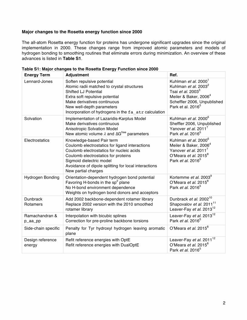
2
Major changes to the Rosetta energy function since 2000 The all-atom Rosetta energy function for proteins has undergone significant upgrades since the original implementation in 2000. These changes range from improved atomic parameters and models of hydrogen bonding to smoothing routines that eliminate errors during minimization. An overview of these advances is listed in Table S1. Table S1: Major changes to the Rosetta Energy Function since 2000
Energy Term Adjustment Ref. Lennard-Jones Soften repulsive potential
Atomic radii matched to crystal structures Shifted LJ Potential Extra soft repulsive potential Make derivatives continuous New well-depth parameters Incorporation of hydrogens in the fa_atr calculation
Kuhlman et al. 20001 Kuhlman et al. 20032 Tsai et al. 20033 Meiler & Baker, 20064 Scheffler 2006, Unpublished Park et al. 20165
Solvation Implementation of Lazaridis-Karplus Model Make derivatives continuous Anisotropic Solvation Model New atomic volume 𝜆 and ∆Gfree parameters
Kuhlman et al. 20006 Sheffler 2006, Unpublished Yanover et al. 20117 Park et al. 20165
Electrostatics Knowledge-based Pair term Coulomb electrostatics for ligand interactions Coulomb electrostatics for nucleic acids Coulomb electrostatics for proteins Sigmoid dielectric model Avoidance of dipole splitting for local interactions New partial charges
Kuhlman et al. 20006 Meiler & Baker, 20064 Yanover et al. 20117 O’Meara et al. 20158 Park et al. 20165
Hydrogen Bonding Orientation-dependent hydrogen bond potential Favoring H-bonds in the sp2 plane No H-bond environment dependence Weights on hydrogen bond donors and acceptors
Kortemme et al. 20039 O’Meara et al. 20158 Park et al. 20165
Dunbrack Rotamers
Add 2002 backbone-dependent rotamer library Replace 2002 version with the 2010 smoothed rotamer library
Dunbrack et al. 200210 Shapovalov et al. 201111 Leaver-Fay et al. 201312
Ramachandran & p_aa_pp
Interpolation with bicubic splines Correction for pre-proline backbone torsions
Leaver-Fay et al. 201312 Park et al. 20165
Side-chain specific Penalty for Tyr hydroxyl hydrogen leaving aromatic plane
O’Meara et al. 20158
Design reference energy
Refit reference energies with OptE Refit reference energies with DualOptE
Leaver-Fay et al. 201112 O’Meara et al. 20158 Park et al. 20165

3
Table S2: Rosetta revisions corresponding to major energy function updates Version Rosetta Revisiona Public Versionb
Score12 pre-#55611 Pre-Rosetta 3.5 Talaris2013 #55611 Rosetta 3.5 Talaris2014 #58602 v2016.13-dev58602 REF2015 #59248 v2017.05-dev59248 a Internal code revision number available to member institutions of the Rosetta Commons. b Download the public Rosetta release from http://www.rosettacommons.org.
Data for calibrating Rosetta energies to kcal/mol The parameters and weights in REF2015 were recently fit5 such that Rosetta simulations reproduce high-resolution protein structural data and thermodynamic data for small molecules from Jorgensen et al.13 Thus, the Rosetta energy is now expressed in kcal/mol. In support, Figure S1 compares experimental data and Rosetta predictions of density, heat of vaporization (∆Hvap) and heat capacity (Cp(l)) for seventeen molecules: ethane, propane, isobutene, cyclohexane, benzene, toluene, phenol, methanol, ethanol, 2-propanol, tert-Butyl alcohol, methane thiol, ethane thiol, dimethyl sulfide, acetamide, N-methylamide, N-methylformamide, dimethyl ether, ethanol and propanone.
Figure S1: Comparison of Rosetta simulations with experimental thermodynamic data Comparison between Rosetta predictions and experimental thermodynamic measurements for seventeen small molecules (A) Density (B) Heat Capacity and (C) Heat of vaporization.

4
Additional energy function details Parameters for the Lennard-Jones and Lazaridis-Karplus energies New experiments and numerical methods to optimize the energy function have led to updated atomic-parameters used by the Lennard-Jones14,15 and Lazaridis-Karplus16 potentials. The updated parameters are in the following Rosetta database files: Table S3: Location of Rosetta atom type parameters Parameter Database File Radii, 𝛥𝐺free, 𝜖 chemical/atom_type_sets/fa_standard/atom_properties.txt
Partial charges chemical/residue_type_sets/residue_types/l-caa/*.params lk_ball weights chemical/atom_type_sets/fa_standard/extras/lk_ball_wtd.txt
hbond parameters scoring/score_functions/hbonds/*
rama scoring/score_functions/rama/*
p_aa_pp scoring/score_functions/P_AA_pp/*
omega scoring/score_functions/omega/*
fa_dun rotamer/*
Tables S4-S6 present a comparison between selected atomic parameters between the original source publication and the values in Rosetta energy functions, Talaris2014 and REF2015.

5
Table S4: Atomic radii values from the Neria et al. force field, Talaris2014, and REF2015 Atom Type Neria et al.17
Radius (Å) Talaris2014 Radius (Å)
REF20155 Radius (Å)
CAbb 2.3650 2.0000 2.0112 CH1 2.3650 2.0000 2.0112 CH2 2.2350 2.0000 2.0112 CH3 2.1650 2.0000 2.0112 CNH2 -- 2.0000 1.9922 COO 2.1000 2.0000 1.9649 CObb -- 2.0000 1.9649 aroC 2.1000 2.0000 1.9859 NH2O -- 1.7500 1.7632 Narg 1.6000 1.7500 1.7632 Nbb 1.6000 1.7500 1.7632 Nhis 1.6000 1.7500 1.7632 Nlys 1.6000 1.7500 1.7632 Npro 1.6000 1.7500 1.7632 Ntrp 1.6000 1.7500 1.7632 OCbb 1.6000 1.5500 1.5268 OH 1.6000 1.5500 1.5354 ONH2 -- 1.5500 1.5760 OOC -- 1.5500 1.4492 S 0.0430 1.9000 2.0171 HNbb -- 1.0000 0.8773 Hapo -- 1.2000 1.4634 Haro -- 1.2000 1.3778 Hpol 0.8000 1.0000 0.8773

6
Table S5: Well-depth parameters from the Neria et al. force field, Talaris2014, and REF2015 Atom Type Neria et al.17
𝝐 (kcal/mol) Talaris2014 𝝐 (kcal/mol)
REF20155 𝝐 (kcal/mol)
CAbb 0.0486 0.0486 0.0626 CH1 0.0486 0.0486 0.0626 CH2 0.1142 0.1142 0.0626 CH3 0.1811 0.1811 0.0626 CNH2 -- 0.1200 0.0626 COO 0.1200 0.1200 0.0946 CObb -- 0.1400 0.1418 aroC 0.1200 0.1200 0.1418 NH2O -- 0.2834 0.0688 Narg 0.2384 0.2834 0.1617 Nbb 0.2384 0.2834 0.1617 Nhis 0.2384 0.2834 0.1617 Nlys 0.2384 0.2834 0.1617 Npro 0.2384 0.2834 0.1617 Ntrp 0.2384 0.2834 0.1617 OCbb 0.1591 0.1617 OH 0.1591 0.1591 0.1617 ONH2 -- 0.1591 0.1424 OOC -- 0.2100 0.1619 S 0.0430 0.1600 0.1829 SH1 -- -- 0.0999 HNbb -- 0.0500 0.4560 HS -- -- 0.4560 Hapo -- 0.0500 0.0050 Haro -- 0.0500 0.0508 Hpol -- 0.0500 0.0218

7
Table S6: ∆Gfree parameters from Lazaridis & Karplus, Talaris2014 and REF2015 Atom Type Lazarids-Karplus16
∆𝑮free (kcal/mol) Talaris2014,
∆𝑮free (kcal/mol) REF20155
∆𝑮𝐟𝐫𝐞𝐞(kcal/mol) CAbb -0.2500 1.0000 2.5338 CH0 -0.2500 -0.2500 1.4093 CH1 -0.2500 -0.2500 -3.5384 CH2 0.5200 0.5200 -1.8547 CH3 1.5000 1.5000 7.2929 CNH2 -- 0.0000 3.0770 COO 0.1200 -1.4000 -3.3326 CObb -- 1.0000 3.1042 aroC 0.8000 0.0800 1.7979 NH2O -- -7.8000 -8.1016 Narg -10.0000 -10.0000 -8.9684 Nbb -7.8000 -5.0000 -9.9695 Nhis -4.0000 -4.0000 -9.7396 Nlys -20.000 -20.000 -20.865 Npro -1.5500 -1.5500 -0.9846 Ntrp -8.9000 -8.9000 -8.4131 OCbb -10.0000 -5.0000 -8.0068 OH -6.7000 -6.7000 -8.1335 ONH2 -- -5.8500 -6.5916 OOC -- -10.0000 -9.2398 S -4.1000 -4.1000 -1.7072 SH1 -2.7000 -- 3.2916

8
Analytical form of the hydrogen bonding potential To avoid expensive table lookups, the hydrogen bonding potential (Eq. 21-22 in the main text) is given by component energies with simple analytical forms. For completeness, we detail these analytical forms below. The first two components, 𝐸hbond67 𝑑67 and 𝐸hbond679 𝜃679 are polynomial functions, 𝑓poly(𝑃, 𝑥) where the polynomial 𝑃 depends on the atom type of the acceptor and donor, and the order 𝑛 varies from 6 to 10 (Eq. S1). The forms of 𝐸hbond67 𝑑67 and 𝐸hbond679 𝜃679 are given by Eq. S2-3.
𝑓poly 𝑃, 𝑥 = 𝐶F + 𝐶H𝑥 + 𝐶I𝑥I + ⋯+ 𝐶KLH𝑥KLH + 𝐶K𝑥K (S1)
𝐸hbond67 𝑑67 = 𝐹67 ∙ 𝑓poly(𝑃, 𝑑67) (S2)
𝐸hbond679 𝜃679 = 𝐹679 ∙ 𝑓poly(𝑃(𝜃679)) (S3) The third component, 𝐸hbond
OPO76 𝜌, 𝜙OPO76, 𝜃O76 , is dependent on the hybridization of the acceptor, 𝜌. For sp2 hybridized acceptors, the potential is given as a combination of cosine and polynomial functions (Eq. S4-5) controlled by a cosine switch function (Eq. S6). The functional forms are also shown in Fig. S2.
𝐹 𝜙 =
SIcos 3(𝜋 − 𝜙) + SLH
IIYZ< 𝜙
\Icos H
]𝜋 − IY
Z𝜙 + \LH
IIYZ− 𝑙 ≤ 𝜙 ≤ IY
Z
𝑚 − HI
𝜙 < IYZ− 𝑙
(S4)
𝐺 𝜙 =
𝑑 − HI
IYZ< 𝜙
\LSIcos 𝜋 − H
]𝜋 − IY
Z𝜙 + \aSaH
IIYZ− 𝑙 ≤ 𝜙 ≤ IY
Z
𝑚 − HI
𝜙 < IYZ− 𝑙
(S5)
𝐻 𝜙 = cde If aH
I (S6)
For sp3 hybridized acceptors, the potential is modeled as a composition of sine and cosine functions. If the acceptor is attached to a ring, the potential is modeled with a simple cosine function. The overall energy is given in Eq. S7.
𝐸hbondOPO76 𝜌, 𝜙OPO76, 𝜃O76 =
𝐻 𝜙OPO76 𝐹 𝜙OPO76 + 1 − 𝐻 𝜙OPO76 𝐺 𝜙OPO76 𝜌~spI
𝑓poly cos 𝜃O76 + Hi1 + cos 𝜙OPO76 𝜌~spZ
𝑓poly cos 𝜃O76 𝜌~ring
(S7)
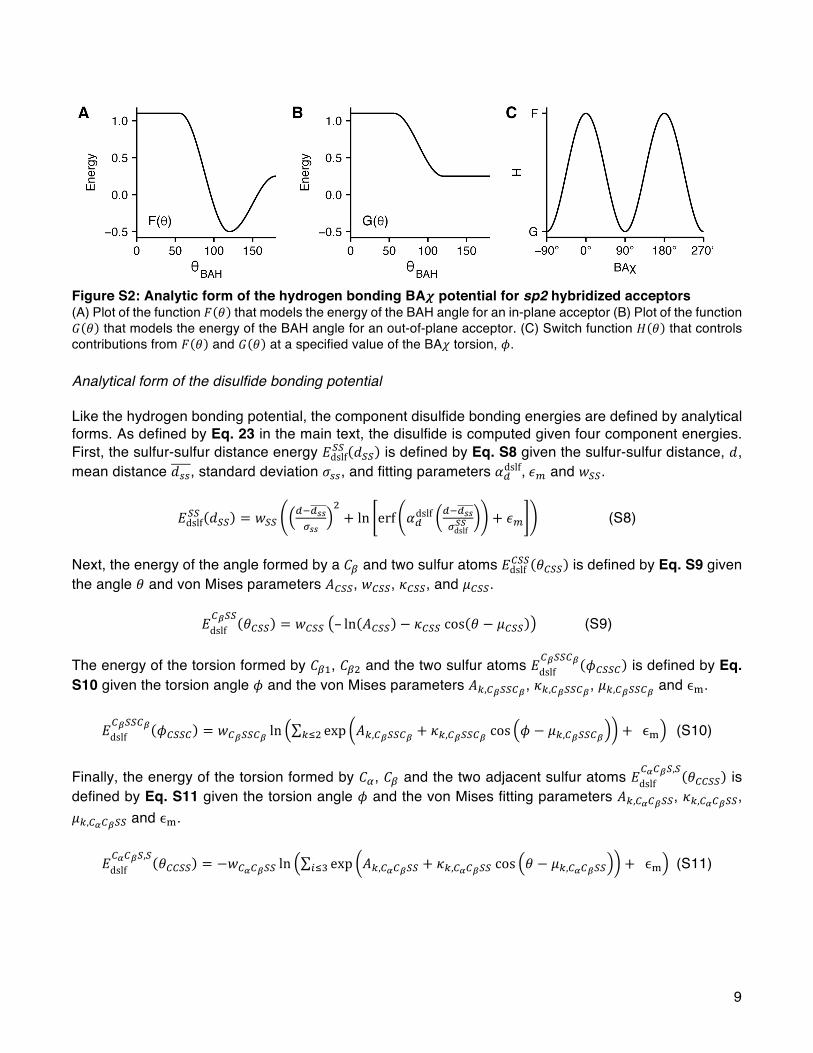
9
Figure S2: Analytic form of the hydrogen bonding BA𝝌 potential for sp2 hybridized acceptors (A) Plot of the function 𝐹 𝜃 that models the energy of the BAH angle for an in-plane acceptor (B) Plot of the function 𝐺 𝜃 that models the energy of the BAH angle for an out-of-plane acceptor. (C) Switch function 𝐻 𝜃 that controls contributions from 𝐹 𝜃 and 𝐺 𝜃 at a specified value of the BA𝜒 torsion, 𝜙. Analytical form of the disulfide bonding potential Like the hydrogen bonding potential, the component disulfide bonding energies are defined by analytical forms. As defined by Eq. 23 in the main text, the disulfide is computed given four component energies. First, the sulfur-sulfur distance energy 𝐸dslfnn 𝑑nn is defined by Eq. S8 given the sulfur-sulfur distance, 𝑑, mean distance 𝑑oo, standard deviation 𝜎oo, and fitting parameters 𝛼Sdslf,𝜖\ and 𝑤nn.
𝐸dslfnn 𝑑nn = 𝑤nn
SLSsstss
I+ ln erf 𝛼Sdslf
SLSsstdslfuu + 𝜖\ (S8)
Next, the energy of the angle formed by a 𝐶v and two sulfur atoms 𝐸dslfwnn 𝜃wnn is defined by Eq. S9 given the angle 𝜃 and von Mises parameters 𝐴wnn, 𝑤wnn, 𝜅wnn, and 𝜇wnn.
𝐸dslfw{nn 𝜃wnn = 𝑤wnn – ln 𝐴wnn − 𝜅wnncos 𝜃 − 𝜇wnn (S9)
The energy of the torsion formed by 𝐶vH, 𝐶vI and the two sulfur atoms 𝐸dslf
w{nnw{ 𝜙wnnw is defined by Eq. S10 given the torsion angle 𝜙 and the von Mises parameters 𝐴},w{nnw{, 𝜅},w{nnw{, 𝜇},w{nnw{ and ϵ�.
𝐸dslfw{nnw{ 𝜙wnnw = 𝑤w{nnw{ ln exp 𝐴},w{nnw{ + 𝜅},w{nnw{cos 𝜙 − 𝜇},w{nnw{}�I + ϵ� (S10)
Finally, the energy of the torsion formed by 𝐶�, 𝐶v and the two adjacent sulfur atoms 𝐸dslf
w�w{n,n 𝜃wwnn is defined by Eq. S11 given the torsion angle 𝜙 and the von Mises fitting parameters 𝐴},w�w{nn, 𝜅},w�w{nn, 𝜇},w�w{nn and ϵ�.
𝐸dslfw�w{n,n 𝜃wwnn = −𝑤w�w{nn ln exp 𝐴},w�w{nn + 𝜅},w�w{nncos 𝜃 − 𝜇},w�w{nn��Z + ϵ� (S11)

10
Statistical potentials: Interpolation of energies rather than probabilities The Rosetta energy function uses probabilities from the Dunbrack backbone-dependent rotamer library18 to derive torsional energies 𝐸 using the inverted Boltzmann relation the probability 𝑃 (Eq. S12):
𝐸 = −𝑘𝑇 ln 𝑃 (S12) Prior to 2012, the probabilities for the 𝜙, 𝜓-dependent terms were stored on a 10° x 10° grid used for energy calculations. These probabilities were calculated using bilinear interpolation and then converted to energies using Eq. S12 and the derivatives were calculated by linearly interpolating 1/𝑃 and 𝑑𝑃/𝑑𝑥 to compute 𝑑 − log 𝑃 𝑑𝑥 = −(1/𝑃)𝑑𝑃/𝑑𝑥 with 𝑥 = 𝜙 or𝜓. This method resulted in large inaccuracies because 𝑃 can vary by orders of magnitude over very short ranges of 𝜙 and𝜓. In addition, the linearly interpolated derivatives are constant between grid points, so that gradient-based minimization results in moving structures to the nearest grid point where the derivative changes sign. Therefore, it is more accurate to provide 𝑃 and 𝐸 = − ln 𝑃 at each grid point and then interpolate the energies using bicubic interpolation. Here we demonstrate why interpolating energies is better than interpolating the probabilities. Figure S3 compares the different interpolation strategies for a toy problem: a one-dimensional probability distribution with a discrete rotamer modeled with the following von Mises function (Eq. S13). Here, the location constant 𝜇 = 180˚, the concentration constant 𝜅 = 20, 𝑥 = 𝜙 or𝜓 and 𝐼F(𝜅) is the modified Bessel function of order zero needed to normalize the distribution.
𝑃 𝑥 = ��� �Lcde �L�IY��(�)
(S13) First, Figure S3A shows the probability distribution, 𝑃 and its linear interpolation based on the 10° x 10° grid. Here, the difference between the curves demonstrate the effect of approximating 𝑃 by linear interpolation. This effect would be more severe for steeper functions such as the Ramachandran probability density function. Figure S3B compares the 𝐸 = − ln 𝑃 calculation with two approaches to interpolating the function: interpolate 𝑃 and then compute the energies versus compute the energies at the grid points and then interpolate. The second scenario clearly mitigates several errors which can be further improved using cubic rather than linear interpolation. Like the first panel, the benefits of cubic interpolation are clearer with steeper functions. Figures S3C and S3D demonstrate that the effects of interpolating energies are more pronounced for the derivatives of 𝑃 and 𝐸 respectively. Previously, Rosetta computed the derivative of 𝑃 as 𝑑𝑃/𝑑𝑥 =[𝑃(𝑥 + 10) − 𝑃(𝑥)]/10 (Fig. S3C). The linear interpolation of this derivative includes noticeable artifacts. Figure S3D shows the four energy derivative curves: (1) the exact analytical expression 𝑑𝐸/𝑑𝑥 =−(1/𝑃)𝑑𝑃/𝑑𝑥 where 𝑃 is interpolated and 𝑑𝑃/𝑑𝑥 is the step function shown in Fig. S3C (2) 𝑑𝐸/𝑑𝑥 =−(1/𝑃)𝑑𝑃/𝑑𝑥 where 𝑃 and 𝑑𝑃/𝑑𝑥 are interpolated from the grid values, (3) 𝑑𝐸/𝑑𝑥 = −(1/𝑃)𝑑𝑃/𝑑𝑥 where both 𝑃 and 𝑑𝑃/𝑑𝑥 are interpolated from the grid values and (4) calculation of 𝐸 and 𝑑𝐸/𝑑𝑥 on the grid followed by interpolation of 𝑑𝐸/𝑑𝑥 in between the grid points. The linear interpolation of 𝑑𝐸/𝑑𝑥provides the closest match to the analytical expression. The current Rosetta energy function interpolates energies rather than probabilities: both 𝑃 and 𝐸 are stored in database files, 𝑑𝐸/𝑑𝑥 is calculated on the grid points, and then 𝑃, 𝐸 and 𝑑𝐸/𝑑𝑥 are computed by bicubic spline interpolation.
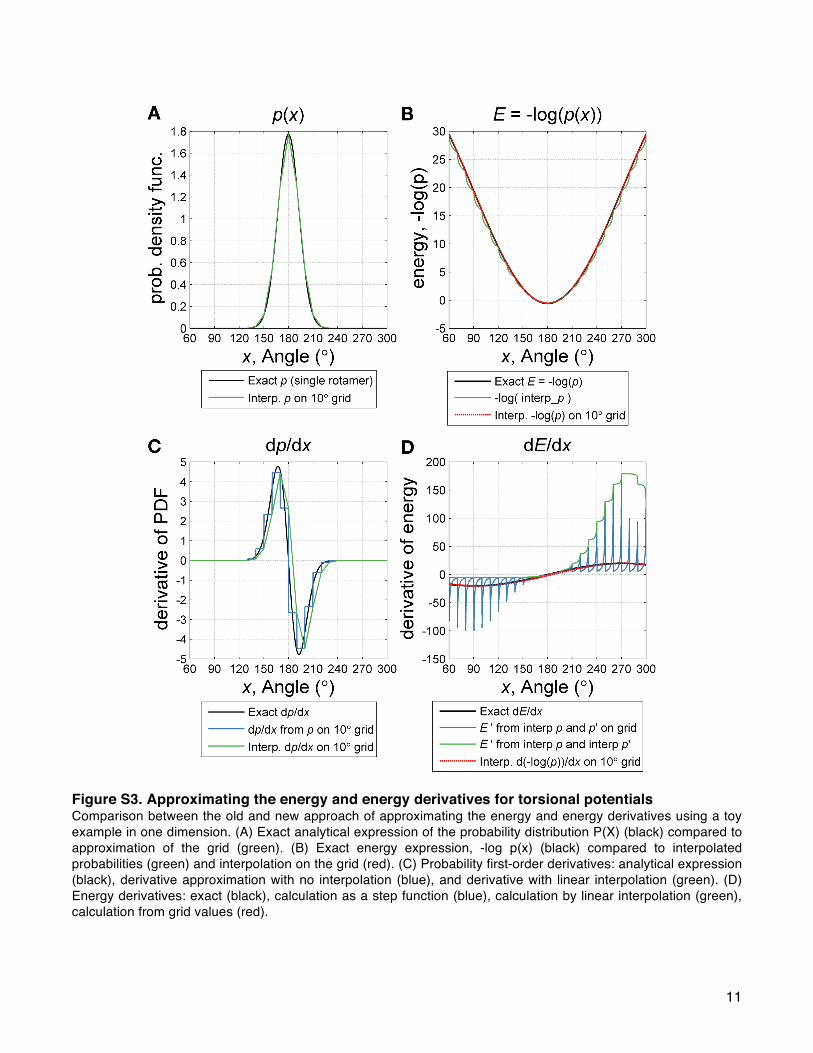
11
Figure S3. Approximating the energy and energy derivatives for torsional potentials Comparison between the old and new approach of approximating the energy and energy derivatives using a toy example in one dimension. (A) Exact analytical expression of the probability distribution P(X) (black) compared to approximation of the grid (green). (B) Exact energy expression, -log p(x) (black) compared to interpolated probabilities (green) and interpolation on the grid (red). (C) Probability first-order derivatives: analytical expression (black), derivative approximation with no interpolation (blue), and derivative with linear interpolation (green). (D) Energy derivatives: exact (black), calculation as a step function (blue), calculation by linear interpolation (green), calculation from grid values (red).
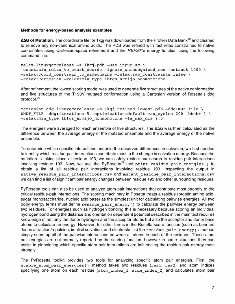
12
Methods for energy-based analysis examples ∆∆G of Mutation. The coordinate file for 1kgj was downloaded from the Protein Data Bank19 and cleaned to remove any non-canonical amino acids. The PDB was refined with fast relax constrained to native coordinates using Cartesian-space refinement and the REF2015 energy function using the following command line: relax.linuxgccrelease –s 1kgj.pdb –use_input_sc \ –constrain_relax_to_start_coords –ignore_unrecognized_res –nstruct 1000 \ –relax:coord_constrain_to_sidechains –relax:ram_constraints false \ –relax:Cartesian –relax:min_type lbfgs_armijo_nonmonotone After refinement, the lowest scoring model was used to generate five structures of the native conformation and five structures of the T193V mutated conformation using a Cartesian version of Rosetta’s ddg protocol.20 cartesian_ddg.linuxgccrelease –s 1kgj_refined_lowest.pdb –ddg:mut_file \ $MUT_FILE –ddg:iterations 5 –optimization:default-max_cycles 200 –bbnbr 1 \ –relax:min_type lbfgs_armijo_nonmonotone –fa_max_dis 9.0 The energies were averaged for each ensemble of five structures. The ∆∆G was then calculated as the difference between the average energy of the mutated ensemble and the average energy of the native ensemble. To determine which specific interactions underlie the observed differences in solvation, we first needed to identify which residue-pair interactions contribute most to the change in solvation energy. Because the mutation is taking place at residue 193, we can safely restrict our search to residue-pair interactions involving residue 193. Now, we use the PyRosetta21 tool print_residue_pair_energies() to obtain a list of all residue pair interactions involving residue 193. Inspecting the output in native_residue_pair_interactions.csv and mutant_residue_pair_interactions.csv we can find a list of significant pair energy changes between residue 193 and other surrounding residues. PyRosetta tools can also be used to analyze atom-pair interactions that contribute most strongly to the critical residue-pair interactions. The scoring machinery in Rosetta treats a residue (protein amino acid, sugar monosaccharide, nucleic acid base) as the simplest unit for calculating pairwise energies. All two body energy terms must define residue_pair_energy() to calculate the pairwise energy between two residues. For energies such as hydrogen bonding this is necessary because scoring an individual hydrogen bond using the distance and orientation dependent potential described in the main text requires knowledge of not only the donor hydrogen and the acceptor atoms but also the acceptor and donor base atoms to calculate an energy. However, for other terms in the Rosetta score function (such as Lennard Jones attraction/repulsion, implicit solvation, and electrostatics) the residue_pair_energy() method simply sums up all of the pairwise interactions between all atoms in each of the residues. These atom pair energies are not normally reported by the scoring function, however in some situations they can assist in pinpointing which specific atom pair interactions are influencing the residue pair energy most strongly. The PyRosetta toolkit provides two tools for analyzing specific atom pair energies. First, the etable_atom_pair_energies() method takes two residues (res1, res2) and atom indices specifying one atom on each residue (atom_index_1, atom_index_2) and calculates atom pair

13
energies for Lennard Jones attractive/repulsive, implicit solvation, and electrostatics using a specified score function (sfxn). The second tool, print_atom_pair_energy_table(), is designed to output energies for all pairwise atom pair interactions between two specified residues. For ease of viewing this tool outputs the pairwise energies as a table formatted in a .csv file. The tool takes a score_type and score function (sfxn) as inputs in addition to two specified residues (res1, res2) and a specified output_filename. Docking. The coordinate file for 1ztx was downloaded from the Protein Data Bank and cleaned to remove any non-canonical amino acids. The structure was first refined to remove significant clashes in the structure using the following command line: relax.linuxgccrelease -s 1ztx_unbound.pdb -relax:ramp_constriants false \ -relax:constrain_relax_to_start_coords -ex1 -ex2 -use_input_sc -flip_HNQ \ -no_optH false Next, the structure was prepacked and then docked using the procedure described in Chaudhury et al.22 using the REF2015 energy function. docking_prepack_protocollinux -s 1ztx_relaxed.pdb -partners LH_G \ -dock_rtmin -docking:sc_min docking_protocol.linuxgccrelease –s 1ztx_unbound_prepacked.pdb –native \ 1ztx_native.pdb -ignore_unrecognized_res -ex1 -ex2aro -dock_pert 3 8 \ -partners LH_G -nstruct 1000 Finally, the interface scores were extracted from the output score file for analysis.
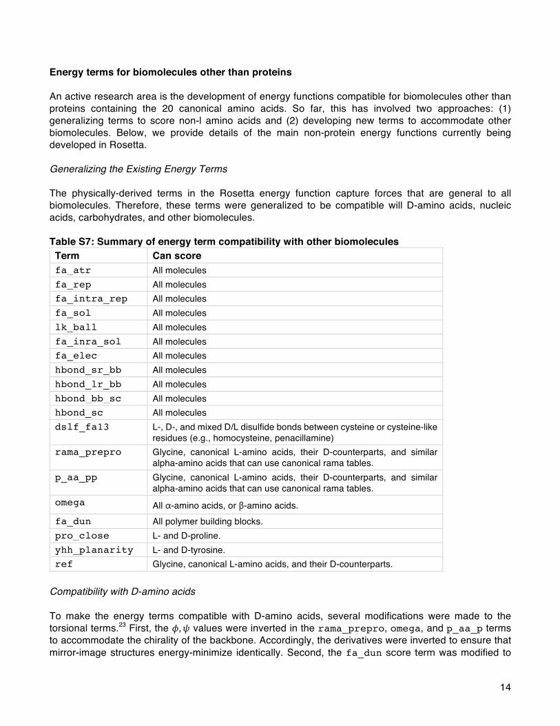
14
Energy terms for biomolecules other than proteins An active research area is the development of energy functions compatible for biomolecules other than proteins containing the 20 canonical amino acids. So far, this has involved two approaches: (1) generalizing terms to score non-l amino acids and (2) developing new terms to accommodate other biomolecules. Below, we provide details of the main non-protein energy functions currently being developed in Rosetta. Generalizing the Existing Energy Terms The physically-derived terms in the Rosetta energy function capture forces that are general to all biomolecules. Therefore, these terms were generalized to be compatible will D-amino acids, nucleic acids, carbohydrates, and other biomolecules. Table S7: Summary of energy term compatibility with other biomolecules Term Can score fa_atr All molecules fa_rep All molecules fa_intra_rep All molecules fa_sol All molecules lk_ball All molecules fa_inra_sol All molecules fa_elec All molecules hbond_sr_bb All molecules hbond_lr_bb All molecules hbond_bb_sc All molecules hbond_sc All molecules dslf_fa13 L-, D-, and mixed D/L disulfide bonds between cysteine or cysteine-like
residues (e.g., homocysteine, penacillamine) rama_prepro Glycine, canonical L-amino acids, their D-counterparts, and similar
alpha-amino acids that can use canonical rama tables. p_aa_pp Glycine, canonical L-amino acids, their D-counterparts, and similar
alpha-amino acids that can use canonical rama tables. omega All α-amino acids, or β-amino acids. fa_dun All polymer building blocks. pro_close L- and D-proline. yhh_planarity L- and D-tyrosine. ref Glycine, canonical L-amino acids, and their D-counterparts.
Compatibility with D-amino acids To make the energy terms compatible with D-amino acids, several modifications were made to the torsional terms.23 First, the 𝜙, 𝜓 values were inverted in the rama_prepro, omega, and p_aa_p terms to accommodate the chirality of the backbone. Accordingly, the derivatives were inverted to ensure that mirror-image structures energy-minimize identically. Second, the fa_dun score term was modified to

15
invert main chain and side-chain torsional values. Special amino acid-specific score terms, such as pro_close and yhh_planarity, were updated to recognize D-proline and D-tyrosine, respectively. The dslf_fa13 term was symmetrized to ensure that mirror-image conformations of mixed D/L disulfides score identically. Finally, the ref term was altered to ensure that D-amino acids have a reference energy penalty or bonus identical to that of their L-counterparts. All other score terms were compatible with arbitrary molecules without modification. Energy terms for non-canonical amino acids Toward the goal of designing proteins with non-canonical amino acids, Renfrew et al. implemented an energy function with terms derived from molecular mechanics. This energy function, called mm_std, removes the terms that depend on residue identity (i.e. rama_prepro, p_aa_pp, omega, and fa_dun) and replaces them with terms that capture the internal and torsional energy preferences: mm_lj_intra_rep, mm_lj_intra_atr, and mm_twist. The ref term is replaced by either a term that explicitly models the unfolded state, (unfolded), or a pair of terms that capture the change in energy experienced by an atom of a specific type going from an unfolded to folded environment (split_unfolded_1b and split_unfolded_2b). These terms were developed toward the goal of designing proteins containing non-canonical alpha-amino acid residues. It has also been used to model oligo-oxypiperizines (OOPs),24 hydrogen bond surrogates (HBS), oligo-peptoids,25 and hybrid molecules. Intra-residue van der Waals interactions are calculated between atom pairs from the same residue using a Lennard-Jones 6-12 potential. Like fa_rep and fa_atr, the potential is divided between attractive (mm_lj_intra_atr) and repulsive (mm_lj_intra_rep) components that can be weighted separately. The two terms have the same functional form as the inter-reside terms (Eq. 3 and 4 in the main text) but with the following differences. The summed atomic radii, 𝜎��, and the geometric mean of atomic well-depths, 𝜖��, are based on the CHARMM 2426 parameters. The terms are applied to all atom pairs in a residue with a bond separation of 3 or more. Some atom-type pairs have different parameters when separated by 3 bonds (and involved in a proper torsion) and when separated by 4 of more bonds, but no connectivity weight is applied. Both attractive and repulsive energies are calculated for hydrogens. The attractive potential is not smoothed and consequently is evaluated to 8 Å. The torsional term, called mm_twist (Eq. S14), is a molecular mechanics torsion term. It is evaluated for all atom quads involved in proper torsions. To match the intra-residue van der Waals term the parameters for 𝐾� and 𝑛 come from CHARMM 24. A given set of 4 atoms types may have multiple 𝐾� and 𝑛 parameters that are summed in a Fourier series to more accurately describe the rotation about the central bond of the torsion.
𝐸twist = 𝐾� 1 − cos 𝑛𝜃� (S14) Explicit Unfolded State Energy (EUSE) represents the unfolded energy of the protein and compensates for the difficultly in packing large side chains (Eq. S15). The ref term is fit during the weight optimization protocol which is only trained on protein data and therefore incompatible with non-protein residues. The EUSE is the sum over each residue and each term in the energy function where 𝑈 AA�, 𝑡 is the unfolded reference value of residue type, AA�, of residue, 𝑟, and energy term 𝑡. The unfolded reference values are the Boltzmann weighted average energies of the central residue of 5-mer fragments of high quality protein structures. The central residue of each fragment was mutated to the residue of choice, repacked and scored and the Boltzmann weighted average for each energy term, 𝑡, for each residue type is stored. For peptoids, only XXGPX fragments were used to mimic an oligo-peptoid environment.27
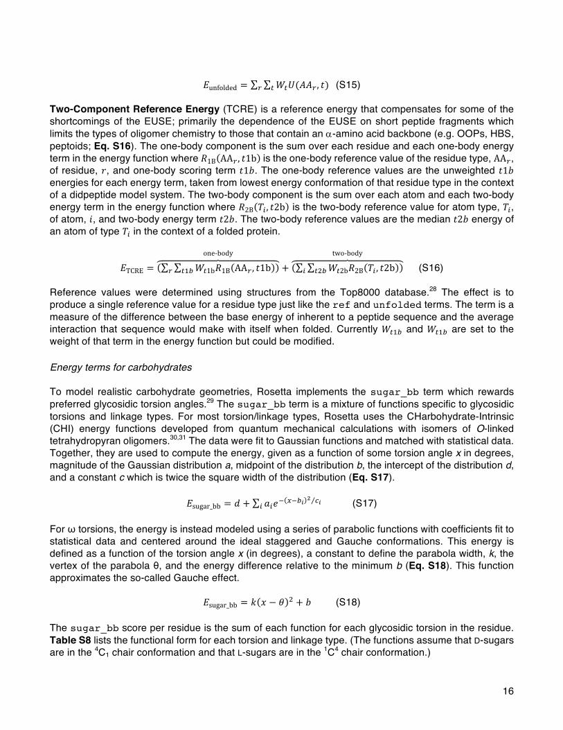
16
𝐸unfolded = 𝑊¤𝑈(𝐴𝐴�, 𝑡)¤� (S15)
Two-Component Reference Energy (TCRE) is a reference energy that compensates for some of the shortcomings of the EUSE; primarily the dependence of the EUSE on short peptide fragments which limits the types of oligomer chemistry to those that contain an a-amino acid backbone (e.g. OOPs, HBS, peptoids; Eq. S16). The one-body component is the sum over each residue and each one-body energy term in the energy function where 𝑅1B AA�, 𝑡1b is the one-body reference value of the residue type, AA�, of residue, 𝑟, and one-body scoring term 𝑡1𝑏. The one-body reference values are the unweighted 𝑡1𝑏 energies for each energy term, taken from lowest energy conformation of that residue type in the context of a didpeptide model system. The two-body component is the sum over each atom and each two-body energy term in the energy function where 𝑅2B 𝑇�, 𝑡2b is the two-body reference value for atom type, 𝑇�, of atom, 𝑖, and two-body energy term 𝑡2𝑏. The two-body reference values are the median 𝑡2𝑏 energy of an atom of type 𝑇� in the context of a folded protein.
𝐸TCRE = 𝑊¤1b𝑅1B AA�, 𝑡1b¤H�
one-body
+ 𝑊¤2b𝑅2B 𝑇�, 𝑡2b¤I�
two-body
(S16) Reference values were determined using structures from the Top8000 database.28 The effect is to produce a single reference value for a residue type just like the ref and unfolded terms. The term is a measure of the difference between the base energy of inherent to a peptide sequence and the average interaction that sequence would make with itself when folded. Currently 𝑊¤H and 𝑊¤H are set to the weight of that term in the energy function but could be modified. Energy terms for carbohydrates To model realistic carbohydrate geometries, Rosetta implements the sugar_bb term which rewards preferred glycosidic torsion angles.29 The sugar_bb term is a mixture of functions specific to glycosidic torsions and linkage types. For most torsion/linkage types, Rosetta uses the CHarbohydrate-Intrinsic (CHI) energy functions developed from quantum mechanical calculations with isomers of O-linked tetrahydropyran oligomers.30,31 The data were fit to Gaussian functions and matched with statistical data. Together, they are used to compute the energy, given as a function of some torsion angle x in degrees, magnitude of the Gaussian distribution a, midpoint of the distribution b, the intercept of the distribution d, and a constant c which is twice the square width of the distribution (Eq. S17).
𝐸sugar_bb = 𝑑 + 𝑎�𝑒L �L³ P ´³� (S17) For ω torsions, the energy is instead modeled using a series of parabolic functions with coefficients fit to statistical data and centered around the ideal staggered and Gauche conformations. This energy is defined as a function of the torsion angle x (in degrees), a constant to define the parabola width, k, the vertex of the parabola θ, and the energy difference relative to the minimum b (Eq. S18). This function approximates the so-called Gauche effect.
𝐸sugar_bb = 𝑘 𝑥 − 𝜃 I + 𝑏 (S18)
The sugar_bb score per residue is the sum of each function for each glycosidic torsion in the residue. Table S8 lists the functional form for each torsion and linkage type. (The functions assume that D-sugars are in the 4C1 chair conformation and that L-sugars are in the 1C4 chair conformation.)

17
Table S8: Functional form of the sugar backbone energy for each torsion and linkage type Angle Ax./eq. designation Stereoisomer Exocyclic Range Functional form
φ
axial (α) D — −180°–180°
Gaussian
eqiuatorial (β) D — −180°–180°
Gaussian
axial (α) L — −180°–180°
Gaussian, x=−φ
equatorial (β) L — −180°–180°
Gaussian, x=−φ
ψ
ax. (parent at odd O) D (parent) no 0–360° Gaussian eq. (parent at odd O) D (parent) no 0–360° Gaussian ax. (parent at even O) D (parent) no 0–360° Gaussian eq. (parent at even O) D (parent) no 0–360° Gaussian ax. (parent at odd O) L (parent) no 0–360° Gaussian, 360°−ψ eq. (parent at odd O) L (parent) no 0–360° Gaussian, 360°−ψ ax. (parent at even O) L (parent) no 0–360° Gaussian, 360°−ψ eq. (parent at even O) L (parent) no 0–360° Gaussian, 360°−ψ axial (α) D yes 0–360° Gaussian equatorial (β) D yes 0–360° Gaussian axial (α) L yes 0–360° Gaussian, 360°−ψ equatorial (β) L yes 0–360° Gaussian, 360°−ψ
ω
axial (parent O4) D (parent) yes 0–120° parabolic axial (parent O4) D (parent) yes 120°–240° parabolic axial (parent O4) D (parent) yes 240°–360° parabolic eq. (parent O4) D (parent) yes 0–120° parabolic eq. (parent O4) D (parent) yes 120°–240° parabolic eq. (parent O4) D (parent) yes 240°–360° parabolic axial (parent O4) L (parent) yes 0–360° parabolic, 360°−ω axial (parent O4) L (parent) yes 120°–240° parabolic, 360°−ω axial (parent O4) L (parent) yes 240°–360° parabolic, 360°−ω eq. (parent O4) L (parent) yes 0–120° parabolic, 360°−ω eq. (parent O4) L (parent) yes 120°–240° parabolic, 360°−ω eq. (parent O4) L (parent) yes 240°–360° parabolic, 360°−ω

18
Energy terms for nucleic acids The Rosetta energy function captures van der Waals and electrostatic forces general to all biomolecules. However, these terms do not capture rules specific to the geometry and pairing of nucleic acid bases. Therefore, Das and coworkers have implemented terms to explicitly capture these rules. Electrostatics. The standard Rosetta electrostatic potential (fa_elec) disfavors Watson-Crick base pairs due to repulsion between the fixed positive charges on the hydrogen atoms in close proximity in G-C and A-U pairs. To alleviate this problem, Rosetta uses two modified terms to evaluate electrostatics involving RNA bases. First, electrostatic interactions between phosphate atoms are evaluated using the standard fa_elec potential (Eq. 10 in the main text), via a term called fa_elec_rna_phos_phos. Second, electrostatic interactions between RNA bases are captured using the stack_elec term.32 This term scales the fa_elec potential as a function of the angle (𝜅�) between the normal to the plane of the base (𝑧�) and the vector 𝑑�,� between base heavy atoms 𝑖 and 𝑗 in residues 𝑟H and 𝑟I, respectively (Figure S4). The equation for stack_elec is given by Eq. S19.
𝐸stack_elec = 𝑓(�,�� ¹�P 𝜅�, 𝜅�)𝐸fa_elec (S19) The scaling function 𝑓(𝜅�, 𝜅�) suppresses the electrostatic energy to zero when the bases are coplanar and maintains the full value of the energy when the bases are stacked (Eq. S20; Fig. S4B).
𝑓 𝜅�, 𝜅� = 𝑐𝑜𝑠I 𝜅� + 𝑐𝑜𝑠I 𝜅� (S20)
Figure S4. Electrostatic and stacking energies for RNA. (A) fa_stack and stack_elec are scaled as a function of the angle, κi, between the normal to the base, 𝑧�, and the distance vector between atoms 𝑖 and 𝑗. (B) The scaling function takes the form 𝑓 𝜅� = cosI(𝜅𝑖), such that the weight is equal to 1.0 when the bases are stacked and 0 when they are coplanar. (C) The fa_stack energy for stacked bases (when 𝑓 𝜅� = 1.0). Base stacking. 𝜋 − 𝜋 stacking interactions are not explicitly captured by fa_atr; thus, Rosetta includes an additional stacking bonus term, called fa_stack.33 The fa_stack term applies a constant bonus for base atoms less than 4 Å from each other to reward neighboring stacked bases. Like the stack_elec term, fa_stack also depends on the angle (𝜅�) between the normal to the plane of the base (𝑧�) and the distance vector from atoms i to j (𝑑�,�), such that stacked, but not coplanar bases receive this bonus (Eq. S19: Fig. 4C). The potential is smoothed to zero between 4 Å and 6 Å using a smoothing function given in Eq. S21-S23.
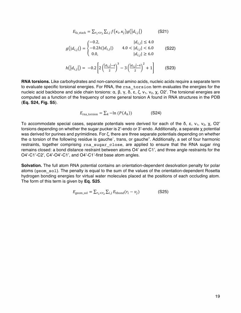
19
𝐸fa_stack = 𝑓 𝜅�, 𝜅� 𝑔 𝑑�,��,�� ¹�P (S21)
𝑔 |𝑑�,�| =−0.2,|𝑑�,�| ≤ 4.0−0.2ℎ(|𝑑�,�|)4.0 < |𝑑�,�| < 6.00.0,|𝑑�,�| ≥ 6.0
(S22)
ℎ 𝑑�,� = −0.2 2 S³,Ä LiI
Z− 3 S³,Ä Li
I
I+ 1 (S23)
RNA torsions. Like carbohydrates and non-canonical amino acids, nucleic acids require a separate term to evaluate specific torsional energies. For RNA, the rna_torsion term evaluates the energies for the nucleic acid backbone and side chain torsions: α, β, γ, δ, ε, ζ, ν1, ν2, χ, O2’. The torsional energies are computed as a function of the frequency of some general torsion A found in RNA structures in the PDB (Eq. S24, Fig. S5).
𝐸rna_torsion = −ln(𝑃 𝐴} )} (S24) To accommodate special cases, separate potentials were derived for each of the δ, ε, ν1, ν2, χ, O2’ torsions depending on whether the sugar pucker is 2’-endo or 3’-endo. Additionally, a separate χ potential was derived for purines and pyrimidines. For ζ, there are three separate potentials depending on whether the α torsion of the following residue is gauche–, trans, or gauche+. Additionally, a set of four harmonic restraints, together comprising rna_sugar_close, are applied to ensure that the RNA sugar ring remains closed: a bond distance restraint between atoms O4’ and C1’, and three angle restraints for the O4’-C1’-C2’, C4’-O4’-C1’, and O4’-C1’-first base atom angles. Solvation. The full atom RNA potential contains an orientation-dependent desolvation penalty for polar atoms (geom_sol). The penalty is equal to the sum of the values of the orientation-dependent Rosetta hydrogen bonding energies for virtual water molecules placed at the positions of each occluding atom. The form of this term is given by Eq. S25.
𝐸geom_sol = 𝐸hbond(𝑟��,�� ¹�P − 𝑣�) (S25)

20
Figure S5. Torsion potentials for RNA RNA torsional potential for (A) α, (B) β, (C) γ, (D) δ, (E) ε, (F) ζ when the α torsion of the following residue is gauche– (orange), trans (cyan), or gauche+ (purple) (G) χ for purines (lighter red and blue) and pyrimidines (darker red and blue), (H) ν1, (I) ν2. Potentials when the sugar pucker is C2’-endo are shown in red and C3’-endo shown in blue.

21
References (1) Kuhlman, B.; Baker, D. Native Protein Sequences Are close to Optimal for Their Structures. Proc
Natl Acad Sci U S A 2000, 97 (19), 10383–10388. (2) Kuhlman, B.; Dantas, G.; Ireton, G. C.; Varani, G.; Stoddard, B. L.; Baker, D. Design of a Novel
Globular Protein Fold with Atomic-Level Accuracy. Science (80-. ). 2003, 302 (5649), 1364–1368. (3) Tsai, J.; Bonneau, R.; Morozov, A. V.; Kuhlman, B.; Rohl, C. A.; Baker, D. An Improved Protein
Decoy Set for Testing Energy Functions for Protein Structure Prediction. Proteins Struct. Funct. Bioinforma. 2003, 53 (1), 76–87.
(4) Meiler, J.; Baker, D. ROSETTALIGAND: Protein-Small Molecule Docking with Full Side-Chain Flexibility. Proteins Struct. Funct. Bioinforma. 2006, 65 (3), 538–548.
(5) Park, H.; Bradley, P.; Greisen, P.; Liu, Y.; Kim, D. E.; Baker, D.; DiMaio, F. Simultaneous Optimization of Biomolecular Energy Function on Features from Small Molecules and Macromolecules. J. Chem. Theory Comput. 2016, 12 (12), 6201–6212.
(6) Kuhlman, B.; Baker, D. Native Protein Sequences Are close to Optimal for Their Structures. Proc. Natl. Acad. Sci. U. S. A. 2000, 97 (19), 10383–10388.
(7) Yanover, C.; Bradley, P. Extensive Protein and DNA Backbone Sampling Improves Structure-Based Specificity Prediction for C2H2 Zinc Fingers. Nucleic Acids Res. 2011, 39 (11), 4564–4576.
(8) O’Meara, M. J.; Leaver-Fay, A.; Tyka, M. D.; Stein, A.; Houlihan, K.; DiMaio, F.; Bradley, P.; Kortemme, T.; Baker, D.; Snoeyink, J.; Kuhlman, B. Combined Covalent-Electrostatic Model of Hydrogen Bonding Improves Structure Prediction with Rosetta. J. Chem. Theory Comput. 2015, 11 (2), 609–622.
(9) Kortemme, T.; Morozov, A. V; Baker, D. An Orientation-Dependent Hydrogen Bonding Potential Improves Prediction of Specificity and Structure for Proteins and Protein-Protein Complexes. J. Mol. Biol. 2003, 326 (4), 1239–1259.
(10) Dunbrack, R. L.; Cohen, F. E.; Cohen, F. E. Bayesian Statistical Analysis of Protein Side-Chain Rotamer Preferences. Protein Sci. 1997, 6 (8), 1661–1681.
(11) Shapovalov, M. V; Dunbrack, R. L. A Smoothed Backbone-Dependent Rotamer Library for Proteins Derived from Adaptive Kernel Density Estimates and Regressions. Structure 2011, 19 (6), 844–858.
(12) Leaver-Fay, A.; O’Meara, M. J.; Tyka, M.; Jacak, R.; Song, Y.; Kellogg, E. H.; Thompson, J.; Davis, I. W.; Pache, R. A.; Lyskov, S.; Gray, J. J.; Kortemme, T.; Richardson, J. S.; Havranek, J. J.; Snoeyink, J.; Baker, D.; Kuhlman, B. Scientific Benchmarks for Guiding Macromolecular Energy Function Improvement. Methods Enzymol. 2013, 523, 109–143.
(13) William L. Jorgensen, *; David S. Maxwell, and; Tirado-Rives, J. Development and Testing of the OPLS All-Atom Force Field on Conformational Energetics and Properties of Organic Liquids. 1996.
(14) Lennard-Jones, J. On the Determination of Molecular Fields II: From the Variation of Viscosity of a Gas with Temperature. R. Soc. London, Ser. A, Contain. Pap. a Math. Phys. Character 1924, 106, 464–477.
(15) Lennard-Jones, J. On the Determination of Molecular Fields I: From the Variation of Viscosity of a Gas with Temperature. R. Soc. London, Ser. A, Contain. Pap. a Math. Phys. Character 1924, 106, 441–462.
(16) Lazaridis, T.; Karplus, M. Effective Energy Function for Proteins in Solution. Proteins 1999, 35 (2), 133–152.
(17) Neria, E.; Fischer, S.; Karplus, M. Simulation of Activation Free Energies in Molecular Systems. J. Chem. Phys. 1996, 105 (5), 1902–1921.
(18) Shapovalov, M. V; Dunbrack Jr., R. L. A Smoothed Backbone-Dependent Rotamer Library for Proteins Derived from Adaptive Kernel Density Estimates and Regressions. Structure 2011, 19
22
(6), 844–858. (19) Berman, H. M.; Westbrook, J.; Feng, Z.; Gilliland, G.; Bhat, T. N.; Weissig, H.; Shindyalov, I. N.;
Bourne, P. E. The Protein Data Bank. Nucleic Acids Res. 2000, 28 (1), 235–242. (20) Kellogg, E. H.; Leaver-Fay, A.; Baker, D. Role of Conformational Sampling in Computing Mutation-
Induced Changes in Protein Structure and Stability. Proteins 2011, 79 (3), 830–838. (21) Chaudhury, S.; Lyskov, S.; Gray, J. J. PyRosetta: A Script-Based Interface for Implementing
Molecular Modeling Algorithms Using Rosetta. Bioinformatics 2010, 26 (5), 689–691. (22) Chaudhury, S.; Berrondo, M.; Weitzner, B. D.; Muthu, P.; Bergman, H.; Gray, J. J. Benchmarking
and Analysis of Protein Docking Performance in Rosetta v3.2. PLoS One 2011, 6 (8), e22477. (23) Bhardwaj, G.; Mulligan, V. K.; Bahl, C. D.; Gilmore, J. M.; Harvey, P. J.; Cheneval, O.; Buchko, G.
W.; Pulavarti, S. V. S. R. K.; Kaas, Q.; Eletsky, A.; Huang, P.-S.; Johnsen, W. A.; Greisen, P. J.; Rocklin, G. J.; Song, Y.; Linsky, T. W.; Watkins, A.; Rettie, S. A.; Xu, X.; Carter, L. P.; Bonneau, R.; Olson, J. M.; Coutsias, E.; Correnti, C. E.; Szyperski, T.; Craik, D. J.; Baker, D. Accurate de Novo Design of Hyperstable Constrained Peptides. Nature 2016, 538 (7625), 329–335.
(24) Lao, B. B.; Drew, K.; Guarracino, D. A.; Brewer, T. F.; Heindel, D. W.; Bonneau, R.; Arora, P. S. Rational Design of Topographical Helix Mimics as Potent Inhibitors of Protein–Protein Interactions. J. Am. Chem. Soc. 2014, 136 (22), 7877–7888.
(25) Drew, K.; Renfrew, P. D.; Craven, T. W.; Butterfoss, G. L.; Chou, F.-C.; Lyskov, S.; Bullock, B. N.; Watkins, A.; Labonte, J. W.; Pacella, M.; Kilambi, K. P.; Leaver-Fay, A.; Kuhlman, B.; Gray, J. J.; Bradley, P.; Kirshenbaum, K.; Arora, P. S.; Das, R.; Bonneau, R. Adding Diverse Noncanonical Backbones to Rosetta: Enabling Peptidomimetic Design. PLoS One 2013, 8 (7), e67051.
(26) MacKerell, A. D.; Bashford, D.; Bellott, M.; Dunbrack, R. L.; Evanseck, J. D.; Field, M. J.; Fischer, S.; Gao, J.; Guo, H.; Ha, S.; Joseph-McCarthy, D.; Kuchnir, L.; Kuczera, K.; Lau, F. T. K.; Mattos, C.; Michnick, S.; Ngo, T.; Nguyen, D. T.; Prodhom, B.; Reiher, W. E.; Roux, B.; Schlenkrich, M.; Smith, J. C.; Stote, R.; Straub, J.; Watanabe, M.; Wiórkiewicz-Kuczera, J.; Yin, D.; Karplus, M. All-Atom Empirical Potential for Molecular Modeling and Dynamics Studies of Proteins. J. Phys. Chem. B 1998, 102 (18), 3586–3616.
(27) Renfrew, P. D.; Craven, T. W.; Butterfoss, G. L.; Kirshenbaum, K.; Bonneau, R. A Rotamer Library to Enable Modeling and Design of Peptoid Foldamers. J. Am. Chem. Soc. 2014, 136 (24), 8772–8782.
(28) Richardson, J. S.; Keedy, D. A.; Richardson, D. C. In Biomolecular Forms and Functions: A Celebration of 50 Years of the Ramachandran Map. World Sci. Publ. Co. Pte. Ltd Singapore 2013, 46–61.
(29) Labonte, J. W.; Aldof-Bryfogle, J.; Schief, W. R.; Gray, J. J. Residue-Centric Modeling and Design of Saccharide and Glycoconjugate Structures. J. Comput. Chem. 2017, 38 (5), 276–287.
(30) Nivedha, A. K.; Thieker, D. F.; Makeneni, S.; Hu, H.; Woods, R. J. Vina-Carb: Improving Glycosidic Angles during Carbohydrate Docking. J. Chem. Theory Comput. 2016, 12 (2), 892–901.
(31) Nivedha, A. K.; Makeneni, S.; Foley, B. L.; Tessier, M. B.; Woods, R. J. Importance of Ligand Conformational Energies in Carbohydrate Docking: Sorting the Wheat from the Chaff. J. Comput. Chem. 2014, 35 (7), 526–539.
(32) Chou, F.-C.; Kladwang, W.; Kappel, K.; Das, R. Blind Tests of RNA Nearest-Neighbor Energy Prediction. Proc. Natl. Acad. Sci. U. S. A. 2016, 113 (30), 8430–8435.
(33) Sripakdeevong, P.; Kladwang, W.; Das, R. An Enumerative Stepwise Ansatz Enables Atomic-Accuracy RNA Loop Modeling. Proc. Natl. Acad. Sci. U. S. A. 2011, 108 (51), 20573–20578.


![[8] Dipolar Couplings in Macromolecular Structure ... · [8] DIPOLAR COUPLINGS AND MACROMOLECULAR STRUCTURE 127 [8] Dipolar Couplings in Macromolecular Structure Determination By](https://img.pdfslide.us/doc/110x75/605c24b70c5494344557be4f/8-dipolar-couplings-in-macromolecular-structure-8-dipolar-couplings-and.jpg)