Embed Size (px)
Citation preview

1
Ph.D. thesis
The role of Wnt signalling in thymic senescence
Zoltán Varecza M.Sc.
Ph.D. Supervisor: Dr. Judit E. Pongrácz
Ph.D. Program Leader: Prof. Dr. Péter Németh
University of Pécs
Institute for Immunology and Biotechnology
Department of Medical Biotechnology
Pécs
2011

2
1. Contents
1. Contents ........................................................................................................................................... 2 2. Abbreviations ................................................................................................................................... 4 3. Introduction ...................................................................................................................................... 6 3.1. Ageing in focus ............................................................................................................................. 6
3.1.1. Ageing and society ................................................................................................................. 6 3.1.2. Ageing of the immune system ............................................................................................... 6 3.1.3. Significance of thymic involution studies .............................................................................. 7
3.2. T-cell development in the thymus ................................................................................................. 7 3.3. The role of thymic microenvironment in de novo T-cell production ............................................ 8 3.4. Thymic involution during ageing .................................................................................................. 9
3.5. Trans-differentiation of fibroblasts into adipocytes .................................................................... 10 3.6. Wnt signalling ............................................................................................................................. 10
3.6.1. Wnt molecules and pathways ............................................................................................... 10 3.6.2. Canonical Wnt-pathway ....................................................................................................... 11 3.6.3. Non-canonical Wnt-pathways .............................................................................................. 13 3.6.4. Inhibitory Wnt pathway ....................................................................................................... 14 3.6.5. Wnts in ageing ..................................................................................................................... 15
3.6.6. Wnts in the thymus .............................................................................................................. 16 3.7. PKC-s in the thymus ................................................................................................................... 16
3.8. PKCs in Wnt signalling............................................................................................................... 17 3.9. Steroids and ageing ..................................................................................................................... 18 4. Aims of the study ........................................................................................................................... 20
5. Materials and Methods ................................................................................................................... 21 5.1. Antibodies ................................................................................................................................... 21
5.1.1. Western blot analysis ........................................................................................................... 21 5.1.2. Fluorescent microscopy ....................................................................................................... 21
5.2. Animals ....................................................................................................................................... 21
5.3. Cell cultures. ............................................................................................................................... 22 5.3.1 Cell lines ............................................................................................................................... 22
5.3.2 Primary thymic epithelial cells ............................................................................................. 22 5.3.3 Dexamethasone treatment of cells and animals .................................................................... 22
5.4 Manipulation of gene expression ................................................................................................. 23 5.4.1. Retroviral Constructs ........................................................................................................... 23
5.4.2. Transient transfection of siRNA PKCδ ............................................................................... 23
5.5. Detection of gene transcription ................................................................................................... 24 5.5.1. cDNA generation ................................................................................................................. 24
5.5.2. Standard Reverse Transcription Polymerase Chain Reaction (RT-PCR) ............................ 24 5.5.3. Microarray analysis .............................................................................................................. 24 5.5.4. Real-time qRT-PCR ............................................................................................................. 25
5.6. Cell sorting .................................................................................................................................. 25 5.7. PKCδ activation assay ................................................................................................................. 25 5.8. Purification of proteins from cell membrane and cytosol ........................................................... 26 5.9. Immuno-precipitation.................................................................................................................. 26
5.10. Western blotting ........................................................................................................................ 26 5.11. Immuno-histochemistry ............................................................................................................ 27 5.12. Statistical analysis ..................................................................................................................... 27
6. Results ............................................................................................................................................ 29 6.1. Physiological thymic senescence ................................................................................................ 30

3
6.1.1. Disintegration of the epithelial network ............................................................................... 30 6.1.2. Adipose involution ............................................................................................................... 32 6.1.3. Gene expression changes in the thymic epithelium during ageing ...................................... 33 6.1.4. Studies of LAP2α and Wnt4 effects on TEC ...................................................................... 34 6.1.5. Fz-4 and Fz-6 levels are affected by age.............................................................................. 35 6.1.6. Active receptor signalling is indicated by PKCδ translocation ........................................... 38 6.1.7. Identification of Wnt4 target genes in TECs using microarray analysis ............................. 41 6.1.8. PKCδ in Wnt4 signalling ..................................................................................................... 42 6.1.9. Co-immuno-precipitation of PKCδ, with Dvl, Fz-4 and Fz-6 ............................................. 45 6.1.10. Increased expression of CTGF and Fz-8 ............................................................................ 47
6.2. Steroid induced thymic senescence ............................................................................................. 48 6.2.1. Effects of single-dose GC administration ............................................................................ 48 6.2.2. Effects of sustained GC administration ............................................................................... 49 6.2.3. Wnt4-mediated inhibition of steroid-induced adipose trans-differentiation ........................ 50
7. Discussion ...................................................................................................................................... 52 8. Conclusions .................................................................................................................................... 55 9. References ...................................................................................................................................... 56 10. List of Publications ...................................................................................................................... 62 10.1. Publications related to the thesis: .............................................................................................. 62
10.1.1. Papers: ................................................................................................................................ 62
10.1.2. Poster presentations related to the thesis: .......................................................................... 63 10.2. Further publications .................................................................................................................. 64 11. Acknowledgements ...................................................................................................................... 67

4
2. Abbreviations
Abs- Antibodies
ADRP- Adipose differentiation-related protein
ANKRD- Ankyrin Repeat Domain
AP-1- Activator protein-1
APC- Adenomatous polyposis coli
BMP- Bone morphogenetic protein
CaMKII- Ca-Calmodulin Kinase II
CTGF- Connective tissue growth factor
DAG- Diacylglycerol
DKK- Dickkopf
DN1, 2, 3 Double negative
DP- Double positive
Dvl- Dishevelled
DX- Dexamethasone
EpCAM1- Epithelial cellular adhesion molecule
Fz- Frizzled
GH- Glucocorticoid hormone
GSK-3- Glycogen synthase kinase 3
IER- Immediate-early response
IGF-I- Insulin like growth factor 1
JNK- c-Jun N-terminal kinases
LAP2α- Lamina associated polypeptide
LEF1- Lymphoid Enhancer-binding Factor 1
LRP5/6- Low density lipoprotein Related Protein 5 and 6
MHC- Major Histocompatibility Complex
NFAT- Nuclear Factor of Activating T- cells
NFκB- Nuclear Factor kappa B
Nkd-1,Nkd-2- Naked 1, 2
NLK- Nemo-Like Kinase
pAb- Polyclonal antibody
PCP- Planar cell polarity pathway

5
PCR- Polymerase chain reaction
PKC- Protein kinase C
PLC- Phospholipase C
PPARγ Peroxisome proliferator-activated receptor
qRT-PCR- Quantitative real-time polymerase chain reaction
RT- Reverse transcription
SEMA- Semaphorin domain
TAK1- Transforming growth factor β–Activated Kinase 1
TCF1, TCF3- T-cell Factor 1, 3
TCR- T-cell receptor
TEC- Thymic epithelial cell
TMB- 3,3’,5,5’-Tetra-Methyl-Benzidine

6
3. Introduction
3.1. Ageing in focus
3.1.1. Ageing and society
Ageing of the population is one of the most important challenges for the developed world to face
over the next fifty years. The current demographic trends and consequent shrinkage of the active
workforce will put enormous pressure on the financing of social protection and health systems,
which likely to reduce living standards. Coupled with increased migration and emergence of novel
infectious diseases, broad-scale provision of immunological protection by vaccination constitutes a
strategic aim for longer and healthier lifespan.
3.1.2. Ageing of the immune system
Impaired immunological responsiveness in the elderly poses a major difficulty for achieving
efficient immunization. The immunological competence of an individual is determined by the
presence of mature lymphocytes formed in primary lymphoid organs, and specialized secondary
lymphoid tissues performing diverse immune responses. Thus at systems level the maintenance of
immunological equilibrium requires steady lymphocyte output, and controlled expansion coupled
with continuous replacement. Lymphostromal interactions in both primary and secondary lymphoid
tissues play essential roles in the development and function of lymphocyte subsets in adaptive
immune responses. The thymic and lymphnode stromal microenvironments thus represent key
elements in the development of the adaptive immune system. Consequently, impairment of the
lymphoid microenvironment will ultimately lead to insufficient primary and secondary immune
responses or to the decline of thymic selection, manifesting in late-onset autoimmune disorders,
often observed in elderly. Self-tolerant cytotoxic and helper T-lymphocytes, the crucial regulator
cells in adaptive immune responses, develop in the specialized epithelial network of the thymus.
The thymus, however, gradually loses its capacity to support lymphopoiesis in a programmed
involution process that results in a decline of de novo T-cell production.

7
3.1.3. Significance of thymic involution studies
In contrast to the extensive studies addressing haematopoietic cells, the in-depth analysis of
determinants for stromal competence during immunological ageing is largely absent, despite its
clear significance related to immunological responsiveness in the elderly. A more thorough
understanding of molecular mechanisms responsible for stromal senescence of the thymus may lead
in aged individuals for the restoration of immunological competence by a controlled reversion of
the involuted thymic epithelium, and consequent strengthening capacity of peripheral lymphoid
tissues to support T-cell dependent antibody-mediated immune responses.
3.2. T-cell development in the thymus
T-cell progenitors migrate to the thymus from the bone marrow where they undergo an extensive
differentiation and selection process. After entering the thymus, thymocytes representing different
stages of development occupy distinct regions of the thymus. The earliest CD4-CD8-CD44+CD25-
thymocyte progenitors, referred to as double negative 1 (DN1) cells are found near their site of
entry at the cortico-medullary junction. The slightly more mature CD4-CD8-CD44+CD25+ (DN2)
subset is found throughout the cortex, whereas CD4-CD8-CD44-CD25+ (DN3) subset is
concentrated below the capsule. Following rearrangement of antigen receptor (TCR) genes (He and
Kappes 2006) CD4+CD8+ (double positive or DP) thymocytes undergo positive (functional TCR)
and negative (self-reactive TCR) selection in the cortex and medulla, to finally as CD4-CD8+
(cytotoxic) or CD4+CD8- (helper) single positive (SP), mature, naïve T-cells leave the thymus for
the periphery. Figure 1 shows a schematic diagram of T-cell development.
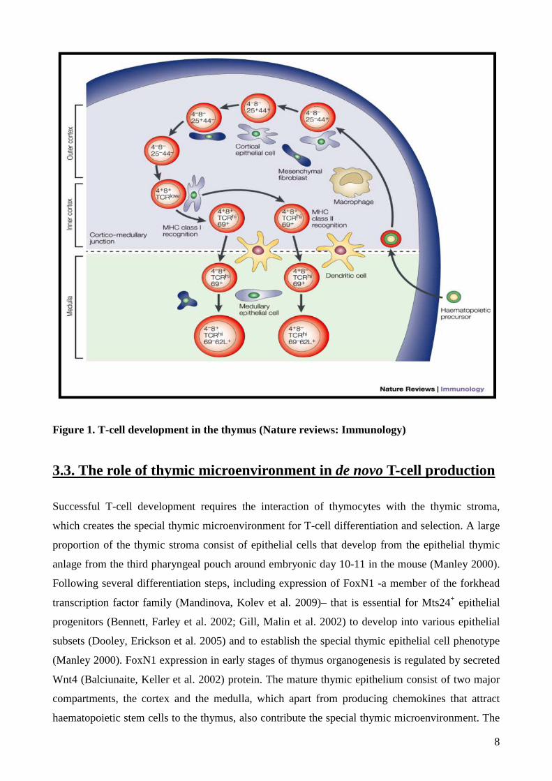
8
Figure 1. T-cell development in the thymus (Nature reviews: Immunology)
3.3. The role of thymic microenvironment in de novo T-cell production
Successful T-cell development requires the interaction of thymocytes with the thymic stroma,
which creates the special thymic microenvironment for T-cell differentiation and selection. A large
proportion of the thymic stroma consist of epithelial cells that develop from the epithelial thymic
anlage from the third pharyngeal pouch around embryonic day 10-11 in the mouse (Manley 2000).
Following several differentiation steps, including expression of FoxN1 -a member of the forkhead
transcription factor family (Mandinova, Kolev et al. 2009)– that is essential for Mts24+ epithelial
progenitors (Bennett, Farley et al. 2002; Gill, Malin et al. 2002) to develop into various epithelial
subsets (Dooley, Erickson et al. 2005) and to establish the special thymic epithelial cell phenotype
(Manley 2000). FoxN1 expression in early stages of thymus organogenesis is regulated by secreted
Wnt4 (Balciunaite, Keller et al. 2002) protein. The mature thymic epithelium consist of two major
compartments, the cortex and the medulla, which apart from producing chemokines that attract
haematopoietic stem cells to the thymus, also contribute the special thymic microenvironment. The

9
thymic epithelial network through release of cytokines (e.g. interleukin-7(Alves, Richard-Le Goff et
al. 2009)), secretion of extracellular matrix components, and establishment of adhesive interactions
(Crisa L, Cirulli V et al. 1996) (Schluns KS, Cook JE et al. 1997) regulate homing, intrathymic
migration, and differentiation of developing T-lymphocytes. Thymocytes bearing diverse TCR
repertoire are selected on MHC (major-histocompatibility-complex) and MHC bound-antigens
presented to them by the thymic stroma, including epithelial cells. During T-cell development,
which is characterised by the progression through phenotypically distinct stages (Lind, Prockop et
al. 2001), thymocytes occupy spatially restricted domains of the mature thymus. T-cell precursors
enter the thymus at the cortico-medullary junction (Blackburn and Manley 2004), then migrate to
the subcapsular zone of the outer cortex, back through the cortex, then to the medulla, where they
finally exit to the periphery (Blackburn and Manley 2004). Functional studies have shown, that the
cortex is important in producing chemokines, which attract pro-thymocytes (Bleul and Boehm
2000) and are also essential for mediating positive selection (Anderson, Owen et al. 1994).
Meanwhile the medullary epithelium has been implicated in driving the final stages of thymocyte
maturation (Ge and Chen 2000) and has a crucial role in tolerance induction (Farr and Rudensky
1998; Derbinski, Schulte et al. 2001). Additionally, the thymic epithelium is also the source of other
secreted and cell surface proteins that regulate T-cell development. These proteins include bone
morphogenic protein (BMP) (Bleul and Boehm 2005), Notch (Valsecchi C 1997), and Wnt
(Pongracz, Hare et al. 2003) family members.
3.4. Thymic involution during ageing
In comparison to other organs, ageing of the thymus is an accelerated process in all mammals. In
humans, thymic senescence begins early, around late puberty and by 50 years of age 80% of the
thymic stroma is converted to adipose tissue (Dixit 2010). As the thymic epithelium is replaced by
adipose tissue, the whole process is called adipose involution (Marinova 2005). Due to decrease in
thymic epithelial tissue mass, the thymus can no longer support the same output of T-cell
production (Ribeiro and Perelson 2007). T-lymphocyte composition in the periphery therefore
exhibits the dominance of memory T-lymphocytes resulting in impaired responses towards novel,
particularly viral infections (Chidgey, Dudakov et al. 2007; Gui, Zhu et al. 2007; Grubeck-
Loebenstein 2009). Since the thymic epithelium has a key role in deleting auto-reactive T-cell
clones, functional impairment increases the chances of developing auto-immune disease (Hsu and
Mountz 2003). One of the transcription factors, FoxN1 that is characteristic in thymus development
is also affected by age. FoxN1 (Mandinova, Kolev et al. 2009) is not only essential for progenitor
epithelial cells of the thymic rudiment to develop into various epithelial subsets (Dooley, Erickson

10
et al. 2005) but also to maintain TEC (Thymic Epithelial Cell) identity in the differentiated, adult
thymus. Decreased levels of FoxN1 expression in the adult TECs result in accelerated thymic
involution (Chen, Xiao et al. 2009; Cheng, Guo et al. 2010).
3.5. Trans-differentiation of fibroblasts into adipocytes
The nuclear lamina consists of a two-dimensional matrix of proteins located next to the inner
nuclear membrane. The lamin family of proteins make up the matrix and are highly conserved in
evolution. The family of lamin associated proteins (LAP) has several members with similar
functions. Studies with fibroblast cells have revealed that fibroblast to pre-adipocyte transformation
is strongly connected to LAP2α, the member of the LAP2 protein family (Dorner, Vlcek et al.
2006). While most splice variants associate with the nuclear envelope, LAP2α is involved in several
nucleoplasmic activities including cell-cycle control and differentiation (Berger, Theodor et al.
1996; Hutchison, Alvarez-Reyes et al. 2001). LAP2α is synthesized in the cytoplasm and is then
transported into the nucleus by a PKC-dependent mechanism (Dreger, Otto et al. 1999). The mere
over-expression of LAP2α in fibroblasts is known to directly up-regulate PPARγ (Peroxisome
proliferator-activated receptor γ) expression, an acknowledged marker and key transcription factor
of pre-adipocyte differentiation (Dorner, Vlcek et al. 2006). In pre-adipocytes PPARγ expression is
followed by an increase of ADRP expression (adipose differentiation-related protein) a known
direct target gene of PPARγ. Although LAP2α over-expression alone initiates pre-adipocyte
differentiation in fibroblasts, it is not sufficient to complete the adipocyte differentiation program in
the absence of additional stimuli (Dorner, Vlcek et al. 2006).
3.6. Wnt signalling
3.6.1. Wnt molecules and pathways
The Wnt family of 19 secreted glycoproteins control a variety of developmental processes including
cell fate specification, cell proliferation, cell polarity and cell migration. There are two main
signalling pathways involved in the signal transduction process from the Wnt receptor (Frizzled)
complex: the canonical or β-catenin dependent and the non-canonical pathway, which diverses into
the planar cell polarity (PCP) or c-Jun-N-Terminal Kinase (JNK)/Activating Protein (AP1)
dependent and the Ca++ or Protein kinase C (PKC)/Calmodulin Kinase (CaMKII)/Nuclear Factor of
Activating T-cells (NFAT) dependent signalling pathways. Based on their ability to activate a
particular Wnt pathway, Wnt molecules have been grouped as canonical (Wnt1, Wnt3, Wnt3a,

11
Wnt7a, Wnt7b, Wnt8) (Torres, Yang-Snyder et al. 1996) and non-canonical pathway activators
(Wnt5a, Wnt4, Wnt11) (Torres, Yang-Snyder et al. 1996), although promiscuity is a feature of both
ligands and receptors alike.
3.6.2. Canonical Wnt-pathway
The canonical or β-catenin/TCF dependent Wnt pathway is extensively investigated, and has been
shown to be present in the thymus both in developing thymocytes (Ioannidis, Beermann et al.
2001; Staal 2001; Xu, Banerjee et al. 2003) as well as in the thymic epithelium (Balciunate, et al
2001, Pongracz et al 2003). Generally, in the absence of canonical Wnts, glycogen synthase kinase-
3β (GSK-3β) is active and phosphorylates β-catenin in the scaffolding protein complex of
adenomatous polyposis coli (APC) and axin (Ikeda 1998; Yamamoto 1999). The phosphorylated β-
catenin is targeted for ubiquitination and 26S proteasome-mediated degradation, thereby decreasing
the cytosolic level of β-catenin (Aberle 1997; Akiyama 2000). In the presence of Wnts, signals
from the Wnt-Fz-LRP6 complex lead to the phosphorylation of three domains of Dishevelled (Dvl),
a family of cytosolic signal transducer molecules (Noordermeer 1994). Activation of Dvl ultimately
leads to phosphorylation and consequently inhibition of GSK-3β. Inhibition of GSK-3β results in
stabilisation and finally cytosolic accumulation of β-catenin, which then translocates to the nucleus
(Fig. 2), where is required to form active transcription complexes with members of the T-Cell
Factor (LEF1, TCF1, TCF3, TCF4) transcription factor family (Staal and Clevers 2003) and
transcription initiator p300 (Labalette, Renard et al. 2004).

12
Figure 2. Canonical Wnt signalling (Pongracz&Stockley, 2006)
Successful assembly of the transcription complex leads to various target gene activation including
cyclin-D1 (Shtutman, Zhurinsky et al. 1999; Tetsu and McCormick 1999), c-myc (He, Sparks et al.
1998), c-jun (Mann, Gelos et al. 1999), Fra-1 (Mann, Gelos et al. 1999), VEGFR (Zhang, Gaspard
et al. 2001), etc. (Further target genes can be viewed at Nusse’s Wnt website:
http://www.stanford.edu/~rnusse/wntwindow.html).
13
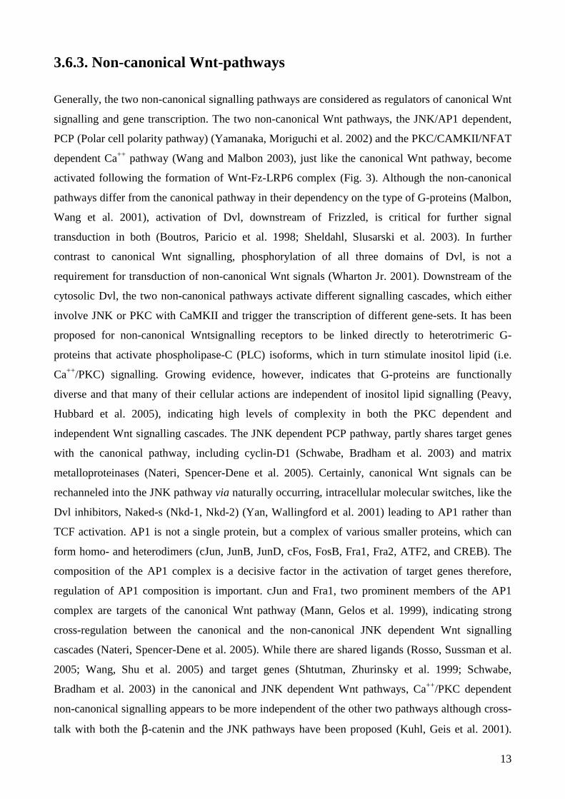
3.6.3. Non-canonical Wnt-pathways
Generally, the two non-canonical signalling pathways are considered as regulators of canonical Wnt
signalling and gene transcription. The two non-canonical Wnt pathways, the JNK/AP1 dependent,
PCP (Polar cell polarity pathway) (Yamanaka, Moriguchi et al. 2002) and the PKC/CAMKII/NFAT
dependent Ca++ pathway (Wang and Malbon 2003), just like the canonical Wnt pathway, become
activated following the formation of Wnt-Fz-LRP6 complex (Fig. 3). Although the non-canonical
pathways differ from the canonical pathway in their dependency on the type of G-proteins (Malbon,
Wang et al. 2001), activation of Dvl, downstream of Frizzled, is critical for further signal
transduction in both (Boutros, Paricio et al. 1998; Sheldahl, Slusarski et al. 2003). In further
contrast to canonical Wnt signalling, phosphorylation of all three domains of Dvl, is not a
requirement for transduction of non-canonical Wnt signals (Wharton Jr. 2001). Downstream of the
cytosolic Dvl, the two non-canonical pathways activate different signalling cascades, which either
involve JNK or PKC with CaMKII and trigger the transcription of different gene-sets. It has been
proposed for non-canonical Wntsignalling receptors to be linked directly to heterotrimeric G-
proteins that activate phospholipase-C (PLC) isoforms, which in turn stimulate inositol lipid (i.e.
Ca++/PKC) signalling. Growing evidence, however, indicates that G-proteins are functionally
diverse and that many of their cellular actions are independent of inositol lipid signalling (Peavy,
Hubbard et al. 2005), indicating high levels of complexity in both the PKC dependent and
independent Wnt signalling cascades. The JNK dependent PCP pathway, partly shares target genes
with the canonical pathway, including cyclin-D1 (Schwabe, Bradham et al. 2003) and matrix
metalloproteinases (Nateri, Spencer-Dene et al. 2005). Certainly, canonical Wnt signals can be
rechanneled into the JNK pathway via naturally occurring, intracellular molecular switches, like the
Dvl inhibitors, Naked-s (Nkd-1, Nkd-2) (Yan, Wallingford et al. 2001) leading to AP1 rather than
TCF activation. AP1 is not a single protein, but a complex of various smaller proteins, which can
form homo- and heterodimers (cJun, JunB, JunD, cFos, FosB, Fra1, Fra2, ATF2, and CREB). The
composition of the AP1 complex is a decisive factor in the activation of target genes therefore,
regulation of AP1 composition is important. cJun and Fra1, two prominent members of the AP1
complex are targets of the canonical Wnt pathway (Mann, Gelos et al. 1999), indicating strong
cross-regulation between the canonical and the non-canonical JNK dependent Wnt signalling
cascades (Nateri, Spencer-Dene et al. 2005). While there are shared ligands (Rosso, Sussman et al.
2005; Wang, Shu et al. 2005) and target genes (Shtutman, Zhurinsky et al. 1999; Schwabe,
Bradham et al. 2003) in the canonical and JNK dependent Wnt pathways, Ca++/PKC dependent
non-canonical signalling appears to be more independent of the other two pathways although cross-
talk with both the β-catenin and the JNK pathways have been proposed (Kuhl, Geis et al. 2001).

14
Generally, Ca++ and PKC-dependent signals are frequently linked to AP1, NFκB and NFAT
activation. Gene transcription, however, which is direct result of Ca++ dependent Wnt signalling has
not been identified.
Figure 3. Non-canonical pathways of Wnt signalling (Pongracz&Stockley, 2006)
3.6.4. Inhibitory Wnt pathway
Besides the canonical and non-canonical Wnt pathways, inhibitory Fz pathways have also been
described. Fz1 and Fz-6 are, for example, able to transduce inhibitory Wnt signals. While Fz1
inhibits Wnt signal transduction via a G-protein dependent manner (Roman-Roman, Shi et al. 2004)
(Zilberberg, Yaniv et al. 2004), Fz-6 (Golan, Yaniv et al. 2004) inhibits Wnt dependent gene
transcription by activating the Transforming growth factor β–activated kinase 1 (TAK1), a member
of the MAPKKK family, and Nemo-Like Kinase (NLK) (Ishitani, Kishida et al. 2003; Smit, Baas et
al. 2004) via a Ca++ dependent signalling cascade. NLK phosphorylates TCF that as a result cannot
bind to β-catenin, consequently formation of the active transcription complex is inhibited (Smit,
Baas et al. 2004) (Fig. 4).

15
Figure 4. Inhibitory Wnt pathway (Pongracz&Stockley, 2006)
3.6.5. Wnts in ageing
As Wnts are important regulators of stem cell survival and differentiation, recent studies have
started to investigate the Wnt family members in ageing. Most studies confirmed that drastically
reduced Wnt levels can trigger ageing as tissue specific stem cells are depleted as a result of low
Wnt signals. In contrast, the KLOTHO mouse, that carries a single gene mutation in KLOTHO, an
endogenous Wnt antagonist also shows signs of accelerated ageing (Liu, Fergusson et al. 2007). It
has been proposed that increased Wnt signalling leads to continuous stem cell proliferation which
finally results in depletion of the stem cell pool causing accelerated ageing (Brack, Conboy et al.
2007).

16
3.6.6. Wnts in the thymus
The main source of Wnt glycoproteins in the thymus is the thymic epithelium, where 14 members
of the Wnt family together with all 10 known Wnt receptors of the seven-loop transmembrane
receptor family, Frizzleds (Fz) have been identified (Pongracz, Hare et al. 2003). That is a striking
difference compared with thymocytes where developmentally regulated receptor expression is
limited to Fz-5 and Fz-6 (Pongracz, Hare et al. 2003). The assembly of an active Wnt-Fz receptor
complex also requires the presence of a co-receptor, the low density lipoprotein related protein 5
and 6 (LRP5/6) (Pinson 2000; Tamai 2000; Wehrli 2000), which is expressed both in thymocytes
and thymic epithelial cells, indicating full ability in both cell types to respond to Wnt signals. Initial
experiments, by manipulating the level of some Wnts and soluble Fz-s, have shown perturbation of
T-cell development (Staal 2001; Mulroy 2002), highlighting the importance of Wnt dependent
signalling for T-cell proliferation and differentiation, but not until data from Pongracz et al
(Pongracz, Hare et al. 2003) revealed differential expression of Wnt ligands and receptors in thymic
cell types, was it considered, that T-cell development may be influenced by indirect events triggered
by Wnt signalling within the thymic epithelium. The canonical pathway has been shown to have an
important role in thymocyte development regulating survival and differentiation (Ioannidis,
Beermann et al. 2001; Staal 2001; Pongracz, Hare et al. 2003; Xu, Banerjee et al. 2003). In a thymic
epithelial cell study, transgenic expression of cyclin-D1, one of the principal target genes of Wnt
signalling, has lead to the expansion of the entire epithelial compartment (Klug, Crouch et al. 2000)
suggesting that canonical Wnt signalling is involved in thymic epithelial cell proliferation,
strengthening the argument, that thymic epithelial development is regulated by Wnts. So far,
signalling studies have revealed, that Wnt4 can activate both the canonical (Lyons, Mueller et al.
2004) and the non-canonical (Torres, Yang-Snyder et al. 1996) (Chang, Sonoyama et al. 2007; Kim,
Clark et al. 2009) Wnt-pathways.
3.7. PKC-s in the thymus
Members of the PKC family regulate a wide variety of cellular processes including proliferation
(Clemens and Trayner 1992), differentiation (Clemens and Trayner 1992) and apoptotic death
(Pongracz, Johnson et al. 1994; Pongracz, Tuffley et al. 1995). The PKC family comprises at least
ten isoenzymes of serine/threonine protein kinases with a broad range of tissue distribution and
differential cellular localisation (Dekker and Parker 1994). Based on their cofactor requirements for
optimal catalytic activity, PKC-s are grouped into three main categories (Yamamoto, Takai et al.
1977; Saito, Kikkawa et al. 2002). While the classical (cPKC: α, βI-βII (splice variants) and γ), and

17
novel (nPKC: δI-III, ε, η, and θI-II) PKC-s bind diacylglycerol (DAG) that stimulates kinase
activity, atypical (aPKC: ζ, PKMz (catalytic fragment of PKCζ), and ι/λ) PKC-s do not interact
with DAG (Newton 2001). Ca++ is an additional requirement of cPKC-s but not for nPKC-s.
Despite that most PKC-s are fully phosphorylated shortly after translation, they remain catalitically
inactive due to the pseudosubstrate domain. Upon binding of lipid second messengers, like DAG
and inositol triphosphate, two products of phospholipase-C (PLC) activity, the molecular
conformation changes and the active centre of the enzymes become exposed and accessible for
substrates. Catalytically active PKCs usually relocate from the cytosol to cellular or nuclear
membranes (Giorgione, Hysell et al. 2003). Although PKC-s have been described as having a non-
redundant role in signal transduction of various immune cell types including mature T-cells
(Monks, Kupfer et al. 1997; Bi K, Tanaka Y et al. 2001) and developing thymocytes (Sun, Arendt et
al. 2000; Michie, Soh et al. 2001), the expression of PKC family members and their function in the
thymic epithelium remains obscure.
3.8. PKCs in Wnt signalling
Due to complexity of Wnt signalling as well as PKC activation and regulation, studies targeting
PKC involvement in Wnt signalling are limited. Wnts of the Ca++ -dependent pathway have been
demonstrated to trigger PLC activity that is responsible for the generation of lipid second
messengers, like DAG and inositol phosphates (Cai Y, Stafford LJ et al. 2005). Recently, the
orphan nuclear receptor RORα-mediated inhibition of canonical Wnt signalling has been identified
(Lee, Kim et al.) to directly involve PKC. Inhibition of Wnt/β-catenin target genes is achieved by
Wnt5a/PKCα-dependent phosphorylation of RORα on serine-35 residue. Additionally, Wnt-5a can
mediate chondro-stimulatory effect of TGF-β3 through upregulation of PKCα and p38MAPK
signalling (Jin, Park et al. 2006). While PKCδ has been identified as an essential activator of Dvl
function (Kinoshita, Iioka et al. 2003) a main signal transducer from Fz-s, PKCζ is proposed to
regulate GSK-3 phosphorylation and activity (Ossipova, Bardeesy et al. 2003). As GSK-3 mediated
phosphorylation leads to proteosomal degradation of β-catenin (Moon, Bowerman et al. 2002),
PKCζ modulates the activity of the canonical Wnt pathway (McManus, Sakamoto et al. 2005) (Fig.
5).

18
PKC
CamK
Gββββ
Wnt
Cellular response
Frizzled
PLCIP3, DAG
[Ca++]
GtGo
PKCζ
PKCα
PKCδ
RORα
GSK3β
Dvl
WntWnt
Wnt Wnt
Figure 5. The role of PKC isoenzymes in signalling pathways
3.9. Steroids and ageing
Physiological steroids are implicated in the regulation of ageing. For example both surgical or
chemical castration have been demonstrated to decrease the progression of ageing (Qiao, Chen et al.
2008) indicating that high steroid levels would accelerate the ageing process. Still, steroids used in
therapy have not been fully investigated for their effects on ageing. Autoimmune diseases and
haematological malignancies are treated by steroids, as they effectively promote apoptosis of
leukaemia cells and trigger complex anti-inflammatory actions (Stahn, Lowenberg et al. 2007).
Apart from triggering decreased expression of cytokines and MHC class II (MHC II) molecules,
glucocorticoid (GC) analogues also induce apoptotic death of peripheral (Wust, van den Brandt et
al. 2008) and developing T-cells. In mouse models, GCs cause massive thymocyte depletion,
especially in the CD4+CD8+ (DP) thymocyte population, (Wiegers, Knoflach et al. 2001; Berki,
Palinkas et al. 2002; Jondal, Pazirandeh et al. 2004) blocking de novo T-cell production.
Experiments have also demonstrated that high-dose GCs induce a dramatic (Blomgren and

19
Andersson 1970) and apoptosis-associated (Boersma, Betel et al. 1979) involution of the thymus,
and not only thymocytes but also TECs are seriously affected (Dardenne, Itoh et al. 1986). Recent
reports (Fletcher, Lowen et al. 2009) have highlighted that TEC depletion appears reversible, and
thymic epithelial stem cells play an important role in this process.

20
4. Aims of the study
I. To study the role of Wnt signalling in physiological thymic senescence:
1. Wnt4 induced signalling and gene expression patterns were investigated in the thymic
epithelium. Identification of potential molecular targets of Wnt4 can reveal proliferation,
differentiation –especially trans-differentiation - patterns within the thymic epithelial network.
2. As Wnt4 can activate both canonical and non-canonical Wnt signalling pathways and regulate
TEC identity, identification of signalling elements and their role -especially PKCδ- in Wnt4
signalling can aid better understanding of regulatory mechanisms of thymic senescence.
II. To compare physiological and induced thymic senescence:
3. Comparison of molecular mechanisms of physiological and GC induced thymic senescence can
reveal, whether molecular features of the two processes are shared or independent. It can also help
to identify molecular targets to alleviate side effects of GC therapy and to identify potential
therapeutic targets to avoid down-regulation of de novo T-cell production.
21
5. Materials and Methods
5.1. Antibodies
5.1.1. Western blot analysis
For western blot analysis rabbit polyclonal anti-PKCδ (C-17), goat polyclonal anti-Dvl (all from
Santa Cruz) and rat monoclonal anti-Fz-6 (R&D Systems) were used as primary and donkey HRP-
conjugated anti-rabbit, donkey HRP-conjugated anti-goat (Santa Cruz) and rabbit HRP-conjugated
anti-rat as secondary antibodies.
5.1.2. Fluorescent microscopy
For fluorescent microscopic studies primary Abs were: anti-PKCδ (658-676) pAb (Calbiochem),
anti-Dvl (C-19) (Santa Cruz), anti-PKCδ (C-17) antibody (Santa Cruz), anti-Fz-4 and anti-Fz-6
(R&D systems Inc.,), anti-Ly51-PE (clone 6C3), anti-EpCAM-FITC (clone G8.8, American Type
Culture Collection (ATCC)), DAPI (Serva), ER-TR7-PE (kind gift from professor William van
Ewijk, Laboratory for Lymphocyte Development, RIKEN Research Center for Allergy and
Immunology, Yokohama, Japan) and LipidTox Red (Invitrogen).
Secondary antibodies were Northern Lights donkey anti-goat IgG-NL493, Northern Lights donkey
anti-goat IgG557, Northern Lights donkey anti-rabbit IgG-NL557, Northern Lights donkey anti-rat
IgG-NL493 and anti-rat-Ig-PE secondary antibody (all from R&D Systems).
5.2. Animals
Balb/c mice were purchased from Charles River Hungary Breeding Ltd. The mice were kept under
standardized conditions where tap water and food was provided ad libitum. Animals were allowed
to age for 1, 3, 6, 9, 12, 18 months. During this time mice received human care according to the
Guide for the Care and Use of Laboratory Animals published by the NIH (USA), and the
experiment was approved by the Animal Research Review Committee of the University of Pecs,
Medical School (BA02/2000-2/2006).

22
5.3. Cell cultures.
5.3.1 Cell lines
TEP1 (thymic epithelial)(Beardsley, Pierschbacher et al. 1983) (Tanaka, Mamalaki et al. 1993) and
293 and Phoenix (PHX) human kidney epithelial cell lines were cultured in DMEM supplemented
with 10% FCS and 100 µg of penicillin and streptomycin (PAA).
5.3.2 Primary thymic epithelial cells
Balb/c mouse thymi were the source of primary cell material. Primary TECs were purified based on
their expression of EpCAM1 cell surface marker using anti-EpCAM1-FITC Ab and magnetic cell
sorter (Miltenyi Biotech). Thymic lobes were from adult Balb/c mice at 24h, 1 week, 1, 3, 6, 9, 12
or 18 month(s) of age, and from 1.5 year old GFP-transgenic BALB/c-mice. The GFP-transgenic
BALB/c model was created using lentiviral transgenesis (Kvell, Czompoly et al.).
5.3.3 Dexamethasone treatment of cells and animals
TEP1 cell line was maintained and used for experiments as described in chapter 5.3.1., Wnt4 over-
expressing TEP1 cell line was generated as described in section 5.4.1. Cell lines were treated with
DX (Sigma, dissolved in DMSO until use) with a final concentration of 1 µM for 1 week or solvent,
respectively. After incubation, the reaction was stopped by placing the tubes in liquid nitrogen (for
Western blots) or with ice-cold PBS-azide (for microscopy) (Bartis, Boldizsar et al. 2006). 4 week-
old BALB/c mice were used for the experiments. Animals received a single dose (20 mg/kg)
Dexamethasone (DX, Oradexon, Organon) injection intraperitoneally (i.p.) in PBS, then were
sacrificed 24 and 168 hours after injection (control animals received PBS). Another group of mice
received PBS and DX for 3 months, respectively. There was also a group of mice receiving once
high dose DX, then continuously low dose DX (2 mg/kg) in every second day for a month, to
mimic the therapeutic regimen of autoimmune diseases (Buttgereit, da Silva et al. 2002; Buttgereit,
Straub et al. 2004)

23
5.4 Manipulation of gene expression
5.4.1. Retroviral Constructs
Wnt4: The Wnt4 sequence was purchased and subcloned from an Origene (Origene) vector
containing human full-length Wnt4 cDNA.
LAP2α: The full-length murine LAP2α cDNA containing plasmid was a kind gift of Dr. Simon
Amos (Institute of Haematology, Chaim Sheba Medical Center, Tel-Hashomer, Israel)
PKCδ: PKCδ sequence in a pHACE vector was a kind gift of Dr. Jae-Won Soh, Professor of
Biochemistry at Inha University, Korea.
The GFP (mock), LAP2α or Wnt4 over-expressing TEP1 cell lines were generated using retroviral
vectors that were prepared as described previously (Kvell, Nguyen et al. 2005).
Wnt4 and PKCδ sequences were amplified and cloned into the MIGRI retroviral vector ( a kind gift
from W.S. Pear, Department of Pathology and Laboratory Medicine, University of Pennsylvania,
PA). Retrovirus was produced by transfecting the plasmid DNA into the Phoenix packageing cell
line (American Type Cell Culture Collection) using Lipofectamine 2000 (Invitrogen).
5.4.2. Transient transfection of siRNA PKCδδδδ
siRNA specific for PKCδ was purchased from Santa Cruz. TEP1 cells were grown to 80%
confluency then siRNA and control siRNA was delivered using Lipofectamine according to
manufacturer’s recommendation. PKCδ mRNA levels were monitored by qRT-PCR prior Wnt
treatment.

24
5.5. Detection of gene transcription
5.5.1. cDNA generation
cDNA was generated both from cell lines and primary cells by isolating total RNA either by TRI-
reagent (MRC) or by using an RNA isolating kit (Macherey Nagel). DNA contamination was
eliminated by a DNA digestion step using RNase free DNaseI. cDNA was made using the high
capacity RNA to cDNA kit (Life Technologies Inc.). Reverse transcription of 0.5 µg of total RNA
was performed in 50 µl total volume using random hexamer primers.
5.5.2. Standard Reverse Transcription Polymerase Chain Reaction
(RT-PCR)
RT-PCR was conducted using Reddymix (ABgene) master mix solution and target sequence
specific primer pairs as described previously (Moore, Anderson et al. 1993). Samples were matched
for β-actin to ensure equal cDNA loading. Primer sequences used in PCR reactions are summarized
in Table1 (p. 28.).
5.5.3. Microarray analysis
Microarray Sample Preparation, Hybridization, and Image Analysis - Starting with 100 ng of
poly(A)+ RNA, one round of RNA amplification was performed with the MessageAmp aRNA
amplification kit (Ambion) using a 4:1 amino-allyl UTP: UTP ratio for aRNA incorporation. For
each sample, 8 µg of aRNA was coupled to N-hydroxysuccinimidyl esters of cyanine-3 or cyanine-
5 (Amersham Biosciences). Following a clean up step, treated and untreated aRNA samples with
opposing cyanine labels were combined, concentrated, and treated with a fragmentation reagent
(Ambion) according to the manufacturer's protocol. For each slide, 4 µg of both treated and
untreated cyanine-labelled aRNA samples were combined with a hybridization buffer (2.3x SSC, 18
mM HEPES, 0.2 mg/ml bovine serum albumin, 0.6 mg/ml poly(A), 0.2% SDS), heat-denatured for
3 min at 95 °C, and applied to microarrays under a LifterSlip coverslip (Erie Scientific). The slides
were placed in a hybridization chamber (Dietech) and incubated in a 63 °C water bath for 16 h.
Following hybridization, the slides were successively washed in 0.6x SSC with 0.025% SDS, 0.05x
SSC, and water then dried. The microarrays were scanned with an Axon 4000B scanner and

25
adaptive spot segmentation performed with GenePix Pro software (version 5.0) (Axon Instruments).
For each treated sample, three independent replicate microarray experiments were performed.
Microarray Data Analysis - Triplicate dye-swap, background-subtracted median intensity values
were used as input to the LIMMA analysis package in Bioconductor (Gentleman RC, Carey VJ et
al. 2004), and average LOESS-corrected log2 ratios were used to estimate differential gene
expression. Microarray and microarray data analysis was performed by the Center for Genomics,
University of Debrecen, Hungary.
5.5.4. Real-time qRT-PCR
Using SYBR Green PCR master mix in reagents and 100 nM sequence specific primers (Table 1) ,
PCR reactions were set up in the ABI Prism 7900HT sequence detection system.( 95 °C incubation
for 10 min, then 40 cycles (95 °C/15 s; 60 °C/1 min)). Threshold cycles (CT) for three replicate
reactions were determined using Sequence Detection System software (version 2.2.2), and relative
transcript abundance was calculated following normalization with a β-actin PCR amplicon.
Amplification of only a single species was verified by a dissociation curve for each reaction.
5.6. Cell sorting
TEP1 cells were infected with recombinant retroviruses containing MIG-WT- PKCδ-GFP or Wnt4-
GFP and cells were sorted based on GFP expression by FACSVantage Cell Sorter (BD). Sorted
cells were collected for mRNA and protein extraction, microarray, qRT-PCR and Western-blot
analysis.
5.7. PKCδ activation assay
Cells were lysed in RIPA buffer (Sigma–Aldrich) supplemented with protease, and phosphatase
inhibitors (Sigma–Aldrich) and immunoprecipitated with rabbit anti-PKCδ (658-676) pAb Lot#
D28896 (Calbiochem) and protein-G resin (Sigma–Aldrich) overnight at 4oC. The kinase assay was
performed using the HTScan Kinase –assay Kit (Cell Signaling Technology Inc.) using a
biotinylated substrate peptide in the presence of PKCδ diluted in kinase buffer (25 mM Tris-HCl
pH 7.5 containing 10 mM MgCl2, 0.1 µM Na3VO4, 5 µM β-glycerophosphate, 2 µM dithiothreitol
(DTT)). Active PKCδ kinase GST fusion protein (162ng/µl (54ng/well) was supplied to the kit as
positive control. PKCδ specific activity was quantified in a colorimetric ELISA Assay using 96-

26
well streptavidin-coated plates (Lot# 70850 (Institute of Isotopes, Budapest, Hungary, Soft Flow
Hungary Kft. Pecs, Hungary). Phosphorylation level of biotinylated substrates from each kinase
reaction mixes were measured using a rabbit anti-phospho-antibody (1:1000) detected by a HRP-
labelled anti-rabbit (1:1000) secondary antibody (both antibodies provided with the kit) in the
presence of 3,3’,5,5’-tetramethylbenzidine (TMB) substrate. The optical density (absorbance) was
read in an iEMS Reader MF V2.9 (Thermo Scientific) spectrophotometer using a bi-chromatic
measurement system at 450nm and using 620 nm as reference.
5.8. Purification of proteins from cell membrane and cytosol
TEP1 cells were treated with Wnt4 containing SN-s (supernatants) for 5 minutes. Wnt4 treated cells
as well as the Wnt4 over-expressing cell line were pelletted at 10000g for 1 min at 4 oC and
resuspended in 500µl of ice cold buffer H (20 mM Tris pH 7,5 containing 0,25 M Sucrose, 10 mM
DTT, 2 mM EDTA, 2 mM EGTA) supplemented with PMSF (1nM) and Leupeptin (100 µg/ml).
Samples were sonicated on ice three times for 10 sec-s and pelletted (30 000g, 30min, 4oC).
Supernatants containing the cytosolic fraction were kept at -80 oC until used. Pellets were
resuspended in 375 µl of ice cold buffer H supplemented with PMSF (1nM), Leupeptin (100 µg/ml)
and 0,5% of Triton X-100 then vortexed. Samples were incubated on ice for 10 min then
centrifuged at 30000g for 10min at 4oC for collecting membrane proteins. Proteins of cytosolic and
membrane fractions were separated on 10% SDS PAGE and PKCδ protein was detected using
Western blotting.
5.9. Immuno-precipitation
TEP1 cells were lysed in RIPA-buffer (Sigma–Aldrich) supplemented with protease, and
phosphatase inhibitors (Sigma–Aldrich) then anti-Fz-6 or anti-Fz-4 Abs and protein G resin
(Sigma–Aldrich) were added to the protein mix and incubated overnight at 4oC Resins were then
pelleted, washed and prepared for western blotting.
5.10. Western blotting
TEP1 cells were collected (2000rpm, 5 min, 4oC) and lysed in RIPA-buffer containing a
Phosphatase Inhibitor Cocktail (Sigma-Aldrich) and RQ1 RNase free DNase (Promega).

27
After adding equal amount of 2× sample buffer (125mM Tris, 4% SDS, 10% glycerol, 0.006%
Bromo-phenol-blue and 10% mercapto-ethanol), whole cell lysates were resolved in 10% SDS–
PAGE. Gels were blotted onto nitrocellulose membranes using BioRad MiniProtean electrophoresis
equipment (Bio-Rad), then blocked in TBS-T buffer (10mM Tris, pH 7.4, 100mM NaCl and 0.1%
Tween 20) containing 3% fat-free dried milk. Membranes were then incubated in primary Ab
(1:1000 dilution) for 2 h at room temperature, then washed three times in TBS-T and incubated in
secondary Ab of HRP-conjugated donkey anti-rabbit Ig (1:1000 dilution). Proteins were visualised
by enhanced chemiluminescence as described in the manufacturer’s instructions (SuperSignal West
Pico Chemiluminescent substrate (Pierce) and analysed by Fuji LAS4000 image station using
different exposure times from 0.5 – 6 min for best quality.
5.11. Immuno-histochemistry
Frozen thymic sections (7-10 µm thick) were fixed in cold acetone or in 4% paraformaldehyde, then
dried and blocked using 5% bovine serum albumin (BSA in PBS for 20 min) before staining with
the appropriate antibodies for 30 min at RT. For histology fluorescent antibodies were used as
described previously. The sections were analyzed by an Olympus Fluoview 300 confocal
microscope with an Olympus Fluoview FV1000S-IX81 system or an Olympus BX61 microscope
equipped with CCD-camera and AnalySIS software.
5.12. Statistical analysis
All experiments were performed minimum three separate occasions. Where appropriate,
representative experiments or images are shown or Student t-test was performed and data are
presented as mean ± 1 SD by error bars. Asterixes represent significant changes with (*p<0.05)
unless otherwise mentioned.

28
Table 1. List of PCR primers
Gene Forward primer Reverse primer
β-actin TGG CGC TTT TGA CTC AGG A GGG AGG GTG AGG GAC TTC C
Wnt4 cloning
primers
gaagatcttc
ATGAGTCCCCGCTCGTGC
ccgctcgagcgg
TCATCGGCACGTGTGCAA
Wnt4 PCR
primers
CTC AAA GGC CTG ATC CAG AG TCA CAG CCA CAC TTC TCC AG
CTGF GGCCTCTTCTGCGATTTCG CCATCTTTGGCAGTGCACACT
PKCδδδδ AGGCCGTGTTATCCAGATTG CGGTTCATGGTTGGAAACTT
Fz-4 TCTGCTTCATCTCCACCACCTT GCGCTCAGGGTAAGAAAACCT
Fz-6 GCGGCGTTTGCTTCGTT CACAGAGGCAGAAGGACGAAGT
LAP2α TGA ACT GCA GGC AGC TAA GA TCA TAG CTA GAC TCT GAG G
Lamin1 TGA GTA CAA CCT GCG CTC AC TGA CTA GGT TGT C CC CGA AG
PPARγ CCC AAT GGT TGC TGA TTA CAA A AAT AAT AAG GTG GAG ATG
CAG GTT CT
ADRP CGC CAT CGG ACA CTT CCT TA GTG ATG GCA GGC GAC ATC T
E-cadherin AAG TGA CCG ATG ATG ATG CC CTT CAT TCA C GT CTA CCA CGT
N-cadherin GTG GAG GCT TCT GGT GAA AT CTG CTG GCT C GC TGC TT
FoxN1 Applied Biosystems TaqMan probe
PN4351272 (Mm00477457_m1)
29
6. Results
Summary of Results
I. Physiological thymic senescence
1. The mouse thymus undergoes morphological changes
2. Wnt4 is down-regulated while LAP2α is up-regulated during the process
3. Adipose involution is regulated by LAP2α, ADRP and PPARγ
4. Adipose involution is preceded by EMT marked by E-cadherin down-regulation
5. Wnt4 can reduce the expression of genes responsible for adipocyte-type trans-differentiation
6. Wnt4 receptors that transduce β-catenin pathway activator (Fz-4) and inhibitor (Fz-6)
signals are up-regulated at early stages of senescence
7. PKCδ is activated by Wnt4 signals
8. PKCδ associates with both receptors but preferentially with Fz-6
9. Wnt4 target gene CTGF and one of its receptors, Fz8 are involved in a negative feedback
loop regulating the canonical Wnt pathway
II. Steroid induced thymic senescence
1. Similarly to physiological senescence, Wnt4 is down-regulated while LAP2α is up-regulated
during DX induced thymic involution
2. Wnt4 can protect against DX induced adipoid trans-differentiation

30
6.1. Physiological thymic senescence
6.1.1. Disintegration of the epithelial network
Senescence exhibits characteristic histological changes in both the human and mouse thymus
(Oksanen 1971; Marinova 2005). In order to demonstrate this process in mice, thymic lobes of 1
month and 1 year old BALB/c mice were analyzed. In young adult mice, histology revealed strict
segregation of epithelial cell compartments by staining for medullary (EpCAM1++, Ly51-) and
cortical (EpCAM1+, Ly51++) epithelial cellular subsets. Thymic morphology shows high level of
integrity just preceding puberty/early adulthood (Fig. 6). However, the highly organized structure
disintegrates and becomes chaotic by the age of 1 year. By this age the previously shown strict
cortico-medullary delineation becomes disintegrated, degenerative vacuoles appear surrounded by
areas showing strong co-staining with both epithelial markers. There are also other large cellular
areas that lack staining with either epithelial markers, a pattern completely absent at the young adult
age. Staining of extracellular matrix components of fibroblast origin (ER-TR7++) on cryostate
thymic sections of 2 month and 9 month old BALB/c mice were performed to identify epithelial and
mesenchymal elements in young adult and ageing thymic lobes. The above ages were selected to
check additional time points and more precisely map the timeframe of thymic physiological
senescence. The staining patterns are strikingly different at the two ages examined. In the 2 month
old thymic tissue section, anti-EpCAM1 and anti-ER-TR7- show little tendency for co-localization.
In stark contrast, by the age of 9 months anti-EpCAM1 and ER-TR7-staining show significant
overlap within the thymic medulla, a phenomenon completely absent at earlier ages.

31
Figure 6. Disintegration of the epithelial network
Figure 6A demonstrates a cryostat section of 1 month, whereas figure 6B presents cryostat section of 1 year old
BALB/c mouse thymus. Staining pattern: anti-EpCAM1-FITC (green), anti-Ly51-PE (red), DAPI (blue). ‘M’ marks
medullary (EpCAM1++, Ly51-), while ‘C’ marks cortical (EpCAM1+, Ly51++) epithelial compartments on Figure 6A.
Single asterisk (*) marks degenerative vacuoles, while double asterisk (**) mark the loss of epithelial staining on Figure
6B. Figure 6C (lower left) shows cryostate section of 2 month, whereas figure 6D (lower right) demonstrates cryostate
section of 9 month old BALB/c mouse thymus. Staining pattern: anti-EpCAM1-FITC (green), anti-ER-TR7-PE (red),
DAPI (blue). The EpCAM1++ thymic medulla is outlined by continuous line on Figures 6C and 6D for easier
visualization.

32
6.1.2. Adipose involution
To demonstrate how the disorganization of thymic epithelial network is followed by the emergence
of adipocytes, thymic sections of 1.5 year old GFP-transgenic BALB/c mice were analyzed. This
mouse strain develops and reproduces exactly like control BALB/c mice, and the thymic epithelial
function and thymocyte maturation is indistinguishable from wild type controls (Kvell, Czompoly
et al.). However, due to the ubiquitous and strong EF1 promoter-driven transgene transcription,
bright GFP expression offers a native, green-coloured, cytoplasmic staining for all the cells in these
mice. Thymic sections of senescent GFP-transgenic mice were co-stained with LipidTox Red to
identify adipocytes. Histology shows the presence of relatively large, inflated cells in which the
green-coloured (GFP-containing) cytoplasm is pushed to the periphery by red-staining neutral lipid
deposits, a pattern characteristic of adipose cells (Fig. 7).
Figure 7. Adipose involution
Figure 7. shows adipose involution over cryostat section of 1.5 year old GFP-transgenic BALB/c mouse thymus.
Staining pattern: GFP (green), LipidTox Red (red).

33
6.1.3. Gene expression changes in the thymic epithelium during ageing
To investigate the underlying molecular events of thymic epithelial senescence, gene expression
changes were investigated in TECs purified from 1 month and 1 year old BALB/c mice based on
EpCAM1 expression (MACS separation). Following cDNA synthesis, quantitative RT-PCR
analysis was performed. Several genes including Wnt4, FoxN1, PPARγ, ADRP, lamin1 and LAP2α
were tested. The expression of both Wnt4 and FoxN1 decreases in thymic epithelial cells. Highly
decreased level (or total absence in some cases) of FoxN1 could be the consequence of strong Wnt4
down-regulation by the age of 1 year, indicating that TECs can down-regulate FoxN1 expression
while maintaining that of epithelial cell surface markers like EpCAM1 (Balciunaite, Keller et al.
2002). At the same time, mRNA levels of pre-adipocyte differentiation markers PPARγ and ADRP
rise with age in the same, EpCAM1-positive cell population. This finding is in harmony with
histological data demonstrating the emergence of adipocytes in the thymic lobes of senescent mice.
The expression of lamin1, a key component of the nuclear lamina remains unaffected during
senescence in thymic epithelial cells; whereas, the expression of LAP2α increases significantly.
This degree of dissociation between lamin1 and LAP2α expression is of note and suggests
functional differences despite conventionally anticipated association of lamin1 and LAP2 molecular
family members. The measured LAP2α up-regulation associated with age-related adipose
involution is, however, in perfect agreement with other literature data suggesting the pre-adipocyte
differentiation-promoting effect of LAP2α in fibroblasts (Dorner, Vlcek et al. 2006). The first report
to show that such, normally fibroblast associated molecular changes occur in purified TECs was
first performed in our laboratory.
As in the literature, epithelial mesenchymal transition (EMT) is associated with differential
expression of E- (decrease) and N-cadherin (increase) (Seike, Mizutani et al. 2009). TECs were
tested for these markers to investigate whether the first step towards pre-adipocyte differentiation is
the EMT of epithelial cells. In purified TECs while E-cadherin mRNA levels significantly
decreased, N-cadherin gene expression showed a slight increase during ageing, indicating that EMT
might be the initial step in epithelial cell transition to become pre-adipocytes (Fig. 8).

34
Figure 8. Molecular changes in thymic epithelium
Figures 8A–D demonstrate gene expression changes of MACS purified thymic epithelial cells measured by qRT-PCR.
Please note that the Y-axis scale is logarithmic.
6.1.4. Studies of LAP2α and Wnt4 effects on TEC
To study the hypothesis that both LAP2α and Wnt4 play important but opposite roles in thymic
senescence, stable LAP2α over-expressing or Wnt4-secreting transgenic TEP1 cell lines were
established using lentiviral transgenesis. The use of a primary-derived model cell line provides the
advantage of absolute purity, the complete lack of other cell types that could potentially affect the
gene expression profile of epithelial cells (Beardsley, Pierschbacher et al. 1983). The established
transgenic cell lines proliferated normally and did not show obvious signs of phenotypic changes
(data not shown). In contrast to morphology, quantitative RT-PCR analysis revealed that LAP2α
over-expression triggers an immense surge of PPARγ expression (Fig. 9). Such an increase in
mRNA level suggests that this is not a plain quantitative, but rather a qualitative change. ADRP a
direct target gene of PPARγ was also up-regulated although to a lesser extent. On the other hand in
Wnt4-secreting TEP1 cells the mRNA level of both PPARγ and ADRP was decreased (Fig. 9). In
the TEP1 cell line the expression of FoxN1 could not be addressed as it was very low and
consequently undetectable (data not shown).

35
Figure 9. Gene expressions in stable LAP2α and Wnt4 over-expressing cell lines
Gene expression changes of LAP2α and Wnt4 over-expressing transgenic TEP1 cells measured by qRT-PCR. The Y-
axis scale is logarithmic.
6.1.5. Fz-4 and Fz-6 levels are affected by age
Once the preventive role of Wnt4 was established in adipocyte-type trans-differentiation of TECs,
receptor associated signalling studies have ensued to investigate what signal modifications can lead
to Wnt4 effects. Initially, expression levels of Wnt4 receptors, Fz-4 and Fz-6 were analysed in
thymi of 1 month and 9 month old Balb/c mice. qRT-PCR analysis of purified EpCAM1+ TECs
showed increased expression of both Fz-4 and Fz-6 mRNA (Fig. 10A and B) by 9 months of age.
Immuno-histochemistry using Fz-4 and Fz-6 specific antibodies confirmed elevated levels of both
receptor proteins (Fig. 11A-D). Additionally, differential expression pattern of Fz-4 and Fz-6 was
also observed in the thymic medulla and cortex. While in the young thymus the medulla
(EpCAM1++/Ly51-) was strongly stained for Fz-4 and Fz-6, the cortex (EpCAM1+/Ly51+) only
faintly stained for these receptors. In the 9 month old thymus the medulla is less pronounced and in
contrast to the young tissue, the whole section including the cortex became increasingly positive for
both receptors.

Figure 10. Fz-4 and Fz-6 expression during thymic senescence.
(A, B) QRT-PCR analysis of Fz-4 and Fz
epithelium. Data was normalised to β-actin.
A.
6 expression during thymic senescence.
4 and Fz-6 expression in young (1month) and ageing (9 months) mouse thymic
actin.
B.
36
6 expression in young (1month) and ageing (9 months) mouse thymic

37
1 month
9 months
EpCAM1 (green) Ly51(red) Fz4(red)
EpCAM1 (green)
+Fz4 (red)
100 µm
EpCAM1 (green) Ly51(red) Fz4(red)
EpCAM1 (green)
+Fz4 (red)
100 µm 100 µm
100 µm 100 µm 100 µm 100 µm
100 µm
EpCAM1 (green) Ly51(yellow) Fz6(red)
EpCAM1 (green)
+Fz6 (red)
EpCAM1 (green) Ly51(yellow) Fz6(red)
EpCAM1 (green)
+Fz6 (red)
1 month
9 months
100 µm 100 µm 100 µm 100 µm
100 µm 100 µm 100 µm 100 µm
Figure 11. Changes in Frizzled receptor expression pattern during ageing
B.
A.
C.
D.

Figure 11. Changes in Frizzled receptor expression pattern during ageing (previous page)
Figure 11A-D shows the expression level and staining pattern of Fz
NL663 and anti-Fz-6- NL663 antibodies, respectively. Thymic morphology was displayed
EpCAM1-FITC and anti-Ly51-PE TEC markers. S
stainings are shown from a minimum of five repeats (bar=100µ
6.1.6. Active receptor signalling is indicated by PKC
As Wnt4 levels as well as its receptors are modulated during the ageing process, further studies
were necessary to investigate active recept
level of phosphorylation of receptor associated signalling molecules. As Fz
that are phosphorylated by the
overexpressing TEP1 cell line was established by retroviral transfection and validated by RT
(Fig. 12A) and western blot analysis (Fig. 12B).
indicate stabilization of β-catenin that phenomenon is assoc
test the involvement of PKCδ in Wnt4 signal transduction, increased Wnt4 levels were achieved by
treatment using the supernatant of Wnt4
Figure 12. Generation of Wnt4 over
(A) RT-PCR analysis of Wnt4 over-expressing transgenic cells compared to mock (GFP) transfected TEP1 cells. (B)
Verification of Wnt4 functionality by western blot analysis
analysis of Wnt4 expression in Wnt4-GFP over
significant differences are marked by asterisks.
A.
C.
Figure 11. Changes in Frizzled receptor expression pattern during ageing (previous page)
D shows the expression level and staining pattern of Fz-4 and Fz-6 assayed by histology
NL663 antibodies, respectively. Thymic morphology was displayed via
PE TEC markers. Size marker is shown in the corner of the figure. Characteristic
from a minimum of five repeats (bar=100µm).
6.1.6. Active receptor signalling is indicated by PKC δδδδ translocation
As Wnt4 levels as well as its receptors are modulated during the ageing process, further studies
were necessary to investigate active receptor signalling that is invariably associated with modified
level of phosphorylation of receptor associated signalling molecules. As Fz-
that are phosphorylated by the δ isoform of PKCs, PKCδ activity was investigated.
cell line was established by retroviral transfection and validated by RT
(Fig. 12A) and western blot analysis (Fig. 12B). Decreased level of β-catenin phosphorylation
catenin that phenomenon is associated with canonical Wnt signalling.
in Wnt4 signal transduction, increased Wnt4 levels were achieved by
treatment using the supernatant of Wnt4-transgenic TEP1 cell line (Fig. 12C).
12. Generation of Wnt4 over-expressing TEP1 cell line by retroviral gene transfer
expressing transgenic cells compared to mock (GFP) transfected TEP1 cells. (B)
Verification of Wnt4 functionality by western blot analysis of p-β-catenin and total β-catenin levels. (C)
GFP over-expressing compared to GFP transfected TEP1 cells.
significant differences are marked by asterisks.
B.
38
Figure 11. Changes in Frizzled receptor expression pattern during ageing (previous page)
6 assayed by histology using anti-Fz-4-
via staining with anti-
ize marker is shown in the corner of the figure. Characteristic
translocation
As Wnt4 levels as well as its receptors are modulated during the ageing process, further studies
or signalling that is invariably associated with modified
-s associate with Dvls
activity was investigated. Wnt4
cell line was established by retroviral transfection and validated by RT-PCR
catenin phosphorylation
iated with canonical Wnt signalling. To
in Wnt4 signal transduction, increased Wnt4 levels were achieved by
).
expressing TEP1 cell line by retroviral gene transfer
expressing transgenic cells compared to mock (GFP) transfected TEP1 cells. (B)
catenin levels. (C) qRT-PCR
expressing compared to GFP transfected TEP1 cells. Statistically

Wild type TEP1 cells were exposed to SNs
cells for 1 hr, then cytosolic and membrane fractions were isolated from cell lysates. Similarly to
previous studies with Wnt-5a (Giorgione, Hysell et al. 2003)
within one hour of Wnt4 exposure PKC
the cleavage products (Kanthasamy et al, 2006) characteristic of PKC
Densitometric analysis of total and cleaved PKC
activation upon Wnt4 exposure. Additionally, increased membrane localisation of PKC
detected (39 fold increase) in the Wnt4
Figure 13. Intracellular translocation of PKCδ
Cytosolic and membrane proteins were separated from control,
Western blot analysis demonstrated PKC
Ponceau red stained total protein. Representative
As for Wnt4 specific receptor expression, both Fz
assumed that active receptor signalling might require more PKC
localisation of PKCδ to the membrane fraction (Fig. 13), up
at both mRNA (Fig.14A) and protein level (Fig.14B and C
characteristic cortico-medullary PKC
PKCδ was preferentially expressed in the cortex (EpCAM1
Wild type TEP1 cells were exposed to SNs of control (TEP1-GFP) and Wnt4 (TEP1
cells for 1 hr, then cytosolic and membrane fractions were isolated from cell lysates. Similarly to
(Giorgione, Hysell et al. 2003), Western blot analysis revealed that
within one hour of Wnt4 exposure PKCδ translocated into the membrane fraction (Fig. 13) where
the cleavage products (Kanthasamy et al, 2006) characteristic of PKCδ activation were detected.
Densitometric analysis of total and cleaved PKCδ demonstrated a 2 fold increase in PKC
Wnt4 exposure. Additionally, increased membrane localisation of PKC
detected (39 fold increase) in the Wnt4-overexpressing cell line (Fig. 13).
13. Intracellular translocation of PKCδ during active receptor signalling
d membrane proteins were separated from control, Wnt4 treated and Wnt4 overexpressing TEP1 cells.
estern blot analysis demonstrated PKCδ translocation. Loading controls are shown below the Western blot as
Representative blots are shown from three repeats.
As for Wnt4 specific receptor expression, both Fz-4 and Fz-6 levels increased with age, it was
assumed that active receptor signalling might require more PKCδ during ageing. Indeed, apart from
he membrane fraction (Fig. 13), up-regulation of PKC
protein level (Fig.14B and C) in the ageing thymi.
medullary PKCδ pattern has also emerged. In both young and ageing
was preferentially expressed in the cortex (EpCAM1+/Ly51++) (Fig. 14B
39
GFP) and Wnt4 (TEP1-Wnt4-GFP)
cells for 1 hr, then cytosolic and membrane fractions were isolated from cell lysates. Similarly to
estern blot analysis revealed that
translocated into the membrane fraction (Fig. 13) where
activation were detected.
demonstrated a 2 fold increase in PKCδ
Wnt4 exposure. Additionally, increased membrane localisation of PKCδ was also
δ during active receptor signalling
4 overexpressing TEP1 cells.
Loading controls are shown below the Western blot as
6 levels increased with age, it was
during ageing. Indeed, apart from
regulation of PKCδ was also detected
) in the ageing thymi. Interestingly, a
pattern has also emerged. In both young and ageing thymi
B and C).

40
EpCAM1 (green) +PKCδδδδ(red)EpCAM1 (green) PKCδδδδ(red)
150 µm150 µm
EpCAM1 (green) +PKCδδδδ(red)EpCAM1 (green) PKC δδδδ(red)
Ly51(red)
Ly51(red)
1 month
9 months
100 µm 100 µm 100 µm 100 µm
100 µm 100 µm 100 µm 100 µm
Figure 14. PKCδ expression pattern changes during ageing
(A) Gene expression of PKCδ changes with age by qRT-PCR. TECs were purified from young (1 month) and ageing (9
months) mice. Data were normalized to β-actin housekeeping gene. Statistically significant differences are marked by
asterisks. (B, C) Age associated changes in PKCδ expression by histology. Cryostate sections of 1 month and 9 months
old mouse thymi were stained with anti- PKCδ− NL663, anti-EpCAM1-FITC and anti-Ly51-PE. Characteristic
stainings are shown from a minimum of five repeats (bar=100µm).
Rel
ativ
eP
KC
δδ δδex
pres
sion
1 month 9 months
0
0,5
1
1,5
2
2,5
3
1
A.
B.
C.

41
6.1.7. Identification of Wnt4 target genes in TECs using microarray
analysis
To be able to identify molecular interactions within the Wnt4 specific signal transduction process,
modification of specific signalling molecules became necessary. However, to detect Wnt4 specific
effects, identification of reliable read-out markers became essential.
To investigate Wnt4 ligand specific gene activity, TEP1 cells were exposed to Wnt4 enriched
medium for 1hr after a thorough wash with protein free medium. Microarray analysis of gene
expression changes identified several genes that were affected by Wnt4 treatment (Table 2).
TABLE 2. A selected list of gene expressions modulated by Wnt4
Gene Fold change upon Wnt4 exposure
CTGF 4,31±0,20
c-Jun 2,15±0,11
IER2 1,87±0,14
IER3 2,52±0,17
ANKRD 2,11±0,10
LNFR 0,51±0,05
PHLDA1 4,58±0,22
SEMA-Domain 0,84±0,05
Wnt4 specific effect on gene expression modification was tested by depletion of Wnt4 from the
medium using a Wnt4 specific antibody (Abcam). The Wnt4 depletion assay demonstrated
abrogation of increased gene transcription indicating that gene expression changes were triggered
by Wnt4 (ANKRD: from 2.1 times induction to 1.5 times, c-jun: from 2.5 times induction to 1.2
times etc.).
Since connecting tissue growth factor (CTGF) showed significant and reliable regulatory pattern in
the experimental system, it was chosen as a read-out gene in Wnt4 signalling studies. To
investigate, whether CTGF is only an immediate early gene following Wnt4 exposure or its
expression is prolonged in a Wnt4 rich environment, gene expression profile of the Wnt4 over-
expressing cell line was studied (Fig. 15). As CTGF transcript was detectable above control levels,
CTGF was deemed suitable as a read-out gene for signalling studies during ageing.

Figure 15. Analysis of Wnt4 target gene (CTGF) expression
QRT-PCR analysis of CTGF expression in control, Wnt 4
significant differences are marked by asterisks.
6.1.8. PKCδ δ δ δ in Wnt4 signalling
To investigate PKCδ involvement in Wnt4 signalling, PKC
expression of wild type PKCδ (Fig. 16
available siPKCδ (Santa Cruz) (Fig. 1
TEP1 cells with increased or decreased PKC
1 hr, and then CTGF expression was analysed (Fig. 17A
15. Analysis of Wnt4 target gene (CTGF) expression
analysis of CTGF expression in control, Wnt 4-treated, Wnt4 over-expressing TEP
significant differences are marked by asterisks.
Wnt4 signalling
involvement in Wnt4 signalling, PKCδ activity was
(Fig. 16A-C) or by silencing PKCδ translation using commercially
(Santa Cruz) (Fig. 16D). CTGF was used as a read-out gene in the experiments.
TEP1 cells with increased or decreased PKCδ levels were exposed to control and Wnt4 rich SNs for
ression was analysed (Fig. 17A-B).
42
expressing TEP1 cells. Statistically
activity was modified by over-
translation using commercially
out gene in the experiments.
levels were exposed to control and Wnt4 rich SNs for

Figure 16. Gene-expression levels of PKCδ
(A) PKCδ expression by qRT-PCR in control and PKC
blot using control and PKCδ overexpressing
assay, relative absorbance values of controls, GFP only and PKC
RNAi silencing measured by qRT-PCR
significant differences are marked by asterisks.
A.
B.
C.
D.
expression levels of PKCδ in over-expressing and silenced TEP1 cell line
in control and PKCδ over-expressing TEP1 cells. (B) PKC
overexpressing TEP1 cells. (C) PKCδ activity was also measured by colorimetric ELISA
assay, relative absorbance values of controls, GFP only and PKCδ over-expressing cells are shown. (D)
in mock and PKCδ specific siRNA-transfected TEP1 cells.
significant differences are marked by asterisks.
43
expressing and silenced TEP1 cell line
1 cells. (B) PKCδ expression by Western
activity was also measured by colorimetric ELISA
expressing cells are shown. (D) PKCδ specific
transfected TEP1 cells. Statistically

Figure 17. CTGF expression changes in Wnt4 signalling in TEP1 cell lines
(A) qRT-PCR analysis of CTGF expression in control and Wnt4 treated PKC
qRT-PCR analysis of CTGF expression in
were normalized to β-actin. Statistically significant differences are marked by asterisks.
Surprisingly, although over-expression of PKC
transcription (Fig. 17A), even moderate down
CTGF expression in the presence of Wnt4 (Fig. 17B), indicating that PKC
negative regulatory loop.
A.
B.
. CTGF expression changes in Wnt4 signalling in TEP1 cell lines
analysis of CTGF expression in control and Wnt4 treated PKCδ over-expressing TEP
analysis of CTGF expression in control and Wnt4 treated in TEP1 cell lines pre-treated with si
actin. Statistically significant differences are marked by asterisks.
expression of PKCδ had no radical effect on Wnt4 target gene
transcription (Fig. 17A), even moderate down-regulation of PKCδ was able to significantly increase
CTGF expression in the presence of Wnt4 (Fig. 17B), indicating that PKCδ might be involved
44
. CTGF expression changes in Wnt4 signalling in TEP1 cell lines
expressing TEP1 cell line. (B)
treated with siPKCδ. Data
had no radical effect on Wnt4 target gene
was able to significantly increase
might be involved in a

6.1.9. Co-immuno-precipitation of PKC
As Fz-6 has been implicated in previous studies as a negative regulator of
signalling, it was important to determine whether PKC
Wnt4 receptors. Co-immuno-precipitation patterns of PKCδ
control and Wnt4 SN treated cells revealing association of PKC
with both Fz-4 (1.2 fold, 2 fold and 1.4 fold, respectively) and Fz
detectable over non-detectable, respectively) upon Wnt4 treatment (Fig. 1
Figure 18. Analysis of Fz-4, Fz-
(A, B) Western blot analysis following
increased association of Fz-6 with PKCδ
repeats.
To find out whether PKCδ co-localises with Fz
was performed. Experiments demonstrated age dependent increase of both Fz
as co-localisation of Fz-6 and PKC
and PKCδ co-localisation is more pronounced in the thymic cortex (Fig. 1
it is the medulla that exhibits stronger staining for both proteins (Fig. 1
A.
precipitation of PKCδδδδ with Dvl, Fz-4 and Fz
6 has been implicated in previous studies as a negative regulator of
signalling, it was important to determine whether PKCδ is preferentially associated with either
precipitation patterns of PKCδ with Fz-4 and Fz
control and Wnt4 SN treated cells revealing association of PKCδ and its active cleavage products
, 2 fold and 1.4 fold, respectively) and Fz-6 (1.4 fold, 1.5 fold and strongly
detectable, respectively) upon Wnt4 treatment (Fig. 18A and
-6, Dvl and PKCδ co-localisation
(A, B) Western blot analysis following immuno-precipitation using a-Fz-6-antibody (A) or a-
δ and Dvl upon Wnt4 treatment. Representative blots are shown from two
localises with Fz-6 in primary thymic tissue, immuno
was performed. Experiments demonstrated age dependent increase of both Fz
6 and PKCδ staining (Fig. 19A and B). While in the young thy
localisation is more pronounced in the thymic cortex (Fig. 19A), in the ageing thymus
it is the medulla that exhibits stronger staining for both proteins (Fig. 19B).
B.
45
4 and Fz-6
6 has been implicated in previous studies as a negative regulator of β-catenin dependent
is preferentially associated with either
4 and Fz-6 were analysed in
and its active cleavage products
6 (1.4 fold, 1.5 fold and strongly
and B).
-Fz-4-antibody (B) shows
treatment. Representative blots are shown from two
6 in primary thymic tissue, immuno-histochemistry
was performed. Experiments demonstrated age dependent increase of both Fz-6 and PKCδ as well
). While in the young thymus Fz-6
), in the ageing thymus

46
EpCAM1(green) Fz6(red)EpCAM1 (green) +Fz6(red)+PKC δδδδ(blue)PKCδδδδ (blue)
EpCAM1(green) Fz6(red)EpCAM1 (green) +Fz6(red)+PKC δδδδ (blue)PKCδδδδ(blue)
1 month
9 months
150 µm 150 µm 150 µm
150 µm 150 µm 150 µm
150 µm
150 µm
Figure 19. Immuno-histochemical analysis of Fz-6 and PKCδ co-localisation
(A, B) Histology of 1 month and 9 months old mouse thymi using anti-PKCδ− NL663, anti-Fz-6-NL557 and anti-
EpCAM1-FITC antibodies. Size markers are shown in the corner of all histology figures. Characteristic stainings are
shown from a minimum of five repeats (bar=150µm).
A.
B.

6.1.10. Increased expression of CTGF and Fz
While increased expression and activity of the Fz
signalling pathway explains some aspects of uneven target gene transcription following
manipulation of PKCδ activity, parallel changes like up
that might add to the complexity of the signalling process. Increase in Fz
correlated with increased CTGF gene expression (Fig.
If Fz-6 that also increases during senescence is truly a suppressor of
CTGF expression should have decreased or remained unchanged as Fz
have been quenched by Fz-6 signalling. To test the above hypothesis, we have considered the
following: CTGF has recently been reported to
blocking β-catenin stabilisation
degradation of β-catenin (Luo, Kang et al. 2004)
feed-back loop. As CTGF is a secreted protein, expression of Fz
recently reported receptor for CTGF was analysed in purified TECs of 1 and 9 months old thymi
using qRT-PCR. As parallel with CTGF, Fz
while the direct target of β-catenin dependent Wnt4
FoxN1became undetectable (data no
additional negative feed-back loop.
Figure 20. CTGF and Fz8 expression during thymic senescence
(A, B) qRT-PCR analysis of CTGF and Fz
epithelium. Data was normalised to β-actin. Statistically significant differences are marked by asterisks.
A.
6.1.10. Increased expression of CTGF and Fz-8
While increased expression and activity of the Fz-6 receptor, a suppressor of the canonical Wnt
explains some aspects of uneven target gene transcription following
activity, parallel changes like up-regulation of Fz-4 also occur during ageing
that might add to the complexity of the signalling process. Increase in Fz-4 levels in a
correlated with increased CTGF gene expression (Fig. 20A).
6 that also increases during senescence is truly a suppressor of β-catenin signalling then
CTGF expression should have decreased or remained unchanged as Fz-4 transmitted signals would
6 signalling. To test the above hypothesis, we have considered the
following: CTGF has recently been reported to negatively regulate canonical Wnt signalling by
via GSK3β activation leading to phosphorylation and consequent
(Luo, Kang et al. 2004), indicating that CTGF might be part of a negative
TGF is a secreted protein, expression of Fz-8 (Mercurio, Latinkic et al. 2004)
recently reported receptor for CTGF was analysed in purified TECs of 1 and 9 months old thymi
. As parallel with CTGF, Fz-8 mRNA levels increased (Fig.
catenin dependent Wnt4 signalling (Balciunaite et al., 2002)
FoxN1became undetectable (data not shown) (Kvell et al., 2010), indicating the existence of an
back loop.
. CTGF and Fz8 expression during thymic senescence
analysis of CTGF and Fz-8 expression in young (1 month) and ageing (9 months) mouse thymic
actin. Statistically significant differences are marked by asterisks.
B.
47
6 receptor, a suppressor of the canonical Wnt
explains some aspects of uneven target gene transcription following
4 also occur during ageing
4 levels in ageing mice
catenin signalling then
4 transmitted signals would
6 signalling. To test the above hypothesis, we have considered the
negatively regulate canonical Wnt signalling by
activation leading to phosphorylation and consequent
, indicating that CTGF might be part of a negative
(Mercurio, Latinkic et al. 2004) a
recently reported receptor for CTGF was analysed in purified TECs of 1 and 9 months old thymi
8 mRNA levels increased (Fig. 20B) in ageing mice,
signalling (Balciunaite et al., 2002)
indicating the existence of an
month) and ageing (9 months) mouse thymic
actin. Statistically significant differences are marked by asterisks.

6.2. Steroid induced thymic senescence
A commonly held view is that the
studies showing that growth hormone (GH) and sex steroids can
the thymus and that their concentrations change with age
2006), GH and insulin-like growth factor
et al. 1999) and their production declines with age. However, it is the increased production of sex
steroids at puberty that is most often associated with thymic involution.
thesis describes mechanisms associated physiological thymic senescence. As steroids are frequently
applied medications, investigations were extended to identify similarities in induced and
physiological senescence and potential mechanisms that mi
of the thymus.
6.2.1. Effects of single-dose GC administration
Similarly to physiological senescence, the level of both FoxN1 the transcription factor essential for
thymic organogenesis and maintenance and its reg
24 hrs following a single dose DX injection and remained at low levels over 1 week (Fig. 2
Figure 21. Dexamethasone (DX)
Wnt4 (A) and FoxN1 (B) expressions in
Gray bars represent gene expressions 168 h after DX treatment and asterisks indicate significant differences
Down-regulation of FoxN1 and Wnt4 correlated wit
LAP2α and PPARγ (Fig. 22), indicating
senescence.
A.
Steroid induced thymic senescence
ly held view is that the thymus involutes at puberty, and this model is based primarily on
studies showing that growth hormone (GH) and sex steroids can affect
and that their concentrations change with age (Min, Montecino
like growth factor-I (IGF-I) as well have thymopoietic effects
and their production declines with age. However, it is the increased production of sex
steroids at puberty that is most often associated with thymic involution. The previous par
thesis describes mechanisms associated physiological thymic senescence. As steroids are frequently
applied medications, investigations were extended to identify similarities in induced and
physiological senescence and potential mechanisms that might be able to reduce adipoid involution
dose GC administration
Similarly to physiological senescence, the level of both FoxN1 the transcription factor essential for
thymic organogenesis and maintenance and its regulator Wnt4 decreased in EpCAM1
24 hrs following a single dose DX injection and remained at low levels over 1 week (Fig. 2
. Dexamethasone (DX)-induced effects on Wnt4 and FoxN1 expressions
expressions in DX-treated thymic epithelial cells (TEC) control (
168 h after DX treatment and asterisks indicate significant differences
regulation of FoxN1 and Wnt4 correlated with up-regulation of pre
), indicating further molecular similarities with physiological
B.
48
involutes at puberty, and this model is based primarily on
affect T-cell production in
(Min, Montecino-Rodriguez et al.
thymopoietic effects (Burgess, Liu
and their production declines with age. However, it is the increased production of sex
The previous part of this
thesis describes mechanisms associated physiological thymic senescence. As steroids are frequently
applied medications, investigations were extended to identify similarities in induced and
ght be able to reduce adipoid involution
Similarly to physiological senescence, the level of both FoxN1 the transcription factor essential for
ulator Wnt4 decreased in EpCAM1+ TECs within
24 hrs following a single dose DX injection and remained at low levels over 1 week (Fig. 21).
induced effects on Wnt4 and FoxN1 expressions
(TEC) control (white) and treated (black).
168 h after DX treatment and asterisks indicate significant differences.
regulation of pre-adipocyte markers
further molecular similarities with physiological

Figure 22. Dexamethasone (DX)
Expression of LAP2α (A) and PPARγ (B)
elevated in DX-treated samples (black) compared to control
Results are representative of three independent
6.2.2. Effects of sustained GC administration
In clinical treatments GC analogues are widely used for extended periods of time. To mimic this
pattern of clinical application, mice were injected with DX repeatedly for a time course of 1
Both Wnt4 and FoxN1 levels were drastically down
while the adipocyte differentiation fact
significantly increased by this advanced time point (Fig. 2
type trans-differentiation is completed over a time period where the early trigger signal is an
increase in LAP2α expression that is followed by PPARγ
A.
. Dexamethasone (DX)-induced effects on expressions of LAP2αααα
(B) were tested in TECs 168 h after a single DX injection and were found to be
) compared to control (white). Gene expression was normalized t
f three independent experiments.
6.2.2. Effects of sustained GC administration
In clinical treatments GC analogues are widely used for extended periods of time. To mimic this
pattern of clinical application, mice were injected with DX repeatedly for a time course of 1
Both Wnt4 and FoxN1 levels were drastically down-regulated measured by
adipocyte differentiation factor ADRP, down-stream target of LAP2α γ
significantly increased by this advanced time point (Fig. 23). The results indicated that adipocyte
differentiation is completed over a time period where the early trigger signal is an
α expression that is followed by PPARγ, then ADRP up-regulation.
B.
49
αααα and PPARγγγγ
were tested in TECs 168 h after a single DX injection and were found to be
. Gene expression was normalized to 18S rRNA.
In clinical treatments GC analogues are widely used for extended periods of time. To mimic this
pattern of clinical application, mice were injected with DX repeatedly for a time course of 1 month.
measured by qRT-PCR (Fig. 23),
LAP2α and PPARγ was
esults indicated that adipocyte-
differentiation is completed over a time period where the early trigger signal is an
regulation.

Figure 23. Effect of sustained DX
Wnt4, FoxN1, LAP2α, PPARγ, and ADRP expression of control and DX
and LAP2α were found to be reduced after repeated
ADRP was found to be elevated in DX-
6.2.3. Wnt4-mediated inhibition of steroid
differentiation
To test whether Wnt4 can prevent adipocyte
cell line was exposed to solvent or DX for a week then gene expressions were analysed compared
with control TEP1 cell line. While in the control cell line DX exposure induced up
adipoid trans-differentiation markers, within the Wnt4 over
trans-differentiation markers were up
against adipoid trans-differentiation.
As the TEP1 cell line does not ex
the absence of Wnt4 alone is sufficient to allow pre
apparently FoxN1 has no significant role in the process.
. Effect of sustained DX administration (1 month) on TEC gene expressions
, and ADRP expression of control and DX-treated TECs. The levels of Wnt4, FoxN
were found to be reduced after repeated in vivo DX injections, whereas PPARγ was unaltered.
-treated samples. Note the scale differences for gene expression.
mediated inhibition of steroid-induced adipose trans
ther Wnt4 can prevent adipocyte-type trans-differentiation, Wnt4 overexpressing TEP1
cell line was exposed to solvent or DX for a week then gene expressions were analysed compared
While in the control cell line DX exposure induced up
ifferentiation markers, within the Wnt4 over-expressing cell line, none of the adipoid
differentiation markers were up-regulated (Fig. 24) indicating that Wnt4 can protect TECs
differentiation.
As the TEP1 cell line does not express FoxN1 (unpublished observation), it has become evident that
the absence of Wnt4 alone is sufficient to allow pre-adipocyte-type trans
apparently FoxN1 has no significant role in the process.
50
administration (1 month) on TEC gene expressions
. The levels of Wnt4, FoxN1,
was unaltered. Additionally,
treated samples. Note the scale differences for gene expression.
induced adipose trans-
differentiation, Wnt4 overexpressing TEP1
cell line was exposed to solvent or DX for a week then gene expressions were analysed compared
While in the control cell line DX exposure induced up-regulation of
expressing cell line, none of the adipoid
) indicating that Wnt4 can protect TECs
it has become evident that
type trans-differentiation, and

Figure 24. Wnt4 overexpression re
TEP1 cells
Gene expression changes in Wnt4 over-
and ADRP expressions of solvent and DX
indicate solvent-treated control and black bars show the values of DX
three independent experiments. Note the different scales for gene expression.
. Wnt4 overexpression rescues the DX-induced adipocyte-related gene expressions in
-expressing TEP1 - cell line following 168 h of DX-treatment. LAP2
and ADRP expressions of solvent and DX-treated control and Wnt4 over-expressing TEP1 cell lines.
treated control and black bars show the values of DX-treated samples. Results are representative of
three independent experiments. Note the different scales for gene expression.
51
related gene expressions in
treatment. LAP2α, PPARγ,
expressing TEP1 cell lines. White bars
treated samples. Results are representative of
52
7. Discussion
Wnts are involved in many cellular mechanisms in various organs including the thymus. Wnt4 is
expressed abundantly in the thymus and regulates cell-cell interactions, migration, proliferation, and
activates different target gene expressions during thymic organogenesis and physiological function
of the developed thymus. The sequence of Wnt4 is highly conserved in mammals and shows
multiple roles in organogenesis and homeostasis as well as differentiation. While Wnt4 can elicit its
effects via the non-canonical signalling network, within the thymus Wnt4 has been regarded as the
canonical - β-catenin signalling – pathway activator. As Wnts in general and Wnt4 in particular are
regulators of both thymocyte development and maintenance of TEC identity, the question has risen
naturally, what role Wnts have in regulating thymic senescence?
The model system was chosen carefully, as rodent tissues do not necessarily mimic age associated
alterations in human beings. Based on our immuno-histochemical analysis of thymic tissues in
ageing mice, our experiments provided evidence that in mice the thymic structure becomes just as
disorganized with age as it does in the human tissue. The border of medullary and cortical area
becomes less defined and the medullary region occupies less space. Using GFP transgenic animals
and adipose tissue staining, the mechanism was proved to be highly similar to adipose involution
that is normally detected in ageing human thymi.
Molecular analysis of ageing thymic tissue revealed that morphological changes were associated
with down-regulation of Wnt4 and up-regulation of molecules (LAP2α, PPARγ and ADRP)
responsible for adipoid trans-differentiation. To be able to determine whether TECs can directly
change into adipocytes or they turn into fibroblasts first, a simple immuno-fluorescent staining
experiment was performed, where co-localization of the epithelial marker EpCAM1 and the
fibroblast marker ER-TR-7 was detected indicating gradual changes during the trans-differentiation
process. Our experiments have provided evidence that adipocytes do not migrate into the thymus
from perithymic area during ageing, instead they are produced locally via multiple cellular trans-
differentiation steps.
In summary, dedifferentiation of TECs triggers EMT first, then the resulting fibroblasts undergo the
conventional route of differentiation program towards adipocyte-lineage commitment regulated by
the continually changing ratio of Wnt4 and LAP2α. Our current understanding of the adipoid
involution process is summarized in figure 25.

53
Figure 25. Model of adipose involution in thymic ageing
Interestingly, GCs induce similar molecular changes observed during physiological senescence and
also lead to adipose involution. Wnt4 plays a central role in the involution process, as loss of TEC
identity and adipose trans-differentiation are tightly associated with decreasing Wnt4 and FoxN1
levels. As the involution process can be reversed and TEC identity restored by the up-regulation or
added Wnt4, these results highlighted the regulatory role of Wnt signalling in thymic involution.
While the physiological model is important, the question remains: what signals trigger down-
regulation of the β-catenin dependent Wnt pathway in such a forceful way that allows the initiation
of thymic involution?
Signalling studies using cloned Wnt4 have ensued with determination of Wnt4 specific target genes
to provide a reliable read-out system. Modification of signalling molecules associated with Wnt4
signals transmitted from Fz-4 and Fz-6 receptors became the target of the investigation. It has
become evident that PKCδ is involved in signal transduction from both receptors and that PKCδ
preferentially associates with Fz-6, a negative regulator of β-catenin dependent signalling.
Additionally, our studies have revealed that down-regulation of β-catenin dependent Wnt signalling
is progressively down-regulated during ageing via activation of multiple negative feed-back loops
in the following steps: During the ageing process, Wnt4 levels decrease, while receptor expression

54
increase with proportionally higher expression of Fz-6. The β-catenin dependent Fz-4 signals lead
to increased expression of CTGF, a β-catenin dependent target gene that is also part of a negative-
feedback loop. CTGF receptor Fz-8 is also up-regulated leading to enhanced activation of GSK3β
that phosphorylates β-catenin accelerating proteosomal β-catenin degradation in the cytosol. All
these signalling events lead to FoxN1 down-regulation, loss of TEC characteristics and provide an
opening for molecular events leading up to adipocyte type trans-differentiation. The molecular
events associated with thymic ageing are summarized in figure 26.
Gene transcription
Wnt4
ββββ-catenin
Fz 6
PKCδδδδFz 4
Wnt4
Wnt4Wnt4
Fz 6Fz 6
PKCδδδδPKCδδδδ
PKCδδδδFz 4
Fz 6
PKCδδδδ
YOUNG AGEING
P-TCF
Wnt4
Wnt4
Wnt4
ββββ-catenin
CTGF CTGFCTGF
CTGF
P-TCF
P-TCFP-TCF
TCF
TCF
CTGF
Fz 8
Fz 8
GSK3ββββ
Fz 8Fz 8
Fz 8
GSK3ββββ
CTGF
CTGF
CTGFCTGF
Fz 4
ββββ-catenin
PKCδδδδ
PKCδδδδ
FoxN1
Figure 26. Model of molecular mechanisms in thymic ageing
It has been demonstrated in the present work that Wnt4 and LAP2α are key regulators in
physiological and induced ageing. The tightly regulated balance of these two factors determines the
rate of thymic senescence. Details of the signal transduction of the ageing process raises the
possibility to find molecular targets for restoration of the physiological T-cell out-put or to find
therapeutic targets in laminopathies, where lamins including LAP2α − a member of lamin protein
family – play an important role.

55
8. Conclusions
Main conclusions of the thesis
1. In mice, similarly to human thymic tissue, the highly organized structure of the thymic
epithelium becomes disorganized followed by adipose involution making the mouse thymus a
suitable model to study the molecular background of thymic senescence.
2. Both physiological and glucocorticoid induced senescence are regulated by decreased Wnt4 and
increased LAP2α signalling.
3. Up-regulation of Wnt4 can protect against adipoid trans-differentiation and thymic involution.
4. Down-regulation of Wnt4 levels initiate EMT (Fig. 25).
5. Up-regulation of LAP2α initiates adipocyte type trans-differentiation (Fig. 25) in the fibroblast
like cells results of the EMT process.
6. PKCδ is involved in Wnt4 signalling from both Wnt4 receptors, Fz-4 and Fz-6. Nevertheless,
PKCδ preferentially associates with Fz-6 that receptor transmits negative, β-catenin inhibitory
signals that leads to suppression of β-catenin dependent gene transcription.
7. The β-catenin target gene, CTGF, and one of its receptors, Fz-8 are up-regulated during thymic
senescence. As CTGF-Fz-8 signals are involved in a negative feedback loop inhibiting β-catenin
dependent signalling, this signalling pathway contributes to down-regulation of TEC identity.
8. Thymic ageing is a continuous, multi-component process that is regulated by complex molecular
interactions leading to suppression of the canonical Wnt pathway allowing adipocyte-type trans-
differentiation (Fig. 26).

56
9. References
Aberle, H., Bauer, A., Stappert, J., Kispert, A., and Kemler, R. (1997). "b-Catenin is a target for the ubiquitin-proteosome pathway." EMBO J. 16: 3797-3804.
Akiyama, T. (2000). "Wnt/b-catenin signaling." Cytokine&Growth Factor Rev. 11: 273-282. Alves, N. L., O. Richard-Le Goff, et al. (2009). "Characterization of the thymic IL-7 niche in vivo."
Proc Natl Acad Sci U S A 106(5): 1512-7. Anderson, G., J. J. T. Owen, et al. (1994). "Thymic epithelial cells provide unique signals for
positive selection of CD4+CD8+ thymocytes in vitro." J. Exp. Med. 179: 2027-2031. Balciunaite, G., M. Keller, et al. (2002). "Wnt glycoproteins regulate the expression of FoxN1, the
gene defective in nude mice." Nat. Immunol. 3(11): 1102-1108. Bartis, D., F. Boldizsar, et al. (2006). "Dexamethasone induces rapid tyrosine-phosphorylation of
ZAP-70 in Jurkat cells." J Steroid Biochem Mol Biol 98(2-3): 147-54. Beardsley, T. R., M. Pierschbacher, et al. (1983). "Induction of T-Cell Maturation by a Cloned Line
of Thymic Epithelium (TEPI) 10.1073/pnas.80.19.6005." Proceedings of the National Academy of Sciences 80(19): 6005-6009. Bennett, A., A. Farley, et al. (2002). "Identification and characterization of thymic epithelial
progenitor cells." Immunity 16(6): 803-814. Berger, R., L. Theodor, et al. (1996). "The characterization and localization of the mouse
thymopoietin/lamina-associated polypeptide 2 gene and its alternatively spliced products." Genome Res 6(5): 361-70.
Berki, T., L. Palinkas, et al. (2002). "Glucocorticoid (GC) sensitivity and GC receptor expression differ in thymocyte subpopulations." Int Immunol 14(5): 463-9.
Bi K, Tanaka Y, et al. (2001). "Antigen-induced translocation of PKC-theta to membrane rafts is required for T cell activation." Nat. Immunol. 2(6): 556-563.
Blackburn, C. and N. Manley (2004). "Developing a new paradigm for thymus organogenesis." Nat Rev Immunol 4(4): 278-289.
Bleul, C. and T. Boehm (2000). "Chemokines define distinct microenvironments in the developing thymus." Eur. J. Immunol. 30: 3371-3379.
Bleul, C. and T. Boehm (2005). "BMP signaling is required for normal thymus development." J Immunol 175: 5213-5221.
Blomgren, H. and B. Andersson (1970). "Characteristics of the immunocompetent cells in the mouse thymus: cell population changes during cortisone-induced atrophy and subsequent regeneration." Cell Immunol 1(5): 545-60.
Boersma, W., I. Betel, et al. (1979). "Thymic regeneration after dexamethasone treatment as a model for subpopulation development." Eur J Immunol 9(1): 45-52.
Boutros, M., N. Paricio, et al. (1998). "Dishevelled activates JNK and discriminates between JNK pathways in planar polarity and wingless signaling." Cell 94: 109-118.
Brack, A. S., M. J. Conboy, et al. (2007). "Increased Wnt Signaling During Aging Alters Muscle Stem Cell Fate and Increases Fibrosis." Science 317(5839): 807-810.
Burgess, W., Q. Liu, et al. (1999). "The Immune-Endocrine Loop during Aging: Role of Growth Hormone and Insulin-Like Growth Factor-I." Neuroimmunomodulation 6(1-2): 56-68.
Buttgereit, F., J. A. da Silva, et al. (2002). "Standardised nomenclature for glucocorticoid dosages and glucocorticoid treatment regimens: current questions and tentative answers in rheumatology." Ann Rheum Dis 61(8): 718-22.
Buttgereit, F., R. H. Straub, et al. (2004). "Glucocorticoids in the treatment of rheumatic diseases: an update on the mechanisms of action." Arthritis Rheum 50(11): 3408-17.
Cai Y, Stafford LJ, et al. (2005). "G-protein-activated phospholipase C-beta, new partners for cell polarity proteins Par3 and Par6." Oncogene 24(26): 4293-4300.

57
Chang, J., W. Sonoyama, et al. (2007). "Noncanonical Wnt-4 signaling enhances bone regeneration of mesenchymal stem cells in craniofacial defects through activation of p38 MAPK." Journal of Biological Chemistry 282(42): 30938-30948.
Chen, L. Z., S. Y. Xiao, et al. (2009). "Foxn1 is required to maintain the postnatal thymic microenvironment in a dosage-sensitive manner." Blood 113(3): 567-574.
Cheng, L. L., J. F. Guo, et al. (2010). "Postnatal Tissue-specific Disruption of Transcription Factor FoxN1 Triggers Acute Thymic Atrophy." Journal of Biological Chemistry 285(8): 5836-5847.
Chidgey, A., J. Dudakov, et al. (2007). "Impact of niche aging on thymic regeneration and immune reconstitution." Semin Immunol 19(5): 331-40.
Clemens, M. and I. Trayner (1992). "The role of protein kinase C isoenzymes in the regulation of cell proliferation and differentiation." J. Cell Sci. 103: 881-887.
Crisa L, Cirulli V, et al. (1996). "Cell adhesion and migration are regulated at distinct stages of thymic T cell development: the roles of fibronectin, VLA4, and VLA5." J Exp Med 184(1): 21-28.
Dardenne, M., T. Itoh, et al. (1986). "Presence of glucocorticoid receptors in cultured thymic epithelial cells." Cell Immunol 100(1): 112-8.
Dekker, L. and P. Parker (1994). "Protein kinase C--a question of specificity." Trends Biochem. Sci. 19(2): 73-77.
Derbinski, J., A. Schulte, et al. (2001). "Promiscuous gene expression in medullary thymic epithelial cells mirrors the peripheral self." Nat. Immunol. 2(11): 1032-1039.
Dixit, V. D. (2010). "Thymic fatness and approaches to enhance thymopoietic fitness in aging." Curr Opin Immunol 22(4): 521-8.
Dooley, J., M. Erickson, et al. (2005). "Nude thymic rudiment lacking functional foxn1 resembles respiratory epithelium." Dev Dyn. 233(4): 1605-1612.
Dorner, D., S. Vlcek, et al. (2006). "Lamina-associated polypeptide 2alpha regulates cell cycle progression and differentiation via the retinoblastoma-E2F pathway." J Cell Biol 173(1): 83-93.
Dreger, M., H. Otto, et al. (1999). "Identification of phosphorylation sites in native lamina-associated polypeptide 2 beta." Biochemistry 38(29): 9426-34.
Farr, A. and A. Rudensky (1998). "Medullary thymic epithelium: a mosaic of epithelial "self"?" J. Exp. Med. 188(1): 1-4.
Fletcher, A. L., T. E. Lowen, et al. (2009). "Ablation and regeneration of tolerance-inducing medullary thymic epithelial cells after cyclosporine, cyclophosphamide, and dexamethasone treatment." J Immunol 183(2): 823-31.
Ge, Q. and W. Chen (2000). "Effect of murine thymic epithelial cell line (MTEC1) on the functional expression of CD4(+)CD8(-) thymocyte subgroups." Int. Immunol. 12(8): 1127-1133.
Gentleman RC, Carey VJ, et al. (2004). "Bioconductor: open software development for computational biology and bioinformatics." Genome Biol 5(10): R80.
Gill, J., M. Malin, et al. (2002). "Generation of a complete thymic microenvironment by MTS24(+) thymic epithelial cells." Nat. Immunol. 3(7): 635-642.
Giorgione, J., M. Hysell, et al. (2003). "Contribution of the C1A and C1B domains to the membrane interaction of protein kinase C." Biochemistry 42(38): 11194-11202.
Giorgione, J., M. Hysell, et al. (2003). "Contribution of the C1A and C1B domains to the membrane interaction of protein kinase C." Biochemistry 42(38): 11194-202.
Golan, T., A. Yaniv, et al. (2004). "The human frizzled 6 (HFz6) acts as a negative regulator of the canonical Wnt b-catenin signaling cascade." J. Biol. Chem. 279(15): 14879-14888.
Grubeck-Loebenstein, B. (2009). "Fading Immune Protection in Old Age: Vaccination in the Elderly." J Comp Pathol.
Gui, J., X. Zhu, et al. (2007). "The aged thymus shows normal recruitment of lymphohematopoietic progenitors but has defects in thymic epithelial cells." Int Immunol 19(10): 1201-11.

58
He, T., A. Sparks, et al. (1998). "Identification of c-MYC as a target of the APC pathway." Science 281(5382): 1509-1512.
He, X. and D. J. Kappes (2006). "CD4/CD8 lineage commitment: light at the end of the tunnel?" Curr Opin Immunol 18(2): 135-42.
Hsu, H. C. and J. D. Mountz (2003). "Origin of late-onset autoimmune disease." Immunol Allergy Clin North Am 23(1): 65-82, vi.
Hutchison, C. J., M. Alvarez-Reyes, et al. (2001). "Lamins in disease: why do ubiquitously expressed nuclear envelope proteins give rise to tissue-specific disease phenotypes?" J Cell Sci 114(Pt 1): 9-19.
Ikeda, S., Kishida, S., Yamamoto, H., Murai, H., Koyama, S., Kikuchi, A. (1998). "Axin, a negative regulator of the Wnt signaling pathway, forms a complex with GSK3b and b-catenin and promotes GSK-3b-dependent phosphorylation of b-catenin." The EMBO J. 17: 1371-1384.
Ioannidis, V., F. Beermann, et al. (2001). "The b-catenin-TCF1 pathway ensures CD4+CD8+ thymocyte survival." Nature Immunology 2: 691-697.
Ishitani, T., S. Kishida, et al. (2003). "The TAK1-NLK Mitogen-activated protein kinase cascade functions in the Wnt-5a/Ca2+ pathway to antagonize Wnt/b-catenin signaling." Mol. Cell Biol. 23(1): 131-139.
Jin, E. J., J. H. Park, et al. (2006). "Wnt-5a is involved in TGF-beta3-stimulated chondrogenic differentiation of chick wing bud mesenchymal cells." Int J Biochem Cell Biol 38(2): 183-95.
Jondal, M., A. Pazirandeh, et al. (2004). "Different roles for glucocorticoids in thymocyte homeostasis?" Trends Immunol 25(11): 595-600.
Kim, Y. C., R. J. Clark, et al. (2009). "Wnt4 is not sufficient to induce lobuloalveolar mammary development." BMC Dev Biol 9: 55.
Kinoshita, N., H. Iioka, et al. (2003). "PKC delta is essential for Dishevelled function in a noncanonical Wnt pathway that regulates Xenopus convergent extension movements." Genes Dev. 17: 1663-1676.
Klug, D., E. Crouch, et al. (2000). "Transgenic expression of cyclin D1 in thymic epithelial precursors promotes epithelial and T cell development." J. Immunol. 164: 1881-1888.
Kuhl, M., K. Geis, et al. (2001). "Antagonistic regulation of convergent extension movements in Xenopus by Wnt/beta-catenin and Wnt/Ca2+ signalling." Mech. Dev. 106: 61-76.
Kvell, K., T. Czompoly, et al. "Characterisation of eGFP-transgenic BALB/c mouse strain established by lentiviral transgenesis." Transgenic Res 19(1): 105-12.
Kvell, K., T. H. Nguyen, et al. (2005). "Transduction of CpG DNA-stimulated primary human B cells with bicistronic lentivectors." Mol Ther 12(5): 892-9.
Labalette, C., C. Renard, et al. (2004). "Interaction and functional cooperation between the LIM protein FHL2, CBP/p300, and beta-catenin." Mol. Cell Biol. 24(24): 10689-10702.
Lee, J. M., I. S. Kim, et al. "RORalpha attenuates Wnt/beta-catenin signaling by PKCalpha-dependent phosphorylation in colon cancer." Mol Cell 37(2): 183-95.
Lind, E., S. Prockop, et al. (2001). "Mapping precursor movement through the postnatal thymus reveals specific microenvironments supporting defined stages of early lymphoid development." J. Exp. Med. 194(2): 127-134.
Liu, H., M. M. Fergusson, et al. (2007). "Augmented Wnt Signaling in a Mammalian Model of Accelerated Aging." Science 317(5839): 803-806.
Luo, Q., Q. Kang, et al. (2004). "Connective tissue growth factor (CTGF) is regulated by Wnt and bone morphogenetic proteins signaling in osteoblast differentiation of mesenchymal stem cells." J Biol Chem 279(53): 55958-68.
Lyons, J. P., U. W. Mueller, et al. (2004). "Wnt-4 activates the canonical beta-catenin-mediated Wnt pathway and binds Frizzled-6 CRD: functional implications of Wnt/beta-catenin activity in kidney epithelial cells." Exp Cell Res 298(2): 369-87.
Malbon, C., H. Wang, et al. (2001). "Wnt signaling and heterotrimeric G-proteins: strange bedfellows or a classic romance?" Biochem. Biophys. Res. Commun. 287(3): 589-93.

59
Mandinova, A., V. Kolev, et al. (2009). "A positive FGFR3/FOXN1 feedback loop underlies benign skin keratosis versus squamous cell carcinoma formation in humans." J Clin Invest 119(10): 3127-37.
Manley, N. R. (2000). "Thymus organogenesis and molecular mechanisms of thymic epithelial cell differentiation." Sem. Immunol. 12: 421-428.
Mann, B., M. Gelos, et al. (1999). "Target genes of beta-catenin-T cell-factor/lymphoid-enhancer-factor signaling in human colorectal carcinomas." Proc. Natl. Acad. Sci U.S.A. 96(4): 1603-1608.
Marinova, T. T. (2005). "Epithelial framework reorganization during human thymus involution." Gerontology 51(1): 14-8.
McManus, E. J., M. Sakamoto, et al. (2005). "Role that phosphorylation of GSK3 plays in insulin and Wnt signalling defined by knockin analysis " EMBO J. 24: 1571-1583.
Mercurio, S., B. Latinkic, et al. (2004). "Connective-tissue growth factor modulates WNT signalling and interacts with the WNT receptor complex." Development 131(9): 2137-47.
Michie, A., J. Soh, et al. (2001). "Allelic exclusion and differentiation by protein kinase C-mediated signals in immature thymocytes." Proc. Natl. Acad. Sci U.S.A. 98(2): 609-614.
Min, H., E. Montecino-Rodriguez, et al. (2006). "Reassessing the role of growth hormone and sex steroids in thymic involution." Clinical Immunology 118(1): 117-123.
Monks, C., H. Kupfer, et al. (1997). "Selective modulation of protein kinase C theta during T-cell activation." Nature 385: 83-86.
Moon, R. T., B. Bowerman, et al. (2002). "The promise and perils of Wnt signaling through beta-catenin." Science 296: 1644-1646.
Moore, N. C., G. Anderson, et al. (1993). "Analysis of cytokine gene expression in subpopulations of freshly isolated thymocytes and thymic stromal cells using semiquantitative polymerase chain reaction." Eur. J. Immunol. 23: 922-927.
Mulroy, T., McMahon, J.A., Burakoff, S.J., McMahon, A.P., and Sen, J. (2002). "Wnt-1 and Wnt-4 regulated thymic cellularity." Eur. J. Immunol. 32: 967-971.
Nateri, A., B. Spencer-Dene, et al. (2005). "Interaction of phosphorylated c-Jun with TCF4 regulates intestinal cancer development." Nature July, Epub.
Newton, A. (2001). "Protein kinase C: structural and spatial regulation by phosphorylation, cofactors, and macromolecular interactions." Chem. Rev. 101: 2353-2364.
Noordermeer, J. K., J., Perrimon, N., and Nusse, R. (1994). "Dishevelled and armadillo act in the wingless signalling pathway in Drosophila." Nature 367: 80-83.
Oksanen, A. (1971). "Multilocular fat in thymuses of rats and mice associated with thymus involution: a light- and electron-microscope and histochemical study." J Pathol 105(3): 223-6.
Ossipova, O., N. Bardeesy, et al. (2003). "LKB1 (XEEK1) regulates Wnt signalling in vertebrate development." Nat Cell Biol. 5: 889-894.
Peavy, R. D., K. B. Hubbard, et al. (2005). "Differential effects of Gq alpha, G14 alpha, and G15 alpha on vascular smooth muscle cell survival and gene expression profiles." Mol Pharmacol. 67(6): 2102-21014.
Pinson, K. I., Brennan, J., Monkley, S., Avery, B.J., Skarnes, W.C. (2000). "An LDL-receptor-related protein mediates Wnt signalling in mice." Nature 407(6803): 535-538.
Pongracz, J., K. Hare, et al. (2003). "Thymic epithelial cells provide Wnt signals." Eur. J. Immunol. 33: 1949-1956.
Pongracz, J., G. Johnson, et al. (1994). "The role of protein kinase C in myeloid cell apoptosis." Biochem. Soc. Trans. 22: 593-597.
Pongracz, J., W. Tuffley, et al. (1995). "Changes in protein kinase C isoenzyme expression associated with apoptosis in U937 myelomonocytic cells." Exp Cell Res 218: 430-438.
Qiao, S., L. Chen, et al. (2008). "Age-related synthesis of glucocorticoids in thymocytes." Exp Cell Res 314(16): 3027-35.

60
Ribeiro, R. M. and A. S. Perelson (2007). "Determining thymic output quantitatively: using models to interpret experimental T-cell receptor excision circle (TREC) data." Immunol Rev 216: 21-34.
Roman-Roman, S., D. Shi, et al. (2004). "Murine Frizzled-1 behaves as an antagonist of the canonical Wnt/beta-catenin signaling." J Biol Chem. 279: 5725-5733.
Rosso, S., D. Sussman, et al. (2005). "Wnt signaling through Dishevelled, Rac and JNK regulates dendritic development." Nat. Neuroscience 8: 34-42.
Saito, N., U. Kikkawa, et al. (2002). "The family of protein kinase C and membrane lipid mediators." J. Diabetes Complications 16: 4-8.
Schluns KS, Cook JE, et al. (1997). "TGF-beta differentially modulates epidermal growth factor-mediated increases in leukemia-inhibitory factor, IL-6, IL-1 alpha, and IL-1 beta in human thymic epithelial cells." J Immunol 158(6): 2704-12.
Schwabe, R., C. Bradham, et al. (2003). "c-Jun-N-terminal kinase drives cyclin D1 expression and proliferation during liver regeneration." Hepatology 37(4): 824-832.
Seike, M., H. Mizutani, et al. (2009). "Epithelial to mesenchymal transition of lung cancer cells." J Nippon Med Sch 76(4): 181.
Sheldahl, L., D. Slusarski, et al. (2003). "Dishevelled activates Ca2+ flux, PKC, and CamKII in vertebrate embryos." J. Cell Biol. 161(4): 767-777.
Shtutman, M., J. Zhurinsky, et al. (1999). "The cyclin D1 gene is a target of the beta-catenin/LEF-1 pathway." Proc. Natl. Acad. Sci U.S.A. 96(10): 5522-5527.
Smit, L., A. Baas, et al. (2004). "Wnt activates the Tak1/Nemo-like kinase pathway." J Biol Chem 279(17): 17232-40.
Smit, L., A. Baas, et al. (2004). "Wnt activates the Tak1/Nemo-like kinase pathway." J Biol Chem. 279: 17232-17240.
Staal, F. J. and H. Clevers (2003). "Wnt signaling in the thymus." Curr. Opin. Immunol. 15(2): 204-208.
Staal, F. J. T., Meeldijk, J., Moerer, P., Jay, P., van de Weerdt, B.C.M., Vainio, S., Nolan, G.P., Clevers, H. (2001). "Wnt signaling is required for thymocyte development and activates Tcf-1 mediated transcription." Eur. J. Immunol. 31.: 285-293.
Stahn, C., M. Lowenberg, et al. (2007). "Molecular mechanisms of glucocorticoid action and selective glucocorticoid receptor agonists." Mol Cell Endocrinol 275(1-2): 71-8.
Sun, Z., C. Arendt, et al. (2000). "PKC-theta is required for TCR-induced NF-kappaB activation in mature but not immature T lymphocytes." Nature 404(6776): 402-407.
Tamai, K., Semenov, M., Kato, Y., Spkony, r., Chumming, L., Katsuyama, Y., Hess, F., Saint-jeannet, J.-P., He, X. (2000). "LDL-receptor-related proteins in Wnt signal transduction." Nature 407: 530-535.
Tanaka, Y., C. Mamalaki, et al. (1993). "In vitro negative selection of alpha beta T cell receptor transgenic thymocytes by conditionally immortalized thymic cortical epithelial cell lines and dendritic cells." Eur. J. Immunol. 23(10): 2614-2621.
Tetsu, O. and F. McCormick (1999). "Beta-catenin regulates expression of cyclin D1 in colon carcinoma cells." Nature 398(6726): 422-426.
Torres, M., J. Yang-Snyder, et al. (1996). "Activities of the Wnt-1 class of secreted signaling factors are antagonized by the Wnt-5A class and by a dominant negative cadherin in early Xenopus development." J. Cell Biol. 133(5): 1123-1137.
Valsecchi C, G. C., Ballabio A, Rugarli EI. (1997). "JAGGED2: a putative Notch ligand expressed in the apical ectodermal ridge and in sites of epithelial-mesenchymal interactions." Mech. Dev. 69(1-2): 203-207.
Wang, H. and C. Malbon (2003). "Wnt signaling, Ca2+, and cyclic GMP: visualizing frizzled functions." Science 300: 1529-1530.
Wang, Z., W. Shu, et al. (2005). "Wnt7b activates canonical signaling in epithelial and vascular smooth muscle cells through interactions with Fzd1, Fzd10, and LRP5." Mol. Cell Biol. 25: 5022-5030.

61
Wehrli, M., Dougan, S.T., Caldwell, K., O'Keefe, L., Schwartz, S., Vaizel-Ohayon, D., Schejter, E., Tomlinson, A., DiNardo, S. (2000). "Arrow encodes an LDL-receptor-related protein essential for Wingless signalling." Nature 407(6803): 527-530.
Wharton Jr., K. A., Zimmermann, G., Rousset, R., Scott, M.P. (2001). "Vertebrate proteins related to Drosophila naked cuticle bind dishevelled and antagonize Wnt signaling." Developmental Biology 234: 93-106.
Wiegers, G. J., M. Knoflach, et al. (2001). "CD4(+)CD8(+)TCR(low) thymocytes express low levels of glucocorticoid receptors while being sensitive to glucocorticoid-induced apoptosis." Eur J Immunol 31(8): 2293-301.
Wust, S., J. van den Brandt, et al. (2008). "Peripheral T cells are the therapeutic targets of glucocorticoids in experimental autoimmune encephalomyelitis." J Immunol 180(12): 8434-43.
Xu, Y., D. Banerjee, et al. (2003). "Deletion of b-catenin impairs T cell development." Nature Immunol. 4: 1177-1182.
Yamamoto, H., Kishida, S., Kishida, M., Ikeda, S., Takada, S., and Kikuchi, A. (1999). "Phosphorylation of axin, a Wnt signal negative regulator, by glycogen synthase kinase-3beta regulates its stability." EMBO J. 274: 10681-10684.
Yamamoto, M., Y. Takai, et al. (1977). "Intrinsic activity of guanosine 3'5'-monophosphate-dependent protein kinase similar to adenosine 3'5'-monophosphate-dependent protein kinase I. Phosporylation of histone fractions." J. Biochem. 81 (Tokyo): 1857-1862.
Yamanaka, H., T. Moriguchi, et al. (2002). "JNK functions in the non-canonical Wnt pathway to regulate convergent extension movements in vertebrates." EMBO Rep. 3(1): 69-75.
Yan, D., J. Wallingford, et al. (2001). "Cell autonomous regulation of multiple Dishevelled-dependent pathways by mammalian Nkd." Proc. Natl. Acad. Sci U.S.A. 98(7): 3802-3807.
Zhang, X., J. Gaspard, et al. (2001). "Regulation of vascular endothelial growth factor by the Wnt and K-ras pathways in colonic neoplasia." Cancer Res. 61(16): 6050-6054.
Zilberberg, A., A. Yaniv, et al. (2004). "The low density lipoprotein receptor-1, LRP1, iteracts with the human Frizzled-1 (HFz1) and down-regulates the canonical Wnt signaling pathway." J Biol Chem. 279: 17535-17542.

62
10. List of Publications
10.1. Publications related to the thesis:
10.1.1. Papers:
Varecza, Z., Kvell, K., Talaber, G., Miskei, G., Parnell, S.M., Anderson, G., Jenkinson, E.J.
Pongrácz, J.E.: Multiple suppression pathways of canonical Wnt signalling control thymic epithelial
senescence.
2011. Mech Ageing Dev. (in press, available online 27. April 2011) (IF: 4.2)
Talabér, G., Kvell, K., Varecza, Z., Anderson, G.,Jenkinson, J.E., Boldizsar, F., Berki, T.,
Pongrácz, J.E.:Wnt4 protects thymic epithelial cells against Dexamethasone-induced senescence.
2011. Rejuv.Res., 14(3) Epub (IF: 4.2)
Kvell, K., Varecza, Z., Bartis, D., Hesse, S., Parnell, S., Anderson, G., Jenkinson, E.J., Pongracz
J.E.: Wnt4 and LAP2alpha as pacemakers of thymic epithelial senescence.
2010. PLoS One, 5(5):e10701 (IF: 4.35)
Impact factor: 12.75
Total impact factor: 20.75
Total citations: 36

63
10.1.2. Poster presentations related to the thesis:
Varecza, Z, Kvell, K, Miskei, G., Parnell, S.M., Anderson, G. Jenkinson E.J. and Pongracz, J.E.
Wnt Modulates Notch Pathway Associated Gene Expressions in Primary Thymic Epithelium
of Balb/c Mice
2009 Wnt meeting, LOMBARDI COMPREHENSIVE CANCER CENTER,
GEORGETOWN UNIVERSITY, Washington DC, USA, 11-14 June 2009.
Varecza, Z, Kvell, K Miskei, G., Parnell, S.M., Anderson, G. Jenkinson E.J. and Pongracz, J.E.
Novel and atypical PKCs are involved in non-canonical Wnt signaling
Wnt Signaling in Development and Disease, Max Delbrück Communications Center, Berlin-Buch,
12 – 15 September 2007.
Varecza, Z, Kvell, K Miskei, G., Parnell, S.M., Anderson, G. Jenkinson E.J. and Pongracz, J.E.
Novel PKCs are involved in Wnt signaling
IV. International Conference on Molecular Recognition (Pecs), Aug. 15-18, 2007.
Varecza, Z, Kvell, K Miskei, G., Parnell, S.M., Anderson, G. Jenkinson E.J. and Pongracz, J.E.
Novel and atypical PKCs are differentially involved in non-canonical Wnt signalling in thymic
epithelial development in mice
The traditional Wnt meeting, UCSD, La Jolla (California), June 21-23, 2007.
Kvell, K, Varecza, Z, Miskei, G., Parnell, S.M., Anderson, G. Jenkinson E.J. and Pongracz, J.E.
Wnt glycoprotein-triggered changes of gene expression in murine thymic epithelial cells The
traditional Wnt meeting, UCSD, La Jolla (California), June 21-23, 2007.
Varecza, Z, Kvell, K, Miskei, G. Anderson, G, Jenkinson E.J. , Pongrácz, J.E.
PKCs differentially regulate Wnt signalling in thymic epithelium in mice
Annual meeting of the Hungarian Society for Physiology, Pecs, June 5-8, 2007.
Varecza, Z, Kvell, K, Miskei, G, Anderson, G, Jenkinson E.J, Pongrácz, J.E.
Novel and atypical PKCs are involved in Wnt signalling in thymic epithelium in mice
Annual meeting of the Hungarian Society for Membrane Transport, Sümeg, May 22-25, 2007.
Varecza, Z, Kvell, K Miskei, G., Parnell, S.M., Anderson, G. Jenkinson E.J. and Pongracz, J.E.
Protein kinase C dependent expression of AIRE transcription factor regulates autoimmunity
Annual meeting of PhD students, Semmelveis University, Budapest, Apr. 12-13, 2007.

64
10.2. Further publications
Papers:
Varecza Z, Elekes K, László T, Perkecz A, Pintér E, Sándor Z, Szolcsányi J, Keszthelyi D, Szabó
A, Sándor K, Molnár TF, Szántó Z, Pongrácz JE, Helyes Z.:
Expression of the Somatostatin Receptor Subtype 4 in Intact and Inflamed Pulmonary
Tissues.
2009. J Histochem Cytochem., 57(12):1127-1137. (IF:2.5)
Jakab F., Horvát G., Ferenczic E., Sebők J., Varecza Z.,Szűcs G.,
Detection of Dobrava hantaviruses in Apodemus agrarius mice in the Transdanubian region
of Hungary
Virus Res. 2007 Sep;128(1-2):149-52. Epub 2007 May 23 (IF:2.56)
Pócsi I, Molnár Z, Pusztahelyi T, Varecza Z, Emri T.
Yeast-like cell formation and glutathione metabolism in autolysing cultures of Penicillium
chrysogenum.
Acta Biol Hung. 2007 Dec;58(4):431-40 (IF:0.55)
Varecza Z., Emri T., Pusztahelyi T., and Pócsi I.
A novel aspect of NADPH production in ageing Penicillium chrysogenum.
Acta Biol Hung. 2006 Mar;57(1):115-21. (IF:0.55)
Emri T., Molnár Z., Pusztahelyi T., Varecza Z., and Pócsi I.
The FluG-BrlA pathway is involved in the regulation of autolsys in Aspergillus nidulans
Mycol Res. 2005 Jul;109(Pt 7):757-63 (IF:2.92)
Sámi, L., Pusztahelyi, T., Emri, T., Varecza, Z., Grallert, Á., Karányi, Zs., Kiss, L. and Pócsi, I.
Autolysis and ageing of Penicillium chrysogenum cultures under carbon starvation: Chitinase
production and antifungal effect of allosamidin.
(2001) J. Gen. Appl. Microbiol. , 47, pp. 201-211 (IF:0.95)

65
Book chapters:
Pócsi I., Emri T., Varecza Z., Sámi L. and Pusztahelyi T.
Allosamidin inhibits the fragmentation and autolysis of Penicillium chrysogenum.
(2000) In: Advances in Chitin Science, Vol. 4. Eds. Peter, M.G., Domard, A. and Muzzarelli,
R.A.A. pp. 558-564.
Further poster presentations:
Bovari J., Miskei G., Kvell K., Dorn A., Demeter A., Varecza Z., Thickett D. and Pongracz J.E.
Salbutamol differentially regulates the expression of Wnts and their target genes in A549 cells
Wnt Signaling in Development and Disease, Max Delbrück Communications Center, Berlin-Buch,
12 – 15 September 2007.
Pongrácz J.E., Miskei G., Bartis D., Molnár T., László T., Kvell K., Varecza Z., Stockley R.A.,
Tickett D.
Inflammatory mediator production is mediated by Wnts in primary human lung cell tissue.
Wnt signaling in Development and Disease, Berlin, Germany, 2007.
Miskei G., Bartis D., Kvell K., Molnár T., Varecza Z., Balasa T., Kovács B., László T., Pongrácz
J.E.:
Retinoic acid affects Wnt signalling both in the alveolar epithelial cell line and in primary
human lung tissue.
Wnt signaling in Development and Disease, Berlin, Germany, 2007.
Varecza Z., Emri T., Molnár Z., Pusztahelyi T. and Pócsi I.
Yeast like cell formation and gluthatione metabolism in Penicillium chrysogenum and
Aspergillus nidulans
X. Fermentation Kollokvium, Keszthely (2004)
Varecza Z., Emri T. and Pócsi I.
A new aspect of NADPH production in ageing Penicillium chrysogenum
15th International Congress of the Hungarian Society for Microbiology, Keszthely (2004)

66
Emri T., Pusztahelyi T., Molnár Z., Varecza Z. and Pócsi I.
The protein FluG is indispensable in the initialization of autolysis in Aspergillus nidulans
15th International Congress of the Hungarian Society for Microbiology, Keszthely (2004)
Varecza Z., Nagy I. and Jap B. K.
Cloning, expression and purification of the mysterious presenilin 1 in yeast expression system
14th International Congress of the Hungarian Society for Microbiology, Balatonfüred (2003)
Varecza Z., Yannone S. M., Jap B. K. and Chen D. J.
Co-infection of Sf9 cells with baculovirus harbouring Ku70/80 and TRF1
14th International Congress of the Hungarian Society for Microbiology, Balatonfüred (2003)
Varecza Z., Yannone S. M., Jap B. K. and Chen D. J.
Optimization of the expression of Ku 70/80 protein in baculovirus expression system
Department of Energy Collaboration Meeting, Lawrence Berkeley National Laboratory, (2001)
Pócsi I., Emri T., Sámi L., Varecza Z., Pusztahelyi T., Grallert Á., and Kiss L.
Effects of allosamidin on the fragmentation, autolysis and cryptic growth of ageing cultures of
Penicillium chrysogenum
15. Conference of the Hungarian Chemotherapic Society, Hajdúszoboszló (2000).
Sámi, L., Emri, T., Varecza, Z., Pusztahelyi, T., Pócsi, I.,
Fragmentation, autolysis and cryptic growth in ageing Penicillium chrysogenum cultures
17. Meeting Hungarian Society for Microbiology, Keszthely (2000)
Sámi L., Varecza Z., Pusztahelyi T., Emri T., Pócsi I.
Morphological and physiological changes in ageing cultures of Penicillium chrysogenum
First Joint Meeting of Hungarian Society for Microbiology and Slovenian Society for
Microbiology, Keszthely (2000)
Varecza Z., Emri T., Pusztahelyi T. and Pócsi I.
Allosamidin inhibits the fragmentation and autolysis of hyphae in ageing cultures of
Penicillium chrysogenum
First Hungarian Conference of Mycology, Budapest (1999)

67
11. Acknowledgements
I would like to express my gratitude to my mentor Dr. Judit E. Pongrácz, who stood beside me
during my work and studies with guidance in research and personal matters.
I am thankful to Prof. Dr. Peter Nemeth who made my work possible in the Institute of
Immunology and Biotechnology.
I also thank my closest colleagues Dr. Krisztián Kvell and Dr. Gergely Talabér whom we spent a lot
of busy hours in the lab as well as discussing research results and the meaning of life during
barbecue.
I thank all my colleagues in the Institute of Immunology and Biotechnology for their warm
welcome and help during these years.
I would like to thank Dr. Gergely Berta, who helped a lot in confocal microscopic studies, and my
friend Dr. Gábor Pauler for his help in microarray analysis.
I am especially thankful to my parents my Mom who has inspired me all the time, my Dad who
taught me how to be patient and become a scientist, my beautiful fiancé Janka, my beloved aunts
Terka and Edi, my great cousins Csaba for the long discussions, Joco who started my biology career
and Feri for helping me at the beginning of my career and all their families, who provided me the
best atmosphere and solid background for my research.

![Lymphosarcoma: Virus-induced Thymic ... - Cancer Research · [CANCER RESEARCH 30, 2213-2222, August 1970] Lymphosarcoma: Virus-induced Thymic-independent Disease in Mice1 Herbert](https://img.pdfslide.us/doc/110x75/5fd343694fa1b372eb7f08e2/lymphosarcoma-virus-induced-thymic-cancer-research-cancer-research-30-2213-2222.jpg)