Embed Size (px)
Citation preview

Developmental Immunology, 1996, Vol 5, pp. 17-24Reprints available directly from the publisherPhotocopying permitted by license only
(C) 1996 OPA (Overseas Publishers Association)Amsterdam B.V. Published in The Netherlands
by Harwood Academic Publishers GmbHPrinted in Singapore
In Vitro and In Situ Characterization ofFish Thymic Nurse CellsEMILIO FLAlqO,.** FRANCISCO/LVAREZ,.* PILAR LOPEZ-FIERRO, BLANCA E. RAZQUIN,ALBERTO J. VILLENA, and AGUSTN G. ZAPATA*
tDepartamento de Biologfa Celular y Anatomfa, Facultad de Biologfa, Universidad de Le6n, 24071 Le6n, SpainCDepartamento de Biologfa Celular, Facultad de Ciencias Biol6gicas, Universidad Complutense de Madrid, 28040 Madrid, Spain
We present an enzyme- and immuno-cytochemical, and ultrastructural characterization oftrout thymic nurse cells (TNCs). Our data suggest that isolated trout thymic multicellularcomplexes are epithelial cells with acidic compartments that may be involved in the process-ing of antigens and in the generation of the MHC-II proteins that these cell express, andalso that isolated TNCs are the in vitro equivalent of the pale and intermediate electron-lucent epithelial cells located in the inner zone of the trout thymus, constituting indirectevidence of the phylogenetical relationships of the inner zone of the teleost thymus withthe thymic cortex of higher vertebrates.
KEYWORDS: Thymic epithelium, thymic nurse cells, trout thymus.
INTRODUCTION
Multicellular complexes of thymocytes and stromalcells, including both epithelial cells and bone-marrow-derived cells, can be isolated in vitro fromthymus. Several years ago, Wekerle et al. (1980) iso-lated in vitro from the motlse thymus thymocyte-epithelial-cell complexes, named thymic nurse cells(TNCs), in which the lymphocytes are enclosedwithin vacuoles lined by the epithelial-cell mem-brane. It is generally assumed that in vitro TNCsrepresent the in vivo association of epithelial cellswith cortical thymocytes. Some authors have never-theless reported lymphocyte-epithelial-cell com-plexes in lymphoid organs other than the thymus(Gerdes et al., 1983; Manconi et al., 1984; Wick andOberhuber, 1986; Tsunoda and Kojima, 1987). Theepithelial nature of TNCs was established by thepresence of keratin bundles (Vakharia, 1983; de WaalMalefijt et al., 1986; Toussaint-Demylle et al., 1993),whereas thymocytes within TNCs are mainly imma-ture double-positive CD4 CD8 cells (Ritter et al.,1981; Kyewski and Kaplan, 1982; Van Vliet et al.,1984; Kyewski et al., 1987), although some authorshave reported more mature cells in the complexes
*Corresponding author.
(Vakharia, 1983; Hugo et al., 1988) and others alsoinclude double-negative CD4- CD8- cells (Singeret al., 1986; Kyewski, 1986; Wood et al., 1988).Although TNCs have repeatedly been claimed to
play a decisive role in T-cell maturation, their truefunctional significance is a matter of discussion. Theyhave been involved in the establishment of positive(Farr and Anderson, 1985; Kyewski, 1986; Owenet al., 1986; Ron et al., 1986) and negative selection(Wick and Oberhuber, 1986; Lorenz and Allen, 1989;Speiser et al., 1992), although their formation is notdependent on TCR-MHC interaction (Boyd et al.,1993; Aguilar et al., 1994). Recently, Aguilar et al.(1994) have concluded their participation in the pro-cess of thymocyte apoptosis or in the clearing ofapoptotic thymocytes.TNCs have been found in the thymus of all higher
vertebrates studied (Wick and Oberhuber, 1986;Penninger et al., 1990, 1994; Boyd et al., 1993), butdirect evidence of their existence in ectotherms islacking. In Rana pipiens tadpole thymi, Holtfreter andCohen (1987) described thymocyte-stromal-cell com-plexes with regard only to their external appearance,size, and number of internalized thymocytes, includ-ing TNCs, and also thymic rosette cells, which con-tained macrophages and presumably dendriticlikecells. In the teleost thymus, morphological stu-dies have emphasized the intimate association
17

18 FLANO et al.
between the epithelial cells and thymocytes(Pulsford et al., 1991; lvarez, 1993), whereasthymocyte-macrophage complexes have been re-ported in both elasmobranches (Pulsford et al., 1984;Navarro, 1987) and teleosts (Finge and Pulsford,1985).
In this paper, we report for the first time thein vitro isolation of TNCs from the adult trout thy-mus, analyzing their cytochemical and ultra-structural characteristics and their presumptiverelationships with thymocyte-epithelial-cell com-plexes observed in situ.
RESULTS
Light Microscopy
Isolated lymphoid-epithelial-cell complexes ap-peared as large round cells with a regular outlinecontaining I to 11 (average 3 to 4) round lymphocyticnuclei. The nuclei of the epithelial cells were irregu-lar in shape, eccentric, and poorly stained (Fig. 1).
Enzyme- and Immuno-Cytochemical Analysis
Enzyme-Cytochemical Analysis
The lymphoid-epithelial-cell complexes showedACPH (Fig. 2), AKPH (Fig. 3), and ANAE (Fig. 4)enzyme activities. Controls for the enzymatic activi-ties assayed were always negative.
Immuno-Cytochemical Analysis
The lymphoid-epithelial-cell complexes showedcertain cross-reactivity with a mouse monoclonalantibody (myc-16/) raised against chicken Ia anti-gens (Fig. 5) and were strongly positive for keratin(Fig. 6). Cell complexes incubated without primaryantibodies were always negative.
Transmission Electron Microscopy
Isolated Cells
Trout thymic lymphoid-epithelial-cell complexes
2
4 6
FIGURES to 6 (1) TNC stained with haematoxilin-eosin (xl,150). (2) TNC stained for ACPH activity (xl,150). (3) TNC stained for AKPHactivity (xl,150). (4) TNC stained for ANAE activity (xl,050). (5) TNC positive for the myc-16 monoclonal antibody (xl,150). (6) TNC positivefor the pan-keratin polyclonal antibody (x 1,150).
FISH TNCs 19
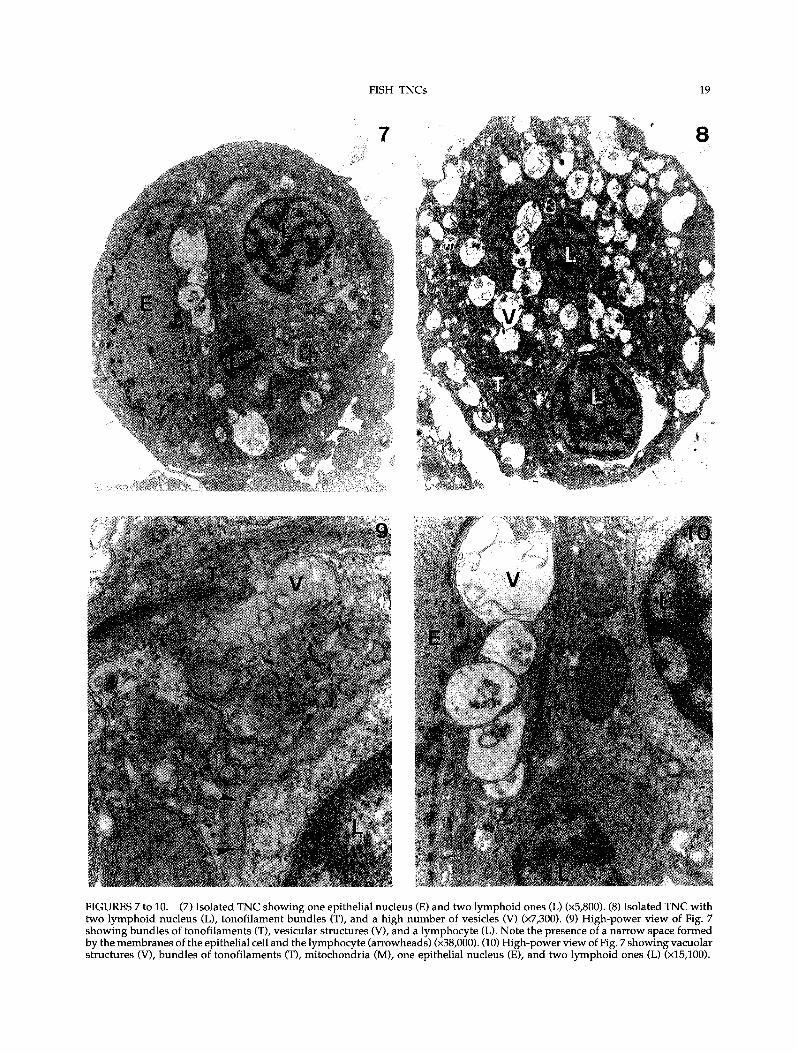
FIGURES 7 to 10. (7) Isolated TNC showing one epithelial nucleus (E) and two lymphoid ones (L) (x5,800). (8) Isolated TNC withtwo lymphoid nucleus (L), tonofilament bundles (T), and a high number of vesicles (V) (x7,300). (9) High-power view of Fig. 7showing bundles of tonofilaments (T), vesicular structures (V), and a lymphocyte (L). Note the presence of a narrow space formedby the membranes of the epithelial cell and the lymphocyte (arrowheads) (x38,000). (10) High-power view of Fig. 7 showing vacuolarstructures (V), bundles of tonofilaments (T), mitochondria (M), one epithelial nucleus (E), and two lymphoid ones (L) (x15,100).
20 FLANO et al.
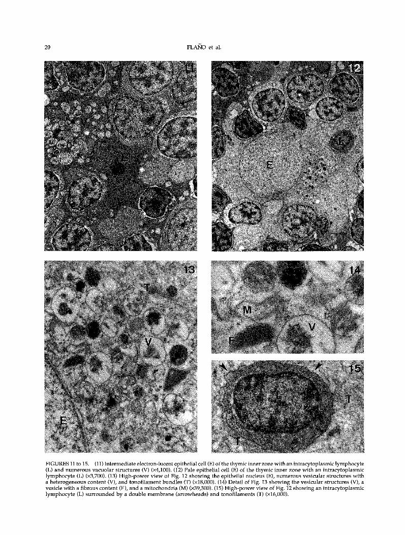
FIGURES 11 to 15. (11) Intermediate electron-lucent epithelialcell (E) of the thymic inner zone with an intracytoplasmic lymphocyte(L) and numerous vacuolar structures (V) (x4,100). (12) Pale epithelial cell (E) of the thymic inner zone with an intracytoplasmiclymphocyte (L) (x3,700). (13) High-power view of Fig. 12 showing the epithelial nucleus (E), numerous vesicular structures witha heterogeneous content (V), and tonofilament bundles (T) (x18,000). (14) Detail of Fig. 13 showing the vesicular structures (V), avesicle with a fibrous content (F), and a mitochondria (M) (x39,300). (15) High-power view of Fig. 12 showing an intracytoplasmiclymphocyte (L) surrounded by a double membrane (arrowheads) and tonofilaments (T) (x16,000).
FISH TNCs 21
consisted of one epithelial cell that embraced sev-eral lymphoid cells. The epithelial cell showed aeuchromatic or moderately heterochromatic nucleusovoid or irregular in shape, sometimes with inden-tations (Figs 7 and 8). The cytoplasm containednumerous mitochondria, free ribosomes, cisternaeof rough endoplasmic reticulum, and large tonofil-ament bundles that frequently surrounded thevacuoles containing thymocytes (Figs 9 and 10). Inaddition, the epithelial cell showed numerous largeelectron-lucent and electron-dense membranousvesicles resembling both lysosomes and endosomes(Figs 7 to 10). Some vacuoles with internal membra-nous structures were also present throughout thecytoplasm (Figs 9 and 10).Within the TNCs, several thymocytes were indi-
vidually enclosed in vacuoles lined by the epithelial-cell membrane. Membranes of both cell types werein close contact, forming a narrow intercellular space(Fig. 9). Intra-TNC thymocytes, without signs ofapoptosis or degeneration, had round or avoidheterochromatic nuclei and homogeneous, electron-dense cytoplasm containing numerous free ribo-somes and mitochondria (Figs 7 and 9).
In Situ Lymphoid-Epithelial Cell Complexes
Previously, we had defined in situ seven differenttypes of epithelial cells in the trout thymus accord-ing to their location in the organ, morphology,histochemical reactivity, and ontogenetical develop-ment (Castillo et al., 1990, 1991). Two of these epithe-lial cell types, pale and intermediate electron-lucent,which occupied the so-called inner zone of the thy-mus, appeared frequently in close association withthymocytes apparently enclosed in their cytoplasm(Figs 11 and 12). The intermediate electron-lucentepithelial cells had numerous cell processes extend-ing between neighboring thymocytes and otherepithelial cells (Fig. 11). Their cytoplasm containedmicrofilaments, elements of the Golgi apparatus,profiles of rough endoplasmic reticulum, a few elec-tron-dense vesicles, as well as numerous vesicleswith a heterogeneous electron-pale content (Fig. 11).The nucleus, irregular in shape, was moderatelyheterochromatic, with a prominent nucleolus. En-gulfed thymocytes frequently appeared in the epi-thelial cytoplasm in close contact with pale vesicles(Fig. 11). On the other hand, pale epithelial cells hadeuchromatic nuclei and ovoid electron-lucent cyto-plasm. They contained free ribosomes, mitochondria,some groups of dictyosomes, rough endoplasmic
reticulum, and large vesicles with a heterogeneouscontent (Figs 12 to 15). In addition, bundles oftonofilaments, sometimes closely apposed tothymocyte-containing vacuoles (Fig. 15), and vesi-cles with a fibrous content, appeared in these cells(Fig. 14).
Intraepithelial thymocytes were individually en-closed in vacuoles lined by the epithelial-cell mem-brane (Fig. 15). They showed no signs of celldegeneration, containing a large number of freeribosomes and mitochondria.
DISCUSSION
Our results demonstrate for the first time that thetrout thymus contains thymocyte-epithelial cell com-plexes, which can be isolated in vitro, resemblingmorphologically the TNCs described in higher ver-tebrates. As previously reported in both mammalsand birds (Wekerle et al., 1980; Vakharia, 1983;Penninger et al., 1994), the teleost TNCs consist ofepithelial cells with numerous mitochondria, pro-files of rough endoplasmic reticulum, large bundlesof tonofilaments, and membranous organelles re-sembling endo- and lysosomes. In addition, they arekeratin-positive and exhibit acid and alkalinephosphatase and nonspecific esterase activities.These morpho-cytochemical features suggest thattrout TNCs have a high metabolic activity and con-tain presumably acidic cytoplasmic compartments,important for antigen processing, as recently dem-onstrated in chicken TNCs (Penninger et al., 1994).On the other hand, our in situ electron microscopy
study confirms previous results (Pulsford et al.,1991; ,lvarez, 1993) concerning the occurrence ofthymocyte-epithelial-cell complexes in the teleostthymus. They correspond to both pale and inter-mediate electron-lucent epithelial cells of the innerzone of trout thymus. Indeed, they resemble mor-phologically the so-called type 2 (pale) and type 3(intermediate) epithelial cells of the human thymus(Wijngaert et al., 1984), which have been proposedas the TNCs found in suspensions (Van de Wijngaertet al., 1984; Boyd et al., 1993). Thus, because mam-malian TNCs seem to correspond mainly to corticalepithelial cells (Ritter et al., 1981; Kyewski andKaplan, 1982; Van Vliet et al., 1984; Nabarra andAdrianarison, 1987), although their relationship tosubcapsular (Kyewski and Kaplan, 1982; Kaneshimaet al., 1987; Mizutami et al., 1987) and medullar epi-thelium has also been mentioned (Hugo et al., 1988),
22 FLANO et al.
our results indirectly demonstrate the phylogeneticalrelationships of the inner zone of the teleost thymuswith the thymic cortex of higher vertebrates, a factrepeatedly suggested by our group (Zapata, 1981;Castillo et al., 1990, 1991). It is, however, difficult,from a merely morphological study, to establish cor-relations between the in vitro isolated TNCs andthese lymphocyte-epithelial-cell clusters identifiedin situ. Moreover, some authors have mentioned acertain heterogeneity in enriched TNCs from thehuman thymus (Ritter et al., 1981).
All phenotypical studies remark on the expressionof class I and class II MHC on higher-vertebrateTNCs (Wekerle et al., 1980; Ritter et al., 1981; Boydet al., 1984), although the use of trypsine treatmentduring the isolation of cell clusters modifies thisexpression (Kyewski and Kaplan, 1982). Unfortu-nately, there are no available reagents to detect MHCmolecules in teleost fish, although recent moleculardata demonstrated the existence of both class I andclass II antigens in them (Kromenberg et al., 1994).
Ltd., Bale, Switzerland), blood was extracted fromthe caudal sinus, and the thymi were dissected.
Cell Isolation
The thymi were mechanically minced in ice-coldphosphate-buffered saline (PBS), and the super-natant containing free cells was discarded. The re-maining tissue fragments were incubated in PBS for10 min at 15C with stirring, and the supernatantdiscarded again. After this mechanical dissociation,the tissue fragments were digested with 0.6 mg/mltype Ia collagenase (Sigma, St. Louis) in PBS for15 min at 15C under agitation. The supernatant wasdiscarded and this step was repeated three times,the supernatants of the two last steps being har-vested. After collagenase digestion, the remainingtissue fragments were trypsinized (1% trypsin-EDTAin PBS; Boehringer-Mannheim) for 30 min at 15Cunder gentle agitation. Collection of TNC wasachieved using a modification of the method of
By using cross-reactive monoclonal antibodies, Wekerle et al. (1980). TNC-containing fractions wereKaufman et al. (1990) detected MHC-like moleculesin some nonmammalian vertebrates. On this samebasis, we have demonstrated a slight expression ofMHC class II antigens in trout TNCs using an anti-chicken class II MHC molecule monoclonal antibody.The expression of class II molecules and the ex-
istence of an acidic cytoplasmic compartment, asindicated by our enzyme-histochemical and ultra-structural results, stggest some capacity of teleostTNCs for antigenic processing and presentation, asrecently reported for chicken TNCs (Penninger et al.,1994). Nevertheless, the TCR-MHC interactions areapparently not necessary for the formation of mouseTNCs (Boyd et al., 1993) and, although most authorsemphasize a role for TNCs in intra-thymic T-cellmaturation, their functional significance is reallyobscure.
MATERIALS AND METHODS
Animals
Thymi from rainbow trout, Oncorhynchus mykiss, andbrown trout, Salmo trutta, were used for cell isola-tion and histological examination. Fish were agedby scalimetry and were between 1 and 3 years. Nodifferences were seen between the two species stud-ied in any of the experimental systems used. Fishwere anesthesized with MS-222 (Sandoz Pharma
enriched for TNC by one 1-g sedimentation over FCSat 4C for 15 min. After sedimentation, the top layerwas discarded, and the rest of the suspension wascentrifuged at 250 g for 10 min at 4C and resus-pended in PBS.
Routine Histology
The isolated cells were placed in drops onto slides,air dried, and stained with haematoxylin-eosin forroutine examination and identification of multi-cellular complexes.
Enzyme- and Immuno-Cytochemical Analysis
The assayed enzymatic activities and the substratesused are summarized in Table 1. Negative controlsfor the specificity of the enzymatic reactions wereestablished using incubation media in which the cor-responding substrates were lacking.
For immunodetection, cells were fixed in acetonefor 10 min at room temperature, air dried, and pro-cessed by the indirect immunoperoxidase techni-que using 3,3-diaminobenzidine-tetrahydrochloride(Sigma) as a coupling reagent. Controls were sys-tematically performed by omission of the first anti-body, and endogenous peroxidase activity waspreviously inhibited by incubation in 0.3% hydro-gen peroxide in methanol for 10 min at room
FISH TNCs 23
temperature. Keratins were identified using a pan-keratin rabbit polyclonal antibody (Dako, Denmark),and MHC class II proteins were detected using cross-reactivity of a mouse monoclonal antibody myc-16raised against chicken Ia antigens (kindly providedby Dr M. Cooper). As secondary antibodies, a goatanti-rabbit Ig (Sigma) and a rabbit anti-mouse IgGantiserum (Dako), peroxidase-conjugated, were used,respectively. Before their use on isolated cells, allantibodies were checked for their specificity in vivoon acetone-fixed cryosections of trout thymus.
Transmission Electron Microscopy
Isolated cells and thymi were fixed in 2% gluta-raldehyde in 0.1 M cacodylate buffer, pH 7.2,postfixed with 1% osmium tetroxide in the samebuffer, dehydrated in acetone series, counterstainedwith 1% uranyl acetate in 70% acetone, and embed-ded in Araldite (Durcupan, ACM, Fluka, Switzer-land). One-micrometer thick sections were stainedwith an aqueous solution of toluidine blue in boraxto select the most suitable areas. Ultrathin sectionswere obtained with a Reichert-Jung UM-3 ultratome,counterstained with lead citrate, and examined in a
JEOL-EM1010 electron microscope at 60 kV.
ACKNOWLEDGMENTS
The authors thank Dr M. Cooper for kindly pro,viding themyc-16 monoclonal antibody. E. Flafio and F. Alvarez are
supported by EC contract AIR1 CT92 0036.
(Received September 6, 1995)
(Accepted January 3, 1996)
REFERENCES
Aguilar L.K., Aguilar-Cordova E., Cartwright J. Jr., and BelmontJ.W. (1994). Thymic nurse cells are sites of thymocyte apoptosis.
J. Immunol. 152: 2645-2651.lvarez F. (1993). Immunobiologia de salm6nidos. Anlisis
morfol6gico de los mecanismos de defensa de Salmo truttacontra la infecci6n por Saprolegnia sp. Estudio del tegumentoy los 6rganos linfoides. Doctoral thesis, University of Le6n,Le6n, Spain.
Barka T., and Anderson P.J. (1962). Histochemical methods foracid phosphatase using hexazonium pararosanilin as coupler.J. Histochem. Cytochem. 10: 741-753.
Boyd R.L., Oberhuber G., Hala K., and Wick G. (1984). Obesestrain (os) chickens with spontaneous autoimmune tyroiditishave a deficiency in thymic nurse cells. J. Immunol. 132:718-724.
Boyd R.L., Tucek C.L., Godfrey D.I., et al. (1993). The thymicmicroenvironment. Immunol. Today 14: 445-459.
Burstone M.S.(1958). Histochemical comparison of naphthol AS-phosphates for the demonstration of phosphatases. J. NatlCancer Inst. 20: 601-615.
Castillo A., L6pez-Fierro P., Zapata A., Villena A., and RazquinB. (1991). Post-hatching development of the thymic epithelialcells in the rainbow trout Salmo gairdneri: An ultrastructuralstudy. Amer. J. Anat. 190: 299-307.
Castillo A., Razquin B.E., L6pez-Fierro P., lvarez F., Zapata A.,and Villena A. (1990). Enzyme- and immuno-histochemicalstudy of the thymic stroma in the rainbow trout, Salmo gairdneriRich. Thymus 15: 153-166.
De Waal Malefijt R., Leene W., Rohall P.J.M., Wormmeester J.,and Hoeben K.A. (1986). T cell differentiation within thymicnurse cells. Lab. Invest. 55: 25-34.
F/inge R. and Pulsford A. (1985). The thymus of the angler fishLophius piscatorius: An ultrastructural study. In: Fish immunol-ogy, Manning M.J., and Tatner M.F., Eds. (London: AcademicPress), pp. 293-311.
Farr A.G., and Anderson S.K. (1985). Epithelial cell heterogene-ity in the murine thymus: Fucose-specific lectins bind medul-lary epithelial cells. J. Immunol. 134: 2971-2977.
Gerdes J., Stein H., Masm D.Y., and Ziegler A. (1983). Humandendritic reticulum cells of lymphoid follicles: Their antigenicprofile and their identification as multinucleated giant cells.Virchows Arch. (Cell. Pathol.) 42: 161-172.
Holtfreter H.B., and Cohen N. (1987). In vitro behavior of thymicnurse cell-like complexes from mechanically and enzymaticallydissociated frog tadpole thymus. Amer. J. Anat. 179: 342-355.
Hugo P., St-Pierre Y., and Potworowsky E.F. (1988). Characteri-zation of the in vitro interaction between thymocytes and amedullary thymic epithelial cell line. Thymus 12: 27-37.
Kaneshima H., Ito M., Asai J., Taguchi O., and Hiai H. (1987).Thymic epithelial cell subpopulations in mice defined bymonoclonal antibodies. Lab. Invest. 56: 372-380.
Kaufman J., Ferrone S., Flajnik M., Kilb M., V61k H., and ParisotR. (1990). MHC-like molecules in some non-mammalian ver-tebrates can be detected by some cross-reactive monoclonalantibodies. J. Immunol. 144: 2273-2280.
Kronenberg M., Brines R., and Kaufman J. (1994). MHC evolu-tion: A long term investment in defense. Immunol. Today 4:4-6.
TABLE
Enzymatic activity Substrate Dose(mg/ml) Incubation medium Reference
ACPH Naphthol AS-BI phosphate 0.5 pH 5 Barka and Anderson, 1962
AKPH Naphthol AS-BI phosphate 0.1 pH 8 Burstone, 1958
ANAE c-naphthyl acetate 0.25 pH 6.5 Pearse, 1972
aACPH: acid phosphatase. AKPH: alkaline phosphatase. ANAE: nonspecific a-naphthyl acetate esterase.
24 FLANO et al.
Kyewski B.A., and Kaplan H.S. (1982). Lymphoepithelialinterations in the mouse thymus: Phenotypic and kinetic stud-ies on thymic nurse cells. J. Immunol. 128: 2287-2294.
Kyewski B.A. (1986). Thymic nurse cells: Possible sites of T-cellselection. Immunol. Today 7: 374-379.
Kyewski B.A., Momburg F., and Schirrmacher V. (1987). Pheno-type of stromal cell-associated thymocytes in situ is compati-ble with selection of the T cell repertoire at an "inmature"stage of thymic T cell differentiation. Eur. J. Immunol. 17:961-967.
Lorenz R.G., and Allen P.M. (1989). Thymic cortical epithelialcells can present self antigens in vivo. Nature 337: 560-562.
Manconi P.E., Ennas M.G., Murrn M.R. Caddedu G., Tore G.,and Lantini M.S. (1984). Epithelial-like cells containinglymphocytes (nurse cells) in human adenoids and tonsils.Thymus 6: 351-357.
Mizutami S., Watt S.M., Robertson D., Hussein S., Healy L.E.,Furley A.J.V., and Greaves M.F. (1987). Cloning of humanthymic subcapsular-cortex epithelial cells with T-lymphocytebinding sites and hemopoietic growth factor acticity. Proc. NatlAcad. Sci. USA 84: 4999-5003.
Nabarra B., and Adrianarison I. (1987). Ultrastructural studiesof thymic reticulum. I. Epithelial component. Thymus 9:95-121.
Navarro R. (1987). Ontogenia de los 6rganos linfoides deScyliorhinus canicula. Estudio ultraestructural. Master thesis,Complutense University, Madrid.
Owen J.J.T., Jenkison E.J., and Kingston R. (1986). Thymic stemcells: Their interaction with the thymic stroma and toleranceinduction. Curr. Topics Microbiol. Immunol. 126: 35-41.
Pearse A.G.E. (1972). Histochemistry. Theoretical and applied(Edinburg: Churchill Livingstone).
Penninger J., Hala K., and Wick G. (1990). Intrathymic nurse celllymphocyte can induce a specific graft-versus-host reaction.J. Exp. Med. 172: 521-529.
Penninger J., Rieker T., Romani N., et al. (1994). Ultrastructuralanalysis of thymic nurse cell epithelium. Eur. J. Immunol. 24:222-228.
Pulsford A., F/inge R., and Zapata A. (1991). The thymicmicroenvironment of the common sole, Solea solea. Acta Zool.(Stockholm) 72: 209-216.
Pulsford A., Morrow W.J.W., and Fringe R. (1984). Ultrastructuralstudies on the thymus of the dogfish, Scyliorhinus canicula L.J.Fish Biol. 25: 353-360.
Ritter M.A., Sauvage C.A., and Cotmore S.F. (1981). The humanthymus microenvironment: In vivo identification of thymicnurse cells and other antigenically-distinct subpopulations ofepithelial cells. Immunology 44: 439-446.
Ron Y., Lo D., and Spent J. (1986). T cell specificity in twice-irradiated Fl-into-parent bone marrow chimeras: Failure to de-tect a role for immigrant marrow-derived cells in imprintingintrathymic H-2 restriction. J. Immunol. 137: 1764-1771.
Singer K.H., Wolf L:S., Lobach D.F., Denning S.M., Tuck D.T.,Robertso A.L., and HaynesB.E (1986). Human thymocytes bindto autologus and allogenic thymic epithelial cells in vitro. Proc.Natl Acad. Sci. USA 83: 6588-6592.
Speiser D.E., Pircher H., Ohashi P.S., Kyburz D., Hengartner H.,and Zinkernagel R.M. (1992). Clonal deletion induced by ei-ther radioresistant thymic host cells or lymphohemopoieticdonor cells at different stages of class I-restricted T-cellontogeny. J. Exp. Med. 175: 1277-1284.
Toussaint-Demylle D., Scheiff J.M., and Haumont S. (1993).Thymic nurse cells in culture: Morphological and antigeniccharacterization. Cell Tis. Res. 272: 343-354.
Tsunoda R., and Kojima M. (1987). A light microscopical studyof isolated follicular dendritic cell-clusters in human tonsils.Acta Pathol. Jpn 37: 575-586.
Vakharia D.D. (1983). Demonstration of keratin filaments inthymic nurse cells and alloreactivity of TNC-T cell popula-tion. Thymus 5: 43-52.
Van de Wijngaert F.P., Kendall M.D., Schuurman H.J.,Rademakers L.H.P.M., and Kater L. (1984). Heterogeneity ofepithelial cells in the human thymus. An ultrastructural study.Cell Tissue Res. 237: 227-237.
Van Vliet E., Melis M., and Van Ewijk W. (1984). Immunohistologyof thymic nurse cells. Cell. Immunol. 87: 101-109.
Wekerle H., Ketelsen U.P., and Ernst M. (1980). Thymic nursecells. Lymphoepithelial cell complexes in murine thymuses:Morphological and Serological characterization. J. Exp. Med.151: 925-944.
Wick G., and Oberhuber G. (1986). Thymic nurse cells: A schoolfor alloreactive and autoreactive cortical thymocytes? Eur. J.Immunol. 16: 855-858.
Wood G.W., Mauser L., Fleming R.H., Greenwood J., andSandford T.H. (1988). J. Immunol. 141: 2221-2229.
Zapata A. (1981). Lymphoid organs of teleost fish. I. Ultra-structure of the thymus of Rutilus rutilus. Dev. Comp. Immunol.5: 427-436.


![Lymphosarcoma: Virus-induced Thymic ... - Cancer Research · [CANCER RESEARCH 30, 2213-2222, August 1970] Lymphosarcoma: Virus-induced Thymic-independent Disease in Mice1 Herbert](https://img.pdfslide.us/doc/110x75/5fd343694fa1b372eb7f08e2/lymphosarcoma-virus-induced-thymic-cancer-research-cancer-research-30-2213-2222.jpg)