Embed Size (px)
Citation preview
http://www.diva-portal.org
This is the published version of a paper published in PLoS ONE.
Citation for the original published paper (version of record):
Skogberg, G., Gudmundsdottir, J., van der Post, S., Sandström, K., Bruhn, S. et al. (2013)
Characterization of human thymic exosomes.
PLoS ONE, 8(7): e67554
http://dx.doi.org/10.1371/journal.pone.0067554
Access to the published version may require subscription.
N.B. When citing this work, cite the original published paper.
Permanent link to this version:http://urn.kb.se/resolve?urn=urn:nbn:se:umu:diva-79252
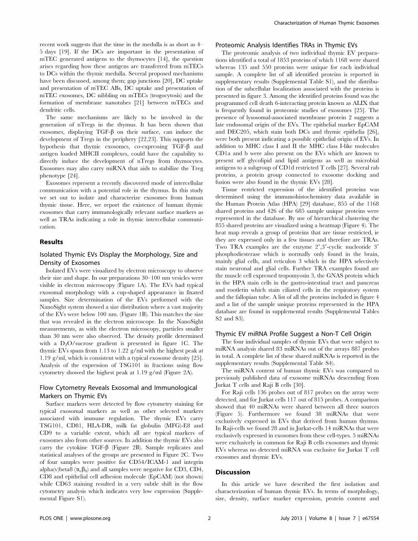
Characterization of Human Thymic ExosomesGabriel Skogberg1*, Judith Gudmundsdottir1,2, Sjoerd van der Post3, Kerstin Sandstrom4, Soren Bruhn5,
Mikael Benson5,6, Lucia Mincheva-Nilsson7, Vladimir Baranov7, Esbjorn Telemo1, Olov Ekwall1,2
1Dept of Rheumatology and Inflammation Research at the Institute of Medicine, University of Gothenburg, Gothenburg, Sweden, 2Dept of Pediatrics at the Institute of
Clinical Sciences, University of Gothenburg, Gothenburg, Sweden, 3 The Proteomics Core Facility, University of Gothenburg, Gothenburg, Sweden, 4Department of
Pediatric Anesthesia and Intensive Care at the Sahlgrenska Academy, University of Gothenburg, Gothenburg, Sweden, 5 The Center for Invidualized Medication, Linkoping
University, Linkoping, Sweden, 6Department of Pediatrics, Linkoping University, Linkoping, Sweden, 7Department of Clinical Microbiology Division of Clinical
Immunology, Umea University, Umea, Sweden
Abstract
Exosomes are nanosized membrane-bound vesicles that are released by various cell types and are capable of carryingproteins, lipids and RNAs which can be delivered to recipient cells. Exosomes play a role in intercellular communication andhave been described to mediate immunologic information. In this article we report the first isolation and characterization ofexosomes from human thymic tissue. Using electron microscopy, particle size determination, density gradientmeasurement, flow cytometry, proteomic analysis and microRNA profiling we describe the morphology, size, density,protein composition and microRNA content of human thymic exosomes. The thymic exosomes share characteristics withpreviously described exosomes such as antigen presentation molecules, but they also exhibit thymus specific featuresregarding surface markers, protein content and microRNA profile. Interestingly, thymic exosomes carry proteins that have atissue restricted expression in the periphery which may suggest a role in T cell selection and the induction of centraltolerance. We speculate that thymic exosomes may provide the means for intercellular information exchange necessary fornegative selection and regulatory T cell formation of the developing thymocytes within the human thymic medulla.
Citation: Skogberg G, Gudmundsdottir J, van der Post S, Sandstrom K, Bruhn S, et al. (2013) Characterization of Human Thymic Exosomes. PLoS ONE 8(7): e67554.doi:10.1371/journal.pone.0067554
Editor: Cheryl A. Stoddart, University of California, San Francisco, United States of America
Received November 21, 2012; Accepted May 21, 2013; Published July 2, 2013
Copyright: � 2013 Skogberg et al. This is an open-access article distributed under the terms of the Creative Commons Attribution License, which permitsunrestricted use, distribution, and reproduction in any medium, provided the original author and source are credited.
Funding: This project was supported by the Swedish Research Council (contract 80409601), Region Vastra Gotaland (ALFGBG-771712), AFA Forsakring (contract100258), IngaBritt and Arne Lundbergs Research Foundation and The Gothenburg Medical Society (all grants received by OE). The funders had no role in studydesign, data collection and analysis, decision to publish, or preparation of the manuscript.
Competing Interests: The authors have declared that no competing interests exist.
* E-mail: [email protected]
Introduction
Extracellular vesicles (EVs) are membrane bound vesicles shed
from various types of cells into the extracellular space. Exosomes,
ectosomes and apoptotic bodies (ABs) are all examples of EV
subgroups [1]. Exosomes are defined as 40–100 nm vesicles with a
density ranging from 1.10–1.21 g/ml that carry proteins such as
tumor susceptibility gene 101 (TSG101), which indicate an
endocytic origin [1]. Exosomes are formed from inward budding
of late endosomes and are released at the cell surface when
multivesicular endosomes fuse with the outer cell membrane.
Several recent studies have focused on a diverse set of exosomal
functions such as antigen presentation and microRNA (miRNA)
transfer between cells [2], induction of transplant tolerance [3],
tolerance to fed antigens [4], tumor immunosuppression [5],
transporting vesicles used by retroviruses [6] and prions [7,8] and
their ability to cross the blood brain barrier [9].
In mice, thymic exosome-like particles (ELPs) have been
demonstrated to express membrane bound transforming growth
factor beta (TGF-b) and MHC class II [10]. These thymic ELPs
seem to posses the ability to induce the formation of natural
regulatory T cells (nTregs) from thymocytes. Interestingly,
intercellular transfer of thymic stromal material to thymic
dendritic cells (DCs) has also been observed in mice [11]. To
our knowledge, there is no previous report on the existence,
characterization or function of exosomes from the human thymus.
In the thymus progenitor T cells go through sequential selection
and maturation steps that determine the fate of the individual
thymocyte. This process is orchestrated by surrounding cells and
leads to the final export of mature CD4 and CD8 single positive T
cells to the periphery. During this process nTregs, capable of
regulating other T cells, are also formed. The intercellular
communication, which is pivotal for this process is classically
based on direct cell-cell contact, and the production of cytokines
and chemokines. However, the precise mechanisms that regulate
thymic T cell maturation, are still not completely clear. One
crucial event in this process is the presentation of tissue restricted
antigens (TRAs) to maturing thymocytes. The TRAs mirror parts
of the self-antigen repertoire and are presented to maturing
thymocytes during the selection process. Medullary thymic
epithelial cells (mTECs) express TRAs [12] under the influence
of the autoimmune regulator protein [13]. However, there is an
ongoing discussion as to what extent mTECs are able to present
antigens directly to thymocytes or if DCs are needed as a route to
aid the presentation of mTEC derived antigens to thymocytes
[14,15,16]. Also, adding to the complexity of antigen presence in
the thymic medulla, not every mTEC express every TRA at a
given timepoint [12], [17] which lowers the probability that all
TRAs are to be presented by mTECs directly to the thymocytes.
This could in part be compensated by the long, up to two weeks
time [18], that thymocytes reside in the thymic medulla. However,
PLOS ONE | www.plosone.org 1 July 2013 | Volume 8 | Issue 7 | e67554
recent work suggests that the time in the medulla is as short as 4–
5 days [19]. If the DCs are important in the presentation of
mTEC generated antigens to the thymocytes [14], the question
arises regarding how these antigens are transferred from mTECs
to DCs within the thymic medulla. Several proposed mechanisms
have been discussed, among them; gap junctions [20], DC uptake
and presentation of mTEC ABs, DC uptake and presentation of
mTEC exosomes, DC nibbling on mTECs (trogocytosis) and the
formation of membrane nanotubes [21] between mTECs and
dendritic cells.
The same mechanisms are likely to be involved in the
generation of nTregs in the thymus. It has been shown that
exosomes, displaying TGF-b on their surface, can induce the
development of Tregs in the periphery [22,23]. This supports the
hypothesis that thymic exosomes, co-expressing TGF-b and
antigen loaded MHCII complexes, could have the capability to
directly induce the development of nTregs from thymocytes.
Exosomes may also carry miRNA that aids to stabilize the Treg
phenotype [24].
Exosomes represent a recently discovered mode of intercellular
communication with a potential role in the thymus. In this study
we set out to isolate and characterize exosomes from human
thymic tissue. Here, we report the existence of human thymic
exosomes that carry immunologically relevant surface markers as
well as TRAs indicating a role in thymic intercellular communi-
cation.
Results
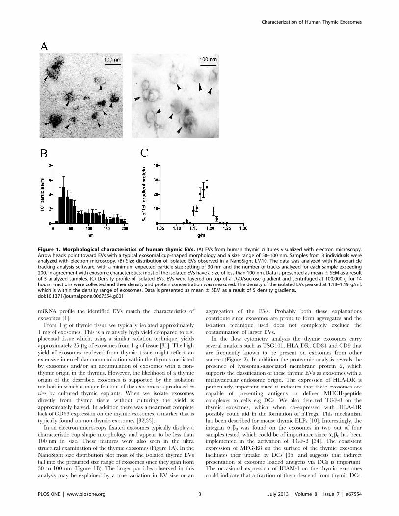
Isolated Thymic EVs Display the Morphology, Size andDensity of ExosomesIsolated EVs were visualized by electron microscopy to observe
their size and shape. In our preparations 30–100 nm vesicles were
visible in electron microscopy (Figure 1A). The EVs had typical
exosomal morphology with a cup-shaped appearance in fixated
samples. Size determination of the EVs performed with the
NanoSight system showed a size distribution where a vast majority
of the EVs were below 100 nm. (Figure 1B). This matches the size
that was revealed in the electron microscope. In the NanoSight
measurements, as with the electron microscopy, particles smaller
than 30 nm were also observed. The density profile determined
with a D2O/sucrose gradient is presented in figure 1C. The
thymic EVs spans from 1.13 to 1.22 g/ml with the highest peak at
1.19 g/ml, which is consistent with a typical exosome density [25].
Analysis of the expression of TSG101 in fractions using flow
cytometry showed the highest peak at 1.19 g/ml (Figure 2A).
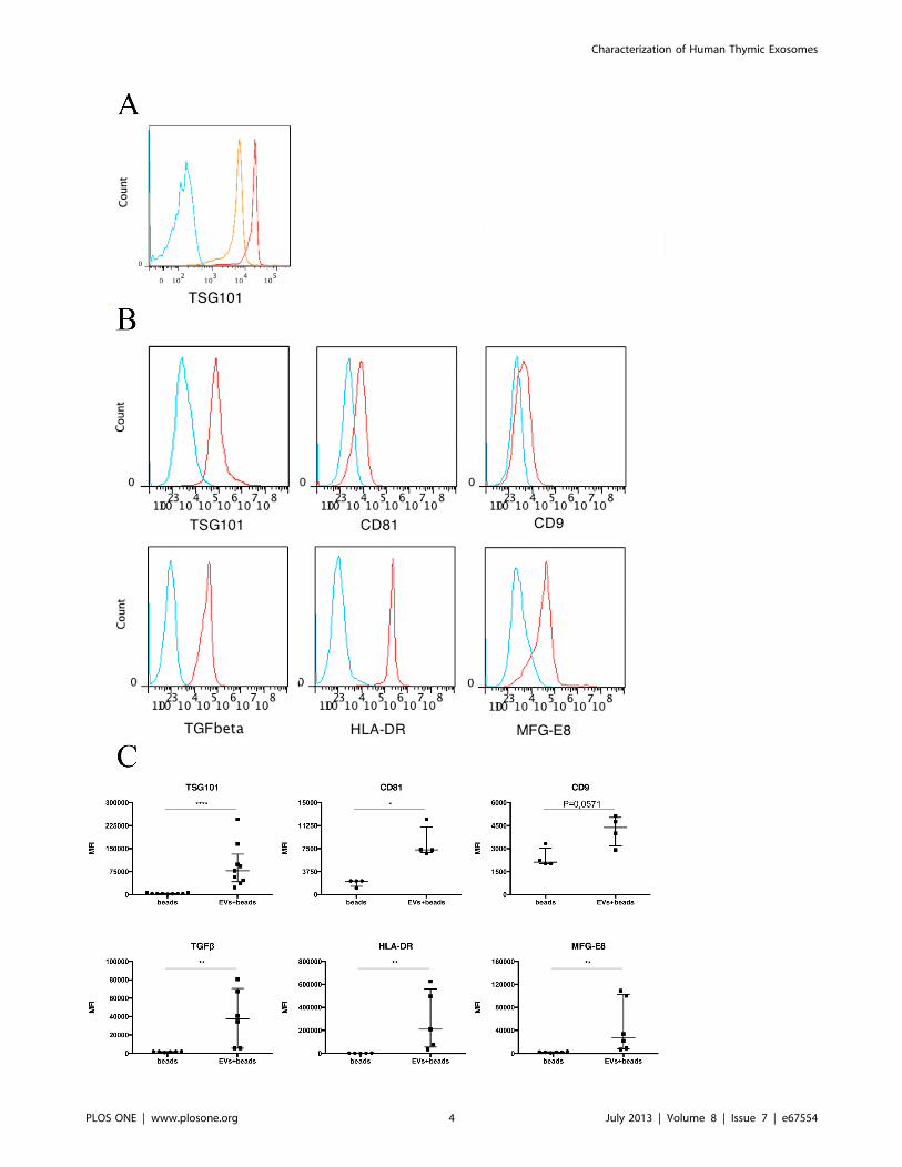
Flow Cytometry Reveals Exosomal and ImmunologicalMarkers on Thymic EVsSurface markers were detected by flow cytometry staining for
typical exosomal markers as well as other selected markers
associated with immune regulation. The thymic EVs carry
TSG101, CD81, HLA-DR, milk fat globulin (MFG)-E8 and
CD9 to a variable extent, which all are typical markers of
exosomes also from other sources. In addition the thymic EVs also
carry the cytokine TGF-b (Figure 2B). Sample replicates and
statistical analyses of the groups are presented in Figure 2C. Two
of four samples were positive for CD54/ICAM-1 and integrin
alpha(v)beta8 (avb8) and all samples were negative for CD3, CD4,
CD8 and epithelial cell adhesion molecule (EpCAM) (not shown)
while CD63 staining resulted in a very subtle shift in the flow
cytometry analysis which indicates very low expression (Supple-
mental Figure S1).
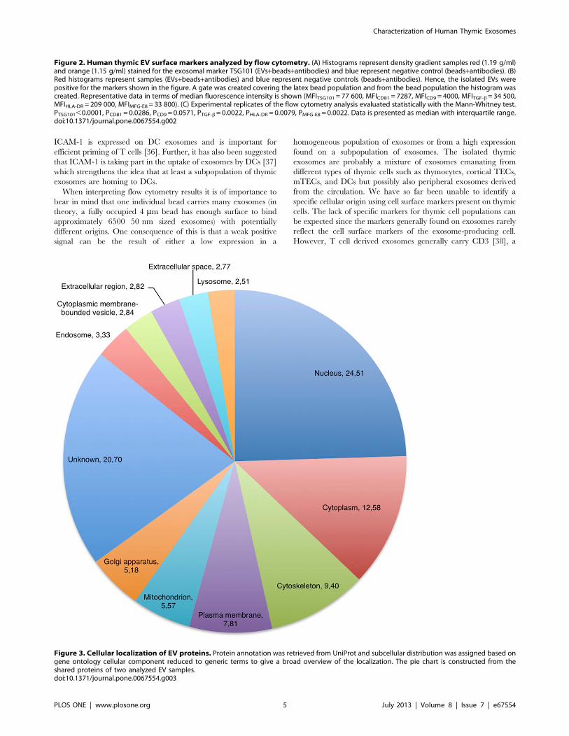
Proteomic Analysis Identifies TRAs in Thymic EVsThe proteomic analysis of two individual thymic EV prepara-
tions identified a total of 1853 proteins of which 1168 were shared
whereas 135 and 550 proteins were unique for each individual
sample. A complete list of all identified proteins is reported in
supplementary results (Supplemental Table S1), and the distribu-
tion of the subcellular localization associated with the proteins is
presented in figure 3. Among the identified proteins found was the
programmed cell death 6-interacting protein known as ALIX that
is frequently found in proteomic studies of exosomes [25]. The
presence of lysosomal-associated membrane protein 2 suggests a
late endosomal origin of the EVs. The epithelial marker EpCAM
and DEC205, which stain both DCs and thymic epithelia [26],
were both present indicating a possible epithelial origin of EVs. In
addition to MHC class I and II the MHC class I-like molecules
CD1a and b were also present on the EVs which are known to
present self glycolipid and lipid antigens as well as microbial
antigens to a subgroup of CD1d restricted T cells [27]. Several rab
proteins, a protein group connected to exosome docking and
fusion were also found in the thymic EVs [28].
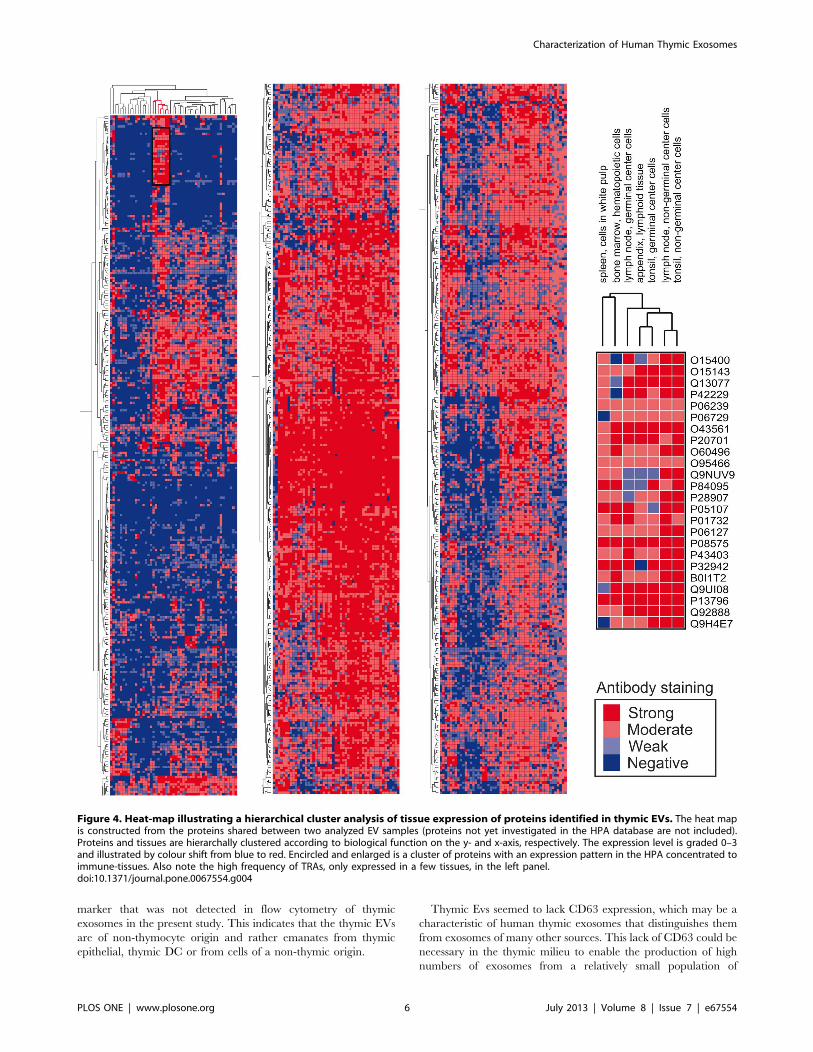
Tissue restricted expression of the identified proteins was
determined using the immunohistochemistry data available in
the Human Protein Atlas (HPA) [29] database, 855 of the 1168
shared proteins and 426 of the 685 sample unique proteins were
represented in the database. By use of hierarchical clustering the
855 shared proteins are visualized using a heatmap (Figure 4). The
heat map reveals a group of proteins that are tissue restricted, ie
they are expressed only in a few tissues and therefore are TRAs.
Two TRA examples are the enzyme 29,39-cyclic nucleotide 39
phosphodiesterase which is normally only found in the brain,
mainly glial cells, and reticulon 3 which in the HPA selectively
stain neuronal and glial cells. Further TRA examples found are
the muscle cell expressed tropomyosin 3, the GNAS protein which
in the HPA stain cells in the gastro-intestinal tract and pancreas
and rootletin which stain ciliated cells in the respiratory system
and the fallopian tube. A list of all the proteins included in figure 4
and a list of the sample unique proteins represented in the HPA
database are found in supplemental results (Supplemental Tables
S2 and S3).
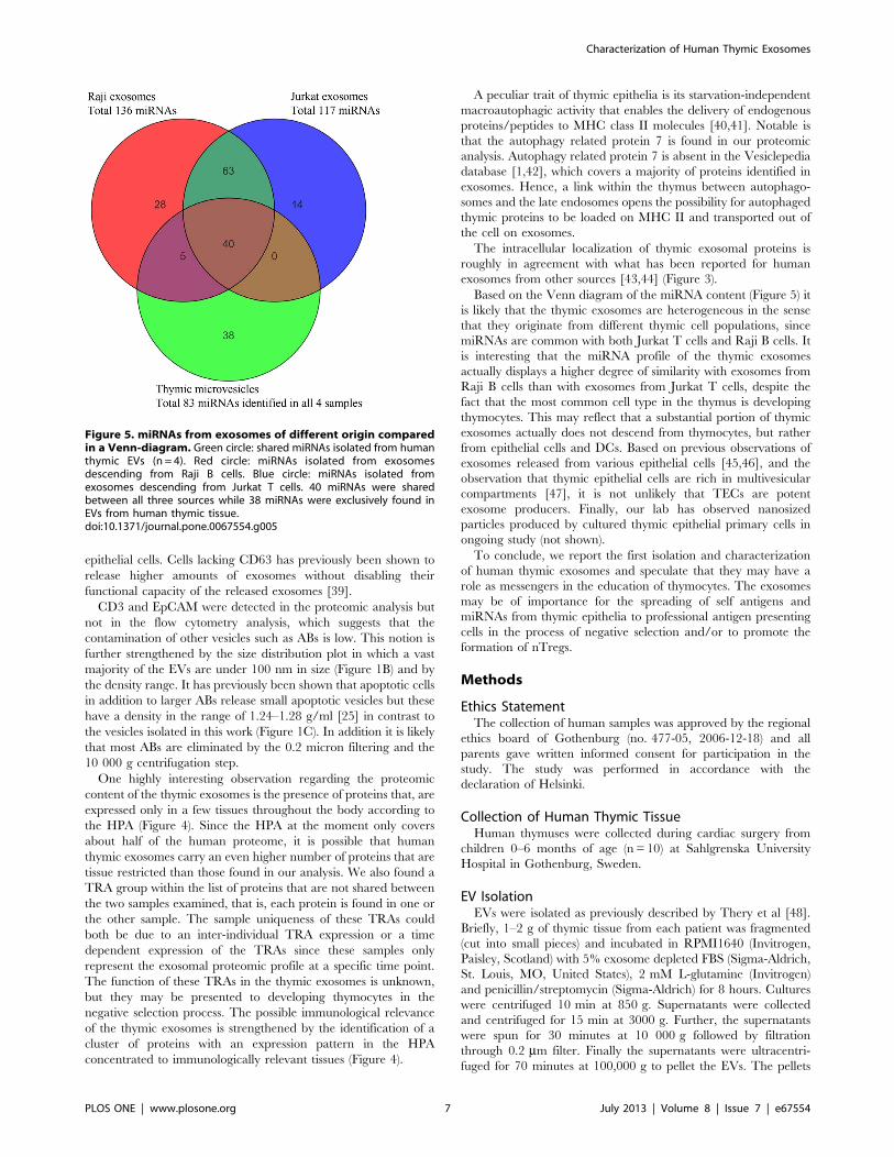
Thymic EV miRNA Profile Suggest a Non-T Cell OriginThe four individual samples of thymic EVs that were subject to
miRNA analysis shared 83 miRNAs out of the arrays 887 probes
in total. A complete list of these shared miRNAs is reported in the
supplementary results (Supplemental Table S4).
The miRNA content of human thymic EVs was compared to
previously published data of exosome miRNAs descending from
Jurkat T cells and Raji B cells [30].
For Raji cells 136 probes out of 817 probes on the array were
detected, and for Jurkat cells 117 out of 815 probes. A comparison
showed that 40 miRNAs were shared between all three sources
(Figure 5). Furthermore we found 38 miRNAs that were
exclusively expressed in EVs that derived from human thymus.
In Raji-cells we found 28 and in Jurkat-cells 14 miRNAs that were
exclusively expressed in exosomes from these cell-types. 5 miRNAs
were exclusively in common for Raji B cells exosomes and thymic
EVs whereas no detected miRNA was exclusive for Jurkat T cell
exosomes and thymic EVs.
Discussion
In this article we have described the first isolation and
characterization of human thymic EVs. In terms of morphology,
size, density, surface marker expression, protein content and
Characterization of Human Thymic Exosomes
PLOS ONE | www.plosone.org 2 July 2013 | Volume 8 | Issue 7 | e67554
miRNA profile the identified EVs match the characteristics of
exosomes [1].
From 1 g of thymic tissue we typically isolated approximately
1 mg of exosomes. This is a relatively high yield compared to e.g.
placental tissue which, using a similar isolation technique, yields
approximately 25 mg of exosomes from 1 g of tissue [31]. The high
yield of exosomes retrieved from thymic tissue might reflect an
extensive intercellular communication within the thymus mediated
by exosomes and/or an accumulation of exosomes with a non-
thymic origin in the thymus. However, the likelihood of a thymic
origin of the described exosomes is supported by the isolation
method in which a major fraction of the exosomes is produced ex
vivo by cultured thymic explants. When we isolate exosomes
directly from thymic tissue without culturing the yield is
approximately halved. In addition there was a nearmost complete
lack of CD63 expression on the thymic exosomes, a marker that is
typically found on non-thymic exosomes [32,33].
In an electron microscopy fixated exosomes typically display a
characteristic cup shape morphology and appear to be less than
100 nm in size. These features were also seen in the ultra
structural examination of the thymic exosomes (Figure 1A). In the
NanoSight size distribution plot most of the isolated thymic EVs
fall into the presumed size range of exosomes since they span from
30 to 100 nm (Figure 1B). The larger particles observed in this
analysis may be explained by a true variation in EV size or an
aggregation of the EVs. Probably both these explanations
contribute since exosomes are prone to form aggregates and the
isolation technique used does not completely exclude the
contamination of larger EVs.
In the flow cytometry analysis the thymic exosomes carry
several markers such as TSG101, HLA-DR, CD81 and CD9 that
are frequently known to be present on exosomes from other
sources (Figure 2). In addition the proteomic analysis reveals the
presence of lysosomal-associated membrane protein 2, which
supports the classification of these thymic EVs as exosomes with a
multivesicular endosome origin. The expression of HLA-DR is
particularly important since it indicates that these exosomes are
capable of presenting antigens or deliver MHCII-peptide
complexes to cells e.g DCs. We also detected TGF-ß on the
thymic exosomes, which when co-expressed with HLA-DR
possibly could aid in the formation of nTregs. This mechanism
has been described for mouse thymic ELPs [10]. Interestingly, the
integrin avb8 was found on the exosomes in two out of four
samples tested, which could be of importance since avb8 has beenimplemented in the activation of TGF-b [34]. The consistent
expression of MFG-E8 on the surface of the thymic exosomes
facilitates their uptake by DCs [35] and suggests that indirect
presentation of exosome loaded antigens via DCs is important.
The occasional expression of ICAM-1 on the thymic exosomes
could indicate that a fraction of them descend from thymic DCs.
Figure 1. Morphological characteristics of human thymic EVs. (A) EVs from human thymic cultures visualized with electron microscopy.Arrow heads point toward EVs with a typical exosomal cup-shaped morphology and a size range of 50–100 nm. Samples from 3 individuals wereanalyzed with electron microscopy. (B) Size distribution of isolated EVs observed in a NanoSight LM10. The data was analyzed with Nanoparticletracking analysis software, with a minimum expected particle size setting of 30 nm and the number of tracks analyzed for each sample exceeding200. In agreement with exosome characteristics, most of the isolated EVs have a size of less than 100 nm. Data is presented as mean 6 SEM as a resultof 5 analyzed samples. (C) Density profile of isolated EVs. EVs were layered on top of a D2O/sucrose gradient and centrifuged at 100,000 g for 14hours. Fractions were collected and their density and protein concentration was measured. The density of the isolated EVs peaked at 1.18–1.19 g/ml,which is within the density range of exosomes. Data is presented as mean 6 SEM as a result of 5 density gradients.doi:10.1371/journal.pone.0067554.g001
Characterization of Human Thymic Exosomes
PLOS ONE | www.plosone.org 3 July 2013 | Volume 8 | Issue 7 | e67554
Characterization of Human Thymic Exosomes
PLOS ONE | www.plosone.org 4 July 2013 | Volume 8 | Issue 7 | e67554
ICAM-1 is expressed on DC exosomes and is important for
efficient priming of T cells [36]. Further, it has also been suggested
that ICAM-1 is taking part in the uptake of exosomes by DCs [37]
which strengthens the idea that at least a subpopulation of thymic
exosomes are homing to DCs.
When interpreting flow cytometry results it is of importance to
bear in mind that one individual bead carries many exosomes (in
theory, a fully occupied 4 mm bead has enough surface to bind
approximately 6500 50 nm sized exosomes) with potentially
different origins. One consequence of this is that a weak positive
signal can be the result of either a low expression in a
homogeneous population of exosomes or from a high expression
found on a subpopulation of exosomes. The isolated thymic
exosomes are probably a mixture of exosomes emanating from
different types of thymic cells such as thymocytes, cortical TECs,
mTECs, and DCs but possibly also peripheral exosomes derived
from the circulation. We have so far been unable to identify a
specific cellular origin using cell surface markers present on thymic
cells. The lack of specific markers for thymic cell populations can
be expected since the markers generally found on exosomes rarely
reflect the cell surface markers of the exosome-producing cell.
However, T cell derived exosomes generally carry CD3 [38], a
Figure 2. Human thymic EV surface markers analyzed by flow cytometry. (A) Histograms represent density gradient samples red (1.19 g/ml)and orange (1.15 g/ml) stained for the exosomal marker TSG101 (EVs+beads+antibodies) and blue represent negative control (beads+antibodies). (B)Red histograms represent samples (EVs+beads+antibodies) and blue represent negative controls (beads+antibodies). Hence, the isolated EVs werepositive for the markers shown in the figure. A gate was created covering the latex bead population and from the bead population the histogram wascreated. Representative data in terms of median fluorescence intensity is shown (MFITSG101 = 77 600, MFICD81 = 7287, MFICD9 = 4000, MFITGF-b = 34 500,MFIHLA-DR = 209 000, MFIMFG-E8 = 33 800). (C) Experimental replicates of the flow cytometry analysis evaluated statistically with the Mann-Whitney test.PTSG101,0.0001, PCD81 = 0.0286, PCD9 = 0.0571, PTGF-b = 0.0022, PHLA-DR = 0.0079, PMFG-E8 = 0.0022. Data is presented as median with interquartile range.doi:10.1371/journal.pone.0067554.g002
Figure 3. Cellular localization of EV proteins. Protein annotation was retrieved from UniProt and subcellular distribution was assigned based ongene ontology cellular component reduced to generic terms to give a broad overview of the localization. The pie chart is constructed from theshared proteins of two analyzed EV samples.doi:10.1371/journal.pone.0067554.g003
Characterization of Human Thymic Exosomes
PLOS ONE | www.plosone.org 5 July 2013 | Volume 8 | Issue 7 | e67554
marker that was not detected in flow cytometry of thymic
exosomes in the present study. This indicates that the thymic EVs
are of non-thymocyte origin and rather emanates from thymic
epithelial, thymic DC or from cells of a non-thymic origin.
Thymic Evs seemed to lack CD63 expression, which may be a
characteristic of human thymic exosomes that distinguishes them
from exosomes of many other sources. This lack of CD63 could be
necessary in the thymic milieu to enable the production of high
numbers of exosomes from a relatively small population of
Figure 4. Heat-map illustrating a hierarchical cluster analysis of tissue expression of proteins identified in thymic EVs. The heat mapis constructed from the proteins shared between two analyzed EV samples (proteins not yet investigated in the HPA database are not included).Proteins and tissues are hierarchally clustered according to biological function on the y- and x-axis, respectively. The expression level is graded 0–3and illustrated by colour shift from blue to red. Encircled and enlarged is a cluster of proteins with an expression pattern in the HPA concentrated toimmune-tissues. Also note the high frequency of TRAs, only expressed in a few tissues, in the left panel.doi:10.1371/journal.pone.0067554.g004
Characterization of Human Thymic Exosomes
PLOS ONE | www.plosone.org 6 July 2013 | Volume 8 | Issue 7 | e67554
epithelial cells. Cells lacking CD63 has previously been shown to
release higher amounts of exosomes without disabling their
functional capacity of the released exosomes [39].
CD3 and EpCAM were detected in the proteomic analysis but
not in the flow cytometry analysis, which suggests that the
contamination of other vesicles such as ABs is low. This notion is
further strengthened by the size distribution plot in which a vast
majority of the EVs are under 100 nm in size (Figure 1B) and by
the density range. It has previously been shown that apoptotic cells
in addition to larger ABs release small apoptotic vesicles but these
have a density in the range of 1.24–1.28 g/ml [25] in contrast to
the vesicles isolated in this work (Figure 1C). In addition it is likely
that most ABs are eliminated by the 0.2 micron filtering and the
10 000 g centrifugation step.
One highly interesting observation regarding the proteomic
content of the thymic exosomes is the presence of proteins that, are
expressed only in a few tissues throughout the body according to
the HPA (Figure 4). Since the HPA at the moment only covers
about half of the human proteome, it is possible that human
thymic exosomes carry an even higher number of proteins that are
tissue restricted than those found in our analysis. We also found a
TRA group within the list of proteins that are not shared between
the two samples examined, that is, each protein is found in one or
the other sample. The sample uniqueness of these TRAs could
both be due to an inter-individual TRA expression or a time
dependent expression of the TRAs since these samples only
represent the exosomal proteomic profile at a specific time point.
The function of these TRAs in the thymic exosomes is unknown,
but they may be presented to developing thymocytes in the
negative selection process. The possible immunological relevance
of the thymic exosomes is strengthened by the identification of a
cluster of proteins with an expression pattern in the HPA
concentrated to immunologically relevant tissues (Figure 4).
A peculiar trait of thymic epithelia is its starvation-independent
macroautophagic activity that enables the delivery of endogenous
proteins/peptides to MHC class II molecules [40,41]. Notable is
that the autophagy related protein 7 is found in our proteomic
analysis. Autophagy related protein 7 is absent in the Vesiclepedia
database [1,42], which covers a majority of proteins identified in
exosomes. Hence, a link within the thymus between autophago-
somes and the late endosomes opens the possibility for autophaged
thymic proteins to be loaded on MHC II and transported out of
the cell on exosomes.
The intracellular localization of thymic exosomal proteins is
roughly in agreement with what has been reported for human
exosomes from other sources [43,44] (Figure 3).
Based on the Venn diagram of the miRNA content (Figure 5) it
is likely that the thymic exosomes are heterogeneous in the sense
that they originate from different thymic cell populations, since
miRNAs are common with both Jurkat T cells and Raji B cells. It
is interesting that the miRNA profile of the thymic exosomes
actually displays a higher degree of similarity with exosomes from
Raji B cells than with exosomes from Jurkat T cells, despite the
fact that the most common cell type in the thymus is developing
thymocytes. This may reflect that a substantial portion of thymic
exosomes actually does not descend from thymocytes, but rather
from epithelial cells and DCs. Based on previous observations of
exosomes released from various epithelial cells [45,46], and the
observation that thymic epithelial cells are rich in multivesicular
compartments [47], it is not unlikely that TECs are potent
exosome producers. Finally, our lab has observed nanosized
particles produced by cultured thymic epithelial primary cells in
ongoing study (not shown).
To conclude, we report the first isolation and characterization
of human thymic exosomes and speculate that they may have a
role as messengers in the education of thymocytes. The exosomes
may be of importance for the spreading of self antigens and
miRNAs from thymic epithelia to professional antigen presenting
cells in the process of negative selection and/or to promote the
formation of nTregs.
Methods
Ethics StatementThe collection of human samples was approved by the regional
ethics board of Gothenburg (no. 477-05, 2006-12-18) and all
parents gave written informed consent for participation in the
study. The study was performed in accordance with the
declaration of Helsinki.
Collection of Human Thymic TissueHuman thymuses were collected during cardiac surgery from
children 0–6 months of age (n = 10) at Sahlgrenska University
Hospital in Gothenburg, Sweden.
EV IsolationEVs were isolated as previously described by Thery et al [48].
Briefly, 1–2 g of thymic tissue from each patient was fragmented
(cut into small pieces) and incubated in RPMI1640 (Invitrogen,
Paisley, Scotland) with 5% exosome depleted FBS (Sigma-Aldrich,
St. Louis, MO, United States), 2 mM L-glutamine (Invitrogen)
and penicillin/streptomycin (Sigma-Aldrich) for 8 hours. Cultures
were centrifuged 10 min at 850 g. Supernatants were collected
and centrifuged for 15 min at 3000 g. Further, the supernatants
were spun for 30 minutes at 10 000 g followed by filtration
through 0.2 mm filter. Finally the supernatants were ultracentri-
fuged for 70 minutes at 100,000 g to pellet the EVs. The pellets
Figure 5. miRNAs from exosomes of different origin comparedin a Venn-diagram. Green circle: shared miRNAs isolated from humanthymic EVs (n = 4). Red circle: miRNAs isolated from exosomesdescending from Raji B cells. Blue circle: miRNAs isolated fromexosomes descending from Jurkat T cells. 40 miRNAs were sharedbetween all three sources while 38 miRNAs were exclusively found inEVs from human thymic tissue.doi:10.1371/journal.pone.0067554.g005
Characterization of Human Thymic Exosomes
PLOS ONE | www.plosone.org 7 July 2013 | Volume 8 | Issue 7 | e67554
were washed in PBS and repelleted by an additional 100,000 g
centrifugation.
Electron Microscopy of the Isolated EVsDrops of 15 ml of the isolated EVs in PBS were placed on 2%
agarose to concentrate the content. Formvar/carbon-coated nickel
grids were placed on top of the EV - containing drops and allowed
to stand for 5–10 min to absorb the EVs on the grids and get rid of
excess fluid. The grids with adherent EVs were then washed by
transferring them several times to 50 ml drops of PBS for 10 min.
Thereafter, the EVs on the grids were fixed in 2% paraformal-
dehyde in PBS for 10 min. Negative contrast staining was
performed by incubating the grids with 25 ml drops of 1.9%
methylcellulose (Sigma-Aldrich) containing 0.3% uranyl acetate
(Ted Pella Inc., Redding, CA, United States) for 10 min on ice.
Excess fluid was removed and the grids were allowed to dry before
examination in a Zeiss EM 900 electron microscope (Carl Zeiss,
Oberkochen, Germany).
Size Distribution Measurement of EVsSize distribution was estimated by the Brownian motion of the
particles in a NanoSight LM10 instrument with the Nanoparticle
Tracking Analysis software (NanoSight, Amesbury, UK). Samples
were diluted with PBS in the optical chamber to reach a suitable
concentration for the analysis. Particle concentration was evalu-
ated in intervals of 10 nm.
EV Density in D2O/sucrose GradientEVs were layered on top of a sucrose gradient (D2O/sucrose)
(both from Sigma-Aldrich) with a density ranging from 1.12–1.25
and centrifuged at 100,000 g for 14 hours. Fractions (1 ml each)
were collected from which the sucrose content was measured with
a refractometer (VMR International, Stockholm, Sweden) giving
the relative density (the relative density was also validated by
weighing the fractions). The protein concentration in each fraction
was measured with the Bradford protein concentration assay
according to manufacturers instructions (Bio-rad Laboratories,
Hercules, CA, United States).
Flow Cytometry Analysis of EV Surface MarkersEVs were coupled to 4 mm latex beads (Invitrogen) over night at
4uC during gentle agitation. Based on the Bradford protein
concentration assay, 5 mg of EVs were used together with 0.125 mlof latex beads per staining. After incubation the unspecific
antibody binding to the latex beads was blocked with 0.5% BSA
(Sigma-Aldrich) followed by Fc-blocking (Biolegend, San Diego,
CA, United States). The beads were then stained with primary
antibodies to TSG101 (Abnova, Jhongli City, Taiwan), CD9,
CD81, CD63 (Becton Dickinson, Franklin Lakes, NJ, United
States.), TGF-b, HLA-DR, MFG-E8, avb8 (all R&D Systems,
Minneapolis, MN, United States), ICAM-1 (eBioscience, San
Diego, CA, United States), CD3, CD4, CD8 (BD Biosciences, San
Jose, CA, United States) and EpCAM (Abcam, Cambridge, UK),
followed by a FITC labeled secondary antibody (Sigma-Aldrich).
The samples were analyzed on an Eclipse flow cytometer (iCyt,
Champaign, IL, United States) using FlowJo 7.6.1 software and
evaluated using median fluorescence intensity.
Protein Identification by Tandem Masspectrometry50 mg of the EV sample was separated by one-dimensional
SDS-PAGE (4–12% Bis-Tris Novex mini-gel, Invitrogen) and
visualized by Coomassie staining (Novex, Invitrogen). The
complete gel lanes were excised and divided into equal slices
and subjected to in-gel protein digestion with trypsin overnight at
37uC [49]. Peptides were extracted with 50% acetonitrile in 1%
formic acid and the supernatant was lyophilized in a vacuum
centrifuge and reconstituted in 0.2% formic acid. Two-microliter
sample injections were made with an HTC-PAL autosampler
(CTC Analytics AG, Zwingen, Switzerland) connected to an
Agilent 1200 binary pump (Agilent Technologies, Palo Alto, CA,
USA). The peptides were trapped on a precolumn (45 x
0.075 mm i.d.) and separated on a 200 x 0.050 mm column
packed with 3 mm Reprosil-Pur C18-AQ particles (Dr. Maisch,
Ammerbuch, Germany). The flow through of the analytical
column was passively split to approximately 100 nl/min. A 40 min
gradient 5–35% acetonitrile in 0.2% formic acid was applied for
peptide separation. The LTQ-Orbitrap was operated in a data-
dependent mode automatically switching between MS and MS/
MS mode. Full MS scans were acquired in the orbitrap (from m/z
400 to 2000) with a resolution of 60.000 at m/z 400. The top six
most intense double or triple protonated ions were selected for
fragmentation in the linear ion trap using collision induced
dissociation fragmentation. All tandem mass spectra were searched
using MASCOT (v.2.3, Matrix Science, London, UK) against the
SwissProt database (release 2011_04) concatenated with a reversed
version of all entries. The search parameters were set to: species
Human, MS accuracy 5 ppm, MS/MS accuracy 0.5 Da, enzyme
trypsin allowing one missed cleavage, fixed modification of
propionamide on cysteine and variable modifications of oxidized
methionine and acetylation protein N-terminal. The false discov-
ery rate threshold for protein identification was set to,1% at both
peptide and protein level, corresponding to a minimum peptide
score of 19. Protein identifications are based on a minimum of one
unique peptide. Protein annotation was retrieved from UniProt
[50] and subcellular distribution was assigned based on gene
ontology cellular component reduced to generic terms to give a
broad overview of the localization. Tissue specific expression for
the identified proteins in two different patient samples was
extracted from the HPA when available, converted to numerical
values (0–3 for none, low, medium and high expression respec-
tively) and evaluated by hierarchical cluster analysis, using
‘‘Euclidean distance’’ as similarity metric combined with complete
linkage clustering. The proteomic data has been submitted to the
Vesiclepedia database, http://www.microvesicles.org/(accession
number: Vesiclepedia_350).
Analysis of EV miRNA ContentThe miRNA-expression analysis of human EVs was performed
on samples from four individuals. 60 ng of total RNA was isolated
with Qiagen miRNA mini kit, (Qiagen, Hilden, Germany) each
sample was then dephosphorylated and labeled with the miRNA
complete labeling kit, (Agilent) all according to the manufacturers
instructions. The labeled RNA was desalted with MicroBioSpin 6
Columns (Bio-Rad) and dried in a vacuum concentrator for 90
minutes at 55uC. The dried samples were resuspended in 18 mlnuclease-free water, incubated for 5 minutes at 100uC and then
transferred to ice water bath for 5 minutes. Each array (Agilent
human miRNA microarray release 14.0, 8615K), representing
894 human miRNAs was loaded with a sample volume of 45 mland hybridized in an oven for 20 hours at 55uC with 20 rpm. After
hybridization, the microarray-slides were washed, scanned (Agi-
lent, G2505C) and extracted (Feature Extraction 10.7.3.1)
according to the manufacturers instructions. The raw data was
analyzed with GeneSpring 11.5.1, compromised and undetected
miRNA probes were excluded. In the same way exosome derived
miRNA from Jurkat [30] and Raji-cells [30] was analyzed and
compared with miRNA from thymic EVs.
Characterization of Human Thymic Exosomes
PLOS ONE | www.plosone.org 8 July 2013 | Volume 8 | Issue 7 | e67554
Statistical AnalysisContinous variables are presented with mean 6 SEM.
Statistical evaluation was performed, using Prism version 6.0b
(GraphPad Software), with two tailed Mann-Whitney test to
calculate a P-value which was considered significant if less than
0.05 and higly significant if less than 0.01.
Supporting Information
Figure S1 Flow cytometry staining for CD63. Red:
(EVs+beads+antiCD63), blue: negative control (beads+antiCD63).
(TIF)
Table S1 All proteins found in the two thymic exosomalsamples.(PDF)
Table S2 Shared proteins between the two individualexosomal samples.(PDF)
Table S3 Proteins found in one or the other of the twoinduvidual exosomal samples.
(PDF)
Table S4 miRNAs in human thymic exosomes.
(PDF)
Acknowledgments
We wish to thank Birgitta Romlin and Arvid Otterlind at the Queen Silvia
Children9s Hospital for assistance with collection of thymic material and
the Proteomics Core Facility at Sahlgrenska Academy, Gothenburg
University for performing proteomic analyses.
Author Contributions
Conceived and designed the experiments: GS JG KS MB LMN ET OE.
Performed the experiments: GS JG SB VB. Analyzed the data: GS JG
SvdP SB ET OE. Contributed reagents/materials/analysis tools: KS.
Wrote the paper: GS JG ET OE.
References
1. Kalra H, Simpson RJ, Ji H, Aikawa E, Altevogt P, et al. (2012) Vesiclepedia: acompendium for extracellular vesicles with continuous community annotation.
PloS Biol 10: e1001450.
2. Valadi H, Ekstrom K, Bossios A, Sjostrand M, Lee JJ, et al. (2007) Exosome-
mediated transfer of mRNAs and microRNAs is a novel mechanism of geneticexchange between cells. Nat Cell Biol 9: 654–659.
3. Peche H, Renaudin K, Beriou G, Merieau E, Amigorena S, et al. (2006)
Induction of tolerance by exosomes and short-term immunosuppression in afully MHC-mismatched rat cardiac allograft model. Am J Transplant 6: 1541–
1550.
4. Karlsson M, Lundin S, Dahlgren U, Kahu H, Pettersson I, et al. (2001)
"Tolerosomes" are produced by intestinal epithelial cells. Eur J Immunol 31:2892–2900.
5. Yang C, Kim SH, Bianco NR, Robbins PD (2011) Tumor-derived exosomes
confer antigen-specific immunosuppression in a murine delayed-type hypersen-sitivity model. PLoS One 6: e22517.
6. Gould SJ, Booth AM, Hildreth JE (2003) The Trojan exosome hypothesis. ProcNatl Acad Sci USA 100: 10592–10597.
7. Fevrier B, Vilette D, Archer F, Loew D, Faigle W, et al. (2004) Cells release
prions in association with exosomes. Proc Natl Acad Sci USA 101: 9683–9688.
8. Kujala P, Raymond CR, Romeijn M, Godsave SF, van Kasteren SI, et al. (2011)
Prion uptake in the gut: identification of the first uptake and replication sites.PLoS Pathog 7: e1002449.
9. Alvarez-Erviti L, Seow Y, Yin H, Betts C, Lakhal S, et al. (2011) Delivery of
siRNA to the mouse brain by systemic injection of targeted exosomes. NatBiotechnol 29: 341–345.
10. Wang GJ, Liu Y, Qin A, Shah SV, Deng ZB, et al. (2008) Thymus exosomes-like
particles induce regulatory T cells. J Immunol 181: 5242–5248.
11. Humblet C, Rudensky AY, Kyewski B (1994) Presentation and intercellular
transfer of self antigen within the thymic microenvironment: expression of the Ealpha peptide-I-Ab complex by isolated thymic stromal cells. Int Immunol 6:
1949–1958.
12. Derbinski J, Schulte A, Kyewski B, Klein L (2001) Promiscuous gene expression
in medullary thymic epithelial cells mirrors the peripheral self. Nat Immunol 2:1032–1039.
13. Anderson MS, Venanzi ES, Klein L, Chen Z, Berzins SP, et al. (2002) Projection
of an immunological self shadow within the thymus by the aire protein. Science298: 1395–1401.
14. Gallegos AM, Bevan MJ (2004) Central tolerance to tissue-specific antigensmediated by direct and indirect antigen presentation. J Exp Med 200: 1039–
1049.
15. Koble C, Kyewski B (2009) The thymic medulla: a unique microenvironmentfor intercellular self-antigen transfer. J Exp Med 206: 1505–1513.
16. Hubert FX, Kinkel SA, Davey GM, Phipson B, Mueller SN, et al. (2011) Aire
regulates the transfer of antigen from mTECs to dendritic cells for induction of
thymic tolerance. Blood 118: 2462–2472.
17. Derbinski J, Pinto S, Rosch S, Hexel K, Kyewski B (2008) Promiscuous geneexpression patterns in single medullary thymic epithelial cells argue for a
stochastic mechanism. Proc Natl Acad Sci USA 105: 657–662.
18. Scollay R, Godfrey DI (1995) Thymic emigration: conveyor belts or lucky dips?
Immunol Today 16: 268–273.
19. McCaughtry TM, Wilken MS, Hogquist KA (2007) Thymic emigrationrevisited. J Exp Med 204: 2513–2520.
20. Neijssen J, Herberts C, Drijfhout JW, Reits E, Janssen L, et al. (2005) Cross-
presentation by intercellular peptide transfer through gap junctions. Nature 434:
83–88.
21. Millet V, Naquet P, Guinamard RR (2008) Intercellular MHC transfer betweenthymic epithelial and dendritic cells. Eur J Immunol 38: 1257–1263.
22. Szajnik M, Czystowska M, Szczepanski MJ, Mandapathil M, Whiteside TL(2010) Tumor-derived microvesicles induce, expand and up-regulate biological
activities of human regulatory T cells (Treg). PLoS One 5: e11469.
23. Clayton A, Mitchell JP, Court J, Mason MD, Tabi Z (2007) Human tumor-
derived exosomes selectively impair lymphocyte responses to interleukin-2.Cancer Res 67: 7458–7466.
24. Rouas R, Fayyad-Kazan H, El Zein N, Lewalle P, Rothe F, et al. (2009) Humannatural Treg microRNA signature: role of microRNA-31 and microRNA-21 in
FOXP3 expression. Eur J Immunol 39: 1608–1618.
25. Thery C, Boussac M, Veron P, Ricciardi-Castagnoli P, Raposo G, et al. (2001)
Proteomic analysis of dendritic cell-derived exosomes: a secreted subcellularcompartment distinct from apoptotic vesicles. J Immunol 166: 7309–7318.
26. Jiang W, Swiggard WJ, Heufler C, Peng M, Mirza A, et al. (1995) The receptor
DEC-205 expressed by dendritic cells and thymic epithelial cells is involved in
antigen processing. Nature 375: 151–155.
27. Barral DC, Brenner MB (2007) CD1 antigen presentation: how it works. NatRev Immunol 7: 929–941.
28. Simpson RJ, Jensen SS, Lim JW (2008) Proteomic profiling of exosomes: currentperspectives. Proteomics 8: 4083–4099.
29. Uhlen M, Oksvold P, Fagerberg L, Lundberg E, Jonasson K, et al. (2010)Towards a knowledge-based Human Protein Atlas. Nat Biotechnol 28: 1248–
1250.
30. Mittelbrunn M, Gutierrez-Vazquez C, Villarroya-Beltri C, Gonzalez S,
Sanchez-Cabo F, et al. (2011) Unidirectional transfer of microRNA-loadedexosomes from T cells to antigen-presenting cells. Nat Commun 2: 282.
31. Hedlund M, Stenqvist AC, Nagaeva O, Kjellberg L, Wulff M, et al. (2009)Human placenta expresses and secretes NKG2D ligands via exosomes that
down-modulate the cognate receptor expression: evidence for immunosuppres-sive function. J Immunol 183: 340–351.
32. Escola JM, Kleijmeer MJ, Stoorvogel W, Griffith JM, Yoshie O, et al. (1998)Selective enrichment of tetraspan proteins on the internal vesicles of
multivesicular endosomes and on exosomes secreted by human B-lymphocytes.J Biol Chem 273: 20121–20127.
33. Admyre C, Grunewald J, Thyberg J, Gripenback S, Tornling G, et al. (2003)Exosomes with major histocompatibility complex class II and co-stimulatory
molecules are present in human BAL fluid. Eur Respir J 22: 578–583.
34. Aluwihare P, Mu Z, Zhao Z, Yu D, Weinreb PH, et al. (2009) Mice that lack
activity of alphavbeta6- and alphavbeta8-integrins reproduce the abnormalitiesof Tgfb1- and Tgfb3-null mice. J Cell Sci 122: 227–232.
35. Veron P, Segura E, Sugano G, Amigorena S, Thery C (2005) Accumulation ofMFG-E8/lactadherin on exosomes from immature dendritic cells. Blood Cells
Mol Dis 35: 81–88.
36. Segura E, Nicco C, Lombard B, Veron P, Raposo G, et al. (2005) ICAM-1 on
exosomes from mature dendritic cells is critical for efficient naive T-cell priming.Blood 106: 216–223.
37. Segura E, Guerin C, Hogg N, Amigorena S, Thery C (2007) CD8+ dendritic
cells use LFA-1 to capture MHC-peptide complexes from exosomes in vivo.
J Immunol 179: 1489–1496.
38. Blanchard N, Lankar D, Faure F, Regnault A, Dumont C, et al. (2002) TCRactivation of human T cells induces the production of exosomes bearing the
TCR/CD3/zeta complex. J Immunol 168: 3235–3241.
39. Petersen SH, Odintsova E, Haigh TA, Rickinson AB, Taylor GS, et al. (2011)
The role of tetraspanin CD63 in antigen presentation via MHC II.
Eur J Immunol 41: 2556–2561.
Characterization of Human Thymic Exosomes
PLOS ONE | www.plosone.org 9 July 2013 | Volume 8 | Issue 7 | e67554
40. Nedjic J, Aichinger M, Emmerich J, Mizushima N, Klein L (2008) Autophagy in
thymic epithelium shapes the T-cell repertoire and is essential for tolerance.Nature 455: 396–400.
41. Kasai M, Tanida I, Ueno T, Kominami E, Seki S, et al. (2009) Autophagic
compartments gain access to the MHC class II compartments in thymicepithelium. J Immunol 183: 7278–7285.
42. Mathivanan S, Simpson RJ (2009) ExoCarta: A compendium of exosomalproteins and RNA. Proteomics 9: 4997–5000.
43. Mathivanan S, Lim JW, Tauro BJ, Ji H, Moritz RL, et al. (2010) Proteomics
analysis of A33 immunoaffinity-purified exosomes released from the humancolon tumor cell line LIM1215 reveals a tissue-specific protein signature. Mol
Cell Proteomics 9: 197–208.44. Pisitkun T, Shen RF, Knepper MA (2004) Identification and proteomic profiling
of exosomes in human urine. Proc Natl Acad Sci USA 101: 13368–13373.45. van Niel G, Raposo G, Candalh C, Boussac M, Hershberg R, et al. (2001)
Intestinal epithelial cells secrete exosome-like vesicles. Gastroenterology 121:
337–349.
46. Kapsogeorgou EK, Abu-Helu RF, Moutsopoulos HM, Manoussakis MN (2005)
Salivary gland epithelial cell exosomes: A source of autoantigenic ribonucleo-
proteins. Arthritis Rheum 52: 1517–1521.
47. Milicevic Z, Milicevic NM, Laan M, Peterson P, Kisand K, et al. (2010)
Ultrastructure of medullary thymic epithelial cells of autoimmune regulator
(Aire)-deficient mice. Immunol Cell Biol 88: 50–56.
48. Thery C, Amigorena S, Raposo G, Clayton A (2006) Isolation and
characterization of exosomes from cell culture supernatants and biological
fluids. Curr Protoc Cell Biol Chapter 3: Unit 3 22.
49. Shevchenko A, Wilm M, Vorm O, Mann M (1996) Mass spectrometric
sequencing of proteins silver-stained polyacrylamide gels. Anal Chem 68: 850–
858.
50. UniProt-Consortium (2012) Reorganizing the protein space at the Universal
Protein Resource (UniProt). Nucleic Acids Res 40: D71–75.
Characterization of Human Thymic Exosomes
PLOS ONE | www.plosone.org 10 July 2013 | Volume 8 | Issue 7 | e67554