Embed Size (px)
Citation preview
Antigen recognition and thymic maturation
of human TCR Vgamma9-Vdelta2 cells
Inauguraldissertation
zur Erlangung der Würde eines Doktors der Philosophie
vorgelegt der Philosophisch-Naturwissenschaftlichen Fakultät
der Universität Basel
von
Magdalena Kistowska aus Pozna ń, Polen
Basel 2007

Genehmigt von der Philosophisch-Naturwissenschaftlichen Fakultät
auf Antrag von
Prof. Antonius Rolink (Fakultätsverantwortlicher)
Prof. Gennaro De Libero (Dissertationleiter)
Prof. Ed Palmer (Korreferent)
Basel, den 13.11.2007
Prof. Dr. Hans-Peter Hauri, Dekan

Niniejszą pracę dedykuję moim Rodzicom
(I dedicate this work to my Parents)

Acknowledgments
Foremost, I would like to thank my supervisor Gennaro for giving me the
opportunity to work in his lab. Thanks for the guidance, support, inspiration and
encouragement all throughout my thesis work. To have had a teacher with such a
passion for science will surely play an important role in motivating me in my future
career.
I also would like to thank everyone who shared with me the time in the institute,
in particular those who worked with me in “Little Italy”. Special words of gratitude go to
those who directly helped me with this work: Hans-Jürgen, Lucia, Lena and Nino.
Moreover I would like to thank my friends with whom I have shared unforgettable
moments both within and outside of the lab:
Zaima, one of the most positive people I have ever met, for making beginning much
easier;
Gabriel, the “coffee angel”, for always having something good to say;
Sabry, my hood-mate, for all the successful operations and for hours of valuable talking;
Manu, my “personal” biochemist, for all his help and for his contagious zest for life;
Fede, the “shopping queen”, for her almost unbreakable optimism that helped me in
many moments;
Sami, the chemotaxis expert, for all the support;
In addition I would like to thank Vreni, the cell-sorter master and a good friend.
Last but not least I would like to thank one very special person, my best friend
and life companion, Łukasz:
Dziękuję za to, Ŝe zawsze jesteś przy mnie...
Table of content
Abbreviations 10
Summary 13
Introduction 17
Genetic organization of TCR γ and δ loci 17
Assembly of TCR γδ chains 19
TCR γδ structure 20
TCR γδ-CD3 complex 21
Development of TCR γδ cells 22
TCR γδ cell tissue distribution 27
TCR γδ stimulatory antigens 28
Natural non-peptidic phosphorylated antigens 28
Synthetic stimulatory ligands for TCR Vγ9-Vδ2 cells 32
Alkylamines 34
TCR Vγ9-Vδ2 antigen recognition 35
TCR γδ cells reactivity to MHC and MHC–like molecules 35
CD1c restricted TCR γδ cells 35
MIC and ULBP reactive TCR γδ cells 36
Other molecules 37
Stimulation by bacterial superantigens 38
Effector functions of TCR γδ cells 38
Role of TCR γδ cells in microbial infections 38
Tumor surveillance 41
Tissue homeostasis and repair 43
TCR γδ cells in autoimmune diseases and inflammation 44
Part 1
Intracellular endogenous ligands activating TCR Vγ9-Vδ2 cells 45
Results 47
Active HMGR in tumor cells is required for activation of
TCR γδ cells 47
HMGR overexpressing cells are potent TCR γδ cells stimulators 50
Nitrogen-containing bisphosphonates treated APC activate
TCR γδ cells 51
nBP have different mechanisms of action than IPP 53
nBP require internalization for their activity 55
nBP induce accumulation of endogenous TCR γδ ligands 57
Identification of metabolites important for tumor cell recognition 59
Discussion 62
Part 2
Transient dysregulation of the mevalonate pathway during early
bacterial infection leads to TCR Vγ9-Vδ2 cells activation 65
Results 66
Stimulation of TCR Vγ9-Vδ2 cells by bacteria-infected APC is
MEP pathway independent 66
Endogenous mevalonate pathway is involved in generation
of TCR γδ ligands during infection 68
Bacterial infections modulate HMGR protein levels and
phosphorylation state 72
Increased PP2A activity leads to HMGR dephosphorylation
induced by bacterial infection 75
HMGR activity is increased during early times of
bacterial infection 77
Activity of MVK, PMVK and MVD is not changed during
bacterial infection 79

Increased HMGR activity during bacterial infection is MyD88
independent 80
Discussion 83
Part 3
Multi-drug related protein 5 (MRP5, ABCC5) is involved in
trafficking of phosphorylated mevalonate metabolites 88
Results 88
Transfer of TCR γδ stimulatory ligands 88
Involvement of ATP-binding cassette transporter-C
(ABC-C) in transport of the TCR γδ ligands 90
MRP5 overexpression increases stimulation of TCR γδ cells 98
MRP5 downmodulation affects stimulation of TCR γδ cells 100
Discussion 104
Part 4
Thymic development of TCR Vγ9-Vδ2 cells 109
Results 109
Localization of Tg T cell in lymphoid organs 109
Tg thymocytes have a semi-mature phenotype 112
Tg TCR is functional and induces T cell activation in vitro 114
DN TCR γδ thymocytes proliferate upon in vitro TCR stimulation 118
Triggering of Tg TCR in vivo induces maturation of Tg thymocytes 119
Upon TCR triggering TCR γδ T cells exit the thymus and colonize
peripheral lymphoid organs 122
Discussion 125
Conclusions 130
Materials and methods 132
Bacteria 132
Cell culture reagents 132
Cells 133
Freezing and thawing of primary cells and cell lines 133
Preparation of human monocytes and dendritic cells (DCs) 134
Expansion of human thymic epithelial cells (TEC) 134
Generation of human T cell clones 135
Maintenance of human T cell clones 135
T cell stimulation assays 136
Experiments with bisphosphonates 136
Bacterial infection experiments 136
Ligand transfer experiments 137
Experiments with drugs inhibiting transport proteins 137
Cytokine determination by Enzyme Linked Immunosorbent Assay
(ELISA) 139
Recombinant cytokines production 140
Generation of stable transfectants 140
Generation of MRP5 shRNA interference constructs 141
RT-PCR analysis of HMGR 142
Real-time quantitative PCR of MRP4 and MRP5 143
Immunoprecipitation of HMGR 144
Electrophoresis, transfer and western blotting 145
HMGR phosphorylation studies 147
PP2A activity assay 147
Calcium flux measurement 148
HMGR activity assay 148
LC-MS analysis of HMGR products 149
Mevalonate kinase, phosphomevalonate kinase and
diphosphomevalonate decarboxylase activity assays 150
Induction of mevalonate pathway products in cell lysates 151
Separation of the mevalonate metabolites by HPLC 152
Structural identification of the antigenic fraction by
mass-spectroscopy 153
14C-ZOL uptake 154
Mice 155
Screening of transgenic mice 156
Intrathymic injections 157
Preparation of mouse lymphoid cells 157
Preparation of mouse bone marrow derived dendritic cells 158
Activation assays with Tg T cells 158
Cell surface markers staining 158
Intracellular staining 159
Flow cytometry 159
Chemotaxis assay 160
Production of monoclonal antibodies from hybrydoma 161
Biotinylation of purified antibodies 162
Statistical analysis 162
References 163
Appendix 189
Functional CD1a is stabilized by exogenous lipids 189
Curriculum vitae 199
Abbrevations
10
Abbreviations
7-DHC 7-dehydrocholesterol
ABC ATP-binding cassette transporters
AP alkaline phosphatase
APC antigen presenting cell
APM antigen-presenting molecule
APS ammoniumpersulfate
ATP adenosine triphosphate
BrHPP bromohydrine pyrophosphate
BSA bovine serum albumin
CA calyculin A
CCR7 CC-chemokine receptor 7
CFTR cystic fibrosis transmembrane conductance regulator
cDNA complemantary deoxyribonucleic acid
CDR complementarity determining region
cpm counts per minute
DC dendritic cells
DETC dendritic epithelial T cells
DIDS 4,4'-diisothiocyanatostilbene-2,2'-disulfonic acid
DMAPP dimethylallylpyrophosphate
DMSO dimethylsulfoxide
DN double negative
DP double positive
EDTA ethylenediamine-tetraacetic acid
ELISA Enzyme Linked Immunosorbent Assay
FACS fluorescence activated cell sorting
FCS fetal calf serum
FPP farnesylpyrophosphate
GGPP geranylgeranylpyrophosphate
GM-CSF granulocyte-macrophage colony-stimulating-factor
cGMP cyclic guanosine monophosphate
GPP geranylpyrophosphate
h hour(s)

Abbrevations
11
HLA human leukocyte antigen
HMB-PP (E)-4-hydroxy-3-methyl-but-2-enyl pyrophosphate
HMGR 3-hydroxymethyl-3-glutaryl-CoenzymeA-reductase
HPLC high performance liquid chromatography
HRP horse radish peroxidase
HS human serum
HSA human serum albumin
IELs intraepithelial lymphocytes
IFNγ interferon gamma
Ig immunoglobulin
IL interleukin
IPP isopentenylpyrophosphate
i.t. intra thymic
kDa kilo Dalton
KGF keratinocyte growth factor
LC-ESI-MS liquid chromatography-electrospray-mass spectrometry
mAb monoclonal antibody
MCT monocarboxylate transporters
MEP 2-C-methyl-D-erythritol 4-phosphate
MEV mevastatin
MHC major histocompatibility complex
min minute(s)
MOI multiplicity of infection
Mon monensin
MDR multi-drug resistance protein
MRP multi-drug related protein
MVD diphosphomevalonate decarboxylase
MVK mevalonate kinase
MVL mevalonolacton
nBP nitrogen-containing bisphosphonate drugs
NK natural killer
ND not determined
OA okadaic acid
OATP organic anion-transpoting polypeptide
Abbrevations
12
PAM pamidronate
PBMC peripheral blood mononuclear cells
PBS phosphate buffered saline solution
PCR polymerase chain reaction
PHA phytohemagglutinin
PMSF phenylmethylsulfonylfluoride
PMVK phosphomevalonate kinase
Rag recombination activating gene
SBA sec-butylamine
SCID severe combined immunodeficiency
SDS sodiumdodecylsulfate
shRNA small hairpin RNA
SLC secondary lymphoid-tissue chemokine
SP single positive
SUR sulfonylurea receptors
TAP trasporter associated with antigen processing
TCR T cell receptor
TE Tris-EDTA buffer
TEC thymic epithelial cells
TEA triethylammonium-acetate
TEMED N,N,N',N'-tetramethylethylenediamine
Tg transgenic
TNFα tumor necrosis factor alpha
U unit(s)
Wt wild type
ZOL zoledronate
Summary
13
Summary
T cells are divided into two populations according to the type of TCR used
for antigen recognition. One population uses a TCR heterodimer, which is
composed by the non-covalently associated alpha and beta chains. This TCR
recognizes protein and lipid antigens, which are presented by MHC and CD1
antigen-presenting molecules, respectively. A second population uses a TCR
heterodimer composed by the gamma and delta chains and recognizes non-
peptidic ligands in the absence of MHC and CD1 restriction. In humans the major
population of TCR γδ cells uses the Vγ9-Vδ2 TCR. This is a unique population
because it is present only in primates and constitutes >50% of peripheral TCR γδ
cells. TCR Vγ9-Vδ2 cells are activated by microbial phosphorylated metabolites
and by so far unknown ligands expressed by a group of tumor cells. The principal
microbial antigen is (E)-4-hydroxy-3-methyl-but-2-enyl pyrophosphate (HMB-PP),
an intermediate metabolite generated in 2-C-methyl-D-erythritol 4-phosphate
(MEP) pathway of isoprenoids biosynthesis.
Despite these cells were described in 1986, many aspects remain unclear,
including the nature of the stimulatory ligands present in tumor cells, the
mechanisms of their activation during infection, the molecular mechanisms
involved in antigen presentation, and the requirements for thymic maturation. In
this dissertation we have addressed these important issues using ex vivo cells,
biochemical approaches for ligand identification, T cell activation assays and
generation of transgenic mice expressing this human TCR.
Summary
14
We have identified endogenous metabolites generated in the mevalonate
pathway as the tumor ligands which stimulate TCR Vγ9-Vδ2 lymphocytes. We
have found that tumor cells show altered mevalonate pathway which leads to
accumulation of intermediate metabolites. This is novel mechanism utilized by
the immune system to monitor the metabolic integrity of cells and to react to
those which have a dysregulation of this important metabolic pathway.
In a second series of studies we have investigated how TCR Vγ9-Vδ2 cells
are activated during bacterial infections. Despite published studies identified
HMB-PP as a potent stimulatory ligand in vitro, there was no formal evidence that
this compound participates in cell activation during infection. Unexpectedly, we
found that HMB-PP is not the major stimulatory ligand during infection and
instead endogenous mevalonate metabolites are the stimulatory ligands. We
describe how infection modifies the 3-hydroxy-3-methyl-glutaryl-CoA reductase
(HMGR), which is the key enzyme of the mevalonate pathway, and promotes
increased synthesis of stimulatory metabolites. We show that infection induces a
transient increase in HMGR protein levels and dephosphorylation, leading to
increased enzymatic activity. This alteration occurs already within 1 hour after
infection, thus representing a rapid mechanism reacting to infection. Thus, like
with recognition of tumor cells, also during infection, the immune system of
primates utilizes a mechanism which detects alterations of an important
metabolic pathway.
We also investigated the mechanisms how mevalonate metabolites traffic
within cells. We found that these ligands, which are generated within the
Summary
15
cytoplasm, are transported to the cell surface, where they interact with the TCR
γδ, by the MRP5 transporter. We showed that MRP5-blocking drugs inhibit
presentation to TCR γδ cells that over expression and knocking down of MRP5
protein increase and inhibit ligand presentation, respectively. These results show
that like peptides, which are transported from cytoplasm to the ER through the
ABC transporters TAP1 and TAP2, also TCR γδ ligands utilize ABC transporters
to become immunogenic. We also found that MRP5 is not involved in forming
complexes presented to the TCR γδ and that other unknown ubiquitous and non-
polymorphic molecules are involved in this process.
In the last part of these studies we investigated the requirements for
thymic maturation and peripheral expansion of TCR Vγ9-Vδ2 cells. We generated
a transgenic (Tg) mouse model in which T cells express a TCR composed by
human Vγ9-Vδ2 chains. Tg thymocytes express molecules characteristic of
partially mature thymocytes together with high levels of Tg TCR. Tg cells do not
acquire a mature phenotype and do not exit the thymus in the absence of TCR
triggering. However, upon injection of TCR-specific mAbs, Tg thymocytes
undergo maturation and colonize peripheral lymphoid organs. Mature Tg T cells
remain in the periphery for up to 6 months, with a phenotype of naïve T cells and
strongly react to physiological ligands when stimulated by human antigen-
presenting cells, which express the restriction element. Thus, Tg T cells
expressing the human TCR Vγ9-Vδ2 resemble TCR αβ cells since they also
require selection events during thymic maturation.
Summary
16
Our studies suggest that TCR Vγ9-Vδ2 cells by reacting to cells
which accumulate mevalonate metabolites provide an early immune response
during the time when antigen-specific TCR αβ cells have not yet been recruited
and expanded. Thus, TCR γδ cells fulfill the role of sentinel cells which monitor
the metabolic integrity of other cells. These cells undergo thymic selection events
and require the presence of unique molecules for efficient antigen presentation.
Our studies indicate novel aspects of some of these important processes.
Introduction
17
Introduction
TCR γδ cells represent a lymphocyte population phenotypically and
functionally diverse from TCR αβ cells. They have been found in all vertebrates
examined so far including humans (Brenner et al., 1986), monkeys (Malkovsky et
al., 1992), mice (Saito et al., 1984; Raulet et al., 1991), rats (Lawetzky et al.,
1990), rabbits (Isono et al., 1995), sheep (Hein and Mackay, 1991), cattle
(Mackay and Hein, 1989), horses (Schrenzel and Ferrick, 1995), pigs (Hirt et al.,
1990; Saalmuller et al., 1990) and chicken (Bucy et al., 1988; Sowder et al.,
1988).
They usually represent a small proportion (1-10%) of circulating
lymphocytes in most adult animals, while they represent a major proportion in
certain extra-lymphoid sites. TCR γδ cells differ from classical MHC restricted T
cells in terms of TCR diversity, requirements for antigen recognition and their role
in immunity and tissue homoeostasis.
Genetic organization of TCR γγγγ and δδδδ loci
TCR γ and TCR δ chains genes, like TCR α, TCR β and immunoglobulins
chain genes, are assembled during somatic rearrangement processes.
In human the TCR γ locus maps on chromosome 7 (Murre et al., 1985) and is
composed of two constant gene segments (Cγ), five joining elements (Jγ) and
fourteen variable (Vγ) genes of which six encode functional proteins and eight are
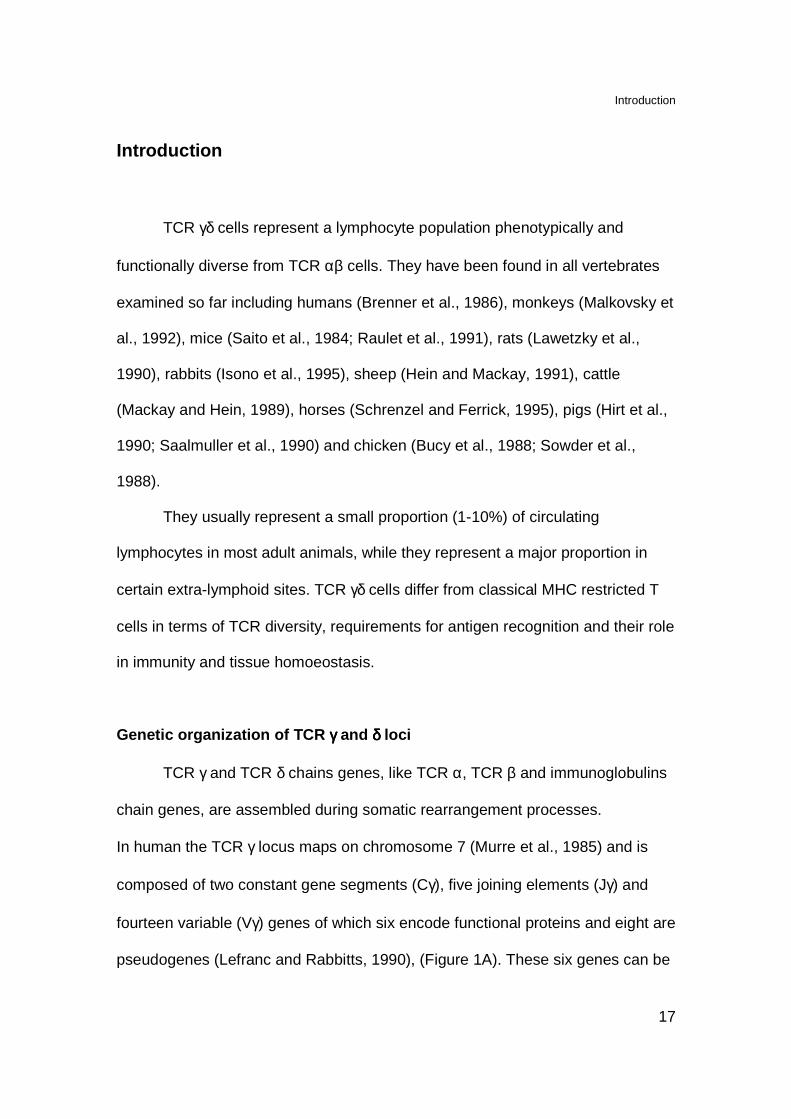
pseudogenes (Lefranc and Rabbitts, 1990), (Figure 1A). These six genes can be
Introduction
18
subdivided into two families: the VγI family composed of Vγ2, 3, 4, 5 and 8 genes
and VγII consisting of Vγ9 (Vγ2 in other nomenclature) gene. The TCR γ chain
undergoes Vγ-Jγ rearrangement and its variability at junctions is crated by
addition of not germline encoded nucleotides (N nucleotides) during
recombination process by terminal deoxynucleotidyl transferase (Tdt), (Strauss et
al., 1987; Huck et al., 1988).
Vα Vα Vα VδVα/δ VδDδ1-3 Jδ1-4 Cδ CαJαn
Vγ Jγ1 Jγ2Cγ1 Cγ2
A
BVα Vα Vα VδVα/δ VδDδ1-3 Jδ1-4 Cδ CαJαn
Vγ Jγ1 Jγ2Cγ1 Cγ2
A
B
Figure 1. Genetic organization of human (A) TCR γ locus and (B) TCR δ locus adapted
from (Hayday, 2000). TCR V segments are green and pseudogens are grey; C
segments are red; J segments are blue and D segments are yellow. Only some Vγ and
Vα are indicated.
TCR δ locus is closely linked to TCR α locus in contemporary mammals
(Hayday et al., 1985; Hayday, 2000). In humans the δ locus is located within the
TCR α locus on chromosome 14 (Collins et al., 1985), between Jα and Vα gene
segments (Griesser et al., 1988), (Figure 1B). TCR δ chain is assembled via V-D-
J rearrangement. The TCR δ locus is composed of single Cδ gene segment, four
different Jδ segments preceded by three diversity (Dδ) elements (Chien et al.,
Introduction
19
1987; Hata et al., 1987; Takihara et al., 1989). Some of V segments are used
both as Vδ and Vα (Guglielmi et al., 1988; Takihara et al., 1989). Although eight
distinct Vδ genes have been localized, only six of them were found expressed on
the cell surface (Arden et al., 1995; Migone et al., 1995). The variability of TCR δ
chain can be extremely high due to the Dδ segments that can undergo tandem
rearrangement (Boehm et al., 1988) and flexible reading frame usage (Hata et
al., 1988) creating diverse length and composition of the joining regions.
Therefore, despite the limited number of germline encoded elements for the TCR
γδ, the potential repertoire of this TCR is at least three orders of magnitude
higher than TCR αβ repertoire due to the extremely high variability in the CDR3
regions (Davis and Bjorkman, 1988; Hata et al., 1988). Comparison of the CDR3
length reveled that TCR γδ is more similar to immunoglobulins than to TCR αβ
(Rock et al., 1994).
Assembly of TCR γγγγδδδδ chains
The particular feature of TCR γδ is the preferential association of Vδ
chains with certain Vγ chains. In humans Vδ2 chain is usually associated with
unique Vγ chain: Vγ9-JP-Cγ1 while Vδ1 and Vδ3 chains are mostly paired with
various Vγ elements from VγI gene family using Cγ2 (Sturm et al., 1989; Hayday,
2000). Cγ1 gene segment usage allows formation of disulphate bond between
TCR chains while usage of Cγ2 results in non-disulphate linkage (Krangel et al.,
1987; Littman et al., 1987).
Introduction
20
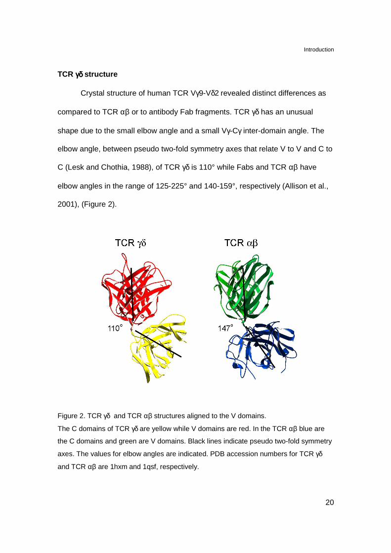
TCR γδ γδ γδ γδ structure
Crystal structure of human TCR Vγ9-Vδ2 revealed distinct differences as
compared to TCR αβ or to antibody Fab fragments. TCR γδ has an unusual
shape due to the small elbow angle and a small Vγ-Cγ inter-domain angle. The
elbow angle, between pseudo two-fold symmetry axes that relate V to V and C to
C (Lesk and Chothia, 1988), of TCR γδ is 110° while Fabs and TCR αβ have
elbow angles in the range of 125-225° and 140-159°, respectively (Allison et al.,
2001), (Figure 2).
Figure 2. TCR γδ and TCR αβ structures aligned to the V domains.
The C domains of TCR γδ are yellow while V domains are red. In the TCR αβ blue are
the C domains and green are V domains. Black lines indicate pseudo two-fold symmetry
axes. The values for elbow angles are indicated. PDB accession numbers for TCR γδ
and TCR αβ are 1hxm and 1qsf, respectively.

Introduction
21
The inter-domain angles, between the long axis of the C domains and long
axis of the V domains, of TCR γδ have 42° for Cγ-Vγ and 101° for Cδ-Vδ. In
contrast antibodies and TCR αβ have average inter-domain angles of 92° (VL-CL,
σ=9°), 76° (VH-CH, σ=11°), 100° (Cα-Vα, σ=4°) and 67° (Cβ-Vβ, σ=3°).
Therefore, the 42° of Cγ-Vγ is the smallest inter-domain angle among these
receptors. Moreover TCR γδ differ from TCR αβ also in the structure of C
domains. The FG loop of Cγ is much smaller that the one of Cβ suggesting
different binding with CD3ε subunit. The secondary structure of Cδ is composed
of a regular immunoglobulin-like domain with three-stranded β-sheet as its outer
face and therefore differs from the one or Cα (Allison et al., 2001)
The structural difference between TCR αβ and TCR γδ most likely reflects
that these receptors recognize structurally different molecules.
TCR γγγγδδδδ-CD3 complex
The main difference between TCR αβ- and TCR γδ-CD3 complexes is
their requirement for CD3δ chain. TCR γδ-CD3 complex, unlike TCR αβ, does not
associate with CD3δ and contains only CD3γε dimers (Dave et al., 1997; Hayes
and Love, 2002). The lack of CD3δ chains, unlikely for TCR αβ (Delgado et al.,
2000), does not affect the ERK activation occurring upon TCR stimulation (Hayes
and Love, 2002).
The difference in the recruitment of signaling molecules provides a
difference in signaling potential of these T cell receptors. In fact TCR γδ cells,
Introduction
22
upon CD3ε stimulation, have better proliferative response than TCR αβ cells
(Hayes and Love, 2002). Therefore enhanced signaling capacity of TCR γδ cells
together with their localization and recognition of native antigens allows these
cells to respond rapidly and acquire effector functions faster than TCR αβ cells
(Hiromatsu et al., 1992; Ferrick et al., 1995; Hayday, 2000; Hayes and Love,
2002).
Development of TCR γγγγδδδδ cells
TCR αβ and γδ cells develop in the thymus from the pluripotent CD34+
precursor cells deriving from bone marrow but distinct from stem cells (Res et al.,
1996). In humans immature thymocytes can be divided accordingly to the
expression of CD34, CD38 and CD1a (Spits et al., 1998; Spits, 2002). The
earliest thymic progenitors are CD34+CD38-CD1a-, followed by
CD34+CD38+CD1a- and CD34+CD38+CD1a+ cells, with CD1a expression
correlating with T linage commitment (Sanchez et al., 1994). In the next stage
cells start to express CD4, but not CD8, and they are referred to as CD4+
immature single positive (CD4 ISP) cells (Kraft et al., 1993; Spits, 2002).
Importantly, this population contains precursors for both TCR αβ and γδ cells
meaning that these cells are before β-selection checkpoint (Ramiro et al., 1996;
Blom et al., 1999). The CD4 ISP stage is followed by cells that express CD4 and
CD8α chain, and referred to as early double positive (EDP) cells (Spits, 2002).
Recently it has been shown that within the population of EDP there are still
Introduction
23
present cells uncommitted to the linage. The TCR γδ developmental potential is
only lost on double positive (DP) stage (Joachims et al., 2006).
Up to date there are no clear evidences that TCR γδ cells are undergoing
selection process in the thymus. However, the TCR γδ repertoire generated in the
human thymus is much more diverse than the one present in the periphery where
TCR Vγ9 chain pairs only with TCR Vδ2 chain (Casorati et al., 1989; Krangel et
al., 1990), thus suggesting that certain selection of TCR γδ cells takes place in
the thymus.
More extensive studies concerning TCR γδ cells development have been
performed using mouse models. In mice TCR γδ cells and TCR αβ cells also
develop from a common thymic double negative (DN) precursor but they diverge
into separate lineages very early in ontogeny (Petrie et al., 1992; Dudley et al.,
1995).
Immature αβ lineage cells expressing pre-TCR undergo proliferation and
transition to CD4 CD8 DP stage (Fehling et al., 1995). DP thymocytes which
express mature TCR αβ, follow positive and/or negative selection and emerge as
CD4 or CD8 single positive (SP) thymocytes (Fehling et al., 1995 ). In contrast γδ
lineage cells during differentiation do express mature TCR γδ complex, remain
mainly DN and undergo limited proliferation (Pardoll et al., 1988).
The developmental stage at which γδ lineage diverges from αβ lineage
presumably occurs between the CD44+CD25+ (DN2) and CD44-CD25- (DN4)
stage (Shortman et al., 1991; Petrie et al., 1992; Kang et al., 2001). There are
indications that TCR γδ-dependent developmental checkpoint take place already

Introduction
24
at DN3 stage (Prinz et al., 2006; Taghon et al., 2006), (Figure 3). Progression
through this checkpoint is marked by high expression of CD27 on TCR γδ cells
(Taghon et al., 2006).
DN1DN1 DN2DN2 DN3aDN3a
DN3bDN3b
DN3bDN3b
γδγδγδγδγδγδγδγδ
DN4DN4 DPDP
CD4+CD4+
CD8+CD8+
TCRγ, TCRδ, TCRβrearrangements
TCRα rearrangement
γδ-se
lection
β-selectionTCR αβ-positive
selection
Figure 3. Schematics of thymic T cell development of TCR αβ and TCR γδ cells modified
from (Hayday and Pennington, 2007).
TCR γ and δ gene rearrangements are initiated at DN2 stage whereas
rearrangement of TCR β gene is slightly delayed and begins between DN2 and
DN3 stages (Livak et al., 1999).
The αβ/γδ lineage choice is mediated by single TCR and regulated by the
strength of TCR signal (Hayes et al., 2003; Haks et al., 2005; Hayes et al., 2005)
and by Notch signaling (Garbe and von Boehmer, 2007). Strong TCR signals
through TCR γδ or TCR αβ direct either the development of DN γδ lineage cells
or MHC independent development of TCR αβ cells with γδ DN phenotype
Introduction
25
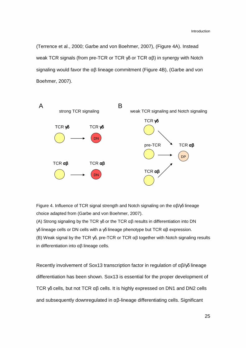
(Terrence et al., 2000; Garbe and von Boehmer, 2007), (Figure 4A). Instead
weak TCR signals (from pre-TCR or TCR γδ or TCR αβ) in synergy with Notch
signaling would favor the αβ lineage commitment (Figure 4B), (Garbe and von
Boehmer, 2007).
TCR γδγδγδγδ TCR γδγδγδγδ
TCR αβαβαβαβ TCR αβαβαβαβ
DN
strong TCR signaling weak TCR signaling and Notch signaling
pre-TCR TCR αβαβαβαβ
DP
TCR γδγδγδγδ
TCR αβαβαβαβ
A B
DN
Figure 4. Influence of TCR signal strength and Notch signaling on the αβ/γδ lineage
choice adapted from (Garbe and von Boehmer, 2007).
(A) Strong signaling by the TCR γδ or the TCR αβ results in differentiation into DN
γδ lineage cells or DN cells with a γδ lineage phenotype but TCR αβ expression.
(B) Weak signal by the TCR γδ, pre-TCR or TCR αβ together with Notch signaling results
in differentiation into αβ lineage cells.
Recently involvement of Sox13 transcription factor in regulation of αβ/γδ lineage
differentiation has been shown. Sox13 is essential for the proper development of
TCR γδ cells, but not TCR αβ cells. It is highly expressed on DN1 and DN2 cells
and subsequently downregulated in αβ-lineage differentiating cells. Significant
Introduction
26
levels of Sox13 are maintained in peripheral TCR γδ cells. Since Sox 13
expression by DN2 cells is heterogenous (50% of DN cells are positive for
Sox13) it has been suggested that some lineage separation occurs even before
TCR rearrangement (Melichar et al., 2007).
TCR γδ cells developing in adult, but not fetal thymus, require a significant
number of DP thymocytes which trans regulate differentiation of TCR γδ cells
through involvement of transcription factor RORγt and lymphotoxin β receptor
(LTβR). The proper signaling from LTβR is essential for correct TCR γδ biased
gene expression which is required for the proper function of TCR γδ cells. Thus
the trans conditioning overall influences rather cell’s functional competences than
commitment to the lineage (Pennington et al., 2003; Silva-Santos et al., 2005;
Hayday and Pennington, 2007).
Another factor important for the proper development of TCR γδ cells is IL-7
receptor signaling which promotes the expansion and survival of TCR γδ cells in
the thymus. Moreover, it is also required for the proper recombination of TCR γ
locus (Ikuta et al., 2001).
An important question which remains to be answered is whether TCR γδ
requires ligand engagement for the proper TCR γδ cells development. Up to date
only indirect evidence suggest the requirement for ligand-mediated positive
selection in fetal thymus of the dendritic epidermal T cells (DETC), (Xiong et al.,
2004; Lewis et al., 2006). This subset of TCR γδ cells populates mouse skin.

Introduction
27
TCR γγγγδδδδ cell tissue distribution
TCR γδ cells comprise 1-5% of circulating T cells while they are very
abundant in tissues. In human TCR γδ cells expressing TCR Vδ1 or Vδ3 chains
are predominant in the epithelium of the intestine (De Libero et al., 1993) where
they comprise the majority of intraepithelial lymphocytes (IELs). Cells bearing
TCR Vδ2 chain localize mainly in secondary lymphoid organs, tonsils and
peripheral blood where they represent 5-10% of total lymphocytes (Casorati and
Migone, 1990; Parker et al., 1990; Haas et al., 1993). Importantly, in humans and
some primates about 50-80% of all TCR γδ express restricted TCR composed by
the Vγ9 and Vδ2 chains (Porcelli et al., 1991). In the postnatal thymus TCR Vγ9-
Vδ2 cells constitute a minor population (up to 1% of total thymocytes), (Casorati
et al., 1989; Falini et al., 1989) but they expand in the periphery, probably due to
the continuous stimulation by unknown factors (Parker et al., 1990).
Also in mice certain populations of TCR γδ cells localize in epithelia of
particular organs. In the epidermis DETC account for ~100% of resident IELs.
They have TCR composed of Vγ3 and Vδ1 chains lacking junctional diversity
(Havran and Allison, 1990). TCR Vγ4 chain is predominantly expressed in the
reproductive tract, lung and tongue (Itohara et al., 1990). In the small intestine
TCR γδ cells preferentially use Vγ5 and Vγ1.1 chains (Pereira et al., 2000) while
in the secondary lymphoid organs TCR γδ cells mainly express Vγ2, Vγ1.1 and
Vγ1.2 chains (Raulet et al., 1991; Pereira et al., 2000).
Introduction
28
The tissue specific distribution of cells expressing particular TCR γδ
heterodimers most likely is associated with recognition of ligands present at the
localization site.
TCR γγγγδδδδ stimulatory antigens
Natural non-peptidic phosphorylated antigens
The main population of human TCR γδ cells expressing TCR Vγ9-Vδ2
respond in vitro to pathogen derived (both bacterial and parasite) extracts (Morita
et al., 2000). The stimulatory components, obtained form mycobacteria cell
lysates, have small molecular weight (less than 500 Da), are protease-resistant
and contain critical phosphate residue (Pfeffer et al., 1990; Constant et al., 1994;
Schoel et al., 1994; Tanaka et al., 1994). The analysis of Mycobacterium
smegmatis culture supernatants resulted in identification of
isopentenylpyrophosphate (IPP) and its hydroxymethyl derivatives as natural
TCR γδ ligands (Tanaka et al., 1995). These compounds are intermediate
metabolites of isoprenoids biosynthesis. In eukaryotes, archaebacteria and
certain eubacteria the biosynthesis of IPP proceeds via mevalonate pathway,
while in many eubacteria and plastids of algae and higher plants, IPP is supplied
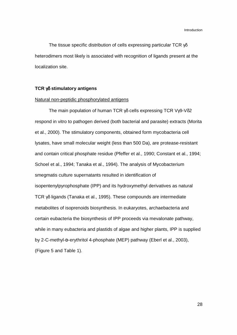
by 2-C-methyl-D-erythritol 4-phosphate (MEP) pathway (Eberl et al., 2003),
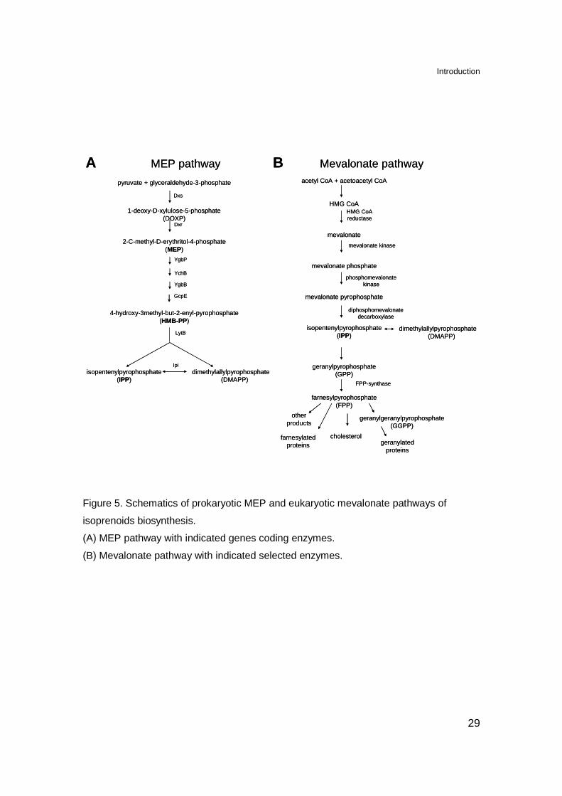
(Figure 5 and Table 1).
Introduction
29
Mevalonate pathwayacetyl CoA + acetoacetyl CoA
HMG CoA
mevalonate
mevalonate phosphate
mevalonate pyrophosphate
isopentenylpyrophosphate(IPP)
geranylpyrophosphate(GPP)
farnesylpyrophosphate(FPP)
cholesterolgeranylated
proteins
farnesylatedproteins
other products
HMG CoAreductase
dimethylallylpyrophosphate(DMAPP)
FPP-synthase
geranylgeranylpyrophosphate(GGPP)
B
mevalonate kinase
phosphomevalonatekinase
diphosphomevalonatedecarboxylase
MEP pathway
pyruvate + glyceraldehyde-3-phosphate
1-deoxy-D-xylulose-5-phosphate(DOXP)
2-C-methyl-D-erythritol-4-phosphate(MEP)
4-hydroxy-3methyl-but-2-enyl-pyrophosphate(HMB-PP)
dimethylallylpyrophosphate(DMAPP)
isopentenylpyrophosphate(IPP)
Dxs
Dxr
YgbP
YchB
YgbB
GcpE
LytB
Ipi
A Mevalonate pathwayacetyl CoA + acetoacetyl CoA
HMG CoA
mevalonate
mevalonate phosphate
mevalonate pyrophosphate
isopentenylpyrophosphate(IPP)
geranylpyrophosphate(GPP)
farnesylpyrophosphate(FPP)
cholesterolgeranylated
proteins
farnesylatedproteins
other products
HMG CoAreductase
dimethylallylpyrophosphate(DMAPP)
FPP-synthase
geranylgeranylpyrophosphate(GGPP)
B
mevalonate kinase
phosphomevalonatekinase
diphosphomevalonatedecarboxylase
MEP pathway
pyruvate + glyceraldehyde-3-phosphate
1-deoxy-D-xylulose-5-phosphate(DOXP)
2-C-methyl-D-erythritol-4-phosphate(MEP)
4-hydroxy-3methyl-but-2-enyl-pyrophosphate(HMB-PP)
dimethylallylpyrophosphate(DMAPP)
isopentenylpyrophosphate(IPP)
Dxs
Dxr
YgbP
YchB
YgbB
GcpE
LytB
Ipi
A
Figure 5. Schematics of prokaryotic MEP and eukaryotic mevalonate pathways of
isoprenoids biosynthesis.
(A) MEP pathway with indicated genes coding enzymes.
(B) Mevalonate pathway with indicated selected enzymes.
Introduction
30
Organism MEP pathway Mevalonate pathway
Prokaryotes Eubacteria
Mycobacterium tuberculosis + - Mycobacterium leprae + -
Escherichia coli + - Haemophilus influenzae + - Chlamydia pneumoniae + -
Pseudomonas aeruginosa + + Listeria monocytogenes + + Staphylococcus aureus - +
Streptococcus pneumoniae - + Borrelia burgdorferi - +
Archaebacteria Pyrococcus horikowskii - +
Methanobacterium - + Eukaryotes Apicomplexan parasites
Plasmodium farciparum + - Plants
Arabidopsis thaliana + + Fungi
Saccharomyces ceravisiae - + Mammals - +
Table 1. Utilization of MEP and mevalonate pathways by selected organisms, adapted
from (Morita et al., 2000).
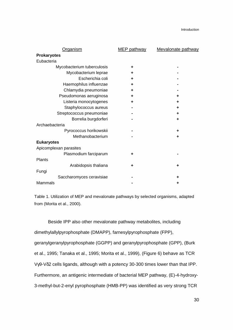
Beside IPP also other mevalonate pathway metabolites, including
dimethylallylpyrophosphate (DMAPP), farnesylpyrophosphate (FPP),
geranylgeranylpyrophosphate (GGPP) and geranylpyrophosphate (GPP), (Burk
et al., 1995; Tanaka et al., 1995; Morita et al., 1999), (Figure 6) behave as TCR
Vγ9-Vδ2 cells ligands, although with a potency 30-300 times lower than that IPP.
Furthermore, an antigenic intermediate of bacterial MEP pathway, (E)-4-hydroxy-
3-methyl-but-2-enyl pyrophosphate (HMB-PP) was identified as very strong TCR
Introduction
31
γδ antigen (Hintz et al., 2001), (Figure 6). The reactivity of human peripheral
blood mononuclear cells (PBMC) towards HMB-PP is restricted to TCR Vγ9-Vδ2
cells and leads to upregulation of activation markers, secretion of pro-
inflammatory cytokines and expansion of these TCR γδ cells (Hintz et al., 2001;
Eberl et al., 2002; Reichenberg et al., 2003).
The stimulatory capacity of HMB-PP for TCR Vγ9-Vδ2 cells is
approximately four orders of magnitude higher than IPP. Therefore it was
suggested that HMB-PP is the key activator of TCR Vγ9-Vδ2 cells during
bacterial infection (Eberl et al., 2003). This aspect will be further discussed in this
work.
Figure 6. Structures of selected natural prenyl pyrophosphates.
Introduction
32
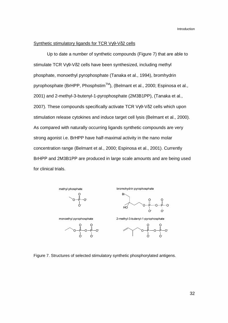
Synthetic stimulatory ligands for TCR Vγ9-Vδ2 cells
Up to date a number of synthetic compounds (Figure 7) that are able to
stimulate TCR Vγ9-Vδ2 cells have been synthesized, including methyl
phosphate, monoethyl pyrophosphate (Tanaka et al., 1994), bromhydrin
pyrophosphate (BrHPP, PhosphstimTM), (Belmant et al., 2000; Espinosa et al.,
2001) and 2-methyl-3-butenyl-1-pyrophosphate (2M3B1PP), (Tanaka et al.,
2007). These compounds specifically activate TCR Vγ9-Vδ2 cells which upon
stimulation release cytokines and induce target cell lysis (Belmant et al., 2000).
As compared with naturally occurring ligands synthetic compounds are very
strong agonist i.e. BrHPP have half-maximal activity in the nano molar
concentration range (Belmant et al., 2000; Espinosa et al., 2001). Currently
BrHPP and 2M3B1PP are produced in large scale amounts and are being used
for clinical trials.
Figure 7. Structures of selected stimulatory synthetic phosphorylated antigens.
Introduction
33
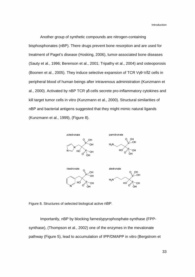
Another group of synthetic compounds are nitrogen-containing
bisphosphonates (nBP). There drugs prevent bone resorption and are used for
treatment of Paget’s disease (Hosking, 2006), tumor-associated bone diseases
(Sauty et al., 1996; Berenson et al., 2001; Tripathy et al., 2004) and osteoporosis
(Boonen et al., 2005). They induce selective expansion of TCR Vγ9-Vδ2 cells in
peripheral blood of human beings after intravenous administration (Kunzmann et
al., 2000). Activated by nBP TCR γδ cells secrete pro-inflammatory cytokines and
kill target tumor cells in vitro (Kunzmann et al., 2000). Structural similarities of
nBP and bacterial antigens suggested that they might mimic natural ligands
(Kunzmann et al., 1999), (Figure 8).
Figure 8. Structures of selected biological active nBP.
Importantly, nBP by blocking farneslypyrophosphate-synthase (FPP-
synthase), (Thompson et al., 2002) one of the enzymes in the mevalonate
pathway (Figure 5), lead to accumulation of IPP/DMAPP in vitro (Bergstrom et
Introduction
34
al., 2000). Therefore, nBP could increase levels of intracellular ligands
responsible for activation of TCR γδ cells (see this work).
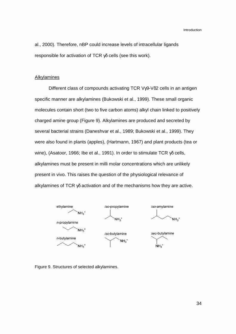
Alkylamines
Different class of compounds activating TCR Vγ9-Vδ2 cells in an antigen
specific manner are alkylamines (Bukowski et al., 1999). These small organic
molecules contain short (two to five carbon atoms) alkyl chain linked to positively
charged amine group (Figure 9). Alkylamines are produced and secreted by
several bacterial strains (Daneshvar et al., 1989; Bukowski et al., 1999). They
were also found in plants (apples), (Hartmann, 1967) and plant products (tea or
wine), (Asatoor, 1966; Ibe et al., 1991). In order to stimulate TCR γδ cells,
alkylamines must be present in milli molar concentrations which are unlikely
present in vivo. This raises the question of the physiological relevance of
alkylamines of TCR γδ activation and of the mechanisms how they are active.
Figure 9. Structures of selected alkylamines.
Introduction
35
TCR Vγγγγ9-Vδδδδ2 antigen recognition
TCR Vγ9-Vδ2 cells are activated in a crossreactive manner by variety of
ligands like IPP, DMAPP, 2,3-diphosphogyceric acid (DPG), glycerol-3-
phosphate (G3P), xylose-1-phosphate (Xyl-1P) , ribose-1-phosphate (Rib-1-P),
(Burk et al., 1995). Optimal stimulation of TCR γδ cell is driven by cells of human
origin (De Libero et al., 1991). The recognition of phosphate-containing antigens
requires cell-cell interactions without the need for antigen processing (De Libero
et al., 1991; Kabelitz et al., 1991; Lang et al., 1995; Morita et al., 1995). Taken
together this suggests the existence of a dedicated antigen-presenting molecule.
TCR Vγ9-Vδ2 cells recognize phosphorylated antigens in the absence of MHC or
CD1 restriction since APC lacking MHC class I, β2-microglobulin, CD1, or MHC
class II molecules are able to activate TCR Vγ9-Vδ2 cells (Morita et al., 1995).
Moreover, tumor cell lines and normal PBMCs are able to activate TCR Vγ9-Vδ2
cells form different donors (Morita et al., 1995). Thus, the putative antigen-
presenting molecule has rather limited or no polymorphism and is constitutively
expressed in a variety of tissues.
TCR γγγγδδδδ cells reactivity to MHC and MHC–like molecules
CD1c restricted TCR γδ cells
Human TCR γδ composed of Vδ1 chain paired with either Vγ1 or Vγ2
chains recognize CD1c molecule which is mainly expressed on immature DC and
B cells (Faure et al., 1990; Spada et al., 2000). CD1c is a member of CD1 family
molecules which are non-MHC-encoded proteins sharing structural similarities

Introduction
36
with MHC class I molecules. CD1 antigen-presenting molecules have little
polymorphism and are specialized in the presentation of lipids and glycolipids
(Beckman et al., 1994; Matsuda and Kronenberg, 2001; De Libero and Mori,
2003).
This recognition occurs in the absence of exogenous foreign antigen
(Spada et al., 2000; Vincent et al., 2002) suggesting reactivity against self-lipid
loaded into CD1c molecule (De Libero and Mori, 2003). TCR γδ cells upon
recognition of CD1c secrete pro-inflammatory cytokines which contribute to the
DC maturation process (Ismaili et al., 2002; Leslie et al., 2002). In addition, they
have a cytolytic, Th1 effector phenotype and produce granulysin (Spada et al.,
2000).
MIC and ULBP reactive TCR γδ cells
Subset of TCR Vδ1+ cells have been found to interact with cells
expressing stress-induced MHC class I-related chain (MICA and MICB)
molecules (Groh et al., 1998) and UL16-binding protein (ULBP family) molecules
(Poggi et al., 2004). MIC are encoded within the MHC locus (Bahram et al.,
1994) while ULBPs are encoded by genes on chromosome 6q25. MIC proteins
are upregulated in response to cellular stress like heat shock in intestinal
epithelium and on epithelial tumors (Groh et al., 1996; Groh et al., 1999) while
ULBPs are expressed on various cancers of hematopoietic origin including acute
myeloid and lymphoblastic leukemias (Salih et al., 2003).

Introduction
37
Despite the structural similarities between MIC and MHC class I
molecules, MIC do not associate with β2-microglobulin and do not present
peptides, probably due to the limited size of the putative peptide binding grove
and conformational differences in α1 and α2 domains (Groh et al., 1998; Li et al.,
1999). TCR Vδ1+ cells interact with MIC trough the TCR (TCR-dependent signal
1) and through natural killer activating receptor NKG2D (NKG2D dependent
costimulatory signal 2), (Wu et al., 2002) while ULBPs most likely interact only
through NK receptors (Cosman et al., 2001).
Other molecules
Among human TCR γδ cells a few T cell clones, expressing TCR Vδ1
chain, react to allo–MHC molecules including HLA-A2 (Spits et al., 1990), HLA-
A24 (Ciccone et al., 1989), HLA-B27 (Del Porto et al., 1994) and some
unspecified class I molecules (Rivas et al., 1989). In al these studies a cognate
interaction of the TCR γδ with MHC molecules was not formally demonstrated.
In mouse TCR γδ cells recognizing T10/T22 MHC class Ib molecules
(Matis et al., 1989; Van Kaer et al., 1991) or I-Ek MHC class II molecule (Matis et
al., 1989) have been described. Recognition of these molecules is independent
from the peptide binding and antigen processing (Schild et al., 1994; Crowley et
al., 2000). In the case of I-Ek recognition depends on the post-translational
changes in its glycosilation (Hampl et al., 1999).
Introduction
38
Stimulation by bacterial superantigens
Bacterial superantigens are toxins secreted by several bacterial species,
which activate T cells by binding to the non-polymorphic region of MHC class II
molecules outside of the antigen binding groove (Kozono et al., 1995) and to the
Vβ domain of the TCR αβ (Li et al., 1998). TCR Vγ9+ cells are activated by the
superantigen staphylococcal enterotoxin A (SEA), (Loh et al., 1994; Morita et al.,
2001). This stimulation occurs in a MHC class II dependent but Vδ independent
manner. TCR γδ cell require higher concentrations of SEA for stimulation as
compared to TCR αβ cells (Morita et al., 2001). Therefore, due to the structural
similarity between TCR Vγ and TCR Vβ (Allison et al., 2001) and comparable
concentrations required for T cell stimulation (Surman et al., 1994) interaction of
Vγ with SEA might resemble that of Vβ with staphylococcal enterotoxin B.
Effector functions of TCR γγγγδδδδ cells
Role of TCR γδ cells in microbial infections
TCR γδ cells were found to expand to high levels during a variety of
bacterial, viral and protozoan infections. Elevated levels of TCR Vγ9-Vδ2 cells in
the peripheral blood were observed in patients infected with Mycobacterium
tuberculosis (Barnes et al., 1992), Mycobacterium leprae (Modlin et al., 1989),
Listeria monocytogenes (Jouen-Beades et al., 1997), Francisella tularensis
(Poquet et al., 1998), Brucella melitensis (Bertotto et al., 1993), Salmonella
typhimurium (Hara et al., 1992), Ehrlichia (Caldwell et al., 1995). Also patients
with the following parasite infections have increased number of TCR γδ cells:

Introduction
39
Leishmania donovani (Raziuddin et al., 1992), Toxoplasma ssp (Scalise et al.,
1992), Plasmodium falciparum (Ho et al., 1990). Moreover TCR Vγ9-Vδ2 cells
may increase in case of Epstein-Barr virus (EBV), (De Paoli et al., 1990) and
Herpes simplex virus (HSV), (Bukowski et al., 1994) infections.
During certain microbial infections TCR Vγ9-Vδ2 cells expand even up to
50-fold (Table 2).
Infectious disease % of TCR γδ cells Reference
bacterial
tuberculosis 14 (35) (Balbi et al., 1993)
tularemia 33 (Sumida et al., 1992; Poquet et al., 1998)
salmonellosis 18 (48) (Hara et al., 1992)
brucellosis 29 (48) (Bertotto et al., 1993)
ehrlichiosis 57 (97) (Caldwell et al., 1995)
H. influence/meningitis 27 (37) (Raziuddin et al., 1994)
N. meningititis/meningitis 25 (42) (Raziuddin et al., 1994)
S. pneumoniae/meningitis 35 (46) (Raziuddin et al., 1994)
legionellosis 15 (Kroca et al., 2001)
listeriosis 12 (33) (Jouen-Beades et al., 1997)
Coxiella brunetii/Q-fever 16 (30) (Schneider et al., 1997)
parasite
acute malaria (non endemic) 18 (46) (Schwartz et al., 1996)
toxoplasmosis 9 (15) (Scalise et al., 1992)
leishmaniases 13 (18) (Russo et al., 1993)
Table 2. Examples of human TCR γδ cells expansion in response to infection.
Percentage of TCR γδ cells shows mean values detected in patients. Maximal number of
detected cells is shown in brackets.

Introduction
40
Expanded and activated TCR Vγ9-Vδ2 cells may directly participate in
anti-microbial immune responses inducing killing of bacteria, through granulysine
release, and bacteria-infected cells, through preforin and/or Fas-Fas ligand
interactions (Hara et al., 1992; Dieli et al., 2000; Ottones et al., 2000). Activated
TCR Vγ9-Vδ2 cells are able to produce significant amounts of Th1 cytokines:
IFNγ and TNFα providing an important stimulus for macrophages attraction
during the early stage of infection (Garcia et al., 1997; Wang et al., 2001).
Moreover, TCR Vγ9-Vδ2 cells release large quantities of the β-chemokines such
as macrophage inflammatory protein-1α (MIP-1α, CCL3) and MIP-1β (CCL4),
(Cipriani et al., 2000). In vitro MIP-1α and MIP-1β attract TCR αβ CD4+ and TCR
αβ CD8+ cells, respectively (Schall et al., 1993; Taub et al., 1993). Therefore,
chemokines release by TCR Vγ9-Vδ2 cells might contribute to the pro-
inflammatory microenvironment at the sites of infection.
The levels of TCR Vδ1/Vδ3 are elevated in renal allograph recipients
developing cytomegalovirus (CMV) infection (Dechanet et al., 1999a). Moreover
TCR Vδ2- cells activated by CMV-infected fibroblasts produce large amounts of
TNFα and kill infected cells. This recognition is mediated by TCR independently
of MHC class I presentation and without NKG2D engagement (Halary et al.,
2005). Infection with human immunodeficiency virus-1 (HIV-1) leads to
proliferation of TCR Vδ1 cells in the peripheral blood (Hinz et al., 1994). Most
likely, these TCR γδ cells are activated in the intestinal epithelia and then migrate
to the peripheral blood (Dechanet et al., 1999b).
Introduction
41
In mice TCR γδ cells expand in response to mycobacteria (Janis et al.,
1989), listeria (Hiromatsu et al., 1992) and salmonella (Emoto et al., 1992)
infections. Importantly, mice lacking TCR γδ cells develop enhanced inflammation
characterized by disruption of macrophage homeostasis and liver necrosis
(Carding and Egan, 2002) and they do not survive infections with Listeria
monocytogenes (Skeen et al., 2001) or Klebsiella pneumoniae (Moore et al.,
2000). Furthermore, in TCR αβ deficient mice, TCR γδ cells provide early
protective immune responses against listeriosis (Mombaerts et al., 1993) and
malaria (Tsuji et al., 1994).
Tumor surveillance
TCR Vγ9-Vδ2 cells have been shown to recognize and kill tumor
transformed cells like B cell lymphomas (Fisch et al., 1990; Selin et al., 1992),
thymic lymphomas (De Libero et al., 1991) and erythroleukemia cells (Di Fabrizio
et al., 1991). Moreover, this population of TCR γδ cells is expanded in blood
and/or intra-lesions of patients with hemopoietic and solid tumors (Bonneville and
Fournie, 2005).
The potent anti-tumor activities of TCR γδ cells have recently stimulated
great interest in of TCR γδ cells cell-based cancer immunotherapy. TCR Vγ9-Vδ2
cells expanded ex vivo, with BrHPP and IL-2 and derived from patients with
metastatic form of renal cell carcinoma have the ability to kill autologus primary
renal tumor cells (Viey et al., 2005). Furthermore, nBP activated TCR Vγ9-Vδ2
cells produce cytokines, exhibit specific cytotoxicity against myeloma cell lines,

Introduction
42
and lead to reduced survival of autologous myeloma cells (Kunzmann et al.,
2000). TCR Vγ9-Vδ2 cells expanded in vitro in the presence of aledronate
(another nBP) and IL-2 maintain their anti-tumor activity in vivo after adoptive
transfer into mice with severe combined immunodeficiency (SCID), (Kabelitz et
al., 2004).
Promising results in the treatment of patients with low-grade non-Hodgkin
lymphoma and multiple myeloma were achieved by in vivo stimulation of TCR γδ
cells using pamidronate and low-dose IL-2. This type of immunotherapy resulted
in tumor regression (Wilhelm et al., 2003).
TCR Vγ9-Vδ2 cells have a phenotype of memory cells (Miyawaki et al.,
1990; De Rosa et al., 2004) and the capacity to promptly release IFNγ and TNFα.
These characteristics implicate that they can be rapidly recruited to the site of
tumorgenesis and therefore contribute to early immune protection.
Human TCR Vδ1 cells exhibit a selective lytic activity against various
tumor cell lines like colorectal cancer, esophageal cancer, renal cell cancer,
pancreatic cancer, lung cancer (Ferrarini et al., 1994; Zocchi et al., 1994;
Choudhary et al., 1995; Maeurer et al., 1996; Groh et al., 1999; Thomas et al.,
2001). The recognition of tumor cells occurs via MIC or ULBP molecules which
interact directly with NKG2D and possibly with TCR present on TCR Vδ1 cells
(Groh et al., 1999; Wu et al., 2002; Poggi et al., 2004). Recently, it was reported
that upon MICA-NKG2D interactions the antigen-dependent effector functions of
TCR Vγ9-Vδ2 cells can be enhanced (Das et al., 2001a).
Introduction
43
Tissue homeostasis and repair
TCR γδ cells from both human and mouse are able to produce
keratinocyte growth factor (KGF), (Boismenu and Havran, 1994; Workalemahu et
al., 2003), a cytokine promoting epithelial cell growth (Visco et al., 2004). Human
TCR γδ cells from bronchoalveolar lavage in the presence of IPP are able to
secrete fibroblast growth factor 9 (FGF-9), associated with epithelial cell
proliferation (Workalemahu et al., 2004).
Furthermore, upon activation with IPP TCR γδ cells secrete metalloproteinase 7
(MMP7) which serves a key role in epithelium repair (Workalemahu et al., 2006).
Therefore TCR γδ cells contribute to the maintenance of epithelial homeostasis
and play a role in the restoring epithelial integrity.
Mouse DETCs can be activated in vitro by stressed or damaged
keratinocytes. Upon activation they secrete KGF which implicates their role in the
response to skin damage. They also participate in keratinocyte survival by
constitutively secreting insulin-like growth factor 1 (IGF-1). In addition TCR δ-/-
mice have problems with tissue repair and keratinocyte homeostasis. The lung
and intestine TCR γδ cells, exhibit similar roles in the maintenance epithelium
integrity (Witherden et al., 2000; Jameson and Havran, 2007).
The fact that TCR γδ cells reside in epithelial tissues of all mammals,
suggest that they play a conserved role in the monitoring of tissue integrity.
Introduction
44
TCR γδ cells in autoimmune diseases and inflammations
There are findings suggesting that TCR γδ cells could be involved in the
pathogenesis of autoimmune diseases. Accumulation of TCR γδ cells has been
observed in the inflamed synovium of patients with rheumatoid arthritis
(Holoshitz, 1999). By using a mouse model of collagen induced arthritis it was
demonstrated that TCR γδ cells can have pro-inflammatory functions early in the
disease but anti-inflammatory during the late stage of the disease (Peterman et
al., 1993).
The number of TCR Vγ9-Vδ2 cells is increased in the peripheral blood of patients
with diabetes mellitus (Lang et al., 1991) and in pre-diabetic and diabetic children
(Gyarmati et al., 1999). The regulatory role of TCR γδ cells in diabetes was
studied by using non-obese diabetic (NOD) mouse model. The intranasal
inhalation of pro-insulin leads to the generation of a population of regulatory TCR
γδ cells that can suppress the development of diabetes (Harrison et al., 1996).
In case of patients with multiple sclerosis (MS) elevated levels of TCR Vδ1 cells
were found in the brain lesions (Wucherpfennig et al., 1992) and cerebrospinal
fluid (Nick et al., 1995). The same population of TCR γδ cells has been found to
be increased in the blood and intestine of patients with inflammatory bowel
disease (Soderstrom et al., 1996; McVay et al., 1997).
Part 1
45
Part 1
Intracellular endogenous ligands activating TCR V γγγγ9-Vδδδδ2 cells
(These results have been published in The Journal of Experimental Medicine,
2003, 197, 163-168)
TCR Vγ9-Vδ2 cells recognize bacteria phosphorylated metabolites and
also react to bone marrow-derived tumor cells such as Daudi Burkitt’s lymphoma
line (Fisch et al., 1990; Malkovska et al., 1992). In order to identify the nature of
the tumor antigens we investigated whether TCR γδ cells recognize
phosphorylated nonpeptidic ligands resembling those produced by microbes.
One of the potent identified bacterial antigens is IPP, an intermediate product of
isoprenoids biosynthesis which is present in prokaryotic as well as in eukaryotic
cells (Burk et al., 1995; Tanaka et al., 1995). Bacteria produce IPP in either MEP
or mevalonate pathways (Morita et al., 2000), (Figure 5), whereas in eukaryotes
IPP is generated exclusively in the mevalonate pathway (Figure 5). It was
previously reported that in some hematological malignancies (Harwood et al.,
1991) and mammary carcinomas (Asslan et al., 1999) expression and function of
3-hydroxymethyl-3-glutaryl-CoenzymeA-reductase (HMGR), the rate-limiting
enzyme in the mevalonate pathway, is increased. Based on these findings we
investigated whether dysregulation of mevalonate pathway in tumors may lead to
accumulation of mevalonate metabolites thus resulting in activation of TCR Vγ9-
Part 1
46
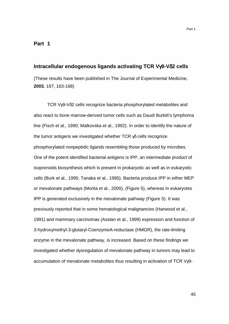
Vδ2 cells. In the following studies we took advantage of drugs that influence
enzymes of the mevalonate pathway (Figure 10).
Figure 10. Mevalonate pathway with indicated compounds used in this study and the
affected enzymes.
Part 1, Results
47
Results
Active HMGR in tumor cells is required for activati on of TCR γγγγδδδδ cells
Various cell lines of human origin were tested for the capacity to activate
TCR Vγ9-Vδ2 cells in the presence or absence of exogenous IPP and after
treatment with ZOL (Table 3).
APC Cell type Medium IPP ZOL
Daudi Bone marrow, B-cell lymphoma 2527* 14342 5647
THP-1 Bone marrow, monocytes 197 3135 16393
CEM 1.3 Bone marrow, T-cell lymphoma 310 6586 1564
K562 Bone marrow, erythroleukemia 36 2762 1145
A-375 Skin, melanoma 188 9169 15842
A-431 Skin, epidermoid carcinoma 89 3269 3067
Colo 201 Colon epithelia, coloncarcinoma 260 3103 4374
HEP G2 Liver parenchyma, hepatocarcinoma 96 12040 12417
HuH6 Liver parenchyma, hepatoblastoma 80 4329 3451
A-243 Central nervous system, astrocytoma 23 5415 7141
U118 Central nervous system, glioblastoma 33 2114 5575
BS 125.3.2 Central nervous system, glioblastoma 46 13942 8440
MRK-nu-1 Mammary gland, mammary carcinoma 35 3003 1965
HMC-1-8 Pleural effusion, mammary carcinoma 17 2011 1845
YMB-1 Mammary gland, mammary carcinoma 1162 3695 1525
Fibroblasts isolated from primary, lung connective tissue 175 5085 8768
Table 3. Tumor cell lines of different tissue origin and primary lung fibroblasts were used
as APC. Cells were either incubated with medium or IPP (10 µM) or were pulsed with
ZOL (zoledronate, 50 µg/ml) for 3 h before T cell were added. * Numbers represent
mean values in pg/ml of TNFα release by the G2B9 TCR γδ clone.
Part 1, Results
48
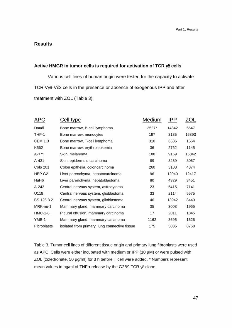
We found that not only Daudi cell but also YMB-1 cells, a solid breast carcinoma
activate TCR γδ cells (Table 3). In order to investigate whether HMGR is involved
in generation of TCR γδ stimulatory antigens we treated both, Daudi and YMB-1
cells with mevastatin (MEV), and inhibitor of the HMGR catalytic site (Istvan and
Deisenhofer, 2001) 2 h before TCR γδ cells addition. Activation of TCR Vγ9-Vδ2
was strongly reduced in the presence of MEV treated Daudi or YMB-1 cells
(Figure 11). To rule out toxicity and unspecific inhibitory effect of MEV stimulation
with exogenous IPP and PHA was also performed (Figure 11).
Figure 11. TCR γδ cell activation, by tumor cells, depends on active HMGR.
Daudi or YMB-1 cells, in the absence or presence of MEV, were used as APC in TCR γδ
cell activation assay. In control experiment stimulation with exogenously added IPP or
PHA was performed.
HMGR is one of the most tightly regulated enzyme in the cells (Goldstein
and Brown, 1990). The activity of HMGR is controlled through protein synthesis,
degradation and phosphorylation (Cheng et al., 1999). In order to further
investigate the involvement of HMGR in the generation of TCR γδ stimulatory
Part 1, Results
49
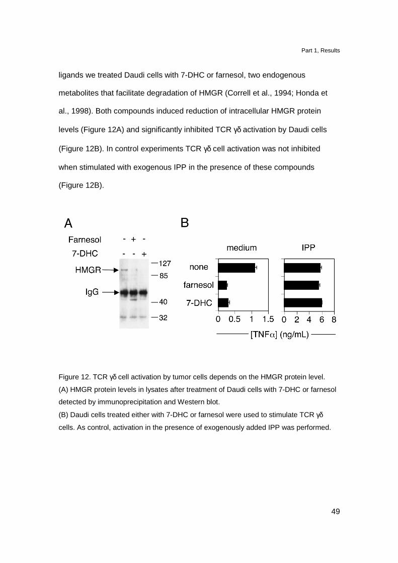
ligands we treated Daudi cells with 7-DHC or farnesol, two endogenous
metabolites that facilitate degradation of HMGR (Correll et al., 1994; Honda et
al., 1998). Both compounds induced reduction of intracellular HMGR protein
levels (Figure 12A) and significantly inhibited TCR γδ activation by Daudi cells
(Figure 12B). In control experiments TCR γδ cell activation was not inhibited
when stimulated with exogenous IPP in the presence of these compounds
(Figure 12B).
Figure 12. TCR γδ cell activation by tumor cells depends on the HMGR protein level.
(A) HMGR protein levels in lysates after treatment of Daudi cells with 7-DHC or farnesol
detected by immunoprecipitation and Western blot.
(B) Daudi cells treated either with 7-DHC or farnesol were used to stimulate TCR γδ
cells. As control, activation in the presence of exogenously added IPP was performed.

Part 1, Results
50
These experiments show that active HMGR is important for stimulation of TCR γδ
cells by Daudi and YMB-1 cells. Moreover, the data suggest that indeed
phosphorylated metabolites generated in the mevalonate pathway are the
stimulatory ligands for TCR Vγ9-Vδ2 cells.
HMGR overexpressing cells are potent TCR γγγγδδδδ cells stimulators
Transfected cells expressed high levels of HMGR as shown by
intracellular staining with a specific HMGR mAb (Figure 13)
Figure 13. HMGR overexpression in transfected Daudi cells.
Intracellular level of HMGR protein in HMGR-trasfected (bold line) or non-transfected
(thin line) Daudi cells was determined by staining with anti-HMGR specific mAb. Control
staining was performed with irrelevant mAb (dotted lines).
These cells were used as APC in TCR γδ activation assay. The capacity to
stimulate TCR γδ cells by Daudi-HMGR transfected cells was significantly
increased as compared to the wild type Daudi cells (Figure 14). Moreover, when
APC were treated with suboptimal doses of MEV, T cell activation was strongly

Part 1, Results
51
reduced. The complete inhibition, in case of Daudi-HMGR cells, was achieved
only when MEV was added at least 12 h before the assay (Figure 14). HMGR-
transfected cells might require longer incubation with inhibitor since the HMGR
protein level in these cells is increased and also mevalonate metabolites might
reach higher concentrations.
Figure 14. HMGR overexpression increases TCR γδ cell activation.
HMGR-transfected (closed bars) or wt Daudi cells (open bars) were used as APC in
TCR γδ stimulation assay. A suboptimal dose (10 µM) of MEV was added 2, 6 or 12 h
before incubation with T cells. Stimulation with exogenouse IPP was used as positive
control.
Nitrogen-containing bisphosphonates treated APC act ivate TCR γγγγδδδδ cells
The involvement of endogenous mevalonate pathway in the generation of
TCR γδ stimulatory ligands was further investigated in a series of experiments
with nitrogen-containing bisphosphonate drugs (nBP). These drugs have a
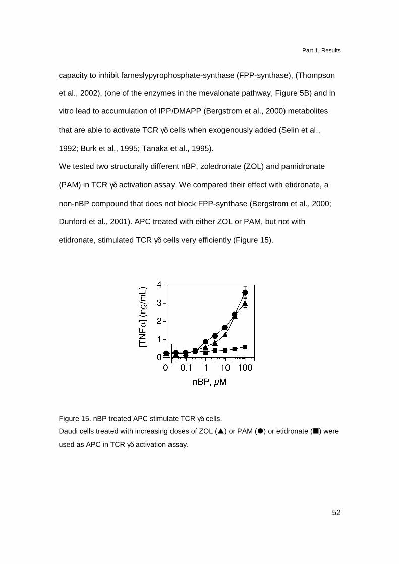
Part 1, Results
52
capacity to inhibit farneslypyrophosphate-synthase (FPP-synthase), (Thompson
et al., 2002), (one of the enzymes in the mevalonate pathway, Figure 5B) and in
vitro lead to accumulation of IPP/DMAPP (Bergstrom et al., 2000) metabolites
that are able to activate TCR γδ cells when exogenously added (Selin et al.,
1992; Burk et al., 1995; Tanaka et al., 1995).
We tested two structurally different nBP, zoledronate (ZOL) and pamidronate
(PAM) in TCR γδ activation assay. We compared their effect with etidronate, a
non-nBP compound that does not block FPP-synthase (Bergstrom et al., 2000;
Dunford et al., 2001). APC treated with either ZOL or PAM, but not with
etidronate, stimulated TCR γδ cells very efficiently (Figure 15).
Figure 15. nBP treated APC stimulate TCR γδ cells.
Daudi cells treated with increasing doses of ZOL (�) or PAM (�) or etidronate (�) were
used as APC in TCR γδ activation assay.
Part 1, Results
53
These results show that not all compounds containing a phosphate group induce
stimulation of TCR γδ cells and confirm previous studies performed with other
nBP (Kunzmann et al., 2000; Das et al., 2001b).
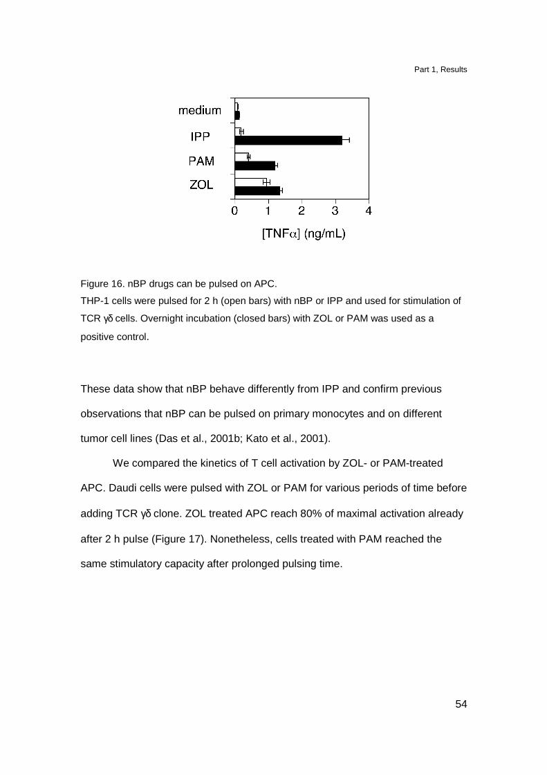
nBP have different mechanisms of action than IPP
When stimulated with exogenously added phosphorylated metabolites
TCR γδ cells require their permanent presence during the assay. When IPP is
added to APC and then cells are washed, the capacity of APC to stimulate TCR
γδ cells is lost. Presumably the activatory compounds are not stably associated
on the surface of APC (Morita et al., 1995). We tested whether the same rule
applies to nBP treated cells.
THP-1 cells were pulsed for 2 h with ZOL, PAM or IPP and then extensively
washed before adding TCR γδ cells. After pulsing with ZOL or PAM, but not with
IPP, THP-1 cells strongly activated TCR γδ cells (Figure 16). In control
experiments all compounds were stimulatory when they were kept during the
whole assay (Figure 16).
Part 1, Results
54
Figure 16. nBP drugs can be pulsed on APC.
THP-1 cells were pulsed for 2 h (open bars) with nBP or IPP and used for stimulation of
TCR γδ cells. Overnight incubation (closed bars) with ZOL or PAM was used as a
positive control.
These data show that nBP behave differently from IPP and confirm previous
observations that nBP can be pulsed on primary monocytes and on different
tumor cell lines (Das et al., 2001b; Kato et al., 2001).
We compared the kinetics of T cell activation by ZOL- or PAM-treated
APC. Daudi cells were pulsed with ZOL or PAM for various periods of time before
adding TCR γδ clone. ZOL treated APC reach 80% of maximal activation already
after 2 h pulse (Figure 17). Nonetheless, cells treated with PAM reached the
same stimulatory capacity after prolonged pulsing time.
Part 1, Results
55
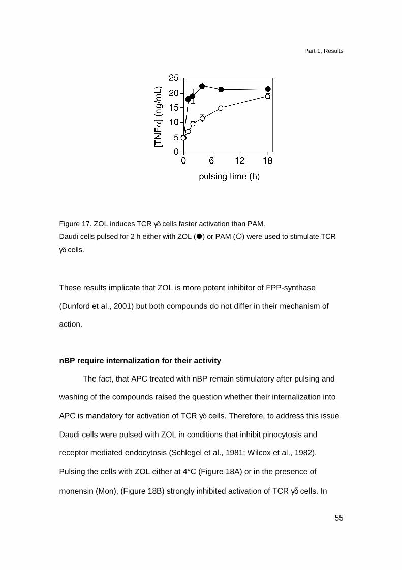
Figure 17. ZOL induces TCR γδ cells faster activation than PAM.
Daudi cells pulsed for 2 h either with ZOL (�) or PAM (�) were used to stimulate TCR
γδ cells.
These results implicate that ZOL is more potent inhibitor of FPP-synthase
(Dunford et al., 2001) but both compounds do not differ in their mechanism of
action.
nBP require internalization for their activity
The fact, that APC treated with nBP remain stimulatory after pulsing and
washing of the compounds raised the question whether their internalization into
APC is mandatory for activation of TCR γδ cells. Therefore, to address this issue
Daudi cells were pulsed with ZOL in conditions that inhibit pinocytosis and
receptor mediated endocytosis (Schlegel et al., 1981; Wilcox et al., 1982).
Pulsing the cells with ZOL either at 4°C (Figure 18A) or in the presence of
monensin (Mon), (Figure 18B) strongly inhibited activation of TCR γδ cells. In
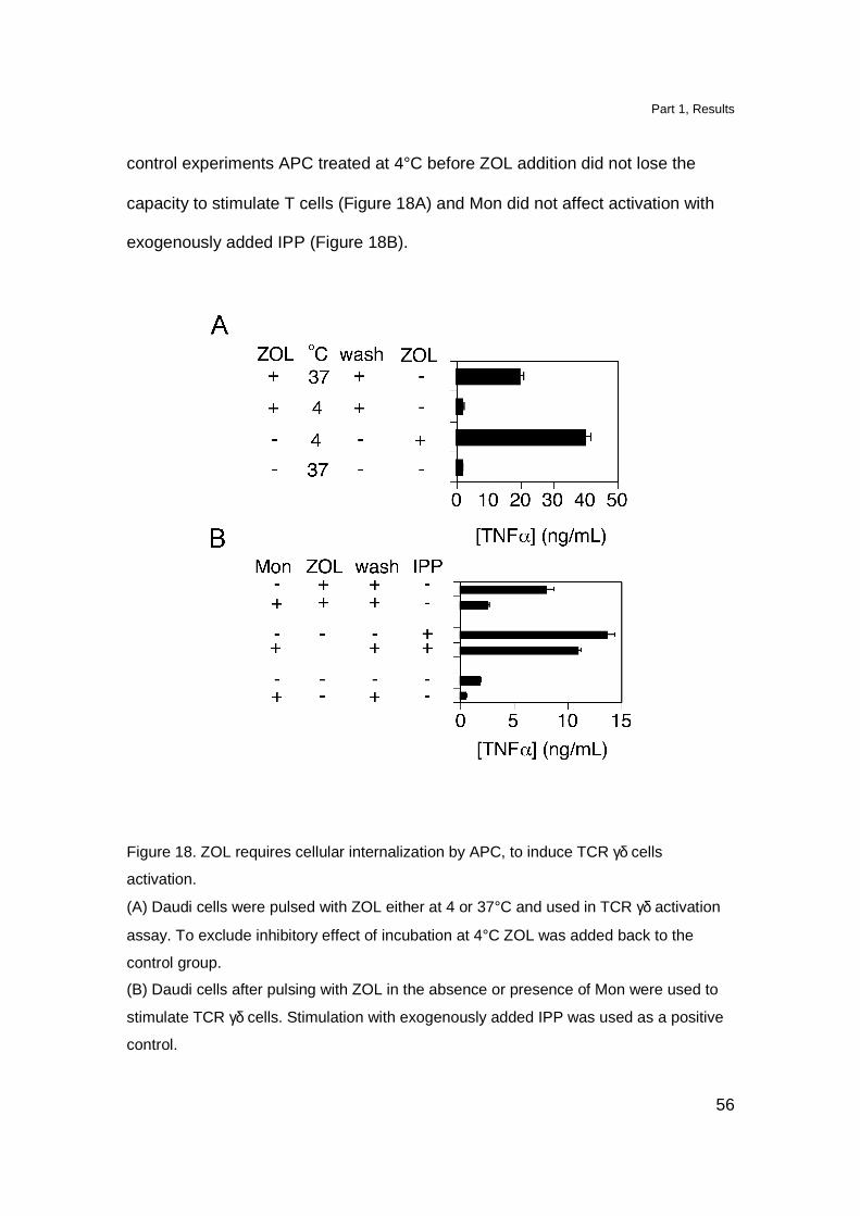
Part 1, Results
56
control experiments APC treated at 4°C before ZOL addition did not lose the
capacity to stimulate T cells (Figure 18A) and Mon did not affect activation with
exogenously added IPP (Figure 18B).
Figure 18. ZOL requires cellular internalization by APC, to induce TCR γδ cells
activation.
(A) Daudi cells were pulsed with ZOL either at 4 or 37°C and used in TCR γδ activation
assay. To exclude inhibitory effect of incubation at 4°C ZOL was added back to the
control group.
(B) Daudi cells after pulsing with ZOL in the absence or presence of Mon were used to
stimulate TCR γδ cells. Stimulation with exogenously added IPP was used as a positive
control.

Part 1, Results
57
In order to further proof that nBP are internalized we pulsed Daudi cells
with 14C-labeled ZOL under similar experimental conditions and in the presence
of NaN3 in order to block ATP-dependent active uptake during pulsing time.
Incubation either at 4°C or in the presence of monensin completely abolished the
uptake of 14C-ZOL (Figure 19).
Figure 19. Intracellular uptake of 14C-labeled ZOL by Daudi cells.
Daudi cells were pulsed with 14C-labeled ZOL at 37°C in the absence or presence of
Mon. Incubation at 4°C was performed in order to exclude surface binding of ZOL.
These data demonstrate that nBP need internalization in order to become active.
This is another difference between this class of compounds and IPP which is
active without being internalized (Morita et al., 1995).
nBP induce accumulation of endogenous TCR γγγγδδδδ ligands
The previous experiments strongly suggest that nBP induce modification
in APC, which then become stimulatory for TCR γδ cells. Therefore, we

Part 1, Results
58
investigated the hypothesis that nBP induce intracellular accumulation of
stimulatory metabolites.
In order to further examine this hypothesis we performed TCR γδ cells stimulation
by Daudi cells pulsed with ZOL in the presence of MEV. The complete inhibition
of TCR γδ cells activation was observed when ZOL was added simultaneously
with MEV (Figure 20). One hour interval between ZOL and MEV addition reduced
the inhibitory effect to 50%. When MEV was added 3 h after ZOL the inhibition
was not observed anymore suggesting that accumulation of TCR γδ cells
stimulatory ligands had already taken place.
Figure 20. MEV inhibits T cell activation induced by nBP when added simultaneously
with ZOL.
Daudi cells were pulsed with ZOL in the presence of MEV which was added at indicated
time points after ZOL. Daudi cells were washed and used to stimulate TCR γδ cells.
This result suggests that TCR γδ cells stimulatory ligands, generated in the
mevalonate pathway, must be downstream products of HMGR and upstream
Part 1, Results
59
intermediates of FPP-synthase. Therefore activatory metabolites are most likely
prenylated pyrophosphates or mevalonic acid pyrophosphate derivatives.
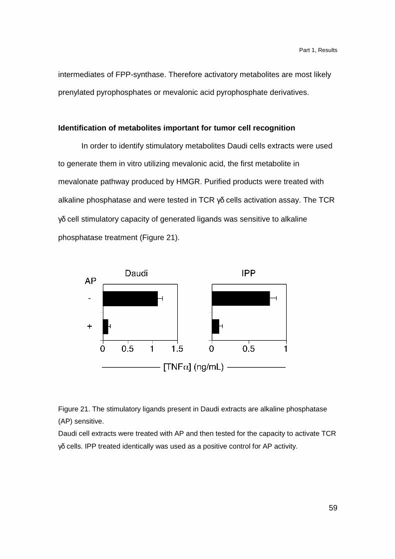
Identification of metabolites important for tumor c ell recognition
In order to identify stimulatory metabolites Daudi cells extracts were used
to generate them in vitro utilizing mevalonic acid, the first metabolite in
mevalonate pathway produced by HMGR. Purified products were treated with
alkaline phosphatase and were tested in TCR γδ cells activation assay. The TCR
γδ cell stimulatory capacity of generated ligands was sensitive to alkaline
phosphatase treatment (Figure 21).
Figure 21. The stimulatory ligands present in Daudi extracts are alkaline phosphatase
(AP) sensitive.
Daudi cell extracts were treated with AP and then tested for the capacity to activate TCR
γδ cells. IPP treated identically was used as a positive control for AP activity.
Part 1, Results
60
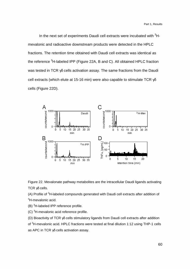
In the next set of experiments Daudi cell extracts were incubated with 3H-
mevalonic and radioactive downstream products were detected in the HPLC
fractions. The retention time obtained with Daudi cell extracts was identical as
the reference 3H-labeled IPP (Figure 22A, B and C). All obtained HPLC fraction
was tested in TCR γδ cells activation assay. The same fractions from the Daudi
cell extracts (which elute at 15-16 min) were also capable to stimulate TCR γδ
cells (Figure 22D).
Figure 22. Mevalonate pathway metabolites are the intracellular Daudi ligands activating
TCR γδ cells.
(A) Profile of 3H-labeled compounds generated with Daudi cell extracts after addition of 3H-mevalonic acid.
(B) 3H-labeled IPP reference profile.
(C) 3H-mevalonic acid reference profile.
(D) Bioactivity of TCR γδ cells stimulatory ligands from Daudi cell extracts after addition
of 3H-mevalonic acid. HPLC fractions were tested at final dilution 1:12 using THP-1 cells
as APC in TCR γδ cells activation assay.
Part 1, Results
61
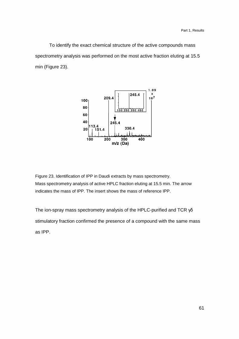
To identify the exact chemical structure of the active compounds mass
spectrometry analysis was performed on the most active fraction eluting at 15.5
min (Figure 23).
Figure 23. Identification of IPP in Daudi extracts by mass spectrometry.
Mass spectrometry analysis of active HPLC fraction eluting at 15.5 min. The arrow
indicates the mass of IPP. The insert shows the mass of reference IPP.
The ion-spray mass spectrometry analysis of the HPLC-purified and TCR γδ
stimulatory fraction confirmed the presence of a compound with the same mass
as IPP.

Part 1, Discussion
62
Discussion
The population of TCR γδ cells expressing TCR composed of Vγ9 and Vδ2
chains is activated by small phosphorylated nonpeptidic metabolites of bacterial
origin. In prokaryotes these stimulatory antigens are generated either in the MEP
or mevalonate pathway of isoprenoids biosynthesis. Moreover TCR Vγ9-Vδ2 cells
have also ability to recognize and eliminate certain lymphomas in vitro and in a
SCID animal model in vivo (Fisch et al., 1990; Malkovska et al., 1992).
Since mevalonate pathway is present in all eukaryotic cells we investigated the
involvement of this pathway in generation of TCR γδ stimulatory ligands.
Two tumor cell lines of different origin i.e. Daudi Burkitt’s lymphoma and
YMB-1 solid breast carcinoma lose their capacity to activate TCR Vγ9-Vδ2 cells
when treated with mevastatin a potent inhibitor of the HMGR catalytic site (Istvan
and Deisenhofer, 2001). In addition treatment with endogenous metabolites, 7-
DHC and farnesol, which, facilitate HMGR degradation by negative feedback
mechanism (Correll et al., 1994; Honda et al., 1998) reduced the level of
endogenous HMGR and significantly inhibited TCR γδ activation. Upregulation of
HMGR protein level, by overexpresion of the enzyme in Daudi cells, resulted in
significant increase of TCR Vγ9-Vδ2 cells activation. These results indicate that
active HMGR is important for generation of TCR γδ stimulatory ligands.
Furthermore the involvement of endogenous mevalonate pathway in
generation of phosphorylated antigens was investigated using nBP drugs which
inhibit FPP synthase (Bergstrom et al., 2000). The treatment with nBP increases

Part 1, Discussion
63
the capacity of various cells types (including human primary lung fibroblasts) to
activate TCR Vγ9-Vδ2 cells. All tested cell lines were also very stimulatory when
exogenous IPP was added. Presentation of both exogenous and endogenous
ligands by many different cell types indicates the involvement of non polymorphic
molecule(s) with a broad tissue distribution. However, the fact that nBP, in
contrast to IPP, can be pulsed on antigen presenting cells suggest that they differ
in their mechanisms of action. Indeed we showed that nBP in order to be active
require internalization. Moreover, the effect of nBP depends on active HMGR
since it is blocked when cells are treated with MEV. These results suggest that
nBP by blocking FPP, induce accumulation of mevalonate metabolites which are
directly responsible for activation of TCR γδ cells. Further analysis revealed that
these metabolites contain phosphate residue and their mass corresponds to the
one of IPP. However, since detected IPP originates from an in vitro enzymatic
assay, it cannot be excluded that in vivo other phosphorylated metabolites of the
mevalonate pathway might be involved in TCR γδ activation. Therefore, we can
conclude that detection of certain tumors by TCR Vγ9-Vδ2 cells is mediated by
cognate recognition of endogenous metabolites generated in these tumor cells.
Currently we cannot discriminate whether in tumor cells generation of TCR
γδ activatory ligands occurs due to the increased activity of HMGR, or due to the
impairment in the metabolite utilization, or both. Nonetheless, the recognition of
cells with altered mevalonate pathway that overproduce phosphorylated
metabolites may allow the immune system to target the cells with metabolic
abnormalities. Importantly, dysregulation of mevalonate pathway leading to TCR

Part 1, Discussion
64
γδ cells activation can be pharmacologically induced with nBP providing a novel
approach in tumor immunotherapy.
Part 2
65
Part 2
Transient dysregulation of the mevalonate pathway d uring early
bacterial infection leads to TCR V γγγγ9-Vδδδδ2 cells activation
Elevated levels of TCR Vγ9-Vδ2 cells in the peripheral blood of patients
(Morita et al., 1999) and macaques (Shen et al., 2002) have been described in a
variety of bacterial and protozoan infections. These TCR γδ cells have pro-
inflammatory functions and it has been suggested that they participate in
protective immunity (Morita et al., 1999).
The expansion of TCR Vγ9-Vδ2 cells has been associated with the
production of HMB-PP by microbes. Indeed, infections displaying in vivo
expansion of TCR γδ cells are caused by microorganisms that also utilize the
MEP pathway (Eberl et al., 2003). The possibility that TCR Vγ9-Vδ2 cells are
stimulated by HMB-PP during infections has not yet been tested experimentally
and it has been assumed that only APC infected with HMB-PP-producing
microorganisms stimulate TCR γδ cells (Eberl et al., 2003). Here we investigate
the mechanisms by which TCR γδ cells get activated by bacteria-infected APC.

Part 2, Results
66
Results
Stimulation of TCR V γγγγ9-Vδδδδ2 cells by bacteria-infected APC is MEP pathway
independent
Two types of human cells, namely freshly isolated CD14+ monocytes and
immature dendritic cells (DC) were infected and used as antigen-presenting cells
(APC) to stimulate TCR Vγ9-Vδ2 T cells. Infecting agents were the gram-negative
Escherichia coli, which utilizes the MEP pathway, or the gram-positive
Staphylococcus aureus, which lacks the MEP pathway and utilizes the
mevalonate pathway (Morita et al., 2000), (Table 1). TCR Vγ9-Vδ2 cells were
efficiently activated by infected APC (Figure 24A). The activation was inoculum-
dependent and at a multiplicity of infection (MOI, bacteria: APC) of 0.1 an
efficient stimulation could already be observed when using both bacteria species.
Surprisingly, monocytes and DC infected with S. aureus were also strong
stimulators, showing that MEP pathway negative bacteria also have the capacity
to activate TCR γδ cells. MHC class II-deficient APC infected with
S. aureus were also stimulatory, thus ruling out a superantigen effect during
infection with these bacteria (Rust et al., 1990), (Figure 24B).
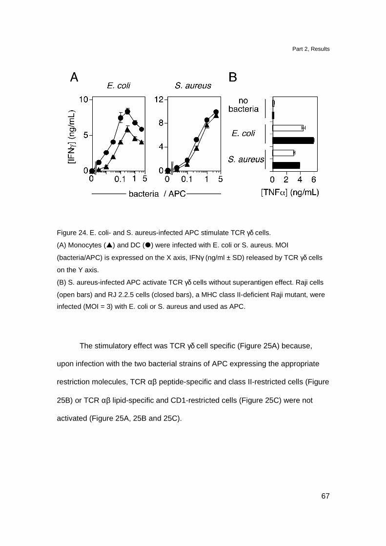
Part 2, Results
67
Figure 24. E. coli- and S. aureus-infected APC stimulate TCR γδ cells.
(A) Monocytes (�) and DC (�) were infected with E. coli or S. aureus. MOI
(bacteria/APC) is expressed on the X axis, IFNγ (ng/ml ± SD) released by TCR γδ cells
on the Y axis.
(B) S. aureus-infected APC activate TCR γδ cells without superantigen effect. Raji cells
(open bars) and RJ 2.2.5 cells (closed bars), a MHC class II-deficient Raji mutant, were
infected (MOI = 3) with E. coli or S. aureus and used as APC.
The stimulatory effect was TCR γδ cell specific (Figure 25A) because,
upon infection with the two bacterial strains of APC expressing the appropriate
restriction molecules, TCR αβ peptide-specific and class II-restricted cells (Figure
25B) or TCR αβ lipid-specific and CD1-restricted cells (Figure 25C) were not
activated (Figure 25A, 25B and 25C).
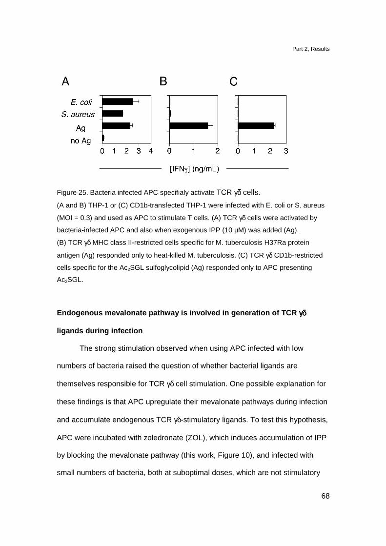
Part 2, Results
68
Figure 25. Bacteria infected APC specifialy activate TCR γδ cells.
(A and B) THP-1 or (C) CD1b-transfected THP-1 were infected with E. coli or S. aureus
(MOI = 0.3) and used as APC to stimulate T cells. (A) TCR γδ cells were activated by
bacteria-infected APC and also when exogenous IPP (10 µM) was added (Ag).
(B) TCR γδ MHC class II-restricted cells specific for M. tuberculosis H37Ra protein
antigen (Ag) responded only to heat-killed M. tuberculosis. (C) TCR γδ CD1b-restricted
cells specific for the Ac2SGL sulfoglycolipid (Ag) responded only to APC presenting
Ac2SGL.
Endogenous mevalonate pathway is involved in genera tion of TCR γγγγδδδδ
ligands during infection
The strong stimulation observed when using APC infected with low
numbers of bacteria raised the question of whether bacterial ligands are
themselves responsible for TCR γδ cell stimulation. One possible explanation for
these findings is that APC upregulate their mevalonate pathways during infection
and accumulate endogenous TCR γδ-stimulatory ligands. To test this hypothesis,
APC were incubated with zoledronate (ZOL), which induces accumulation of IPP
by blocking the mevalonate pathway (this work, Figure 10), and infected with
small numbers of bacteria, both at suboptimal doses, which are not stimulatory
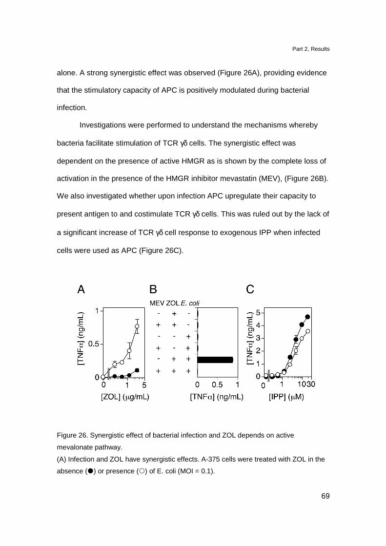
Part 2, Results
69
alone. A strong synergistic effect was observed (Figure 26A), providing evidence
that the stimulatory capacity of APC is positively modulated during bacterial
infection.
Investigations were performed to understand the mechanisms whereby
bacteria facilitate stimulation of TCR γδ cells. The synergistic effect was
dependent on the presence of active HMGR as is shown by the complete loss of
activation in the presence of the HMGR inhibitor mevastatin (MEV), (Figure 26B).
We also investigated whether upon infection APC upregulate their capacity to
present antigen to and costimulate TCR γδ cells. This was ruled out by the lack of
a significant increase of TCR γδ cell response to exogenous IPP when infected
cells were used as APC (Figure 26C).
Figure 26. Synergistic effect of bacterial infection and ZOL depends on active
mevalonate pathway.
(A) Infection and ZOL have synergistic effects. A-375 cells were treated with ZOL in the
absence (�) or presence () of E. coli (MOI = 0.1).

Part 2, Results
70
(B) Synergistic effect of infection is HMGR-dependent. A-375 cells were treated with
mevastatin (MEV) 2 h prior infection (MOI = 0.1) and addition of 2.5 µg/ml ZOL.
(C) Infection does not increase the APC antigen presentation capacity. IPP presentation
by A-375 cells in the absence (�) or presence () of E. coli (MOI = 0.1).
Moreover we tested whether bacterial growth is modified in the presence of MEV
and ZOL. None of the compounds influenced the growth of the bacteria used in
the infection experiments (Figure 27)
Figure 27. Mevalonate pathway modulators do not affect bacterial growth.
ZOL or MEV were incubated with S. aureus or E. coli and bacterial growth was
determined by CFU counts.
Next the hypothesis was tested whether infection modulates HMGR
activity in APC by using wild type or HMGR-transfected A-375 cells infected with
either E. coli or with S. aureus (Figure 28A). HMGR-transfected APC showed a
stronger stimulation capacity than wild type cells at all MOI and with both
bacteria, suggesting the importance of an active endogenous mevalonate
pathway. Moreover, MEV strongly reduced T cell activation in these experimental
settings, thus revealing the importance of endogenous eukaryotic ligands (Figure
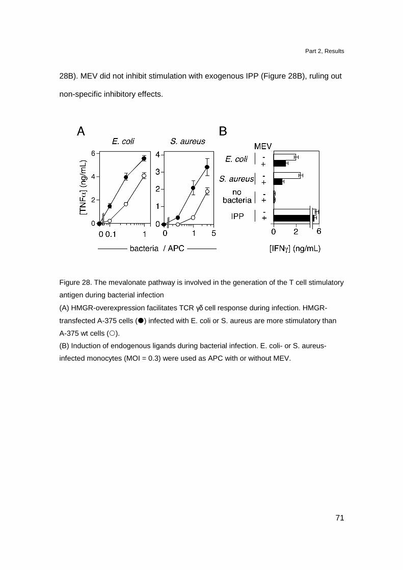
Part 2, Results
71
28B). MEV did not inhibit stimulation with exogenous IPP (Figure 28B), ruling out
non-specific inhibitory effects.
Figure 28. The mevalonate pathway is involved in the generation of the T cell stimulatory
antigen during bacterial infection
(A) HMGR-overexpression facilitates TCR γδ cell response during infection. HMGR-
transfected A-375 cells (�) infected with E. coli or S. aureus are more stimulatory than
A-375 wt cells ().
(B) Induction of endogenous ligands during bacterial infection. E. coli- or S. aureus-
infected monocytes (MOI = 0.3) were used as APC with or without MEV.

Part 2, Results
72
Bacterial infections modulate HMGR protein levels a nd phosphorylation
state
As HMGR is the rate-limiting enzyme of the mevalonate pathway, we
investigated whether bacteria directly influence the intracellular levels and the
function of HMGR. HMGR protein levels were evaluated by densitometry after
immunoprecipitation and Western blotting of APC lysates. After infection with E.
coli or with S. aureus (Figure 29A) a slight, but consistent, increase in the total
levels of HMGR was already observable 1 h after infection, and was followed by
a decline 24 h after infection. Analysis of HMGR gene transcription by semi-
quantitative RT-PCR showed that HMGR mRNA levels do not change
immediately after infection and that they increase significantly after 24 h (Figure
29B). This rapid increase in HMGR protein level at 1 h post infection, without a
concomitant increase in mRNA levels, suggests that the regulation may occur
through a reduced degradation of HMGR.

Part 2, Results
73
Figure 29. Bacterial infection modifies HMGR protein levels.
(A) HMGR was immunoprecipitated from THP-1 cells infected (MOI = 1) with E. coli or S.
aureus for 1, 2, 4 and 24 h (gentamycin was added after 4 h) and analyzed by Western
blot. The bands indicated with the arrow correspond to intact HMGR (97 kDa). They
were scanned and the values compared to the one obtained in uninfected cells (time 0).
(B) Bacterial infection influences HMGR gene transcription at later time points. Semi-
quantitative RT-PCR analysis of HMGR (upper panel) was performed on THP-1 cells
infected with E. coli (MOI = 1) for different time points. Serial 1:3 dilutions of cDNA are
shown. As a control β-actin RT-PCR (lower panel) was performed. * indicates that the
difference between time 0 and 24 h is significant (p<0.05).
As dephosphorylated HMGR is more active and generates more
mevalonate than phosphorylated forms (Omkumar et al., 1994; Istvan and
Deisenhofer, 2000), the possibility that rapid HMGR dephosphorylation occurs
during bacterial infection was investigated. HMGR was immunoprecipitated from
E. coli- or S. aureus-infected cells as well as from control cells and detected by
Western blotting by using a phosphoserine-specific mAb (Figure 30A). The
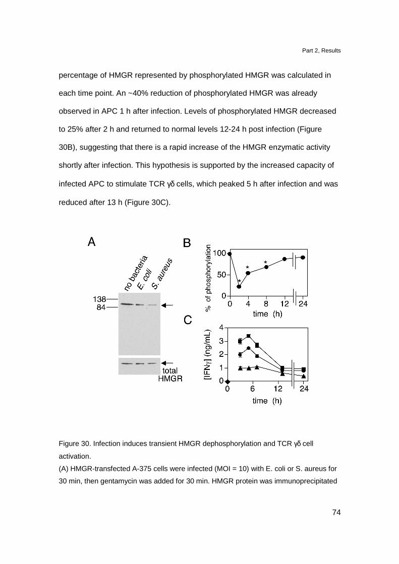
Part 2, Results
74
percentage of HMGR represented by phosphorylated HMGR was calculated in
each time point. An ~40% reduction of phosphorylated HMGR was already
observed in APC 1 h after infection. Levels of phosphorylated HMGR decreased
to 25% after 2 h and returned to normal levels 12-24 h post infection (Figure
30B), suggesting that there is a rapid increase of the HMGR enzymatic activity
shortly after infection. This hypothesis is supported by the increased capacity of
infected APC to stimulate TCR γδ cells, which peaked 5 h after infection and was
reduced after 13 h (Figure 30C).
Figure 30. Infection induces transient HMGR dephosphorylation and TCR γδ cell
activation.
(A) HMGR-transfected A-375 cells were infected (MOI = 10) with E. coli or S. aureus for
30 min, then gentamycin was added for 30 min. HMGR protein was immunoprecipitated
Part 2, Results
75
and phosphorylation was analyzed by Western blot. Control total HMGR protein levels
are shown in the lower panel.
(B) HMGR phosphorylation is modulated during infection. HMGR-transfected A375 were
infected with E. coli (MOI = 10) and HMGR phosphorylation and protein levels were
analyzed. Results are expressed as a % of phosphorylated HMGR standardized for total
HMGR signal in infected and uninfected cells (time 0, 100%).
(C) Duration of APC stimulatory capacity after bacterial infection. DC were infected with
E. coli at MOI 1 (�), 0.3 (�), 0.1 (�) or uninfected (�). Gentamycin was applied after 3
h and T cells added at the indicated time points.
Increased PP2A activity leads to HMGR dephosphoryla tion induced by
bacterial infection
The mechanism for serine 872 dephosphorylation in HMGR was
investigated by using phosphatase inhibitors. Within the serine/threonine
phosphatase family, PP2A is an enzyme with an ubiquitous distribution, large
abundance and broad specificity (Wera and Hemmings, 1995). We took
advantage of okadaic acid (OA), and calyculin A (CA) drugs, which block PP2A
activity (Favre et al., 1997) to determine whether PP2A is required for activation
of TCR γδ cells. We could not use the bacteria infection model to stimulate TCR
γδ cells because preincubation of APC with these drugs leads to altered
phagocytosis (Zhang et al., 2000; Sosroseno et al., 2003). Therefore, Daudi
cells, which stimulate TCR Vγ9-Vδ2 cells without addition of exogenous ligands
(Fisch et al., 1990), were used. Daudi cells pretreated with OA and CA were no
longer able to activate TCR γδ cells as revealed by Ca2+ mobilization in the T
cells (Figure 31A), showing the requirement for active PP2A. Unspecific effects
of phosphatase inhibitors on antigen presentation were ruled out by showing that
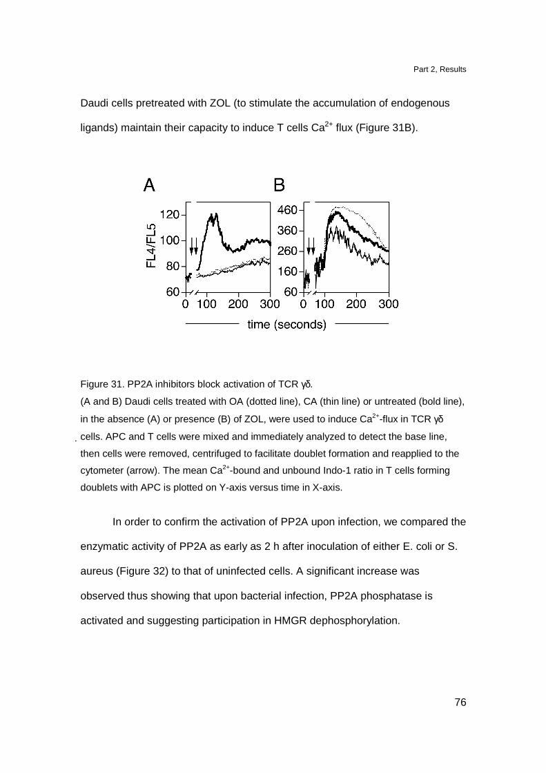
Part 2, Results
76
Daudi cells pretreated with ZOL (to stimulate the accumulation of endogenous
ligands) maintain their capacity to induce T cells Ca2+ flux (Figure 31B).
Figure 31. PP2A inhibitors block activation of TCR γδ.
(A and B) Daudi cells treated with OA (dotted line), CA (thin line) or untreated (bold line),
in the absence (A) or presence (B) of ZOL, were used to induce Ca2+-flux in TCR γδ
cells. APC and T cells were mixed and immediately analyzed to detect the base line,
then cells were removed, centrifuged to facilitate doublet formation and reapplied to the
cytometer (arrow). The mean Ca2+-bound and unbound Indo-1 ratio in T cells forming
doublets with APC is plotted on Y-axis versus time in X-axis.
In order to confirm the activation of PP2A upon infection, we compared the
enzymatic activity of PP2A as early as 2 h after inoculation of either E. coli or S.
aureus (Figure 32) to that of uninfected cells. A significant increase was
observed thus showing that upon bacterial infection, PP2A phosphatase is
activated and suggesting participation in HMGR dephosphorylation.

Part 2, Results
77
Figure 32. PP2A activity is increased upon bacterial infection.
Cell lysates from HMGR-transfected A-375 cells infected (MOI = 10) with E. coli or S.
aureus for 2 h (30 min after infection gentamycin was added) were used to determine
PP2A activity. Results are represented as % of PP2A activity. 100% was calculated for
uninfected cells. Significant differences are indicated by *(p<0.05) or **(p<0.005).
HMGR activity is increased during early times of ba cterial infection
To examine whether HMGR dephosphorylation and increased protein
levels might be responsible for increased HMGR enzymatic activity, infected and
control cells were lysed and the crude extracts were tested in vitro for their
content of active HMGR, i.e. for the capacity to utilize the substrate
hydroxymethylglutaryl-CoA (HMG-CoA), using NADPH as a co-substrate. The
product of the reaction, mevalonic acid was monitored by liquid chromatography-
electrospray-mass spectrometry (LC-ESI-MS) after conversion into the more
stable mevalonolactone (MVL), (Gerber et al., 2004). In preliminary experiments
aimed at determining the enzyme kinetics we found that a 60 min enzymatic
reaction was optimal to generate measurable metabolite without reaching a
plateau, thus permitting exact MVL quantization (Figure 33A and 33B). In the
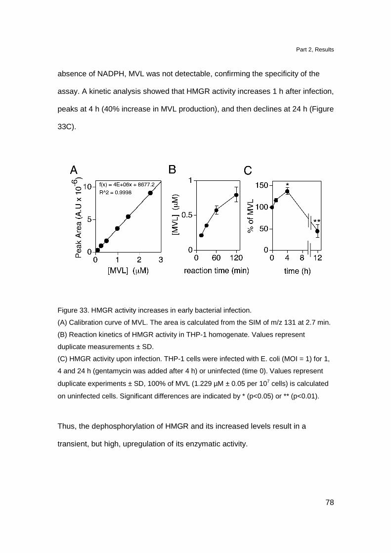
Part 2, Results
78
absence of NADPH, MVL was not detectable, confirming the specificity of the
assay. A kinetic analysis showed that HMGR activity increases 1 h after infection,
peaks at 4 h (40% increase in MVL production), and then declines at 24 h (Figure
33C).
Figure 33. HMGR activity increases in early bacterial infection.
(A) Calibration curve of MVL. The area is calculated from the SIM of m/z 131 at 2.7 min.
(B) Reaction kinetics of HMGR activity in THP-1 homogenate. Values represent
duplicate measurements ± SD.
(C) HMGR activity upon infection. THP-1 cells were infected with E. coli (MOI = 1) for 1,
4 and 24 h (gentamycin was added after 4 h) or uninfected (time 0). Values represent
duplicate experiments ± SD, 100% of MVL (1.229 µM ± 0.05 per 107 cells) is calculated
on uninfected cells. Significant differences are indicated by * (p<0.05) or ** (p<0.01).
Thus, the dephosphorylation of HMGR and its increased levels result in a
transient, but high, upregulation of its enzymatic activity.
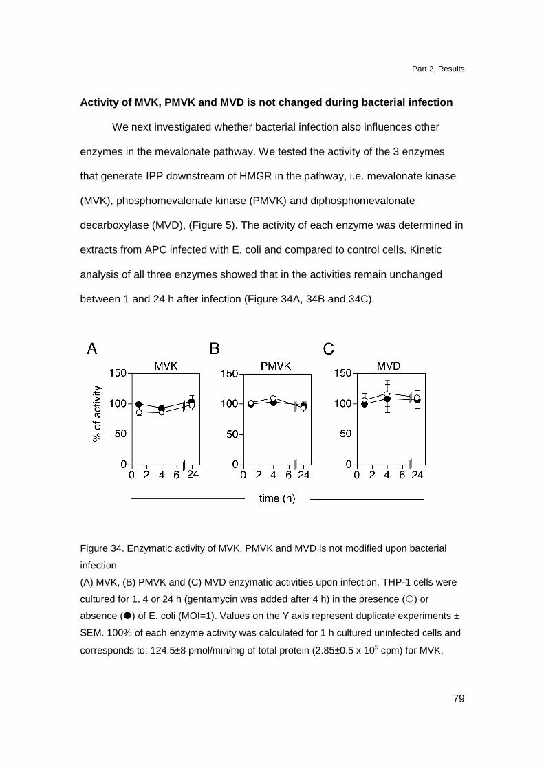
Part 2, Results
79
Activity of MVK, PMVK and MVD is not changed during bacterial infection
We next investigated whether bacterial infection also influences other
enzymes in the mevalonate pathway. We tested the activity of the 3 enzymes
that generate IPP downstream of HMGR in the pathway, i.e. mevalonate kinase
(MVK), phosphomevalonate kinase (PMVK) and diphosphomevalonate
decarboxylase (MVD), (Figure 5). The activity of each enzyme was determined in
extracts from APC infected with E. coli and compared to control cells. Kinetic
analysis of all three enzymes showed that in the activities remain unchanged
between 1 and 24 h after infection (Figure 34A, 34B and 34C).
Figure 34. Enzymatic activity of MVK, PMVK and MVD is not modified upon bacterial
infection.
(A) MVK, (B) PMVK and (C) MVD enzymatic activities upon infection. THP-1 cells were
cultured for 1, 4 or 24 h (gentamycin was added after 4 h) in the presence () or
absence (�) of E. coli (MOI=1). Values on the Y axis represent duplicate experiments ±
SEM. 100% of each enzyme activity was calculated for 1 h cultured uninfected cells and
corresponds to: 124.5±8 pmol/min/mg of total protein (2.85±0.5 x 10⁵ cpm) for MVK,
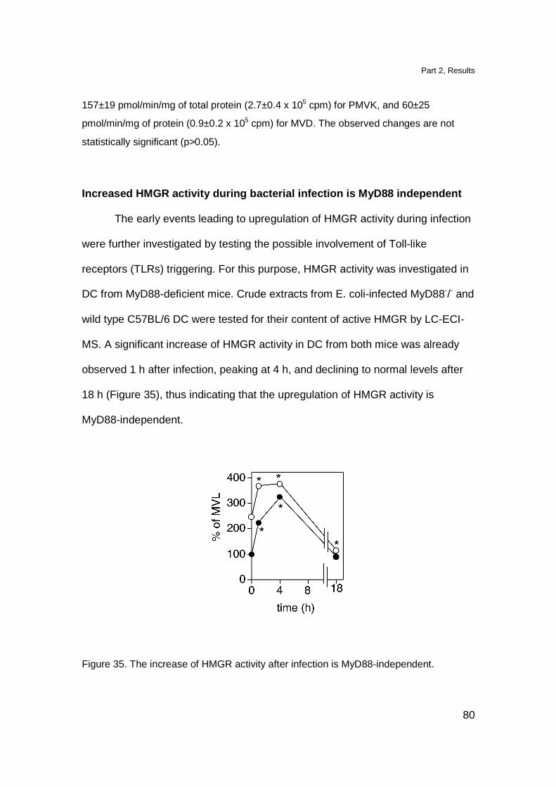
Part 2, Results
80
157±19 pmol/min/mg of total protein (2.7±0.4 x 10⁵ cpm) for PMVK, and 60±25
pmol/min/mg of protein (0.9±0.2 x 10⁵ cpm) for MVD. The observed changes are not
statistically significant (p>0.05).
Increased HMGR activity during bacterial infection is MyD88 independent
The early events leading to upregulation of HMGR activity during infection
were further investigated by testing the possible involvement of Toll-like
receptors (TLRs) triggering. For this purpose, HMGR activity was investigated in
DC from MyD88-deficient mice. Crude extracts from E. coli-infected MyD88-/- and
wild type C57BL/6 DC were tested for their content of active HMGR by LC-ECI-
MS. A significant increase of HMGR activity in DC from both mice was already
observed 1 h after infection, peaking at 4 h, and declining to normal levels after
18 h (Figure 35), thus indicating that the upregulation of HMGR activity is
MyD88-independent.
Figure 35. The increase of HMGR activity after infection is MyD88-independent.

Part 2, Results
81
DC from MyD88-/- () or C57BL/6 (�) mice were infected with E. coli (MOI = 1) for 1, 4
and 24 h (gentamycin was added after 4 h) or uninfected (time 0). Values on Y axis
represent % of MVL production. 100% is calculated on C57BL/6 uninfected cells and
corresponds to 0.4 µM of MVL per 107 cells. Significant differences are indicated by *
(p<0.01).
From these experiments we conclude that HMGR enzymatic activity is
increased in a MyD88-independent manner after bacterial infection, while the
activities of other enzymes involved in IPP generation are unchanged.
Altogether these results point to the following chronological order of
events occurring in bacteria infected cells. At the early time points of bacterial
infection (1h) HMGR activity increases without concomitant increase of mRNA
synthesis. Later on (24 h) the activity of HMGR is decreased and it is
accompanied by an increase of mRNA levels. This suggests that HMGR
feedback regulatory mechanism occurs. Immediately after infection bacteria-
infected cells augment PP2A phosphatase activity, increase the amounts of
dephosphorylated and total HMGR, raise the production of TCR γδ-stimulatory
ligands and acquire the capacity to stimulate TCR γδ cells, which then declines
24 h post-infection (Figure 36).
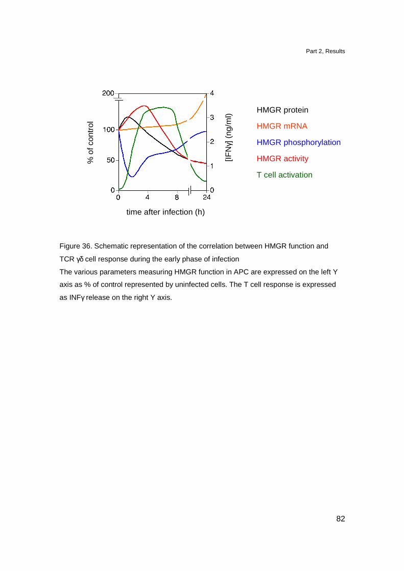
Part 2, Results
82
% o
f con
trol
time after infection (h)
[IFN
γ] (
ng/m
l)
HMGR protein
HMGR mRNA
HMGR phosphorylation
HMGR activity
T cell activation
Figure 36. Schematic representation of the correlation between HMGR function and
TCR γδ cell response during the early phase of infection
The various parameters measuring HMGR function in APC are expressed on the left Y
axis as % of control represented by uninfected cells. The T cell response is expressed
as INFγ release on the right Y axis.
Part 2, Discussion
83
Discussion
The selective expansion of TCR Vγ9-Vδ2 cells during infection with certain
bacteria or parasites suggests that these T cells have a role in immune defense
against pathogens. Whether endogenous or bacterial products stimulate TCR
Vγ9-Vδ2 cells during infection was unknown. The discovery of HMB-PP, a
metabolite product of the MEP pathway, as the most potent TCR γδ stimulatory
ligand implicated its major role in T cell activation during microbial infection (Eberl
et al., 2003). However, despite these compounds are active at low
concentrations, it is questionable whether they reach the minimal concentration
required for TCR γδ activation. Here we find that different phagocytic cell types,
including freshly isolated monocytes and immature DC, immediately increase the
production of mevalonate metabolites when infected with either MEP-positive
E. coli or MEP-negative S. aureus bacteria and, as a consequence, acquire the
capacity to stimulate TCR Vγ9-Vδ2 cells.
The TCR γδ-stimulatory ligands are not produced by the bacteria themselves, but
rather by the host mevalonate metabolic pathway. In fact infection of APC with
very low MOI efficiently stimulates TCR γδ cells. It is possible that bacterial HMB-
PP represents an additional stimulatory ligand in a later phase of infection when
large numbers of MEP-positive bacteria accumulate.
In this study we show that bacterial infections naturally modulate the mevalonate
metabolism of APC and suggest that endogenous mevalonate metabolites
accumulating within infected cells stimulate TCR γδ cells. Upon infection of APC

Part 2, Discussion
84
with low doses of bacteria, and in the presence of small amounts of ZOL, both at
suboptimal and inactive doses, there is a strong stimulation of TCR γδ cells. Thus
bacterial infection has a synergistic activity on the ZOL effects. In addition, the
treatment of infected APC with mevastatin prevents the generation of the cellular
stimulatory ligands and inhibits TCR γδ cell activation. Therefore, the HMGR
activity in APC sets the threshold of TCR γδ response to infection.
Infection promotes two different mechanisms, both of which augment the function
of this key enzyme. Firstly, there is a rapid rise in the HMGR protein levels within
the first hour post-infection. Since there is no concomitant increase in HMGR
mRNA levels, it is likely that HMGR protein rises as a consequence of its
reduced degradation in infected cells. Secondly, infection also leads to increased
amounts of the dephosphorylated form of HMGR, which is followed by enhanced
enzymatic activity. This is in agreement with other studies showing that HMGR
activity is negatively influenced by phosphorylation of serine 872 by AMP-
activated protein kinase (Omkumar et al., 1994; Istvan and Deisenhofer, 2000).
Mass Spectrometric quantitative analysis confirmed that there was increased
HMGR activity in infected phagocytic cells, as compared to non-infected cells.
The early modifications in HMGR protein levels and dephosphorylated forms
immediately precede maximal T cell activation, which shows a peak 4-8 h after
infection in our experimental system. This suggests a direct correlation of the
three phenomena, which appear in a temporally ordered fashion (Figure 36). The
other enzymes involved in IPP synthesis (i.e. mevalonate kinase,
phosphomevalonate kinase and diphosphomevalonate decarboxylase), do not
Part 2, Discussion
85
increase their activity upon infection, which confirms the key role of HMGR as the
main regulatory enzyme in the mevalonate pathway (Goldstein and Brown,
1990).
Our attempts to outline the mechanisms leading to HMGR dephosphorylation
indicate an increased activity of PP2A in the early phases of bacterial infection
and suggest that HMGR is a PP2A substrate. Activity of this serine/threonine
phosphatase is required to maintain the stimulatory capacity of Daudi cells, as
shown by inhibition with PP2A-blocking drugs, and this reveals a previously
unrecognized role of this phosphatase in stimulating TCR γδ cells. PP2A is
probably involved in the production of TCR γδ stimulatory ligands and not in
antigen presentation, as indicated by experiments with ZOL-treated Daudi cells.
Indeed, these cells accumulate endogenous ligands and maintain a strong TCR
γδ cell stimulatory capacity in the presence of PP2A inhibitory drugs.
Previous studies have shown that other phosphatases, such as dual specificity
phosphatases, are activated during innate immune responses (Liu et al., 2007).
Importantly, these phosphatases are not active on serine as substrate as is the
case with PP2A and therefore are not candidate to dephosphorylate HMGR in
vivo.
HMGR activation following infection is independent from the MyD88 pathway,
thus implying that this phenomenon is not associated with microbial activation of
Toll-like receptors which utilize MyD88. Whether, other adaptor proteins of innate
immunity are involved in HMGR activation remains to be investigated.

Part 2, Discussion
86
Different stimuli, including cholesterol depletion (Goldstein et al., 2006),
growth factors and insulin signaling (Demoulin et al., 2004; Nadeau et al., 2004)
exposure to hypotonic media, and ER calcium depletion (Lee and Ye, 2004),
have been reported to induce upregulation of HMGR transcription. Interestingly,
HMGR gene transcription also increases after internalization of latex beads
(Castoreno et al., 2005), which, to some extent mimics phagocytosis of bacteria.
Moreover, membrane pore formation by the bacterial aerolysin toxin, leads to
upregulation of HMGR transcription and to increased cholesterol synthesis as a
defence mechanism facilitating membrane repair (Gurcel et al., 2006). Therefore,
upregulation of mevalonate pathway occurs after a variety of cell stressing
signals, as a result of increased HMGR gene transcription and enzymatic activity.
Here we add a novel mechanism involving HMGR dephosphorylation and PP2A
activation occurring during the early phases of infection. Whether the other
physiological conditions which upregulate HMGR transcription also induce
stimulation of TCR γδ cells has not been investigated.
Overall these findings have revealed two important aspects relating to the
manner in which infected cells activate TCR Vγ9-Vδ2 cells and therefore to the
possible relevance of this T cell population in immune responses in vivo. Firstly,
HMGR dephosphorylation and accumulation of endogenous (self) ligands is fast,
being detectable within the first hour post infection. This rapid mechanism may
contribute to the stimulation of TCR Vγ9-Vδ2 cell response during the very early
stages of infection, when very low numbers of bacteria are present and when
other bacteria-specific T cells are not available at the site of infection.
Part 2, Discussion
87
Another important issue is that the mevalonate dysregulation following infection
is transient. This probably occurs because of the feedback regulation of both the
mevalonate pathway and cholesterol metabolism, both of which are tightly
controlled within cells (Goldstein and Brown, 1990). The temporary alteration of
the mevalonate pathway is necessary to avoid continuous stimulation of TCR
Vγ9-Vδ2 cells, which are very abundant and thus have to be activated with
caution. Despite its short duration, the transient activation may explain the
observed expansion of this TCR γδ population in the periphery during early post-
natal life (Parker et al., 1990). This could be the result of repeated infections
naturally occurring in this age.
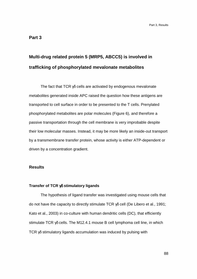
Part 3, Results
88
Part 3
Multi-drug related protein 5 (MRP5, ABCC5) is invol ved in
trafficking of phosphorylated mevalonate metabolite s
The fact that TCR γδ cells are activated by endogenous mevalonate
metabolites generated inside APC raised the question how these antigens are
transported to cell surface in order to be presented to the T cells. Prenylated
phosphorylated metabolites are polar molecules (Figure 6), and therefore a
passive transportation through the cell membrane is very improbable despite
their low molecular masses. Instead, it may be more likely an inside-out transport
by a transmembrane transfer protein, whose activity is either ATP-dependent or
driven by a concentration gradient.
Results
Transfer of TCR γδ γδ γδ γδ stimulatory ligands
The hypothesis of ligand transfer was investigated using mouse cells that
do not have the capacity to directly stimulate TCR γδ cell (De Libero et al., 1991;
Kato et al., 2003) in co-culture with human dendritic cells (DC), that efficiently
stimulate TCR γδ cells. The M12.4.1 mouse B cell lymphoma cell line, in which
TCR γδ stimulatory ligands accumulation was induced by pulsing with
Part 3, Results
89
zoledronate (ZOL), were mixed with DC cells and used in T cells activation
assay. We observed a significant increase of TCR γδ cells activation (Figure 37).
Figure 37. Transfer of endogenously generated antigens from mouse to human cells.
Mouse M12.4.1 cells pulsed with ZOL were mixed with DC cells and used to stimulate
TCR γδ clone. As a control not treated M12.4.1 cells mixed with DC cells were used.
We interpreted this result with a probable transfer of activatory metabolites from
mouse cells to human APC.
The synthesis of TCR γδ stimulatory ligands takes place in the cytoplasm
(Hogenboom et al., 2004c; Hogenboom et al., 2004b; Hogenboom et al., 2004a)
where they need to be transported from in order to be presented. Thus, we
hypothesized that the transport through the membrane of phosphorylated
antigenic metabolites is mediated by a transporter protein located at the cell
surface.
Part 3, Results
90
Involvement of ATP-binding cassette transporter-C ( ABC-C) in transport of
the TCR γγγγδδδδ ligands
In order to further investigate the protein mediating the transmembrane
transport for TCR γδ ligands, a series of drugs inhibiting different transport
proteins were used to block TCR γδ cell activation. The main targets of our
investigation were the: ATP-binding cassette transporters (ABC), the
monocarboxylate transporters (MCT) and the inorganic transporters. In addition,
we also considered different classes of ATPases in our studies.
Firstly, we established a TCR γδ assay in the presence of both APC
accumulating endogenous mevalonate metabolites and drugs known as
inhibitors of different classes of transporters. Table 4 lists the drugs used in the
assays and their target transporters.

Part 3, Results
91
Compound Inhibited transporter
Isopentenol
Geraniol
Farnesol
Transporter for IPP/GPP/FPP?
Foscarnet inorganic phosphate transporter
Lactate
Phenylpyruvate
Phloridzin
DIDS
various MCT
MCT1
various MCT
MCT2/MCT4/ABC??, irreversible chloride antiporter inhibitor
ARL-67156
β,γ−MAT
Oligomycin
Omeprazol
Bafilomycin A1
Ouabain
ecto-ATPase inhibitor
ecto-ATPase inhibitor
mitochondrial F-type ATPase inhibitor, intracellular ATP-depletor
gastric H+-ATPase inhibitor
V-type ATPase inhibitor
Na/K transport ATPase inhibitor
Glybenclamide
PSC-833
PKF-274
Niflumic acid
Quercetin
Sulfinpyrazone
Benzbromarone
Probenecid
Methotrexate
MK-571
MDR1, MRP2, OATP1/2, SUR1/2
MDR1, MDR2, many MRP
MDR1, MDR2, many MRP
MCT1, CFTR
MCT1, MDR1, many MRP
MRP1-5
MRP1-5
MRP1, 2, 3
MRP1, 2, 3, MRP4?
MRP1, 2, 4, 5
Table 4. List of tested compounds with indicated groups of inhibited transporters.
4,4'-diisothiocyanatostilbene-2,2'-disulfonic acid (DIDS), multi-drug resistance protein
(MDR), multi-drug related protein (MRP), organic anion-transpoting polypeptide (OATP),
sulfonylurea receptors (SUR), cystic fibrosis transmembrane conductance regulator
(CFTR).
Endogenous antigens in Daudi cells were increased by treatment with
ZOL and in some experiments also with alkylamines (sec-butylamine, SBA,
Figure 9). In control experiments activation in the presence of exogenously
Part 3, Results
92
added phytohemagglutinin (PHA) and activation of a CD1a-restricted TCR αβ
clone was performed to exclude drug toxicity. Specific inhibition of TCR γδ
activation was considered when activation with PHA or activation of TCR αβ
clone was not affected. When drugs showed to be toxic when kept continuously
in culture during entire assay, pulsing experiments were performed. All drugs
were used at concentrations shown to be active in the literature. The percentage
of inhibition of T cell activation was calculated for each tested drug considering
the response in the absence of drugs as 100% (Table 5).
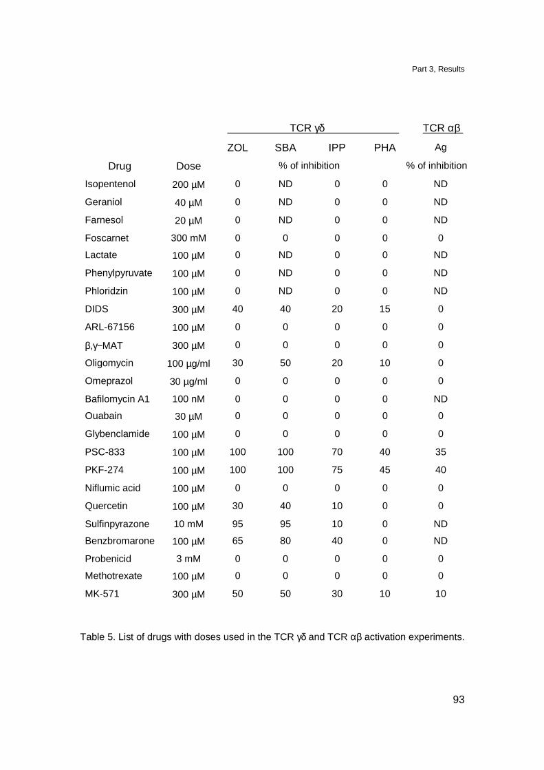
Part 3, Results
93
TCR γδ TCR αβ
ZOL SBA IPP PHA Ag
Drug Dose % of inhibition % of inhibition
Isopentenol 200 µM 0 ND 0 0 ND
Geraniol 40 µM 0 ND 0 0 ND
Farnesol 20 µM 0 ND 0 0 ND
Foscarnet 300 mM 0 0 0 0 0
Lactate 100 µM 0 ND 0 0 ND
Phenylpyruvate 100 µM 0 ND 0 0 ND
Phloridzin 100 µM 0 ND 0 0 ND
DIDS 300 µM 40 40 20 15 0
ARL-67156 100 µM 0 0 0 0 0
β,γ−MAT 300 µM 0 0 0 0 0
Oligomycin 100 µg/ml 30 50 20 10 0
Omeprazol 30 µg/ml 0 0 0 0 0
Bafilomycin A1 100 nM 0 0 0 0 ND
Ouabain 30 µM 0 0 0 0 0
Glybenclamide 100 µM 0 0 0 0 0
PSC-833 100 µM 100 100 70 40 35
PKF-274 100 µM 100 100 75 45 40
Niflumic acid 100 µM 0 0 0 0 0
Quercetin 100 µM 30 40 10 0 0
Sulfinpyrazone 10 mM 95 95 10 0 ND
Benzbromarone 100 µM 65 80 40 0 ND
Probenicid 3 mM 0 0 0 0 0
Methotrexate 100 µM 0 0 0 0 0
MK-571 300 µM 50 50 30 10 10
Table 5. List of drugs with doses used in the TCR γδ and TCR αβ activation experiments.
Part 3, Results
94
Specific inhibition of TCR γδ cells activation was determined by comparing activation in
the absence and presence of the drug, (ND, not determined).
The activation of TCR γδ cells was not affected when isopentenol, geraniol
or farnesol, the alcohol analogs of IPP, GPP and FPP, respectively, were used.
This suggests that the putative transporter recognizes the phosphate group
which is present in active ligands (Pfeffer et al., 1990; Constant et al., 1994;
Schoel et al., 1994; Tanaka et al., 1994). Therefore, the inhibitors of inorganic
phosphate transporters, foscarnet (Yusufi et al., 1986; Timmer and Gunn, 1998),
and inhibitors of MTC, lactate, phloridzin, phenylpyruvate (Halestrap and Price,
1999), were tested. These compounds also did not show inhibition of TCR γδ
cells activation. In contrast, two inhibitors of ABC transporters, PSC-833 and
PKF-274, preferentially decreased activation of TCR γδ cells induced by ZOL or
SBA. These findings indicated involvement of the protein(s) from the ABC-
transporters family in the TCR γδ activation.
PSC-833 and PKF-274 drugs, which were chemically designed to inhibit multi-
drug resistant protein 1 (MDR1), (Boesch et al., 1991; Lum et al., 1993), a
member of the B subfamily of ABC transporters, also inhibit other ABC
transporters (Paul et al., 1996). Therefore, their broad activity does not allow
discrimination of the transporter involved in TCR γδ cells activation.
We have found two other drugs, sulfinpyrazone and benzbromarone, that
specifically inhibit TCR γδ cells activation. The fact that these drugs inhibit
various anion transporters belonging to the ABC family (Bakos et al., 2000; Evers
et al., 2000) supported the hypothesis that the transporters involved in TCR γδ
Part 3, Results
95
activation recognize the anionic part of the stimulatory ligands. Additionally we
found specific inhibition in the presence of quercetin, an inhibitor of ABC-C
transporters (Walgren et al., 2000). Therefore, we narrowed our studies to the
subfamily C of ABC transporters.
This subfamily is composed of 12 characterized members: 9 multi-drug related
proteins (MRP1-9), two sulfonylurea receptors (SUR1/2) and a cystic fibrosis
transmembrane conductance regulator (CFTR). The MRPs studied so far,
MRP1-8, are all organic anion pumps but they differ in substrate specificity and
tissue distribution. CFTR is a chloride channel (Schwiebert et al., 1999) and
SURs are the regulatory subunits of the ATP- sensitive potassium (KATP) channel
(Aguilar-Bryan et al., 1998). Table 6 lists the members of the ABC-C family of
transporters, their chromosomal localization, tissue distribution and substrate
specificity.
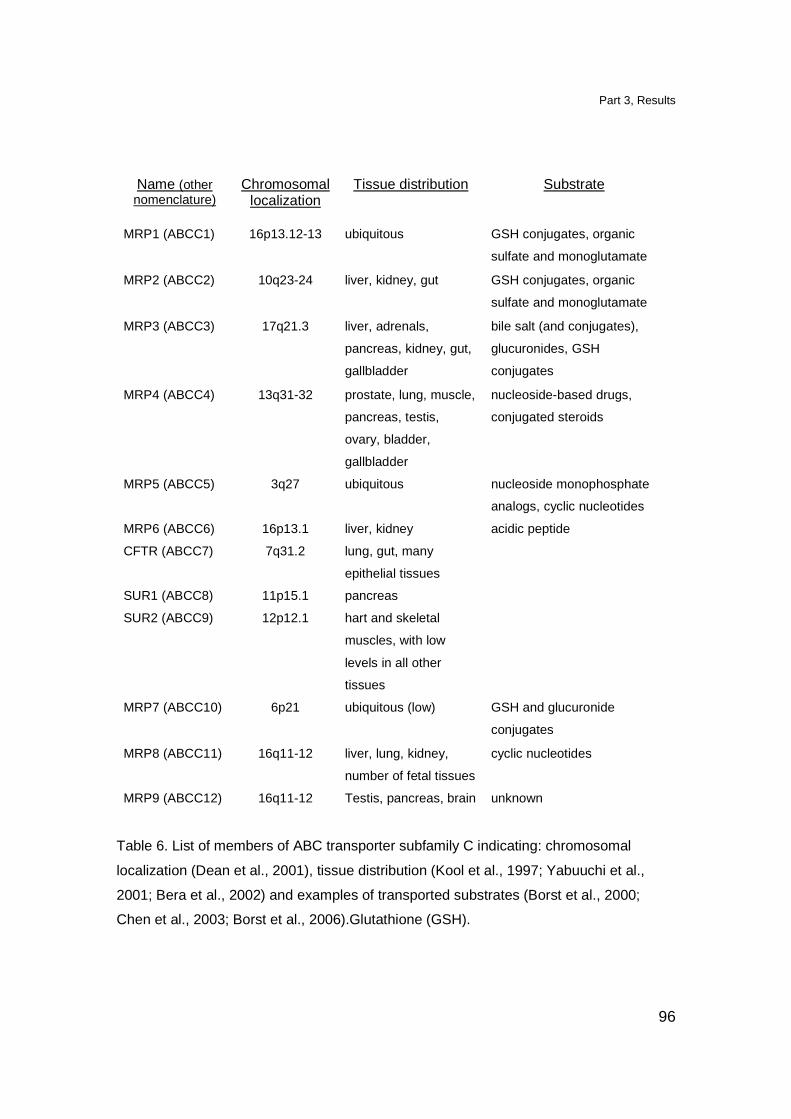
Part 3, Results
96
Name (other nomenclature)
Chromosomal localization
Tissue distribution Substrate
MRP1 (ABCC1)
16p13.12-13
ubiquitous
GSH conjugates, organic
sulfate and monoglutamate
MRP2 (ABCC2) 10q23-24 liver, kidney, gut GSH conjugates, organic
sulfate and monoglutamate
MRP3 (ABCC3) 17q21.3 liver, adrenals,
pancreas, kidney, gut,
gallbladder
bile salt (and conjugates),
glucuronides, GSH
conjugates
MRP4 (ABCC4) 13q31-32 prostate, lung, muscle,
pancreas, testis,
ovary, bladder,
gallbladder
nucleoside-based drugs,
conjugated steroids
MRP5 (ABCC5) 3q27 ubiquitous nucleoside monophosphate
analogs, cyclic nucleotides
MRP6 (ABCC6) 16p13.1 liver, kidney acidic peptide
CFTR (ABCC7) 7q31.2 lung, gut, many
epithelial tissues
SUR1 (ABCC8) 11p15.1 pancreas
SUR2 (ABCC9) 12p12.1 hart and skeletal
muscles, with low
levels in all other
tissues
MRP7 (ABCC10) 6p21 ubiquitous (low) GSH and glucuronide
conjugates
MRP8 (ABCC11) 16q11-12 liver, lung, kidney,
number of fetal tissues
cyclic nucleotides
MRP9 (ABCC12) 16q11-12 Testis, pancreas, brain unknown
Table 6. List of members of ABC transporter subfamily C indicating: chromosomal
localization (Dean et al., 2001), tissue distribution (Kool et al., 1997; Yabuuchi et al.,
2001; Bera et al., 2002) and examples of transported substrates (Borst et al., 2000;
Chen et al., 2003; Borst et al., 2006).Glutathione (GSH).

Part 3, Results
97
Since TCR γδ cells are activated by many cell types from different tissue
origin (Table 3), we assumed that the transporter involved in the antigen
presentation has rather broad tissue distribution. Therefore, we reduced the
number of candidate ABC-C transporters to those that are ubiquitously
expressed. Furthermore, we excluded CFTR as a candidate because, niflumic
acid, a potent inhibitor of CFTR (Scott-Ward et al., 2004) did not affect the
activation of TCR γδ cells. We also eliminated MRP7 due to its low level of
expression and our investigation focused on MRP1, MRP4 and MRP5.
The MK-571, an analog of leukotriens which specifically inhibits the transport
activity of MRP1, MRP4 and MRP5 (Reid et al., 2003) reduced the activation of
TCR γδ cells. However, other drugs which inhibit only MRP1, such as probenicid
and methotraxate (Bakos et al., 2000) did not affect TCR γδ cell activation. Thus,
we concluded that MRP1 is not responsible for IPP transport.
These results narrowed the studies to MRP4 and MRP5. Importantly, the tissue
distribution of MRP5 was described as ubiquitous while MRP4 has been found in
many but not all tissues (Kool et al., 1997). To further discriminate between
MRP4 and MRP5, a real-time quantitative PCR analysis was performed to
compare the mRNA levels of these two transporters in cell lines from various
tissues (Table 7), which all stimulate TCR γδ cells when pulsed with ZOL.
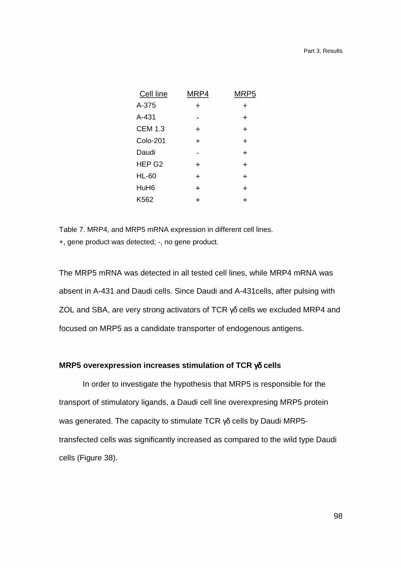
Part 3, Results
98
Cell line MRP4 MRP5 A-375 + + A-431 - + CEM 1.3 + + Colo-201 + + Daudi - + HEP G2 + + HL-60 + + HuH6 + + K562 + +
Table 7. MRP4, and MRP5 mRNA expression in different cell lines.
+, gene product was detected; -, no gene product.
The MRP5 mRNA was detected in all tested cell lines, while MRP4 mRNA was
absent in A-431 and Daudi cells. Since Daudi and A-431cells, after pulsing with
ZOL and SBA, are very strong activators of TCR γδ cells we excluded MRP4 and
focused on MRP5 as a candidate transporter of endogenous antigens.
MRP5 overexpression increases stimulation of TCR γδγδγδγδ cells
In order to investigate the hypothesis that MRP5 is responsible for the
transport of stimulatory ligands, a Daudi cell line overexpresing MRP5 protein
was generated. The capacity to stimulate TCR γδ cells by Daudi MRP5-
transfected cells was significantly increased as compared to the wild type Daudi
cells (Figure 38).
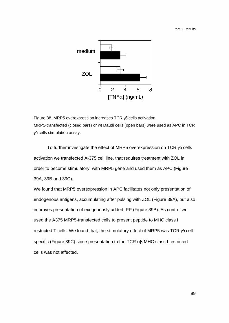
Part 3, Results
99
Figure 38. MRP5 overexpression increases TCR γδ cells activation.
MRP5-transfected (closed bars) or wt Daudi cells (open bars) were used as APC in TCR
γδ cells stimulation assay.
To further investigate the effect of MRP5 overexpression on TCR γδ cells
activation we transfected A-375 cell line, that requires treatment with ZOL in
order to become stimulatory, with MRP5 gene and used them as APC (Figure
39A, 39B and 39C).
We found that MRP5 overexpression in APC facilitates not only presentation of
endogenous antigens, accumulating after pulsing with ZOL (Figure 39A), but also
improves presentation of exogenously added IPP (Figure 39B). As control we
used the A375 MRP5-transfected cells to present peptide to MHC class I
restricted T cells. We found that, the stimulatory effect of MRP5 was TCR γδ cell
specific (Figure 39C) since presentation to the TCR αβ MHC class I restricted
cells was not affected.

Part 3, Results
100
Figure 39. MRP5 participates in the transport of endogenous and exogenous TCR γδ
ligands.
(A) MRP5 overexpression facilitates TCR γδ cells activation by ZOL treated APC. MRP5
transfected A-375 cells (�) stimulate TCR γδ more efficiently than A-375 wt cells ().
(B) Exogenously added IPP is better presented by MRP5 transfected A-375 cells (�)
than A-375 wt cells ().
(C) MRP5 ovexpression does not influence stimulation of MHC class I restricted, Mart
peptide specific TCR αβ clone. MRP5 transfected A-375 cells (�) and A-375 wt cells ()
were used as APC.
MRP5 downmodulation affects stimulation of TCR γδ γδ γδ γδ cells
In order to further confirm the involvement of MRP5 in the presentation of
TCR γδ stimulatory ligands we knocked down the MRP5 protein and performed
antigen presentation experiment. We generated four different small hairpin RNA
(shRNA) interference constructs targeting MRP5 mRNA. A-375 cells were
transfected with each of these constructs and were used as APC in TCR γδ
activation experiments. We found that A-375 cells transfected with MRP5 shRNA
interference constructs, but not with control firefly luciferase shRNA interference
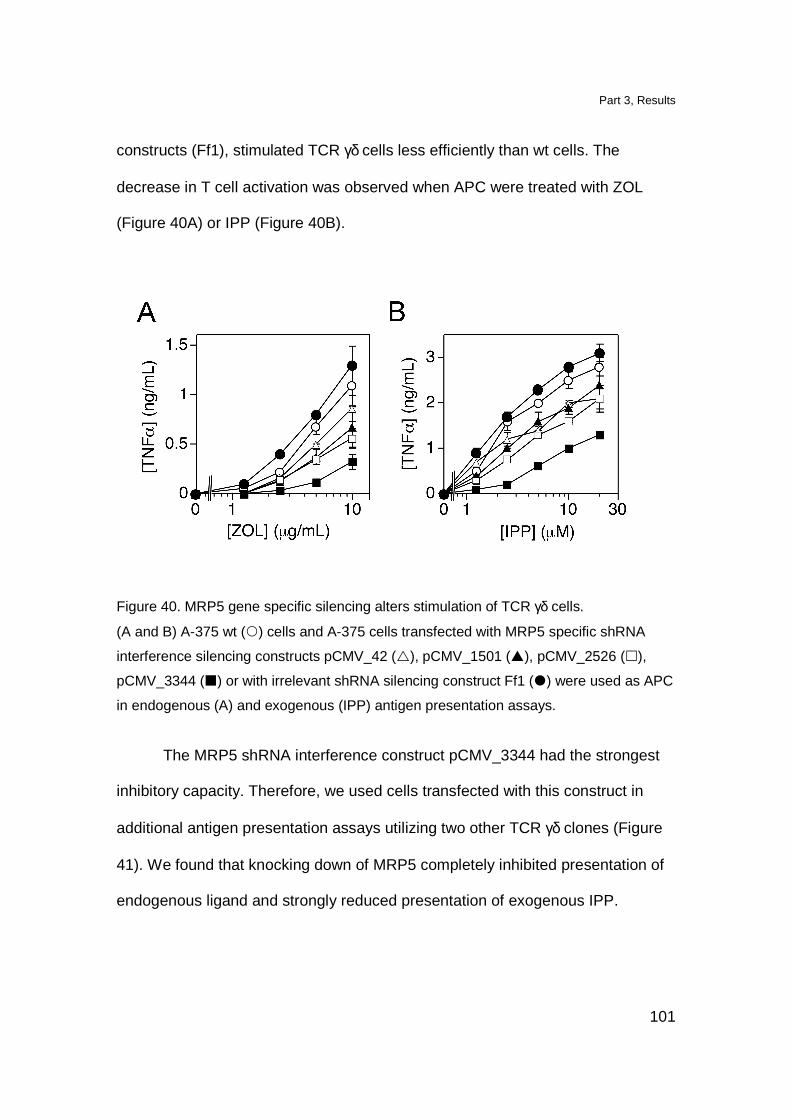
Part 3, Results
101
constructs (Ff1), stimulated TCR γδ cells less efficiently than wt cells. The
decrease in T cell activation was observed when APC were treated with ZOL
(Figure 40A) or IPP (Figure 40B).
Figure 40. MRP5 gene specific silencing alters stimulation of TCR γδ cells.
(A and B) A-375 wt () cells and A-375 cells transfected with MRP5 specific shRNA
interference silencing constructs pCMV_42 (�), pCMV_1501 (�), pCMV_2526 (�),
pCMV_3344 (�) or with irrelevant shRNA silencing construct Ff1 (�) were used as APC
in endogenous (A) and exogenous (IPP) antigen presentation assays.
The MRP5 shRNA interference construct pCMV_3344 had the strongest
inhibitory capacity. Therefore, we used cells transfected with this construct in
additional antigen presentation assays utilizing two other TCR γδ clones (Figure
41). We found that knocking down of MRP5 completely inhibited presentation of
endogenous ligand and strongly reduced presentation of exogenous IPP.
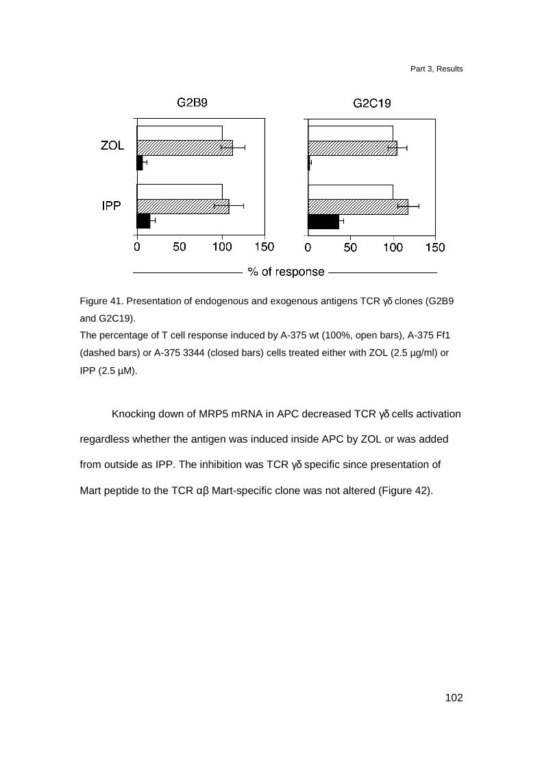
Part 3, Results
102
Figure 41. Presentation of endogenous and exogenous antigens TCR γδ clones (G2B9
and G2C19).
The percentage of T cell response induced by A-375 wt (100%, open bars), A-375 Ff1
(dashed bars) or A-375 3344 (closed bars) cells treated either with ZOL (2.5 µg/ml) or
IPP (2.5 µM).
Knocking down of MRP5 mRNA in APC decreased TCR γδ cells activation
regardless whether the antigen was induced inside APC by ZOL or was added
from outside as IPP. The inhibition was TCR γδ specific since presentation of
Mart peptide to the TCR αβ Mart-specific clone was not altered (Figure 42).
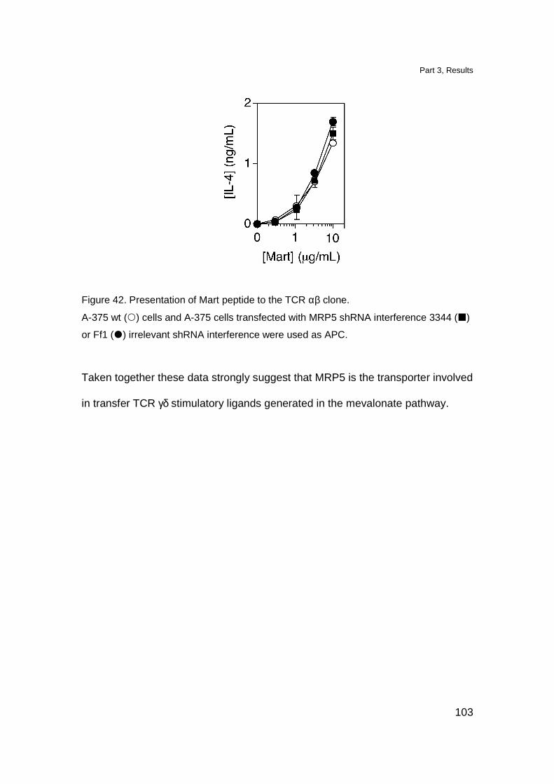
Part 3, Results
103
Figure 42. Presentation of Mart peptide to the TCR αβ clone.
A-375 wt () cells and A-375 cells transfected with MRP5 shRNA interference 3344 (�)
or Ff1 (�) irrelevant shRNA interference were used as APC.
Taken together these data strongly suggest that MRP5 is the transporter involved
in transfer TCR γδ stimulatory ligands generated in the mevalonate pathway.
Part 3, Discussion
104
Discussion
The fact that intracellular metabolites activate TCR Vγ9-Vδ2 cells raised
the question of how they are transported to the cell surface in order to establish a
cognate interaction with TCR. One possibility could be that inside APC,
mevalonate ligands bind to a dedicated presenting molecule and then are
transported on the cell surface. Thus it would resemble the biology of MHC-
peptide and CD1-lipid complexes. However, the inability of IPP to bind to putative
antigen-presenting molecules in a stable manner (IPP cannot be pulsed, Figure
16) makes this mechanism unlikely. If this would be the case, we might speculate
that the antigen presenting molecule is loaded intracellularly, forms immunogenic
complexes with mevalonate metabolites in a stable manner, and then traffick on
the cell surface, where ligands are degraded or dissociate with a very fast
kinetics.
Another possibility is that the mevalonate ligands are translocated across
membranes by specific transporter proteins. Indeed, the polarity and high charge
of pyrophosphorylated metabolites make unlikely a passive diffusion through
membranes.
Here we identified MRP5, a member of ABC-C transporter family, as a
protein required for activation of TCR Vγ9-Vδ2 cells. MRP5 is a multispecific
organic anion pump which transports nucleotide analogs (Jedlitschky et al., 2000;
Wijnholds et al., 2000). This protein has 12 transmembrane helices in each of
two membrane spanning domains forming P-glycoprotein-like core (Persson and
Argos, 1994; Belinsky et al., 1998). Therefore, together with MRP4 and MRP8,
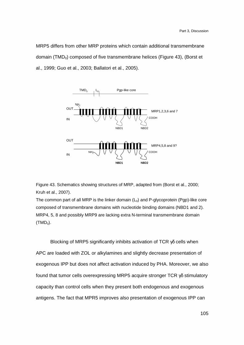
Part 3, Discussion
105
MRP5 differs from other MRP proteins which contain additional transmembrane
domain (TMD0) composed of five transmembrane helices (Figure 43), (Borst et
al., 1999; Guo et al., 2003; Ballatori et al., 2005).
OUT
OUT
IN
IN
NBD1 NBD2
NBD1 NBD2NBD1 NBD2
TMD0 L0 Pgp-like core
MRP1,2,3,6 and 7
MRP4,5,8 and 9?
COOH
COOHNH2
NH2
Figure 43. Schematics showing structures of MRP, adapted from (Borst et al., 2000;
Kruh et al., 2007).
The common part of all MRP is the linker domain (L0) and P-glycoprotein (Pgp)-like core
composed of transmembrane domains with nucleotide binding domains (NBD1 and 2).
MRP4, 5, 8 and possibly MRP9 are lacking extra N-terminal transmembrane domain
(TMD0).
Blocking of MRP5 significantly inhibits activation of TCR γδ cells when
APC are loaded with ZOL or alkylamines and slightly decrease presentation of
exogenous IPP but does not affect activation induced by PHA. Moreover, we also
found that tumor cells overexpressing MRP5 acquire stronger TCR γδ stimulatory
capacity than control cells when they present both endogenous and exogenous
antigens. The fact that MPR5 improves also presentation of exogenous IPP can

Part 3, Discussion
106
be explained with two mechanismis i) MRP5 might participate in the loading of
putative antigen presenting molecule directly on the cell surface or ii) IPP might
partially internalized inside APC and then reexpressed on the cell surface with
the help of MRP5.
The involvement of MRP5 in TCR γδ antigen presentation has been further
confirmed by experiments with MRP5 specific shRNA interference. Cell lines with
knock down MRP5 have much lower capacity to stimulate TCR γδ cells with
endogenous and exogenous antigens. However, the decrease in presentation of
exogenous IPP, when both specific MRP5 inhibitors and specific shRNA
interference were used, is much smaller as compared with endogenous ligands.
Therefore, it is likely that our second hypothesis is more accurate. If this is the
case, then MRP5 would be involved in the presentation of the internalized pool of
IPP.
Up to date the physiological function of MRP5 is not clear. It was
described as transporter for cyclic nucleotides, especially cGMP and was
suggested that MRP5 removes cGMP from the cytosol during signal transduction
(Jedlitschky et al., 2000). It is possible that IPP and other prenylated metabolites
generated in the mevalonate pathway mimic cGMP. Therefore, the role of MRP5
would be to recognize these substrates and facilitate their removal when they
accumulate in the cytosol. It is unlikely that MRP5 behaves as an antigen-
presenting molecule for the TCR γδ cells because the predicted extracellular
MRP5 domains (Persson and Argos, 1994; Belinsky et al., 1998) are too small.
We therefore, prefer the hypothesis that independent antigen-presenting
Part 3, Discussion
107
molecule interacts with TCR γδ. MRP5 may participate in antigen presentation to
TCR γδ cells by transporting the mevalonate metabolites from the cytoplasm,
where the metabolites are synthetised. MRP5 protein is localized on the cell
membrane (McAleer et al., 1999) and perhaps in small amounts intracellularly
(Wijnholds et al., 1997). These localizations might mediate two different
mechanisms of action. In the first case, MRP5 may transport mevalonate
metabolites outside the cell where they finally interact with antigen-presenting
molecules. A second possibility is that MRP5 facilitates traffic of stimulatory
metabolites from cytosol inside other compartments, i.e. ER. After transport in
this compartment, metabolites could bind to the antigen-presenting molecules,
form a complex and then be transported to the cell surface. This second
mechanism of action would resemble that of ATP-dependent trasporters
associated with antigen processing (TAP) involved in peptide traffic from cytosol
into ER (Neefjes et al., 1993; Momburg et al., 1994). Interestingly, TAP
molecules are part of complex protein multimers composed of several proteins
which facilitate the assembly of MHC class I proteins and also provide transport
and loading of peptides on these presenting molecules (Cresswell et al., 1999).
At present it is not clear whether MRP5 constitutes multi-subunit complexes with
other proteins. Interestingly, other transporters are associated with a second
protein and form complexes which are localized on the cell surface. This is the
case of MCT4- and MCT1 which form dimers with CD147 on the cell surface
(Kirk et al., 2000).

Part 3, Discussion
108
Taken together our data show that MRP5 is involved in the presentation of
TCR γδ stimulatory ligand generated in the mevalonate pathway. MRP5 is most
likely participates in transfer of these phosphorylated ligands from the cytoplasm,
where they are synthesized, to the cell surface where they are presented to the
TCR γδ cells.
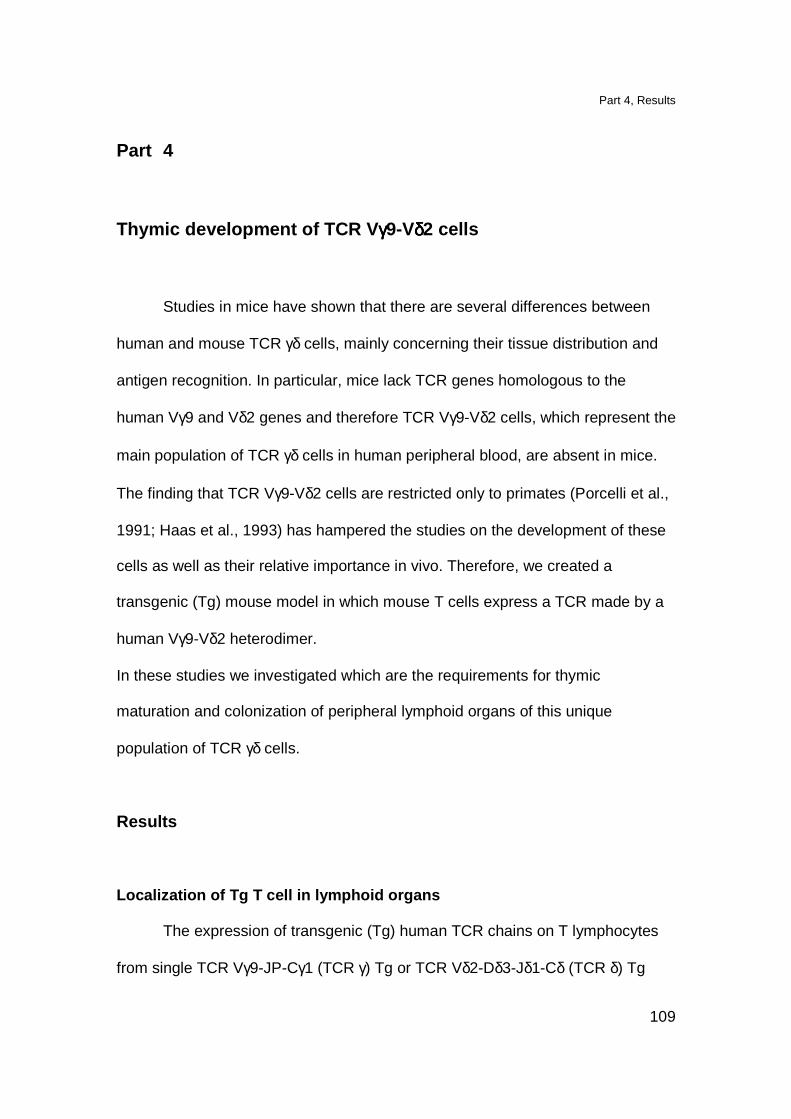
Part 4, Results
109
Part 4
Thymic development of TCR V γγγγ9-Vδδδδ2 cells
Studies in mice have shown that there are several differences between
human and mouse TCR γδ cells, mainly concerning their tissue distribution and
antigen recognition. In particular, mice lack TCR genes homologous to the
human Vγ9 and Vδ2 genes and therefore TCR Vγ9-Vδ2 cells, which represent the
main population of TCR γδ cells in human peripheral blood, are absent in mice.
The finding that TCR Vγ9-Vδ2 cells are restricted only to primates (Porcelli et al.,
1991; Haas et al., 1993) has hampered the studies on the development of these
cells as well as their relative importance in vivo. Therefore, we created a
transgenic (Tg) mouse model in which mouse T cells express a TCR made by a
human Vγ9-Vδ2 heterodimer.
In these studies we investigated which are the requirements for thymic
maturation and colonization of peripheral lymphoid organs of this unique
population of TCR γδ cells.
Results
Localization of Tg T cell in lymphoid organs
The expression of transgenic (Tg) human TCR chains on T lymphocytes
from single TCR Vγ9-JP-Cγ1 (TCR γ) Tg or TCR Vδ2-Dδ3-Jδ1-Cδ (TCR δ) Tg
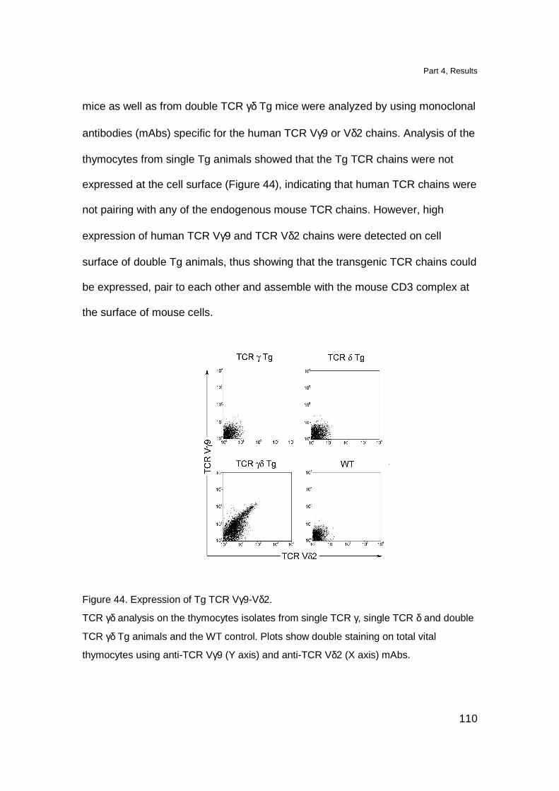
Part 4, Results
110
mice as well as from double TCR γδ Tg mice were analyzed by using monoclonal
antibodies (mAbs) specific for the human TCR Vγ9 or Vδ2 chains. Analysis of the
thymocytes from single Tg animals showed that the Tg TCR chains were not
expressed at the cell surface (Figure 44), indicating that human TCR chains were
not pairing with any of the endogenous mouse TCR chains. However, high
expression of human TCR Vγ9 and TCR Vδ2 chains were detected on cell
surface of double Tg animals, thus showing that the transgenic TCR chains could
be expressed, pair to each other and assemble with the mouse CD3 complex at
the surface of mouse cells.
Figure 44. Expression of Tg TCR Vγ9-Vδ2.
TCR γδ analysis on the thymocytes isolates from single TCR γ, single TCR δ and double
TCR γδ Tg animals and the WT control. Plots show double staining on total vital
thymocytes using anti-TCR Vγ9 (Y axis) and anti-TCR Vδ2 (X axis) mAbs.
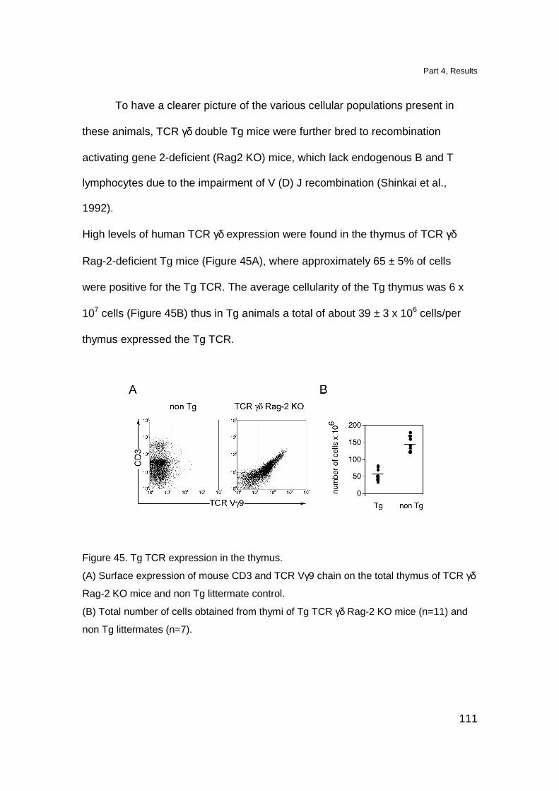
Part 4, Results
111
To have a clearer picture of the various cellular populations present in
these animals, TCR γδ double Tg mice were further bred to recombination
activating gene 2-deficient (Rag2 KO) mice, which lack endogenous B and T
lymphocytes due to the impairment of V (D) J recombination (Shinkai et al.,
1992).
High levels of human TCR γδ expression were found in the thymus of TCR γδ
Rag-2-deficient Tg mice (Figure 45A), where approximately 65 ± 5% of cells
were positive for the Tg TCR. The average cellularity of the Tg thymus was 6 x
107 cells (Figure 45B) thus in Tg animals a total of about 39 ± 3 x 10⁶ cells/per
thymus expressed the Tg TCR.
Figure 45. Tg TCR expression in the thymus.
(A) Surface expression of mouse CD3 and TCR Vγ9 chain on the total thymus of TCR γδ
Rag-2 KO mice and non Tg littermate control.
(B) Total number of cells obtained from thymi of Tg TCR γδ Rag-2 KO mice (n=11) and
non Tg littermates (n=7).
Part 4, Results
112
When we analyzed peripheral lymphoid organs, we found that, in the spleen
(Figure 46) and in the lymph nodes (data not shown), only 0.1-0.9% cells bearing
human TCR γδ were detected (the average of Tg T cells was 1.4 x 10⁴/per
spleen).
Figure 46. Surface expression of mouse CD3 and TCR Vγ9 chain on splenocytes.
Analyzed cells were gated as Mac-1 and B220 negative from TCR γδ Rag-2 KO and non
Tg control mice.
Tg thymocytes have a semi-mature phenotype
Impairment in the colonization of peripheral lymphoid organs prompted us
to investigate whether Tg thymocytes were arrested during their development.
Thymocytes from Rag-2-deficient TCR γδ Tg mice were stained with mAbs
specific for surface markers characteristic of thymocyte differentiation. While Tg
animals have similar percentages of thymocytes with DP phenotype, as
compared to non Tg littermates, they have slightly increased numbers of DN
cells, compensated by a decrease in the single CD4 and single CD8 positive
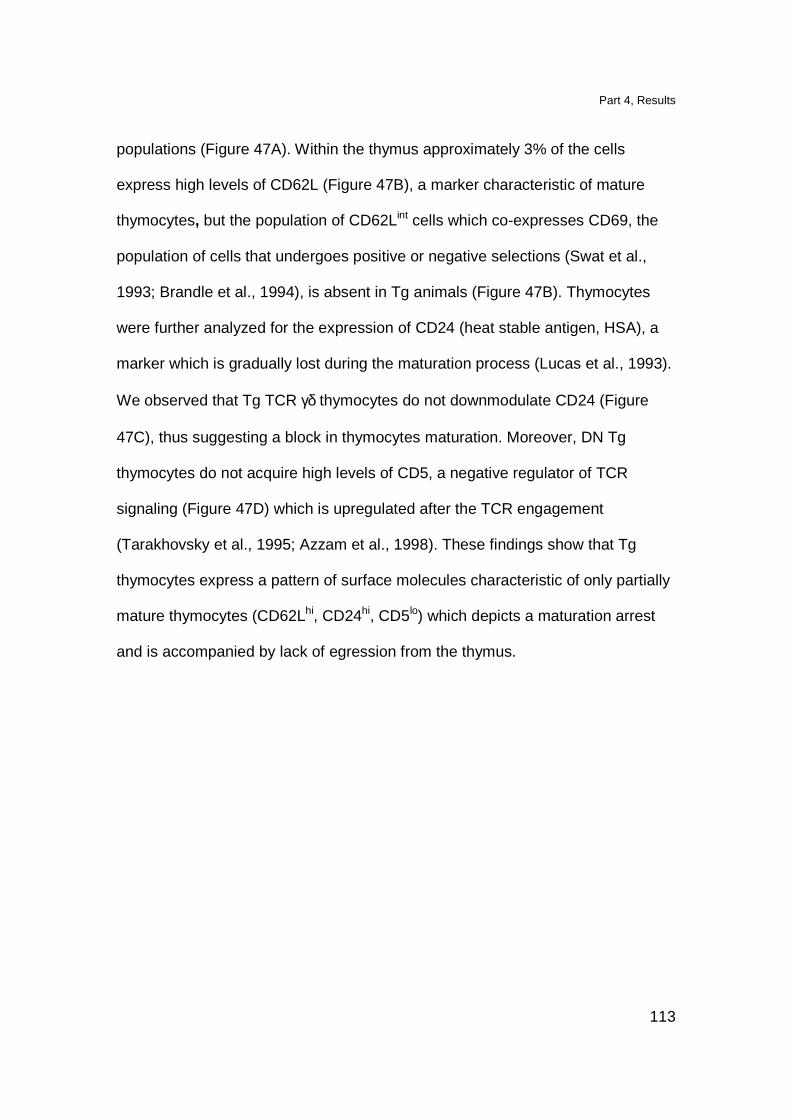
Part 4, Results
113
populations (Figure 47A). Within the thymus approximately 3% of the cells
express high levels of CD62L (Figure 47B), a marker characteristic of mature
thymocytes, but the population of CD62Lint cells which co-expresses CD69, the
population of cells that undergoes positive or negative selections (Swat et al.,
1993; Brandle et al., 1994), is absent in Tg animals (Figure 47B). Thymocytes
were further analyzed for the expression of CD24 (heat stable antigen, HSA), a
marker which is gradually lost during the maturation process (Lucas et al., 1993).
We observed that Tg TCR γδ thymocytes do not downmodulate CD24 (Figure
47C), thus suggesting a block in thymocytes maturation. Moreover, DN Tg
thymocytes do not acquire high levels of CD5, a negative regulator of TCR
signaling (Figure 47D) which is upregulated after the TCR engagement
(Tarakhovsky et al., 1995; Azzam et al., 1998). These findings show that Tg
thymocytes express a pattern of surface molecules characteristic of only partially
mature thymocytes (CD62Lhi, CD24hi, CD5lo) which depicts a maturation arrest
and is accompanied by lack of egression from the thymus.
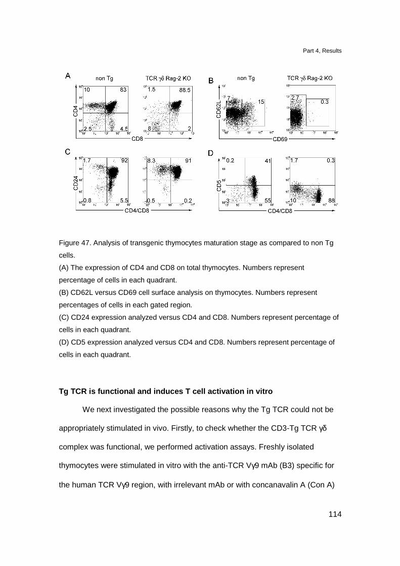
Part 4, Results
114
Figure 47. Analysis of transgenic thymocytes maturation stage as compared to non Tg
cells.
(A) The expression of CD4 and CD8 on total thymocytes. Numbers represent
percentage of cells in each quadrant.
(B) CD62L versus CD69 cell surface analysis on thymocytes. Numbers represent
percentages of cells in each gated region.
(C) CD24 expression analyzed versus CD4 and CD8. Numbers represent percentage of
cells in each quadrant.
(D) CD5 expression analyzed versus CD4 and CD8. Numbers represent percentage of
cells in each quadrant.
Tg TCR is functional and induces T cell activation in vitro
We next investigated the possible reasons why the Tg TCR could not be
appropriately stimulated in vivo. Firstly, to check whether the CD3-Tg TCR γδ
complex was functional, we performed activation assays. Freshly isolated
thymocytes were stimulated in vitro with the anti-TCR Vγ9 mAb (B3) specific for
the human TCR Vγ9 region, with irrelevant mAb or with concanavalin A (Con A)
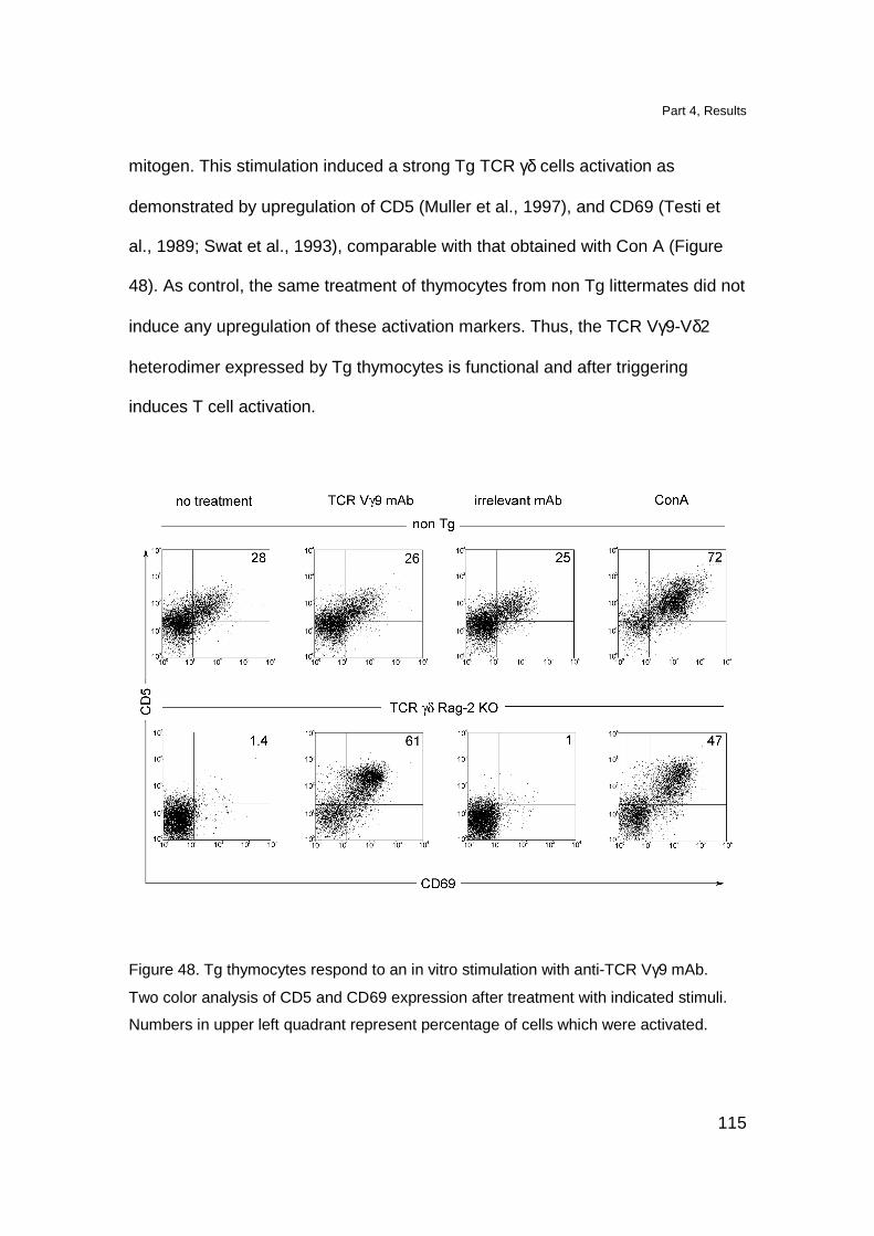
Part 4, Results
115
mitogen. This stimulation induced a strong Tg TCR γδ cells activation as
demonstrated by upregulation of CD5 (Muller et al., 1997), and CD69 (Testi et
al., 1989; Swat et al., 1993), comparable with that obtained with Con A (Figure
48). As control, the same treatment of thymocytes from non Tg littermates did not
induce any upregulation of these activation markers. Thus, the TCR Vγ9-Vδ2
heterodimer expressed by Tg thymocytes is functional and after triggering
induces T cell activation.
Figure 48. Tg thymocytes respond to an in vitro stimulation with anti-TCR Vγ9 mAb.
Two color analysis of CD5 and CD69 expression after treatment with indicated stimuli.
Numbers in upper left quadrant represent percentage of cells which were activated.
Part 4, Results
116
We further investigated whether Tg T cells were able to respond to the
stimuli that activate human TCR Vγ9-Vδ2 cells. Various cell types of human or
mouse origin, were treated with zoledronate (ZOL) and were used as APC. The
stimulation of Tg T cells was evaluated by checking the expression of CD5 and
CD69 activation markers. Tg thymocytes were activated by human DC and
human thymic epithelial cells (TEC) but not by human thymocytes (Figure 49).
Figure 49. Human APC activate TRC γδ transgenic thymocytes in vitro.
Two color analysis of CD5 and CD69 expression on Tg thymocytes stimulated with ZOL-
treated APC (DC, TEC and thymocytes).
Several other established tumor cell lines as well as freshly isolated cells
of both human and mouse origin were also tested. Since only cells of human and
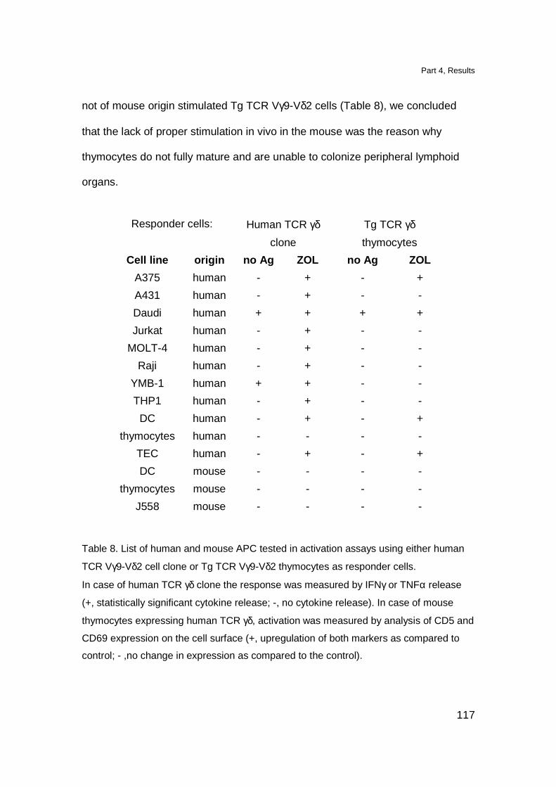
Part 4, Results
117
not of mouse origin stimulated Tg TCR Vγ9-Vδ2 cells (Table 8), we concluded
that the lack of proper stimulation in vivo in the mouse was the reason why
thymocytes do not fully mature and are unable to colonize peripheral lymphoid
organs.
Responder cells: Human TCR γδ
clone
Tg TCR γδ
thymocytes
Cell line origin no Ag ZOL no Ag ZOL
A375 human - + - +
A431 human - + - -
Daudi human + + + +
Jurkat human - + - -
MOLT-4 human - + - -
Raji human - + - -
YMB-1 human + + - -
THP1 human - + - -
DC human - + - +
thymocytes human - - - -
TEC human - + - +
DC mouse - - - -
thymocytes mouse - - - -
J558 mouse - - - -
Table 8. List of human and mouse APC tested in activation assays using either human
TCR Vγ9-Vδ2 cell clone or Tg TCR Vγ9-Vδ2 thymocytes as responder cells.
In case of human TCR γδ clone the response was measured by IFNγ or TNFα release
(+, statistically significant cytokine release; -, no cytokine release). In case of mouse
thymocytes expressing human TCR γδ, activation was measured by analysis of CD5 and
CD69 expression on the cell surface (+, upregulation of both markers as compared to
control; - ,no change in expression as compared to the control).
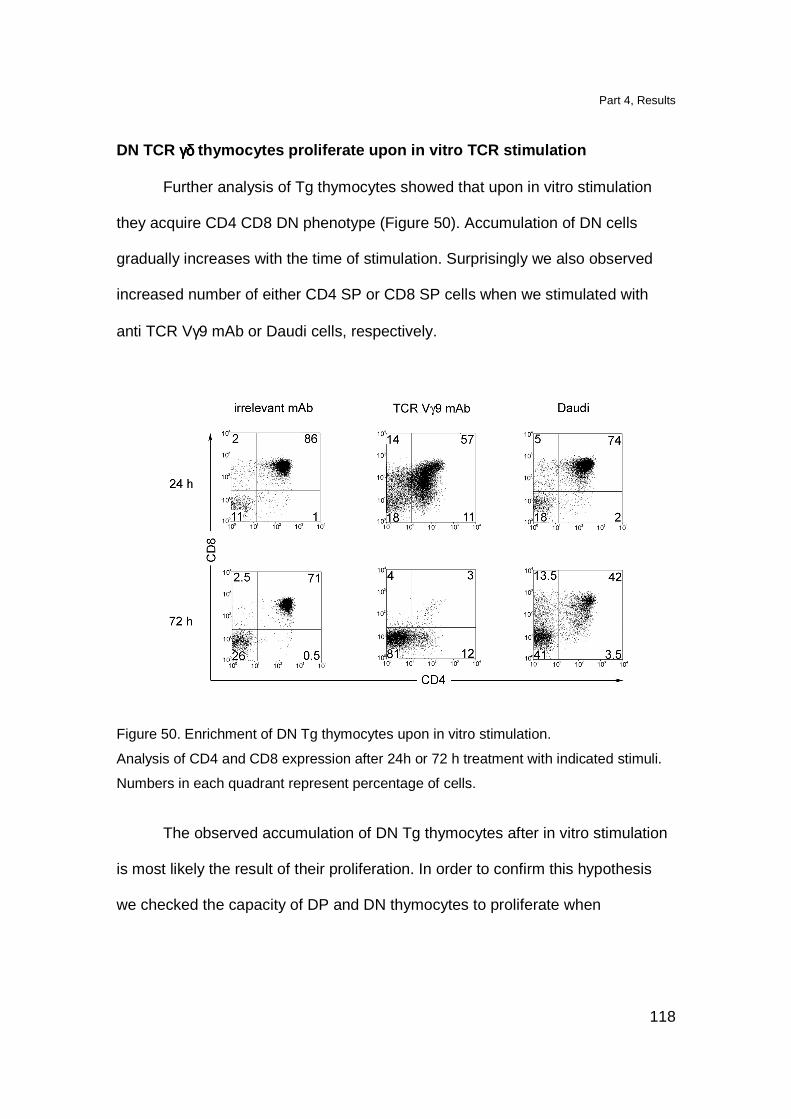
Part 4, Results
118
DN TCR γδγδγδγδ thymocytes proliferate upon in vitro TCR stimulation
Further analysis of Tg thymocytes showed that upon in vitro stimulation
they acquire CD4 CD8 DN phenotype (Figure 50). Accumulation of DN cells
gradually increases with the time of stimulation. Surprisingly we also observed
increased number of either CD4 SP or CD8 SP cells when we stimulated with
anti TCR Vγ9 mAb or Daudi cells, respectively.
Figure 50. Enrichment of DN Tg thymocytes upon in vitro stimulation.
Analysis of CD4 and CD8 expression after 24h or 72 h treatment with indicated stimuli.
Numbers in each quadrant represent percentage of cells.
The observed accumulation of DN Tg thymocytes after in vitro stimulation
is most likely the result of their proliferation. In order to confirm this hypothesis
we checked the capacity of DP and DN thymocytes to proliferate when
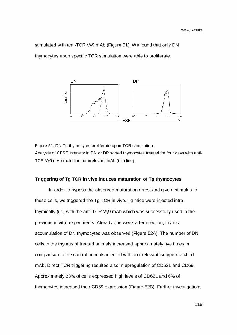
Part 4, Results
119
stimulated with anti-TCR Vγ9 mAb (Figure 51). We found that only DN
thymocytes upon specific TCR stimulation were able to proliferate.
Figure 51. DN Tg thymocytes proliferate upon TCR stimulation.
Analysis of CFSE intensity in DN or DP sorted thymocytes treated for four days with anti-
TCR Vγ9 mAb (bold line) or irrelevant mAb (thin line).
Triggering of Tg TCR in vivo induces maturation of Tg thymocytes
In order to bypass the observed maturation arrest and give a stimulus to
these cells, we triggered the Tg TCR in vivo. Tg mice were injected intra-
thymically (i.t.) with the anti-TCR Vγ9 mAb which was successfully used in the
previous in vitro experiments. Already one week after injection, thymic
accumulation of DN thymocytes was observed (Figure 52A). The number of DN
cells in the thymus of treated animals increased approximately five times in
comparison to the control animals injected with an irrelevant isotype-matched
mAb. Direct TCR triggering resulted also in upregulation of CD62L and CD69.
Approximately 23% of cells expressed high levels of CD62L and 6% of
thymocytes increased their CD69 expression (Figure 52B). Further investigations
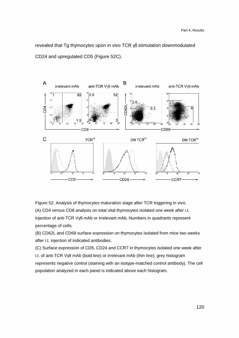
Part 4, Results
120
revealed that Tg thymocytes upon in vivo TCR γδ stimulation downmodulated
CD24 and upregulated CD5 (Figure 52C).
Figure 52. Analysis of thymocytes maturation stage after TCR triggering in vivo.
(A) CD4 versus CD8 analysis on total vital thymocytes isolated one week after i.t.
injection of anti-TCR Vγ9 mAb or irrelevant mAb. Numbers in quadrants represent
percentage of cells.
(B) CD62L and CD69 surface expression on thymocytes isolated from mice two weeks
after i.t. injection of indicated antibodies.
(C) Surface expression of CD5, CD24 and CCR7 in thymocytes isolated one week after
i.t. of anti-TCR Vγ9 mAb (bold line) or irrelevant mAb (thin line); grey histogram
represents negative control (staining with an isotype-matched control antibody). The cell
population analyzed in each panel is indicated above each histogram.
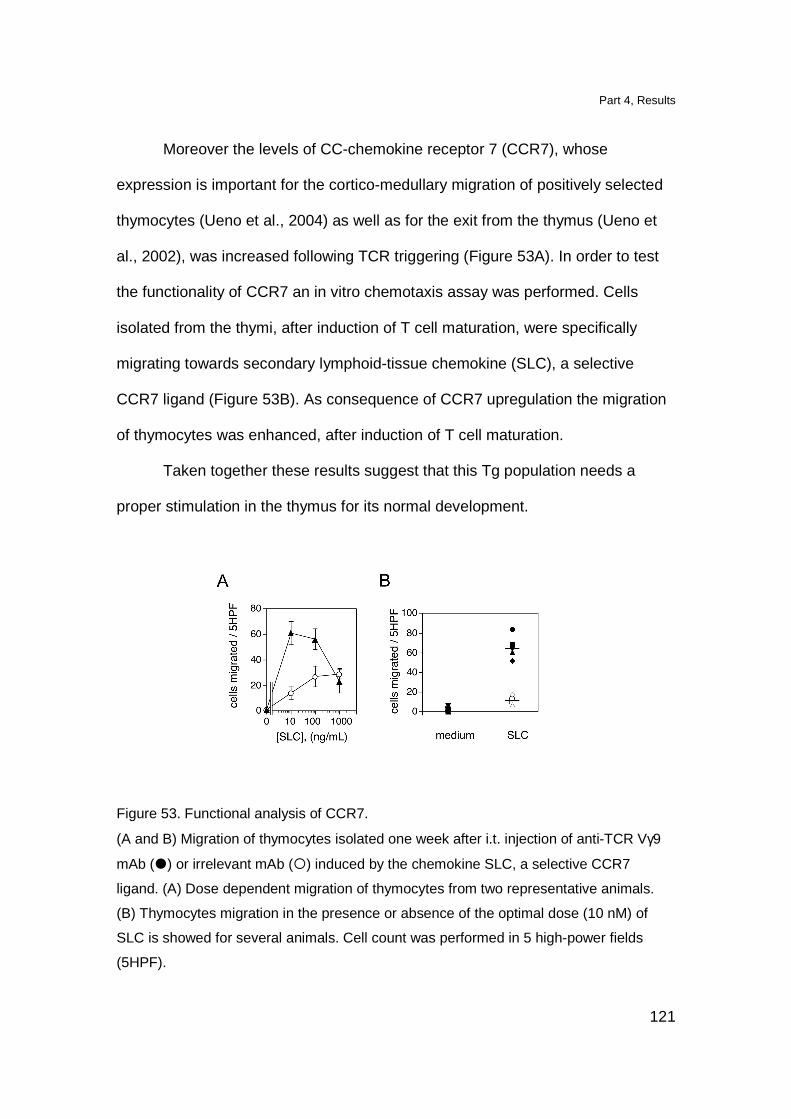
Part 4, Results
121
Moreover the levels of CC-chemokine receptor 7 (CCR7), whose
expression is important for the cortico-medullary migration of positively selected
thymocytes (Ueno et al., 2004) as well as for the exit from the thymus (Ueno et
al., 2002), was increased following TCR triggering (Figure 53A). In order to test
the functionality of CCR7 an in vitro chemotaxis assay was performed. Cells
isolated from the thymi, after induction of T cell maturation, were specifically
migrating towards secondary lymphoid-tissue chemokine (SLC), a selective
CCR7 ligand (Figure 53B). As consequence of CCR7 upregulation the migration
of thymocytes was enhanced, after induction of T cell maturation.
Taken together these results suggest that this Tg population needs a
proper stimulation in the thymus for its normal development.
Figure 53. Functional analysis of CCR7.
(A and B) Migration of thymocytes isolated one week after i.t. injection of anti-TCR Vγ9
mAb (�) or irrelevant mAb (�) induced by the chemokine SLC, a selective CCR7
ligand. (A) Dose dependent migration of thymocytes from two representative animals.
(B) Thymocytes migration in the presence or absence of the optimal dose (10 nM) of
SLC is showed for several animals. Cell count was performed in 5 high-power fields
(5HPF).
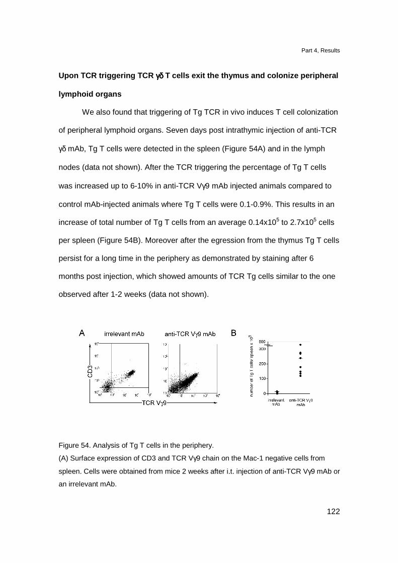
Part 4, Results
122
Upon TCR triggering TCR γγγγδδδδ T cells exit the thymus and colonize peripheral
lymphoid organs
We also found that triggering of Tg TCR in vivo induces T cell colonization
of peripheral lymphoid organs. Seven days post intrathymic injection of anti-TCR
γδ mAb, Tg T cells were detected in the spleen (Figure 54A) and in the lymph
nodes (data not shown). After the TCR triggering the percentage of Tg T cells
was increased up to 6-10% in anti-TCR Vγ9 mAb injected animals compared to
control mAb-injected animals where Tg T cells were 0.1-0.9%. This results in an
increase of total number of Tg T cells from an average 0.14x10⁵ to 2.7x10⁵ cells
per spleen (Figure 54B). Moreover after the egression from the thymus Tg T cells
persist for a long time in the periphery as demonstrated by staining after 6
months post injection, which showed amounts of TCR Tg cells similar to the one
observed after 1-2 weeks (data not shown).
Figure 54. Analysis of Tg T cells in the periphery.
(A) Surface expression of CD3 and TCR Vγ9 chain on the Mac-1 negative cells from
spleen. Cells were obtained from mice 2 weeks after i.t. injection of anti-TCR Vγ9 mAb or
an irrelevant mAb.

Part 4, Results
123
(B) Total number of Tg T cells was calculated per each analyzed spleen of mice injected
i.t. with either irrelevant mAb (n=8) or with anti-TCR Vγ9 mAb (n=8). Horizontal lines
represent average number of cell in each group.
The vast majority of Tg T cells that colonize peripheral lymphoid organs is
DN, expresses CD5 and has non activated CD25- CD69low phenotype (Figure
55).
Figure 55. Phenotypic analysis of Tg T cells in the spleen.
Surface expression of CD4/CD8, CD5, CD25, CD69 and TCR Vγ9 two weeks after i.t.
injection of anti-TCR Vγ9 mAb. Mac-1 positive cells were excluded from the analysis.
The functionality of the cells that colonize peripheral lymphoid organs was
determined in an in vitro stimulation assay. Peripheral TCR γδ cells were
activated in the presence of specific anti-TCR γδ mAb as well as in the presence
of Daudi cells. Activated T cells upregulated CD25 and CD69 markers (Figure
56A) and also released IFNγ (Figure 56B). These data show that cells which
colonize the periphery are functional.
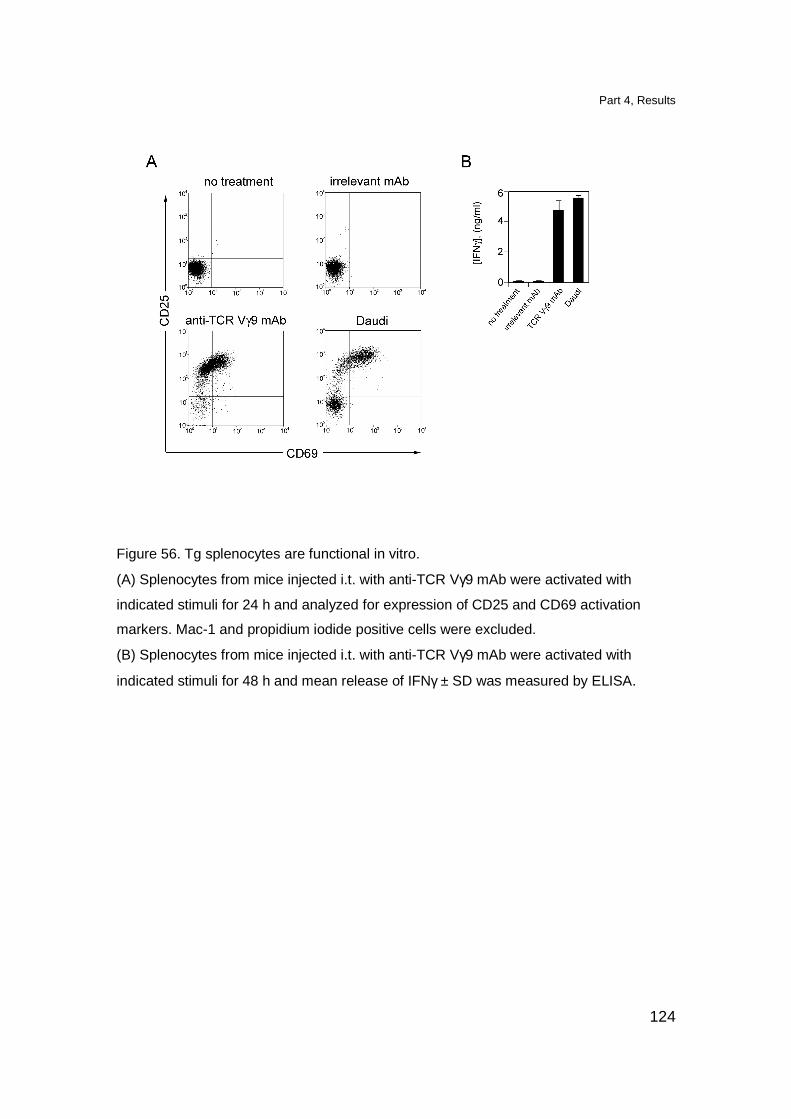
Part 4, Results
124
Figure 56. Tg splenocytes are functional in vitro.
(A) Splenocytes from mice injected i.t. with anti-TCR Vγ9 mAb were activated with
indicated stimuli for 24 h and analyzed for expression of CD25 and CD69 activation
markers. Mac-1 and propidium iodide positive cells were excluded.
(B) Splenocytes from mice injected i.t. with anti-TCR Vγ9 mAb were activated with
indicated stimuli for 48 h and mean release of IFNγ ± SD was measured by ELISA.
Part 4, Discussion
125
Discussion
The mechanisms governing the development of human TCR γδ cells
remain largely unknown. The TCR γδ cells development has been mainly studied
using two mouse TCR γδ Tg models, G8 and KN6. Tg TCR γδ cells in G8 and
KN6 mouse models carry the rearranged TCR γδ reactive to the MHC class Ib
T22 molecule (Bluestone et al., 1988; Bonneville et al., 1989; Ito et al., 1990). So
far it is not clear whether the selection process of these TCR γδ cells requires
ligand engagement since contradictory results have been published (Wells et al.,
1991; Pereira et al., 1992; Schweighoffer and Fowlkes, 1996; Haks et al., 2005).
More recent reports provide evidence for ligand-mediated positive selection of
murine DETC population (Xiong et al., 2004; Lewis et al., 2006).
In order to study TCR Vγ9-Vδ2 cells in vivo we have generated a
transgenic mouse model in which human TCR γδ is expressed on mouse T cells.
Tg TCR was cloned under the control of human CD2 promoter that assured early
expression of transgene during ontogeny (Duplay et al., 1989; Sen et al., 1989;
Yagita et al., 1989). Cells expressing the human Tg TCR are very abundant in
the thymus of Tg mice and they represent the only T cell population present in
the thymus of Tg mice with Rag-2-deficient background. However, in the
peripheral lymphoid organs only few cells bearing Tg TCR are present.
The unusual tissue distribution of Tg T cells could be the result of impaired
thymic maturation and/or selection. In the thymi of Tg mice we observed only a
small increase in the percentage of DN cells (up to 10%), together with a reduced
Part 4, Discussion
126
number of CD4 SP and CD8 SP cells, as compared to non Tg littermates. This is
in contrast with other Tg models namely the Tg G8 TCR γδ mice and Tg Vγ6-Vδ1
TCR γδ mice in which, DN cells reach approximately 50% of total thymocytes
(Livak et al., 1997; Hayes et al., 2005). The DP cell compartment was
comparable to the one observed in non Tg littermates. The presence of DP
thymocytes in Tg mice is not completely surprising since also in the thymus of
TCR β -/- mice, where TCR αβ development is arrested, DP cells are present in
variable numbers (Mombaerts et al., 1993; Passoni et al., 1997). However, the
DP cells in TCR β -/- mice derive from TCR γδ-expressing progenitor cells but are
committed to the αβ T cell linage (Kang et al., 1998).
According to the signal-strength model, in which strong TCR signals promote
γδ lineage development while weak TCR signals in synergy with Notch signaling
promote αβ T cell differentiation through DP intermediates (Hayes et al., 2003;
Haks et al., 2005; Hayes et al., 2005; Garbe and von Boehmer, 2007; Hayday
and Pennington, 2007). The acquisition of DP phenotype might result from
impaired stimulation of the Tg TCR γδ. This is also in agreement with the findings
in Tg KN6 TCR γδ mice in which blocking of the TCR engagement is followed by
expression of DP phenotype in 90% of thymocytes (Haks et al., 2005).
Our studies revealed that Tg thymocytes express a pattern of molecules
characteristic for partially mature thymocytes (CD62Lhi, CD24hi, CD5lo, CCR7-).
TCRhi DN cells express high levels of CD62L, a marker characteristic of mature
thymocytes, whereas the CD24 molecule, which is low in mature cells, is not
downmodulated. In Tg KN6 TCR γδ mice the absence of specific TCR γδ ligand
Part 4, Discussion
127
skewed DN thymocytes towards immature CD24hi phenotype while the presence
of the specific ligand lead to CD24 downmodulation and maturation of TCR γδ
DN thymocytes (Haks et al., 2005). Additionally we found that, the expression
CD5, which is upregulated after pre-TCR and TCR engagement (Azzam et al.,
1998), is maintained at low levels on Tg TCR Vγ9-Vδ2 thymocytes.
During maturation processes T cells upregulate transiently CD69 very
early after TCR triggering and decline the expression of this surface marker
shortly after stimuli withdrawal (Testi et al., 1989). In the thymus DP thymocytes
that are undergoing selection also transiently express CD69 (Swat et al., 1993;
Brandle et al., 1994) and after the selection they lose the CD69 expression and
gain expression of the peripheral homing marker CD62L (Gabor et al., 1997).
Therefore, the absence in Tg TCR Vγ9-Vδ2 mice of cells expressing CD69 in
another piece of evidence indicating that Tg thymocytes abort their maturation
process.
Importantly, Tg thymocytes are able to respond in vitro to various stimuli
capable of activating human TCR Vγ9-Vδ2 cells in a specific manner. Both anti-
TCR Vγ9 mAb and human APC presenting endogenous antigens were able to
activate Tg thymocytes indicating that the Tg human TCR associated with mouse
CD3 complex is functional. Additional analysis of CD4 and CD8 expression
showed that activated in vitro DN, but not DP, Tg thymocytes are able to
proliferate. Thus DN Tg thymocytes in terms of phenotype and functionality
reflect human TCR Vγ9-Vδ2.
Part 4, Discussion
128
Taken together these findings suggest that Tg TCR γδ thymocytes in vivo
do not receive an appropriate TCR stimulation and therefore their thymic
development is arrested. This is supported by our finding that shortly after in vivo
TCR stimulation a significant change in the phenotype of Tg T cells is observed
as compared with controls. Firstly, there was approximately five fold increase in
the percentage of DN cells meaning that potentially there are more cells that can
mature into TCR γδ cells. We also observed an increased percentage (from 3%
to 23%) of cell expressing high levels of CD62L and appearance of CD69
positive cells. Moreover, the same DN cells which express high levels of Tg TCR
and CD62L, also dowmodulated CD24 expression and upregulated CD5. Finally
triggered Tg thymocytes upregulataed functional CCR7. This chemokine receptor
is expressed in response to TCR engagement and is involved in the emigration
of newly generated mature thymocytes to the peripheral lymphoid organs (Ueno
et al., 2002).
The direct in vivo triggering of Tg TCR γδ induced change from immature
(CD62Lhi, CD24hi, CD5lo, CCR7-) to mature (CD62Lhi, CD24lo, CD5hi, CCR7+)
phenotype of Tg thymocytes, which become able of homing to peripheral
lymphoid organs. The T cells that colonize the spleen have quiescent phenotype
(CD25lo CD69lo) and respond in vitro to human TCR γδ stimuli. Moreover, Tg T
cells persist at least six months (data not shown) in the spleen after the i.t.
injection of anti-TCR Vγ9 mAb despite the absence of stimulation.
In our studies we generated a mouse model in which human TCR Vγ9-Vδ2
cells are present in the thymus but do not exit to the periphery. We have shown
Part 4, Discussion
129
that inefficient maturation resulting in the thymic arrest of Tg thymocytes occurs
due to the lack of appropriate stimulation. By providing specific TCR γδ
stimulation, maturation and egression of Tg thymocytes was induced. However,
we cannot conclude whether TCR signaling promotes proper γδ linage choice or
drives positive selection of Tg TCR γδ cell or both. Importantly, results obtained in
in vitro stimulation assays might suggest involvement of human DC and/or thymic
epithelial cells, but not thymocytes, in these processes.
Conclusions
130
Conclusions
This work focuses on the mechanisms responsible for TCR Vγ9-Vδ2 cells
activation and mechanisms required for the thymic maturation of TCR Vγ9-Vδ2
cells.
We have demonstrated that in tumor cells and bacteria infected cells
alterations in the mevalonate pathway result in activation of TCR Vγ9-Vδ2 cells.
Therefore, mevalonate dysregulation may serve as a novel type of danger signal
(Seong and Matzinger, 2004) alerting the immune system of primates.
Metabolites generated in mevalonate pathway are necessary for sterol synthesis,
cell growth and membrane integrity (Edwards and Ericsson, 1999). As the
mevalonate pathway is ubiquitous because it is essential to cell survival,
mevalonate-deficient cells cannot be generated by selection-induced
mechanisms. This makes detection of cells with mevalonate pathway alterations
an optimal system for surveying the metabolic integrity of cells that may be
altered, either upon tumor transformation or infection. This alert system is unique
to primates, the only species so far known to have a population of T cells
reacting to phosphorylated mevalonate metabolites. Although a selective
advantage of this immune response remains obscure, it is tempting to speculate
that it is an important protective mechanism during the initial phases of infection
when antigen specific TCR αβ cells have not been recruited and expanded (De
Libero, 1997). Therefore, this mechanism should not be inhibited and might be
exploited for novel immunotherapy. An important consequence of these findings

Conclusions
131
is that the use of drugs that efficiently inhibit cholesterol synthesis by blocking
HMGR enzymatic activity (Grundy, 1988; Greenwood et al., 2006) deserves
careful evaluation in patients with infections
Up to date mechanisms of cellular trafficking and loading, on the putative
antigen-presenting molecule, of TCR γδ stimulatory ligands has not been
described.
Here we demonstrated that MRP5, an ATP-dependent transporter, participates in
TCR γδ activation process by transporting mevalonate metabolites. Since MRP5
is known to transport cyclic nucleotides, it could contribute to the clinical
resistance to nucleoside and nucleotide analogues which are used in anticancer
and antiviral therapy (Kruh et al., 2001; Adachi et al., 2002). Therefore, targeting
MRP5 has to be carefully planed since it might hamper the recognition of tumors
by TCR Vγ9-Vδ2 cells.
Our results obtained with TCR γδ Tg mice show that the lack of unique
restriction element in the mouse thymus results in maturation arrest of Tg TCR
Vγ9-Vδ2 cells. We have shown that when the proper TCR γδ stimulation is
provided, Tg T cells mature and colonize peripheral lymphoid organs. This
suggests that TCR Vγ9-Vδ2 cells for the proper thymic development require
selection process that might be of similar complexities as those required for the
positive selection TCR αβ cells.
Materials and Methods
132
Materials and Methods
Bacteria
The following bacteria were used in infection experiments: E. coli
DH5α and S. aureus Newman. Bacterial cultures in the logarithmic phase were
obtained by growing E. coli in LB broth (Difco) and S. aureus in TSB broth (Difco)
at 37°C. Series of 1/10 dilution was prepared and b acteria cell number was
determined by CFU count after overnight cultures on LB-agar or MH-agar (Difco)
plates.
Cell culture reagents
RPMI 1640 medium (BioWhittaker) was supplemented with 1 mM sodium-
pyruvate (Gibco), 50 µM non-essential amino acid (Gibco), 2 mM UltraGlutamine
II solution (Gibco) and 100 µg/ml Kanamycin (Gibco) and used as base cell
culture medium. Base medium was supplemented with 10% fetal calf serum
(FCS, BioWhittaker, Europe) for tumor cell line cultures or with 5% human serum
(HS, from healthy, blood group AB+ donors, Blutspendezentrum Basel) and 100
U/ml human recombinant IL-2 for T cell clone cultures. For mouse cell culture the
D-MEM medium (Gibco) was used to prepare the base medium.
All the cell culture media were filtered through Stericup GP Express PLUS 0.22
µM membrane filters (Millipore).

Materials and Methods
133
Cells
The following cell lines were used: A-431, an epidermoid cancer; Daudi, a
Burkitt’s lymphoma; THP-1, a monocytic leukaemia; CEM 1.3, a T cell
lymphoma; HeLa, a cervix epithelial carcinoma; Colo-201, a coloncarcinoma line;
HEP-G2, a hepatocarcinoma line, K562 an erythroleukemia line, Raji Burkitt’s
lymphoma, RJ 2.2.5 MHC class II-deficient mutant of Raji and J558, a mouse
myeloma cell line all obtained from American Type Culture Collection (ATCC).
YMB-1, HMC-1-8, and MRK-nu-1 mammary carcinomas were obtained from
Health Science Research Resources Bank, Osaka, Japan. Primary human lung
fibroblasts were obtained from M. Bihl; A-375 cells, a malignant melanoma from
G. Spagnoli, HuH6 hepatoblastoma cell line from E. Köhler; glioblastomas U118
and BS125.3.2 from A. Merlo (all from University Hospital, Basel, Switzerland);
astrocytoma A-243 was established from an anaplastic astrocytoma (WHO grade
III). THP-1 cells transfected with CD1B gene were generated as described
(Gilleron et al., 2004).
The following TCR Vγ9-Vδ2 cell clones were used: G2B2, G2B9, G2C19
obtained as described bellow.
Freezing and thawing of primary cells and cell line s
Cells were washed once with PBS (BioWhittaker), then resuspended in
freezing medium containing 90% FCS and 10% dimethylsulfoxide (DMSO,
Sigma-Aldrich). Samples were stored in liquid nitrogen containing tanks. In order
to thaw cells, frozen vials were incubated at 37°C until they were thawed and
Materials and Methods
134
then cells were transferred to 15 ml tubes, slowly buffered with PBS and
centrifuged.
Preparation of human monocytes and dendritic cells (DCs)
Monocytes and monocyte-derived DCs from healthy donors were obtained from
peripheral blood mononuclear cells (PBMC). PBMC were purified from heparinized
blood of healthy donors or buffy coat (Blutspendezentrum Basel) using a density
gradient (Ficoll-Paque, Pharmacia). In brief, blood samples were diluted in PBS (two
volumes) and slowly applied on a ficoll gradient (3 volumes blood: 1 volume ficoll).
PBMC were separated by centrifugation at 1000 rpm, room temperature for 20 min
with slow acceleration and slow breaking rate. Monocytes were sorted from PBMC
using anti-CD14-labeled magnetic beads (MACS, Miltenyi) according to the product
protocol. Isolated monocytes were either directly used in antigen presentation assays
or cultured in 6 well plates (3 ml/well) for 5 days in base medium supplemented with
10% FCS, GM-CSF (50 ng/ml) and IL-4 (1000 U/ml) at a concentration of 4 x
105 cells/ml to induce their differentiation into DCs.
The DC differentiation was checked after 5 days of culture by surface staining
evaluating expression of antigen-presenting molecules (CD1a, CD1b, MHC class I
and MHC class II-DR) and costimulation molecules (CD80, CD86).
Expansion of human thymic epithelial cells (TEC)
TEC were expanded on a layer of irradiated feeder 3T3-J2 cells. Irradiated
feeder cells (6000 RAD) 1.8 x 10⁶ were plated in 75 cm2 flasks in base D-MEM/F12
Materials and Methods
135
medium supplemented with 10% FCS, 5 µg/ml insulin (Sigma), 5 µg/ml human
transferrin (Behring Diagnostic), 2 nM 3,3’,5 triiodo-L-thyronine sodium salt (T3,
Sigma-Aldrich), 0.4 µg/ml hydrocortisone (Behring Diagnostic), 100 nM cholera toxin
(Scharz-Mann), 18 mM adenine (Boehringer Mannheim) ,10 ng/ml epithelial growth
factor (human recombinant EGF, Chiron Corp.). After 3 h at 37°C TEC (2 x 10⁵/flask)
were added and grow until confluency.
Generation of human T cell clones
TCR γδ clones were established from PBMC of healthy donors. Cells
expressing TCR γδ were sorted using pan anti-δ mAb (δ1) and cloned by limiting
dilutions in Terasaki plates. Sorted cells were incubated in the presence of 5 x
105/ml irradiated PBMC (1500 RAD), in 20 µl of base medium supplied with 5%
HS, 1000 U/ml IL-2 and 1 µg/ml phytohemaglutinin (PHA; Murex, Dartford). After
10-15 days growing cultures were transferred to 96 well-plates, expanded and
tested for antigen specificity.
Maintenance of human T cell clones
Human T cell clones were cultured in base cell culture medium containing
5% HS and IL-2 at 100 U/ml and expanded by restimulation every 25-35 days.
Restimulation was performed in the presence of irradiated (1500 RAD) PBMC
(1:1 ratio) in the same cell culture medium supplied with 1 µg/ml PHA. For
antigen dependent activation experiments, restimulated T cell clones were used
12-18 days after restimulation.

Materials and Methods
136
T cell stimulation assays
Experiments with bisphosphonates
APC were incubated with different doses of bisphosphonates for 3 h at
37°C or at 4°C (pulsing) followed by three washes i n PBS before plating (5 x 10⁴
cells/well) and subsequent addition of T cells (5 x 10⁴ cells/well). In some
experiments 25 µM mevastatin (MEV, Sigma-Aldrich) was added 2 h before
bisphosphonates and again after removal of bisphosphonates to maintain a
constant concentration during incubation with T cells. Farnesol (Sigma-Aldrich)
and 7-dehydrocholesterol (7-DHC, Sigma-Aldrich) were pulsed on APC for 12 h
and rinsed out before addition of T cells. Stimulation with IPP (10 µM, Sigma-
Aldrich) and PHA (1 µg/ml) served as positive controls to exclude unspecific or
toxic effects of the tested drugs. To study the kinetics of activation induced by
nitrogen-containing bisphosphonate drugs (nBP), Daudi cells were pulsed with
50 µM pamidronate (PAM), or zoledronate (ZOL), or etidronate (all supplied by
Novartis) for selected periods (1-18 h), washed three times, and incubated with
the TCR γδ clone G2B9 for 12 h. T cell activation was estimated by measuring
cytokine release.
Bacterial infection experiments
APC were incubated for indicated time periods at 37°C with different MOI
(range from 0.003 to 3 bacteria: 1 APC), after 3-4 h gentamycin (15 µg/ml,
Invitrogen) was applied 2 h later TCR γδ cells were added and incubated for 12-

Materials and Methods
137
46 h. In HMGR-blocking experiments, MEV (50 µM) was added 2 h before ZOL
or bacterial infection and kept in culture during the whole T cell stimulation assay.
Four different TCR Vγ9-Vδ2 cell clones were used with similar results. All
experiments illustrated in the figures were carried out with the G2C19 TCR γδ cell
clone and were repeated at least three times.
In control experiments the following TCR αβ clones were used: MHC class II-
restricted clone G15A75 specific for a M. tuberculosis peptide antigen (M.
tuberculosis H37Ra, 50 µg/ml, Difco) and CD1b-restricted clone Z4B27 specific
for diacylsulphoglycolipids (Ac2SGL, 10 µg/ml), (Gilleron et al., 2004).
Ligand transfer experiment
The mouse B cell lymphoma cell line M12.4.1 was pulsed with 100 µg/ml
ZOL of for 2 h at 37°C, washed three times with PBS, and plated (25 x 104
cells/well) in 96 well plates. DC cells (5 x 10⁴cells/well) and the TCR γδ clone
G2B9 (5 x 10⁴cells/well) were added. Activation of the TCR γδ clone was
detected by measuring the TNFα release after 12 h culture.
Experiments with drugs inhibiting transport proteins
Series of drugs were tested to determine the type of transport protein
involved in transport of endogenous phosphorylated TCR γδ antigens. In these
experiments Daudi cells were used as APC. The treatment with ZOL (5 µg/ml) or
sec-butylamine (SBA, 4 mM, Fluka) was used to induce accumulation of

Materials and Methods
138
endogenous ligands. Activation with exogenously added IPP or PHA was used to
control the specificity of the inhibition. Additionally TCR αβ clone CD1a restricted
and sulfatide specific was used to determined specificity of inhibition to TCR γδ
cells.The transporter inhibitors were purchased from Sigma-Aldrich accept: PSC-
883 and PKF-274-266 provided by Novartis; methotrexate and MK-571
purchased from Alexis.
APC were treaded with ZOL or SBA for 3h, then drugs were applied to the
culture and 3 h later T cells were added. In parallel the pulsing experiments were
performed. In case of pulsing experiments APC were treated with ZOL or SBA for
3 h then drugs were added for additional 6 h. Then APC were washed and T
cells were added and incubated for 6 h. T cell activation was determined by
measurement of TNFα release. When toxic effects where occurring by keeping
the drugs overnight, only the data from the experiments with pulsed drugs were
considered. A series of concentrations were tested for each drug, according to
the literature. A positive inhibition of antigen transport was considered only at
drug concentrations, inhibiting activation by SBA and ZOL, but not exogenous
IPP, PHA activation, nor activation of the TCR αβ clone.
Some drugs showed at each concentration some toxic effects together with a
strong inhibition of activation by ZOL or SBA. In these cases the highest value of
inhibition detected with IPP, PHA or the TCR αβ clone was subtracted from the
value of ZOL inhibition. Specific inhibition was expressed as percent of inhibition
in relation to TCR γδ cell activation in the absence of drug (100% response).

Materials and Methods
139
Cytokine determination by Enzyme Linked Immunosorbe nt Assay (ELISA)
Released cytokines from T cell activation assays were measured by
ELISA. ELISA plates (Maxisorb, Nunc) were coated with the capture antibodies
for 12 h at 4°C, dissolved in PBS at a concentratio n from 1-10 µg/ml, containing
0.02% NaN3 (Fluka). The incubation of the coating antibody was done using 70
µl of antibody solution per well. After coating, the ELISA plates were washed
once with washing buffer containing PBS and 0.05% Tween 20 (Fluka) on a plate
washer (ELX50 autostrip washer, Polygon Science) and wells were blocked with
200 µl of blocking buffer containing PBS, 0.05% Tween 20 and 1% bovine serum
albumine (BSA, Sigma-Aldrich) for at least 1 h at room temperature. After
blocking, plates were washed and incubated for 2 h with 70 µl of sample or the
standard recombinant cytokines. Then plates were washed three times with
washing buffer and incubated for at least 1 h with 70 µl of the detection antibody,
diluted in blocking buffer at 1-5 µg/ml. After incubation plates were washed again
three times and incubated with 70 µl of Streptavidin-HRP (Zymax Streptavidin-
HRP conjugate, Zymed) diluted in blocking buffer 1:4000. After 1 h incubation,
plates were washed five times and incubated with 90 µl of the substrate solution
(fast o-phenylenediamine dihydrochloride tablet, Sigma-Aldrich). The substrate
incubation was done for 5-10 min then stopped by addition of 10% H2SO4 (50
µl/well, Fluka). The absorption was read at 490 nm, with an ELISA reader
(Spectra Max 190, Molecular Devices). The samples were expressed as
duplicates or triplicates and concentration of the cytokine was calculated with a
Materials and Methods
140
standard curve made by serial dilutions of the appropriate recombinant cytokine
(Pharmingen).
The following coating antibodies were used: MAb1 (BD, Pharmingen) for human
TNFα; HB 8700 (ATCC) for human IFNγ; 8D48 antibody (BD Biosciences) for
human IL-4 and JES6-1A12 (BD Biosciences) for mouse IL-2.
The following revealing biotinylated antibodies were used: MAb11 (BD
Biosciences) for human TNFα; γ69-2GV for human IFNγ; MP4-25D2 (BD
Biosciences) for human IL-4 and JES6-5H4 (BD Biosciences) for mouse IL-2.
Recombinant cytokines production
Human cytokines IL-4, IL-6, IL-2, GM-CSF and mouse GM-CSF were
obtained by culturing J558 cells transfected with plasmid containing the
appropriate human cytokine cDNA in base cell culture medium supplemented
with 10% FCS. Cell-free culture supernatants were harvested from cell cultures
containing 1-2 x 10⁷ cells/ml. The concentration of hGM-CSF, hIL-4, hIL-6 and
mGM-CSF were determined by ELISA. The active units of human IL-2 were
measured using the standard IL-2-dependent cytotoxic T cell line (CTLL-2)
proliferation assay.
Generation of stable transfectants
Hamster HMGR cDNA (pRed-227;ATCC), was amplified with the following
primers:
5’HamHMG_XhoI_ ATCTCGAGGGACCGAGTGGCTACAATG;
3’HamHMG_HincII_ATGTCGACAGCTGACTTCTTGGTGCACG;
Materials and Methods
141
and subcloned (XhoI/ HincII) into pBluescript II KS (Stratagene). The gene was
tagged at the 3’ end with a sequence for 19 amino-acid long BirA peptide
(KLGGGLNDIFEAQKIEWHE) and was further subcloned (XhoI/NotI) into
BCMGSNeo expression vector (Karasuyama et al., 1990).
pABC11 (McAleer et al., 1999) plasmid construct containing MRP5 fused to GFP
(N. Matthews Yamanouchi Research Institute, United Kingdom and P.Artursson
Department of Pharmacy, Uppsala University, Sweden) was used to transfect
different cell lines. A-375 cells were transfected with 200 ng DNA using Effectene
(Qiagen) and Daudi cells were tansfected with 10 (g of DNA by electoporation.
For both cell lines permanent transfectants were selected with 0.8 mg/ml of G418
(Calbiochem).
Genaration of MRP5 shRNA interference constructs
PSHAG-1 and pSHAG-Ff1 plasmids (Paddison et al., 2002) containing U6
promoter only and firefly luciferase gene specific small hairpine RNA (shRNA)
interference, respectively were kindly provided by G.J. Hannon (Watson School
of Biological Sicences, Cold Spring Harbor, USA). The following oligonucleotide
pairs were annealed and inserted into pSHAG-1 plasmid (BseR I/BamH I) in
order to generate MRP5 sequence specific shRNA interference constructs:
42_forward_TTCTCTCCCTCACACTTCTATACCCAGGGAAGCTTGCTTGGGTG
TAGAAGTGTGAGGGGGAGAATCATTTTTT;
42_reverse_GATCAAAAAATGATTCTCCCCCTCACACTTCTACACCCAAGCAA
GCTTCCCTGGGTATAGAAGTGTGAGGGAGAGAACG;

Materials and Methods
142
1501_forward_GGCGAGTTCTGGATACTGGAGTGGGAGGGAAGCTTGCTTCT
CATTCTAGTATCCAGAACTCGCCCAATTTTTT;
1501_reverse_GATCAAAAAATTGGGCGAGTTCTGGATACTAGAATGAGAAGC
AAGCTTCCCTCCCACTCCAGTATCCAGAACTCGCCCG;
2526_forward_CCTGGATGTAGACACCATATACTGACCAGAAGCTTGTGGTTA
GTATGTGGTGTCTGCATCCGGGCTGTTTTTT;
2526_reverse_GATCAAAAAACAGCCCGGATGCAGACACCACATACTAACCAC
AAGCTTCTGGTCAGTATATGGTGTCTACATCCAGGCG;
3344_forward_AATCTGCCCGTGCATAAGAACGATCATCGAAGCTTGGGTGGT
CGTTCTTGTGCACGGGCAGGTTCCCTTTTTT;
3344_reverse_GATCAAAAAAGGCAACCTGCCCGTGCACAAGAACGACCACC
CAAGCTTCGATGATCGTTCTTATGCACGGGCAGATTCG;
The name of each construct contains the number, which correspomds to the 5’
binding position within the MRP5 mRNA sequence.
Further subcloning (EcoR V/Not I) into the pCMV/Zeo (Invitrogen) expression
vector was performed. Obtained constructs were used to generate stable
transfectants as before.
RT-PCR analysis of HMGR
Total RNA was extracted (RNeasy, Qiagen) from THP-1 cells infected with
E. coli (MOI=1) for the time points indicated in Figure 29B (Part 2) and from A375
wt cells and A375 cells transfected with MRP5 shRNA interference constructs
and control Ff1 construct. Two µg of RNA were used for cDNA synthesis
(Superscript III, Invitrogen) and PCR was then performed on series of 1:3 cDNA
dilutions using Taq Polymerase (NEB). The following primers were used:
HMGR: 5’for_TGGCTGAAACAGATACCCCAAAC;
3’rev_CACCTCCACCAAGACCTATTGC;
Materials and Methods
143
β-actin: 5’for_CACAGAGCCTCGCCTTTG;
3’rev_TGGATAGCAACGTACATG;
PCR conditions were adjusted according to the sequence of primers and the
length of generated products. Bands were scanned, HMGR and MRP5 products
were compared with β-actin level for all time points and dilutions.
Real-time quantitative PCR of MRP4 and MRP5
MRP4 and MRP5 mRNA expression levels were checked by real-time
quantitative PCR as described (Pfrunder et al., 2003). Briefly, total RNA was
isolated from different cell lines (RNeasy, Qiagen) and 1 µg of RNA was used for
cDNA synthesis (Superscript III, Invitrogen) using random hexamers as primers.
A total of 25 ng cDNA was used as a template for real-time quantitative PCR. To
standardize the amount of sample cDNA added to the reaction the calculated
amount of the gene of interest was compared with the amount of constitutively
expressed glyceraldehyde-3-phosphate dehydrogenase (GAPDH) gene.
The following primers and probes were used:
for MRP4:
Forward_ACCAGGAGGTGAAGCCCAAC;
Probe_CGCTGCAGGACGCGAACATCTG;
Reverse_AGGGATTGAGCCACCAGAAGA;
for MRP5:
Forward_CTGCAGTACAGCTTGTTGTTAGTGC;
Probe_CTGACGGAAATCGTGCGGTCTTGG;
Reverse_TCGGTAATTCAATGCCCAAGTC;
GAPDH:

Materials and Methods
144
Forward_GGTGAAGGTCGGAGTCAACG;
Probe_CGCCTGGTCACCAGGGCTGC;
Reverse_ACCATGTAGTTGAGGTCAATGAAGG;
Immunoprecipitation of HMGR
Daudi or THP-1 cells (5-2 x 10⁷) either untreated or treated with: farnesol
(50 µM) or 7-DHC (100 µg/ml) for 12 h or infected with bacteria (MOI=1) for
different time points. Cells from each group were washed three times with PBS,
counted and lysed on ice in lysis buffer containing 50 mM KH2PO4 (MERK), 150
mM NaCl, 5 mM EDTA (Sigma-Aldrich), 1% Triton X-100 (Fluka), 5 mM PMSF
(Axon Lab AG), 10 µg/ml Leupeptin (Sigma-Aldrich), 50 µl Inhibitor mix
(Boehringer Mannheim) at pH 7.4. Lysates were centrifuged at 15000 rpm for 15
min at 4°C to remove insoluble material. Supernatan ts were transferred to new
eppendorf tubes on ice.
In order to perform an immunoprecipitation lysates were firstly pre-cleared with
60 µl of protein G-sepharose (50% in lysis buffer, GE-Healthcare Biosciences
AG) for 1 h on a turning wheel (Bender Hobein Scientific Instruments, Inc.) at
4°C. After the pre-clearing, lysates from each samp le were incubated with anti-
HMG-CoA reductase rabbit polyclonal antiserum (R. Simoni, Stanford University,
Palo Alto, CA and J. Roitelman, Sheba Medical Center, Tel Hashomer, Israel) for
12 h at 4°C under rotation on a turning wheel. Then 60 µl of protein G-sepharose
were added and incubated for 3 h on the turning wheel, to precipitate the
antibody. After this step protein G-sepharose was centrifuged as above and

Materials and Methods
145
subsequently washed 2 times with 1 ml of lysis buffer and 2 times with 1 ml of
PBS.
Electrophoresis, transfer and western blotting
Precipitated antigen was released from the antibody and protein G-
sepharose pellet, by addition of 40 µl of 2x concentrated reducing sample buffer
containing 60 mM Tris HCl pH 6.8 (Fluka), 30% glycerol (Fluka), 10% sodium
dodecyl sulfatide (SDS, Sigma-Aldrich), 4 mM Na2EDTA (Fluka), 4% 2-
mercaptoethanol (Sigma-Aldrich) and 0.005% bromophenol blue (Fluka).
Samples were incubated at 95°C for 5 min. Then the sepharose-residues were
removed by centrifugation at 14000 rpm for 10 min at room temperature and
supernatant applied on an electrophoresis gel.
20 µl from each sample were applied with Hamilton syringes (Hamilton Bonaduz)
in the slots of a stacking polyacrylamide gel (5% polyacrylamide content, 0.75
mm thickness), overlaid above a separating acrylamide gel (8.5% polyacrylamide
content, 0.75 mm thickness) in an electrophoresis aperture (Mini Protean IITM).
After application of the samples and a molecular weight marker (Rainbow,
Biorad), the gel electrophoresis aparatus was connected with a power supply
(Model 1000/500, Biorad) and voltage was applied at 20 Volt during run in the
stacking gel and 40 Volt during the run in the separating gel.
Composition of running buffer for electrophoresis (pH 8.8): 3 g of Tris, 14.4 g of
glycin (Fluka), 1 g of SDS per 1 l; 5% stacking gel: 2.3 ml of Tris (0.5 M in water,
pH 6.8), 100 µl of SDS (10% in water), 1.5 ml acrylamide (30%, Biorad), 100 µl
APS (10% in water, Fluka), 10 µl of TEMED (Fluka) and water up to 9 ml; 8.5%
Materials and Methods
146
separating gel: 2.5 ml of Tris (1.5 M in water, pH 8.8), 100 µl of SDS (10% in
water), 2.9 ml acrylamide (30%), 50 µl APS (10% in water), 5 µl of TEMED and
water up to 10 ml. After separation proteins were transferred to a nitrocellulose
membrane. The separating gel was removed from the apparatus and
preequilibrated in transfer buffer for 15 – 30 min. Then the gel was attached to a
nitrocellulose membrane (0.2 µM Hybond-C extra, Amersham) of identical size
and inserted in a transblot apparatus (Mini protean II, Biorad) supported by filter
papers (GB 002 Gel-Blotting-Paper, Schleicher & Schuell) and sponges. The
transfer apparatus was filled with transfer buffer, connected to a power supply
(Model 1000/500, Biorad) and transferred under constant current conditions (200
mM) for 4 h at 4°C. Buffer was cooled by ice and stirred with a magnetic stirrer
during transfer. When transfer was finished, nitrocellulose membrane was
removed, washed in PBS and immersed in a solution of 5% gelatin (Top Block,
Juro) dissolved in PBST (PBS, 0.05% Tween 20) at 4°C overnight for blocking.
Composition of transfer buffer: 3 g of Tris, 14.4 g of glycin, 20 ml of CH3OH
(Fluka) per 1l.
After blocking, the membrane was washed three times for 5 min with an excess
of PBST, then incubated with the mAb against HMGR (A9-biotinylated, ATCC)
dissolved in PBST, 2.5% gelatin at a concentration of 5 µg/ml for 3 h under
shaking at room temperature. Then the membrane was again washed three
times in PBST and incubated with a Streptavidin-HRP (SBA), diluted 1:2500 in
PBST + 2.5% gelatin for 1 hr. The membrane was washed three times with
PBST, 2 times with PBS, finally the enhanced chemoluminescence reaction
Materials and Methods
147
(ECL-reaction) was performed. The ECL-reaction was done with a supersignal kit
(Pierce). After 5 min incubation the excess of liquid was removed, membrane
was wrapped and exposed to X-ray film (Biomax MR, Kodak) for various times.
The film was developed using X-ray film developer (Curix 60, AGFA).
HMGR phosphorylation studies
A-375 HMGR-transfected cells (107/group) were lysed as before with
addition of phosphatase inhibitor cocktail 1 (Sigma-Aldrich). Immunoprecipitation
was performed as described above using the anti-BirA tag mAb Bir1.4 (Nowbakht
et al., 2005). Blots were incubated with anti-phosphoserine mAb (7F12, Alexis),
HRP-conjugated goat anti-mouse IgG1 (SBA) and chemiluminescence substrate
(Supersignal,Pierce). As a control, membranes were stripped using the
RestoreTM Western Blot Stripping Buffer (Pierce) and total HMGR Western blot
was performed as described previously. In order to calculate % of HMGR
phosphorylation, bands were scanned and the total HMGR protein levels were
compared with corresponding levels of phosphorylated HMGR (time 0 was
considered as 100%).
PP2A activity assay
A-375 HMGR-transfected cells were lysed (25 mM Tris HCl, pH 7.5, 2 mM EDTA,
10 mM 2-mercaptoethanol and protease inhibitors) and PP2A activity was
determined using the Serine/Threonine Phosphatase Assay System (Promega)
according to the manufacturer’s protocol.
Materials and Methods
148
Calcium flux measurement
Daudi cells were labeled with CFSE (0.5 µM, Molecular Probes) 2 days before
the assay. In the control samples a low dose of ZOL (5 µg/ml) was added to
Daudi cells 1 day before the assay to induce accumulation of TCR γδ cells
ligands. Cells were washed and incubated with phosphatase inhibitor: okadaic
acid (OA, 100 nM, Alexis) or calyculin A (CA, 10 nM, Alexis) for 4 h. In control
samples ZOL (50 µg/ml) was added together with inhibitors. Cells were washed
and resuspended in 5% FCS RPMI 1640 without phenol red (fluxing medium) at
6 x 10⁶/ml. TCR γδ cells were labeled with Indo-1 (Sigma-Aldrich) dye according
to standard protocol and resuspended in fluxing medium at 6 x 10⁶/ml. APC and
T cells were mixed (1:1 ratio) and immediately analyzed with FACS VantageTM
(Becton Dickinson), to detect the base line. After 30 seconds cells were removed
and centrifuged for 10 seconds (600 g) in order to facilitate formation of cell
conjugates and reapplied to the cytometer. Acquisition was carried for 5 min. The
ratio of fluorescence emission of Indo-1 Ca2+-bound (405 nm) to unbound (485
nm) was plotted against time. Samples were analyzed using FlowJO software
(Tree Star, Inc.) after gating on doublets containing Daudi cells (CFSE+) and T
cells (Indo-1+).
HMGR activity assay
The assay is a slightly modified derivation of the method previously
described (Park et al., 2001; Gerber et al., 2004). Cells (107/group) were lysed by
seven freeze and thaw cycles using liquid nitrogen and a 37°C water bath.
Materials and Methods
149
Homogenate cell pellets were resuspended in 500 µl of activation buffer
containing 50 mM K2HPO4, 1 mM EDTA, 5 mM DTT (Fluka) at pH 7.5, and left
for 20 min at 37°C to activate HMGR. To perform HMGR enzymatic assay, 250 µl
of reaction buffer (125 mM K2HPO4, 12.5 mM DTT, pH 7.5) were added to 500 µl
of homogenate. The reaction mix containing 15 mM NADPH (Sigma-Aldrich) and
0.4 mM HMG-CoA (Sigma-Aldrich) or water with 0.4 mM HMG-CoA were added
to reach a final volume of 917 µl. The assay was performed at 37°C and stopped
after 60 min by addition of 83 µl of 37% fuming HCl (Fluka). Homogenates were
incubated 15 h at room temperature to promote conversion of mevalonate into
mevalonolactone (MVL) and to precipitate proteins. A calibration curve was
performed on the homogenates treated with water. All samples were filtered
through a Sep-Pak® Classic C18 Cartridges (Waters).
LC-MS analysis of HMGR products
A triple quadrupole TSQ Advantage Max (Finnigan) equipped with an
electrospray source was coupled to a Rheos 2000 HPLC system (Flux). The
HPLC separation was achieved on a reverse-phase Nucleosyl C18 column
(Macherey-Nagel,125 x 2 mm i.d.). Ten µl were injected onto the column and
eluted with 10% acetonitrile (Rathburn) and 0.1% formic acid (AppliChem) in
water, at a flow rate of 0.2 ml/min during 10 min. The nitrogen nebulizing
pressure was 45 psi with a heated capillary temperature at 300°C. The
electrospray needle was set at 4.0 kV. Single ion monitoring (SIM) mode was
selected (m/z 131 ± 10) for MVL in the third quadrupole. MVL detected in each
Materials and Methods
150
sample was quantified by creating a calibration curve by adding increasing
amounts of MVL (0.1-5 µM) to samples containing cell homogenates with water.
The concentration of MVL was determined from the SIR (m/z 131 ± 0.2) peak
area at 2.7 min.
Mevalonate kinase, phosphomevalonate kinase and dip hosphomevalonate
decarboxylase activity assays
The assays are a slightly modified derivation of the method previously
described (Biardi et al., 1994; Krisans et al., 1994). Cells were lysed by seven
freeze and thaw cycles using liquid nitrogen and a 37°C water bath. Homogenate
cell pellets were resuspended in 100 µl of water and 300 µg of total protein from
cell lysates were used in the enzymatic reactions. Radioactive precursors (50
mM, 1.22 x 10⁵ dpm/nmol (RS) [2-14C] mevalonate for MVK activity assay or 50
mM, 2.87 x 10⁵ dpm/nmol (RS) [2-14C] mevalonate-5-P for PMVK and MVD
activity assay, GE Healthcare) were added to the reaction mix containing: 15 mM
CHAPS (Sigma-Aldrich), 0.1 M KPO4 (Fluka), 15 mM ATP (Sigma-Aldrich), 4 mM
MgCl2 (Fluka), 1 mM DTT, 1 mM EDTA at pH 7.4. Control reactions were
performed in the absence of ATP.
Enzymatic reaction was carried out at 37°C and stop ped after 60 min by boiling.
Samples were separated on a 2 ml AG 1-X8 200-400 mesh formate resin column
(Biorad) and products were eluted using: 2 N formic acid (for mevalonate
elution), 4 N formic acid (for phosphomevalonate elution), 0.4 M ammonium
formate in 4 N formic acid (for diphosphomevalonate elution) or 0.8 M
Materials and Methods
151
ammonium formate in 4 N formic acid (for IPP elution). Three ml fractions were
collected for each elution step and each fraction was analyzed on β-counter.
Induction of mevalonate pathway products in cell ly sates
Daudi cells (5 x 10⁸) were lysed on ice in 4 ml detergent free, hypotonic
lysis buffer containing 50 mM HEPES (BioWhittaker), 5 mM MgCl2, 2 mM MnCl2
(Merk) at pH7.5. The lysate was centrifuged at 104 g for 1 h, at 4°C on an
ultracentrifuge (XL-70 K, Beckman-Coulter) using the rotor SW55TI (Beckman-
Coulter), to remove organelles, membranes and insoluble material. Then the cell
lysate was concentrated 4 times with a 10 kDa cutoff centrifugal filter (ultrafree
centrifugal filter device, Millipore) on a centrifuge at 6000 rpm, until a volume of 1
ml was reached. 25 µl of the concentrated lysate was mixed with 75 µl of reaction
buffer containing mevalonic acid (Anawa), (10 mM KH2PO4, 11 mM KF, 3 mM
DTT, 7.5 mM MgCl2, 7.5 mM ATP, 0.1 mM MVA (R, S-), 0.4 µCi MVA (R, S) [5-
3H], pH 7.5) and incubated for 60 min at 37°C. React ion was stopped by addition
of 100 µl methanol (Fluka) and putting on ice for several minutes. The sample
was clarified from the precipitated proteins by centrifugation at 9000 rpm and
stored at -20°C.
Twenty µl of the metabolite mixture were lyophilized in an eppendorf tube,
resuspended in 45 µl water and treated with 6 units of alkaline phosphatase
(Boehringer Mannheim) which cleaves organic pyrophosphate-esters. The
reaction was performed for 3 h at 37°C. To test the biological activity of this
supernatant, 25 µl of phosphatase-treated solution were added to 96 well plates,
Materials and Methods
152
lyophilized and resuspended in culture medium containing 10% FCS. THP-1 cells
were used as APCs (5 x 10⁴ cells/well) and the TCR γδ clone G2B9 (5 x 10⁴
cells/well) as responder cells, in a final volume of 200 µl medium per well. After
12 h, activation was measured by the released of TNFα. Phosphatase-treated
samples were compared with an equal amount of non-phosphatase treated
samples. As control for toxicity, the glycolipid sulfatide was treated using the
same conditions as above and used to activate a sulfatide specific T cell clone.
Separation of the mevalonate metabolites by HPLC
An aliquot of the filtered metabolite mixture was injected in a HPLC
system (PU-980 HPLC pump, Jasco) and the organic components were
separated on a Spherisorb SAX 5µ HPLC column (4.6 mm X 25 cm; VDS
Optilab) using a gradient of two buffers A and B, composed of 78% 10 mM
KH2PO4, 20% acetonitrile, 2% tetrahydrofuran (THF, Merk) at pH 4.5 and of 70%
10 mM KH2PO4, 25% acetonitrile, 5% THF at pH 4.5, respectively. This type of
separation allowed separation of prenylated phosphorylated metabolites from
other organic components in the mixture. The gradient formed by these two
buffers was: 0-2 min 0% B, 2-13.5 min 57% B, 13- 14.5 min 99% B, 14.5-35 min
99% B. The metabolic products were detected by their radioactivity using a β-
scintillation detector (Radiomatic Flow Scintillation Analyzer, Packard) connected
with the HPLC system. As scintillation liquid an Ultima Flo M (Packard Instrument
Co.) was used. In separate HPLC-runs, 3H-IPP (ARC) and 3H-MVA (Anawa
Radiochemicals) were injected and used as reference to correlate the retention

Materials and Methods
153
times of radioactive peaks in the metabolite mixture, with the retention time of
these standard substances. In other separate HPLC-runs, the β-scintillation
detector was disconnected and instead the separated components were
collected in sample tubes (Sarstedt) in intervals of 15 seconds, using a fraction
collector (2211 Superrac, LKB Brommer). The acidic pH of the mobile phase was
immediately neutralized by adding 20 µl of 2 N KOH (Fluka) in each fraction.
From each fraction aliquots were transferred in a 96 well plate (5 µl/well),
lyophilized, resuspended in cell culture medium and used to stimulate the TCR γδ
clone G2B9 (5 x 10⁴ cells/well) in the presence of THP1 cells as APC (1.5 x
104/well). After 12 h, activation was determined by measuring released TNFα by
ELISA assay.
Structural identification of the antigenic fraction by mass-spectroscopy
The active fraction, identified by activation of TCR γδ cells was desalted
before mass-spectroscopy. Desalting was done with an anion-exchange DEAE-
sepharose filled in a cartridge (DEAE-sephacel, Pharmacia). The DEAE-
sepharose (250 µl) was fixed with two filters in a cartridge, washed one time with
10 ml water, subsequently with 10 ml of a TEA-acetate solution (800 mM in
water) and again with 10 ml of water. The active fraction was centrifuged under
vacuum in a speedvac centrifuge (DNA 110, Savant) for 2 h, to remove the
organic solvents THF and acetonitrile. The water/TEA-acetate pretreated
cartridge was slowly loaded with the active fraction. Subsequently the cartridge
was flushed with 20 ml of a 50 mM TEA-acetate solution to remove the inorganic
Materials and Methods
154
phosphate. The stronger bound organic pyrophosphorylated compounds were
eluted with 800 µl of a 200 mM TEA-acetate solution and separated in this way
from inorganic phosphate anions originating from the HPLC-buffer. The TEA-
acetate was lyophilized and the solid substance solubilyzed in 10 µl of a 50%
methanol solution. A few microliters were loaded into a nanospray tip (Protana
Engineering A/S). The spray was initiated at a voltage of 1100 V. Spectra were
recorded on a Finnigan TSQ7000 instrument (Finnigan) in negative ion mode set
to 1 D resolution.
14C-ZOL uptake
Daudi cells were pulsed with 14C-ZOL (25 µg/ml, corresponding to 700
nCi/ml, Novartis) for 3 h at 37°C or at 4°C, and wa shed three times with PBS.
Some experiments included monensin (20 µM, Sigma) and NaN3 (0.05%) during
the ZOL pulse. After the last wash, 106 Daudi cells from each group were
resuspended in 1 ml PBS, transferred to scintillation tubes (Pico Pro Vial,
Packard) and lysed by adding 2 ml of scintillation liquid (IRGA-SAFE PLUS,
Packard Bioscience BV). Radioactivity was counted with a β-counter (TR 1900,
Canberra Packard). Triplicate samples were evaluated.

Materials and Methods
155
Mice
Generation of human TCR γδ tg mice. Full length human cDNA containing
TCR Vγ9-JP-Cγ1 (TCR Vγ9) or TCR Vδ2-Dδ3-Jδ1-Cδ (TCR Vδ2) regions,
obtained by RT-PCR from the previously established TCR γδ clone D1C55, were
cloned into VA expression vector under control of the human CD2 promoter
(Zhumabekov et al., 1995).
Primers used for cloning of human TCRγ and TCRδ genes:
TCR Vγ9 chain: 5’gammaS2_GGGTCGACTGCGGTGATTTCA;
3’gammaN_GCATGCGGCCGCCACCGTC;
TCR Vδ2 chain: 5’deltaS_GGGTCGACGTGGTTGAGAGGC;
3’deltaN_GCATGCGGCCGCTTCCTCATG;
Each construct, devoid of vector sequences, was used for microinjection
into (C57BL/10 x CBA) F2 fertilized oocytes. Mice transgenic for human TCR Vγ9
(B6;CBA-Tg(CD2-TRGV9/JP/C1)7Dfim) or Vδ2 (B6;CBA-Tg(CD2-
TRDV2/D3/J1/C)15Dfim) chains were obtained and inter-bred in order to
generate TCR Vγ9-Vδ2 double transgenic animals. TCR γδ transgenic mice
having RAG-2 deficient background were also obtained by breeding with
B6.129S6-Rag2tm1FwaN12 (Shinkai et al., 1992).
MyD88-/- (Adachi et al., 1998) and C57BL/6 mice were maintained and bred in
the animal facility at our Institute. All animals used for experiments were
sacrificed at 4 -10 weeks of age. This study was approved by the “Kantonales
Veterinaeramt Basel-Stadt”.
Materials and Methods
156
Screening of transgenic mice
Mice were marked for long time identification and tail biopsy was
performed according to the rules of “Kantonales Veterinaeramt Basel-Stadt “. Tail
samples were placed in Eppendorf tube (1.5 ml) and incubated over night at
56°C in 500 µl of lysis buffer containing 100 mM Tris HCl, 5 mM EDTA, 0.2%
SDS, 200 mM NaCl, 0.4 mg/ml Proteinase K (Axon Lab). Lysates were
centrifuged for 10 min at maximal speed and transferred to fresh tubes. DNA was
precipitated with one volume of isopropanol (Fluka) and recovered using glass
capillaries. After placing the capillaries for 5 min in 70% ethanol, DNA was dried
and resuspened in 400 µl of TE (10 mM Tris HCl, 1 mM EDTA, pH 7.5). Obtained
DNA samples were diluted in TE buffer to reach a concentration of 30 µg/ml and
1 µl was used for PCR.
PCR was preformed according to the protocol using Taq Polimerase (NEB).
Primers used for identification of TCR γδ tg mice:
for TCR Vγ9 chain and TCR Vδ2 chain as above;
mRag 2: 5’mRAG2for_TCTAAAGATTCCTGCTACCTCCCAC;
3’mRAG2rev_TGAAAAGACAGCCCATCCTGAAG;
neomycin cassette: 5’NEOfor_TGGGCACAACAGACAATCGG;
3’NEOrev_TGGATACTTTCTCGGCAGGAGC;
PCR conditions were adjusted according to primer sequences and size of the
generated products.

Materials and Methods
157
Intrathymic injections
Animals were anesthetized by i.p. injection of 30 µg/10 g body weight of
ketamine (Ketalar, 50 mg/ml, Parke-Davis): xylazine (Xylasol, 20 mg/ml, Graeub)
mixed 2:1 in sterile 0.9% NaCl solution (Fluka). The upper part of the thoracic
cavity was opened to expose thymic lobes. Lobes were injected with 40 µg of
indicated mAb in sterile PBS solution. Mice were administered s.c. with the
analgesic, 1 µg/10 g body weight Temgesic (Reckitt Benckiser Healthcare, UK).
Mice were sacrificed between 1 and 3 weeks after injection.
Preparation of mouse lymphoid cells
The organs of interest were isolated from sacrificed mice under sterile
conditions and placed in D-MEM (Gibco) medium supplied with 10% FCS, 100
µg/ml Kanamycin and 10 mM HEPES buffer. Cells were isolated by mashing the
organs on metal sieves using 2 ml syringe plunges. Isolated cells were
transferred to 15 ml conical bottom tubes and incubated for 5 min at room
temperature. Clear suspensions were transferred to new tubes avoiding tissue
debris. In case of cell preparation from spleen, red blood cells were lysed by
adding 5 ml of ice cold Guy’s solution to the cell pellet. Cells were incubated on
ice for 2-3 min then lysis was stopped by addition of 10 ml medium containing
10% FCS. Additional debris were removed and cells were washed once with
fresh medium. Isolated cells were either directly used for staining or were
cultivated in base D-MEM medium with 10% FCS.

Materials and Methods
158
Preparation of mouse bone marrow derived dendritic cells
Bone marrows were isolated from mouse femurs and tibias. After the
removal of muscles both ends of bones were cut and marrows were flushed out
with 2 ml of base culture medium using a syringe. Cells were resuspened by
pipetting and then washed. In order to generate the DCs were cultivated for 8
days in base cell culture medium supplied with 5% FCS and 1000 U/ml of mGM-
SCF.
The DC differentiation was checked by surface staining evaluating expression of
CD11c, CD1d and MHC class II.
Activation assays with Tg T cells
Human and mouse APC were incubated with 50 µg/ml of ZOL for 2 h at
37°C then washed twice with PBS. APC (1-1.5 x 10⁶/well) were incubated with
freshly isolated mouse thymocytes or splenocytes (3 x 10⁶/well) for 24 h in 24
well plates and then samples were analyzed by surface staining. In some
experiments anti-TCR mAbs were bound to the plastic at 5 µg/ml for 12 h at 4°C
then wells were washed and thymocytes or splenocytes were added and
incubated as before. As a positive control thymocytes were activated in the
presence of 5 µg/ml of Concanavalin A (ConA, Calbiochem).
Cell surface markers staining
Cells were washed with FACS-buffer (0.5% human albumin, 0.02% NaN3 in
PBS) and incubated with the primary mAbs or with the appropriate isotype-matched
Materials and Methods
159
irrelevant mAb for background determination. After washing with FACS buffer cells
were stained with fluorochrome conjugated secondary reagents. All incubations and
washings were done at 4°C in FACS-buffer.
Intracellular staining
Cells were washed three times with PBS, fixed with 2% paraformaldehyde
(PFA, Merk) solution in PBS for 15 min at room temperature and washed three
times with FACS-buffer. In order to permabilized cell membrane, cells were
incubated in 0.1% saponin (Sigma-Aldrich) FACS-buffer for 5 min at room
temperature. Cells were stained with primary antibodies or isotype matching
control antibodies, then washed three times and incubated with of fluorochrome
conjugated secondary reagents. All incubation steps for staining were done at
room temperature for 30 min and all the reagents were prepared in FACS-buffer
containing 0.1% saponin.
Flow cytometry
The following antibodies specific for human determinants were used: anti-
HMGR (A9) from ATCC, anti-hTCR Vγ9 (B3) and anti-hTCR Vγ2 (4G6)
antibodies as previously described (Carena et al., 1997).
The following antibodies specific for mouse determinants were used: anti-CD3ε
(145-2C11), anti-CD4 (GK1.5), anti-CD8α (53-6.7), anti-CD11c (HL-3) and anti-
CD24 (M1/69) from BD Biosciences; anti-CD5 (CG16), anti-CD69 (H1.2F3), anti-
CD62L (MEL-14) and anti-CD25 (PC61 5.3) from Caltag; anti-CCR7 (4B12),
Materials and Methods
160
(kindly provided by Professor Zwirner, University of Göttingen, Germany), (Ritter
et al., 2004). Anti-CD1d (HB322) and anti-MHC class II (m5 114) purified from
hybrydoma culture supernatants.
BirA-tagged HMGR was followed with Bir1.4 mAb (Nowbakht et al., 2005).
The following secondary reagents were used: Goat Anti-Mouse IgG Alexa 647
(Molecular Probes), Goat Anti-Mouse IgG FITC (SBA), Goat Anti-Mouse IgG PE
(SBA), Streptavidin RPE (SBA), Streptavidin APC and Streptavidin PE-TR
(Caltag) were used as revealing secondary reagents when the mAbs were not
directly labeled to fluorocromes.
Samples were analyzed using CyAn ADP flow cytometer (DakoCytomation,
Baar, Switzerland). Nonviable cells were excluded from the analysis using light
scatter and incorporation of propidium iodide (Fluka). Data were analyzed using
Summit 4.2 software (DakoCytomation).
Chemotaxis assay
Chemotaxis was assyed in 48-well Boyden microchambers (Neuro Probe, Cobin
John, MD) by using polyvinylpryrrolidone-free polycarbonate (Nucleopore)
membranes as previously described (Paoletti et al., 2005). In brief, thymocytes
after selection were cultured for an 18 h before the chemotaxis assay. 1 x 10⁵
cells diluted in chemotaxis buffer (RPMI 1640, 20 mM HEPES, 1% BSA) were
added to the upper wells (30 µl/well). The attractive chemokine SLC (R&D)
diluted in chemotaxis buffer was placed in the lower well at different
concentrations. After 60 min of incubation, the membrane was removed, washed
Materials and Methods
161
in the upper side with PBS, fixed (50% methanol) and stained (Diff-Quick,
DADE). Migrated cells were counted at 1000-folds magnification in 5 randomly
selected fields. All the experiments were performed in triplicate.
Production of monoclonal antibodies from hybrydoma
Hybridoma cells producing monoclonal antibodies (mAb) were cultured
and expanded in medium containing 10% FCS and then adapted to 5% FCS
medium. For final antibody production cells were cultivated in flasks (175 cm2,
BD Falcon) in medium containing 3% low IgG serum (Gibco). mAb were purified
from collected supernatants using 5 ml protein G column (HiTrap ProteinG,
Pharmacia). The column was connected with a peristaltic pump (2132
Mikroperpex peristaltic Pump, LKB Bromma) and equilibrated with PBS, before
the hybridoma supernatant was loaded at a flow rate of approximately 1 ml/min
at 4°C. Each time 500 to 1000 ml of supernatant wer e used for antibody
purifcation. After supernatant loading, the column was connected with an UV-
spectrophotometer (2238 UVICORD SII, LKB Bromma) set at 280 nm and
washed with PBS until all contaminating serum components were removed. Then
the antibody was eluted with a solution of 100 mM glycine dissolved in water at
pH 3.0. The antibody was collected in 15 ml tubes and immediately neutralized
with 500 µl of a Tris solution (1M, pH 7.4). The antibody was filtered, NaN3 was
added at a final concentration of 0.02% and the concentration was determined
with an UV-spectrophotometer (V-530, Jasco) at 280 nm wavelength. An OD of
1.3 was estimated to correspond to 1 mg/ml of antibody concentration.

Materials and Methods
162
Biotinylation of purified antibodies
The purified antibody solution was dialyzed against borate buffer (pH 8.5)
containing 0.2 M H3BO3 (Fluka), 0.1 M KCl (Fluka), in order to remove all small
organic molecules containing free amino groups. Dialysis was performed in
dialysis tubes (3.5 kDa cutoff, Spectra/Por CE Float A Lyzer, Spectrum).
Antibody concentration was adjusted to at least 1 mg/ml. Biotinylation was
performed in the borate buffer pH 8.5 in small glass vials containing a magnetic
stirrer. During stirring at room temperature, biotin-NHS ester (Sigma-Aldrich) was
added stepwise (in 4 steps) at 5 min interval between each addition step. The
amount of biotin was calculated as 20 times molar excess to antibody molecules,
so that ratio between biotin-NHS ester:antibody was 20:1.
Biotin-NHS ester was dissolved in DMSO under nitrogen atmosphere at a
concentration of 10 mg/ml. After the last addition of biotin-NHS ester, the solution
was further stirred for 30 min then dialyzed against PBS. The concentration of
the biotinylated antibody was determined by UV-absorption as described and
success of biotinylation was tested by either FACS staining or by ELISA.
Statistical analysis
The Dunnett Multiple Comparison Test was used to compare groups in the semi-
quantitative RT-PCR experiments, HMGR phosphorylation studies and HMGR,
MVK, PMVK and MVD activity assays. One-tailed P value was calculated for
comparison of PP2A activity between experimental groups.
References
163
References
Adachi, M., Reid, G., and Schuetz, J. D. (2002). Therapeutic and biological importance of getting nucleotides out of cells: a case for the ABC transporters, MRP4 and 5. Adv Drug Deliv Rev 54, 1333-1342.
Adachi, O., Kawai, T., Takeda, K., Matsumoto, M., Tsutsui, H., Sakagami, M., Nakanishi, K., and Akira, S. (1998). Targeted disruption of the MyD88 gene results in loss of IL-1- and IL-18-mediated function. Immunity 9, 143-150.
Aguilar-Bryan, L., Clement, J. P. t., Gonzalez, G., Kunjilwar, K., Babenko, A., and Bryan, J. (1998). Toward understanding the assembly and structure of KATP channels. Physiol Rev 78, 227-245.
Allison, T. J., Winter, C. C., Fournie, J. J., Bonneville, M., and Garboczi, D. N. (2001). Structure of a human gammadelta T-cell antigen receptor. Nature 411, 820-824.
Arden, B., Clark, S. P., Kabelitz, D., and Mak, T. W. (1995). Human T-cell receptor variable gene segment families. Immunogenetics 42, 455-500.
Asatoor, A. M. (1966). Tea as a source of urinary ethylamine. Nature 210, 1358-1360.
Asslan, R., Pradines, A., Pratx, C., Allal, C., Favre, G., and Le Gaillard, F. (1999). Epidermal growth factor stimulates 3-hydroxy-3-methylglutaryl-coenzyme A reductase expression via the ErbB-2 pathway in human breast adenocarcinoma cells. Biochem Biophys Res Commun 260, 699-706.
Azzam, H. S., Grinberg, A., Lui, K., Shen, H., Shores, E. W., and Love, P. E. (1998). CD5 expression is developmentally regulated by T cell receptor (TCR) signals and TCR avidity. J Exp Med 188, 2301-2311.
Bahram, S., Bresnahan, M., Geraghty, D. E., and Spies, T. (1994). A second lineage of mammalian major histocompatibility complex class I genes. Proc Natl Acad Sci U S A 91, 6259-6263.
Bakos, E., Evers, R., Sinko, E., Varadi, A., Borst, P., and Sarkadi, B. (2000). Interactions of the human multidrug resistance proteins MRP1 and MRP2 with organic anions. Mol Pharmacol 57, 760-768.
Balbi, B., Valle, M. T., Oddera, S., Giunti, D., Manca, F., Rossi, G. A., and Allegra, L. (1993). T-lymphocytes with gamma delta+ V delta 2+ antigen receptors are present in increased proportions in a fraction of patients with tuberculosis or with sarcoidosis. Am Rev Respir Dis 148, 1685-1690.
Ballatori, N., Hammond, C. L., Cunningham, J. B., Krance, S. M., and Marchan, R. (2005). Molecular mechanisms of reduced glutathione transport: role of the MRP/CFTR/ABCC and OATP/SLC21A families of membrane proteins. Toxicol Appl Pharmacol 204, 238-255.
Barnes, P. F., Grisso, C. L., Abrams, J. S., Band, H., Rea, T. H., and Modlin, R. L. (1992). Gamma delta T lymphocytes in human tuberculosis. J Infect Dis 165, 506-512.
References
164
Beckman, E. M., Porcelli, S. A., Morita, C. T., Behar, S. M., Furlong, S. T., and Brenner, M. B. (1994). Recognition of a lipid antigen by CD1-restricted alpha beta+ T cells. Nature 372, 691-694.
Belinsky, M. G., Bain, L. J., Balsara, B. B., Testa, J. R., and Kruh, G. D. (1998). Characterization of MOAT-C and MOAT-D, new members of the MRP/cMOAT subfamily of transporter proteins. J Natl Cancer Inst 90, 1735-1741.
Belmant, C., Espinosa, E., Halary, F., Tang, Y., Peyrat, M. A., Sicard, H., Kozikowski, A., Buelow, R., Poupot, R., Bonneville, M., and Fournie, J. J. (2000). A chemical basis for selective recognition of nonpeptide antigens by human delta T cells. Faseb J 14, 1669-1670.
Bera, T. K., Iavarone, C., Kumar, V., Lee, S., Lee, B., and Pastan, I. (2002). MRP9, an unusual truncated member of the ABC transporter superfamily, is highly expressed in breast cancer. Proc Natl Acad Sci U S A 99, 6997-7002.
Berenson, J. R., Vescio, R. A., Rosen, L. S., VonTeichert, J. M., Woo, M., Swift, R., Savage, A., Givant, E., Hupkes, M., Harvey, H., and Lipton, A. (2001). A phase I dose-ranging trial of monthly infusions of zoledronic acid for the treatment of osteolytic bone metastases. Clin Cancer Res 7, 478-485.
Bergstrom, J. D., Bostedor, R. G., Masarachia, P. J., Reszka, A. A., and Rodan, G. (2000). Alendronate is a specific, nanomolar inhibitor of farnesyl diphosphate synthase. Arch Biochem Biophys 373, 231-241.
Bertotto, A., Gerli, R., Spinozzi, F., Muscat, C., Scalise, F., Castellucci, G., Sposito, M., Candio, F., and Vaccaro, R. (1993). Lymphocytes bearing the gamma delta T cell receptor in acute Brucella melitensis infection. Eur J Immunol 23, 1177-1180.
Biardi, L., Sreedhar, A., Zokaei, A., Vartak, N. B., Bozeat, R. L., Shackelford, J. E., Keller, G. A., and Krisans, S. K. (1994). Mevalonate kinase is predominantly localized in peroxisomes and is defective in patients with peroxisome deficiency disorders. J Biol Chem 269, 1197-1205.
Blom, B., Verschuren, M. C., Heemskerk, M. H., Bakker, A. Q., van Gastel-Mol, E. J., Wolvers-Tettero, I. L., van Dongen, J. J., and Spits, H. (1999). TCR gene rearrangements and expression of the pre-T cell receptor complex during human T-cell differentiation. Blood 93, 3033-3043.
Bluestone, J. A., Cron, R. Q., Cotterman, M., Houlden, B. A., and Matis, L. A. (1988). Structure and specificity of T cell receptor gamma/delta on major histocompatibility complex antigen-specific CD3+, CD4-, CD8- T lymphocytes. J Exp Med 168, 1899-1916.
Boehm, T., Buluwela, L., Williams, D., White, L., and Rabbitts, T. H. (1988). A cluster of chromosome 11p13 translocations found via distinct D-D and D-D-J rearrangements of the human T cell receptor delta chain gene. Embo J 7, 2011-2017.
Boesch, D., Gaveriaux, C., Jachez, B., Pourtier-Manzanedo, A., Bollinger, P., and Loor, F. (1991). In vivo circumvention of P-glycoprotein-mediated multidrug resistance of tumor cells with SDZ PSC 833. Cancer Res 51, 4226-4233.
References
165
Boismenu, R., and Havran, W. L. (1994). Modulation of epithelial cell growth by intraepithelial gamma delta T cells. Science 266, 1253-1255.
Bonneville, M., and Fournie, J. J. (2005). Sensing cell stress and transformation through Vgamma9Vdelta2 T cell-mediated recognition of the isoprenoid pathway metabolites. Microbes Infect 7, 503-509.
Bonneville, M., Ito, K., Krecko, E. G., Itohara, S., Kappes, D., Ishida, I., Kanagawa, O., Janeway, C. A., Murphy, D. B., and Tonegawa, S. (1989). Recognition of a self major histocompatibility complex TL region product by gamma delta T-cell receptors. Proc Natl Acad Sci U S A 86, 5928-5932.
Boonen, S., Laan, R. F., Barton, I. P., and Watts, N. B. (2005). Effect of osteoporosis treatments on risk of non-vertebral fractures: review and meta-analysis of intention-to-treat studies. Osteoporos Int 16, 1291-1298.
Borst, P., Evers, R., Kool, M., and Wijnholds, J. (1999). The multidrug resistance protein family. Biochim Biophys Acta 1461, 347-357.
Borst, P., Evers, R., Kool, M., and Wijnholds, J. (2000). A family of drug transporters: the multidrug resistance-associated proteins. J Natl Cancer Inst 92, 1295-1302.
Borst, P., Zelcer, N., van de Wetering, K., and Poolman, B. (2006). On the putative co-transport of drugs by multidrug resistance proteins. FEBS Lett 580, 1085-1093.
Brandle, D., Muller, S., Muller, C., Hengartner, H., and Pircher, H. (1994). Regulation of RAG-1 and CD69 expression in the thymus during positive and negative selection. Eur J Immunol 24, 145-151.
Brenner, M. B., McLean, J., Dialynas, D. P., Strominger, J. L., Smith, J. A., Owen, F. L., Seidman, J. G., Ip, S., Rosen, F., and Krangel, M. S. (1986). Identification of a putative second T-cell receptor. Nature 322, 145-149.
Bucy, R. P., Chen, C. L., Cihak, J., Losch, U., and Cooper, M. D. (1988). Avian T cells expressing gamma delta receptors localize in the splenic sinusoids and the intestinal epithelium. J Immunol 141, 2200-2205.
Bukowski, J. F., Morita, C. T., and Brenner, M. B. (1994). Recognition and destruction of virus-infected cells by human gamma delta CTL. J Immunol 153, 5133-5140.
Bukowski, J. F., Morita, C. T., and Brenner, M. B. (1999). Human gd T cells recognize alkylamines derived from microbes, edible plants, and tea: implications for innate immunity. Immunity 11, 57-65.
Burk, M. R., Mori, L., and De Libero, G. (1995). Human V gamma 9-V delta 2 cells are stimulated in a cross-reactive fashion by a variety of phosphorylated metabolites. Eur J Immunol 25, 2052-2058.
Caldwell, C. W., Everett, E. D., McDonald, G., Yesus, Y. W., and Roland, W. E. (1995). Lymphocytosis of gamma/delta T cells in human ehrlichiosis. Am J Clin Pathol 103, 761-766.

References
166
Carding, S. R., and Egan, P. J. (2002). Gammadelta T cells: functional plasticity and heterogeneity. Nat Rev Immunol 2, 336-345.
Carena, I., Shamshiev, A., Donda, A., Colonna, M., and Libero, G. D. (1997). Major histocompatibility complex class I molecules modulate activation threshold and early signaling of T cell antigen receptor-gamma/delta stimulated by nonpeptidic ligands. J Exp Med 186, 1769-1774.
Casorati, G., De Libero, G., Lanzavecchia, A., and Migone, N. (1989). Molecular analysis of human gamma/delta+ clones from thymus and peripheral blood. J Exp Med 170, 1521-1535.
Casorati, G., and Migone, N. (1990). The TCR V delta repertoire and the restricted TCR V gene expression and pairing. Res Immunol 141, 624-625.
Castoreno, A. B., Wang, Y., Stockinger, W., Jarzylo, L. A., Du, H., Pagnon, J. C., Shieh, E. C., and Nohturfft, A. (2005). Transcriptional regulation of phagocytosis-induced membrane biogenesis by sterol regulatory element binding proteins. Proc Natl Acad Sci U S A 102, 13129-13134.
Chen, Z. S., Hopper-Borge, E., Belinsky, M. G., Shchaveleva, I., Kotova, E., and Kruh, G. D. (2003). Characterization of the transport properties of human multidrug resistance protein 7 (MRP7, ABCC10). Mol Pharmacol 63, 351-358.
Cheng, H. H., Xu, L., Kumagai, H., and Simoni, R. D. (1999). Oligomerization state influences the degradation rate of 3-hydroxy-3-methylglutaryl-CoA reductase. J Biol Chem 274, 17171-17178.
Chien, Y. H., Iwashima, M., Kaplan, K. B., Elliott, J. F., and Davis, M. M. (1987). A new T-cell receptor gene located within the alpha locus and expressed early in T-cell differentiation. Nature 327, 677-682.
Choudhary, A., Davodeau, F., Moreau, A., Peyrat, M. A., Bonneville, M., and Jotereau, F. (1995). Selective lysis of autologous tumor cells by recurrent gamma delta tumor-infiltrating lymphocytes from renal carcinoma. J Immunol 154, 3932-3940.
Ciccone, E., Viale, O., Pende, D., Malnati, M., Battista Ferrara, G., Barocci, S., Moretta, A., and Moretta, L. (1989). Specificity of human T lymphocytes expressing a gamma/delta T cell antigen receptor. Recognition of a polymorphic determinant of HLA class I molecules by a gamma/delta clone. Eur J Immunol 19, 1267-1271.
Cipriani, B., Borsellino, G., Poccia, F., Placido, R., Tramonti, D., Bach, S., Battistini, L., and Brosnan, C. F. (2000). Activation of C-C beta-chemokines in human peripheral blood gammadelta T cells by isopentenyl pyrophosphate and regulation by cytokines. Blood 95, 39-47.
Collins, M. K., Goodfellow, P. N., Spurr, N. K., Solomon, E., Tanigawa, G., Tonegawa, S., and Owen, M. J. (1985). The human T-cell receptor alpha-chain gene maps to chromosome 14. Nature 314, 273-274.
References
167
Constant, P., Davodeau, F., Peyrat, M. A., Poquet, Y., Puzo, G., Bonneville, M., and Fournie, J. J. (1994). Stimulation of human gd T cells by nonpeptidic mycobacterial ligands. Science 264, 267-270.
Correll, C. C., Ng, L., and Edwards, P. A. (1994). Identification of farnesol as the non-sterol derivative of mevalonic acid required for the accelerated degradation of 3-hydroxy-3-methylglutaryl-coenzyme A reductase. J Biol Chem 269, 17390-17393.
Cosman, D., Mullberg, J., Sutherland, C. L., Chin, W., Armitage, R., Fanslow, W., Kubin, M., and Chalupny, N. J. (2001). ULBPs, novel MHC class I-related molecules, bind to CMV glycoprotein UL16 and stimulate NK cytotoxicity through the NKG2D receptor. Immunity 14, 123-133.
Cresswell, P., Bangia, N., Dick, T., and Diedrich, G. (1999). The nature of the MHC class I peptide loading complex. Immunol Rev 172, 21-28.
Crowley, M. P., Fahrer, A. M., Baumgarth, N., Hampl, J., Gutgemann, I., Teyton, L., and Chien, Y. (2000). A population of murine gammadelta T cells that recognize an inducible MHC class Ib molecule. Science 287, 314-316.
Daneshvar, M. I., Brooks, J. B., Malcolm, G. B., and Pine, L. (1989). Analyses of fermentation products of Listeria species by frequency-pulsed electron-capture gas-liquid chromatography. Can J Microbiol 35, 786-793.
Das, H., Groh, V., Kuijl, C., Sugita, M., Morita, C. T., Spies, T., and Bukowski, J. F. (2001a). MICA engagement by human Vgamma2Vdelta2 T cells enhances their antigen-dependent effector function. Immunity 15, 83-93.
Das, H., Wang, L., Kamath, A., and Bukowski, J. F. (2001b). Vgamma2Vdelta2 T-cell receptor-mediated recognition of aminobisphosphonates. Blood 98, 1616-1618.
Dave, V. P., Cao, Z., Browne, C., Alarcon, B., Fernandez-Miguel, G., Lafaille, J., de la Hera, A., Tonegawa, S., and Kappes, D. J. (1997). CD3 delta deficiency arrests development of the alpha beta but not the gamma delta T cell lineage. Embo J 16, 1360-1370.
Davis, M. M., and Bjorkman, P. J. (1988). T-cell antigen receptor genes and T-cell recognition. Nature 334, 395-402.
De Libero, G. (1997). Sentinel function of broadly reactive human gd T cells. Immunol Today 18, 22-26.
De Libero, G., Casorati, G., Giachino, C., Carbonara, C., Migone, N., Matzinger, P., and Lanzavecchia, A. (1991). Selection by two powerful antigens may account for the presence of the major population of human peripheral gamma/delta T cells. J Exp Med 173, 1311-1322.
De Libero, G., and Mori, L. (2003). Self glycosphingolipids: new antigens recognized by autoreactive T lymphocytes. News Physiol Sci 18, 71-76.
References
168
De Libero, G., Rocci, M. P., Casorati, G., Giachino, C., Oderda, G., Tavassoli, K., and Migone, N. (1993). T cell receptor heterogeneity in gamma delta T cell clones from intestinal biopsies of patients with celiac disease. Eur J Immunol 23, 499-504.
De Paoli, P., Gennari, D., Martelli, P., Cavarzerani, V., Comoretto, R., and Santini, G. (1990). Gamma delta T cell receptor-bearing lymphocytes during Epstein-Barr virus infection. J Infect Dis 161, 1013-1016.
De Rosa, S. C., Andrus, J. P., Perfetto, S. P., Mantovani, J. J., Herzenberg, L. A., and Roederer, M. (2004). Ontogeny of gamma delta T cells in humans. J Immunol 172, 1637-1645.
Dean, M., Rzhetsky, A., and Allikmets, R. (2001). The human ATP-binding cassette (ABC) transporter superfamily. Genome Res 11, 1156-1166.
Dechanet, J., Merville, P., Berge, F., Bone-Mane, G., Taupin, J. L., Michel, P., Joly, P., Bonneville, M., Potaux, L., and Moreau, J. F. (1999a). Major expansion of gammadelta T lymphocytes following cytomegalovirus infection in kidney allograft recipients. J Infect Dis 179, 1-8.
Dechanet, J., Merville, P., Pitard, V., Lafarge, X., and Moreau, J. F. (1999b). Human gammadelta T cells and viruses. Microbes Infect 1, 213-217.
Del Porto, P., D'Amato, M., Fiorillo, M. T., Tuosto, L., Piccolella, E., and Sorrentino, R. (1994). Identification of a novel HLA-B27 subtype by restriction analysis of a cytotoxic gamma delta T cell clone. J Immunol 153, 3093-3100.
Delgado, P., Fernandez, E., Dave, V., Kappes, D., and Alarcon, B. (2000). CD3delta couples T-cell receptor signalling to ERK activation and thymocyte positive selection. Nature 406, 426-430.
Demoulin, J. B., Ericsson, J., Kallin, A., Rorsman, C., Ronnstrand, L., and Heldin, C. H. (2004). Platelet-derived growth factor stimulates membrane lipid synthesis through activation of phosphatidylinositol 3-kinase and sterol regulatory element-binding proteins. J Biol Chem 279, 35392-35402.
Di Fabrizio, L., Kimura, Y., Ware, R., Rogozinski, L., and Chess, L. (1991). Specific triggering of gamma, delta T cells by K562 activates the gamma, delta T cell receptor and may regulate natural killer-like function. J Immunol 146, 2495-2503.
Dieli, F., Troye-Blomberg, M., Ivanyi, J., Fournie, J. J., Bonneville, M., Peyrat, M. A., Sireci, G., and Salerno, A. (2000). Vgamma9/Vdelta2 T lymphocytes reduce the viability of intracellular Mycobacterium tuberculosis. Eur J Immunol 30, 1512-1519.
Dudley, E. C., Girardi, M., Owen, M. J., and Hayday, A. C. (1995). Alpha beta and gamma delta T cells can share a late common precursor. Curr Biol 5, 659-669.
Dunford, J. E., Thompson, K., Coxon, F. P., Luckman, S. P., Hahn, F. M., Poulter, C. D., Ebetino, F. H., and Rogers, M. J. (2001). Structure-activity relationships for inhibition of farnesyl diphosphate synthase in vitro and inhibition of bone resorption in vivo by nitrogen-containing bisphosphonates. J Pharmacol Exp Ther 296, 235-242.
References
169
Duplay, P., Lancki, D., and Allison, J. P. (1989). Distribution and ontogeny of CD2 expression by murine T cells. J Immunol 142, 2998-3005.
Eberl, M., Altincicek, B., Kollas, A. K., Sanderbrand, S., Bahr, U., Reichenberg, A., Beck, E., Foster, D., Wiesner, J., Hintz, M., and Jomaa, H. (2002). Accumulation of a potent gammadelta T-cell stimulator after deletion of the lytB gene in Escherichia coli. Immunology 106, 200-211.
Eberl, M., Hintz, M., Reichenberg, A., Kollas, A. K., Wiesner, J., and Jomaa, H. (2003). Microbial isoprenoid biosynthesis and human gd T cell activation. FEBS Lett 544, 4-10.
Edwards, P. A., and Ericsson, J. (1999). Sterols and isoprenoids: signaling molecules derived from the cholesterol biosynthetic pathway. Annu Rev Biochem 68, 157-185.
Emoto, M., Danbara, H., and Yoshikai, Y. (1992). Induction of gamma/delta T cells in murine salmonellosis by an avirulent but not by a virulent strain of Salmonella choleraesuis. J Exp Med 176, 363-372.
Espinosa, E., Belmant, C., Pont, F., Luciani, B., Poupot, R., Romagne, F., Brailly, H., Bonneville, M., and Fournie, J. J. (2001). Chemical synthesis and biological activity of bromohydrin pyrophosphate, a potent stimulator of human gamma delta T cells. J Biol Chem 276, 18337-18344.
Evers, R., de Haas, M., Sparidans, R., Beijnen, J., Wielinga, P. R., Lankelma, J., and Borst, P. (2000). Vinblastine and sulfinpyrazone export by the multidrug resistance protein MRP2 is associated with glutathione export. Br J Cancer 83, 375-383.
Falini, B., Flenghi, L., Pileri, S., Pelicci, P., Fagioli, M., Martelli, M. F., Moretta, L., and Ciccone, E. (1989). Distribution of T cells bearing different forms of the T cell receptor gamma/delta in normal and pathological human tissues. J Immunol 143, 2480-2488.
Faure, F., Jitsukawa, S., Miossec, C., and Hercend, T. (1990). CD1c as a target recognition structure for human T lymphocytes: analysis with peripheral blood gamma/delta cells. Eur J Immunol 20, 703-706.
Favre, B., Turowski, P., and Hemmings, B. A. (1997). Differential inhibition and posttranslational modification of protein phosphatase 1 and 2A in MCF7 cells treated with calyculin-A, okadaic acid, and tautomycin. J Biol Chem 272, 13856-13863.
Fehling, H. J., Krotkova, A., Saint-Ruf, C., and von Boehmer, H. (1995). Crucial role of the pre-T-cell receptor alpha gene in development of alpha beta but not gamma delta T cells. Nature 375, 795-798.
Ferrarini, M., Heltai, S., Chiesa, G., and Sabbadini, M. G. (1994). V delta 1+ gamma/delta T lymphocytes infiltrating human lung cancer express the CD8 alpha/alpha homodimer. Scand J Immunol 40, 363-367.
Ferrick, D. A., Schrenzel, M. D., Mulvania, T., Hsieh, B., Ferlin, W. G., and Lepper, H. (1995). Differential production of interferon-gamma and interleukin-4 in response to Th1- and Th2-stimulating pathogens by gamma delta T cells in vivo. Nature 373, 255-257.

References
170
Fisch, P., Malkovsky, M., Kovats, S., Sturm, E., Braakman, E., Klein, B. S., Voss, S. D., Morrissey, L. W., DeMars, R., Welch, W. J., and et al. (1990). Recognition by human V gamma 9/V delta 2 T cells of a GroEL homolog on Daudi Burkitt's lymphoma cells. Science 250, 1269-1273.
Gabor, M. J., Godfrey, D. I., and Scollay, R. (1997). Recent thymic emigrants are distinct from most medullary thymocytes. Eur J Immunol 27, 2010-2015.
Garbe, A. I., and von Boehmer, H. (2007). TCR and Notch synergize in alphabeta versus gammadelta lineage choice. Trends Immunol 28, 124-131.
Garcia, V. E., Sieling, P. A., Gong, J., Barnes, P. F., Uyemura, K., Tanaka, Y., Bloom, B. R., Morita, C. T., and Modlin, R. L. (1997). Single-cell cytokine analysis of gamma delta T cell responses to nonpeptide mycobacterial antigens. J Immunol 159, 1328-1335.
Gerber, R., Ryan, J. D., and Clark, D. S. (2004). Cell-based screen of HMG-CoA reductase inhibitors and expression regulators using LC-MS. Anal Biochem 329, 28-34.
Gilleron, M., Stenger, S., Mazorra, Z., Wittke, F., Mariotti, S., Bohmer, G., Prandi, J., Mori, L., Puzo, G., and De Libero, G. (2004). Diacylated sulfoglycolipids are novel mycobacterial antigens stimulating CD1-restricted T cells during infection with Mycobacterium tuberculosis. J Exp Med 199, 649-659.
Goldstein, J. L., and Brown, M. S. (1990). Regulation of the mevalonate pathway. Nature 343, 425-430.
Goldstein, J. L., DeBose-Boyd, R. A., and Brown, M. S. (2006). Protein sensors for membrane sterols. Cell 124, 35-46.
Greenwood, J., Steinman, L., and Zamvil, S. S. (2006). Statin therapy and autoimmune disease: from protein prenylation to immunomodulation. Nat Rev Immunol 6, 358-370.
Griesser, H., Champagne, E., Tkachuk, D., Takihara, Y., Lalande, M., Baillie, E., Minden, M., and Mak, T. W. (1988). The human T cell receptor alpha-delta locus: a physical map of the variable, joining and constant region genes. Eur J Immunol 18, 641-644.
Groh, V., Bahram, S., Bauer, S., Herman, A., Beauchamp, M., and Spies, T. (1996). Cell stress-regulated human major histocompatibility complex class I gene expressed in gastrointestinal epithelium. Proc Natl Acad Sci U S A 93, 12445-12450.
Groh, V., Rhinehart, R., Secrist, H., Bauer, S., Grabstein, K. H., and Spies, T. (1999). Broad tumor-associated expression and recognition by tumor-derived gamma delta T cells of MICA and MICB. Proc Natl Acad Sci U S A 96, 6879-6884.
Groh, V., Steinle, A., Bauer, S., and Spies, T. (1998). Recognition of stress-induced MHC molecules by intestinal epithelial gammadelta T cells. Science 279, 1737-1740.
Grundy, S. M. (1988). HMG-CoA reductase inhibitors for treatment of hypercholesterolemia. N Engl J Med 319, 24-33.

References
171
Guglielmi, P., Davi, F., d'Auriol, L., Bories, J. C., Dausset, J., and Bensussan, A. (1988). Use of a variable alpha region to create a functional T-cell receptor delta chain. Proc Natl Acad Sci U S A 85, 5634-5638.
Guo, Y., Kotova, E., Chen, Z. S., Lee, K., Hopper-Borge, E., Belinsky, M. G., and Kruh, G. D. (2003). MRP8, ATP-binding cassette C11 (ABCC11), is a cyclic nucleotide efflux pump and a resistance factor for fluoropyrimidines 2',3'-dideoxycytidine and 9'-(2'-phosphonylmethoxyethyl)adenine. J Biol Chem 278, 29509-29514.
Gurcel, L., Abrami, L., Girardin, S., Tschopp, J., and van der Goot, F. G. (2006). Caspase-1 activation of lipid metabolic pathways in response to bacterial pore-forming toxins promotes cell survival. Cell 126, 1135-1145.
Gyarmati, J., Szekeres-Bartho, J., Fischer, B., and Soltesz, G. (1999). Fetal type lymphocytes in insulin dependent diabetes mellitus. Autoimmunity 30, 63-69.
Haas, W., Pereira, P., and Tonegawa, S. (1993). Gamma/delta cells. Annu Rev Immunol 11, 637-685.
Haks, M. C., Lefebvre, J. M., Lauritsen, J. P., Carleton, M., Rhodes, M., Miyazaki, T., Kappes, D. J., and Wiest, D. L. (2005). Attenuation of gammadeltaTCR signaling efficiently diverts thymocytes to the alphabeta lineage. Immunity 22, 595-606.
Halary, F., Pitard, V., Dlubek, D., Krzysiek, R., de la Salle, H., Merville, P., Dromer, C., Emilie, D., Moreau, J. F., and Dechanet-Merville, J. (2005). Shared reactivity of V{delta}2(neg) {gamma}{delta} T cells against cytomegalovirus-infected cells and tumor intestinal epithelial cells. J Exp Med 201, 1567-1578.
Halestrap, A. P., and Price, N. T. (1999). The proton-linked monocarboxylate transporter (MCT) family: structure, function and regulation. Biochem J 343 Pt 2, 281-299.
Hampl, J., Schild, H., Litzenberger, C., Baron, M., Crowley, M. P., and Chien, Y. H. (1999). The specificity of a weak gamma delta TCR interaction can be modulated by the glycosylation of the ligand. J Immunol 163, 288-294.
Hara, T., Mizuno, Y., Takaki, K., Takada, H., Akeda, H., Aoki, T., Nagata, M., Ueda, K., Matsuzaki, G., Yoshikai, Y., and et al. (1992). Predominant activation and expansion of V gamma 9-bearing gamma delta T cells in vivo as well as in vitro in Salmonella infection. J Clin Invest 90, 204-210.
Harrison, L. C., Dempsey-Collier, M., Kramer, D. R., and Takahashi, K. (1996). Aerosol insulin induces regulatory CD8 gamma delta T cells that prevent murine insulin-dependent diabetes. J Exp Med 184, 2167-2174.
Hartmann, T. (1967). [Detection of n-butylamine in apples]. Experientia 23, 680-681.
Harwood, H. J., Jr., Alvarez, I. M., Noyes, W. D., and Stacpoole, P. W. (1991). In vivo regulation of human leukocyte 3-hydroxy-3-methylglutaryl coenzyme A reductase: increased enzyme protein concentration and catalytic efficiency in human leukemia and lymphoma. J Lipid Res 32, 1237-1252.

References
172
Hata, S., Brenner, M. B., and Krangel, M. S. (1987). Identification of putative human T cell receptor delta complementary DNA clones. Science 238, 678-682.
Hata, S., Satyanarayana, K., Devlin, P., Band, H., McLean, J., Strominger, J. L., Brenner, M. B., and Krangel, M. S. (1988). Extensive junctional diversity of rearranged human T cell receptor delta genes. Science 240, 1541-1544.
Havran, W. L., and Allison, J. P. (1990). Origin of Thy-1+ dendritic epidermal cells of adult mice from fetal thymic precursors. Nature 344, 68-70.
Hayday, A. C. (2000). [gamma][delta] cells: a right time and a right place for a conserved third way of protection. Annu Rev Immunol 18, 975-1026.
Hayday, A. C., and Pennington, D. J. (2007). Key factors in the organized chaos of early T cell development. Nat Immunol 8, 137-144.
Hayday, A. C., Saito, H., Gillies, S. D., Kranz, D. M., Tanigawa, G., Eisen, H. N., and Tonegawa, S. (1985). Structure, organization, and somatic rearrangement of T cell gamma genes. Cell 40, 259-269.
Hayes, S. M., Li, L., and Love, P. E. (2005). TCR signal strength influences alphabeta/gammadelta lineage fate. Immunity 22, 583-593.
Hayes, S. M., and Love, P. E. (2002). Distinct structure and signaling potential of the gamma delta TCR complex. Immunity 16, 827-838.
Hayes, S. M., Shores, E. W., and Love, P. E. (2003). An architectural perspective on signaling by the pre-, alphabeta and gammadelta T cell receptors. Immunol Rev 191, 28-37.
Hein, W. R., and Mackay, C. R. (1991). Prominence of gamma delta T cells in the ruminant immune system. Immunol Today 12, 30-34.
Hintz, M., Reichenberg, A., Altincicek, B., Bahr, U., Gschwind, R. M., Kollas, A. K., Beck, E., Wiesner, J., Eberl, M., and Jomaa, H. (2001). Identification of (E)-4-hydroxy-3-methyl-but-2-enyl pyrophosphate as a major activator for human gd T cells in Escherichia coli. FEBS Lett 509, 317-322.
Hinz, T., Wesch, D., Friese, K., Reckziegel, A., Arden, B., and Kabelitz, D. (1994). T cell receptor gamma delta repertoire in HIV-1-infected individuals. Eur J Immunol 24, 3044-3049.
Hiromatsu, K., Yoshikai, Y., Matsuzaki, G., Ohga, S., Muramori, K., Matsumoto, K., Bluestone, J. A., and Nomoto, K. (1992). A protective role of gamma/delta T cells in primary infection with Listeria monocytogenes in mice. J Exp Med 175, 49-56.
Hirt, W., Saalmuller, A., and Reddehase, M. J. (1990). Distinct gamma/delta T cell receptors define two subsets of circulating porcine CD2-CD4-CD8- T lymphocytes. Eur J Immunol 20, 265-269.

References
173
Ho, M., Webster, H. K., Tongtawe, P., Pattanapanyasat, K., and Weidanz, W. P. (1990). Increased gamma delta T cells in acute Plasmodium falciparum malaria. Immunol Lett 25, 139-141.
Hogenboom, S., Tuyp, J. J., Espeel, M., Koster, J., Wanders, R. J., and Waterham, H. R. (2004a). Human mevalonate pyrophosphate decarboxylase is localized in the cytosol. Mol Genet Metab 81, 216-224.
Hogenboom, S., Tuyp, J. J., Espeel, M., Koster, J., Wanders, R. J., and Waterham, H. R. (2004b). Mevalonate kinase is a cytosolic enzyme in humans. J Cell Sci 117, 631-639.
Hogenboom, S., Tuyp, J. J., Espeel, M., Koster, J., Wanders, R. J., and Waterham, H. R. (2004c). Phosphomevalonate kinase is a cytosolic protein in humans. J Lipid Res 45, 697-705.
Holoshitz, J. (1999). Activation of gammadelta T cells by mycobacterial antigens in rheumatoid arthritis. Microbes Infect 1, 197-202.
Honda, M., Tint, G. S., Honda, A., Nguyen, L. B., Chen, T. S., and Shefer, S. (1998). 7-Dehydrocholesterol down-regulates cholesterol biosynthesis in cultured Smith-Lemli-Opitz syndrome skin fibroblasts. J Lipid Res 39, 647-657.
Hosking, D. (2006). Pharmacological therapy of Paget's and other metabolic bone diseases. Bone 38, S3-7.
Huck, S., Dariavach, P., and Lefranc, M. P. (1988). Variable region genes in the human T-cell rearranging gamma (TRG) locus: V-J junction and homology with the mouse genes. Embo J 7, 719-726.
Ibe, A., Saito, K., Nakazato, M., Kikuchi, Y., Fujinuma, K., and Nishima, T. (1991). Quantitative determination of amines in wine by liquid chromatography. J Assoc Off Anal Chem 74, 695-698.
Ikuta, K., Lee, H. C., and Ye, S. K. (2001). Role of the IL-7 receptor in gamma-delta T cell development. Chem Immunol 79, 29-42.
Ismaili, J., Olislagers, V., Poupot, R., Fournie, J. J., and Goldman, M. (2002). Human gamma delta T cells induce dendritic cell maturation. Clin Immunol 103, 296-302.
Isono, T., Kim, C. J., and Seto, A. (1995). Sequence and diversity of rabbit T-cell receptor gamma chain genes. Immunogenetics 41, 295-300.
Istvan, E. S., and Deisenhofer, J. (2000). The structure of the catalytic portion of human HMG-CoA reductase. Biochim Biophys Acta 1529, 9-18.
Istvan, E. S., and Deisenhofer, J. (2001). Structural mechanism for statin inhibition of HMG-CoA reductase. Science 292, 1160-1164.
References
174
Ito, K., Van Kaer, L., Bonneville, M., Hsu, S., Murphy, D. B., and Tonegawa, S. (1990). Recognition of the product of a novel MHC TL region gene (27b) by a mouse gamma delta T cell receptor. Cell 62, 549-561.
Itohara, S., Farr, A. G., Lafaille, J. J., Bonneville, M., Takagaki, Y., Haas, W., and Tonegawa, S. (1990). Homing of a gamma delta thymocyte subset with homogeneous T-cell receptors to mucosal epithelia. Nature 343, 754-757.
Jameson, J., and Havran, W. L. (2007). Skin gammadelta T-cell functions in homeostasis and wound healing. Immunol Rev 215, 114-122.
Janis, E. M., Kaufmann, S. H., Schwartz, R. H., and Pardoll, D. M. (1989). Activation of gamma delta T cells in the primary immune response to Mycobacterium tuberculosis. Science 244, 713-716.
Jedlitschky, G., Burchell, B., and Keppler, D. (2000). The multidrug resistance protein 5 functions as an ATP-dependent export pump for cyclic nucleotides. J Biol Chem 275, 30069-30074.
Joachims, M. L., Chain, J. L., Hooker, S. W., Knott-Craig, C. J., and Thompson, L. F. (2006). Human alpha beta and gamma delta thymocyte development: TCR gene rearrangements, intracellular TCR beta expression, and gamma delta developmental potential--differences between men and mice. J Immunol 176, 1543-1552.
Jouen-Beades, F., Paris, E., Dieulois, C., Lemeland, J. F., Barre-Dezelus, V., Marret, S., Humbert, G., Leroy, J., and Tron, F. (1997). In vivo and in vitro activation and expansion of gammadelta T cells during Listeria monocytogenes infection in humans. Infect Immun 65, 4267-4272.
Kabelitz, D., Bender, A., Prospero, T., Wesselborg, S., Janssen, O., and Pechhold, K. (1991). The primary response of human gamma/delta + T cells to Mycobacterium tuberculosis is restricted to V gamma 9-bearing cells. J Exp Med 173, 1331-1338.
Kabelitz, D., Wesch, D., Pitters, E., and Zoller, M. (2004). Characterization of tumor reactivity of human V gamma 9V delta 2 gamma delta T cells in vitro and in SCID mice in vivo. J Immunol 173, 6767-6776.
Kang, J., Coles, M., Cado, D., and Raulet, D. H. (1998). The developmental fate of T cells is critically influenced by TCRgammadelta expression. Immunity 8, 427-438.
Kang, J., Volkmann, A., and Raulet, D. H. (2001). Evidence that gammadelta versus alphabeta T cell fate determination is initiated independently of T cell receptor signaling. J Exp Med 193, 689-698.
Karasuyama, H., Kudo, A., and Melchers, F. (1990). The proteins encoded by the VpreB and lambda 5 pre-B cell-specific genes can associate with each other and with mu heavy chain. J Exp Med 172, 969-972.
Kato, Y., Tanaka, Y., Miyagawa, F., Yamashita, S., and Minato, N. (2001). Targeting of tumor cells for human gammadelta T cells by nonpeptide antigens. J Immunol 167, 5092-5098.
References
175
Kato, Y., Tanaka, Y., Tanaka, H., Yamashita, S., and Minato, N. (2003). Requirement of species-specific interactions for the activation of human gamma delta T cells by pamidronate. J Immunol 170, 3608-3613.
Kirk, P., Wilson, M. C., Heddle, C., Brown, M. H., Barclay, A. N., and Halestrap, A. P. (2000). CD147 is tightly associated with lactate transporters MCT1 and MCT4 and facilitates their cell surface expression. Embo J 19, 3896-3904.
Kool, M., de Haas, M., Scheffer, G. L., Scheper, R. J., van Eijk, M. J., Juijn, J. A., Baas, F., and Borst, P. (1997). Analysis of expression of cMOAT (MRP2), MRP3, MRP4, and MRP5, homologues of the multidrug resistance-associated protein gene (MRP1), in human cancer cell lines. Cancer Res 57, 3537-3547.
Kozono, H., Parker, D., White, J., Marrack, P., and Kappler, J. (1995). Multiple binding sites for bacterial superantigens on soluble class II MHC molecules. Immunity 3, 187-196.
Kraft, D. L., Weissman, I. L., and Waller, E. K. (1993). Differentiation of CD3-4-8- human fetal thymocytes in vivo: characterization of a CD3-4+8- intermediate. J Exp Med 178, 265-277.
Krangel, M. S., Band, H., Hata, S., McLean, J., and Brenner, M. B. (1987). Structurally divergent human T cell receptor gamma proteins encoded by distinct C gamma genes. Science 237, 64-67.
Krangel, M. S., Yssel, H., Brocklehurst, C., and Spits, H. (1990). A distinct wave of human T cell receptor gamma/delta lymphocytes in the early fetal thymus: evidence for controlled gene rearrangement and cytokine production. J Exp Med 172, 847-859.
Krisans, S. K., Ericsson, J., Edwards, P. A., and Keller, G. A. (1994). Farnesyl-diphosphate synthase is localized in peroxisomes. J Biol Chem 269, 14165-14169.
Kroca, M., Johansson, A., Sjostedt, A., and Tarnvik, A. (2001). V gamma 9V delta 2 T cells in human legionellosis. Clin Diagn Lab Immunol 8, 949-954.
Kruh, G. D., Guo, Y., Hopper-Borge, E., Belinsky, M. G., and Chen, Z. S. (2007). ABCC10, ABCC11, and ABCC12. Pflugers Arch 453, 675-684.
Kruh, G. D., Zeng, H., Rea, P. A., Liu, G., Chen, Z. S., Lee, K., and Belinsky, M. G. (2001). MRP subfamily transporters and resistance to anticancer agents. J Bioenerg Biomembr 33, 493-501.
Kunzmann, V., Bauer, E., Feurle, J., Weissinger, F., Tony, H. P., and Wilhelm, M. (2000). Stimulation of gammadelta T cells by aminobisphosphonates and induction of antiplasma cell activity in multiple myeloma. Blood 96, 384-392.
Kunzmann, V., Bauer, E., and Wilhelm, M. (1999). gd T-cell stimulation by pamidronate. N Engl J Med 340, 737-738.
Lang, F., Peyrat, M. A., Constant, P., Davodeau, F., David-Ameline, J., Poquet, Y., Vie, H., Fournie, J. J., and Bonneville, M. (1995). Early activation of human Vg9Vd2 T cell
References
176
broad cytotoxicity and TNF production by nonpeptidic mycobacterial ligands. J Immunol 154, 5986-5994.
Lang, F. P., Schatz, D. A., Pollock, B. H., Riley, W. J., Maclaren, N. K., Dumont-Driscoll, M., and Barrett, D. J. (1991). Increased T lymphocytes bearing the gamma-delta T cell receptor in subjects at high risk for insulin dependent diabetes. J Autoimmun 4, 925-933.
Lawetzky, A., Tiefenthaler, G., Kubo, R., and Hunig, T. (1990). Identification and characterization of rat T cell subpopulations expressing T cell receptors alpha/beta and gamma/delta. Eur J Immunol 20, 343-349.
Lee, J. N., and Ye, J. (2004). Proteolytic activation of sterol regulatory element-binding protein induced by cellular stress through depletion of Insig-1. J Biol Chem 279, 45257-45265.
Lefranc, M. P., and Rabbitts, T. H. (1990). A nomenclature to fit the organization of the human T-cell receptor gamma and delta genes. Res Immunol 141, 615-618.
Lesk, A. M., and Chothia, C. (1988). Elbow motion in the immunoglobulins involves a molecular ball-and-socket joint. Nature 335, 188-190.
Leslie, D. S., Vincent, M. S., Spada, F. M., Das, H., Sugita, M., Morita, C. T., and Brenner, M. B. (2002). CD1-mediated gamma/delta T cell maturation of dendritic cells. J Exp Med 196, 1575-1584.
Lewis, J. M., Girardi, M., Roberts, S. J., Barbee, S. D., Hayday, A. C., and Tigelaar, R. E. (2006). Selection of the cutaneous intraepithelial gammadelta+ T cell repertoire by a thymic stromal determinant. Nat Immunol 7, 843-850.
Li, H., Llera, A., and Mariuzza, R. A. (1998). Structure-function studies of T-cell receptor-superantigen interactions. Immunol Rev 163, 177-186.
Li, P., Willie, S. T., Bauer, S., Morris, D. L., Spies, T., and Strong, R. K. (1999). Crystal structure of the MHC class I homolog MIC-A, a gammadelta T cell ligand. Immunity 10, 577-584.
Littman, D. R., Newton, M., Crommie, D., Ang, S. L., Seidman, J. G., Gettner, S. N., and Weiss, A. (1987). Characterization of an expressed CD3-associated Ti gamma-chain reveals C gamma domain polymorphism. Nature 326, 85-88.
Liu, Y., Shepherd, E. G., and Nelin, L. D. (2007). MAPK phosphatases--regulating the immune response. Nat Rev Immunol 7, 202-212.
Livak, F., Tourigny, M., Schatz, D. G., and Petrie, H. T. (1999). Characterization of TCR gene rearrangements during adult murine T cell development. J Immunol 162, 2575-2580.
Livak, F., Wilson, A., MacDonald, H. R., and Schatz, D. G. (1997). Alpha beta lineage-committed thymocytes can be rescued by the gamma delta T cell receptor (TCR) in the absence of TCR beta chain. Eur J Immunol 27, 2948-2958.

References
177
Loh, E. Y., Wang, M., Bartkowiak, J., Wiaderkiewicz, R., Hyjek, E., Wang, Z., and Kozbor, D. (1994). Gene transfer studies of T cell receptor-gamma delta recognition. Specificity for staphylococcal enterotoxin A is conveyed by V gamma 9 alone. J Immunol 152, 3324-3332.
Lucas, B., Vasseur, F., and Penit, C. (1993). Normal sequence of phenotypic transitions in one cohort of 5-bromo-2'-deoxyuridine-pulse-labeled thymocytes. Correlation with T cell receptor expression. J Immunol 151, 4574-4582.
Lum, B. L., Fisher, G. A., Brophy, N. A., Yahanda, A. M., Adler, K. M., Kaubisch, S., Halsey, J., and Sikic, B. I. (1993). Clinical trials of modulation of multidrug resistance. Pharmacokinetic and pharmacodynamic considerations. Cancer 72, 3502-3514.
Mackay, C. R., and Hein, W. R. (1989). A large proportion of bovine T cells express the gamma delta T cell receptor and show a distinct tissue distribution and surface phenotype. Int Immunol 1, 540-545.
Maeurer, M. J., Martin, D., Walter, W., Liu, K., Zitvogel, L., Halusczcak, K., Rabinowich, H., Duquesnoy, R., Storkus, W., and Lotze, M. T. (1996). Human intestinal Vdelta1+ lymphocytes recognize tumor cells of epithelial origin. J Exp Med 183, 1681-1696.
Malkovska, V., Cigel, F. K., Armstrong, N., Storer, B. E., and Hong, R. (1992). Antilymphoma activity of human gamma delta T-cells in mice with severe combined immune deficiency. Cancer Res 52, 5610-5616.
Malkovsky, M., Bartz, S. R., MacKenzie, D., Radtke, B. E., Wallace, M., Manning, J., Pauza, C. D., and Fisch, P. (1992). Are gamma delta T cells important for the elimination of virus-infected cells? J Med Primatol 21, 113-118.
Matis, L. A., Fry, A. M., Cron, R. Q., Cotterman, M. M., Dick, R. F., and Bluestone, J. A. (1989). Structure and specificity of a class II MHC alloreactive gamma delta T cell receptor heterodimer. Science 245, 746-749.
Matsuda, J. L., and Kronenberg, M. (2001). Presentation of self and microbial lipids by CD1 molecules. Curr Opin Immunol 13, 19-25.
McAleer, M. A., Breen, M. A., White, N. L., and Matthews, N. (1999). pABC11 (also known as MOAT-C and MRP5), a member of the ABC family of proteins, has anion transporter activity but does not confer multidrug resistance when overexpressed in human embryonic kidney 293 cells. J Biol Chem 274, 23541-23548.
McVay, L. D., Li, B., Biancaniello, R., Creighton, M. A., Bachwich, D., Lichtenstein, G., Rombeau, J. L., and Carding, S. R. (1997). Changes in human mucosal gamma delta T cell repertoire and function associated with the disease process in inflammatory bowel disease. Mol Med 3, 183-203.
Melichar, H. J., Narayan, K., Der, S. D., Hiraoka, Y., Gardiol, N., Jeannet, G., Held, W., Chambers, C. A., and Kang, J. (2007). Regulation of gammadelta versus alphabeta T lymphocyte differentiation by the transcription factor SOX13. Science 315, 230-233.
References
178
Migone, N., Padovan, S., Zappador, C., Giachino, C., Bottaro, M., Matullo, G., Carbonara, C., Libero, G. D., and Casorati, G. (1995). Restriction of the T-cell receptor V delta gene repertoire is due to preferential rearrangement and is independent of antigen selection. Immunogenetics 42, 323-332.
Miyawaki, T., Kasahara, Y., Taga, K., Yachie, A., and Taniguchi, N. (1990). Differential expression of CD45RO (UCHL1) and its functional relevance in two subpopulations of circulating TCR-gamma/delta+ lymphocytes. J Exp Med 171, 1833-1838.
Modlin, R. L., Pirmez, C., Hofman, F. M., Torigian, V., Uyemura, K., Rea, T. H., Bloom, B. R., and Brenner, M. B. (1989). Lymphocytes bearing antigen-specific gamma delta T-cell receptors accumulate in human infectious disease lesions. Nature 339, 544-548.
Mombaerts, P., Arnoldi, J., Russ, F., Tonegawa, S., and Kaufmann, S. H. (1993). Different roles of alpha beta and gamma delta T cells in immunity against an intracellular bacterial pathogen. Nature 365, 53-56.
Momburg, F., Roelse, J., Hammerling, G. J., and Neefjes, J. J. (1994). Peptide size selection by the major histocompatibility complex-encoded peptide transporter. J Exp Med 179, 1613-1623.
Moore, T. A., Moore, B. B., Newstead, M. W., and Standiford, T. J. (2000). Gamma delta-T cells are critical for survival and early proinflammatory cytokine gene expression during murine Klebsiella pneumonia. J Immunol 165, 2643-2650.
Morita, C. T., Beckman, E. M., Bukowski, J. F., Tanaka, Y., Band, H., Bloom, B. R., Golan, D. E., and Brenner, M. B. (1995). Direct presentation of nonpeptide prenyl pyrophosphate antigens to human gamma delta T cells. Immunity 3, 495-507.
Morita, C. T., Lee, H. K., Leslie, D. S., Tanaka, Y., Bukowski, J. F., and Marker-Hermann, E. (1999). Recognition of nonpeptide prenyl pyrophosphate antigens by human gd T cells. Microbes Infect 1, 175-186.
Morita, C. T., Li, H., Lamphear, J. G., Rich, R. R., Fraser, J. D., Mariuzza, R. A., and Lee, H. K. (2001). Superantigen recognition by gammadelta T cells: SEA recognition site for human Vgamma2 T cell receptors. Immunity 14, 331-344.
Morita, C. T., Mariuzza, R. A., and Brenner, M. B. (2000). Antigen recognition by human gamma delta T cells: pattern recognition by the adaptive immune system. Springer Semin Immunopathol 22, 191-217.
Muller, S., Jungo, M., Aichele, P., and Mueller, C. (1997). CD5- CD8 alpha beta intestinal intraepithelial lymphocytes (IEL) are induced to express CD5 upon antigen-specific activation: CD5- and CD5+ CD8 alpha beta IEL do not represent separate T cell lineages. Eur J Immunol 27, 1756-1761.
Murre, C., Waldmann, R. A., Morton, C. C., Bongiovanni, K. F., Waldmann, T. A., Shows, T. B., and Seidman, J. G. (1985). Human gamma-chain genes are rearranged in leukaemic T cells and map to the short arm of chromosome 7. Nature 316, 549-552.
References
179
Nadeau, K. J., Leitner, J. W., Gurerich, I., and Draznin, B. (2004). Insulin regulation of sterol regulatory element-binding protein-1 expression in L-6 muscle cells and 3T3 L1 adipocytes. J Biol Chem 279, 34380-34387.
Neefjes, J. J., Momburg, F., and Hammerling, G. J. (1993). Selective and ATP-dependent translocation of peptides by the MHC-encoded transporter. Science 261, 769-771.
Nick, S., Pileri, P., Tongiani, S., Uematsu, Y., Kappos, L., and De Libero, G. (1995). T cell receptor gamma delta repertoire is skewed in cerebrospinal fluid of multiple sclerosis patients: molecular and functional analyses of antigen-reactive gamma delta clones. Eur J Immunol 25, 355-363.
Nowbakht, P., Ionescu, M. C., Rohner, A., Kalberer, C. P., Rossy, E., Mori, L., Cosman, D., De Libero, G., and Wodnar-Filipowicz, A. (2005). Ligands for natural killer cell-activating receptors are expressed upon the maturation of normal myelomonocytic cells but at low levels in acute myeloid leukemias. Blood 105, 3615-3622.
Omkumar, R. V., Darnay, B. G., and Rodwell, V. W. (1994). Modulation of Syrian hamster 3-hydroxy-3-methylglutaryl-CoA reductase activity by phosphorylation. Role of serine 871. J Biol Chem 269, 6810-6814.
Ottones, F., Dornand, J., Naroeni, A., Liautard, J. P., and Favero, J. (2000). V gamma 9V delta 2 T cells impair intracellular multiplication of Brucella suis in autologous monocytes through soluble factor release and contact-dependent cytotoxic effect. J Immunol 165, 7133-7139.
Paddison, P. J., Caudy, A. A., Bernstein, E., Hannon, G. J., and Conklin, D. S. (2002). Short hairpin RNAs (shRNAs) induce sequence-specific silencing in mammalian cells. Genes Dev 16, 948-958.
Paoletti, S., Petkovic, V., Sebastiani, S., Danelon, M. G., Uguccioni, M., and Gerber, B. O. (2005). A rich chemokine environment strongly enhances leukocyte migration and activities. Blood 105, 3405-3412.
Pardoll, D. M., Fowlkes, B. J., Lew, A. M., Maloy, W. L., Weston, M. A., Bluestone, J. A., Schwartz, R. H., Coligan, J. E., and Kruisbeek, A. M. (1988). Thymus-dependent and thymus-independent developmental pathways for peripheral T cell receptor-gamma delta-bearing lymphocytes. J Immunol 140, 4091-4096.
Park, E. J., Lee, D., Shin, Y. G., Lantvit, D. D., van Breemen, R. B., Kinghorn, A. D., and Pezzuto, J. M. (2001). Analysis of 3-hydroxy-3-methylglutaryl-coenzyme A reductase inhibitors using liquid chromatography-electrospray mass spectrometry. J Chromatogr B Biomed Sci Appl 754, 327-332.
Parker, C. M., Groh, V., Band, H., Porcelli, S. A., Morita, C., Fabbi, M., Glass, D., Strominger, J. L., and Brenner, M. B. (1990). Evidence for extrathymic changes in the T cell receptor gamma/delta repertoire. J Exp Med 171, 1597-1612.
References
180
Passoni, L., Hoffman, E. S., Kim, S., Crompton, T., Pao, W., Dong, M. Q., Owen, M. J., and Hayday, A. C. (1997). Intrathymic delta selection events in gammadelta cell development. Immunity 7, 83-95.
Paul, S., Breuninger, L. M., and Kruh, G. D. (1996). ATP-dependent transport of lipophilic cytotoxic drugs by membrane vesicles prepared from MRP-overexpressing HL60/ADR cells. Biochemistry 35, 14003-14011.
Pennington, D. J., Silva-Santos, B., Shires, J., Theodoridis, E., Pollitt, C., Wise, E. L., Tigelaar, R. E., Owen, M. J., and Hayday, A. C. (2003). The inter-relatedness and interdependence of mouse T cell receptor gammadelta+ and alphabeta+ cells. Nat Immunol 4, 991-998.
Pereira, P., Hermitte, V., Lembezat, M. P., Boucontet, L., Azuara, V., and Grigoriadou, K. (2000). Developmentally regulated and lineage-specific rearrangement of T cell receptor Valpha/delta gene segments. Eur J Immunol 30, 1988-1997.
Pereira, P., Zijlstra, M., McMaster, J., Loring, J. M., Jaenisch, R., and Tonegawa, S. (1992). Blockade of transgenic gamma delta T cell development in beta 2-microglobulin deficient mice. Embo J 11, 25-31.
Persson, B., and Argos, P. (1994). Prediction of transmembrane segments in proteins utilising multiple sequence alignments. J Mol Biol 237, 182-192.
Peterman, G. M., Spencer, C., Sperling, A. I., and Bluestone, J. A. (1993). Role of gamma delta T cells in murine collagen-induced arthritis. J Immunol 151, 6546-6558.
Petrie, H. T., Scollay, R., and Shortman, K. (1992). Commitment to the T cell receptor-alpha beta or -gamma delta lineages can occur just prior to the onset of CD4 and CD8 expression among immature thymocytes. Eur J Immunol 22, 2185-2188.
Pfeffer, K., Schoel, B., Gulle, H., Kaufmann, S. H., and Wagner, H. (1990). Primary responses of human T cells to mycobacteria: a frequent set of gamma/delta T cells are stimulated by protease-resistant ligands. Eur J Immunol 20, 1175-1179.
Pfrunder, A., Gutmann, H., Beglinger, C., and Drewe, J. (2003). Gene expression of CYP3A4, ABC-transporters (MDR1 and MRP1-MRP5) and hPXR in three different human colon carcinoma cell lines. J Pharm Pharmacol 55, 59-66.
Poggi, A., Venturino, C., Catellani, S., Clavio, M., Miglino, M., Gobbi, M., Steinle, A., Ghia, P., Stella, S., Caligaris-Cappio, F., and Zocchi, M. R. (2004). Vdelta1 T lymphocytes from B-CLL patients recognize ULBP3 expressed on leukemic B cells and up-regulated by trans-retinoic acid. Cancer Res 64, 9172-9179.
Poquet, Y., Kroca, M., Halary, F., Stenmark, S., Peyrat, M. A., Bonneville, M., Fournie, J. J., and Sjostedt, A. (1998). Expansion of Vgamma9 Vdelta2 T cells is triggered by Francisella tularensis-derived phosphoantigens in tularemia but not after tularemia vaccination. Infect Immun 66, 2107-2114.
Porcelli, S., Brenner, M. B., and Band, H. (1991). Biology of the human gamma delta T-cell receptor. Immunol Rev 120, 137-183.
References
181
Prinz, I., Sansoni, A., Kissenpfennig, A., Ardouin, L., Malissen, M., and Malissen, B. (2006). Visualization of the earliest steps of gammadelta T cell development in the adult thymus. Nat Immunol 7, 995-1003.
Ramiro, A. R., Trigueros, C., Marquez, C., San Millan, J. L., and Toribio, M. L. (1996). Regulation of pre-T cell receptor (pT alpha-TCR beta) gene expression during human thymic development. J Exp Med 184, 519-530.
Raulet, D. H., Spencer, D. M., Hsiang, Y. H., Goldman, J. P., Bix, M., Liao, N. S., Zijstra, M., Jaenisch, R., and Correa, I. (1991). Control of gamma delta T-cell development. Immunol Rev 120, 185-204.
Raziuddin, S., Mir, N. A., el-Awad, M. e.-H., Telmesani, A. W., and al-Janadi, M. (1994). Gamma delta T lymphocytes and proinflammatory cytokines in bacterial meningitis. J Allergy Clin Immunol 93, 793-798.
Raziuddin, S., Telmasani, A. W., el-Hag el-Awad, M., al-Amari, O., and al-Janadi, M. (1992). Gamma delta T cells and the immune response in visceral leishmaniasis. Eur J Immunol 22, 1143-1148.
Reichenberg, A., Hintz, M., Kletschek, Y., Kuhl, T., Haug, C., Engel, R., Moll, J., Ostrovsky, D. N., Jomaa, H., and Eberl, M. (2003). Replacing the pyrophosphate group of HMB-PP by a diphosphonate function abrogates Its potential to activate human gammadelta T cells but does not lead to competitive antagonism. Bioorg Med Chem Lett 13, 1257-1260.
Reid, G., Wielinga, P., Zelcer, N., De Haas, M., Van Deemter, L., Wijnholds, J., Balzarini, J., and Borst, P. (2003). Characterization of the transport of nucleoside analog drugs by the human multidrug resistance proteins MRP4 and MRP5. Mol Pharmacol 63, 1094-1103.
Res, P., Martinez-Caceres, E., Cristina Jaleco, A., Staal, F., Noteboom, E., Weijer, K., and Spits, H. (1996). CD34+CD38dim cells in the human thymus can differentiate into T, natural killer, and dendritic cells but are distinct from pluripotent stem cells. Blood 87, 5196-5206.
Ritter, U., Wiede, F., Mielenz, D., Kiafard, Z., Zwirner, J., and Korner, H. (2004). Analysis of the CCR7 expression on murine bone marrow-derived and spleen dendritic cells. J Leukoc Biol 76, 472-476.
Rivas, A., Koide, J., Cleary, M. L., and Engleman, E. G. (1989). Evidence for involvement of the gamma, delta T cell antigen receptor in cytotoxicity mediated by human alloantigen-specific T cell clones. J Immunol 142, 1840-1846.
Rock, E. P., Sibbald, P. R., Davis, M. M., and Chien, Y. H. (1994). CDR3 length in antigen-specific immune receptors. J Exp Med 179, 323-328.
Russo, D. M., Armitage, R. J., Barral-Netto, M., Barral, A., Grabstein, K. H., and Reed, S. G. (1993). Antigen-reactive gamma delta T cells in human leishmaniasis. J Immunol 151, 3712-3718.

References
182
Rust, C. J., Verreck, F., Vietor, H., and Koning, F. (1990). Specific recognition of staphylococcal enterotoxin A by human T cells bearing receptors with the V gamma 9 region. Nature 346, 572-574.
Saalmuller, A., Hirt, W., and Reddehase, M. J. (1990). Porcine gamma/delta T lymphocyte subsets differing in their propensity to home to lymphoid tissue. Eur J Immunol 20, 2343-2346.
Saito, H., Kranz, D. M., Takagaki, Y., Hayday, A. C., Eisen, H. N., and Tonegawa, S. (1984). Complete primary structure of a heterodimeric T-cell receptor deduced from cDNA sequences. Nature 309, 757-762.
Salih, H. R., Antropius, H., Gieseke, F., Lutz, S. Z., Kanz, L., Rammensee, H. G., and Steinle, A. (2003). Functional expression and release of ligands for the activating immunoreceptor NKG2D in leukemia. Blood 102, 1389-1396.
Sanchez, M. J., Muench, M. O., Roncarolo, M. G., Lanier, L. L., and Phillips, J. H. (1994). Identification of a common T/natural killer cell progenitor in human fetal thymus. J Exp Med 180, 569-576.
Sauty, A., Pecherstorfer, M., Zimmer-Roth, I., Fioroni, P., Juillerat, L., Markert, M., Ludwig, H., Leuenberger, P., Burckhardt, P., and Thiebaud, D. (1996). Interleukin-6 and tumor necrosis factor alpha levels after bisphosphonates treatment in vitro and in patients with malignancy. Bone 18, 133-139.
Scalise, F., Gerli, R., Castellucci, G., Spinozzi, F., Fabietti, G. M., Crupi, S., Sensi, L., Britta, R., Vaccaro, R., and Bertotto, A. (1992). Lymphocytes bearing the gamma delta T-cell receptor in acute toxoplasmosis. Immunology 76, 668-670.
Schall, T. J., Bacon, K., Camp, R. D., Kaspari, J. W., and Goeddel, D. V. (1993). Human macrophage inflammatory protein alpha (MIP-1 alpha) and MIP-1 beta chemokines attract distinct populations of lymphocytes. J Exp Med 177, 1821-1826.
Schild, H., Mavaddat, N., Litzenberger, C., Ehrich, E. W., Davis, M. M., Bluestone, J. A., Matis, L., Draper, R. K., and Chien, Y. H. (1994). The nature of major histocompatibility complex recognition by gamma delta T cells. Cell 76, 29-37.
Schlegel, R., Willingham, M., and Pastan, I. (1981). Monensin blocks endocytosis of vesicular stomatitis virus. Biochem Biophys Res Commun 102, 992-998.
Schneider, T., Jahn, H. U., Liesenfeld, O., Steinhoff, D., Riecken, E. O., Zeitz, M., and Ullrich, R. (1997). The number and proportion of Vgamma9 Vdelta2 T cells rise significantly in the peripheral blood of patients after the onset of acute Coxiella burnetii infection. Clin Infect Dis 24, 261-264.
Schoel, B., Sprenger, S., and Kaufmann, S. H. (1994). Phosphate is essential for stimulation of V gamma 9V delta 2 T lymphocytes by mycobacterial low molecular weight ligand. Eur J Immunol 24, 1886-1892.
References
183
Schrenzel, M. D., and Ferrick, D. A. (1995). Horse (Equus caballus) T-cell receptor alpha, gamma, and delta chain genes: nucleotide sequences and tissue-specific gene expression. Immunogenetics 42, 112-122.
Schwartz, E., Shapiro, R., Shina, S., and Bank, I. (1996). Delayed expansion of V delta 2+ and V delta 1+ gamma delta T cells after acute Plasmodium falciparum and Plasmodium vivax malaria. J Allergy Clin Immunol 97, 1387-1392.
Schweighoffer, E., and Fowlkes, B. J. (1996). Positive selection is not required for thymic maturation of transgenic gamma delta T cells. J Exp Med 183, 2033-2041.
Schwiebert, E. M., Benos, D. J., Egan, M. E., Stutts, M. J., and Guggino, W. B. (1999). CFTR is a conductance regulator as well as a chloride channel. Physiol Rev 79, S145-166.
Scott-Ward, T. S., Li, H., Schmidt, A., Cai, Z., and Sheppard, D. N. (2004). Direct block of the cystic fibrosis transmembrane conductance regulator Cl(-) channel by niflumic acid. Mol Membr Biol 21, 27-38.
Selin, L. K., Stewart, S., Shen, C., Mao, H. Q., and Wilkins, J. A. (1992). Reactivity of gamma delta T cells induced by the tumour cell line RPMI 8226: functional heterogeneity of clonal populations and role of GroEL heat shock proteins. Scand J Immunol 36, 107-117.
Sen, J., Arceci, R. J., Jones, W., and Burakoff, S. J. (1989). Expression and ontogeny of murine CD2. Eur J Immunol 19, 1297-1302.
Seong, S. Y., and Matzinger, P. (2004). Hydrophobicity: an ancient damage-associated molecular pattern that initiates innate immune responses. Nat Rev Immunol 4, 469-478.
Shen, Y., Zhou, D., Qiu, L., Lai, X., Simon, M., Shen, L., Kou, Z., Wang, Q., Jiang, L., Estep, J., et al. (2002). Adaptive immune response of Vg2Vd2+ T cells during mycobacterial infections. Science 295, 2255-2258.
Shinkai, Y., Rathbun, G., Lam, K. P., Oltz, E. M., Stewart, V., Mendelsohn, M., Charron, J., Datta, M., Young, F., Stall, A. M., and et al. (1992). RAG-2-deficient mice lack mature lymphocytes owing to inability to initiate V(D)J rearrangement. Cell 68, 855-867.
Shortman, K., Wu, L., Kelly, K. A., and Scollay, R. (1991). The beginning and the end of the development of TCR gamma delta cells in the thymus. Curr Top Microbiol Immunol 173, 71-80.
Silva-Santos, B., Pennington, D. J., and Hayday, A. C. (2005). Lymphotoxin-mediated regulation of gammadelta cell differentiation by alphabeta T cell progenitors. Science 307, 925-928.
Skeen, M. J., Rix, E. P., Freeman, M. M., and Ziegler, H. K. (2001). Exaggerated proinflammatory and Th1 responses in the absence of gamma/delta T cells after infection with Listeria monocytogenes. Infect Immun 69, 7213-7223.

References
184
Soderstrom, K., Bucht, A., Halapi, E., Gronberg, A., Magnusson, I., and Kiessling, R. (1996). Increased frequency of abnormal gamma delta T cells in blood of patients with inflammatory bowel diseases. J Immunol 156, 2331-2339.
Sosroseno, W., Bird, P. S., and Seymour, G. J. (2003). Intracellular proteins involved in Porphyromonas gingivalis-induced opsonophagocytic activities of a murine macrophage cell line (RAW264.7 cells). J Microbiol Immunol Infect 36, 229-235.
Sowder, J. T., Chen, C. L., Ager, L. L., Chan, M. M., and Cooper, M. D. (1988). A large subpopulation of avian T cells express a homologue of the mammalian T gamma/delta receptor. J Exp Med 167, 315-322.
Spada, F. M., Grant, E. P., Peters, P. J., Sugita, M., Melian, A., Leslie, D. S., Lee, H. K., van Donselaar, E., Hanson, D. A., Krensky, A. M., et al. (2000). Self-recognition of CD1 by gamma/delta T cells: implications for innate immunity. J Exp Med 191, 937-948.
Spits, H. (2002). Development of alphabeta T cells in the human thymus. Nat Rev Immunol 2, 760-772.
Spits, H., Blom, B., Jaleco, A. C., Weijer, K., Verschuren, M. C., van Dongen, J. J., Heemskerk, M. H., and Res, P. C. (1998). Early stages in the development of human T, natural killer and thymic dendritic cells. Immunol Rev 165, 75-86.
Spits, H., Paliard, X., Engelhard, V. H., and de Vries, J. E. (1990). Cytotoxic activity and lymphokine production of T cell receptor (TCR)-alpha beta+ and TCR-gamma delta+ cytotoxic T lymphocyte (CTL) clones recognizing HLA-A2 and HLA-A2 mutants. Recognition of TCR-gamma delta+ CTL clones is affected by mutations at positions 152 and 156. J Immunol 144, 4156-4162.
Strauss, W. M., Quertermous, T., and Seidman, J. G. (1987). Measuring the human T cell receptor gamma-chain locus. Science 237, 1217-1219.
Sturm, E., Braakman, E., Bontrop, R. E., Chuchana, P., Van de Griend, R. J., Koning, F., Lefranc, M. P., and Bolhuis, R. L. (1989). Coordinated V gamma and V delta gene segment rearrangements in human T cell receptor gamma/delta+ lymphocytes. Eur J Immunol 19, 1261-1265.
Sumida, T., Maeda, T., Takahashi, H., Yoshida, S., Yonaha, F., Sakamoto, A., Tomioka, H., and Koike, T. (1992). Predominant expansion of V gamma 9/V delta 2 T cells in a tularemia patient. Infect Immun 60, 2554-2558.
Surman, S., Deckhut, A. M., Blackman, M. A., and Woodland, D. L. (1994). MHC-specific recognition of a bacterial superantigen by weakly reactive T cells. J Immunol 152, 4893-4902.
Swat, W., Dessing, M., von Boehmer, H., and Kisielow, P. (1993). CD69 expression during selection and maturation of CD4+8+ thymocytes. Eur J Immunol 23, 739-746.
Taghon, T., Yui, M. A., Pant, R., Diamond, R. A., and Rothenberg, E. V. (2006). Developmental and molecular characterization of emerging beta- and gammadelta-selected pre-T cells in the adult mouse thymus. Immunity 24, 53-64.

References
185
Takihara, Y., Reimann, J., Michalopoulos, E., Ciccone, E., Moretta, L., and Mak, T. W. (1989). Diversity and structure of human T cell receptor delta chain genes in peripheral blood gamma/delta-bearing T lymphocytes. J Exp Med 169, 393-405.
Tanaka, Y., Kobayashi, H., Terasaki, T., Toma, H., Aruga, A., Uchiyama, T., Mizutani, K., Mikami, B., Morita, C. T., and Minato, N. (2007). Synthesis of pyrophosphate-containing compounds that stimulate Vgamma2Vdelta2 T cells: application to cancer immunotherapy. Med Chem 3, 85-99.
Tanaka, Y., Morita, C. T., Nieves, E., Brenner, M. B., and Bloom, B. R. (1995). Natural and synthetic non-peptide antigens recognized by human gamma delta T cells. Nature 375, 155-158.
Tanaka, Y., Sano, S., Nieves, E., De Libero, G., Rosa, D., Modlin, R. L., Brenner, M. B., Bloom, B. R., and Morita, C. T. (1994). Nonpeptide ligands for human gamma delta T cells. Proc Natl Acad Sci U S A 91, 8175-8179.
Tarakhovsky, A., Kanner, S. B., Hombach, J., Ledbetter, J. A., Muller, W., Killeen, N., and Rajewsky, K. (1995). A role for CD5 in TCR-mediated signal transduction and thymocyte selection. Science 269, 535-537.
Taub, D. D., Conlon, K., Lloyd, A. R., Oppenheim, J. J., and Kelvin, D. J. (1993). Preferential migration of activated CD4+ and CD8+ T cells in response to MIP-1 alpha and MIP-1 beta. Science 260, 355-358.
Terrence, K., Pavlovich, C. P., Matechak, E. O., and Fowlkes, B. J. (2000). Premature expression of T cell receptor (TCR)alphabeta suppresses TCRgammadelta gene rearrangement but permits development of gammadelta lineage T cells. J Exp Med 192, 537-548.
Testi, R., Phillips, J. H., and Lanier, L. L. (1989). T cell activation via Leu-23 (CD69). J Immunol 143, 1123-1128.
Thomas, M. L., Badwe, R. A., Deshpande, R. K., Samant, U. C., and Chiplunkar, S. V. (2001). Role of adhesion molecules in recruitment of Vdelta1 T cells from the peripheral blood to the tumor tissue of esophageal cancer patients. Cancer Immunol Immunother 50, 218-225.
Thompson, K., Dunford, J. E., Ebetino, F. H., and Rogers, M. J. (2002). Identification of a bisphosphonate that inhibits isopentenyl diphosphate isomerase and farnesyl diphosphate synthase. Biochem Biophys Res Commun 290, 869-873.
Timmer, R. T., and Gunn, R. B. (1998). Phosphate transport by the human renal cotransporter NaPi-3 expressed in HEK-293 cells. Am J Physiol 274, C757-769.
Tripathy, D., Body, J. J., and Bergstrom, B. (2004). Review of ibandronate in the treatment of metastatic bone disease: experience from phase III trials. Clin Ther 26, 1947-1959.

References
186
Tsuji, M., Mombaerts, P., Lefrancois, L., Nussenzweig, R. S., Zavala, F., and Tonegawa, S. (1994). Gamma delta T cells contribute to immunity against the liver stages of malaria in alpha beta T-cell-deficient mice. Proc Natl Acad Sci U S A 91, 345-349.
Ueno, T., Hara, K., Willis, M. S., Malin, M. A., Hopken, U. E., Gray, D. H., Matsushima, K., Lipp, M., Springer, T. A., Boyd, R. L., et al. (2002). Role for CCR7 ligands in the emigration of newly generated T lymphocytes from the neonatal thymus. Immunity 16, 205-218.
Ueno, T., Saito, F., Gray, D. H., Kuse, S., Hieshima, K., Nakano, H., Kakiuchi, T., Lipp, M., Boyd, R. L., and Takahama, Y. (2004). CCR7 signals are essential for cortex-medulla migration of developing thymocytes. J Exp Med 200, 493-505.
Van Kaer, L., Wu, M., Ichikawa, Y., Ito, K., Bonneville, M., Ostrand-Rosenberg, S., Murphy, D. B., and Tonegawa, S. (1991). Recognition of MHC TL gene products by gamma delta T cells. Immunol Rev 120, 89-115.
Viey, E., Fromont, G., Escudier, B., Morel, Y., Da Rocha, S., Chouaib, S., and Caignard, A. (2005). Phosphostim-activated gamma delta T cells kill autologous metastatic renal cell carcinoma. J Immunol 174, 1338-1347.
Vincent, M. S., Leslie, D. S., Gumperz, J. E., Xiong, X., Grant, E. P., and Brenner, M. B. (2002). CD1-dependent dendritic cell instruction. Nat Immunol 3, 1163-1168.
Visco, V., Belleudi, F., Marchese, C., Leone, L., Aimati, L., Cardinali, G., Kovacs, D., Frati, L., and Torrisi, M. R. (2004). Differential response to keratinocyte growth factor receptor and epidermal growth factor receptor ligands of proliferating and differentiating intestinal epithelial cells. J Cell Physiol 200, 31-44.
Walgren, R. A., Karnaky, K. J., Jr., Lindenmayer, G. E., and Walle, T. (2000). Efflux of dietary flavonoid quercetin 4'-beta-glucoside across human intestinal Caco-2 cell monolayers by apical multidrug resistance-associated protein-2. J Pharmacol Exp Ther 294, 830-836.
Wang, L., Das, H., Kamath, A., and Bukowski, J. F. (2001). Human V gamma 2V delta 2 T cells produce IFN-gamma and TNF-alpha with an on/off/on cycling pattern in response to live bacterial products. J Immunol 167, 6195-6201.
Wells, F. B., Gahm, S. J., Hedrick, S. M., Bluestone, J. A., Dent, A., and Matis, L. A. (1991). Requirement for positive selection of gamma delta receptor-bearing T cells. Science 253, 903-905.
Wera, S., and Hemmings, B. A. (1995). Serine/threonine protein phosphatases. Biochem J 311 ( Pt 1), 17-29.
Wijnholds, J., Evers, R., van Leusden, M. R., Mol, C. A., Zaman, G. J., Mayer, U., Beijnen, J. H., van der Valk, M., Krimpenfort, P., and Borst, P. (1997). Increased sensitivity to anticancer drugs and decreased inflammatory response in mice lacking the multidrug resistance-associated protein. Nat Med 3, 1275-1279.
References
187
Wijnholds, J., Mol, C. A., van Deemter, L., de Haas, M., Scheffer, G. L., Baas, F., Beijnen, J. H., Scheper, R. J., Hatse, S., De Clercq, E., et al. (2000). Multidrug-resistance protein 5 is a multispecific organic anion transporter able to transport nucleotide analogs. Proc Natl Acad Sci U S A 97, 7476-7481.
Wilcox, D. K., Kitson, R. P., and Widnell, C. C. (1982). Inhibition of pinocytosis in rat embryo fibroblasts treated with monensin. J Cell Biol 92, 859-864.
Wilhelm, M., Kunzmann, V., Eckstein, S., Reimer, P., Weissinger, F., Ruediger, T., and Tony, H. P. (2003). Gammadelta T cells for immune therapy of patients with lymphoid malignancies. Blood 102, 200-206.
Witherden, D. A., Rieder, S. E., Boismenu, R., and Havran, W. L. (2000). A role for epithelial gamma delta T cells in tissue repair. Springer Semin Immunopathol 22, 265-281.
Workalemahu, G., Foerster, M., and Kroegel, C. (2004). Expression and synthesis of fibroblast growth factor-9 in human gammadelta T-lymphocytes. Response to isopentenyl pyrophosphate and TGF-beta1/IL-15. J Leukoc Biol 75, 657-663.
Workalemahu, G., Foerster, M., and Kroegel, C. (2006). Expression of metalloproteinase-7 (matrilysin) in human blood and bronchoalveolar gamma/delta T-lymphocytes. Selective upregulation by the soluble non-peptidic mycobacterial phosphoantigen (isopentenyl pyrophosphate). J Cell Physiol 207, 67-74.
Workalemahu, G., Foerster, M., Kroegel, C., and Braun, R. K. (2003). Human gamma delta-T lymphocytes express and synthesize connective tissue growth factor: effect of IL-15 and TGF-beta 1 and comparison with alpha beta-T lymphocytes. J Immunol 170, 153-157.
Wu, J., Groh, V., and Spies, T. (2002). T cell antigen receptor engagement and specificity in the recognition of stress-inducible MHC class I-related chains by human epithelial gamma delta T cells. J Immunol 169, 1236-1240.
Wucherpfennig, K. W., Newcombe, J., Li, H., Keddy, C., Cuzner, M. L., and Hafler, D. A. (1992). Gamma delta T-cell receptor repertoire in acute multiple sclerosis lesions. Proc Natl Acad Sci U S A 89, 4588-4592.
Xiong, N., Kang, C., and Raulet, D. H. (2004). Positive selection of dendritic epidermal gammadelta T cell precursors in the fetal thymus determines expression of skin-homing receptors. Immunity 21, 121-131.
Yabuuchi, H., Shimizu, H., Takayanagi, S., and Ishikawa, T. (2001). Multiple splicing variants of two new human ATP-binding cassette transporters, ABCC11 and ABCC12. Biochem Biophys Res Commun 288, 933-939.
Yagita, H., Asakawa, J., Tansyo, S., Nakamura, T., Habu, S., and Okumura, K. (1989). Expression and function of CD2 during murine thymocyte ontogeny. Eur J Immunol 19, 2211-2217.
References
188
Yusufi, A. N., Szczepanska-Konkel, M., Kempson, S. A., McAteer, J. A., and Dousa, T. P. (1986). Inhibition of human renal epithelial Na+/Pi cotransport by phosphonoformic acid. Biochem Biophys Res Commun 139, 679-686.
Zhang, P., Nelson, S., Summer, W. R., and Spitzer, J. A. (2000). Serine/threonine phosphorylation in cellular signaling for alveolar macrophage phagocytic response to endotoxin. Shock 13, 34-40.
Zhumabekov, T., Corbella, P., Tolaini, M., and Kioussis, D. (1995). Improved version of a human CD2 minigene based vector for T cell-specific expression in transgenic mice. J Immunol Methods 185, 133-140.
Zocchi, M. R., Ferrarini, M., Migone, N., and Casorati, G. (1994). T-cell receptor V delta gene usage by tumour reactive gamma delta T lymphocytes infiltrating human lung cancer. Immunology 81, 234-239.

Appendix
189
Appendix
Appendix
190
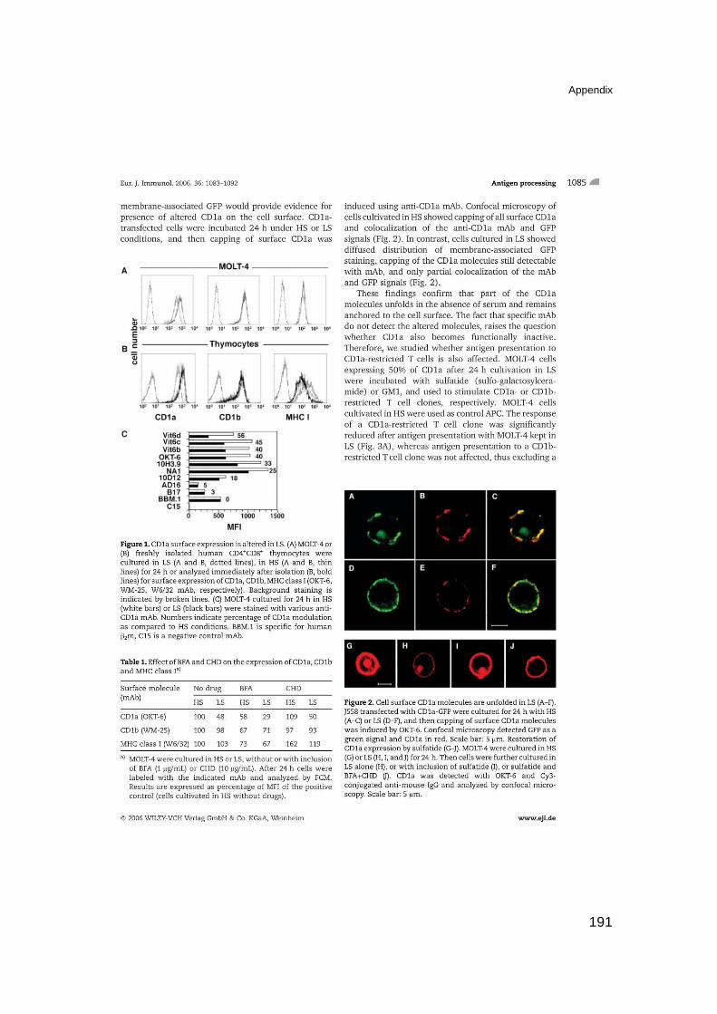
Appendix
191
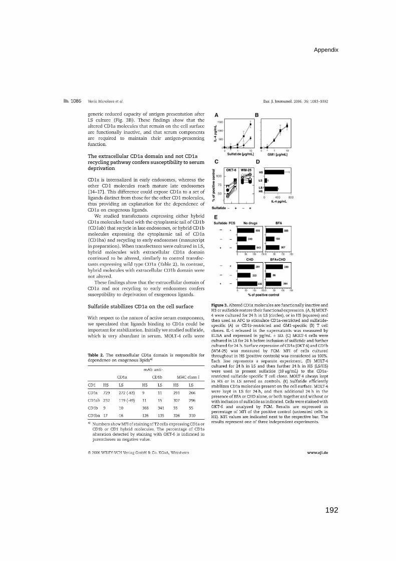
Appendix
192
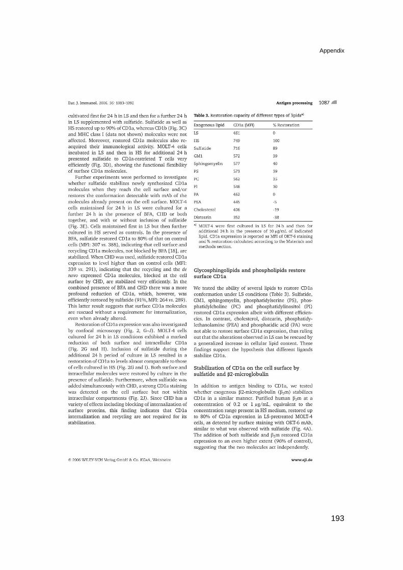
Appendix
193
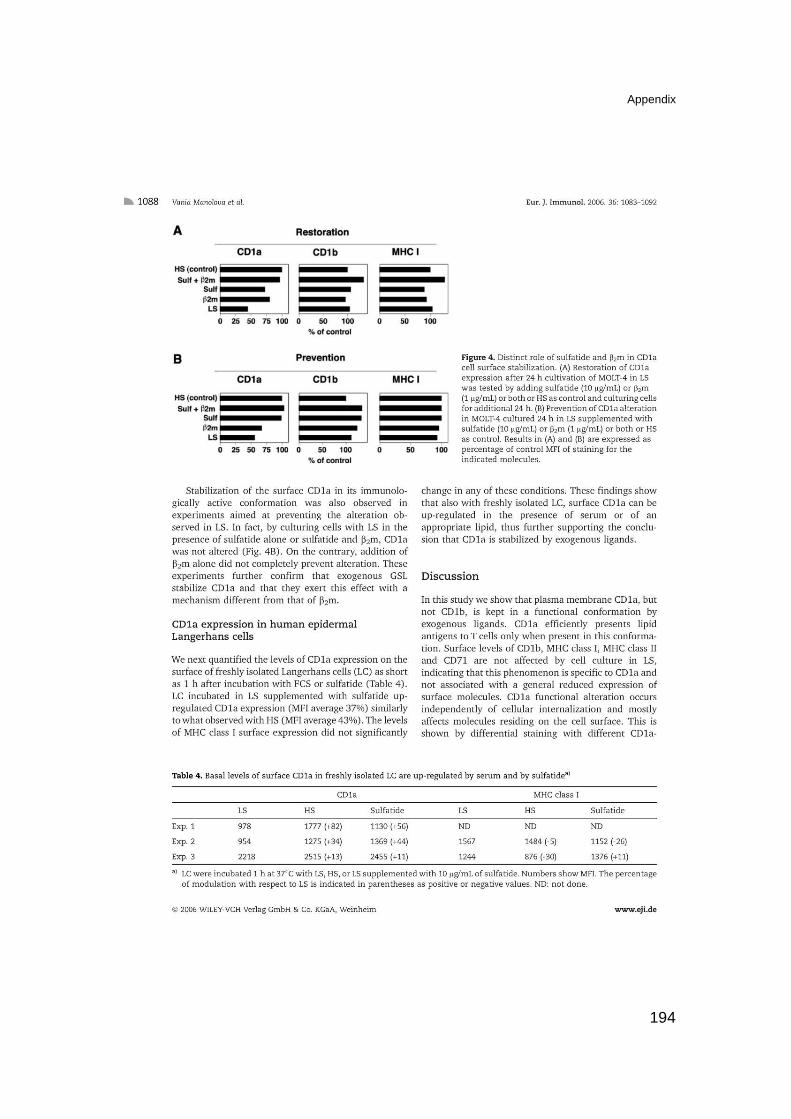
Appendix
194

Appendix
195

Appendix
196
Appendix
197

Appendix
198

CURRICULUM VITAE
Personal information
Name Magdalena Kistowska
Nationality Polish
Address Oetlingerstrasse 50, 4057 Basel, Switzerland
Telephone 0041 765779427
E-mail [email protected]
Date and place of birth 21.05.1977, Poznań, Poland
Marital status single, no children
Working place
University Hospital Basel, Dept. of Research, Experimental Immunology group,
Hebelstrasse 20, 4031 Basel, Switzerland
Education
1992 to 1996 Karol Marcinkowski 1st High School of General Education,
Poznań, Poland
Oct 1996 to Mar 2001 Adam Mickiewicz University, Faculty of Biology,
specialization: Molecular Biology, MSc, graduated: Mar
2001 (grade: very good). Thesis title: Biological and
biochemical properties of milky sap from Chelidonium
maius L.
April 2002 to
December 2007 Post-graduate study in Medical and Biological Research at
the Faculty of Science. University of Basel, under the
supervision of Prof.G. De Libero. Thesis title: Antigen
recognition and thymic maturation of human TCR
Vgamma9-Vdelta2 cells.

Language skills
English: fluent
German: basic
Polish: native speaker
Computer skills
Microsoft Office, Internet, Delta Graph, Photoshop, MacVector, Chem Draw, SoftMax
Pro, Instat, Flow-Jo, Summit, Cell Quest Pro
Professional experience
Oct 1998 to Jun 2000 Young Scientist Society, Molecular Biology Division, at
Adam Mickiewicz University, Poznań, Poland
Oct 1999 to Mar 2001 MSc student, Molecular Virology Group, Dept. of Molecular
Biology and Biotechnology, Adam Mickiewicz University,
Poznań, Poland
0ct 2001 to present Experimental Immunology group, Dept. of Research,
University Hospital Basel
Training courses
Mar 2003 LTK Module 1E: Introductory course in Laboratory Animal
Science
September 2004 FEBS, International Summer School for Immunology,
Ionian Village, Greece
August 2007 Novartis Biotechnology Leadership Camp (Biocamp)
Practical experience
Molecular biology and biochemistry techniques: PCR, RT-PCR, cloning, expression of
recombinant proteins in mammalian system, protein immunoprecipitation, purification
and quantification, SDS-PAGE, SSCP and TGGE analysis, immunobloting; Cellular
biology and immunology techniques: immunofluorescence, FACS (up to seven colors),
ELISA, cell culture (tumor and primary cells), isolation of primary cells from body fluids,
gene transfection, T cell activation assays, T cell clones generation, cell cloning,
collection of mouse organs and cell culture preparation, generation of cell hybrydomas.
In vivo experiments (in mice): anesthesia, intra peritoneal, intra venous, subcutaneous
and intra thymic injections, immunization.
Publications
1. Fik E., Wołuń-Cholewa M., Kistowska M., Warchoł J.B., Goździcka-Józefiak A. Effect
lectin from Chelidonium maius L on normal and cancer cells in culture. Folia
Histichemica et Cytobiologica, (2001) 39(2), 215-217.
2. Pawlaczyk M, Kistowska M., Poreba E, Filas V, Breborowicz J, Gozdzicka-Jozefiak A.
[Analysis of cloning rearrangement of T-cell receptor gamma gene in primary cutaneous
lymphoma] Pol Merkuriusz Lek. 2003 Nov;15(89):420-3. Polish.
3. Gober H-J., Kistowska M., Angman L., Jeno P., Mori L., De Libero G. Human T cell
receptor γδ cells recognize endogenous mevalonate metabolites in tumor cells. J. Exp.
Med. (2003) 197(2), 163-167.
4. Manolova V.*, Kistowska M.*, Paoletti S.*, Baltariu M.B., Bausinger H., Hanau D.,
Mori L., De Libero G. Functional CD1a is stabilazed by exogenous lipids. Eur J Immunol.
2006 May;36(5):1083-92 (* these authors contributed equally).
5. Kistowska M., Rossy E., Sansano S., Gober H-J., Landmann R., Mori L., De Libero G.
Transient dysregulation of the mevalonate pathway during early bacterial infection
activates TCR γδ cells. Manuscript submitted.
6. Kistowska M., Paoletti S., Mariotti S., Donda A., Zwinger J., Wells, D.J., Mori L., De
Libero G. Development and maturation of human TCR γδ cells in transgenic mice.
Manuscript in preparation.
Conferences presentations
1. Swiss Society for Allergology and Immunology (SGAI) Annual Meeting March 30th-31st
2006, Zurich; Title: Requirements for maturation and peripheral expansion of TCR
Vgamma9 /Vdelta2 cells in a mouse model. Selected for an oral presentation
References
Gennaro De Libero, MD, Ph.D University Hospital Basel Dept. of Research Experimental Immunology Hebelstrasse 20 4031 Basel, Switzerland Phone: 0041 61 2652327 Fax: 0041 61 2652350 [email protected]