Embed Size (px)
Citation preview
The role of the inhibitor of apoptosis protein
Survivin in cellular radiation response
Vom Fachbereich Biologie der Technischen Universität Darmstadt
zur Erlangung des akademischen Grades eines Doctor rerum naturalium
genehmigte Dissertation von
M.Sc. Chrysi Eirini Petraki
aus Chania, Griechenland
Darmstadt 2014
D17
1. Referent: Prof. Dr. Markus Löbrich
2. Referent: Prof. Dr. Cristina Cardoso
Tag der Einreichung: 18. März 2014
Tag der mündlichen Prüfung: 06. Juni 2014

ii

iii
Word of honour
I assure herewith on my word of honour, that I wrote this thesis by myself. All quotes,
whether word by word, or in my own words, have been put in quotation marks or otherwise
identified as such. The thesis has not been published anywhere else or has been presented
to any other examination board.
Darmstadt, (date) ………………………………
_________________________________
(Signature)

iv
Contents
List of Figures ....................................................................................................................... vii
List of Tables ......................................................................................................................... ix
Summary ................................................................................................................................ x
Zusammenfassung (summary in German) ............................................................................ xii
1. Introduction .............................................................................................................. 1
1.1 Colorectal cancer ..................................................................................................... 1
1.2 Inhibitor of apoptosis proteins (IAPs) ....................................................................... 3
1.3 DNA damage repair ................................................................................................. 7
1.4 Survivin .................................................................................................................... 8
1.5 Aim of project ..........................................................................................................15
2. Materials and Methods............................................................................................17
2.1 Materials .................................................................................................................17
2.1.1 Appliances/Instruments...........................................................................................17
2.1.2 Consumables ..........................................................................................................18
2.1.3 Chemicals and Media .............................................................................................20
2.1.4 Solutions and Buffers ..............................................................................................22
2.1.5 Antibodies ...............................................................................................................25
2.1.6 Expression plasmids ...............................................................................................26
2.1.7 Specific small interfering RNA.................................................................................27
2.1.8 Commercial Kits......................................................................................................28
2.1.9 Enzymes and respective buffers .............................................................................28
2.1.10 Electrophoresis Markers .........................................................................................29

v
2.1.11 Oligonucleotides .....................................................................................................29
2.2 Methods ..................................................................................................................32
2.2.1 Cell culture..............................................................................................................32
2.2.2 Survivin constructs and stable transfection .............................................................32
2.2.3 Attenuation of endogenous Survivin via siRNA .......................................................39
2.2.4 Irradiation procedure ...............................................................................................40
2.2.5 Three-dimensional colony forming assay ................................................................40
2.2.6 Protein extraction and Western blotting ..................................................................41
2.2.7 Subcellular fractionation and immunoprecipitation ..................................................43
2.2.8 Immunofluorescence staining and imaging .............................................................44
2.2.9 Cell cycle analysis ..................................................................................................47
2.2.10 Measurement of apoptosis and caspase 3/7 assay .................................................48
2.2.11 Transmigration assay .............................................................................................49
2.2.12 Data analysis ..........................................................................................................50
3. Results ...................................................................................................................51
3.1 The impact of Survivin deletion mutants on cellular radiation response ..................51
3.1.1 Survivin BIR, XIAP, Microtubules and Hsp90 deletion mutants failed to reconstitute
irradiation induced cell cycle arrest and apoptosis after knockdown of endogenous
Survivin ...................................................................................................................54
3.1.2 Survivin BIR domain and XIAP binding site are essential for three-dimensional
radiation clonogenic survival ...................................................................................57
3.1.3 Survivin BIR domain and XIAP binding site are essential for regulation of DNA
double-strand break repair ......................................................................................61
3.1.4 The XIAP binding site of Survivin is essential for interaction with DNA-PKcs ..........64
3.2 The impact of Survivin phosphorylation on cellular radiation response ...................65

vi
3.2.1 Phosphorylation of Survivin on T34 is important for 3D radiation clonogenic survival
...............................................................................................................................66
3.2.2 The XIAP binding site and the T34 phosphorylation site of Survivin are essential for
regulation of residual DNA DSBs repair ..................................................................72
3.2.3 Mutation of the Survivin T34 phosphorylation site to its non-phosphorylatable form
T34A prevents interaction with DNA-PKcs ..............................................................74
3.3 Survivin BIR domain, XIAP, Microtubules and Hsp90 binding sites are essential for
transmigration capability of colorectal cancer cells ..................................................76
4. Discussion ..............................................................................................................78
References ...........................................................................................................................88
Appendix ............................................................................................................................ 101
Abbreviations ..................................................................................................................... 106
Acknowledgements ............................................................................................................ 111
Curriculum Vitae ................................................................................................................. 113

vii
List of Figures
Figure 1: Symbolic representation of structural domains of IAPs .......................................... 4
Figure 2: Schematic diagram of Survivin role in radiation response .....................................10
Figure 3: Symbolic illustration of Survivin protein structure ..................................................11
Figure 4: Plasmid map for pEGFP-N1-wild type Survivin .....................................................27
Figure 5: Methodology for 3D radiation clonogenic survival assays. ....................................40
Figure 6: Methodology of 3D IF of H2AX/53BP1 foci ..........................................................46
Figure 7: Methodology of transmigration assay. ..................................................................49
Figure 8: Symbolic diagram of Survivin-EGFP fusion constructs .........................................52
Figure 9: Stable expression of Survivin deletion mutants in SW480 cells. ...........................53
Figure 10: Western blot analysis with cells overexpressing Survivin deletion mutants .........54
Figure 11: Cell cycle analysis of SW480 cells overexpressing Survivin deletion mutants ....55
Figure 12: Sub G1 fraction and caspase 3/7 activity assays of cells overexpressing Survivin
deletion mutants ...................................................................................................................56
Figure 13: 3D clonogenic radiation survival of cells overexpressing Survivin deletion mutants
.............................................................................................................................................58
Figure 14: 3D radiation clonogenic survival assays with cells expressing alternative clones of
Survivin deletion mutants .....................................................................................................60
Figure 15: Immunofluorescence staining of H2AX/53BP1 foci in cells overexpressing
Survivin deletion constructs ..................................................................................................62
Figure 16: 3D Immunofluorescence staining of H2AX/53BP1 foci in cells overexpressing
alternative clones of Survivin deletion constructs ..................................................................63
Figure 17: Immunoprecipitation experiments performed with cells overexpressing wild type
Survivin and its XIAP deletion mutant .................................................................................64
Figure 18: Symbolic representation of Survivin phosphorylation mutants. ...........................66
viii
Figure 19: Western blot conducted with cells overexpressing Survivin phosphorylation
mutants. ...............................................................................................................................67
Figure 20: 3D clonogenic radiation survival of cells overexpressing Survivin phosphorylation
mutants ................................................................................................................................68
Figure 21: 3D clonogenic radiation survival of cells overexpressing alternative clones of
Survivin phospho-mutants ....................................................................................................69
Figure 22: Western blotting performed with cells overexpressing Survivin XIAP deletion
mutant and Survivin T34A or T34D phospho-mutant. ...........................................................70
Figure 23: 3D clonogenic radiation survival of cells overexpressing the indicated Survivin
mutants ................................................................................................................................71
Figure 24: 3D immunofluorescence staining of H2AX/53BP1 foci in cells overexpressing
Survivin deletion and phosphorylation mutants .....................................................................73
Figure 25: Immunoprecipitation experiment with cells overexpressing Survivin phospho-
mutants ................................................................................................................................75
Figure 26: Transmigration assay performed with cells overexpressing Survivin deletion
mutants ................................................................................................................................76
Figure 27: Symbolic diagram of Survivin wt and Surv. XIAP. .............................................83
Figure 28: Survivin as a radiation resistance factor. ............................................................85
Figure 29: Synopsis of the different binding sites of Survivin and their role in cellular
radiation response. ...............................................................................................................86
List of Tables
Table 1: Pipetting scheme for two discontinuous SDS-Electrophoresis gels (8.3 cm x 7.3 cm
x 1 mm) ................................................................................................................................24
Table 2: Characteristics of primary antibodies used for western blotting, immunoprecipitation
and immunofluorescence staining assays ............................................................................25
Table 3: Characteristics of secondary antibodies used for immunofluorescence staining .....26
Table 4: Characteristics of secondary antibodies used for western blotting ..........................26
Table 5: Radiation response variables of 3D grown human SW480 colorectal cancer cells.
........................................................................................................................................... 101
Table 6: Radiation response variables of alternative clones and phospho-mutants of 3D
grown human SW480 colorectal cancer cells. .................................................................... 102
Table 7: Synopsis of observed effects of Survivin mutants on cellular radiation response . 105
x
Summary
Survivin, the smallest and functionally unique member of the inhibitor of apoptosis protein
(IAP) family, is frequently overexpressed in malignant cells and has been acknowledged as a
predictive molecular marker for metastases and cancer patient survival following radiation
therapy. The role of this protein in cellular radiation response, however, far exceeds a simple
inhibition of apoptotic cell death involving non-caspase dependent mechanisms such as
regulation of cell cycle and DNA damage repair. To investigate in more detail the role of
Survivin in cellular radiation response and tumour cell motility, the present study aimed to
establish and stably express several Survivin enhanced green fluorescent protein (EGFP)-
tagged deletion or phosphorylation mutant constructs in SW480 colorectal cancer cells. To
this end, Survivin wild-type (Surv. wt) and recombinant proteins lacking the binding sites for
X-linked IAP (XIAP), Microtubules and heat shock protein 90 (Hsp90) (Survivin
XIAP/MicTub/Hsp90) or the baculovirus IAP repeat (BIR) domain (Survivin BIR) as well
as phosphorylation mutants for the sites S20, T34 and T117 (Survivin
S20A/S20D/T34A/T34D/T117A/T117D) were expressed in a pEGFP-N1 vector system.
Subsequently, these mutant clones were subjected to more physiologic three-dimensional
(3D) colony forming assays, immunofluorescence staining of DNA double strand break
markers H2AX and 53BP1, analysis of cell cycle distribution, induction of apoptosis,
caspase 3/7 activity and transmigration assays following irradiation with doses ranging from
1 to 6 Gy. While knockdown of endogenous Survivin by RNA interference (siRNA) resulted in
a significantly decreased 3D radiation survival in line with elevated numbers of residual
H2AX/53BP1 foci in pEGFP-N1 vector control, Survivin XIAP, BIR and T34A clones, both
radiation clonogenic survival and the capacity to regulate DNA repair was rescued in clones
stably overexpressing Survivin wt, MicTub, Hsp90 and Survivin T34D mutant cells.
Moreover, deletion of the BIR domain, XIAP, Microtubules and Hsp90 binding sites resulted
in a G2/M cell cycle arrest, an elevated percentage of apoptotic SubG1 cells, an increased
caspase 3/7 activity and significantly hampered transmigration activity of SW480 cells
following depletion of endogenous Survivin. On a molecular level, constructs derived from
Survivin XIAP and T34A phospho-mutant overexpressing clones did not co-
immunoprecipitate with the DNA repair protein DNA-dependent protein kinase catalytic
subunit (DNA-PKcs), whereas Survivin wt and T34D constructs co-precipitated with the
protein. These data confirm Survivin to act as a radiation resistance factor modulating
cellular radiation response by multiple mechanisms including DNA DSB repair, induction of
cell cycle arrest and apoptosis. It is for the first time indicated that Survivin’s XIAP binding
and T34 phosphorylation sites, but not its Microtubules and Hsp90 binding sites, are
essential for radiation clonogenic survival and modulation of DNA damage repair, at least in

xi
part by disturbing protein interaction with DNA-PKcs. On the contrary, XIAP, Hsp90 and
Microtubules binding sites as well as the BIR domain were shown to be essential for proper
cell cycle regulation, apoptosis inhibition and transmigration capacity of SW480 colorectal
adenocarcinomas.

Zusammenfassung (summary in German)
Survivin, das kleinste und funktionell einzigartige Mitglied der Inhibitor of Apoptosis Protein
(IAP) Familie ist in der Mehrzahl maligner Zellen überexpremiert und stellt einen
wesentlichen prädiktiven molekularen Marker für eine Metastasierung und das Überleben
von Patienten nach Strahlentherapie dar. In diesem Zusammenhang wurde zudem gezeigt,
dass die Rolle von Survivin in der zellulären Strahlenantwort weit über eine alleinige
Hemmung der apoptotischen Reaktionskaskaden hinausgeht, sondern vielmehr auch
Caspase-unabhängige Mechanismen wie eine Regulation des Zellzyklus und der Reparatur
von DNA-Schäden beinhaltet. Um die Rolle von Survivin in der zellulären Strahlenantwort
und Tumorzellmotilität detaillierter zu untersuchen, war es Ziel der vorliegenden Arbeit, eine
Reihe von grün fluoreszierendes Protein (GFP) markierten Survivin Deletions- und
Phosphorylierungsmutanten zu generieren und stabil exprimierende Klone kolorektaler
SW480 Karzinomzellen herzustellen. Dazu wurden wildtyp-Survivin und rekombinante
Proteine mit fehlenden Bindungsstellen für X-linked IAP (XIAP), Mikrotubuli,
Hitzeschockprotein 90 (Surv-XIAP/MicTub/Hsp90), die Baculovirus IAP Repeat (BIR)
Domäne (Surv-BIR), sowie Mutanten der Phosphorylierungsstellen S20, T34 und T117
(Survivin S20A/ S20D/T34A/T34D/T117A/ T117D) in einem pEGFP-N1 Vektor System zur
Expression gebracht. Anschließend wurden die Zellen unter physiologischen Bedingungen in
drei-dimensionalen (3D) Koloniebildungstests eingesetzt, die Expression der DNA-
Doppelstrangbruchmarker H2AX und 53BP1 mittels Immunfluoreszenz quantifiziert und die
Zellzyklusverteilung, Induktion von Apoptose und die Caspase 3/7 Aktivität bzw. die
Transmigrationsfähigkeit nach Bestrahlung mit Dosen von 1 bis 6 Gy untersucht. Während
die Hemmung von endogenem Survivin durch RNA-Interferenz (siRNA) zu einer
signifikanten Minderung des 3D klonogenen Überlebens und einer erhöhten Anzahl
residueller H2AX/53BP1 Foci in pEGFP-N1 Kontrollvektor, Surv. XIAP, Surv. BIR und
Surv. T34A transfizierten Zellen führte, konnte das radiogene Zellüberleben und die DNA-
Reparaturkapazität in stabil Surv. wt, Surv. MicTub, Surv. Hsp90 und Surv. T34D
überexprimierenden Zellklonen rekonstituiert werden. Darüber hinaus führte die Deletion der
BIR Domäne, der Mikrotubuli und der Hsp90 Bindungsstelle zur Induktion eines G2/M
Zellzyklus-Arrestes, einem erhöhten Anteil apoptotischer SubG1 Zellen, einer gesteigerten
Caspase 3/7 Aktivität und einer signifikanten Hemmung der Transmigration von SW480
Zellen nach Attenuation des endogenen Survivins. Auf molekularer Ebene gelang bei
Konstrukten aus Surv. XIAP and T34A Phospho-mutanten überexpremierenden Zellen
keine Ko-Immunpräzipitation mit dem DNA Reparaturprotein DNA Proteinkinase (DNA-
PKcs), während dies bei wt-Survivin Klonen sowie bei der T34D phospho-mimetischen
Mutante möglich war. Zusammengefaßt bestätigen diese Daten die Aktivität von Survivin als

xiii
Radioresistenzfaktor, der die zelluläre Bestrahlungsantwort durch multiple Mechanismen,
einschließlich der DNA- Doppelstrangbruch-Reparatur und der Induktion eines Zellzyklus
Arrestes und von Apoptose zu modulieren vermag. Darüber hinaus konnte hier erstmals
gezeigt werden, dass die XIAP Bindungs- und T34 Phosphorylierungs-, nicht jedoch die
Microtubuli und Hsp90 Bindungsstellen essentiell für das klonogene Zellüberleben nach
Bestrahlung und die Modulation der DNA Schadensreparatur sind. Dies beruht, zumindest
teilweise, auf der Beeinträchtigung der Interaktion mit dem Protein DNA-PKcs. Im Gegensatz
dazu sind für die Regulation des Zellzklus, die Inhibition der Apoptose und das
Migrationsverhalten der kolorektalen Adenokarzinomlinie SW480 sowohl die XIAP-, die
Hsp90- und die Mikrotubuli-Bindungsstellen wie auch die BIR-Domäne wesentlich.

Introduction 1
1. Introduction
1.1 Colorectal cancer
Colorectal cancer (CRC), the second most common cancer in women and third in men,
accounts for 1,233,700 cancer incidents annually worldwide, 608,700 cases of which
eventually lead to death [1]. More than 95% of CRC cases are adenocarcinomas while 50%
of the patients are diagnosed with distant metastasis or local recurrence with a median
survival time of 4 to 22 months [2]. Although there are some environmental and lifestyle risk
factors it is nowadays generally accepted that colorectal cancer is caused by changes in
various molecular pathogenic pathways, such as CpG island methylator phenotype (CIMP),
chromosomal instability (CIN) or microsatellite instability (MSI) [3].
The CIN pathway is the one via which approximately 85% of CRCs are developed [4] mainly
due to an accumulation of numerical or structural chromosomal anomalies [5]. In this
pathway the earliest detectable lesion is a microscopic mucosal dysplasia named aberrant
crypt focus (ACF) [6] which is followed by the development of a polyp [3, 7]. Typical
aberrations during the CIN pathway include V-Ki-ras2 Kirsten rat sarcoma viral oncogene
homologue (KRAS) oncogene mutation, deletion of 17p chromosome (which entails the
tumour suppressor gene TP53) and loss of 18q chromosome [3, 4]. However, there are
many alternative genetic and epigenetic changes that have recently been described [8] and
will eventually be considered as complementary events to the aforementioned genetic
mutations [3].
A deficiency in the function of the tumour suppressor TP53 gene, typically via allelic loss of
17p [3], is known to characterize 50-75% of CRCs [9]. The TP53 protein is responsible for
the slowdown of cell cycle in order for the DNA damage to be repaired [3] as well as for the
Introduction 2
induction of pro-apoptotic genes in case of an accumulation of unrepaired genetic damage
[10].
Around 60% of CRCs demonstrate an allelic loss of 18p21.1, a site entailing SMAD2 and
SMAD4 [11], both of them being involved in the transforming growth factor- (TGF-)
signaling pathway which is essential for apoptosis and growth regulation [3].
In the case of MSI, microsatellites typically consist of 1-5 nucleotide tandem repeats found
throughout the whole genome and MSI corresponds to an inconsistency in the amount of
those microsatellites between normal and tumour cells [3, 12]. MSI phenotype can arise from
mutations interfering with the mismatch repair (MMR) machinery, a condition present in
approximately 15% of colorectal cancers [13-15]. It is now understood that the colorectal
cancer genome entails probably thousands molecular aberrations certain subsets of which
take part in carcinogenesis [12].
Approximately 15% of all sporadic colorectal cancers are originated by the CIMP pathway
[16, 17]. KRAS, v-Raf murine sarcoma viral oncogene homologue B1 (BRAF) and MSI
mutations often occur in the case of CIMP cancers which are also related to older age,
genetic predisposition or the female gender [18]. In general, genetic and epigenetic
aberrations can take place in numerous combination patterns with some genes exclusively
modified by epigenetic inactivation and others being both genetically and epigenetically
inactivated [18].
Coloretal cancer therapeutic approaches vary depending on numerous factors such as
tumour localization and stage, involvement of tumour positive lymph nodes etc., with the
majority of such therapies to involve surgical resection followed by adjuvant chemotherapy in
case of colon carcinoma. By contrast, neoadjuvant (preoperative) combined radiation and
chemotherapy (comprised of 5-Fluorouracil along with additional agents such as oxaliplatin,
or irinotecan) became standard therapy for patients with locally advanced rectal cancer [19].

Introduction 3
1.2 Inhibitor of apoptosis proteins (IAPs)
In 1993 a baculovirus gene that inhibits apoptosis in virally infected Spodoptera frugiperda
insect cells was discovered [20] and since then several cellular homologues have been
detected in yeast, nematodes, flies and higher vertebrates. It is generally known that IAPs
are important for cell division, morphogenesis, nuclear factor kappa B (NF-B) activation,
heavy metal homeostasis and Mitogen-activated protein (MAP) kinase signaling [21]. IAPs
are characterized by the presence of a variable number of Baculoviral IAP Repeat (BIR)
motifs comprised of approximately 70 amino acids coordinating a zinc ion through cysteine
and histidine residues [21]. Apoptosis being a controlled cell suicide process which plays a
pivotal role in homeostasis and development of the organism, when deregulated can lead to
various pathogenic conditions including cancers, neurological disorders and autoimmunity
[21]. The fact that many neoplasms are found to express a high level of IAPs along with a
genetic translocation regarding the gene encoding cellular IAP2 (c-IAP2) detected in a
subset of B cell lymphomas led to the notion that IAPs contribute to apoptotic cell death
resistance that characterizes numerous cancers [21]. It was originally thought that those
proteins were physical caspase inhibitors representing a cytoprotective factor downstream of
death receptors [22, 23]. It is now obvious that IAPs not only regulate caspases via
mechanisms distinctive for each member of the family [24] but also participate in several
aspects of cellular homeostasis [21, 25].
The function of IAPs depends not only on their distinct domains but also on posttranslational
modifications and changes at the level of expression [21]. In particular, protein-protein
interactions, intracellular localization and IAP stability are influenced by phosphorylation of
some IAPs [26-28] while interactions among IAP molecules can have an effect on their
expression levels [29-32].

Introduction 4
Figure 1: Symbolic representation of structural domains of IAPs. Domains such as the BIR (Baculovirus IAP
repeat) and RING (really interesting new gene) shown here characterize all members of the IAP family (except
Survivin). DIAP1: Drosophila IAP 1; UBA: binding site for polyubiquitinated proteins; CARD: caspase-associated
recruitment domain; c-IAPs: cellular IAPs; XIAP: X-linked inhibitor of apoptosis protein. (Figure adapted from [22])
A structural characteristic common to all IAPs is the BIR domain which can be found either in
a single copy or two to three repeats in the N-terminus along with other functional domains
like a really interesting new gene (RING) or caspase-associated recruitment domain (CARD)
in the proximity of C-terminal region of the proteins [21] (Figure 1). BIR domains fold as
three-stranded -sheets surrounded by four -helices and their signature sequence is
CX2CX16HX6C, where C = cysteine, H = histidine and X stands for any amino acid [33-36]. C-
IAP1 (cellular IAP1), c-IAP2 (cellular IAP2) and XIAP (X-linked IAP) contain three BIRs and
one RING domain, functioning as an E3 ubiquitin ligase (a ubiquitin-associated domain
involved in binding to ubiquitinated proteins) and a CARD in c-IAP1 and c-IAP2 whose
function is not fully understood [22]. In spite of the resemblance all BIR domains bear,
different BIRs identify different intermediates and participate in distinct signaling pathways
[21].
In principal, BIRs are involved in protein-protein interactions as well as protein recognition
[21], as a peptide-binding groove (in the case of XIAP, c-IAP1 and c-IAP2) comprises a
hydrophobic recognition site for IAP-binding motif (IBM) entailing proteins [22]. Certain
XIAP
c-IAPs
DIAP1
Survivin
Livin
BIR1
BIR1
BIR1
BIR2
BIR2
BIR2
BIR
BIR
-helix
BIR3
BIR3
UBA
UBA CARD
RING
RING
RING
RING

Introduction 5
apoptosis inducers such as second mitochondrial-derived activator of caspase (Smac) also
known as direct IAP-binding protein with low isoelectric point (DIABLO) along with initiator
capsase 9 and effector caspase 7 contain an IBM i.e. a tetrapeptide region with an invariant
N-terminal alanine residue and other conserved residues [37, 38]. Short peptides that contain
SMAC IBM can prevent BIR3 from binding to caspase 9 [39]. The binding of XIAP and Smac
is greatly increased due to the tandem presence of BIR2 and BIR3 in XIAP, as Smac forms a
stable complex with XIAP in a ratio of 2:1 via concurrent interaction with both BIR2 and -3
[21]. Not every BIR domain contains an IBM recognition site [40] as for example, the XIAP
BIR1 does not bind caspases or IBM containing proteins, but identifies molecules involved in
NF-B activation instead [41, 42].
In general, the interaction between caspases and IAPs occurs via IBM-dependent
complexes, resulting in the abolishment of enzymatic activity of caspases [22, 38]. While this
was originally thought to be the sole role of IAPs [43], it is now commonly accepted that only
XIAP (from the mammalian IAPs) actually inhibits caspases in vivo [25]. XIAP interacts with
caspases 3, 7 and 9, obliterating their cell killing ability [22]. The XIAP linker region upstream
of BIR2 interacts with the catalytic cleft of caspases 3 and 7, inhibiting substrate accessibility
thus eliminating their activity [44-46]. Caspase 9 is inhibited via XIAP BIR3 as the latter
interferes with the homodimerization domain of the enzyme, abolishing the conformational
change required for activity [47]. XIAP participates also in cell-death regulation through its
RING domain [21] the mechanism of which has not yet been clarified, although recent
studies revealed a cytoprotective role for the RING domain when mice expressing a BIR-only
type of XIAP showed an increased caspase activity and cell death in vivo [48].
Members of the IAP family bind to each other via their BIR domains [21]. For instance, the
BIR1 and -3 from XIAP are responsible for binding with the single BIR of Survivin in order to
form a complex of increased stability against proteasomal degradation and synergistically
inhibit apoptosis [31]. Survivin BIR also binds Aurora B, a serine/threonine kinase involved in
Introduction 6
the regulation of cell division, as well as Borealin and inner centromere protein (INCENP)
thus forming the chromosomal passenger complex (CPC) [49].
The RING domain is characterized by the presence of one or two histidines and six to seven
cysteines comprising a cross-brace structure that coordinates two zinc ions [50]. These
domains mediate ubiquitin protein ligase (E3) activity as the transfer of ubiquitin to target
proteins is facilitated by a ubiquitin activating enzyme (E1) and a ubiquitin conjugating
enzyme (E2) [51]. All IAPs that entail RING domains demonstrate E3 activity involving
apoptotic and signaling molecules as well as themselves in a homo- or heterotypic fashion
[21].
In cells exposed to pro-apoptotic stimuli, XIAP, c-IAP1 and c-IAP2 undergo autoubiquitination
in a RING dependent manner, a process found abrogated in a RING-mutant XIAP lacking E3
activity [52, 53]. Those pro-apoptotic stimuli are able to cause IAP autoubiquitination due to
IBM-containing IAP antagonists that are released [54].
Apart from caspase inhibition, IAPs were also found to modulate NF-B, a transcription factor
pivotal for inflammation, cell survival, migration, invasion and immunity [55-57].
Consequently, IAP-mediated NF-B activation can result in tumour progression and
metastasis [58].
IAPs are found to be overexpressed in many tumour entities as inhibition of apoptosis plays
a major role in carcinogenesis by enabling cell survival in unfavourable conditions typically
hostile to non-cancerous cells. For instance, the fact that Survivin is overexpressed in breast,
colorectal, esophageal/gastric carcinoma, lymphoma and neuroblastoma as well as the poor
prognosis with which this overexpression is associated with, led to the hypothesis that IAP
up-regulation might comprise an oncogenic event [59, 60]. Besides Survivin (which will be
further analyzed in section 1.4), c-IAP1 and c-IAP2 also play a role in mammalian cancers
with high levels of c-IAP1 to promote carcinogenesis [61, 62] and c-IAP2 being correlated
Introduction 7
with a type of neoplasia called MALT (mucosa-associated lymphoid tissue) lymphomas [63].
Another way for tumour cells to evade apoptosis is to down-regulate molecules that cause
suppression of XIAP’s caspase-inhibitory activity, such as XIAP-associating factor 1 (XAF1)
whose levels have been reported to be decreased in some cancer cell lines [64, 65].
As IAPs are implicated in tumourigenic events, their expression has been targeted via RNA
interference methods and alternatively, compounds mimicking naturally occurring IAP
antagonists have been developed in order to neutralize IAP function [66]. For instance,
Survivin suppression has been shown to sensitize tumour cells to both chemo- [59, 67, 68]
and radiation therapy.
1.3 DNA damage repair
Higher eukaryotic cells have in the course of evolution acquired complex molecular
mechanisms to preserve chromosomal integrity in their genomes amid background radiation,
byproducts of cellular metabolism and environmental mutagens [69]. Maintaining genomic
stability is essential for prevention of chromosomal rearrangements that can result in cancer
via altered gene expression [70, 71]. There is a wide range of different types of DNA damage
that can arise within the cell, with the DNA double-strand breaks (DSBs) being of great
importance as they can cause cell death or cancer if improperly repaired [69, 72]. Ionizing
radiation, reactive oxygen species as well as certain types of anti-cancer drugs are able to
give rise to this type of lesion which is met mainly by two different repair mechanisms that
mammalian cells have evolved: homologous recombination (HR) and non-homologous end
joining (NHEJ) [69]. An intact sister chromatid is required for HR to take place which is,
therefore, restricted to the S and G2 cell cycle phases. On the other hand, in the case of
NHEJ the two broken DNA ends are re-joined without the need of a sister chromatid, a fact
Introduction 8
that renders NHEJ functional in all cell cycle phases and thus the major DSBs repair
mechanism in mammalian cells.
A protein kinase central to the damage response signal transduction regarding DSBs is the
ataxia telangiectasia mutated (ATM) [73] which is recruited to DSB lesions via the Mre11-
Rad50-Nbs1 (MRN) complex [74]. Following ATM activation (after being auto-
phosphorylated) [75] another part of this signalling cascade is the phosphorylation of a
histone H2A variant, thus, generating H2AX which in turn facilitates the recruitment of other
key proteins [such as p53-binding protein 1 (53BP1), mediator of DNA damage checkpoint
protein 1 (MDC1), breast cancer 1 early onset (BRCA1), etc.] at DSB sites [76, 77].
NHEJ is the predominant pathway through which irradiation-induced DSBs are repaired [78-
81]. This process is known to commence with the binding of Ku70 and Ku80 proteins, to
double-stranded DNA ends in a manner that allows Ku to translocate along the DNA [82-84]
and eventually recruit a large catalytic subunit of the DNA-dependent protein kinase complex
(DNA-PKcs) [85, 86]. DNA-PKcs seems to be responsible for the regulation of the DNA ends
processing to generate the 3’-OH and 5’-P ends required for ligation [87, 88]. The assembly
of Ku and DNA-PKcs on DNA ends is followed by recruitment of a complex termed DNA
ligase IV-X-ray repair cross-complementing protein 4 (XRCC4) that facilitates the re-joining
step [89-92].
1.4 Survivin
Survivin, a 16.5 kDa protein of 142 amino acid residues encoded by the BIRC5 gene, is a
member of the IAP family firstly described by Ambrosini et al. in 1997 [93]. Survivin is the
smallest member of this protein family consisting of a single BIR domain [36] and an
extended C-terminal -helix [93]. When in solution, Survivin forms a stable homodimer [36]
while clear evidence rendering such an architecture necessary for function is still missing. On
Introduction 9
the other hand, it is the monomeric protein that is involved in nuclear-cytoplasmic shuttling
[94] and key protein recognition such as binding to the chromosomal passenger protein
Borealin [49, 95].
Survivin has been shown to function in a cytoprotective manner when in cytoplasm due to its
anti-apoptotic activities [96] while when located in the nucleus it participates in cell division
[97] and DNA repair regulation [98, 99]. It is expressed in a cell-cycle regulated manner with
a peak at mitosis [97] showing a 40-fold up-regulation in G2/M phase when found to be
localized in the mitotic apparatus [100]. Thus, Survivin is acknowledged to be an
indispensable member of the chromosomal passenger complex [101, 102].
Homozygous deletion of Survivin gene resulted in early embryonic death [103], while mitotic
defects, cell death and tissue involution followed after conditional deletion of Survivin in adult
tissues [104, 105]. Such a scenario has been repeatedly confirmed by studies conducted on
Survivin orthologues in other model systems like C. elegans [106, 107] and yeast [108],
demonstrating pivotal roles of this protein in mitosis regarding chromosomal segregation and
cytokinesis. Survivin physically interacting with other members of the chromosomal
passenger complex such as Borealin, Aurora B and INCENP [49], regulates chromosomal
alignment, cytokinesis as well as chromatin-associated spindle assembly [102], whereas
another pool of this protein stabilizes the mitotic spindle [109]. In order to do this, Survivin
binds to polymerized microtubules through its C-terminal -helices [22] suppressing
microtubule dynamics [110]. Those different pools of Survivin manage to work together
probably due to post-translational modifications such as monoubiquitination by Lysine 48
(L48) and L63, an event necessary for mitotic trafficking [111].

Introduction 10
Figure 2: Schematic diagram of Survivin role in radiation response. When in the cytoplasm Survivin interacts
with other members of the IAP family (such as XIAP) to inhibit apoptosis while when found in the nucleus it
participates in cell division (being a member of the CPC) as well as it regulates DNA repair (interacting with DNA-
PKcs and other members of the DNA repair machinery). XIAP: X-linked IAP; CPC: Chromosomal passenger
complex; DNA-PKcs: DNA-dependent protein kinase catalytic subunit; Smac: Second mitochondria-derived
activator of caspases; (Figure adapted from [112])
Survivin being a multifunctional protein, interacts with a variety of protein partners in order to
co-ordinate its different activities including apoptosis inhibition (via interaction with other
IAPs), cell division (being a member of the CPC) and regulation of DNA DSBs repair
(interacting with DNA-PKcs, Ku-70, 53BP1, etc.) (Figure 2) [98, 99, 112].
Survivin
Survivin
Survivin
SurvivinSurvivin
Survivin
MitochondrionDNA Damage
Smac
XIAP
Regulation ofDNA- Repair
Regulation ofCell Cycle
Microtubules
CPC
H2AX
DNA-PK
Ionizing Radiation
Caspases

Introduction 11
Figure 3: Symbolic illustration of Survivin protein structure. Functional domains, binding sites as well as
phosphorylation sites of Survivin are shown here. XIAP: X-linked inhibitor of apoptosis protein; Smac: Second
mitochondria-derived activator of caspase; INCENP: Inner centromere protein; Hsp90: Heat shock protein 90;
NES: Nuclear export signal; PKA: Protein kinase A; Plk1: Polo-like kinase 1; CDK1: Cyclin-dependent kinase 1;
S20: Serine 20; T34: Threonine 34; T117: Threonine 117. (Figure adapted from [112])
Survivin entails various phosphorylation sites such as Serine 20 (S20), Threonine 34 (T34)
and Threonine 117 (T117) [113-116] via which both protein stability and trafficking among the
subcellular compartments are achieved [112]. There are also several molecules that interact
with this protein including tubulin, heat shock protein 90 (Hsp90) [117], Hsp60 [118], aryl
hydrocarbon receptor-interacting protein (AIP) [119] and other members of the IAP family like
XIAP [58] (Figure 3).
Except for XIAP, all other IAPs including Survivin do not show a direct binding to caspases
[21]. In the case of Survivin, it is believed that apoptosis is inhibited only after its complex-
formation with XIAP [31] where Survivin BIR residues 15-38 [114] interact with BIR1 and
BIR3 of XIAP [31]. Besides caspase inhibition, the Survivin-XIAP complex promotes XIAP
stability (as well as Survivin stability) against ubiquitin-dependent degradation and it also
leads to tumour growth in vivo [114] being involved in XIAP-mediated NF-B activation [58].
The latter event gives rise to NF-B-dependent transcription of fibronectin (an extracellular
matrix protein) [58] which in turn engages 1 integrins at the cell surface activating cell
S20 T34 T117
Aurora kinase BCDK1
PKA & Plk1
BIR-Domain
(89-98)
S20 T34
P P
-helixT117
P
Microtubules (99-142)
NES
INCENP/ Borealin(6-10 & 92-102)
XIAP (15-38)
Smac (64;87)
Hsp90(79-90)

Introduction 12
motility kinases, proto-oncogene c-Sarcoma (Src) and focal adhesion kinase (FAK) resulting
in enhanced tumour cell migration, invasion and metastatic dissemination in vivo [58]. In
order to regulate other functions (besides apoptosis inhibition) such as cytokinesis or mitotic
spindle checkpoint, Survivin co-operates with other IAPs like BRUCE and c-IAP1 [28, 31,
120].
A plethora of pre-clinical studies on various cancer cell lines have shown that down-
regulation or antagonization of Survivin led to inhibition of tumour cell proliferation along with
increased apoptosis as well as greater sensitivity to TNF-related apoptosis-inducing ligand
(TRAIL), tumour necrosis factor- (TNF- ) and chemotherapeutic drugs in both cell culture
and xenograft models [121, 122]. It is further known that Survivin renders a radiation
resistance factor [123-125] the attenuation of which exerts a radiosensitization effect on
tumour cells. For instance, Survivin down-regulation combined with ionizing radiation was
followed by a diminished clonogenic survival in vitro (regarding colorectal carcinoma,
glioblastoma, hepatocellular carcinoma and NSCLC cells) as well as tumour growth
retardation in xenograft models [112]. The mechanisms behind such observations are indeed
complex, by far exceeding a mere irradiation-induced apoptotic cell death [112]. In particular,
inability to repair DNA damage is one of the caspase-independent mechanisms via which
Survivin down-regulation can decrease tumour cell survival after irradiation, firstly described
by Chakravarti et al. [124].
Following this, Survivin-specific siRNA, anti-sense oligonucleuotide (ASO) or a small
molecule inhibitor of Survivin expression called YM155, used in colorectal and non-small-cell
lung carcinoma (NSCLC) cells showed a hampered DNA damage repair [126-128].
Additionally, Survivin was observed to accumulate in the nucleus after radiation exposure as
well as directly interact with DNA DSB repair proteins such as DNA-PKcs, Ku70, MDC1,
H2AX and 53BP1 in colorectal cancer cells and glioblastoma cells [98, 99]. Apart from a
diminished radiation clonogenic survival following Survivin attenuation, both DNA-PKcs
Introduction 13
kinase activity and the level of S2056 autophosphorylation of DNA-PKcs were significantly
decreased [98, 99].
In general, interference with IAP function or expression can seriously affect cellular
homeostasis leading to human diseases, most importantly cancer. In neuroblastoma for
instance, the Survivin locus on 17q25 has often been seen amplified [129] and in genome-
wide studies it was found to be the fourth top ‘transcriptome’ in malignancies of colon, lung,
breast, brain and melanoma while being low or undetectable in the corresponding normal
tissues [130]. However, this ‘cancer-specific’ overexpression of Survivin has not yet been
fathomed. Survivin gene expression is up-regulated probably through various oncogenic
pathways while other mechanisms are responsible for the silencing of Survivin gene via
several tumour suppressor networks [131]. In normal tissues for example, Survivin levels can
be maintained low thanks to activated tumour suppressor mechanisms [132], whereas the
oncogene activation that takes place in transformed cells and is associated with loss of
tumour suppression may be responsible for induction of Survivin gene expression in vivo
[131].
Some of the oncogenic pathways potentially responsible for the up-regulation of Survivin
gene expression are the v-Ha-ras Harvey rat sarcoma viral oncogene homologue (c-Ha-Ras)
[133], v-myc myelocytomatosis viral oncogene homologue (avian) (c-Myc) [134], wingless-
related integration site (WNT)/-catenin/transcription factor 4 (TCF4) [135], neurogenic locus
notch homologue (Notch) [136] as well as transcription factors such as signal transduction
and activator of transcription 3 (STAT 3) [137], E2F activators [138], NF-B [139] and
hypoxia inducible factor 1 (HIF-1) [140].
On the other hand, for the transcriptional repression of Survivin non-mutated tumour
suppressor TP53, wild type adenomatous polyposis coli gene (APC) [141] and phosphatase
and tensin homologue deleted from chromosome ten (PTEN) [142] are among the
Introduction 14
candidates. 82% of rectal and 60% of colonic cancers are characterized by APC mutation
[143] which appears to trigger colorectal carcinogenesis as this mutation results in up-
regulation of Survivin transcription via activation of -catenin/TCF-4 signaling [144]. The
mechanisms however, through which Survivin transcription is eventually repressed or up-
regulated are not yet clear although it is assumed that they include direct binding to the
Survivin promoter or modification of chromatin structure accessibility within the promoter
region in the case of TP53 [145, 146] or attachment of forkhead box O1 (FOXO1) and
FOXO3a transcription factors [142] (or APC protein in the case of PTEN [147]) onto the
Survivin promoter. Another factor that is known to bind the Survivin promoter repressing
transcription via epigenetic chromatin modifications, is the histone deacetylase silent mating
type information regulation 2 homologue 1 (SIRT1) which is de novo transcribed via BRCA1
[148, 149].
In addition, non-transcriptional mechanisms may also interfere with Survivin expression in
tumours, such as stabilization of Survivin mRNA in an mammalian target of rapamycin
(mTOR)-mediated pathway described in prostate cancer [150].
According to numerous retrospective analyses of series of patients and genome-wide
microarray studies, Survivin comprises a therapy resistance factor contributing to overall
dismal disease outcome [59]. In particular, deregulated Survivin has an impact on mitotic
transitions amongst tumour cells thus enabling viability of aneuploid cells [151], overcoming
cell-cycle checkpoints [152], conferring resistance to microtubule-targeting molecules [153],
as well as synergistically interacting with oncogenes such as Myc [154].
At the moment, several phase I and II clinical trials targeting Survivin are either in progress or
already completed, with a focus on the application of a second generation 2’-O-methoxy-
methyl modified ASO (LY2181308 or Gataparsen, Eli Lilly and Company, Indianapolis, USA),

Introduction 15
the transcriptional inhibitor YM155 (Astellas Pharma Inc., Tokyo, Japan) as well as
immunotherapeutic approaches [148].
Patients diagnosed with breast/colon cancer, lymphoma, leukemia or melanoma have
demonstrated spontaneous anti-Survivin T-cell reactivity [155] providing compelling evidence
that Survivin could be recognized as a ‘non-self’ protein in cancer patients mounting an
immune response against it [156]. Thus, Survivin renders a potential vaccination target
against cancer [157], as autologous cytotoxic T lymphocytes (Survivin directed) activated
with Survivin-primed dendritic cells or Survivin peptides are now being used in phase I trials
in recurrent oral/colorectal cancer [158, 159], myeloma [160] and malignant melanoma [161].
Apart from a feasible therapeutic target, Survivin renders a diagnostically relevant prognostic
marker [148] associated with more aggressive clinicopathologic features along with a higher
likelihood of tumour recurrence and reduced disease-free survival rates [59, 148, 162].
1.5 Aim of project
The aim of this project was to further examine the role of Survivin in radiation survival of
tumour cells with respect to clonogenic survival, apoptosis, cell cycle regulation, cell motility
and DNA damage response. The course of action involved the development of specific
Survivin deletion and phosphorylation mutants followed by their functional characterization.
The deletion mutants created were the XIAP, MicTub, Hsp90 and BIR lacking the XIAP,
Microtubules, Hsp90 binding sites and BIR domain of the protein, respectively (Figure 3),
whereas the phosphorylation sites S20, T34 and T117 of Survivin were mutated to Alanine
(kinase-dead) S20A, T34A, T117A or to Aspartic acid S20D, T34D and T117D (phospho-
mimetics). The mutants were subsequently inserted into a C-terminal expression vector,
namely the pEGFP (enhanced green fluorescent protein). The EGFP- tagged deletion as well
Introduction 16
as phosphorylation constructs along with the corresponding Survivin-EGFP wild type (wt)
and empty vector (pEGFP) control were then used to stably transfect the human colorectal
cancer cell line SW480. The cells stably expressing the Survivin-EGFP fusion constructs
were then subjected to various functional assays including more physiological three-
dimensional (3D) clonogenic radiation survival assays, conventional 2D and more
physiological 3D immunofluorescence staining of H2AX/53BP1 foci used as markers for
DNA DSBs, invasion/transmigration assays, cell cycle analysis (flow cytometry), apoptosis
(caspase 3/7) assays as well as co-immunoprecipitation assays following subcellular
fractionations.

Materials and Methods 17
2. Materials and Methods
2.1 Materials
2.1.1 Appliances/Instruments
Appliance Model/Description Company
Agarose- peQLab Biotechnologie GmbH,
Gel electrophoresis- Erlangen
chamber
Linear Accelerators SL-15 Elekta, Crawley, UK
Electrophoresis chamber,
Glass plates, Combs Bio-Rad Laboratories, Munich
ELISA Reader VIKTORTM 1420 Multilabel Counter Wallac, Waltham, MA, USA
Software: Wallac 1420n Manager
Developer Optimax Typ TR HS Laborgeräte, Wiesloch
Flow Cytometer FACSCalibur Becton DICKINSON, Heidelberg
Software: Cell Quest Pro
Water bath Gesellschaft für Labortechnik,
GmbH, Burgwedel
IKA® Shaker MTS 4 IKA Labortechnik, Staufen
Incubator HERA cell 240+240i Thermo Fisher Scientific, Dreieich
Microscope AxioImager Z1 Zeiss, Jena
Axio Vision Imager Softwear 4.6
pH Meter pH Meter 765 Calimatic Knick, Berlin
Photometer Bio Photometer Eppendorf, Hamburg
Shaker-Incubator ES-20 BioSan, Riga, Lettland
Laminar flow hood HERA safe Thermo Fisher Scientific,
Materials and Methods 18
Dreieich
Thermocycler Primus 96 advanced PEQLab Biotechnologie GmbH,
Erlangen
Semi-Dry Transblot SD Bio-Rad Laboratories, München
Transfer Cell
Centrifuges mini Spin Eppendorf, Hamburg
UNIVERSAL 320R Hettich, Tüttlingen
Eppendorf 5810 Eppendorf, Hamburg
Note: The mini Spin is a tabletop centrifuge (suitable for 1.5 ml and 2 ml microcentrifuge
tubes only) with a rotor radius of 6 cm. The UNIVERSAL 320R is a benchtop centrifuge with
a rotor radius of 85 mm for 1.5 ml and 2 ml microcentrifuge tubes and another rotor with a
radius of 141 mm for 15ml and 50ml tubes. In this work, all the room temperature
centrifugation steps of microcentrifuge tubes were performed in the mini Spin centrifuge,
while all the 4°C centrifugation steps were performed in the UNIVERSAL 320R.
2.1.2 Consumables
Description Company
8 Stripwell™ Plates, Corning Inc., NY, USA Flat Bottom, Certified High Binding 96 Well micro-plates Greiner Bio-One, Frickenhausen 100 mm cell culture dishes Sarstedt AG & Co, Nümbrecht
60 mm cell culture dishes Sarstedt AG & Co, Nümbrecht
AmershamTM HyperfilmTM ECL GE Healthcare, BKM, UK High performance chemiluminescence film Blotting membranes GB003 VWR, Darmstadt AmershamTM-HybondTM-ECL GE Healthcare, BKM, UK
Culture slides 8 chambers BD FalconTM, Eremodegem, Belgium CELLSTAR® 6-well cell culture plates Greiner Bio-One, Frickenhausen CELLSTAR® 12-well cell culture plates Greiner Bio-One, Frickenhausen CELLSTAR® 24-well cell culture plates Greiner Bio-One, Frickenhausen

Materials and Methods 19
CELLSTAR® Filter Top cell culture flasks Greiner Bio-One, Frickenhausen Cloning Cylinders SciencewareR, Pequannock, NJ, USA
Cover foil, Easy seal (80x140 mm) Greiner Bio-One, Frickenhausen Culture slides 8 Chambers BD FalconTM, Erembodegem, Belgium 15ml tubes Greiner Bio-One, Frickenhausen 50ml tubes Greiner Bio-One, Frickenhausen Filter paper Whatman, Kent, UK Fuji Medical X-Ray Film / Super RX Fujifilm Holdings Corporation,Dusseldorf Microscopic Slides Thermo Fisher Scientific, Dreieich
Microscope Cover Glasses (24x60mm) Marienfeld, Nordrhein-Westfalen
Microscope Cover Glasses (18x18mm) Marienfeld, Nordrhein-Westfalen
Polystyrene Round-Bottom tubes (14ml) Beckton Dickinson, NJ, USA Reaction tubes (1.5 ml) Sarstedt AG & Co Nümbrecht Reaction tubes (2 ml) Sarstedt AG & Co Nümbrecht FACS tubes, flow cytometry Sarstedt AG & Co, Nümbrecht Pipette-tips, TipOne®, Graduated, Filter Tips STARLAB GmbH, Hamburg Pipette-tips, TipOne®, Graduated, Blue/Yellow/White STARLAB GmbH, Hamburg Pipette-tips with wide aperture STARLAB GmbH, Hamburg
Counting Chamber Slides, Fast read 102TM Immune-systems, Paignton, UK
Glass beakers SCHOTT AG, Mainz
Insulin syringes B.Braun AG, Melsungen
FACS tubes Sarstedt AG & Co, Nümbrecht
BD BioCoatTM MatrigelTM Invasion Chamber BD Biosciences, Heidelberg

Materials and Methods 20
2.1.3 Chemicals and Media
The standard chemicals were obtained from AppliChem GmbH (Darmstadt, Germany),
ROTH ® (Karlsruhe) and SIGMA-ALDRICH ® (Munich).
Description Company
4′.6-Diamidin-2-phenylindol (DAPI) Molecular Probes, Eugen, OR, USA
3D IrECM (Cultrex™) AMS Bio, Abingdon, UK
Agarose ROTH®, Karlsruhe
Albumin Fraction V (pH 7) AppliChem GmbH, Darmstadt
Ammonium peroxodisulfate (APS) ROTH®, Karlsruhe
Adefo Citroline 2000 (Developer) ADEFO-CHEMIE GmbH, Dietzenbach Adefofix (Fixer) ADEFO-CHEMIE GmbH, Dietzenbach
Bromophenol blue AppliChem GmbH, Darmstadt
Bovine Serum Albumin (BSA) AppliChem GmbH, Darmstadt
Dichloroacetic acid (DCA) AppliChem GmbH, Darmstadt
Deoxynucleotides (dNTP) Mix (high conc.) New Englands Biolabs Inc, Frankfurt
Dulbecco´s Phosphate Buffered Saline (PBS) PAA Laboratories GmbH, Pasching,
Austria
Dulbecco's Modified Eagle Medium (DMEM) InvitrogenTM, Darmstadt
Dithiothreitol (DTT) SIGMA-ALDRICH®, Hamburg
Ethanol SIGMA-ALDRICH®, Hamburg
Ethylenediaminetetraacetic acid (EDTA) AppliChem GmbH, Darmstadt
Ethidium bromide (EtBr) ROTH®, Karlsruhe
FACSFlow Becton DICKINSON, Heidelberg

Materials and Methods 21
Foetal Calf Serum Gold (A15-151) PAA Laboratories GmbH, Pasching,
Austria
Geneticin (G418) Applichem GmbH, Darmstadt
Glycine Applichem GmbH, Darmstadt
Glycerine ROTH®, Karlsruhe
HaltTM Protease Inhibitor Single-Use Cocktail Thermo Fisher Scientific, Dreieich
Hydrogen chloride (HCl) AppliChem GmbH, Darmstadt
Isopropanol SIGMA-ALDRICH®, Hamburg
Kanamycin ROTH®, Karlsruhe
Lysogeny broth (LB) medium ROTH®, Karlsruhe
LB agar ROTH®, Karlsruhe
Methylene blue C.I. 52015 AppliChem GmbH, Darmstadt
Methanol SIGMA-ALDRICH®, Hamburg
Milk Powder ROTH®, Karlsruhe
RNAse/DNase-free water InvitrogenTM, Darmstadt
Rubidium Chloride SIGMA-ALDRICH®, Hamburg
Sodium Hydroxide (NaOH) SIGMA-ALDRICH®, Hamburg
Sodium Chloride (NaCl) SIGMA-ALDRICH®, Hamburg
Sodium dodecylsulfate (SDS) ROTH®, Karlsruhe
Non Reducing Lane Marker, Sample Buffer Thermo Fisher Scientific, Dreieich
OPTI-MEM® InvitrogenTM, Darmstadt
Penicillin / Streptomycin (5 U ml-1) InvitrogenTM, Darmstadt
Pierce® ECL, Western Blotting Substrate Thermo Scientific, Karlsruhe
Ponceau S AppliChem GmbH, Darmstadt
Materials and Methods 22
ProSieve® QuadColor™ Protein Marker Lonza, Cologne
Propidium Iodide InvitrogenTM, Darmstadt
Protein G Agarose Beads P7700 SIGMA-ALDRICH®, Hamburg
RestoreTM Western Blot Stripping Buffer Thermo Scientific, Karlsruhe
Ribonuclease A (RNase A; 100 mg ml-1) QIAGEN, Hilden
Roti-Fect PLUS ROTH®, Karlsruhe
SDS-Solution (20 %)
Rotiphoresis gel 30 ROTH®, Karlsruhe
SDS-Pellets ROTH®, Karlsruhe
Triton X-100 AppliChem GmbH, Darmstadt
Sepharose A/G Protein Beads GE Healthcare, Buckinghamshire, UK
Silicon for cloning cylinders Momentive performance materials,
Albany, NY, USA
Tetramethylethylenediamin (TEMED) ROTH®, Karlsruhe
Trypan Blue Stain 0.4 % InvitrogenTM, Darmstadt
Trypsin/Ethylene diamine tetraacetic acid InvitrogenTM, Darmstadt
(EDTA, 0.25 %)
Tween® 20 AppliChem GmbH, Darmstadt
Tris hydroxymethyl aminomethane (Tris) ROTH®, Karlsruhe
Triton X-100 AppliChem GmbH, Darmstadt
Vectashield® Mounting Medium Vector, Burlingame, CA, USA
2.1.4 Solutions and Buffers
4′.6-Diamidine-2-phenylindole (DAPI)-Staining
DAPI concentration 300 ng/ml
Fixing Solution 3.7% Formaldehyde in PBS

Materials and Methods 23
Triton X-Solution 0.25% Triton X-100 in PBS
Cell Lysis
Radio-Immunoprecipitation Assay (RIPA)-Buffer
RIPA-Buffer (10x) 150 mM NaCl
50 mM Tris, pH 8.0
1 % Triton X-100
0.5 % Dichloroacetic acid (DCA)
0.1 % SDS
Polyacrylamide Gel Electrophoresis and Western Blot
1M Tris HCl (pH 8.8) 60.6 g Tris
Adjust volume to 500 ml with distilled water
pH 8.8 with HCl
Electrophor. Buffer 25.0 mM Tris
191 mM Glycine
3.47 mM SDS
Adjust volume to 1 L with distilled water
TBS (10x) 87.7 g NaCl
(pH 7.5) 12.1 g Tris
Adjust volume to 1 L with distilled water
TBS-T 100 ml TBS (10x)
(pH 7.5) 0.1 % Tween 20
Adjust volume to 1 L with distilled water
Milk Powder Solution 5 % (w/v) Milk powder in TBS-T
Ponceau 65.7 µM Ponceau S
37.5 ml Trichloroacetic acid (TCA)
Adjust volume to 250 ml with distilled water
Reducing Electrophoresis Buffer 5 ml 50% Glycerol
0.9255 g 0.6 M DTT (Mr = 154.25)
1.028 g 10.28% SDS
3.5 ml 0.35 M Tris pH6.8
1.2 mg 0.012% Bromophenol bleu

Materials and Methods 24
Adjust volume to 10 ml with distilled water, 2 ml aliquots (-20°C)
Transfer Buffer 3.03 g Tris
12.1 g Glycine
20 % Methanol
0.05 % SDS
Adjust volume to 1 L with distilled water
TAE (Tris-acetate)
Buffer 50x 242 g Tris in 500 ml distilled water
100 ml 0.5 M Na2EDTA (pH 8.0)
57.1 ml glacial acetic acid
Adjust volume to 1 L with distilled water
LB medium 20 g LB medium
1 L distilled water
LB agar plates 35g LB agar
1 L distilled water
DNA loading dye
5x Blue Run (AGS)
for 10 ml 0.25 ml 1 M Tris/HCl pH 7
3 ml 0.5 M EDTA
5 mg Bromophenol bleu
2.5 ml Glycerol
4.25 ml distilled water
Table 1: Pipetting scheme for two discontinuous SDS-Electrophoresis gels (8.3 cm x 7.3 cm
x 1 mm)
Separation gel
10 %
Separation gel
12 %
Collection gel
5 %
Distilled Water 4.7 ml 3.6 ml 5.5 ml
Rotiphoresis Gel 30 5.4 ml 6.5 ml 836 µl
Tris HCl (pH 8.8) 6.0 ml 6.0 ml -
Tris HCl (pH 6.8) - - 626 µl
10 % SDS 162 µl 162 µl 50 µl
20 % APS 54 µl 54 µl 40 µl
TEMED 12 µl 12 µl 5 µl
Materials and Methods 25
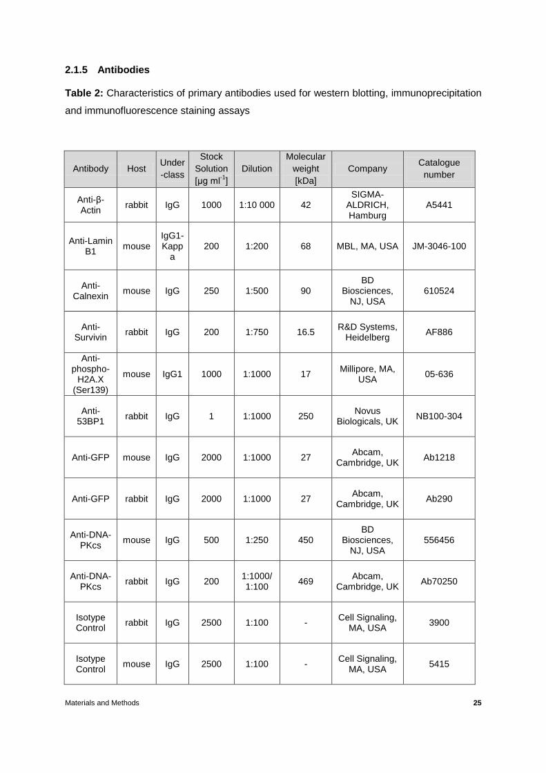
2.1.5 Antibodies
Table 2: Characteristics of primary antibodies used for western blotting, immunoprecipitation
and immunofluorescence staining assays
Antibody Host Under
-class
Stock
Solution
[µg ml-1
]
Dilution
Molecular
weight
[kDa]
Company Catalogue
number
Anti-β-Actin
rabbit IgG 1000 1:10 000 42 SIGMA-
ALDRICH, Hamburg
A5441
Anti-Lamin B1
mouse IgG1-Kapp
a 200 1:200 68 MBL, MA, USA JM-3046-100
Anti-Calnexin
mouse IgG 250 1:500 90 BD
Biosciences, NJ, USA
610524
Anti-Survivin
rabbit IgG 200 1:750 16.5 R&D Systems,
Heidelberg AF886
Anti-phospho-
H2A.X (Ser139)
mouse IgG1 1000 1:1000 17 Millipore, MA,
USA 05-636
Anti-53BP1
rabbit IgG 1 1:1000 250 Novus
Biologicals, UK NB100-304
Anti-GFP mouse IgG 2000 1:1000 27 Abcam,
Cambridge, UK Ab1218
Anti-GFP rabbit IgG 2000 1:1000 27 Abcam,
Cambridge, UK Ab290
Anti-DNA-PKcs
mouse IgG 500 1:250 450 BD
Biosciences, NJ, USA
556456
Anti-DNA-PKcs
rabbit IgG 200 1:1000/ 1:100
469 Abcam,
Cambridge, UK Ab70250
Isotype Control
rabbit IgG 2500 1:100 - Cell Signaling,
MA, USA 3900
Isotype Control
mouse IgG 2500 1:100 - Cell Signaling,
MA, USA 5415

Materials and Methods 26
Table 3: Characteristics of secondary antibodies used for immunofluorescence staining
Antibody Host Under-class
Stock Solution [mg ml
-1]
Dilution Label Company Catalogue
number
Anti-sheep IgG
donkey IgG 2 1:250 Alexa Fluor
R
488 Molecular
Probes, USA A-11015
Anti-sheep IgG
donkey IgG 2 1:250 Alexa Fluor
R
594 Molecular
Probes, USA A-11016
Table 4: Characteristics of secondary antibodies used for western blotting
2.1.6 Expression plasmids
Survivin expression plasmids:
pEGFP-N1 control plasmid, Clontech, Mountain View, CA, USA
(EGFP = Enhanced Green Fluorescent Protein)
pEGFP-N1-Survivin wt (wildtype); made by Dr. S. Hehlgans, C. Petraki, Molecular
Radiobiology, University Hospital Frankfurt
Coupled
enzyme Specificity Host
Under-
class
Stock
solution
[µg ml-1
]
Dilution Company Catalogue
number
Horse radish
peroxidase rabbit goat IgG 400 1:4000 Santa Cruz
Biotechnology,
Heidelberg
Sc-2254
Horse radish
peroxidase mouse goat IgG 400 1:1000 Sc-2255
Materials and Methods 27
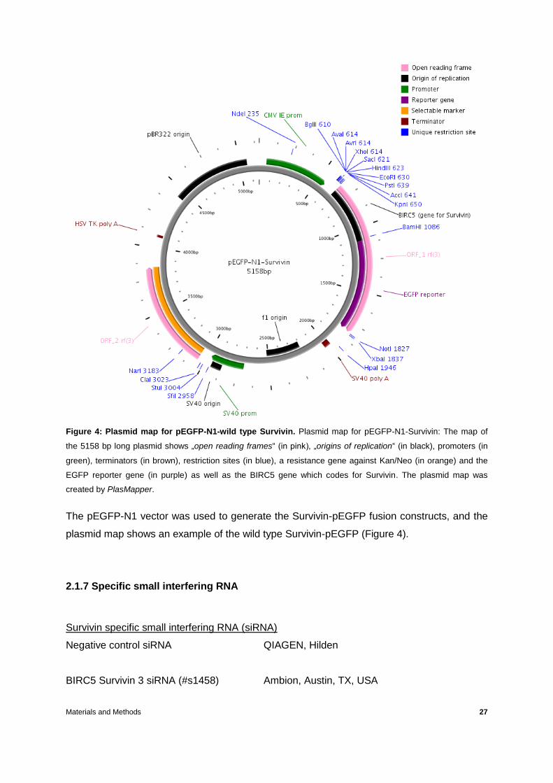
Figure 4: Plasmid map for pEGFP-N1-wild type Survivin. Plasmid map for pEGFP-N1-Survivin: The map of
the 5158 bp long plasmid shows „open reading frames“ (in pink), „origins of replication“ (in black), promoters (in
green), terminators (in brown), restriction sites (in blue), a resistance gene against Kan/Neo (in orange) and the
EGFP reporter gene (in purple) as well as the BIRC5 gene which codes for Survivin. The plasmid map was
created by PlasMapper.
The pEGFP-N1 vector was used to generate the Survivin-pEGFP fusion constructs, and the
plasmid map shows an example of the wild type Survivin-pEGFP (Figure 4).
2.1.7 Specific small interfering RNA
Survivin specific small interfering RNA (siRNA)
Negative control siRNA QIAGEN, Hilden
BIRC5 Survivin 3 siRNA (#s1458) Ambion, Austin, TX, USA
Materials and Methods 28
Sequence (5’ 3’) Sense: GCAGGUUCCUUAUCUGUCAtt
Antisense: UGACAGAUAAGGAACCUGCag
2.1.8 Commercial Kits
Description Company
Micro BCATM Protein Assay Kit Thermo Scientific, Karlsruhe PCR Mycoplasma Test Kit AppliChem GmbH, Darmstadt QIAquick Gel Extraction Kit QIAGEN GmbH, Hilden QIAquick PCR Purification Kit QIAGEN GmbH, Hilden NucleoBond Xtra Midi Plus EF Macherey-Nagel GmbH &co. KG, Dueren QIAprep Spin Miniprep Kit (250) Qiagen GmbH, Hilden PCR clean-up NucleoSpinR Extract II Macherey-Nagel GmbH & co. KG, Dueren Nuclear Complex Co-IP Kit Active Motif, Belgium QuikChange II Site-Directed Agilent Technologies, La Jolla CA, USA Mutagenesis Kit Caspase-GloR 3/7 Assay Promega, WI, USA
2.1.9 Enzymes and respective buffers
Description Company
PfuUltraR High-Fidelity DNA Polymerase Strategene, La Jolla, USA T4-DNA-Ligase New Englands Biolabs Inc, Frankfurt T4 DNA Ligase buffer 10x New Englands Biolabs Inc, Frankfurt Taq-DNA-Polymerase SIGMA Chemical Co, St. Louis, USA DpnI New Englands Biolabs Inc, Frankfurt KpnI New Englands Biolabs Inc, Frankfurt

Materials and Methods 29
XbaI New Englands Biolabs Inc, Frankfurt BamHI New Englands Biolabs Inc, Frankfurt EcoRI New Englands Biolabs Inc, Frankfurt ApaLI New Englands Biolabs Inc, Frankfurt FspI New Englands Biolabs Inc, Frankfurt NEB Buffer 10x New Englands Biolabs Inc, Frankfurt
2.1.10 Electrophoresis Markers
Description Company
100bp DNA-Ladder InvitrogenTM, Gaithersburg, USA 1kb DNA-Ladder InvitrogenTM, Gaithersburg, USA
2.1.11 Oligonucleotides
The melting temperature of all the oligonucleotides used was calculated according to this formula: Tm = 4°C x (G + C) + 2°C x (A + T) from Nelson and Brutlag, 1979. They were provided by Eurofins MWG Operon (Ebersberg, Germany). Primers for PCR cloning
Primers for subcloning of Survivin from pc3-Survivin-GFP into pEGFP-N1
pEGFPC1-fw: GAT CAC TCT CGG CAT GGA C
pEGFPC1-rev: CAT TTT ATG TTT CAG GTT CAG GG
CMV-fw: CGC AAA TGG GCG GTA GGC GTG
pEGFPN1-rev: GTC CAG CTC GAC CAG GAT G
Primers for amplification of Survivin and subcloning in pEGFP-N1/KpnI/BamHI
1-Surv-fw: gg-ggtacc-ggcggc-ATGGGTGCCCCGACGTTGC, KpnI, Tm = 68°C
2-Surv-rev: cg-ggatcc-cg-ATCCATGGCAGCCAGCTGCTC, BamHI, Tm = 68 °C Primers for Survivin S20A mutation

Materials and Methods 30
5-S20A-fw: 5’-tcaaggaccaccgcatcgctacattcaagaactgg-3’, Tm = 79.27°C
6-S20A-rev: 5’-ccagttcttgaatgtagcgatgcggtggtccttga-3’, Tm = 79.27°C
Primers for Survivin S20D mutation
7-S20D-fw: 5’-ctcaaggaccaccgcatcgatacattcaagaactggcc-3’, Tm = 78.97°C
8-S20D-rev: 5’-ggccagttcttgaatgtatcgatgcggtggtccttgag-3’, Tm = 78.97°C
Primers for Survivin T34A mutation
9-T34A-fw: 5’-gctgcgcctgcgccccggagcgg-3, Tm = 81.67°C
10-T34A-rev: 5’-ccgctccggggcgcaggcgcagc-3’, Tm = 81.67°C
Primers for Survivin T34D mutation
11-T34-fw: 5’-gggctgcgcctgcgacccggagcggatg-3’, Tm = 81°C
12-T34D-rev: 5’-catccgctccgggtcgcaggcgcagccc-3’, Tm = 81°C
Primers for Survivin T117A mutation
13-T117A-fw: 5’-gccaagaacaaaattgcaaaggaagccaacaataagaagaaagaat-3’, Tm =
78.02°C
14-T117A-rev: 5’-attctttcttcttattgttggcttcctttgcaattttgttcttggc-3’, Tm = 78.02°C
Primers for Survivin T117D mutation
15-T117D-fw: 5’-gagccaagaacaaaattgcaaaggaagacaacaataagaagaaagaatttgagg-3’,
Tm = 78.96°C
16-T117D-rev: 5’-cctcaaattctttcttcttattgttgtcttcctttgcaattttgttcttggctc-3’, Tm = 78.96°C
Primers for Survivin XIAP mutation
For the 5’ end of deletion mutants XIAP and BIR primers 19 and 20 were annealed and
subsequently used for ligation reactions.
19- XIAP-fw: starts with a.a.1 to 14, KpnI C-ATG GGT GCC CCG ACG TTG CCC
CCT GCC TGG CAG CCC TTT CTC - T XbaI
20- XIAP-rev: starts with a.a. 14 to 1, XbaI CTAGA- GAG AAA GGG CTG CCA GGC
AGG GGG CAA CGT CGG GGC ACC CAT – GGTAC KpnI
Materials and Methods 31
21-XIAP-39fw: starts with a.a.39, XbaI GC-TCT AGA-GCC GAG GCT GGC TTC
ATC CA, Tm = 66°C
For reverse primer see primer no. 2-Surv-rev
Primers for Survivin BIR mutation
For BIR domain deletion for the 5’end refer to primers no. 19-XIAP-fw and 20-XIAP-
rev
22-BIR-fw: starts with a.a. 88, XbaI GC-TCT AGA-TCT GTC AAG AAG CAG TTT
GAA GA, Tm =64°C
For reverse primer see primer no. 2-Surv-rev
Primers for Survivin MicTub mutation
For fw primer see primer no. 1-Surv-fw
23-Mictub-rev: starts with a.a. 98 BamHI cg-ggatcc-AAG GGT TAA TTC TTC AAA
CTG CTT, Tm = 64°C
Primers for Survivin Hsp90 mutation
For fw primer see primer no. 1-Surv-fw
24-Hsp90-rev: starts with a.a. 78 XbaI GC-TCT AGA-TTT ATG TTC CTC TAT GGG
GTC G,Tm = 64°C
For the amplification of the 3’end of Hsp90 deletion mutant primer no. 22-BIR-fw and
2-Surv-rev were used
Primers for sequencing
37-CMV-fw-1: CGTAACAACTCCGCCCCATTG, Tm = 66°C
38-pEGFPN1-rev-1: CTTGTGGCCGTTTACGTCGC, Tm = 64°C
Materials and Methods 32
2.2 Methods
2.2.1 Cell culture
The human colorectal carcinoma cell line SW480 was obtained from the American Type
Culture Collection (ATCC, Promochem, Germany). Cells were cultured in Dulbecco’s
Modified Eagle Medium containing glutamax-I (L-alanyl-L-glutamine) and supplemented with
10% foetal calf serum, 50 U/ml penicillin and 50 µg/ml streptomycin at 37°C in a humidified
atmosphere containing 5% CO2. Cells were passaged on average 1:6 every 3 to 4 days
using 2 ml trypsin after being washed with PBS (see 2.1.3 chemicals and media section) and
plated on T-75 flasks with 12 ml of the medium mentioned above. In the case of the Survivin-
EGFP constructs 750 µg/ml of G418 was added to the medium.
For 3D cell cultures, cells were plated into a mixture of 0.5 mg/ml laminin-rich extracellular
matrix (lrECM; Cultrex 3D Culture Matrix BME Reduced Growth Factor Basement Membrane
Extract; R&D Systems, Wiesbaden, Germany) and DMEM medium as previously described,
on either 96-well plates (for 3D colony forming assays) or 24-well plates (for 3D lysates and
3D immunofluorescence staining assays).
2.2.2 Survivin constructs and stable transfection
For expression of Survivin-EGFP fusion constructs human Survivin cDNA was amplified with
specific primers (listed in section 2.1.11), flanked with KpnI and BamHI restriction sites, from
an expression plasmid provided by R.H. Stauber (University Hospital of Mainz, Germany).
Subsequently, the PCR fragments were digested with appropriate restriction enzymes (see
section 2.1.9) and were inserted into KpnI/BamHI sites of the pEGFP-N1 expression vector.
For deletion mutants, cDNA fragments left and right from the deletion were amplified by PCR
Materials and Methods 33
and ligated using the XbaI restriction site while inserted into the KpnI/BamHI restriction sites
of the pEGFP-N1 vector. The phospho-mutants were generated using the site-directed
mutagenesis kit (section 2.1.8) according to manufacturer’s guidelines using the primers
listed in section 2.1.11.
PCR for amplification of Survivin cDNA for subcloning in pEGFP-N1
pc3-Surv-GFP was used as a template at a working concentration of 50 ng/µl along with (per
reaction): 30µl 10 x Buffer, 7.5 µl of 10µM primers 1 and 2, respectively (for primer names
see section 2.1.11), 7.5 µl of 10 mM dNTPs, 229.5 µl RNase/DNase-free water and 6 µl
PfuUltra HF. The PCR conditions were as follows: 2 min 95°C, 35 x (30 s 95°C, 1 min 62°C,
1 min 72°C), 15 min 72°C, 4°C. The samples were purified using the QIAquick PCR
Purification Kit following manufacturer’s instructions.
Restriction enzyme digestion of Survivin PCR fragment and pEGFP-N1
For restriction enzyme digestion 10 µg of the pEGFP-N1 plasmid or 10 µg of the Survivin
PCR fragment were used for digestion with: 3 µl KpnI + 3 µl BamHI + 1 µl BSA + 10 µl
NEBuffer #1. The reaction volumes were adjusted to 100 µl with RNase/DNase-free water.
The samples were incubated overnight at 37°C.
Gel Purification of plasmid pEGFP-N1
50 ml of 1% agarose gel (supplemented with 3 µl EtBr) in TAE buffer was used (in a gel
electrophoresis chamber) for the loading of 120 µl KpnI/BamHI linearized plasmid for gel
electrophoresis at 80 Volts for 1 hour. The plasmid was then extracted using the QIAquick
Gel Extraction kit following manufacturer’s instructions.

Materials and Methods 34
Ligation of Survivin with the pEGFP-N1
Ligation reaction contained the plasmid DNA pEGFP-N1/KpnI/BamHI (2 µl), the insert
Survivin/KpnI/BamHI (2 µl), T4 DNA Ligase (1µl), 10 x T4 DNA Ligase Buffer (NEB) (1 µl)
and RNase/DNase-free water (4 µl), followed by overnight incubation at 14°C.
PCR for Survivin-EGFP deletion mutants
Survivin cDNA was used as a template at a 50 ng/µl working concentration. For the PCR
reaction forward and reverse primers (for primer names see section 2.1.11) (number 21/2 for
XIAP right, 22/2 for BIR right, 1/28 for MicTub left, 1/24 for Hsp90 left and 22/2 for
Hsp90 right) at a working concentration of 10 µM (1.25 µl each primer per reaction), 10x
NEBuffer (5 µl per reaction), dNTPs 10 mM (1.25 µl per reaction), RNase/DNase-free water
(38.25 µl per reaction) and PfuUltra HF (1 µl per reaction) were added. The PCR conditions
were as follows: 2 min 95°C, 35 x (30 s 95°C, 1 min 62°C, 1 min 72°C), 15 min 72°C, 4°C.
Agarose gel analysis followed (via agarose gel electrophoresis).
Restriction enzyme digestion of Survivin deletion mutants PCR fragments
45 µl of each PCR reaction was digested with 3 µl of each restriction enzyme + 10 µl
NEBuffer #2 or #3 (XIAP right: XbaI/BamHI/#3, BIR right: XbaI/BamHI/#3, MicTub left:
KpnI/BamHI/#3, Hsp90 left: KpnI/XbaI/#2, Hsp90 right: XbaI/BamHI/#3) + 1 µl BSA + 38 µl
RNase/DNase-free water. The samples were incubated overnight at 37°C.
PCR clean-up
Samples were purified using the PCR clean-up NucleoSpinR Extract II kit following
manufacturer’s protocol.

Materials and Methods 35
Ligation reactions for Survivin deletion mutants
XIAP: 2µl pEGFP-N1/KpnI/BamHI, 1µl annealed primers 19 and 20/KpnI/XbaI, 2 µl Surv.
XIAP right/XbaI/BamHI, 1 µl T4 DNA Ligase, 1 µl 10 x T4 DNA Ligase Buffer (NEB), 3 µl
RNase/DNase-free water
BIR: 2µl pEGFP-N1/KpnI/BamHI, 1 µl annealed primers 19 and 20/KpnI/XbaI; 2.5 µl Surv
BIR right/XbaI/BamHI, 1 µl T4 DNA Ligase, 1 µl 10 x T4 DNA Ligase Buffer (NEB), 2.5 µl
RNase/DNase-free water
MicTub: 2µl pEGFP-N1/KpnI/BamHI, 2 µl Surv MicTub left/KpnI/BamHI, 1 µl T4 DNA
Ligase, 1 µl 10 x T4 DNA Ligase Buffer (NEB), 4 µl RNase/DNase-free water
Hsp90: 2µl pEGFP-N1/KpnI/BamHI, 2.5 µl Surv BIR right/XbaI/BamHI, 3 µl Surv Hsp90
left/KpnI/XbaI, 1 µl 10 x T4 DNA Ligase Buffer (NEB), 0.5 µl RNase/DNase-free water
0.2 ml PCR tubes were used for the reactions which were incubated overnight at 14°C.
Transformation
50 µl of competent TOP10 E.coli stored with Rubidium Chloride were used for each
transformation required (everything kept on ice). 3 µl of Dpn I digested DNA was added to 50
µl bacteria (1.5 ml Eppendorf tubes were used) and the tubes were incubated for 20 minutes
on ice before they were subjected to a heat shock at 42°C for 45 seconds and then placed
back on ice for 2 minutes. Then, 250 µl of LB-Medium (warmed at 37°C) was added to each
tube. All samples were incubated at 37°C for 1 hour while being shaked at 250 rpm.
Subsequently, samples were centrifuged at 3,000 rpm for 3 minutes (RT). 200 µl of
supernatants were removed and the pellets were resuspended in the medium that was left.

Materials and Methods 36
The samples were then spread on LB-covered dishes (with 50 µg/ml Kanamycin) and
incubated overnight at 37°C followed by storage at 4°C.
4 ml overnight culture of bacteria
Using 50 ml tubes, 50 ml of LB medium was mixed with 50 µl of Kanamycin and each 4 ml of
this mixture were transferred to 14 ml polystyrene round-bottom tubes (one tube per clone).
Using a 200 µl pipette-tip, E. coli colonies were picked from the LB-covered dishes (following
transformation) and were placed into the round-bottom tubes that were then overnight
incubated at 37°C while being shaken at 250 rpm followed by plasmid mini-preparation.
Plasmid mini-preparation
After transformation of TOP10 bacteria and subsequent inoculation of selected colonies
plasmid mini preparation was performed using the QIAprep Spin Miniprep kit following the
manufacturer’s instructions. The solutions provided by the kit were the following: Buffer P1
(for resuspension) contained 50 mM Tris/HCl, pH 8.0, 10 mM EDTA and 100 µg/ml Rnase A;
buffer P2 (for lysis) contained 200 mM NaOH, 1% SDS; buffer N3 (for neutralisation)
contained 3M KOAc, pH 5.5. Two ml from the content of each round-bottom tube (having
been inoculated) were transferred to 2 ml Eppendorf tubes that were centrifuged at 5,000
rpm (tabletop centrifuge) for 5 minutes (RT). Supernatants were discarded and 250 µl of
buffer P1 was added in each pellet followed by vortexing. Then, 250 µl of buffer P2 was
added to the tubes, the contents were mixed and 350 µl of buffer N3 was also added to each
tube, mixed and centrifuged at 13,000 rpm (tabletop centrifuge) for 10 minutes (RT). All
supernatants (now containing plasmid-DNA) were transferred to Qiaprep spin columns and
centrifuged at 13,000 rpm for 1 minute; all eluates were discarded. The columns were
washed with 0.5 ml buffer PB (provided by the kit) and centrifuged at 13,000 rpm for 1
minute; eluates were discarded. The columns were washed again this time with 0.75 ml
buffer PE and centrifuged at same settings; eluates were discarded. The empty columns
Materials and Methods 37
were dry-centrifuged at the same settings. Subsequently, the columns were placed in new
1.5 ml Eppendorf tubes when 50 µl of buffer EB (for elution – containing 10 mM TrisCl, pH
8.5) was added to each of the column, incubated for 1 minute and centrifuged at same
settings. The concentration was measuered in an Eppendorf Biophotometer (diluting each
plasmid DNA 1:100 with water).
Plasmid midi-preparation
When certain clones per construct were confirmed by sequencing (Eurofins, MWG Operon),
they were followed by midi preparation using the NucleoBond Xtra Midi Plus EF kit according
to manufacturer’s guidelines. Prior to preparation, the according inoculation was first carried
out. In detail, in a 500 ml glass beaker, 100 ml of LB medium was supplemented with 50
µg/ml Kanamycin and inoculated with 100 µl overnight cultures (dilution 1:1000) at 37°C and
175 rpm shaking. On the following day, 50 ml of each of these cultures were transferred to
50 ml tubes. The bacteria were pelleted by centrifugation at 4,000 rpm for 15 minutes at 4°C.
Supernatants were discarded and 8 ml of RES-EF (provided by the kit) was used for
resuspension. Eight ml of Lys-EF was added, mixed and incubated at RT for 5 minutes. 8 ml
of NEU-EF was then added, mixed and incubated on ice for 5 minutes. Subsequently, 15 ml
of EQU-EF was carefully added to the column filters and the lysates were then mixed and
poured into the columns which were then washed with 5 ml FIL-EF before the paper filters
were removed. The columns were washed with 35 ml ENDO-EF followed by washing with 15
ml WASH-EF before they were put in new 50 ml tubes in order to elute the plasmid-DNA with
5 ml ELU-EF. The columns were removed; 3.5 ml of Isopropanol was added to the eluates,
mixed and incubated at RT for 2 minutes before they were syringed through a NucleuoBond
Finalizer in a 30 ml-syringe twice and washed 1 x with 2 ml 70% EtOH (Endotoxin-free from
kit). Then, the NucleoBond Finalizer was placed in a 1 ml-syringe and 0.5 ml H2O-EF was
used to collect the eluate in 1.5 ml Eppendorf tubes. Concentrations were determined using
a Bio photometer.

Materials and Methods 38
Stable transfection
Prior to stable transfection, the constructs were linearized using 30 µg of plasmid DNA, 10 µl
of 10 x NEBuffer, 4 µl of ApaLI enzyme (10 U/µl), 1 µl BSA and the volumes were adjusted to
100 µl with RNase/DNase-free water while the reactions were set up in 1.5 ml tubes
incubated overnight at 37°C. After the confirmation of the linearization via agarose gel
electrophoresis, the linearized plasmids were subjected to PCR clean-up using the
NucleoSpin Extract II kit following manufacturer’s instructions. SW480 cells were then stably
transfected with pEGFP-N1 or Survivin-EGFP expression constructs using 4 µg linearized
plasmid DNA diluted in serum free Opti-MEM and Roti-Fect PLUS transfection reagent
according to manufacturer’s instructions (see section 2.2.3 for method of transfection).
Following transfection, the cells were transferred from 6-well plates to 100 mm petri dishes
where colonies were allowed to grow in DMEM + 10% FCS +1% P/S enriched with 750 µg/ml
G418 for selection. Clones were isolated using cloning cylinders embedded in silicon while
expression of EGFP or Survivin-EGFP constructs was verified by fluorescence microscopy
and Western blotting.
Selection of fluorescent colonies
When the 100 mm petri dishes (mentioned above) contained colonies large enough to be
visible by naked eye, the dishes were washed with PBS and transferred to a fluorescence
microscope where fluorescent colonies were selected and marked (from the outside of the
dish). The dishes were then taken under laminar flow hood where silicon-embedded cloning
cylinders were carefully placed on the selected colonies and 60 µl of trypsin was added to
each cloning cylinder for trypsinization (5 minutes incubation at 37°C/5%CO2 under sterile
conditions). The cells were then resuspended in the cylinders, with 60 µl of DMEM + 10%
FCS +1% P/S and transferred in 12-well plates (one clone per well) along with 1 ml medium
(DMEM + 10% FCS +1% P/S + 750 µg/ml G418) per well. They were then left in those 12-

Materials and Methods 39
well plates to grow to approximately 60-80% confluence before their transfer to T-25 flasks.
Cells from T-25 flasks were later (when 80-100% confluent) harvested for cryostocks.
Fluorescence imaging
Fluorescence images of stably transfected cells were obtained using an Axiovert 40 CFL
microscope (Carl Zeiss, Göttingen, Germany) after staining the nuclei with 4’,6-diamidino-2-
phenylindole (DAPI).
2.2.3 Attenuation of endogenous Survivin via siRNA
For the knockdown of endogenous Survivin, an siRNA targeting the 3’-untranslated region of
Survivin was used: Surv. siRNA: 5’- GCAGGUUCCUUAUCUGUCAtt-3’ (sense), siRNA ID
s1458. The non-specific negative control siRNA was from Qiagen. 24 hours after plating 3.5
x 105 cells of each of the Survivin-EGFP constructs on 6-well plates, both of the siRNAs were
diluted in serum free Opti-MEM to obtain a final concentration of 20 nM, in this case 2.5 µl
siRNA (10µM) plus 122.5 µl Opti-MEM calculated for each well. Roti-Fect PLUS transfection
reagent was used under serum-free conditions at a concentration of 20 nM. Thus, Roti-Fect
was also diluted in serum free Opti-MEM (5 µl Roti-Fect plus 120 µl Opti-MEM calculated per
well) and 5 minutes later it was mixed with the diluted siRNA and incubated for 20 minutes at
RT. In the meanwhile, cells were washed once with PBS and 1 ml of serum free Opti-MEM
was added. Subsequently, the siRNA/Opti-MEM solution was mixed with the Roti-Fect/Opti-
MEM solution, and 250 µl of the mixture was added to each well. The 6-well plates were
incubated for 8 hours at 37°C, 5% CO2, followed by addition of 1.25 ml Opti-MEM/20% FCS
to each well. The 6-well plates were incubated for 24 hours at 37°C 5% CO2 prior to any
further experiments.

Materials and Methods 40
2.2.4 Irradiation procedure
Irradiation of cells with single photon doses ranging from 2 to 6 Gy was performed using a
linear accelerator (SL-15, Elekta, Crawley, UK) with 6 MeV/100 cm focus-surface distance
and a dose rate of 4 Gy/min. Mock-irradiated cultures were kept at room temperature in the
X-ray control room while the other samples were being irradiated.
2.2.5 Three-dimensional colony forming assay
Figure 5: Methodology for 3D radiation clonogenic survival assays. The procedure of a 3D radiation
clonogenic survival assay is briefly depicted here on a symbolic time scale followed by a photograph of colonies
taken under the microscope (2.5x).
24 h after the knockdown of endogenous Survivin via siRNA transfection (Figure 5), the cells
were washed with PBS (1 ml per well) and trypsinized (0.25 ml per well) in order to be
counted with a chamber counter (1:2 dilution with trypan blue) under the microscope. Single
cells were plated in 0.5 mg/ml lrECM/DMEM/10% FCS/1% penicillin/streptomycin on 96-well
plates pre-coated with 50 µl 1% Agarose, by triplets (3 wells per construct per siRNA and 1 x
103 cells per well) in 100 µl cell-matrigel mixture. 200 µl of PBS was added to the wells
around those containing cells. The 96-well plates were incubated for 90 minutes at 37°C, 5%
CO2, followed by addition of 100 µl of DMEM+10% FCS+1%P/S to each well. The plates
were incubated at 37%, 5% CO2 for 24 hours until irradiation (0, 2, 4, 6 Gy) and cell colonies
Counting ofcolonies
Platingof cells
siRNAtransfection
24 h
Plating ofsingle cells
24 h
Start 3D culture
24 h
Irradiation
7 days
100 µm

Materials and Methods 41
(> 50 cells) were microscopically counted 7 days after plating (Figure 5). Plating efficiencies
were calculated as numbers of colonies counted (0Gy) / numbers of cells plated. Surviving
fractions were calculated as numbers of colonies counted / (numbers of cells plated × plating
efficiency). Each point on survival curves represents the mean surviving fraction from 3-4 or
more independent experiments. Survival variables α and β were fitted according to the linear
quadratic equation SF = exp [−α x D − β x D2] with D = dose using EXCEL® software
(Microsoft). The radiation-induced cytotoxicity enhancement factor at 50% cell survival was
calculated by transforming the above mentioned equation using α and β values of the
individual survival curves.
2.2.6 Protein extraction and Western blotting
Protein extraction from 3D cultures
24 hours after the plating of mock- and siRNA-transfected cells in 0.5 µg/µl lrECM on 24-well
plates pre-coated with 300 µl of 1% Agarose (1 x 10⁶ cells per well), the matrigel was
carefully removed and 130 µl of ice-cold RIPA (see section 2.1.4 solution and buffers) was
added to each well and incubated on ice for 30 minutes. The RIPA/cells mixture was
transferred from the 24-well plates to 2 ml Eppendorf tubes, incubated on ice for 10 minutes
followed by passing through a 30-G insulin syringe to optimize protein extraction. The
samples were then incubated on ice for 30 minutes, centrifuged for 15 minutes at 4°C 14,000
rpm followed by transfer of supernatants to new 1.5 ml Eppendorf tubes. For the estimation
of the lysate volume, all samples were filled with RIPA up to the same volume of that with the
largest volume. Loading buffer was then added to all samples (1/5 of the volume of the
largest volume-sample). The samples were then stored at - 80°C before Western blotting.

Materials and Methods 42
Western blotting
Western blotting was performed in the following cases: to confirm the knockdown of
endogenous Survivin, to confirm the correct sizes and expression of Survivin-EGFP mutants,
their relation to other proteins as well as to identify immunoprecipitated complexes. For these
reasons, 10% or 12% gels were prepared for the SDS-gel electrophoresis (see section 2.1.4
table 1) where 20 - 30 µg of protein extract was loaded per lane. Prior to electrophoresis all
samples were heated at 95°C for 10 minutes, shortly (1 minute) centrifuged at 11,000 rpm at
4°C and during electrophoresis (25 mA 400V per gel) 1 L of electrophoresis buffer was used
(see section 2.1.4 for buffers) in the chamber. Following gel-electrophoresis, the gels were
placed on 7 x 9 cm filter papers (see section 2.1.2) soaked in transferring-buffer (see section
2.1.4) then covered with blotting membranes (6 x 8 cm) followed by two more filter papers
and transferred on a semi-dry blotting apparatus for protein transfer at 50 mA per gel for 150
– 210 min. Following blotting, to confirm correct protein transfer and equal loading the
membranes were soaked in Ponceau S solution for 1 minute, subsequently distained with
distilled water and then blocked in 5% milk powder/TBS-T at room temperature for 30
minutes until primary antibodies were applied that were diluted in 5% BSA/TBS-T prior to
application and incubated with the respective membranes at 4°C overnight. The following
primary antibodies were used (for more details see Table 2 and 4): anti-Survivin (AF886),
Anti-Calnexin (BD610524), anti-GFP (ab290 and ab1218), anti-Lamin B1 (JM-3046-100),
anti-DNA-PKcs (BD556456 and ab70250), anti--actin (A5441). For detection of proteins,
horseradish peroxidase-conjugated goat anti-rabbit and goat anti-mouse secondary
antibodies (diluted in 5% milk powder/TBS-T) were used after 3 x washing of the membranes
(using TBS-T). The membranes were then incubated with appropriate dilutions of secondary
antibodies in 5% milk powder/TBS-T for 1 hour followed by 4 washing steps with TBS-T and
one with TBS. The membranes were then incubated for 1 min with working solutions of

Materials and Methods 43
enhanced chemiluminescent (ECL) substrate (Thermo Scientific), placed in plastic sheet
protectors and exposed to Hyperfilm ECL (GE Healthcare) prior to film development.
2.2.7 Subcellular fractionation and immunoprecipitation
Cytoplasmic and nuclear extracts were prepared with the Nuclear Complex Co-IP kit
according to manufacturer’s protocol (all buffers hereby mentioned regarding fractionation
were supplied by the kit) and subjected to co-immunoprecipitation experiments and western
blotting as described.
Subcellular fractionation
Four million cells were plated per T-75 flask, and 4 x T-75 flasks were used per construct in
order to be subjected to fractionation and eventually co-IP. On the second day after plating,
serum starvation was performed, replacing the usual medium (DMEM/10% FCS/1% P/S)
with serum-free medium (DMEM without FCS) in all T-75 flasks. On the day following the
serum starvation, the flasks were washed with ice-cold PBS and scraped with 1 ml 5%
Phosphatase Inhibitor/PBS. The scraped cells were transferred to pre-chilled 15 ml tubes
that were centrifuged at 1,500 rpm at 4°C for 5 minutes. Supernatants were discarded and
each pellet was resuspended in 500 µl ice-cold Hypotonic Buffer (containing Hypotonic
buffer, Phosphatase Inhibitor, Protease Inhibitor and double distilled water) and incubated on
ice for 15 minutes. 25 µl of detergent (provided by the kit) was then added in each sample, all
of which were then centrifuged at 12,000 rpm at 4°C for 30 seconds. All cytoplasmic fractions
were then transferred to 1.5 ml Eppendorf tubes properly labelled for the cytoplasmic
fractions. The pellets were resuspended in 100 µl digestion buffer (containing 0.5% 100 mM
PMSF, 98.5% digestion buffer, 1% Protease Inhibitor Cocktail) and 0.5 µl of enzymatic
shearing cocktail was added to all samples before they were incubated at 4°C for 90
minutes. Following incubation, 2 µl of 0.5M EDTA was added to each sample which were
Materials and Methods 44
then gently vortexed for 2 seconds and incubated for 5 minutes on ice before they were
centrifuged at 12,000 rpm for 10 minutes. The supernatants were collected (i.e. the nuclear
fractions) in new 1.5 ml Eppendorf tubes. Concentrations were determined using the Micro
BCA assay kit according to manufacturer’s instructions.
Immunoprecipitation
For co-immunoprecipitation, 500 µg of nuclear extract (per sample) resuspended in 500 µl
IP-Low buffer (included in the Nuclear Complex Co-IP kit) were incubated overnight at 4°C
with anti-GFP (ab1218) or anti-DNA-PKcs (BD556456) antibodies. A mouse (G3A1) mAb
IgG1 Isotype control antibody served as a control. The final concentration of all antibodies in
the nuclear lysates was 2 µg/ml. Following overnight incubation, 50 µl of slurry protein G
agarose beads (Sigma-Aldrich) was added to the samples and incubated at 4°C for 1 hour
while gently being rotated. Then, the probes were centrifuged at 5,500 rpm for 2 minutes,
supernatants were discarded and samples were washed 3 times with 500 µl IP-High buffer
(included also in the Co-IP kit) and once with PBS at 5,500 rpm for 2 min at 4°C before 30 µl
RIPA and 8 µl loading dye were added to the beads-pellet. The samples were heated for 10
minutes at 95°C and centrifuged at 11,000 rpm for 50 sec prior to SDS-electrophoresis and
eventually western blotting.
2.2.8 Immunofluorescence staining and imaging
Residual DNA double-strand breaks (DSB) of 3D and 2D cell cultures (plated on 24-well
plates) were evaluated using the foci assay.
Two-dimensional immunofluorescence staining
24 hours after the knockdown of endogenous Survivin (via siRNA) on 6-well plates, cells
were harvested and plated on 24-well plates with DMEM/10% FCS/1% P/S for 2D

Materials and Methods 45
immunofluorescence staining (IF). In detail, cover slips were placed on 24-well plates and 75
x 103 cells were plated per well, for control siRNA and Survivin-targeted siRNA per construct.
24 hours after plating, the cells were irradiated with 2 Gy and stained 40 minutes or 24 hours
later (different plates were prepared for different time points) according to the time point of
interest. Cells were then permeabilized and fixed using 0.25% Triton X-100 and 3.7%
formaldehyde in PBS for 10 minutes at RT. Subsequently, blocking took place via incubation
for 1 hour with 3% BSA in PBS followed by staining with anti-phospho-histone H2AX and
anti-53BP1 antibodies 1:1000 diluted in 3% BSA in PBS for 1 hour (50 µl solution per cover
slip was used). Cells were then washed once with PBS and stained with secondary
antibodies i.e. Alexa Fluor 594 goat anti-mouse and Alexa Fluor 488 goat anti-rabbit diluted
1:500 (Invitrogen) for another hour in the dark, followed by 4’,6-diamidino-2-phenylindole
(DAPI; Roth) staining for 5 minutes. Cover slips were then transferred on microscopic slides
after being covered with one drop of Vectashield mounting medium. H2AX/53BP1 foci were
microscopically counted from at least 50 cells per experimental condition.

Materials and Methods 46
Three-dimensional immunofluorescence staining
Figure 6: Methodology of 3D IF of H2AX/53BP1 foci. The procedure of the 3D immunofluorescence staining of
H2AX/53BP1 foci for evaluation of residual DNA DSBs is hereby represented on a symbolic time scale followed
by a photograph of nuclei taken under the microscope (40x). Bar: 10µm
24 hours after the siRNA-mediated attenuation of endogenous Survivin on 6-well plates
(Figure 6), cells were harvested and plated on 24-well plates (pre-covered with 1% agarose)
in a mixture of 0.5 mg/ml laminin-rich extracellular matrix and DMEM/10% FCS/1% P/S for
3D IF. In detail, 0.5 x 10⁶ cells were plated per well, for control siRNA and Survivin-targeted
siRNA per construct. 24 hours after plating, the cells were irradiated with 2 Gy. 24 hours
following irradiation, the cells were transferred with the matrigel from the 24-well plates to 1.5
ml Eppendorf tubes and centrifuged for 3 minutes at 3,500 rpm (talbetop centrifuge).
Supernatants were discarded and cells were permeabilized and fixed using 0.25% Triton X-
100 and 3.7% formaldehyde in PBS (200 µl solution per tube) for 15 minutes at RT. Then,
Staining
Platingof cells
siRNAtransfection
24 h
Plating ofsingle cells
24 h
Start 3D culture
24 h
Irradiation Fixing
24 h
X d
Cells onto microscopicslide
Counting of fociunder fluor. microscope
53BP1
10µm
Materials and Methods 47
the samples were centrifuged again at 4,500 rpm for 3 minutes, supernatants were removed,
and the pellets were resuspended in 50 µl blocking solution (3% BSA in PBS) and left to be
blocked for 1 hour. Subsequently, staining took place with 10 µl (per sample) of 1:500 diluted
in blocking solution anti-phospho-histone H2AX and anti-53BP1 (same dilution) antibodies
for 2 hours. Cells were then washed once with PBS for 10 minutes, centrifuged at 4,500 rpm
for 3 minutes and pellets were resuspended with 50 µl (per pellet) of 1:250 diluted in blocking
solution secondary antibodies i.e. Alexa Fluor 594 goat anti-mouse and Alexa Fluor 488 goat
anti-rabbit and incubated for another hour in the dark. The cells were then washed twice with
1 ml PBS via resuspension followed by 10 minutes incubation and centrifugation at 4,500
rpm for 3 minutes. The pellets were finally resuspended in 15 µl DAPI solution (300 ng/µl)
and incubated in the dark for 5 minutes. The cell suspensions were then transferred on
microscopic slides, along with one drop of Vectashield mounting medium on each sample
while a square cover slip was used to seal the cells. H2AX and 53BP1 foci were
microscopically counted from at least 50 cells per experimental condition (Figure 6).
Fluorescence imaging
Fluorescence images of H2AX and 53BP1 foci were obtained using an Axiovert 40 CFL
microscope (Carl Zeiss).
2.2.9 Cell cycle analysis
24 hours after the siRNA-mediated knockdown of endogenous Survivin, cells were
harvested, counted and 3.5 x 10⁵ cells were plated in triplets on 6-well plates with
DMEM/10% FCS/1% P/S for cell cycle analysis. 24 hours after plating, the supernatants from
all samples were transferred to 15 ml tubes. The cells were then washed with 5 ml PBS and
trypsinized in 1 ml trypsin for 5 minutes at 37°C/5% CO2. Cells were then resuspended in 3

Materials and Methods 48
ml medium and transferred in the 15 ml falcon tubes containing their supernatants. The tubes
were then centrifuged at 1,000 rpm for 5 minutes, washed twice with 1 ml PBS, transferred to
1.5 ml Eppendorf tubes and centrifuged at 3,000 rpm for 5 minutes. Pellets were
resuspended in 200 µl PBS and 1 ml 70% EtOH (cooled at -20°C) was added to each
sample. They were then centrifuged at 2,000 rpm at 4°C for 5 minutes, each pellet was
resuspended in 100 µl PBS followed by addition of 100 µl RNase A (100 µg/ml in PBS) and
100 µl propidium iodide (100 µg/ml in PBS). The samples were then incubated at 37°C for 30
min. Cell suspensions were then transferred in FACS tubes that were measured (12,000
events at 449 nm; FL2-585 Red) using the FACS Calibur flow cytometer and the
CellQuestPro software (Becton Dickinson).
2.2.10 Measurement of apoptosis and caspase 3/7 assay
For quantification of apoptotic cells, subG1 content was analysed by flow cytometry as
described in section 2.2.9. For quantification of caspase 3/7 activity a CASPASE GLOTM-
assay was used according to manufacturer’s instructions. In particular, 24 hours after the
knockdown of endogenous Survivin (via siRNA on 6-well plates) cells were washed,
trypsinized, counted and 6 x 10³ cells were plated per well (in triplets per construct) on three
96-well plates (two for 0 Gy and one for 6 Gy) with DMEM/10% FCS/1% P/S. 24 hours later,
one of the 0 Gy plates was measured after removing 70 µl medium from each well and
adding 30 µl substrate (Caspase-Glo® 3/7 Assay Reagent), then shaking at 300 rpm for 30
seconds followed by 90 minutes incubation at RT. Measurement was conducted using an
ELISA-Reader (Wallac Victor; Protocol: Luciassay 10sec). 24 hours thereafter, the two
remaining plates (the other 0 Gy and the 6 Gy one) were treated and measured likewise.

Materials and Methods 49
2.2.11 Transmigration assay
Figure 7: Methodology of transmigration assay. The procedure of a transmigration assay is hereby depicted
along with the respective the time scale.
For transmigration assays (Figure 7), knockdown cell cultures were washed with PBS,
trypsinized, counted and single cells were plated in a BD BioCoat™ Matrigel™ Invasion
Chamber under serum-free conditions (2.5 x 10⁴ cells). After 24 h of cell attachment, serum-
containing medium used as a chemoattractant was added into the lower buffer chamber and
the cells were further incubated at 37 °C/5% CO2 for 24 h. For analysis of transmigrated
cells, the medium was removed from all wells and the inserts were cleaned from the inside
with a cotton applicator in order to remove matrigel and non-invaded cells. The inserts were
then transferred to another 24-well plate with 750 µl of 70% EtOH in each well for fixation,
and were left there for 20 minutes. The inserts were then transferred to another 24-well plate
with 750 µl PBS in each well for washing. Subsequently, the membranes of the inserts were
removed using a scalpel and soaked in 50 µl of 300 ng/ml DAPI solution for 20 minutes at
37°C (in the dark). Subsequently, the cells were placed on microscopic slides (cells-side on
top) followed by mounting medium and the transmigrated cells were microscopically counted.
Removal of uppercell layer/
DAPI staining/Microsopy
Cell invasionCell plating-FCS
siRNAtransfection
24 h 24 h
Chemoattractant+FCS
24 h

Materials and Methods 50
2.2.12 Data analysis
Means standard deviation (SD) were calculated with reference to controls defined in a 1.0
scale. To test statistical significance, the two-sided unpaired Student´s t-test was applied
using EXCEL® Microsoft software. Results were considered statistically significant if a p-
value of less than 0.05 was reached.

Results 51
3. Results
3.1 The impact of Survivin deletion mutants on cellular radiation response
To analyse the impact of Survivin BIR domain as well as XIAP, Microtubules and Hsp90
binding sites on three-dimensional radiation clonogenic survival, cell cycle distribution,
radiation induced DNA double-strand break repair, apoptosis and transmigration, several
Survivin constructs were established expressing the wild type protein [Surv. wt, comprised of
amino acid residues amino acids (AA) 1-142], XIAP (XIAP; AA 1-14 and 39-142),
Microtubules (MicTub; AA 1-98) or Hsp90 (Hsp90; AA 1-78 and 88-142) binding sites
deletion mutants or a BIR domain deletion mutant (BIR; AA 1-14 and 88-142) fused in-
frame to EGFP at the carboxyterminus of the recombinant proteins (Figure 8). Subsequently,
SW480 colorectal cancer cells were stably transfected and expression of Surv. wt and
deletion mutants was confirmed by western blotting and fluorescence microscopy (Figure 9).
pEGFP-N1 empty vector (EGFP) served as a control.

Results 52
Figure 8: Symbolic diagram of Survivin-EGFP fusion constructs. Schematical representation of enhanced
green fluorescent protein (EGFP), Survivin wild type-EGFP fusion protein (Surv. wt), Survivin XIAP binding site
deletion mutant fused to EGFP (XIAP), Survivin baculovirus inhibitor of apoptosis protein (IAP) repeat (BIR)
domain deletion mutant fused to EGFP (BIR), Survivin Microtubules binding site deletion mutant fused to EGFP
(MicTub), Survivin heat shock protein (Hsp)90 binding site deletion mutant fused to EGFP (Hsp90).
Surv. ΔMicTub-EGFP
Surv. wt-EGFPEGFPXIAP MicTubHsp90
S20 T34 T117
Surv. ΔXIAP-EGFP
Surv. ΔBIR-EGFP
Surv. ΔHsp90-EGFP
EGFPMicTubHsp90
T117
EGFPMicTub
EGFPXIAP Hsp90
S20 T34
T117
EGFPXIAP MicTub
S20 T34 T117
EGFP EGFP

Results 53
a)
b)
Figure 9: Stable expression of Survivin deletion mutants in SW480 cells. (a) Western blot confirmation of
expression of EGFP, Surv. wt, Surv. XIAP, Surv. BIR, Surv. MicTub, Surv. Hsp90 all of them fused to EGFP.
Survivin and GFP antibodies were used to detect recombinant protein expression, β-actin served as loading
control. (b) Fluorescence images of SW480 cells stably transfected with Survivin-EGFP expression constructs
(green). Nuclei were stained with DAPI (blue). Bar: 10 µm.
SW480
WB Detection:
GFP
Survivin
endog. Surv.
Surv.-EGFP
EGFP
-actin
Surv.-EGFP

Results 54
3.1.1 Survivin BIR, XIAP, Microtubules and Hsp90 deletion mutants failed to
reconstitute irradiation induced cell cycle arrest and apoptosis after knockdown of
endogenous Survivin
SW480 cells stably overexpressing Surv. wt and Survivin deletion constructs were subjected
to siRNA-mediated knockdown of endogenous Survivin without affecting expression of
recombinant Survivin-EGFP fusion proteins (Figure 10).
Figure 10: Western blot analysis with cells overexpressing Survivin deletion mutants. Western blot
confirmation of the expression of recombinant Survivin (the indicated Survivin-EGFP fusion constructs stably
expressed by SW480 cells) along with confirmation of the siRNA-mediated knockdown of endogenous Survivin.
Survivin and GFP antibodies were used to detect endogenous and recombinant protein expression, β-actin
served as a loading control. M: Mock-treated cells; co: Control siRNA-treated cells; Surv: cells treated with siRNA
targeted against endogenous Survivin
Former studies have shown that Survivin attenuation (via siRNA) in colorectal cancer cells
yielded an increased G2/M fraction of the cell cycle distribution 24 hours after knockdown,
arresting the cells in a more radioresponsive phase of the cell cycle [127, 163]. In the present
investigation, cell cycle analysis via flow cytometry performed with SW480 cells stably
overexpressing the recombinant Survivin-EGFP fusion constructs confirmed a G2/M arrest
and an increased SubG1 fraction upon knockdown of endogenous Survivin in EGFP control,
Survivin XIAP, BIR, MicTub and Hsp90 mutant transfected cells as compared to mock
or control siRNA treated cells (Figure 11, 12a).
pEGFP
rec. Surv.-EGFP
endog. Surv.
β-actin
M co Surv M co Surv M co Surv M co Surv M co Surv M co Surv siRNA
Surv. wt XIAP BIR MicTub Hsp90

Results 55
Figure 11: Cell cycle analysis of SW480 cells overexpressing Survivin deletion mutants. Cell cycle analysis
using SW480 cells stably expressing Surv. XIAP, Surv. BIR, Surv. MicTub, Surv. Hsp90, Surv. wt and the
empty vector (for control) pEGFP. The experiments were conducted following siRNA-mediated knockdown of
endogenous Survivin (Surv. si), along with control siRNA (co si) treated and mock treated (only transfection
agent) cells that were used for controls. n=4; *P<0.05; **P<0.01
By contrast, stable overexpression of recombinant Survivin wt rescued SW480 cells from the
G2/M arrest induced by knockdown of endogenous Survivin.
Surv. XIAP
Mock co si Surv. si0
20
40
60G1
S
G2/M
Surv. wt
Mock co si Surv. si0
20
40
60
80
pEGFP
% c
ell c
ycle
ph
ase
Mock co si Surv. si0
20
40
60
80
**
*
*
*
*
Surv. Hsp90
Mock co si Surv. si0
20
40
60
80G1
S
G2/M
Surv. MicTub
Mock co si Surv. si0
20
40
60
80
Surv. BIR
% c
ell c
ycle
ph
ase
Mock co si Surv. si0
20
40
60
80
***
** ** *
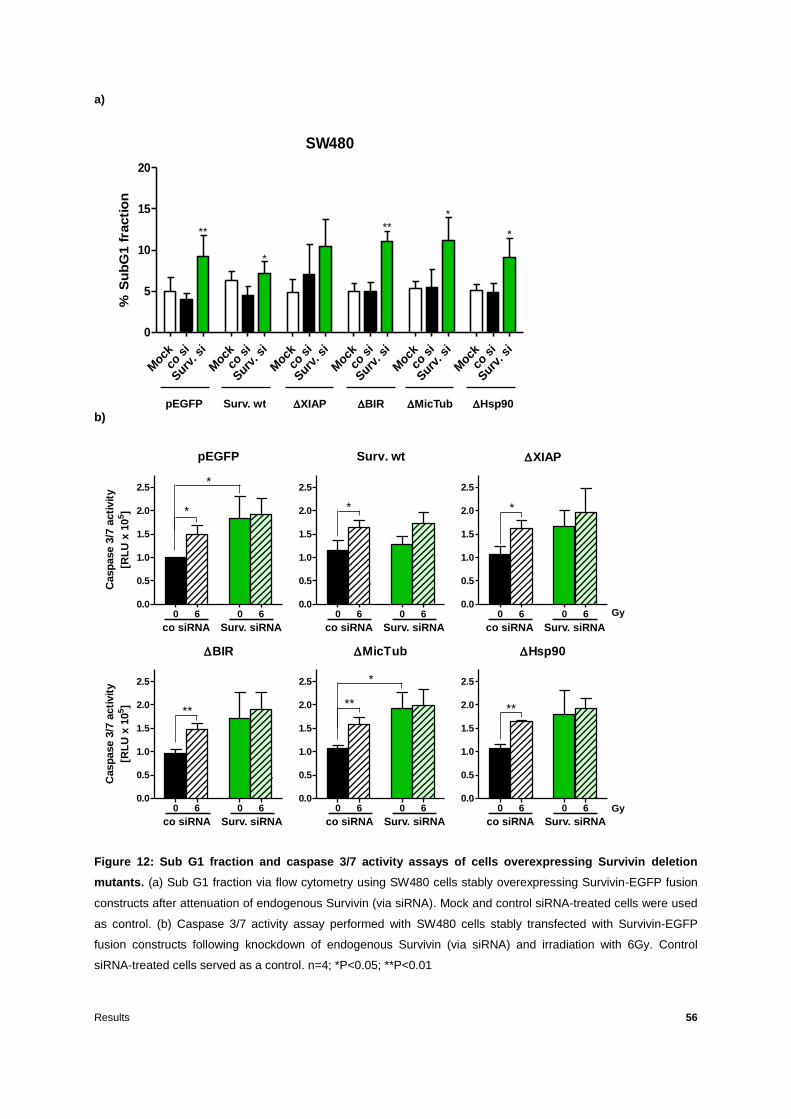
Results 56
a)
b)
Figure 12: Sub G1 fraction and caspase 3/7 activity assays of cells overexpressing Survivin deletion
mutants. (a) Sub G1 fraction via flow cytometry using SW480 cells stably overexpressing Survivin-EGFP fusion
constructs after attenuation of endogenous Survivin (via siRNA). Mock and control siRNA-treated cells were used
as control. (b) Caspase 3/7 activity assay performed with SW480 cells stably transfected with Survivin-EGFP
fusion constructs following knockdown of endogenous Survivin (via siRNA) and irradiation with 6Gy. Control
siRNA-treated cells served as a control. n=4; *P<0.05; **P<0.01
SW480
% S
ub
G1 f
racti
on
Moc
k
co s
i
Sur
v. s
i
Moc
k
co s
i
Sur
v. s
i
Moc
k
co s
i
Sur
v. s
i
Moc
k
co s
i
Sur
v. s
i
Moc
k
co s
i
Sur
v. s
i
Moc
k
co s
i
Sur
v. s
i
0
5
10
15
20
pEGFP Surv. wt XIAP BIR MicTub Hsp90
** ***
*
*
pEGFP
Casp
ase 3
/7 a
cti
vit
y
[RL
U x
10
5]
0 6 0 60.0
0.5
1.0
1.5
2.0
2.5
BIR
Casp
ase 3
/7 a
cti
vit
y
[RL
U x
10
5]
0 6 0 60.0
0.5
1.0
1.5
2.0
2.5
MicTub
0 6 0 60.0
0.5
1.0
1.5
2.0
2.5
Hsp90
0 6 0 60.0
0.5
1.0
1.5
2.0
2.5
Surv. wt
0 6 0 60.0
0.5
1.0
1.5
2.0
2.5
*
*
**
co siRNA Surv. siRNA co siRNA Surv. siRNA
XIAP
0 6 0 60.0
0.5
1.0
1.5
2.0
2.5
co siRNA Surv. siRNA
co siRNA Surv. siRNA co siRNA Surv. siRNA co siRNA Surv. siRNA
Gy
Gy
*
**
*
* *
**

Results 57
Survivin promotes tumour cell survival in part by inhibiting caspases as it has been shown by
previous studies where colorectal cancer cells following treatment with siRNA targeted
against Survivin responded with an increased apoptosis and caspase 3/7 activity [59, 127]. In
the present study, an elevated caspase 3/7 activity was observed upon knockdown of
endogenous Survivin in the deletion mutant transfected cells in comparison with control
siRNA treated cells (Figure 12b). By contrast, knockdown of endogenous Survivin did not
alter G2/M phase (Figure 11), SubG1 fraction or caspase 3/7 activity in Surv. wt
overexpressing cells (Figure 12). Additionally, knockdown of endogenous Survivin in
combination with 6 Gy irradiation did not further increase caspase 3/7 activity as compared to
control siRNA treated cells (Figure 12b).
According to these results, the BIR domain as well as the XIAP, mictrotubules and Hsp90
binding sites of Survivin are important for both cell cycle regulation and apoptosis inhibition.
3.1.2 Survivin BIR domain and XIAP binding site are essential for three-dimensional
radiation clonogenic survival
Previous studies conducted using colorectal carcinoma cells showed a decreased clonogenic
survival following knockdown of Survivin in combination with ionizing radiation [125, 127].
Plating efficiency of non-irradiated 3D cultured cells was significantly diminished after
Survivin knockdown in EGFP control, Survivin XIAP, BIR, MicTub and Hsp90
expressing cells, while recombinant overexpression of Surv. wt rescued the 3D plating
efficiency (Figure 13a). Overexpression of Survivin wt as well as MicTub and Hsp90
deletion mutants rescued 3D clonogenic radiation survival while deletion of the Survivin BIR
domain or XIAP binding site resulted in a significant radiosensitization upon knockdown of
endogenous Survivin (Figure 13b). A summary of radiation response variables is listed in
Table 5 that can be found in the appendix.
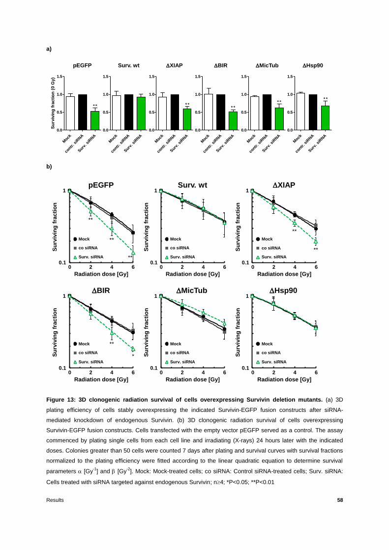
Results 58
a)
b)
Figure 13: 3D clonogenic radiation survival of cells overexpressing Survivin deletion mutants. (a) 3D
plating efficiency of cells stably overexpressing the indicated Survivin-EGFP fusion constructs after siRNA-
mediated knockdown of endogenous Survivin. (b) 3D clonogenic radiation survival of cells overexpressing
Survivin-EGFP fusion constructs. Cells transfected with the empty vector pEGFP served as a control. The assay
commenced by plating single cells from each cell line and irradiating (X-rays) 24 hours later with the indicated
doses. Colonies greater than 50 cells were counted 7 days after plating and survival curves with survival fractions
normalized to the plating efficiency were fitted according to the linear quadratic equation to determine survival
parameters [Gy-1
] and [Gy-2
]. Mock: Mock-treated cells; co siRNA: Control siRNA-treated cells; Surv. siRNA:
Cells treated with siRNA targeted against endogenous Survivin; n4; *P<0.05; **P<0.01
Su
rviv
ing
fra
cti
on
(0 G
y)
Mock
contr
. siR
NA
Surv
. siR
NA
0.0
0.5
1.0
1.5
Mock
contr
. siR
NA
Surv
. siR
NA
0.0
0.5
1.0
1.5
Mock
contr
. siR
NA
Surv
. siR
NA
0.0
0.5
1.0
1.5
Mock
contr
. siR
NA
Surv
. siR
NA
0.0
0.5
1.0
1.5
Mock
contr
. siR
NA
Surv
. siR
NA
0.0
0.5
1.0
1.5
Mock
contr
. siR
NA
Surv
. siR
NA
0.0
0.5
1.0
1.5
pEGFP Surv. wt XIAP BIR MicTub Hsp90
** ****
****
0,1000
1,0000
0 2 4 6
0,1000
1,0000
0 2 4 6
0,1000
1,0000
0 2 4 6
0,1000
1,0000
0 2 4 6
0,1000
1,0000
0 2 4 6
0,1000
1,0000
0 2 4 6
pEGFP
Su
rviv
ing
frac
tio
n
Radiation dose [Gy]
0 2 4 6
1
0.1
Surv. wt
Su
rviv
ing
frac
tio
n
Radiation dose [Gy]
0 2 4 6
1
0.1
XIAP
Su
rviv
ing
frac
tio
n
Radiation dose [Gy]
0 2 4 6
1
0.1
BIR
Su
rviv
ing
frac
tio
n
Radiation dose [Gy]
0 2 4 6
1
0.1
MicTub
Su
rviv
ing
frac
tio
n
Radiation dose [Gy]
0 2 4 6
1
0.1
Hsp90
Su
rviv
ing
frac
tio
n
Radiation dose [Gy]
0 2 4 6
1
0.1
Mock
co siRNA
Surv. siRNA
Mock
co siRNA
Surv. siRNA
Mock
co siRNA
Surv. siRNA
Mock
co siRNA
Surv. siRNA
Mock
co siRNA
Surv. siRNA
Mock
co siRNA
Surv. siRNA
**
**
**
**
**
*
0,1000
1,0000
0 2 4 6
0,1000
1,0000
0 2 4 6
0,1000
1,0000
0 2 4 6
0,1000
1,0000
0 2 4 6
0,1000
1,0000
0 2 4 6
0,1000
1,0000
0 2 4 6
**

Results 59
According to these results, the XIAP binding site of Survivin seems to be indispensable for
three-dimensional radiation clonogenic survival of SW480 colorectal cancer cells.
In order to confirm these results and ensure that the observed effect was not due to any
specific behaviour of the specific SW480 clones used in these experiments, the 3D
clonogenic survival assays were repeated using a set of alternative SW480 clones stably
transfected with the pEGFP control vector, recombinant Survivin wild type and Survivin
XIAP. The radiosensitization effect which was observed in the first place in the case of the
pEGFP vector control and Survivin XIAP (Figure 13b) was similar with that of the alternative
pEGFP #2, Surv. XIAP #2 while the alternative Surv. wt #2 once again rescued the
radiation clonogenic survival following knockdown of the endogenous Survivin (Figure 14).
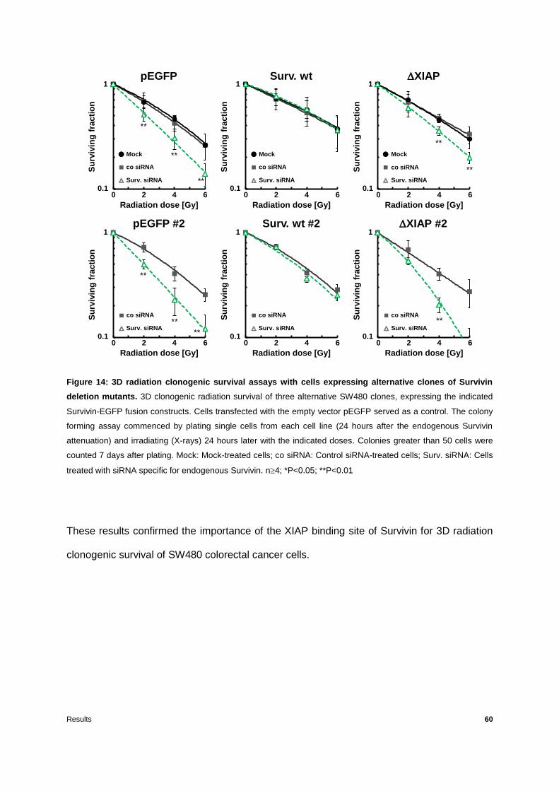
Results 60
Figure 14: 3D radiation clonogenic survival assays with cells expressing alternative clones of Survivin
deletion mutants. 3D clonogenic radiation survival of three alternative SW480 clones, expressing the indicated
Survivin-EGFP fusion constructs. Cells transfected with the empty vector pEGFP served as a control. The colony
forming assay commenced by plating single cells from each cell line (24 hours after the endogenous Survivin
attenuation) and irradiating (X-rays) 24 hours later with the indicated doses. Colonies greater than 50 cells were
counted 7 days after plating. Mock: Mock-treated cells; co siRNA: Control siRNA-treated cells; Surv. siRNA: Cells
treated with siRNA specific for endogenous Survivin. n4; *P<0.05; **P<0.01
These results confirmed the importance of the XIAP binding site of Survivin for 3D radiation
clonogenic survival of SW480 colorectal cancer cells.
0,1000
1,0000
0 2 4 6
0,1000
1,0000
0 2 4 6
Su
rviv
ing
fra
cti
on
Radiation dose [Gy]
0 2 4 6
1
0.1
co siRNA
Surv. siRNA
**
**
**
0,1000
1,0000
0 2 4 6
pEGFP
**
**
Su
rviv
ing
fra
cti
on
Radiation dose [Gy]
0 2 4 6
1
0.1
Mock
co siRNA
Surv. siRNA
**
0,1000
1,0000
0 2 4 6
0,1000
1,0000
0 2 4 6
Surv. wt
Su
rviv
ing
fra
cti
on
Radiation dose [Gy]
0 2 4 6
1
0.1
XIAP
Su
rviv
ing
fra
cti
on
Radiation dose [Gy]
0 2 4 6
1
0.1
Mock
co siRNA
Surv. siRNA
Mock
co siRNA
Surv. siRNA
**
**
pEGFP #2 Surv. wt #2S
urv
ivin
gfr
ac
tio
n
Radiation dose [Gy]
0 2 4 6
1
0.1
co siRNA
Surv. siRNA
0,1000
1,0000
0 2 4 6
Su
rviv
ing
fra
cti
on
Radiation dose [Gy]
0 2 4 6
1
0.1
co siRNA
Surv. siRNA**
XIAP #2
Results 61
3.1.3 Survivin BIR domain and XIAP binding site are essential for regulation of DNA
double-strand break repair
Previous work showed that siRNA-mediated knockdown of Survivin in colorectal cancer as
well as glioblastoma cell lines resulted in increased levels of residual DNA double-strand
breaks [98, 99]. Since the molecular basis for such an effect requires further investigation,
the impact of Survivin deletion mutants on DNA DSB repair was analysed via 2D and 3D
immunofluorescence staining of H2AX and 53BP1 foci of SW480 cells stably transfected
with Survivin-EGFP deletion constructs following siRNA-mediated knockdown of endogenous
Survivin and irradiation with 2 Gy.
Residual DNA damage in both 2D and 3D cell cultures was significantly increased upon
knockdown of endogenous Survivin in empty vector EGFP, Survivin XIAP and BIR
expressing cells, as evaluated by counting H2AX and 53BP1 foci 24 h after irradiation with 2
Gy (Figure 15). On the other hand, overexpression of Survivin wt, MicTub and Hsp90
constructs rescued DNA damage repair after knockdown of endogenous Survivin in both 2D
and 3D IF assays (Figure 15).
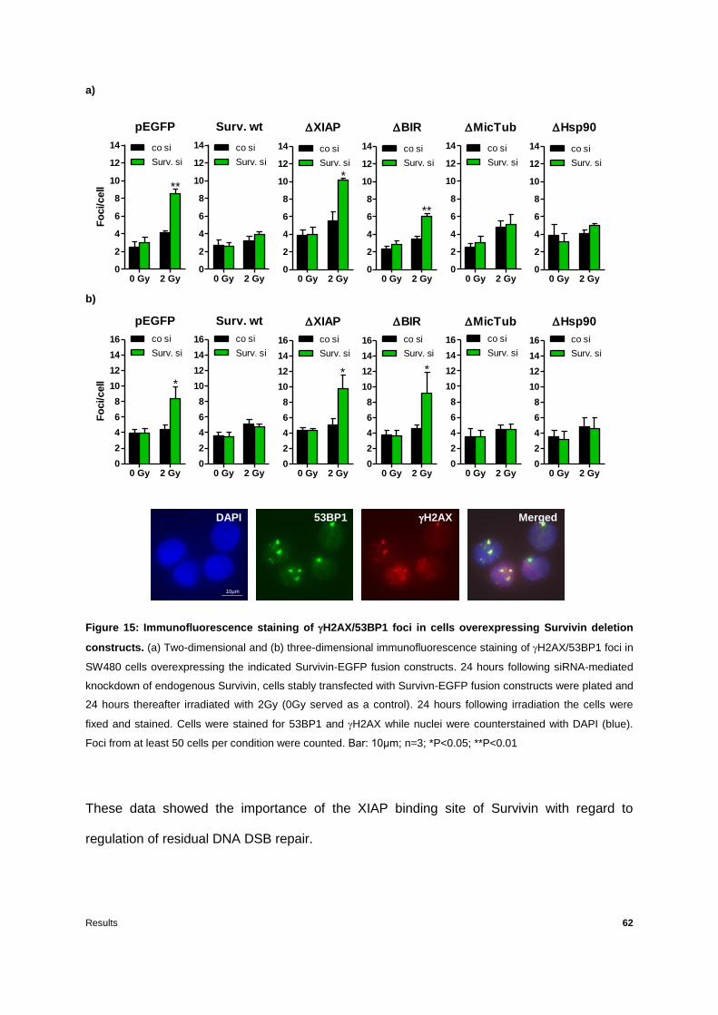
Results 62
a)
b)
Figure 15: Immunofluorescence staining of H2AX/53BP1 foci in cells overexpressing Survivin deletion
constructs. (a) Two-dimensional and (b) three-dimensional immunofluorescence staining of H2AX/53BP1 foci in
SW480 cells overexpressing the indicated Survivin-EGFP fusion constructs. 24 hours following siRNA-mediated
knockdown of endogenous Survivin, cells stably transfected with Survivn-EGFP fusion constructs were plated and
24 hours thereafter irradiated with 2Gy (0Gy served as a control). 24 hours following irradiation the cells were
fixed and stained. Cells were stained for 53BP1 and H2AX while nuclei were counterstained with DAPI (blue).
Foci from at least 50 cells per condition were counted. Bar: 10µm; n=3; *P<0.05; **P<0.01
These data showed the importance of the XIAP binding site of Survivin with regard to
regulation of residual DNA DSB repair.
Hsp90
0 Gy 2 Gy0
2
4
6
8
10
12
14 co si
Surv. si
pEGFP
Fo
ci/cell
0 Gy 2 Gy0
2
4
6
8
10
12
14 co si
Surv. si
Surv. wt
0 Gy 2 Gy0
2
4
6
8
10
12
14 co si
Surv. si
XIAP
0 Gy 2 Gy0
2
4
6
8
10
12
14 co si
Surv. si
BIR
0 Gy 2 Gy0
2
4
6
8
10
12
14 co si
Surv. si
MicTub
0 Gy 2 Gy0
2
4
6
8
10
12
14 co si
Surv. si
***
**
pEGFP
Fo
ci/cell
0 Gy 2 Gy0
2
4
6
8
10
12
14
16 co si
Surv. si
Surv. wt
0 Gy 2 Gy0
2
4
6
8
10
12
14
16 co si
Surv. si
XIAP
0 Gy 2 Gy0
2
4
6
8
10
12
14
16 co si
Surv. si
BIR
0 Gy 2 Gy0
2
4
6
8
10
12
14
16 co si
Surv. si
MicTub
0 Gy 2 Gy0
2
4
6
8
10
12
14
16 co si
Surv. si
Hsp90
0 Gy 2 Gy0
2
4
6
8
10
12
14
16 co si
Surv. si
DAPI 53BP1 H2AX Merged
10µm
* *
*

Results 63
To confirm our results, we tested alternative SW480 clones, stably transfected with pEGFP
empty vector (for control), Survivin wild type and Suvivin XIAP binding site deletion construct
indicated as clones #2. As a result, cells overexpressing the alternative Surv. XIAP #2
along with the ones bearing the alternative empty vector pEGFP #2 showed the same
significant increase of residual DNA DSBs following attenuation of endogenous Survivin and
irradiation with 2Gy, whereas the Surv. wt #2 fully rescued the DNA repair (Figure 16).
Figure 16: 3D Immunofluorescence staining of H2AX/53BP1 foci in cells overexpressing alternative
clones of Survivin deletion constructs. 24 hours following siRNA-mediated knockdown of endogenous Survivin
cells stably transfected with the indicated Survivn-EGFP fusion constructs were plated and 24 hours thereafter
irradiated with 2Gy (0Gy served as a control). 24 hours following irradiation the cells were fixed and stained. Cells
were stained for H2AX and 53BP1 while nuclei were counterstained with DAPI (blue). Foci from at least 50 cells
per condition were counted. n=3; *P<0.05; **P<0.01
In accordance with these results, the XIAP binding site of Survivin is confirmed to be indeed
essential for the regulation of DNA DSBs repair.
pEGFP
Fo
ci/cell
0 Gy 2 Gy0
2
4
6
8
10
12
14
16 co si
Surv. si
Surv. wt
0 Gy 2 Gy0
2
4
6
8
10
12
14
16 co si
Surv. si
XIAP
0 Gy 2 Gy0
2
4
6
8
10
12
14
16 co si
Surv. si
**
pEGFP #2
0 Gy2 Gy0
2
4
6
8
10
12
14
16co si
Surv. si
Surv. wt #2
0 Gy 2 Gy0
2
4
6
8
10
12
14
16 co si
Surv. si
XIAP #2
0 Gy 2 Gy0
2
4
6
8
10
12
14
16 co si
Surv. si
****

Results 64
3.1.4 The XIAP binding site of Survivin is essential for interaction with DNA-PKcs
It has recently been shown that Survivin participates in DNA repair as a member of the non-
homologous end joining (NHEJ) DNA repair machinery co-operating with factors such as
DNA-PKcs [98, 99]. Thus, after having observed the importance of the XIAP binding site of
Survivin in DNA DSB repair (Figures 15-16) the Survivin XIAP mutant was chosen for
further analysis of the interaction between recombinant Survivin and DNA-PKcs via
immunoprecipitation assays that were performed using SW480 cells stably overexpressing
the pEGFP empty vector (for control), Survivin-EGFP wild type and Surv. XIAP mutant
construct.
Figure 17: Immunoprecipitation experiments performed with cells overexpressing wild type Survivin and
its XIAP deletion mutant. Immunoprecipitation (IP) of DNA-PKcs on the left and IP of GFP on the right
performed with nuclear lysates of SW480 cells stably stransfected with the pEGFP empty vector serving as a
control, Survivin wild type and Surv. XIAP deletion mutant after irradiation with 4Gy. Co-immunoprecipitated
proteins were then detected via western blotting. Non-specific isotype antibody (immunoglobulin G or IgG) served
as an additional control to the EGFP empty vector IP. Recombinant Survivin was detected using an anti-GFP
antibody (due to pEGFP fusion) while DNA-PKcs was detected using an anti-DNA-PKcs antibody. Representative
blot of three independent experiments (n=3).
DNA-PKcs
Surv. wt-EGFP
Antibody
DNA PKcs GFP
IgG
IP
XIAP-EGFP
Antibody
EGFP

Results 65
DNA-PKcs was found to precipitate with recombinant Surv. wt (and vice versa) but not with
the XIAP deletion mutant indicating a possibly direct involvement of the XIAP binding site of
Survivin during interaction with DNA-PKcs (Figure 17). Such an interaction can explain the
significantly increased radiation-induced DNA DSBs and the clonogenic radiosensitization
observed in the case of XIAP mutation as opposed to Survivin wt.
3.2 The impact of Survivin phosphorylation on cellular radiation response
In order to further analyse the impact of post-translational modifications of Survivin on
cellular radiation response, several phosphorylation mutants were generated involving Serine
20 (S20), Threonine 34 (T34) and Threonine 117 (T117) phosphorylation sites of the protein.
In particular, those three sites were mutated to Alanine (A) to constitute a non-
phosphorylatable form of the protein and Aspartic acid (D) for a phosphomimetic (Figure 18).
All mutations were confirmed twice, once prior to stable transfections (via sequencing of DNA
from plasmid mini preparations) and once after the stable transfections of SW480 cells via
sequencing of isolated genomic DNA from the SW480 clones.
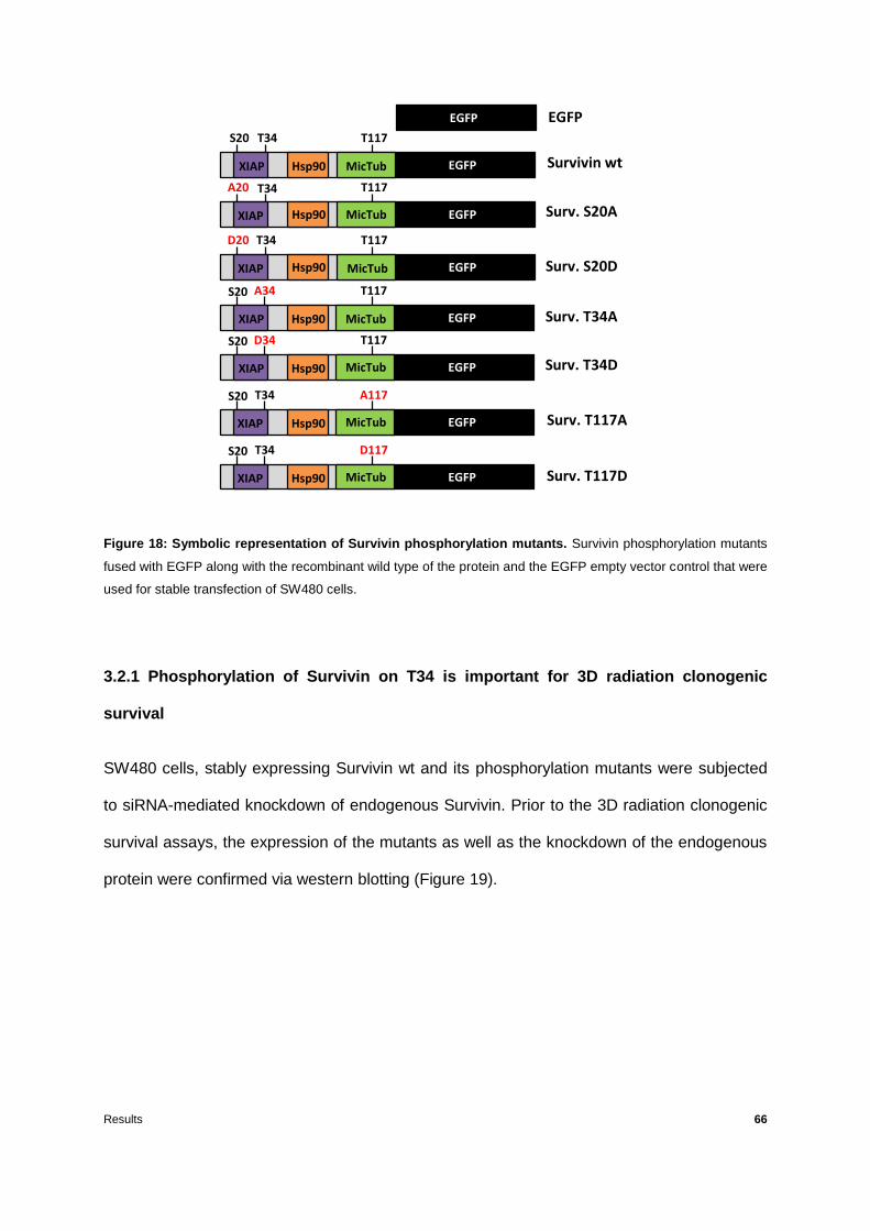
Results 66
Figure 18: Symbolic representation of Survivin phosphorylation mutants. Survivin phosphorylation mutants
fused with EGFP along with the recombinant wild type of the protein and the EGFP empty vector control that were
used for stable transfection of SW480 cells.
3.2.1 Phosphorylation of Survivin on T34 is important for 3D radiation clonogenic
survival
SW480 cells, stably expressing Survivin wt and its phosphorylation mutants were subjected
to siRNA-mediated knockdown of endogenous Survivin. Prior to the 3D radiation clonogenic
survival assays, the expression of the mutants as well as the knockdown of the endogenous
protein were confirmed via western blotting (Figure 19).
EGFPXIAP MicTubHsp90
S20 T34 T117
EGFP
Survivin wt
T34
EGFPXIAP MicTubHsp90
A20 T117
EGFPXIAP MicTubHsp90
D20 T34 T117
EGFPXIAP MicTubHsp90
S20 A34 T117
EGFPXIAP MicTubHsp90
S20 D34 T117
EGFPXIAP MicTubHsp90
S20 T34 A117
EGFPXIAP MicTubHsp90
S20 T34 D117
Surv. S20A
Surv. S20D
Surv. T34A
Surv. T34D
Surv. T117A
Surv. T117D
EGFP

Results 67
Figure 19: Western blot conducted with cells overexpressing Survivin phosphorylation mutants. Western
blot confirmation of recombinant Survivin expression (Survivin-EGFP fusion constructs stably expressed in
SW480 cells) along with confirmation of the siRNA-mediated knockdown of endogenous Survivin. Anti-Survivin
and anti-GFP antibodies were used to detect endogenous and recombinant protein expression while β-actin
served as a loading control. Co: Control siRNA-treated cells; surv: Cells treated with siRNA targeted against
endogenous Survivin; representative blot of three independent experiments (n=3).
Plating efficiency of non-irradiated 3D cultured cells was significantly compromised after
attenuation of endogenous Survivin in EGFP control, Survivin S20A, S20D, T34A, T34D,
T117A and T117D expressing cells, while recombinant overexpression of Surv. wt rescued
the 3D plating efficiency (Figure 20a). On the contrary, overexpression of Surv. wt as well as
S20D, T34D, T117A and T117D phosphorylation mutants rescued 3D clonogenic radiation
survival while the non-phosphorylatable form of Survivin S20A and especially the T34A
resulted in a radiosensitization effect even prior to the knockdown of endogenous Survivin,
thus behaving in a dominant negative manner (Figure 20b).
pEGFP
rec. Surv.-EGFP
endog. Surv.
co Surv co Surv co Surv co Surv co Surv co Surv siRNASurv. wt S20A S20D T34A T34D
β-actin
T117A
co Surv co Surv
T117D
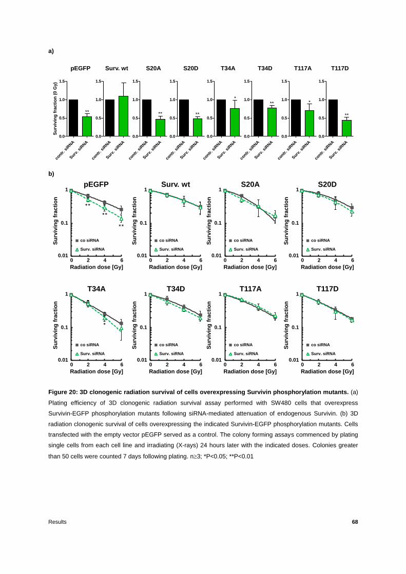
Results 68
a)
b)
Figure 20: 3D clonogenic radiation survival of cells overexpressing Survivin phosphorylation mutants. (a)
Plating efficiency of 3D clonogenic radiation survival assay performed with SW480 cells that overexpress
Survivin-EGFP phosphorylation mutants following siRNA-mediated attenuation of endogenous Survivin. (b) 3D
radiation clonogenic survival of cells overexpressing the indicated Survivin-EGFP phosphorylation mutants. Cells
transfected with the empty vector pEGFP served as a control. The colony forming assays commenced by plating
single cells from each cell line and irradiating (X-rays) 24 hours later with the indicated doses. Colonies greater
than 50 cells were counted 7 days following plating. n3; *P<0.05; **P<0.01
contr
. siR
NA
Surv
. siR
NA
0.0
0.5
1.0
1.5
Su
rviv
ing
fra
cti
on
(0
Gy)
contr
. siR
NA
Surv
. siR
NA
0.0
0.5
1.0
1.5
contr
. siR
NA
Surv
. siR
NA
0.0
0.5
1.0
1.5
contr
. siR
NA
Surv
. siR
NA
0.0
0.5
1.0
1.5
contr
. siR
NA
Surv
. siR
NA
0.0
0.5
1.0
1.5
contr
. siR
NA
Surv
. siR
NA
0.0
0.5
1.0
1.5
pEGFP Surv. wt S20A S20D T34A T34D
****
T117A T117D
contr
. siR
NA
Surv
. siR
NA
0.0
0.5
1.0
1.5
contr
. siR
NA
Surv
. siR
NA
0.0
0.5
1.0
1.5
****
** **
0,0100
0,1000
1,0000
0 2 4 6
co siRNA
Surv. siRNA
0,1000
1,0000
0 2 4 6
Su
rviv
ing
fra
cti
on
Radiation dose [Gy]
0 2 4 6
1
0.01
0.1
0,0100
0,1000
1,0000
0 2 4 6
co siRNA
Surv. siRNA
0,1000
1,0000
0 2 4 6
Su
rviv
ing
fra
cti
on
Radiation dose [Gy]
0 2 4 6
1
0.01
0.1
0,0100
0,1000
1,0000
0 2 4 6
co siRNA
Surv. siRNA
0,1000
1,0000
0 2 4 6
Su
rviv
ing
fra
cti
on
Radiation dose [Gy]
0 2 4 6
1
0.01
0.1
0,0100
0,1000
1,0000
0 2 4 6
co siRNA
Surv. siRNA
0,1000
1,0000
0 2 4 6
Su
rviv
ing
fra
cti
on
Radiation dose [Gy]
0 2 4 6
1
0.01
0.1
0,0100
0,1000
1,0000
0 2 4 6
co siRNA
Surv. siRNA
0,1000
1,0000
0 2 4 6S
urv
ivin
gfr
ac
tio
n
Radiation dose [Gy]
0 2 4 6
1
0.01
0.1
0,0100
0,1000
1,0000
0 2 4 6
co siRNA
Surv. siRNA
0,1000
1,0000
0 2 4 6
Su
rviv
ing
fra
cti
on
Radiation dose [Gy]
0 2 4 6
1
0.01
0.1
0,0100
0,1000
1,0000
0 2 4 6
co siRNA
Surv. siRNA
0,1000
1,0000
0 2 4 6
Su
rviv
ing
fra
cti
on
Radiation dose [Gy]
0 2 4 6
1
0.01
0.1
0,0100
0,1000
1,0000
0 2 4 6
co siRNA
Surv. siRNA
0,1000
1,0000
0 2 4 6
Su
rviv
ing
fra
cti
on
Radiation dose [Gy]
0 2 4 6
1
0.01
0.1
**
**
**
*
pEGFP Surv. wt S20A S20D
T34A T34D T117A T117D

Results 69
A summary of radiation response variables is listed in Table 6 that can be found in the
appendix.
Here it is shown that phosphorylation of Survivin on S20 and T34 is essential for clonogenic
radiation survival (Figure 20b). Both phosphorylation sites are located within the XIAP
binding site of Survivin (Figure 18). However, due to the poor viability of the S20A mutation,
the T34A mutant was chosen for further analysis as well as comparison with the XIAP
construct. Thus, prior to the direct comparison of those two mutations (XIAP and T34A) the
behaviour of the T34A mutant in 3D radiation clonogenic survival assays was confirmed
using alternative SW480 clones stably overexpressing phospho-mutant constructs (Figure
21) in additional clonogenic survival assays. The alternative clone expressing the T34A
mutation named T34A #2 revealed comparable radiosensitization as shown for the first clone
(T34A), while the phosphomimetic Survivin T34D clone #2 (T34D #2) dispayed similar
radioresistance as the first clone (T34D) (Figure 21).
Figure 21: 3D clonogenic radiation survival of cells overexpressing alternative clones of Survivin
phospho-mutants. 3D clonogenic radiation survival assays conducted with SW480 cells stably expressing the
non-phosphorylatable form of Survivin T34A and the phosphomimetic T34D mutants along with the respective
alternative clones (marked with #2) following siRNA-mediated knockdown of the endogenous Survivin. The assay
commenced by plating single cells from each cell line and irradiating (X-rays) with the indicated doses. Colonies
greater than 50 cells were counted 7 days after plating. n3; *P<0.05; **P<0.01
0,0100
0,1000
1,0000
0 2 4 6
co siRNA
Surv. siRNA
0,1000
1,0000
0 2 4 6
Su
rviv
ing
fra
cti
on
Radiation dose [Gy]
0 2 4 6
1
0.01
0.1
0,0100
0,1000
1,0000
0 2 4 6
co siRNA
Surv. siRNA
0,1000
1,0000
0 2 4 6
Su
rviv
ing
fra
cti
on
Radiation dose [Gy]
0 2 4 6
1
0.01
0.1
0,0100
0,1000
1,0000
0 2 4 6
co siRNA
Surv. siRNA
0,1000
1,0000
0 2 4 6
Su
rviv
ing
fra
cti
on
Radiation dose [Gy]
0 2 4 6
1
0.01
0.1
0,0100
0,1000
1,0000
0 2 4 6
co siRNA
Surv. siRNA
0,1000
1,0000
0 2 4 6
Su
rviv
ing
fra
cti
on
Radiation dose [Gy]
0 2 4 6
1
0.01
0.1 *
T34A T34D T34A #2 T34D #2

Results 70
To compare the impact of Survivin T34 phosporylation with the XIAP binding site with regard
to radiation survival, SW480 cells stably transfected wtth Surv. wt, Surv. XIAP, Surv. T34A
and Surv. T34D were subjected to siRNA-mediated knockdown of endogenous Survivin.
Both the expression of the recombinant Survivin and the knockdown of the endogenous
protein were confirmed via western blotting (Figure 22).
Figure 22: Western blotting performed with cells overexpressing Survivin XIAP deletion mutant and
Survivin T34A or T34D phospho-mutant. Western blot confirmation of the expression of recombinant Survivin
along with confirmation of the siRNA-mediated knockdown of endogenous Survivin. Anti-Survivin and anti-GFP
antibodies were used to detect endogenous and recombinant protein expression while β-actin served as a loading
control. Co: Control siRNA-treated cells; Surv: Cells treated with siRNA targeted against endogenous Survivin.
Representative blot of four independent experiments (n=4).
Plating efficiency of non-irradiated 3D cultured cells was significantly diminished after
Survivin knockdown in EGFP control, Survivin XIAP, T34A, and T34D expressing cells
while overexpression of Survivin wt rescued the 3D plating efficiency (Figure 23a). However,
overexpression of Survivin wt as well as T34D phospho-mutants rescued 3D clonogenic
radiation survival while deletion of Survivin XIAP binding site or mutation of Threonine 34 to
Alanine resulted in a radiosensitization upon knockdown of endogenous Survivin in the case
of the XIAP and even prior to the knockdown in the case of T34A mutation (Figure 23b).
pEGFP
rec. Surv.-EGFP
endog. Surv.
co Surv co Surv co Surv co Surv co Surv siRNA
Surv. wt XIAP T34A T34D
β-actin

Results 71
a)
b)
Figure 23: 3D clonogenic radiation survival of cells overexpressing the indicated Survivin mutants. (a) 3D
plating efficiency of cells stably overexpressing Survivin-EGFP fusion constructs namely Surv. wt, Surv. XIAP,
Surv. T34A and Surv. T34D along with the empty vector pEGFP for control. (b) 3D clonogenic radiation survival of
cells overexpressing Survivin-EGFP fusion constructs. The assay commenced by plating single cells from each
cell line and irradiating (X-rays) 24 hours later with the indicated doses. Colonies greater than 50 cells were
counted 7 days after plating. n4; *P<0.05; **P<0.01
contr
. siR
NA
Surv
. siR
NA
0.0
0.5
1.0
1.5
Su
rviv
ing
fra
cti
on
(0
Gy)
contr
. siR
NA
Surv
. siR
NA
0.0
0.5
1.0
1.5
contr
. siR
NA
Surv
. siR
NA
0.0
0.5
1.0
1.5
contr
. siR
NA
Surv
. siR
NA
0.0
0.5
1.0
1.5
contr
. siR
NA
Surv
. siR
NA
0.0
0.5
1.0
1.5
pEGFP Surv. wt XIAP T34A T34D
****
***
0,0100
0,1000
1,0000
0 2 4 6
co siRNA
Surv. siRNA
0,1000
1,0000
0 2 4 6
Su
rviv
ing
frac
tio
n
Radiation dose [Gy]
0 2 4 6
1
0.01
0.1
0,0100
0,1000
1,0000
0 2 4 6
co siRNA
Surv. siRNA
0,1000
1,0000
0 2 4 6
Su
rviv
ing
frac
tio
n
Radiation dose [Gy]
0 2 4 6
1
0.01
0.1 *
T34A T34D
0,0100
0,1000
1,0000
0 2 4 6
co siRNA
Surv. siRNA
0,1000
1,0000
0 2 4 6
Su
rviv
ing
frac
tio
n
Radiation dose [Gy]
0 2 4 6
1
0.01
0.1
**
0,0100
0,1000
1,0000
0 2 4 6
co siRNA
Surv. siRNA
0,1000
1,0000
0 2 4 6
Su
rviv
ing
frac
tio
n
Radiation dose [Gy]
0 2 4 6
1
0.01
0.1
0,0100
0,1000
1,0000
0 2 4 6
co siRNA
Surv. siRNA
0,1000
1,0000
0 2 4 6
Su
rviv
ing
frac
tio
n
Radiation dose [Gy]
0 2 4 6
1
0.01
0.1
**
**
**
pEGFP Surv. wt
**
**
XIAP

Results 72
These results indicate the importance of the T34 phosporylation site of Survivin, while it is
not clear whether XIAP binding or binding of different proteins to the XIAP binding site is
fostered by T34 phosphorylation.
3.2.2 The XIAP binding site and the T34 phosphorylation site of Survivin are essential
for regulation of residual DNA DSBs repair
Following the observation of the impact of Survivin deletion mutant XIAP on DNA DSBs
repair (Figure 15) and the effect of the T34A mutant on 3D radiation clonogenic survival
(Figure 23b), the latter was chosen for further investigation regarding DNA damage repair
along with direct comparison with the XIAP mutant.
Residual DNA damage (as evaluated by counting H2AX and 53BP1 foci in
immunofluorescence staining assays) 24 h after irradiation with 2 Gy in 3D cell cultures was
significantly increased upon knockdown of endogenous Survivin in EGFP and Survivin
XIAP, while T34A expressing cells showed a similar increase of residual DNA damage even
prior to attenuation of endogenous Survivin (Figure 24a).

Results 73
a)
b)
Figure 24: 3D immunofluorescence staining of H2AX/53BP1 foci in cells overexpressing Survivin
deletion and phosphorylation mutants. (a) 3D IF of H2AX/53BP1 foci used as a marker for residual DNA
DSBs. 24 hours following siRNA-mediated knockdown of endogenous Survivin cells stably transfected with the
indincated Survivn-EGFP fusion constructs were plated and 24 hours thereafter irradiated with 2Gy (0Gy served
as a control). 24 hours following irradiation the cells were fixed and stained. Cells were stained for 53BP1 and
H2AX while nuclei were counterstained with DAPI (blue). Foci from at least 50 cells per condition were then
counted. (b) 3D immunofluorescence staining of alternative (#2) Survivin T34A and T34D clones for confirmation
of the previous results. n=3; *P<0.05; **P<0.01
However, overexpression of Survivin wt and T34D constructs rescued the DNA damage
repair after knockdown of endogenous Survivin (Figure 24a).
T34D
0 Gy 2 Gy0
2
4
6
8
10
12
14
16 co si
Surv. si
T34A
0 Gy 2 Gy0
2
4
6
8
10
12
14
16 co si
Surv. si
pEGFPF
oci/cell
0 Gy 2 Gy0
2
4
6
8
10
12
14
16 co si
Surv. si
Surv. wt
0 Gy 2 Gy0
2
4
6
8
10
12
14
16 co si
Surv. si
XIAP
0 Gy 2 Gy0
2
4
6
8
10
12
14
16 co si
Surv. si
**
****
T34D
0 Gy 2 Gy0
2
4
6
8
10
12
14
16 co si
Surv. si
T34A
Fo
ci/cell
0 Gy 2 Gy0
2
4
6
8
10
12
14
16 co si
Surv. si
T34A #2
0 Gy 2 Gy0
2
4
6
8
10
12
14
16 co si
Surv. si
T34D #2
0 Gy 2 Gy0
2
4
6
8
10
12
14
16 co si
Surv. si
********

Results 74
In order to confirm that these results were not clone-dependent, the 3D immunofluorescence
staining assays were repeated using alternative SW480 clones expressing Survivin T34A
and T34D phospho-mutants (Figure 24b). As a result, an alternative SW480 Survivin T34A
#2 clone showed similar significant increase of residual DNA DSBs following irradiation with
2Gy, whereas an alternative SW480 clone expressing the phospho-mimetic Survivin T34D
#2 fully rescued residual DNA DSBs repair (Figure 24b).
3.2.3 Mutation of the Survivin T34 phosphorylation site to its non-phosphorylatable
form T34A prevents interaction with DNA-PKcs
The XIAP binding site as well as the T34 phosphorylation site of Survivin were shown to be
implicated in the repair of residual DNA DSBs (Figure 24a). Thus, the Survivin phospho-
mutant T34A along with its phosphomimetic T34D were further analysed in terms of DNA-
PKcs interaction. In order to investigate such an interaction, SW480 cells stably
overexpressing those Survivin mutants were subjected to subcellular fractionation following
irradiation with 4 Gy and their nuclear fractions were used for GFP immunoprecipitation
assays (due to the Survivin-EGFP fusion) (Figure 25).
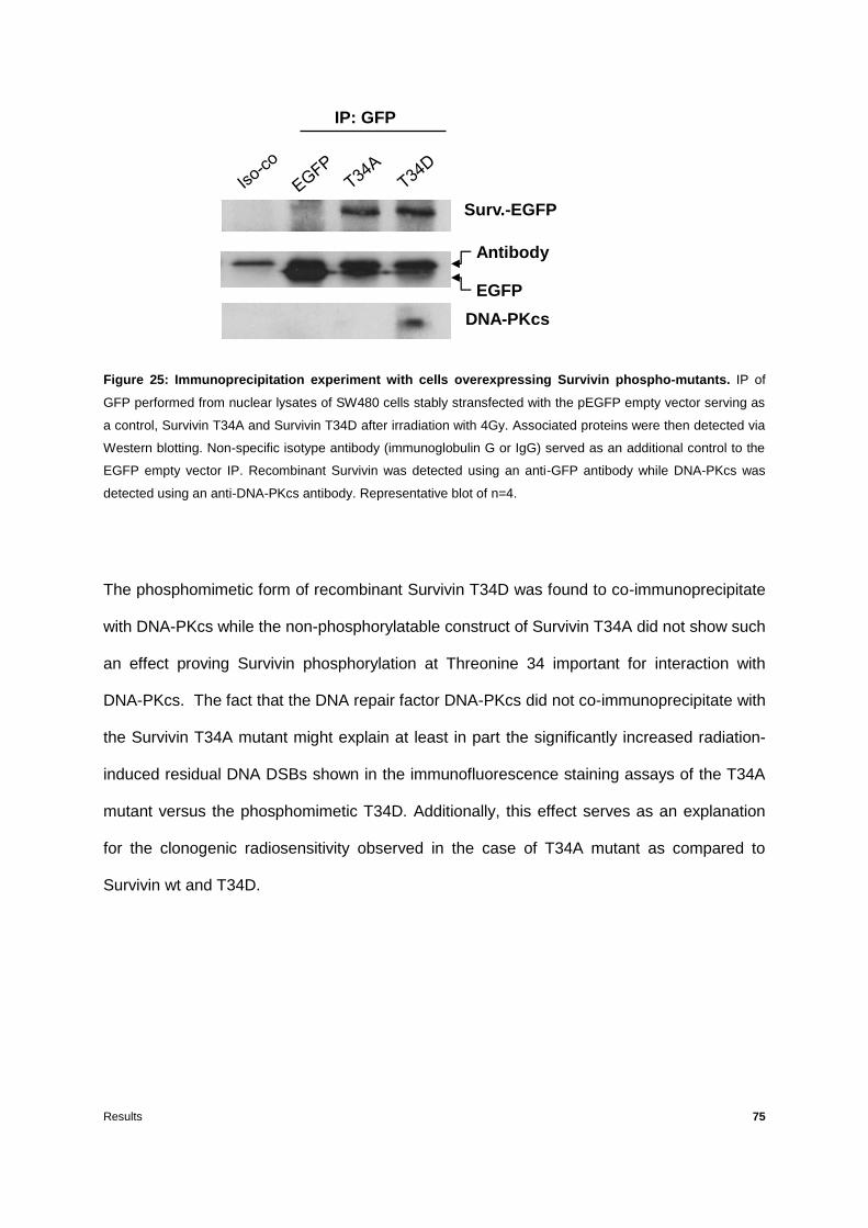
Results 75
Figure 25: Immunoprecipitation experiment with cells overexpressing Survivin phospho-mutants. IP of
GFP performed from nuclear lysates of SW480 cells stably stransfected with the pEGFP empty vector serving as
a control, Survivin T34A and Survivin T34D after irradiation with 4Gy. Associated proteins were then detected via
Western blotting. Non-specific isotype antibody (immunoglobulin G or IgG) served as an additional control to the
EGFP empty vector IP. Recombinant Survivin was detected using an anti-GFP antibody while DNA-PKcs was
detected using an anti-DNA-PKcs antibody. Representative blot of n=4.
The phosphomimetic form of recombinant Survivin T34D was found to co-immunoprecipitate
with DNA-PKcs while the non-phosphorylatable construct of Survivin T34A did not show such
an effect proving Survivin phosphorylation at Threonine 34 important for interaction with
DNA-PKcs. The fact that the DNA repair factor DNA-PKcs did not co-immunoprecipitate with
the Survivin T34A mutant might explain at least in part the significantly increased radiation-
induced residual DNA DSBs shown in the immunofluorescence staining assays of the T34A
mutant versus the phosphomimetic T34D. Additionally, this effect serves as an explanation
for the clonogenic radiosensitivity observed in the case of T34A mutant as compared to
Survivin wt and T34D.
Surv.-EGFP
DNA-PKcs
Antibody
EGFP
IP: GFP

Results 76
3.3 Survivin BIR domain, XIAP, Microtubules and Hsp90 binding sites are essential for
transmigration capability of colorectal cancer cells
It was recently reported that knockdown of Survivin and/or XIAP in many colorectal cancer
cell lines leads to a significantly diminished migration capacity [164] revealing the importance
of Survivin in colorectal cancer cell motility. In this work, attenuation of endogenous Survivin
decreased transmigration of EGFP control, and XIAP expressing cells compared to control
siRNA-treated cells while BIR, MicTub and Hsp90 constructs acted as dominant negative
mutants already significantly repressing transmigration of control cells (Figure 26).
Figure 26: Transmigration assay performed with cells overexpressing Survivin deletion mutants.
Transmigration assay performed with SW480 cells stably transfected with the indicated Survivin-EGFP fusion
constructs. The assay commenced by plating cells in inserts containing a 8 µm pore size membrane covered with
a thin layer of matrigel (a laminin-rich extracellular matrix) under serum-free conditions 24h after siRNA-mediated
knockdown of endogenous Survivin followed by addition of a chemo-attractant (serum-enriched medium). The
cells that went through the membrane of the inserts (transmigrated cells) were fixed and nuclei were
counterstained with DAPI. co siRNA: Control siRNA-treated cells; Surv. siRNA: Cells treated with an siRNA
targeted against endogenous Survivin. n=2; *P<0.05; **P<0.01
Rel. in
vasio
n
co s
iRNA
Surv. s
iRNA
co s
iRNA
Surv. s
iRNA
co s
iRNA
Surv. s
iRNA
co s
iRNA
Surv. s
iRNA
co s
iRNA
Surv. s
iRNA
co s
iRNA
Surv. s
iRNA
0.0
0.5
1.0
1.5
EGFP wt XIAP BIR Mic Hsp90
*
***
**
**

Results 77
Knockdown of endogenous Survivin did not further decrease the transmigration rate of BIR,
MicTub and Hsp90 stably transfected cells. It is hereby shown that not only the binding
site of XIAP, but also those of Microtubules and Hsp90 are involved in the transmigration
process suggesting further mechanisms by which Survivin regulates the motility of cancer
cells.
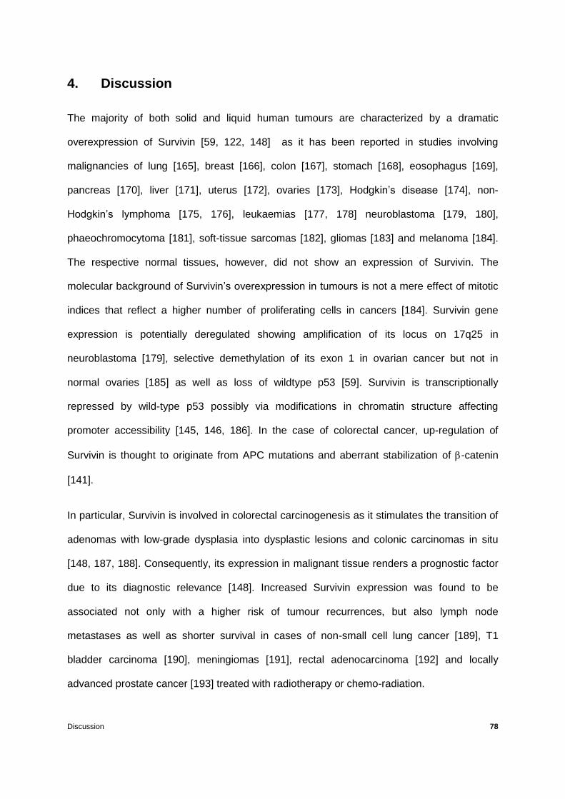
Discussion 78
4. Discussion
The majority of both solid and liquid human tumours are characterized by a dramatic
overexpression of Survivin [59, 122, 148] as it has been reported in studies involving
malignancies of lung [165], breast [166], colon [167], stomach [168], eosophagus [169],
pancreas [170], liver [171], uterus [172], ovaries [173], Hodgkin’s disease [174], non-
Hodgkin’s lymphoma [175, 176], leukaemias [177, 178] neuroblastoma [179, 180],
phaeochromocytoma [181], soft-tissue sarcomas [182], gliomas [183] and melanoma [184].
The respective normal tissues, however, did not show an expression of Survivin. The
molecular background of Survivin’s overexpression in tumours is not a mere effect of mitotic
indices that reflect a higher number of proliferating cells in cancers [184]. Survivin gene
expression is potentially deregulated showing amplification of its locus on 17q25 in
neuroblastoma [179], selective demethylation of its exon 1 in ovarian cancer but not in
normal ovaries [185] as well as loss of wildtype p53 [59]. Survivin is transcriptionally
repressed by wild-type p53 possibly via modifications in chromatin structure affecting
promoter accessibility [145, 146, 186]. In the case of colorectal cancer, up-regulation of
Survivin is thought to originate from APC mutations and aberrant stabilization of -catenin
[141].
In particular, Survivin is involved in colorectal carcinogenesis as it stimulates the transition of
adenomas with low-grade dysplasia into dysplastic lesions and colonic carcinomas in situ
[148, 187, 188]. Consequently, its expression in malignant tissue renders a prognostic factor
due to its diagnostic relevance [148]. Increased Survivin expression was found to be
associated not only with a higher risk of tumour recurrences, but also lymph node
metastases as well as shorter survival in cases of non-small cell lung cancer [189], T1
bladder carcinoma [190], meningiomas [191], rectal adenocarcinoma [192] and locally
advanced prostate cancer [193] treated with radiotherapy or chemo-radiation.

Discussion 79
In addition, there is substantial evidence proving Survivin to be a radiation resistance factor
[123-125] the attenuation of which leads to radiosensitization of tumour cells both in vitro and
in vivo [112, 148]. Studies conducted (in vitro) using glioblastoma, colorectal carcinoma,
hepatocellular carcinoma and NSCLC cells showed a decreased clonogenic survival
following knockdown of Survivin in combination with ionizing radiation as well as reduced
tumour growth in xenograft transplant models [112, 148].
Apart from participating in cell division and apoptosis, Survivin is a nodal tumour protein
involved in a multitude of signaling pathways and transcriptional networks [194]. One striking
feature of this protein is its pronounced ability to co-operate with a plethora of protein
partners such as other members of the IAP family (like XIAP) as well as components of the
DNA DSB repair machinery (such as DNA-PKcs). Thus, a further investigation of its
relationship with members of the IAP family as well as other protein partners (such as
Hsp90) and DNA repair proteins [98] is necessary in order to figure out the regulatory role of
Survivin in terms of tumour cell survival and modulation of treatment response.
The expression of Survivin is controlled at various levels via intracellular sequestration as
well as protein stability and transcription [148]. In non-malignant cells its expression is
transcriptionally regulated in a cell-cycle-dependent manner reaching its peak at mitosis
involving cell-cycle-dependent/cell-cycle gene homology region (CDE/CHR) elements found
in its promoter [102]. On the contrary, in tumour cells its expression is controlled regardless
of mitosis [195] by means of various oncogenic pathways [148]. It has been shown that an
siRNA-mediated knockdown of Survivin in colorectal cancer cells leads to an increased G2/M
fraction of the cell cycle distribution 24 hours following the knockdown, blocking the cells in a
more radiosensitive stage of the cell cycle [127, 163]. In the course of this project, after cell
cycle analysis experiments colorectal cancer cells stably transfected with Survivin lacking its
BIR domain, XIAP, Microtubules or Hsp90 binding sites were shown to accumulate in the

Discussion 80
G2/M phase after knockdown of the endogenous protein revealing an implication of these
particular binding sites of the protein in cell cycle regulation.
It has been demonstrated that colorectal cancer cells following treatment with siRNA targeted
against Survivin respond with increased apoptosis and caspase 3/7 activity supporting the
fact that Survivin promotes tumour cell survival by inhibiting caspases [59, 127]. With the
exception of XIAP, none of the IAPs is able to directly bind caspases [21]. It is thus believed
that Survivin can inhibit apoptosis by interacting with other protein partners, with the Survivin-
XIAP complex being a characteristic example of such an interaction during which the XIAP
binding site of Survivin associates with discontinuous sites in XIAP BIR1 and BIR3 [22, 31].
In this study, it was observed that colorectal cancer cells stably expressing Survivin deletion
mutants (lacking the BIR domain/the XIAP/Microtubules/Hsp90 binding site) showed an
incompetence to inhibit apoptosis following an siRNA-mediated knockdown of the
endogenous protein. Therefore, the BIR domain of the protein as well as the XIAP,
Microtubules and Hsp90 binding sites that were abolished due to the mutations, are
apparently involved in apoptosis inhibition.
The radiation clonogenic survival of colorectal cancer cells has been shown to decrease after
siRNA-mediated attenuation of Survivin [98, 99, 127, 164]. In the present investigation,
colorectal cancer cells transfected with recombinant Survivin lacking its BIR domain or its
XIAP binding site were significantly radiosensitized while transfection of cells with Survivin
MicTub or Hsp90 deletion mutants fully rescued radiation clonogenic survival following the
knockdown of the endogenous protein, demonstrating a radioresistance equal to that of cells
overexpressing wild-type recombinant Survivin. Furthermore, cells expressing the non-
phosphorylatable form of Survivin S20A (Serine 20 Alanine) or T34A (Threonine 34 Alanine),
both of which are located within the XIAP binding site, revealed a radiosensitizing effect even
prior to endogenous Survivin attenuation thus behaving like dominant negative mutants. On
the other hand, cells expressing the phosphomimetic version of Survivin S20D (Serine 20

Discussion 81
Aspartic acid) or T34D (Threonine 34 Aspartic acid) could rescue clonogenic survival.
However, cells transfected with Survivin phospho-mutant T117A or T117D, located on the
Microtubules binding site of the protein, did not show such a radiosensitizing effect.
Consequently, either the interaction between Survivin and XIAP or the XIAP binding site per
se and/or the S20 or T34 phosphorylation sites, located in that region are vital for radiation
clonogenic survival of colorectal cancer cells. These results are in line with previously
published data according to which a phosphomimetic mutant of Survivin namely T34E
(Threonine 34 Glutamic acid) showed a higher radioresistance than the corresponding
kinase-dead mutant T34A in human cervical carcinoma HeLa cells [187]. Additionally,
according to research conducted in our group, depletion of Survivin and/or XIAP resulted in
comparable effects regarding 3D clonongenic radiation survival [164].
Chakravarti and colleagues were the first to observe that Survivin attenuation decreases
tumour cell survival upon irradiation via caspase-independent mechanisms such as DNA
repair deficiency [124]. For instance, following Survivin knockdown via siRNA, ASO (anti-
sense oligonucleotide) or YM155 (a small molecule inhibitor of Survivin expression) in
colorectal and NSCLC cells, a hampered DNA repair was reported [126-128]. In addition, it
has recently been shown that siRNA-mediated attenuation of Survivin followed by irradiation,
resulted in a reduction of Ser2056 autophosphorylation of DNA-PKcs, a decrease of DNA-
PKcs kinase activity and increased radiation-induced residual DNA damage in colorectal
cancer and glioblastoma cells [98, 99]. In order to further investigate the implication of
Survivin in DNA repair regulation, immunofluorescence evaluation of DSBs was performed
with cells overexpressing mutated constructs of this protein. Thus, colorectal cancer cells
transfected with recombinant Survivin following deletion of its XIAP binding site or BIR
domain revealed a significantly increased residual DNA damage following ionizing radiation
as compared to the overexpression of recombinant wild-type Survivin. On the contrary, cells
transfected with Survivin lacking the Microtubules or Hsp90 binding site did not show such an
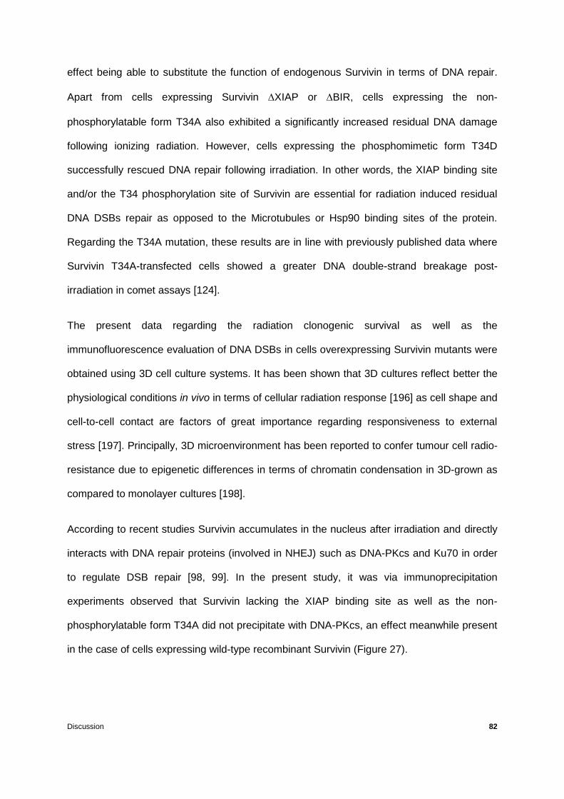
Discussion 82
effect being able to substitute the function of endogenous Survivin in terms of DNA repair.
Apart from cells expressing Survivin XIAP or BIR, cells expressing the non-
phosphorylatable form T34A also exhibited a significantly increased residual DNA damage
following ionizing radiation. However, cells expressing the phosphomimetic form T34D
successfully rescued DNA repair following irradiation. In other words, the XIAP binding site
and/or the T34 phosphorylation site of Survivin are essential for radiation induced residual
DNA DSBs repair as opposed to the Microtubules or Hsp90 binding sites of the protein.
Regarding the T34A mutation, these results are in line with previously published data where
Survivin T34A-transfected cells showed a greater DNA double-strand breakage post-
irradiation in comet assays [124].
The present data regarding the radiation clonogenic survival as well as the
immunofluorescence evaluation of DNA DSBs in cells overexpressing Survivin mutants were
obtained using 3D cell culture systems. It has been shown that 3D cultures reflect better the
physiological conditions in vivo in terms of cellular radiation response [196] as cell shape and
cell-to-cell contact are factors of great importance regarding responsiveness to external
stress [197]. Principally, 3D microenvironment has been reported to confer tumour cell radio-
resistance due to epigenetic differences in terms of chromatin condensation in 3D-grown as
compared to monolayer cultures [198].
According to recent studies Survivin accumulates in the nucleus after irradiation and directly
interacts with DNA repair proteins (involved in NHEJ) such as DNA-PKcs and Ku70 in order
to regulate DSB repair [98, 99]. In the present study, it was via immunoprecipitation
experiments observed that Survivin lacking the XIAP binding site as well as the non-
phosphorylatable form T34A did not precipitate with DNA-PKcs, an effect meanwhile present
in the case of cells expressing wild-type recombinant Survivin (Figure 27).

Discussion 83
Figure 27: Symbolic diagram of Survivin wt and Surv. XIAP. Representation of Survivin wild type (on top)
and Survivin lacking the XIAP binding site (on the bottom). It is suggested that DNA-PKcs interacts with Survivin
within its XIAP binding site and/or only when Survivin is phosphorylated at Threonine 34 while deletion of the
XIAP binding site abolishes such an interaction.
Thus, it can be suggested that the XIAP binding site and/or the T34 phosporylation site of the
protein are of great importance not only for apoptosis inhibition [31] but also for DNA DSB
repair regulation via interaction with DNA-PKcs. The inability of those EGFP-fused constructs
(Survivin XIAP and Survivin T34A) to interact with this repair protein may at least in part
explain both the increased residual DNA damage and the diminished radiation clonogenic
survival of cells overexpressing those constructs.
Aside from its well-known functions in cellular survival regulation, apoptosis and cell cycle,
Survivin has recently been described to promote migration and invasion of different tumour
cells including human LoVo colorectal cancer, breast adenocarcinoma MDA-MB-231,
prostate adenocarcinoma PC3, melanoma cells or melanocytes [58, 199, 200]. It has further
been shown that knockdown of Survivin and/or XIAP in multiple colorectal cancer cell lines
leads to a significantly diminished migration capacity [164]. Principally, the Survivin-XIAP
complex protects XIAP stability from ubiquitin-dependent degradation while increases
BIR-Domain -helixNES XIAP T34
DNA-PKcs
BIR-Domain -helixNES
DNA-PKcs
P
Discussion 84
caspase inhibition [31, 114], enhances tumour growth in vivo [114] and participates in NF-B
activation [58]. NF-B activation leads to a transcriptional up-regulation of Survivin [201]
resulting in an amplification loop that favours cell survival [202] specifically in tumours where
elevated NF-B activity is related to aggressive disease and metastasis [58, 203]. In more
detail, the Survivin-XIAP complex activates NF-B, triggering NF-B-dependent transcription
of fibronectin, an extracellular matrix protein [58] that in turn engages 1 integrins at cell
surface, activating cell motility kinases, Src and focal adhesion kinase (FAK) resulting in
tumour cell migration, invasion and metastatic dissemination in vivo [58]. Despite the
importance of the Survivin-XIAP complex, modulating tumour cell motility factors such as
fibronectin expression and FAK, it is hereby shown that not only the XIAP binding site of
Survivin but also those of Microtubules and Hsp90 are involved in cell migration, suggesting
further mechanisms via which Survivin participates in cancer cell motility.
In brief, these results confirm Survivin to function as a radiation resistance factor modulating
cellular radiation response via DNA DSB repair, induction of cell cycle arrest, apoptosis as
well as cell motility (Figure 28).

Discussion 85
Figure 28: Survivin as a radiation resistance factor. Survivin is here depicted to translocate to the nucleus
following irradiation and regulate DNA repair via interaction with DNA repair proteins; an interaction possible only
after Survivin phosphorylation at Threonine 34 located within the XIAP binding site of the protein.
It is hereby and for the first time shown that Survivin XIAP binding and T34 phosphorylation
sites, but not those of Microtubules and Hsp90, are important for radiation clonogenic
survival and regulation of DNA damage repair, at least in part by disrupting interaction with
DNA-PKcs. In particular, it is suggested that the DNA repair protein DNA-PKcs interacts with
Survivin within the latter’s XIAP binding site (amino acids 15-38) and/or only when Survivin is
phosphorylated at Threonine 34 (Figure 27).
In addition, XIAP, Microtubules and Hsp90 binding sites as well as the BIR domain were
demonstrated to be essential for proper cell cycle regulation, apoptosis inhibition and
transmigration capacity of colorectal cancer cells.
To a certain extent, these findings have functionally distinguished some of the roles of
Survivin BIR domain, XIAP, Microtubules and Hsp90 binding sites regarding radiation
Survivin
nucleus
cytoplasm
Regulation of
DNA repair
Caspases
Smac
SurvivinXIAP
Survivin
P-Survivin
at T34
Survivin
DNA-PKcs
P
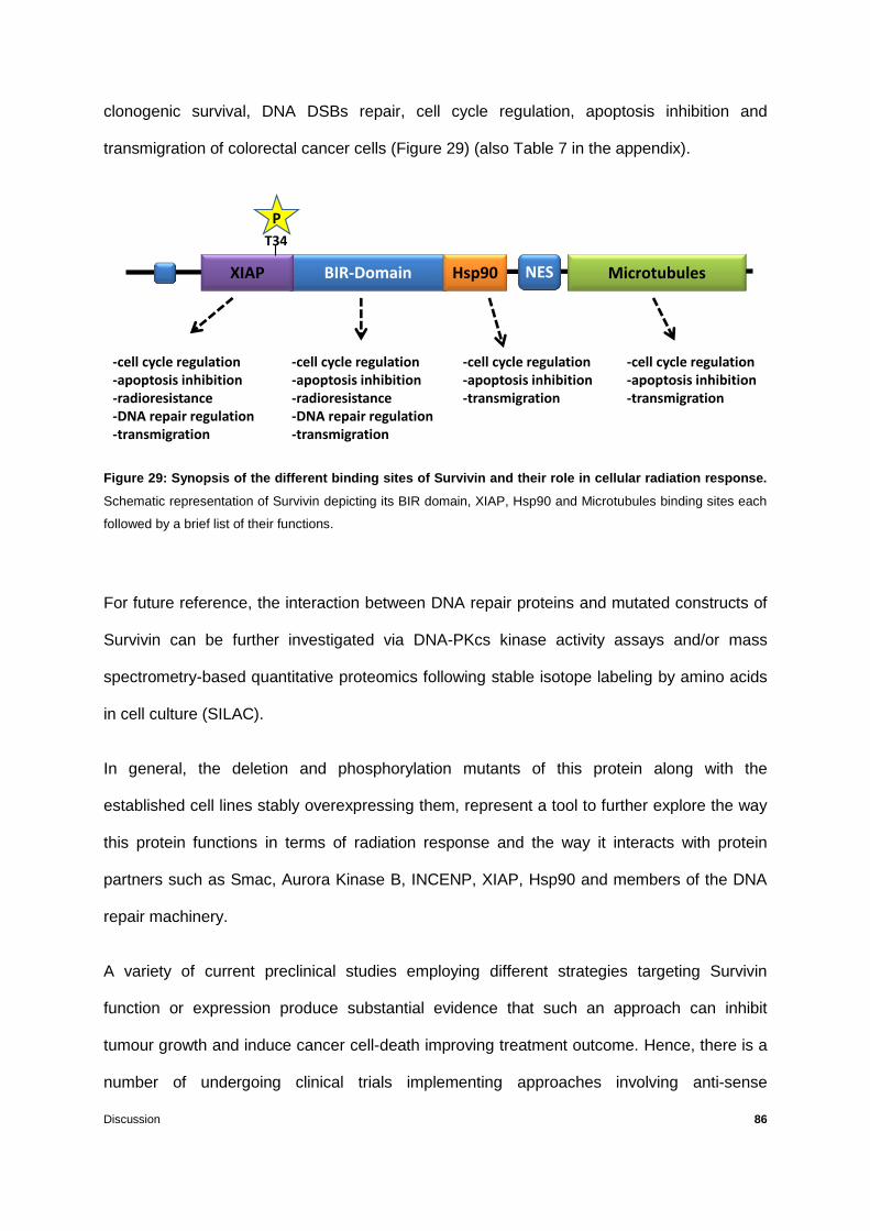
Discussion 86
clonogenic survival, DNA DSBs repair, cell cycle regulation, apoptosis inhibition and
transmigration of colorectal cancer cells (Figure 29) (also Table 7 in the appendix).
Figure 29: Synopsis of the different binding sites of Survivin and their role in cellular radiation response.
Schematic representation of Survivin depicting its BIR domain, XIAP, Hsp90 and Microtubules binding sites each
followed by a brief list of their functions.
For future reference, the interaction between DNA repair proteins and mutated constructs of
Survivin can be further investigated via DNA-PKcs kinase activity assays and/or mass
spectrometry-based quantitative proteomics following stable isotope labeling by amino acids
in cell culture (SILAC).
In general, the deletion and phosphorylation mutants of this protein along with the
established cell lines stably overexpressing them, represent a tool to further explore the way
this protein functions in terms of radiation response and the way it interacts with protein
partners such as Smac, Aurora Kinase B, INCENP, XIAP, Hsp90 and members of the DNA
repair machinery.
A variety of current preclinical studies employing different strategies targeting Survivin
function or expression produce substantial evidence that such an approach can inhibit
tumour growth and induce cancer cell-death improving treatment outcome. Hence, there is a
number of undergoing clinical trials implementing approaches involving anti-sense
BIR-Domain
T34
P
NES XIAP Hsp90 Microtubules
-cell cycle regulation-apoptosis inhibition-radioresistance-DNA repair regulation-transmigration
-cell cycle regulation-apoptosis inhibition-radioresistance-DNA repair regulation-transmigration
-cell cycle regulation-apoptosis inhibition-transmigration
-cell cycle regulation-apoptosis inhibition-transmigration
Discussion 87
oligonucleotides against Survivin (such as Gataparsen) as well as transcriptional inhibitors
(YM155) and immunotherapeutic schemes; the latter using Survivin directed autologous
cytotoxic T lymphocytes in recurrent oral and colorectal cancer, myeloma and malignant
melanoma [158-161]. According to early clinical data, those antagonists revealed a relatively
modest activity having been used as single agents; however, it is believed that if combined
with conventional chemotherapeutic drugs, monoclonal antibodies or irradiation, they may
well be of an enhanced therapeutic advantage [148].
In summary, the present thesis for the first time showed that overexpression of Survivin T34A
or Survivin lacking its XIAP binding site results in a significant radiosensitization and
increased levels of unrepaired DNA damage along with no interaction with DNA-PKcs,
indicating that these mutations interfere with the ability of colorectal cancer cells to repair
DNA damage following irradiation.
To conclude, it is hypothesized that phosphorylation of Survivin on Threonine 34 is essential
for cellular radiation survival proving intervention against Survivin function a promising
approach to improve the outcome of standard radio-chemotherapeutic treatments.

References 88
References
1. Jemal A, Bray F, Center MM, Ferlay J, Ward E, Forman D (2011) Global cancer statistics. CA Cancer J Clin 61, 69-90
2. Kim MS, Lee J, Sidransky D (2010) DNA methylation markers in colorectal cancer. Cancer Metastasis Rev 29, 181-206
3. Worthley DL, Leggett BA (2010) Colorectal cancer: molecular features and clinical opportunities. Clin Biochem Rev 31, 31-38
4. Grady WM (2004) Genomic instability and colon cancer. Cancer Metastasis Rev 23, 11-27
5. Rajagopalan H, Lengauer C (2004) Aneuploidy and cancer. Nature 432, 338-341
6. Freeman DJ, Norrie J, Sattar N, Neely RD, Cobbe SM, Ford I, Isles C, Lorimer AR, Macfarlane PW, McKillop JH, Packard CJ, Shepherd J, Gaw A (2001) Pravastatin and the development of diabetes mellitus: evidence for a protective treatment effect in the West of Scotland Coronary Prevention Study. Circulation 103, 357-362
7. Takayama T, Ohi M, Hayashi T, Miyanishi K, Nobuoka A, Nakajima T, Satoh T, Takimoto R, Kato J, Sakamaki S, Niitsu Y (2001) Analysis of K-ras, APC, and beta-catenin in aberrant crypt foci in sporadic adenoma, cancer, and familial adenomatous polyposis. Gastroenterology 121, 599-611
8. Sjoblom T, Jones S, Wood LD, Parsons DW, Lin J, Barber TD, Mandelker D, Leary RJ, Ptak J, Silliman N, Szabo S, Buckhaults P, Farrell C, Meeh P, Markowitz SD, Willis J, Dawson D, Willson JK, Gazdar AF, Hartigan J, Wu L, Liu C, Parmigiani G, Park BH, Bachman KE, Papadopoulos N, Vogelstein B, Kinzler KW, Velculescu VE (2006) The consensus coding sequences of human breast and colorectal cancers. Science 314, 268-274
9. Leslie A, Carey FA, Pratt NR, Steele RJ (2002) The colorectal adenoma-carcinoma sequence. Br J Surg 89, 845-860
10. Mills AA (2005) p53: link to the past, bridge to the future. Genes Dev 19, 2091-2099
11. Vogelstein B, Fearon ER, Hamilton SR, Kern SE, Preisinger AC, Leppert M, Nakamura Y, White R, Smits AM, Bos JL (1988) Genetic alterations during colorectal-tumor development. N Engl J Med 319, 525-532
12. Schweiger MR, Hussong M, Rohr C, Lehrach H (2013) Genomics and epigenomics of colorectal cancer. Wiley Interdiscip Rev Syst Biol Med 5, 205-219
13. Vilar E, Gruber SB (2010) Microsatellite instability in colorectal cancer-the stable evidence. Nat Rev Clin Oncol 7, 153-162
14. Ribic CM, Sargent DJ, Moore MJ, Thibodeau SN, French AJ, Goldberg RM, Hamilton SR, Laurent-Puig P, Gryfe R, Shepherd LE, Tu D, Redston M, Gallinger S (2003) Tumor microsatellite-instability status as a predictor of benefit from fluorouracil-based adjuvant chemotherapy for colon cancer. N Engl J Med 349, 247-257
15. Esteller M, Levine R, Baylin SB, Ellenson LH, Herman JG (1998) MLH1 promoter hypermethylation is associated with the microsatellite instability phenotype in sporadic endometrial carcinomas. Oncogene 17, 2413-2417
16. Issa JP (2004) CpG island methylator phenotype in cancer. Nat Rev Cancer 4, 988-993
References 89
17. Kondo Y, Issa JP (2004) Epigenetic changes in colorectal cancer. Cancer Metastasis Rev 23, 29-39
18. van Engeland M, Derks S, Smits KM, Meijer GA, Herman JG (2011) Colorectal cancer epigenetics: complex simplicity. J Clin Oncol 29, 1382-1391
19. Rödel C, Sauer R (2004) Radiotherapy and concurrent radiochemotherapy for rectal cancer. Surg Oncol 13, 93-101
20. Crook NE, Clem RJ, Miller LK (1993) An apoptosis-inhibiting baculovirus gene with a zinc finger-like motif. J Virol 67, 2168-2174
21. Srinivasula SM, Ashwell JD (2008) IAPs: what's in a name? Mol Cell 30, 123-135
22. Altieri DC (2010) Survivin and IAP proteins in cell-death mechanisms. Biochem J. 430, 199-205
23. Salvesen GS, Duckett CS (2002) IAP proteins: blocking the road to death's door. Nat Rev Mol Cell Biol 3, 401-410
24. Shi Y (2004) Caspase activation, inhibition, and reactivation: a mechanistic view. Protein Sci 13, 1979-1987
25. Eckelman BP, Salvesen GS, Scott FL (2006) Human inhibitor of apoptosis proteins: why XIAP is the black sheep of the family. EMBO Rep 7, 988-994
26. Kuranaga E, Kanuka H, Tonoki A, Takemoto K, Tomioka T, Kobayashi M, Hayashi S, Miura M (2006) Drosophila IKK-related kinase regulates nonapoptotic function of caspases via degradation of IAPs. Cell 126, 583-596
27. Oshima K, Takeda M, Kuranaga E, Ueda R, Aigaki T, Miura M, Hayashi S (2006) IKK epsilon regulates F actin assembly and interacts with Drosophila IAP1 in cellular morphogenesis. Curr Biol 16, 1531-1537
28. Samuel T, Okada K, Hyer M, Welsh K, Zapata JM, Reed JC (2005) cIAP1 Localizes to the nuclear compartment and modulates the cell cycle. Cancer Res 65, 210-218
29. Arora V, Cheung HH, Plenchette S, Micali OC, Liston P, Korneluk RG (2007) Degradation of survivin by the X-linked inhibitor of apoptosis (XIAP)-XAF1 complex. J Biol Chem 282, 26202-26209
30. Conze DB, Albert L, Ferrick DA, Goeddel DV, Yeh WC, Mak T, Ashwell JD (2005) Posttranscriptional downregulation of c-IAP2 by the ubiquitin protein ligase c-IAP1 in vivo. Mol Cell Biol 25, 3348-3356
31. Dohi T, Okada K, Xia F, Wilford CE, Samuel T, Welsh K, Marusawa H, Zou H, Armstrong R, Matsuzawa S, Salvesen GS, Reed JC, Altieri DC (2004) An IAP-IAP complex inhibits apoptosis. J Biol Chem 279, 34087-34090
32. Silke J, Kratina T, Chu D, Ekert PG, Day CL, Pakusch M, Huang DC, Vaux DL (2005) Determination of cell survival by RING-mediated regulation of inhibitor of apoptosis (IAP) protein abundance. Proc Natl Acad Sci U S A 102, 16182-16187
33. Hinds MG, Norton RS, Vaux DL, Day CL (1999) Solution structure of a baculoviral inhibitor of apoptosis (IAP) repeat. Nat Struct Biol 6, 648-651

References 90
34. Sun C, Cai M, Gunasekera AH, Meadows RP, Wang H, Chen J, Zhang H, Wu W, Xu N, Ng SC, Fesik SW (1999) NMR structure and mutagenesis of the inhibitor-of-apoptosis protein XIAP. Nature 401, 818-822
35. Sun C, Cai M, Meadows RP, Xu N, Gunasekera AH, Herrmann J, Wu JC, Fesik SW (2000) NMR structure and mutagenesis of the third Bir domain of the inhibitor of apoptosis protein XIAP. J Biol Chem 275, 33777-33781
36. Verdecia MA, Huang H, Dutil E, Kaiser DA, Hunter T, Noel JP (2000) Structure of the human anti-apoptotic protein survivin reveals a dimeric arrangement. Nat Struct Biol 7, 602-608
37. Liu Z, Sun C, Olejniczak ET, Meadows RP, Betz SF, Oost T, Herrmann J, Wu JC, Fesik SW (2000) Structural basis for binding of Smac/DIABLO to the XIAP BIR3 domain. Nature 408, 1004-1008
38. Pop C, Salvesen GS (2009) Human caspases: activation, specificity, and regulation. J Biol Chem 284, 21777-21781
39. Srinivasula SM, Hegde R, Saleh A, Datta P, Shiozaki E, Chai J, Lee RA, Robbins PD, Fernandes-Alnemri T, Shi Y, Alnemri ES (2001) A conserved XIAP-interaction motif in caspase-9 and Smac/DIABLO regulates caspase activity and apoptosis. Nature 410, 112-116
40. Lin SC, Huang Y, Lo YC, Lu M, Wu H (2007) Crystal structure of the BIR1 domain of XIAP in two crystal forms. J Mol Biol 372, 847-854
41. Lu M, Lin SC, Huang Y, Kang YJ, Rich R, Lo YC, Myszka D, Han J, Wu H (2007) XIAP induces NF-kappaB activation via the BIR1/TAB1 interaction and BIR1 dimerization. Mol Cell 26, 689-702
42. Rothe M, Pan MG, Henzel WJ, Ayres TM, Goeddel DV (1995) The TNFR2-TRAF signaling complex contains two novel proteins related to baculoviral inhibitor of apoptosis proteins. Cell 83, 1243-1252
43. Deveraux QL, Reed JC (1999) IAP family proteins--suppressors of apoptosis. Genes Dev 13, 239-252
44. Chai J, Shiozaki E, Srinivasula SM, Wu Q, Datta P, Alnemri ES, Shi Y (2001) Structural basis of caspase-7 inhibition by XIAP. Cell 104, 769-780
45. Riedl SJ, Renatus M, Schwarzenbacher R, Zhou Q, Sun C, Fesik SW, Liddington RC, Salvesen GS (2001) Structural basis for the inhibition of caspase-3 by XIAP. Cell 104, 791-800
46. Scott FL, Denault JB, Riedl SJ, Shin H, Renatus M, Salvesen GS (2005) XIAP inhibits caspase-3 and -7 using two binding sites: evolutionarily conserved mechanism of IAPs. EMBO J 24, 645-655
47. Shiozaki EN, Chai J, Rigotti DJ, Riedl SJ, Li P, Srinivasula SM, Alnemri ES, Fairman R, Shi Y (2003) Mechanism of XIAP-mediated inhibition of caspase-9. Mol Cell 11, 519-527
48. Schile AJ, Garcia-Fernandez M, Steller H (2008) Regulation of apoptosis by XIAP ubiquitin-ligase activity. Genes Dev 22, 2256-2266
49. Jeyaprakash AA, Klein UR, Lindner D, Ebert J, Nigg EA, Conti E (2007) Structure of a Survivin-Borealin-INCENP core complex reveals how chromosomal passengers travel together. Cell 131, 271-285

References 91
50. Weissman AM (2001) Themes and variations on ubiquitylation. Nat Rev Mol Cell Biol 2, 169-178
51. Lorick KL, Jensen JP, Fang S, Ong AM, Hatakeyama S, Weissman AM (1999) RING fingers mediate ubiquitin-conjugating enzyme (E2)-dependent ubiquitination. Proc Natl Acad Sci U S A 96, 11364-11369
52. Li X, Yang Y, Ashwell JD (2002) TNF-RII and c-IAP1 mediate ubiquitination and degradation of TRAF2. Nature 416, 345-347
53. Yang Y, Fang S, Jensen JP, Weissman AM, Ashwell JD (2000) Ubiquitin protein ligase activity of IAPs and their degradation in proteasomes in response to apoptotic stimuli. Science 288, 874-877
54. Martin SJ (2002) Destabilizing influences in apoptosis: sowing the seeds of IAP destruction. Cell 109, 793-796
55. Bianchi K, Meier P (2009) A tangled web of ubiquitin chains: breaking news in TNF-R1 signaling. Mol Cell 36, 736-742
56. Karin M, Greten FR (2005) NF-kappaB: linking inflammation and immunity to cancer development and progression. Nat Rev Immunol 5, 749-759
57. Perkins ND (2007) Integrating cell-signalling pathways with NF-kappaB and IKK function. Nat Rev Mol Cell Biol 8, 49-62
58. Mehrotra S, Languino LR, Raskett CM, Mercurio AM, Dohi T, Altieri DC (2010) IAP regulation of metastasis. Cancer Cell 17, 53-64
59. Altieri DC (2003) Validating survivin as a cancer therapeutic target. Nat Rev Cancer 3, 46-54
60. Duffy MJ, O'Donovan N, Brennan DJ, Gallagher WM, Ryan BM (2007) Survivin: a promising tumor biomarker. Cancer Lett 249, 49-60
61. Hunter AM, LaCasse EC, Korneluk RG (2007) The inhibitors of apoptosis (IAPs) as cancer targets. Apoptosis 12, 1543-1568
62. Zender L, Spector MS, Xue W, Flemming P, Cordon-Cardo C, Silke J, Fan ST, Luk JM, Wigler M, Hannon GJ, Mu D, Lucito R, Powers S, Lowe SW (2006) Identification and validation of oncogenes in liver cancer using an integrative oncogenomic approach. Cell 125, 1253-1267
63. Inagaki H (2007) Mucosa-associated lymphoid tissue lymphoma: molecular pathogenesis and clinicopathological significance. Pathol Int 57, 474-484
64. Liston P, Fong WG, Kelly NL, Toji S, Miyazaki T, Conte D, Tamai K, Craig CG, McBurney MW, Korneluk RG (2001) Identification of XAF1 as an antagonist of XIAP anti-Caspase activity. Nat Cell Biol 3, 128-133
65. Plenchette S, Cheung HH, Fong WG, LaCasse EC, Korneluk RG (2007) The role of XAF1 in cancer. Curr Opin Investig Drugs 8, 469-476
66. Wright CW, Duckett CS (2005) Reawakening the cellular death program in neoplasia through the therapeutic blockade of IAP function. J Clin Invest 115, 2673-2678
67. Gordon GJ, Appasani K, Parcells JP, Mukhopadhyay NK, Jaklitsch MT, Richards WG, Sugarbaker DJ, Bueno R (2002) Inhibitor of apoptosis protein-1 promotes tumor cell survival in mesothelioma. Carcinogenesis 23, 1017-1024

References 92
68. McEleny K, Coffey R, Morrissey C, Williamson K, Zangemeister-Wittke U, Fitzpatrick JM, Watson RW (2004) An antisense oligonucleotide to cIAP-1 sensitizes prostate cancer cells to fas and TNFalpha mediated apoptosis. Prostate 59, 419-425
69. Burma S, Chen DJ (2004) Role of DNA–PK in the cellular response to DNA double-strand breaks. DNA Repair (Amst) 3, 909-918
70. Nagasawa H, Brogan JR, Peng Y, Little JB, Bedford JS (2010) Some unsolved problems and unresolved issues in radiation cytogenetics: a review and new data on roles of homologous recombination and non-homologous end joining. Mutat Res 701, 12-22
71. Venkitaraman AR (2009) Linking the cellular functions of BRCA genes to cancer pathogenesis and treatment. Annu Rev Pathol 4, 461-487
72. Khanna KK, Jackson SP (2001) DNA double-strand breaks: signaling, repair and the cancer connection. Nat Genet 27, 247-254
73. Lavin MF (2007) ATM and the Mre11 complex combine to recognize and signal DNA double-strand breaks. Oncogene 26, 7749-7758
74. Uziel T, Lerenthal Y, Moyal L, Andegeko Y, Mittelman L, Shiloh Y (2003) Requirement of the MRN complex for ATM activation by DNA damage. EMBO J 22, 5612-5621
75. Bakkenist CJ, Kastan MB (2003) DNA damage activates ATM through intermolecular autophosphorylation and dimer dissociation. Nature 421, 499-506
76. Celeste A, Petersen S, Romanienko PJ, Fernandez-Capetillo O, Chen HT, Sedelnikova OA, Reina-San-Martin B, Coppola V, Meffre E, Difilippantonio MJ, Redon C, Pilch DR, Olaru A, Eckhaus M, Camerini-Otero RD, Tessarollo L, Livak F, Manova K, Bonner WM, Nussenzweig MC, Nussenzweig A (2002) Genomic instability in mice lacking histone H2AX. Science 296, 922-927
77. Lou Z, Minter-Dykhouse K, Franco S, Gostissa M, Rivera MA, Celeste A, Manis JP, van Deursen J, Nussenzweig A, Paull TT, Alt FW, Chen J (2006) MDC1 maintains genomic stability by participating in the amplification of ATM-dependent DNA damage signals. Mol Cell 21, 187-200
78. Hefferin ML, Tomkinson AE (2005) Mechanism of DNA double-strand break repair by non-homologous end joining. DNA Repair (Amst) 4, 639-648
79. Jeggo P, Lobrich M (2006) Radiation-induced DNA damage responses. Radiat Prot Dosimetry 122, 124-127
80. van Gent DC, van der Burg M (2007) Non-homologous end-joining, a sticky affair. Oncogene 26, 7731-7740
81. Wyman C, Kanaar R (2006) DNA double-strand break repair: all's well that ends well. Annu Rev Genet 40, 363-383
82. Blier PR, Griffith AJ, Craft J, Hardin JA (1993) Binding of Ku protein to DNA. Measurement of affinity for ends and demonstration of binding to nicks. J Biol Chem 268, 7594-7601
83. de Vries E, van Driel W, Bergsma WG, Arnberg AC, van der Vliet PC (1989) HeLa nuclear protein recognizing DNA termini and translocating on DNA forming a regular DNA-multimeric protein complex. J Mol Biol 208, 65-78
84. Smith GC, Jackson SP (1999) The DNA-dependent protein kinase. Genes Dev 13, 916-934

References 93
85. Dvir A, Peterson SR, Knuth MW, Lu H, Dynan WS (1992) Ku autoantigen is the regulatory component of a template-associated protein kinase that phosphorylates RNA polymerase II. Proc Natl Acad Sci U S A 89, 11920-11924
86. Gottlieb TM, Jackson SP (1993) The DNA-dependent protein kinase: requirement for DNA ends and association with Ku antigen. Cell 72, 131-142
87. Cui X, Yu Y, Gupta S, Cho YM, Lees-Miller SP, Meek K (2005) Autophosphorylation of DNA-dependent protein kinase regulates DNA end processing and may also alter double-strand break repair pathway choice. Mol Cell Biol 25, 10842-10852
88. Meek K, Douglas P, Cui X, Ding Q, Lees-Miller SP (2007) trans Autophosphorylation at DNA-dependent protein kinase's two major autophosphorylation site clusters facilitates end processing but not end joining. Mol Cell Biol 27, 3881-3890
89. Calsou P, Delteil C, Frit P, Drouet J, Salles B (2003) Coordinated assembly of Ku and p460 subunits of the DNA-dependent protein kinase on DNA ends is necessary for XRCC4-ligase IV recruitment. J Mol Biol 326, 93-103
90. Chen L, Trujillo K, Sung P, Tomkinson AE (2000) Interactions of the DNA ligase IV-XRCC4 complex with DNA ends and the DNA-dependent protein kinase. J Biol Chem 275, 26196-26205
91. Hsu HL, Yannone SM, Chen DJ (2002) Defining interactions between DNA-PK and ligase IV/XRCC4. DNA Repair (Amst) 1, 225-235
92. Lee JW, Yannone SM, Chen DJ, Povirk LF (2003) Requirement for XRCC4 and DNA ligase IV in alignment-based gap filling for nonhomologous DNA end joining in vitro. Cancer Res 63, 22-24
93. Ambrosini G, Adida C, Altieri DC (1997) A novel anti-apoptosis gene, survivin, expressed in cancer and lymphoma. Nat Med 3, 917-921
94. Engelsma D, Rodriguez JA, Fish A, Giaccone G, Fornerod M (2007) Homodimerization antagonizes nuclear export of survivin. Traffic 8, 1495-1502
95. Bourhis E, Hymowitz SG, Cochran AG (2007) The mitotic regulator Survivin binds as a monomer to its functional interactor Borealin. J Biol Chem 282, 35018-35023
96. Stauber RH, Mann W, Knauer SK (2007) Nuclear and cytoplasmic survivin: molecular mechanism, prognostic, and therapeutic potential. Cancer Res 67, 5999-6002
97. Li F, Ambrosini G, Chu EY, Plescia J, Tognin S, Marchisio PC, Altieri DC (1998) Control of apoptosis and mitotic spindle checkpoint by survivin. Nature 396, 580-584
98. Capalbo G, Dittmann K, Weiss C, Reichert S, Hausmann E, Rödel C, Rödel F (2010) Radiation-induced survivin nuclear accumulation is linked to DNA damage repair. Int J Radiat Oncol Biol Phys 77, 226-234
99. Reichert S, Rödel C, Mirsch J, Harter PN, Tomicic MT, Mittelbronn M, Kaina B, Rödel F (2011) Survivin inhibition and DNA double-strand break repair: a molecular mechanism to overcome radioresistance in glioblastoma. Radiother Oncol 101, 51-58
100. Altieri DC (2001) The molecular basis and potential role of survivin in cancer diagnosis and therapy. Trends Mol Med 7, 542-547
101. Altieri DC (2006) The case for survivin as a regulator of microtubule dynamics and cell-death decisions. Curr Opin Cell Biol 18, 609-615

References 94
102. Lens SM, Vader G, Medema RH (2006) The case for Survivin as mitotic regulator. Curr Opin Cell Biol 18, 616-622
103. Uren AG, Wong L, Pakusch M, Fowler KJ, Burrows FJ, Vaux DL, Choo KH (2000) Survivin and the inner centromere protein INCENP show similar cell-cycle localization and gene knockout phenotype. Curr Biol 10, 1319-1328
104. Gurbuxani S, Xu Y, Keerthivasan G, Wickrema A, Crispino JD (2005) Differential requirements for survivin in hematopoietic cell development. Proc Natl Acad Sci U S A 102, 11480-11485
105. Okada H, Mak TW (2004) Pathways of apoptotic and non-apoptotic death in tumour cells. Nat Rev Cancer 4, 592-603
106. Fraser AG, James C, Evan GI, Hengartner MO (1999) Caenorhabditis elegans inhibitor of apoptosis protein (IAP) homologue BIR-1 plays a conserved role in cytokinesis. Curr Biol 9, 292-301
107. Speliotes EK, Uren A, Vaux D, Horvitz HR (2000) The survivin-like C. elegans BIR-1 protein acts with the Aurora-like kinase AIR-2 to affect chromosomes and the spindle midzone. Mol Cell 6, 211-223
108. Huang HK, Bailis JM, Leverson JD, Gomez EB, Forsburg SL, Hunter T (2005) Suppressors of Bir1p (Survivin) identify roles for the chromosomal passenger protein Pic1p (INCENP) and the replication initiation factor Psf2p in chromosome segregation. Mol Cell Biol 25, 9000-9015
109. Giodini A, Kallio MJ, Wall NR, Gorbsky GJ, Tognin S, Marchisio PC, Symons M, Altieri DC (2002) Regulation of microtubule stability and mitotic progression by survivin. Cancer Res 62, 2462-2467
110. Rosa J, Canovas P, Islam A, Altieri DC, Doxsey SJ (2006) Survivin modulates microtubule dynamics and nucleation throughout the cell cycle. Mol Biol Cell 17, 1483-1493
111. Vong QP, Cao K, Li HY, Iglesias PA, Zheng Y (2005) Chromosome alignment and segregation regulated by ubiquitination of survivin. Science 310, 1499-1504
112. Rödel F, Reichert S, Sprenger T, Gaipl US, Mirsch J, Liersch T, Fulda S, Rödel C (2011) The role of survivin for radiation oncology: moving beyond apoptosis inhibition. Curr Med Chem 18
113. Colnaghi R, Wheatley SP (2010) Liaisons between Survivin and Plk1 during Cell Division and Cell Death. J Biol Chem 285, 22592-22604
114. Dohi T, Xia F, Altieri DC (2007) Compartmentalized phosphorylation of IAP by protein kinase A regulates cytoprotection. Mol Cell 27, 17-28
115. O'Connor DS, Wall NR, Porter AC, Altieri DC (2002) A p34(cdc2) survival checkpoint in cancer. Cancer Cell 2, 43-54
116. Wheatley SP, Barrett RM, Andrews PD, Medema RH, Morley SJ, Swedlow JR, Lens SM (2007) Phosphorylation by aurora-B negatively regulates survivin function during mitosis. Cell Cycle 6, 1220-1230
117. Fortugno P, Beltrami E, Plescia J, Fontana J, Pradhan D, Marchisio PC, Sessa WC, Altieri DC (2003) Regulation of survivin function by Hsp90. Proc Natl Acad Sci U S A 100, 13791-13796
118. Ghosh JC, Dohi T, Kang BH, Altieri DC (2008) Hsp60 regulation of tumor cell apoptosis. J Biol Chem 283, 5188-5194
References 95
119. Kang BH, Altieri DC (2006) Regulation of survivin stability by the aryl hydrocarbon receptor-interacting protein. J Biol Chem 281, 24721-24727
120. Pohl C, Jentsch S (2008) Final stages of cytokinesis and midbody ring formation are controlled by BRUCE. Cell 132, 832-845
121. Kelly RJ, Lopez-Chavez A, Citrin D, Janik JE, Morris JC (2011) Impacting tumor cell-fate by targeting the inhibitor of apoptosis protein survivin. Mol Cancer 10, 35
122. Miura K, Fujibuchi W, Ishida K, Naitoh T, Ogawa H, Ando T, Yazaki N, Watanabe K, Haneda S, Shibata C, Sasaki I (2011) Inhibitor of apoptosis protein family as diagnostic markers and therapeutic targets of colorectal cancer. Surg Today 41, 175-182
123. Asanuma K, Moriai R, Yajima T, Yagihashi A, Yamada M, Kobayashi D, Watanabe N (2000) Survivin as a radioresistance factor in pancreatic cancer. Jpn J Cancer Res 91, 1204-1209
124. Chakravarti A, Zhai GG, Zhang M, Malhotra R, Latham DE, Delaney MA, Robe P, Nestler U, Song Q, Loeffler J (2004) Survivin enhances radiation resistance in primary human glioblastoma cells via caspase-independent mechanisms. Oncogene 23, 7494-7506
125. Rödel C, Haas J, Groth A, Grabenbauer GG, Sauer R, Rödel F (2003) Spontaneous and radiation-induced apoptosis in colorectal carcinoma cells with different intrinsic radiosensitivities: Survivin as a radioresistance factor. Int J Radiat Oncol Biol Phys 55, 1341-1347
126. Iwasa T, Okamoto I, Suzuki M, Nakahara T, Yamanaka K, Hatashita E, Yamada Y, Fukuoka M, Ono K, Nakagawa K (2008) Radiosensitizing effect of YM155, a novel small-molecule survivin suppressant, in non-small cell lung cancer cell lines. Clin Cancer Res 14, 6496-6504
127. Rödel F, Hoffman J, Distel L, Herrmann M, Noisternig T, Papadopoulos T, Sauer R, Rödel C (2005) Survivin as a radioresistance factor, and prognostic and therapeutic target for radiotherapy in rectal cancer. Cancer Res 65, 4881-4887
128. Rödel F, Frey B, Leitmann W, Capalbo G, Weiss C, Rödel C (2008) Survivin antisense oligonucleotides effectively radiosensitize colorectal cancer cells in both tissue culture and murine xenograft models. Int J Radiat Oncol Biol Phys 71, 247-255
129. Bown N (2001) Neuroblastoma tumour genetics: clinical and biological aspects. J Clin Pathol 54, 897-910
130. Velculescu VE, Madden SL, Zhang L, Lash AE, Yu J, Rago C, Lal A, Wang CJ, Beaudry GA, Ciriello KM, Cook BP, Dufault MR, Ferguson AT, Gao Y, He TC, Hermeking H, Hiraldo SK, Hwang PM, Lopez MA, Luderer HF, Mathews B, Petroziello JM, Polyak K, Zawel L, Kinzler KW, et al. (1999) Analysis of human transcriptomes. Nat Genet 23, 387-388
131. Guha M, Altieri DC (2009) Survivin as a global target of intrinsic tumor suppression networks. Cell Cycle 8, 2708-2710
132. Hanahan D, Weinberg RA (2000) The hallmarks of cancer. Cell 100, 57-70
133. Sommer KW, Schamberger CJ, Schmidt GE, Sasgary S, Cerni C (2003) Inhibitor of apoptosis protein (IAP) survivin is upregulated by oncogenic c-H-Ras. Oncogene 22, 4266-4280
134. Fang ZH, Dong CL, Chen Z, Zhou B, Liu N, Lan HF, Liang L, Liao WB, Zhang L, Han ZC (2009) Transcriptional regulation of survivin by c-Myc in BCR/ABL-transformed cells: implications in anti-leukaemic strategy. J Cell Mol Med 13, 2039-2052

References 96
135. Kim PJ, Plescia J, Clevers H, Fearon ER, Altieri DC (2003) Survivin and molecular pathogenesis of colorectal cancer. Lancet 362, 205-209
136. Lee CW, Raskett CM, Prudovsky I, Altieri DC (2008) Molecular dependence of estrogen receptor-negative breast cancer on a notch-survivin signaling axis. Cancer Res 68, 5273-5281
137. Gritsko T, Williams A, Turkson J, Kaneko S, Bowman T, Huang M, Nam S, Eweis I, Diaz N, Sullivan D, Yoder S, Enkemann S, Eschrich S, Lee JH, Beam CA, Cheng J, Minton S, Muro-Cacho CA, Jove R (2006) Persistent activation of stat3 signaling induces survivin gene expression and confers resistance to apoptosis in human breast cancer cells. Clin Cancer Res 12, 11-19
138. Jiang Y, Saavedra HI, Holloway MP, Leone G, Altura RA (2004) Aberrant regulation of survivin by the RB/E2F family of proteins. J Biol Chem 279, 40511-40520
139. Kawakami H, Tomita M, Matsuda T, Ohta T, Tanaka Y, Fujii M, Hatano M, Tokuhisa T, Mori N (2005) Transcriptional activation of survivin through the NF-kappaB pathway by human T-cell leukemia virus type I tax. Int J Cancer 115, 967-974
140. Wu XY, Fu ZX, Wang XH (2010) Effect of hypoxia-inducible factor 1-alpha on Survivin in colorectal cancer. Mol Med Rep 3, 409-415
141. Zhang T, Otevrel T, Gao Z, Ehrlich SM, Fields JZ, Boman BM (2001) Evidence that APC regulates survivin expression: a possible mechanism contributing to the stem cell origin of colon cancer. Cancer Res 61, 8664-8667
142. Guha M, Plescia J, Leav I, Li J, Languino LR, Altieri DC (2009) Endogenous tumor suppression mediated by PTEN involves survivin gene silencing. Cancer Res 69, 4954-4958
143. Jass JR, Whitehall VL, Young J, Leggett BA (2002) Emerging concepts in colorectal neoplasia. Gastroenterology 123, 862-876
144. Li F (2005) Role of survivin and its splice variants in tumorigenesis. Br J Cancer 92, 212-216
145. Hoffman WH, Biade S, Zilfou JT, Chen J, Murphy M (2002) Transcriptional repression of the anti-apoptotic survivin gene by wild type p53. J Biol Chem 277, 3247-3257
146. Mirza A, McGuirk M, Hockenberry TN, Wu Q, Ashar H, Black S, Wen SF, Wang L, Kirschmeier P, Bishop WR, Nielsen LL, Pickett CB, Liu S (2002) Human survivin is negatively regulated by wild-type p53 and participates in p53-dependent apoptotic pathway. Oncogene 21, 2613-2622
147. Gehrke I, Gandhirajan RK, Kreuzer KA (2009) Targeting the WNT/beta-catenin/TCF/LEF1 axis in solid and haematological cancers: Multiplicity of therapeutic options. Eur J Cancer 45, 2759-2767
148. Rödel F, Sprenger T, Kaina B, Liersch T, Rödel C, Fulda S, Hehlgans S (2012) Survivin as a prognostic/predictive marker and molecular target in cancer therapy. Curr Med Chem 19, 3679-3688
149. Wang RH, Zheng Y, Kim HS, Xu X, Cao L, Lahusen T, Lee MH, Xiao C, Vassilopoulos A, Chen W, Gardner K, Man YG, Hung MC, Finkel T, Deng CX (2008) Interplay among BRCA1, SIRT1, and Survivin during BRCA1-associated tumorigenesis. Mol Cell 32, 11-20
150. Vaira V, Lee CW, Goel HL, Bosari S, Languino LR, Altieri DC (2007) Regulation of survivin expression by IGF-1/mTOR signaling. Oncogene 26, 2678-2684
References 97
151. Nguyen HG, Ravid K (2006) Tetraploidy/aneuploidy and stem cells in cancer promotion: The role of chromosome passenger proteins. J Cell Physiol 208, 12-22
152. Vogel C, Hager C, Bastians H (2007) Mechanisms of mitotic cell death induced by chemotherapy-mediated G2 checkpoint abrogation. Cancer Res 67, 339-345
153. Lu J, Tan M, Huang WC, Li P, Guo H, Tseng LM, Su XH, Yang WT, Treekitkarnmongkol W, Andreeff M, Symmans F, Yu D (2009) Mitotic deregulation by survivin in ErbB2-overexpressing breast cancer cells contributes to Taxol resistance. Clin Cancer Res 15, 1326-1334
154. Goga A, Yang D, Tward AD, Morgan DO, Bishop JM (2007) Inhibition of CDK1 as a potential therapy for tumors over-expressing MYC. Nat Med 13, 820-827
155. Schmidt SM, Schag K, Muller MR, Weck MM, Appel S, Kanz L, Grunebach F, Brossart P (2003) Survivin is a shared tumor-associated antigen expressed in a broad variety of malignancies and recognized by specific cytotoxic T cells. Blood 102, 571-576
156. Andersen MH, thor SP (2002) Survivin--a universal tumor antigen. Histol Histopathol 17, 669-675
157. Andersen MH, Svane IM, Becker JC, Straten PT (2007) The universal character of the tumor-associated antigen survivin. Clin Cancer Res 13, 5991-5994
158. Miyazaki A, Kobayashi J, Torigoe T, Hirohashi Y, Yamamoto T, Yamaguchi A, Asanuma H, Takahashi A, Michifuri Y, Nakamori K, Nagai I, Sato N, Hiratsuka H (2011) Phase I clinical trial of survivin-derived peptide vaccine therapy for patients with advanced or recurrent oral cancer. Cancer Sci 102, 324-329
159. Tsuruma T, Hata F, Torigoe T, Furuhata T, Idenoue S, Kurotaki T, Yamamoto M, Yagihashi A, Ohmura T, Yamaguchi K, Katsuramaki T, Yasoshima T, Sasaki K, Mizushima Y, Minamida H, Kimura H, Akiyama M, Hirohashi Y, Asanuma H, Tamura Y, Shimozawa K, Sato N, Hirata K (2004) Phase I clinical study of anti-apoptosis protein, survivin-derived peptide vaccine therapy for patients with advanced or recurrent colorectal cancer. J Transl Med 2, 19
160. Rapoport AP, Aqui NA, Stadtmauer EA, Vogl DT, Fang HB, Cai L, Janofsky S, Chew A, Storek J, Akpek G, Badros A, Yanovich S, Tan MT, Veloso E, Pasetti MF, Cross A, Philip S, Murphy H, Bhagat R, Zheng Z, Milliron T, Cotte J, Cannon A, Levine BL, Vonderheide RH, June CH (2011) Combination immunotherapy using adoptive T-cell transfer and tumor antigen vaccination on the basis of hTERT and survivin after ASCT for myeloma. Blood 117, 788-797
161. Trepiakas R, Berntsen A, Hadrup SR, Bjorn J, Geertsen PF, Straten PT, Andersen MH, Pedersen AE, Soleimani A, Lorentzen T, Johansen JS, Svane IM (2010) Vaccination with autologous dendritic cells pulsed with multiple tumor antigens for treatment of patients with malignant melanoma: results from a phase I/II trial. Cytotherapy 12, 721-734
162. Mita AC, Mita MM, Nawrocki ST, Giles FJ (2008) Survivin: key regulator of mitosis and apoptosis and novel target for cancer therapeutics. Clin Cancer Res 14, 5000-5005
163. Kappler M, Bache M, Bartel F, Kotzsch M, Panian M, Wurl P, Blumke K, Schmidt H, Meye A, Taubert H (2004) Knockdown of survivin expression by small interfering RNA reduces the clonogenic survival of human sarcoma cell lines independently of p53. Cancer Gene Ther 11, 186-193
164. Hehlgans S, Petraki C, Reichert S, Cordes N, Rödel C, Rödel F (2013) Double targeting of Survivin and XIAP radiosensitizes 3D grown human colorectal tumor cells and decreases migration. Radiother Oncol 108, 32-39

References 98
165. Monzo M, Rosell R, Felip E, Astudillo J, Sanchez JJ, Maestre J, Martin C, Font A, Barnadas A, Abad A (1999) A novel anti-apoptosis gene: Re-expression of survivin messenger RNA as a prognosis marker in non-small-cell lung cancers. J Clin Oncol 17, 2100-2104
166. Tanaka K, Iwamoto S, Gon G, Nohara T, Iwamoto M, Tanigawa N (2000) Expression of survivin and its relationship to loss of apoptosis in breast carcinomas. Clin Cancer Res 6, 127-134
167. Kawasaki H, Altieri DC, Lu CD, Toyoda M, Tenjo T, Tanigawa N (1998) Inhibition of apoptosis by survivin predicts shorter survival rates in colorectal cancer. Cancer Res 58, 5071-5074
168. Lu CD, Altieri DC, Tanigawa N (1998) Expression of a novel antiapoptosis gene, survivin, correlated with tumor cell apoptosis and p53 accumulation in gastric carcinomas. Cancer Res 58, 1808-1812
169. Kato J, Kuwabara Y, Mitani M, Shinoda N, Sato A, Toyama T, Mitsui A, Nishiwaki T, Moriyama S, Kudo J, Fujii Y (2001) Expression of survivin in esophageal cancer: correlation with the prognosis and response to chemotherapy. Int J Cancer 95, 92-95
170. Satoh K, Kaneko K, Hirota M, Masamune A, Satoh A, Shimosegawa T (2001) Expression of survivin is correlated with cancer cell apoptosis and is involved in the development of human pancreatic duct cell tumors. Cancer 92, 271-278
171. Ikeguchi M, Ueta T, Yamane Y, Hirooka Y, Kaibara N (2002) Inducible nitric oxide synthase and survivin messenger RNA expression in hepatocellular carcinoma. Clin Cancer Res 8, 3131-3136
172. Saitoh Y, Yaginuma Y, Ishikawa M (1999) Analysis of Bcl-2, Bax and Survivin genes in uterine cancer. Int J Oncol 15, 137-141
173. Yoshida H, Ishiko O, Sumi T, Matsumoto Y, Ogita S (2001) Survivin, bcl-2 and matrix metalloproteinase-2 enhance progression of clear cell- and serous-type ovarian carcinomas. Int J Oncol 19, 537-542
174. Garcia JF, Camacho FI, Morente M, Fraga M, Montalban C, Alvaro T, Bellas C, Castano A, Diez A, Flores T, Martin C, Martinez MA, Mazorra F, Menarguez J, Mestre MJ, Mollejo M, Saez AI, Sanchez L, Piris MA (2003) Hodgkin and Reed-Sternberg cells harbor alterations in the major tumor suppressor pathways and cell-cycle checkpoints: analyses using tissue microarrays. Blood 101, 681-689
175. Adida C, Haioun C, Gaulard P, Lepage E, Morel P, Briere J, Dombret H, Reyes F, Diebold J, Gisselbrecht C, Salles G, Altieri DC, Molina TJ (2000) Prognostic significance of survivin expression in diffuse large B-cell lymphomas. Blood 96, 1921-1925
176. Kuttler F, Valnet-Rabier MB, Angonin R, Ferrand C, Deconinck E, Mougin C, Cahn JY, Fest T (2002) Relationship between expression of genes involved in cell cycle control and apoptosis in diffuse large B cell lymphoma: a preferential survivin-cyclin B link. Leukemia 16, 726-735
177. Adida C, Recher C, Raffoux E, Daniel MT, Taksin AL, Rousselot P, Sigaux F, Degos L, Altieri DC, Dombret H (2000) Expression and prognostic significance of survivin in de novo acute myeloid leukaemia. Br J Haematol 111, 196-203
178. Kamihira S, Yamada Y, Hirakata Y, Tomonaga M, Sugahara K, Hayashi T, Dateki N, Harasawa H, Nakayama K (2001) Aberrant expression of caspase cascade regulatory genes in adult T-cell leukaemia: survivin is an important determinant for prognosis. Br J Haematol 114, 63-69

References 99
179. Islam A, Kageyama H, Takada N, Kawamoto T, Takayasu H, Isogai E, Ohira M, Hashizume K, Kobayashi H, Kaneko Y, Nakagawara A (2000) High expression of Survivin, mapped to 17q25, is significantly associated with poor prognostic factors and promotes cell survival in human neuroblastoma. Oncogene 19, 617-623
180. Adida C, Berrebi D, Peuchmaur M, Reyes-Mugica M, Altieri DC (1998) Anti-apoptosis gene, survivin, and prognosis of neuroblastoma. Lancet 351, 882-883
181. Koch CA, Vortmeyer AO, Diallo R, Poremba C, Giordano TJ, Sanders D, Bornstein SR, Chrousos GP, Pacak K (2002) Survivin: a novel neuroendocrine marker for pheochromocytoma. Eur J Endocrinol 146, 381-388
182. Wurl P, Kappler M, Meye A, Bartel F, Kohler T, Lautenschlager C, Bache M, Schmidt H, Taubert H (2002) Co-expression of survivin and TERT and risk of tumour-related death in patients with soft-tissue sarcoma. Lancet 359, 943-945
183. Chakravarti A, Noll E, Black PM, Finkelstein DF, Finkelstein DM, Dyson NJ, Loeffler JS (2002) Quantitatively determined survivin expression levels are of prognostic value in human gliomas. J Clin Oncol 20, 1063-1068
184. Grossman D, McNiff JM, Li F, Altieri DC (1999) Expression and targeting of the apoptosis inhibitor, survivin, in human melanoma. J Invest Dermatol 113, 1076-1081
185. Hattori M, Sakamoto H, Satoh K, Yamamoto T (2001) DNA demethylase is expressed in ovarian cancers and the expression correlates with demethylation of CpG sites in the promoter region of c-erbB-2 and survivin genes. Cancer Lett 169, 155-164
186. Zhou M, Gu L, Li F, Zhu Y, Woods WG, Findley HW (2002) DNA damage induces a novel p53-survivin signaling pathway regulating cell cycle and apoptosis in acute lymphoblastic leukemia cells. J Pharmacol Exp Ther 303, 124-131
187. Gianani R, Jarboe E, Orlicky D, Frost M, Bobak J, Lehner R, Shroyer KR (2001) Expression of survivin in normal, hyperplastic, and neoplastic colonic mucosa. Hum Pathol 32, 119-125
188. Kawasaki H, Toyoda M, Shinohara H, Okuda J, Watanabe I, Yamamoto T, Tanaka K, Tenjo T, Tanigawa N (2001) Expression of survivin correlates with apoptosis, proliferation, and angiogenesis during human colorectal tumorigenesis. Cancer 91, 2026-2032
189. Shinohara ET, Hallahan DE, Lu B (2004) The Use of Antisense Oligonucleotides in Evaluating Survivin as a Therapeutic Target for Radiation Sensitization in Lung Cancer. Biol Proced Online 6, 250-256
190. Weiss C, von Romer F, Capalbo G, Ott OJ, Wittlinger M, Krause SF, Sauer R, Rödel C, Rödel F (2009) Survivin expression as a predictive marker for local control in patients with high-risk T1 bladder cancer treated with transurethral resection and radiochemotherapy. Int J Radiat Oncol Biol Phys 74, 1455-1460
191. Al-Khalaf HH, Lach B, Allam A, Hassounah M, Alkhani A, Elkum N, Alrokayan SA, Aboussekhra A (2008) Expression of survivin and p16(INK4a)/Cdk6/pRB proteins and induction of apoptosis in response to radiation and cisplatin in meningioma cells. Brain Res 1188, 25-34
192. Rödel C, Grabenbauer GG, Papadopoulos T, Bigalke M, Günther K, Schick C, Peters A, Sauer R, Rödel F (2002) Apoptosis as a cellular predictor for histopathologic response to neoadjuvant radiochemotherapy in patients with rectal cancer. Int J Radiat Oncol Biol Phys 52
193. Zhang M, Ho A, Hammond EH, Suzuki Y, Bermudez RS, Lee RJ, Pilepich M, Shipley WU, Sandler H, Khor LY, Pollack A, Chakravarti A (2009) Prognostic value of survivin in locally

References 100
advanced prostate cancer: study based on RTOG 8610. Int J Radiat Oncol Biol Phys 73, 1033-1042
194. Altieri DC (2008) Survivin, cancer networks and pathway-directed drug discovery. Nat Rev Cancer 8, 61-70
195. Xia F, Altieri DC (2006) Mitosis-independent survivin gene expression in vivo and regulation by p53. Cancer Res 66, 3392-3395
196. Eke I, Cordes N (2011) Radiobiology goes 3D: how ECM and cell morphology impact on cell survival after irradiation. Radiother Oncol 99, 271-278
197. Zschenker O, Streichert T, Hehlgans S, Cordes N (2012) Genome-wide gene expression analysis in cancer cells reveals 3D growth to affect ECM and processes associated with cell adhesion but not DNA repair. PLoS One 7, e34279
198. Storch K, Eke I, Borgmann K, Krause M, Richter C, Becker K, Schrock E, Cordes N (2010) Three-dimensional cell growth confers radioresistance by chromatin density modification. Cancer Res 70, 3925-3934
199. Chu XY, Chen LB, Wang JH, Su QS, Yang JR, Lin Y, Xue LJ, Liu XB, Mo XB (2012) Overexpression of survivin is correlated with increased invasion and metastasis of colorectal cancer. J Surg Oncol 105, 520-528
200. McKenzie JA, Liu T, Goodson AG, Grossman D (2010) Survivin Enhances Motility of Melanoma Cells by Supporting Akt Activation and 5 Integrin Upregulation. Cancer Res 70, 7927-7937
201. Guha M, Xia F, Raskett CM, Altieri DC (2010) Caspase 2-mediated tumor suppression involves survivin gene silencing. Oncogene 29, 1280-1292
202. Baud V, Karin M (2009) Is NF-kappaB a good target for cancer therapy? Hopes and pitfalls. Nat Rev Drug Discov 8, 33-40
203. Grivennikov SI, Greten FR, Karin M (2010) Immunity, inflammation, and cancer. Cell 140, 883-899
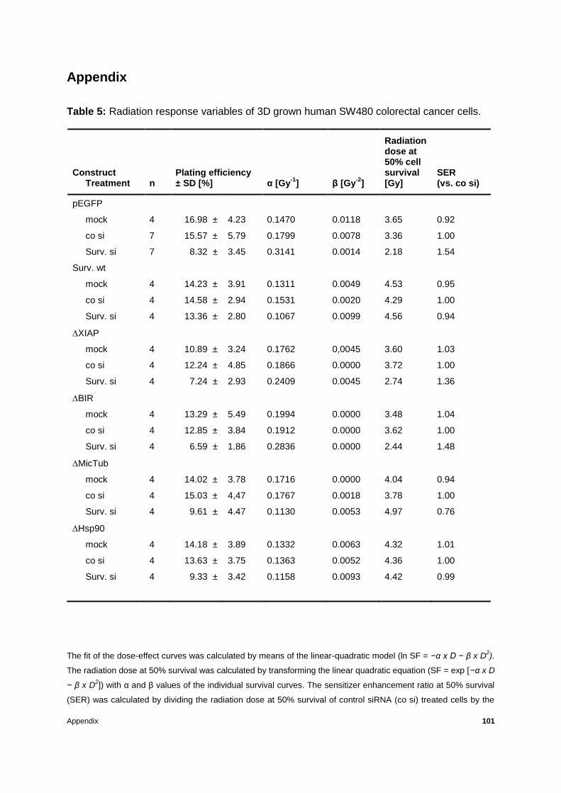
Appendix 101
Appendix
Table 5: Radiation response variables of 3D grown human SW480 colorectal cancer cells.
The fit of the dose-effect curves was calculated by means of the linear-quadratic model (ln SF = −α x D − β x D2).
The radiation dose at 50% survival was calculated by transforming the linear quadratic equation (SF = exp [−α x D
− β x D2]) with α and β values of the individual survival curves. The sensitizer enhancement ratio at 50% survival
(SER) was calculated by dividing the radiation dose at 50% survival of control siRNA (co si) treated cells by the
Construct Treatment
n
Plating efficiency ± SD [%]
α [Gy
-1]
β [Gy
-2]
Radiation dose at 50% cell survival [Gy]
SER (vs. co si)
pEGFP
mock 4 16.98 ± 4.23 0.1470 0.0118 3.65 0.92
co si 7 15.57 ± 5.79 0.1799 0.0078 3.36 1.00
Surv. si 7 8.32 ± 3.45 0.3141 0.0014 2.18 1.54
Surv. wt
mock 4 14.23 ± 3.91 0.1311 0.0049 4.53 0.95
co si 4 14.58 ± 2.94 0.1531 0.0020 4.29 1.00
Surv. si 4 13.36 ± 2.80 0.1067 0.0099 4.56 0.94
XIAP
mock 4 10.89 ± 3.24 0.1762 0,0045 3.60 1.03
co si 4 12.24 ± 4.85 0.1866 0.0000 3.72 1.00
Surv. si 4 7.24 ± 2.93 0.2409 0.0045 2.74 1.36
BIR
mock 4 13.29 ± 5.49 0.1994 0.0000 3.48 1.04
co si 4 12.85 ± 3.84 0.1912 0.0000 3.62 1.00
Surv. si 4 6.59 ± 1.86 0.2836 0.0000 2.44 1.48
MicTub
mock 4 14.02 ± 3.78 0.1716 0.0000 4.04 0.94
co si 4 15.03 ± 4,47 0.1767 0.0018 3.78 1.00
Surv. si 4 9.61 ± 4.47 0.1130 0.0053 4.97 0.76
Hsp90
mock 4 14.18 ± 3.89 0.1332 0.0063 4.32 1.01
co si 4 13.63 ± 3.75 0.1363 0.0052 4.36 1.00
Surv. si 4 9.33 ± 3.42 0.1158 0.0093 4.42 0.99

Appendix 102
radiation dose at 50% survival of mock or Survivin siRNA (Surv. si) treated cells. D, radiation dose [Gy]; n,
number of independent experiments; SD, standard deviation; SF, surviving fraction; pEGFP, enhanced green
fluorescent protein expressing control; Surv. wt, Survivin wild type; XIAP, Survivin X-linked inhibitor of apoptosis
protein binding site; BIR, baculovirus IAP repeat domain; MiCTub, Microtubules binding site; Hsp90, Hsp90
binding site deletion mutants stably expressed in SW480 cells.
Table 6: Radiation response variables of alternative clones and phospho-mutants of 3D
grown human SW480 colorectal cancer cells.

Appendix 103
Construct Treatment
n
Plating efficiency ± SD [%]
α [Gy
-1]
β [Gy
-2]
Radiation dose at 50% cell survival [Gy]
SER (vs. co si)
pEGFP #2
co si 4 11.18 ± 4.15 0.1574 0.0123 3.46 1.00
Surv. si 4 4.14 ± 1.31 0.3477 0.0019 1.97 1.76
Surv. wt #2
co si 4 10.43 ± 3.10 0.1482 0.0124 3.59 1.00
Surv. si 4 9.68 ± 3.76 0.1748 0.0121 3.24 1.11
XIAP #2
co si 4 8.29 ± 1.57 0.2188 0.0008 3.13 1.00
Surv. si 4 3.62 ± 1.96 0.2687 0.0268 2.13 1.47
S20A
co si 3 8.48 ± 3.31 0.1407 0.0371 2.82 1,00
Surv. si 3 3.92 ± 1.63 0.3036 0.0000 2.28 1,24
S20D
co si 3 14.90 ± 3.39 0.0733 0.0220 4.19 1.00
Surv. si 3 7.24 ± 2.39 0.1151 0.0219 3.58 1.17
T34A
co si 6 11.15 ± 3.47 0.3076 0.0064 2.16 1.00
Surv. si 6 8.00 ± 1.59 0.3202 0.0194 1.94 1.11
T34D
co si 6 13.74 ± 5.47 0.1510 0.0155 3.40 1.00
Surv. si 6 10.42 ± 3.84 0.2417 0.0054 2.70 1.26
T34A #2
co si 3 5.37 ± 1.20 0.4427 0.0000 1.57 1.00
Surv. si 3 6.86 ± 2.05 0.3396 0.0263 1.79 0.87
T34D #2
co si 3 11.40 ± 1.07 0.1885 0.0156 2.95 1.00
Surv. si 3 8.97 ± 0.88 0.3153 0.0073 2.10 1.41
T117A
co si 3 0.15 ± 0.02 0.1834 0.0137 3.07 1.00
Surv. si 3 0.11 ± 0.03 0.1296 0.0202 3.47 0.89
T117D
co si 3 0.12 ± 0.03 0.2305 0.0087 2.73 1.00
Surv. si 3 0.05 ± 0.02 0.2404 0.0098 2.61 1.05

Appendix 104
The fit of the dose-effect curves was calculated by means of the linear-quadratic model (ln SF = −α x D − β x D2).
The radiation dose at 50% survival was calculated by transforming the linear quadratic equation (SF = exp [−α x D
− β x D2]) with α and β values of the individual survival curves. The sensitizer enhancement ratio at 50% survival
(SER) was calculated by dividing the radiation dose at 50% survival of control siRNA (co si) treated cells by the
radiation dose at 50% survival of mock or Survivin siRNA (Surv. si) treated cells. D, radiation dose [Gy]; n,
number of independent experiments; SD, standard deviation; SF, surviving fraction; pEGFP, enhanced green
fluorescent protein expressing control; Surv. wt, Survivin wild type; XIAP, Survivin X-linked inhibitor of apoptosis
protein binding site; S20A, S20D, T34A, T34D, T117A, T117D mutants stably expressed in SW480 cells.

Appendix 105
Table 7: Synopsis of observed effects of Survivin mutants on cellular radiation response
Survivin mutation Effects on cellular radiation response or transmigration
Survivin XIAP
G2/M arrest, increased values of caspase 3/7 activity,
increased radiosensitivity, increased levels of DNA DSBs, no
precipitation with DNA-PKcs, decreased levels of
transmigration
Survivin BIR
G2/M arrest, increased values of caspase 3/7 activity,
increased radiosensitivity, increased levels of DNA DSBs,
decreased levels of transmigration
Survivin MicTub G2/M arrest, increased values of caspase 3/7 activity,
decreased levels of transmigration
Survivin Hsp90 G2/M arrest, increased values of caspase 3/7 activity,
decreased levels of transmigration
Survivin S20A radiosensitivity
Survivin S20D -
Survivin T34A increased radiosensitivity, increased levels of DNA DSBs, no
precipitation with DNA-PKcs
Survivin T34D -
Survivin T117A -
Survivin T117D -
Abbreviations 106
Abbreviations
°C degree Celsius
H2AX gamma H2A histone, member X
µg microgram
µl microliter
µm micrometer
µM micromolar
2D two-dimensional
3D three-dimensional
53BP1 p53-binding protein 1
A Alanine
ACF Aberrant crypt focus
AIP Aryl hydrocarbon receptor-interacting protein
APC Adenomatous polyposis coli gene
ASO Anti-sense oligonucleotide
ATM Ataxia telangiectasia mutated
ATR Ataxia telangiectasia rad3-related protein
BSA Bovine serum albumin
BIR Baculovirus IAP repeat
BRCA1 Breast cancer 1
BRAF proto-oncogene B-Raf
CARD Caspase-associated recruitment domain
CDK1 Cyclin-dependent kinase 1
CIN Chromosomal instability
CIMP CpG island methylator phenotype
cIAPs Cellular inhibitor of apoptosis proteins
CRC Colorectal cancer
CO2 Carbon dioxide

Abbreviations 107
CFA Colony forming assay
CPC Chromosomal passenger complex
c-Ha-Ras V-Ha-ras Harvey rat sarcoma viral oncogene homologue
c-Myc V-myc myelocytomatosis viral oncogene homologue (avian)
D Aspartic acid
DAPI 4',6-diamidino-2-phenylindole
DIABLO Direct IAP-binding protein with low pI
DIAP1 Drosophila IAP 1
DMEM Dulbecco’s Modified Eagle Medium
DNA Deoxyribonucleic acid
DNA-PKcs DNA-dependent protein kinase catalytic subunit
DNA DSBs DNA Double-strand breaks
EDTA Ethylenediaminetetraacetic acid
EGFP Enhanced green fluorescent protein
EGFR Epidermal growth factor receptor
FACS Fluorescence-activated cell sorting
FAK Focal adhesion kinase
FOXO1 Forkhead box O1
G2 Growth 2 phase (of the cell cycle)
GTP-binding protein guanosine nucleotide-binding protein
HBXIP Hepatitis B virus X-interacting protein
HIF-1 Hypoxia inducing factor 1 alpha
HR Homologous recombination
Hsp90 Heat shock protein 90
IAP Inhibitor of apoptosis protein
IBM IAP-binding motif
IgG Immunoglobulin G

Abbreviations 108
IP Immunoprecipitation
IF Immunofluorescence
INCENP Inner centromere protein
KRAS V-Ki-ras2 Kirsten rat sarcoma (viral oncogene homolog)
LB medium Luria Bertani medium
Lys Lysine
M Mitotic phase of the cell cycle
MALT Mucosa-associated lymphoid tissue
MAPK Mitogen-activated protein kinase
MDC1 Mediator of DNA damage checkpoint protein 1
mg milligram
min minute
MicTub Microtubules
MMR Mismatch repair
MRN Mre11, Rad50 and Nbs1 complex
mRNA Messenger RNA
MSI Microsatellite instability
mTOR Mammalian target of rapamycin
n Number of experiments
NES Nuclear export signal
ng nanogram
NF-B nuclear factor kappa-light-chain-enhancer of activated B cells
NHEJ Non-homologous end joining
NSCLC Non-small cell lung cancer
p53 Tumour suppressor protein 53
PCR Polymerase Chain Reaction
PKA Protein kinase A

Abbreviations 109
PBS Phosphate buffered saline
PCR Polymerase chain reaction
Plk1 Polo-like kinase 1
PTEN Phosphatase and tensin homolog deleted from chromosome ten
RNA Ribonucleic acid
rpm Rounds per minute
RT Room temperature
RING Really Interesting Gene
S serine
SDS Sodium dodecylsulfate
siRNA Small interfering RNA
SIRT1 Silent mating type information regulation 2 homolog 1
Smac second mitochondria-derived activator of caspases
SMAD2 (or SMAD4) Mothers against decapentaplegic homolog 2 (or 4)
Src proto-oncogene c-Src
STAAT3 Signal transduction and activator of transcription 3
UBA Binding site for polyubiquitinated proteins
TBS buffer Tris-buffered saline
TAE buffer Tris-Acetate-EDTA buffer
TCF4 Transcription factor 4
TGF- Transforming growth factor beta
TNF- Tumour necrosis factor alpha
TRAIL TNF-related apoptosis-inducing ligand
TRIS Tris hydroxymethyl aminomethane
TP53 Tumour protein 53 (gene)
wt Wild-type

Abbreviations 110
Wnt wingless-related integration site
XAF1 XIAP-associating factor 1
XIAP X-chromosome linked inhibitor of apoptosis protein
XRCC4 X-ray repair cross-complementing protein 4

Acknowledgements 111
Acknowledgements
I would now like to thank all those who each in their own way supported me in this work.
First of all, I wish to thank Prof. Dr. Franz Rödel for entrusting me with one of his research
projects, for his guidance, patience, valuable discussions and understanding as well as for
offering me the chance to publish this work.
I am grateful to my first reviewer Prof. Dr. Markus Löbrich for his support, comments and
suggestions concerning this project during the midterm GRK 1657 meetings as well as for
presenting me the opportunity to participate in this particular graduate school.
I am thankful to my second reviewer Prof. Dr. Cristina Cardoso for her advice and
suggestions with regard to my work. I am also thankful to all the members of the examination
board.
Additionally, I wish to thank Prof. Dr. Claus Rödel for giving me the opportunity to work at the
facilities of the Radiotherapy and Oncology Department at the University Hospital Frankfurt,
Germany. Many thanks also to the Medical Physics Division for taking care of the irradiation
settings of the LINACS during the experiments.
Thanks a million to Dr. Stephanie Hehlgans for her outstanding know-how, unlimited practical
and inspirational aid as well as encouragement, guidance and inestimable advice that was at
all times so open-heartedly given. I would also like to express my gratitude to Martin Large,
Julius Oppermann and Dr. Sebastian Reichert for the constructive discussions, their
generous assistance throughout my whole project and for creating an amiable working
environment. Thank you all for such a great time in such a lovely team!
Moreover, I wish to thank Prof. Dr. Klaus Strebhardt and his group at the Molecular
Gynecology Lab as well as Dr. Juping Yuan and her group at the Obstetrics Lab of the
University Hospital Frankfurt, for their comments and suggestions during our weekly
seminars along with the kind sharing of their research facilities.
A special thanks to all scholars, fellows, professors and other members of the GRK 1657
graduate school at the Technical University of Darmstadt, for their helpful suggestions during
our meetings and the great time we had during the excursions organized by the graduate
school. Financial support given from the GRK 1657 is gratefully acknowledged. Many thanks

Acknowledgements 112
to Ms Susanna Türr for the help she patiently provided during the three years of my GRK
membership.
Last but not least, I want to thank my father Nikolaos Petrakis and mother Anna Petraki for
the moral and financial backing as well as my brother, partner, relatives and friends for all the
encouragement during my studies.
Curriculum Vitae 113
Curriculum Vitae
Name: Chrysi Eirini Surname: Petraki
Nationality: Greek Place of birth: Chania, Crete, Greece
Date of birth: 05/05/1987 Email: [email protected]
Education
Doctoral student (GRK 1657 scholar) at the Faculty of Biology at Technical University of Darmstadt, Germany; subject: The role of the inhibitor of apoptosis protein Survivin in cellular radiation response; supervised by: Prof. M. Löbrich and Prof. C. Cardoso; principal investigator: Prof. F. Rödel; project accomplished at the Molecular Radiation Biology laboratory at the Dept. of Radiotherapy and Oncology, University Hospital Frankfurt, Germany, Apr 2011 – Mar 2014
Master of Science in Radiation Biology at University College London, United Kingdom; thesis title: Effects of irradiation on endothelial cells of subcutaneous tissue obtained from a repair blastema induced in C57BL/6 mice; Sep 2009 – Oct 2010
Bachelor of Science in Biological Sciences at London Metropolitan University, United Kingdom; thesis title: Measurement of intracellular calcium concentration changes following histamine treatment in UMR 106 rat osteosarcoma cells using Fura 2-AM as a calcium dye; Sep 2005 – Jun 2008
Apolytirion at the 7th General Lyceum of Chania, Crete, Greece; GPA: 83.5%; Sep 2002 – Jun 2005
Publications
Hehlgans S, Petraki C, Reichert S, Cordes N, Rödel C, Rödel F. Double Targeting of Surviving and XIAP Radiosensitizes 3D Grown Human Colorectal Tumor Cells and Decreases Migration. Radiotherapy and Oncology 108 (2013) 32–39.
Petraki C, Rödel C, Rödel F, Hehlgans S. The XIAP binding site of Survivin is essential for 3D radiation survival of colorectal cancer cells. In preparation (2014)
Attended Conferences
GBS Society for Radiation Biology Research; abstract selected for proffered paper and speech. Darmstadt, Germany, 2013
International Wolfsberg Meeting on Molecular Radiation Biology/Oncology; abstract and poster presentation. Wolfsberg, Switzerland, 2013
UCT University Cancer Centre; abstract and poster presentation. Frankfurt, Germany, 2013
Curriculum Vitae 114
DEGRO German Society for Radiation Oncology; abstract and poster presentation. Berlin, Germany, 2013
GBS Society for Radiation Biology Research; DGDR German Society for Research on DNA repair; abstract and poster presentation. Munich, Germany, 2012
UCT (Universitäres Centrum Tumorerkrankungen Frankfurt) University Cancer Centre; abstract and poster presentation. Frankfurt, Germany, 2012
DEGRO (Deutsche Gesellschaft für Radioonkologie) German Society for Radiation Oncology; abstract and poster presentation. Wiesbaden, Germany, 2012
GBS (Gesellschaft für Biologische Strahlenforschung) Society for Radiation Biology Research. Rheinbach, Germany, 2011
Further Skills
Languages: Greek, English, French, German
Computer programs: MS Office

![Research Paper HSP90 Inhibitor Encapsulated Photo ...synergistic. CI was calculated with Compusyn software based on Chou-Talalay method . [23, 24] Regarding apoptosis assay, PC3 cells](https://img.pdfslide.us/doc/110x75/60e4fea2389ac00bd571afd5/research-paper-hsp90-inhibitor-encapsulated-photo-synergistic-ci-was-calculated.jpg)