Embed Size (px)
Citation preview
The Protein Synthesis Inhibitor Anisomycin InducesMacrophage Apoptosis in Rabbit Atherosclerotic Plaquesthrough p38 Mitogen-Activated Protein Kinase
Valerie Croons, Wim Martinet, Arnold G. Herman, Jean-Pierre Timmermans, andGuido R. Y. De MeyerDivision of Pharmacology (V.C., W.M., A.G.H., G.R.Y.D.M.) and Laboratory of Cell Biology and Histology (J.-P.T.), University ofAntwerp, Wilrijk, Belgium
Received December 16, 2008; accepted March 12, 2009
ABSTRACTBecause macrophages play a major role in atheroscleroticplaque destabilization, selective removal of macrophages rep-resents a promising approach to stabilize plaques. We showedrecently that the protein synthesis inhibitor cycloheximide, incontrast to puromycin, selectively depleted macrophages inrabbit atherosclerotic plaques without affecting smooth musclecells (SMCs). The mechanism of action of these two translationinhibitors is dissimilar and could account for the differentialeffects on SMC viability. It is not known whether selectivedepletion of macrophages is confined to cycloheximide orwhether it can also be achieved with translation inhibitors thathave a similar mechanism of action. Therefore, in the presentstudy, we investigated the effect of anisomycin, a translationinhibitor with a mechanism of action similar to cycloheximide,on macrophage and SMC viability. In vitro, anisomycin inducedapoptosis of macrophages in a concentration-dependent man-ner, whereas SMCs were only affected at higher concentra-tions. In vivo, anisomycin selectively decreased the macro-phage content of rabbit atherosclerotic plaques through
apoptosis. The p38 mitogen-activated protein kinase (MAPK)inhibitor SB202190 [4-(4-fluorophenyl)-2-(4-hydroxyphenyl)-5-(4-pyridyl)-1H-imidazole] prevented anisomycin-induced mac-rophage death, without affecting SMC viability. SB202190 de-creased anisomycin-induced p38 MAPK phosphorylation, didnot alter c-Jun NH2-terminal kinase (JNK) phosphorylation, andincreased extracellular signal-regulated kinase (ERK) 1/2 phos-phorylation. The latter effect was abolished by the mitogen-activated protein kinase kinase 1/2 inhibitor U0126 [1,4-diamino-2,3-dicyano-1,4-bis(2-aminophynyltio)butadiene ethanolate],although the prevention of anisomycin-induced macrophagedeath by SB202190 remained unchanged. The JNK phosphor-ylation inhibitor SP600125 did not affect anisomycin-inducedmacrophage or SMC death. In conclusion, anisomycin selec-tively decreased the macrophage content in rabbit atheroscle-rotic plaques, indicating that this effect is not confined to cy-cloheximide. p38 MAPK, but not ERK1/2 or JNK, plays a majorrole in anisomycin-induced macrophage death.
Atherosclerosis is a chronic inflammatory disease of thelarge- and medium-sized arteries and is characterized by theformation of plaques in the intima. Advanced atheroscleroticplaques have a large necrotic core that is surrounded bynumerous macrophage-derived foam cells and is separatedfrom the lumen by a protective fibrous cap consisting ofsmooth muscle cells (SMCs) and extracellular matrix (Lusis,
2000; Libby and Theroux, 2005). Because macrophages are aprominent feature in all stages of atherosclerosis and be-cause their numbers are highly increased in shoulder regionsof plaques that tend to rupture (van der Wal et al., 1994),macrophages are believed to play a role in plaque destabili-zation (Takahashi et al., 2002). They produce matrix metal-loproteinases (Galis and Khatri, 2002; Johnson, 2007) andinduce SMC death (Boyle et al., 2001), resulting in decreasedsynthesis of interstitial collagen and further thinning of theextracellular matrix. In line with these findings, selectiveremoval of macrophages from atherosclerotic plaques viamacrophage-specific initiation of cell death represents apromising strategy to stabilize rupture-prone plaques. We
This work was supported by the Fonds voor Wetenschappelijk Onderzoek(FWO)-Flanders (Belgium) [Projects G.0308.04, G.0113.06, and G.0112.08];the University of Antwerp (BOF); and the Bekales Foundation. W.M. is apostdoctoral fellow of the FWO-Flanders (Belgium).
Article, publication date, and citation information can be found athttp://jpet.aspetjournals.org.
doi:10.1124/jpet.108.149948.
ABBREVIATIONS: SMC, smooth muscle cell; MAPK, mitogen-activated protein kinase; SB202190, 4-(4-fluorophenyl)-2-(4-hydroxyphenyl)-5-(4-pyridyl)-1H-imidazole; MEK, mitogen-activated protein kinase kinase; U0126, 1,4-diamino-2,3-dicyano-1,4-bis(2-aminophynyltio)butadiene ethanolate;JNK, c-Jun NH2-terminal kinase; SAPK, stress-activated protein kinase; ERK, extracellular signal-regulated kinase; H/E, hematoxylin/eosin; BAL,bronchoalveolar; AN, anisomycin; ANOVA, analysis of variance.
0022-3565/09/3293-856–864$20.00THE JOURNAL OF PHARMACOLOGY AND EXPERIMENTAL THERAPEUTICS Vol. 329, No. 3Copyright © 2009 by The American Society for Pharmacology and Experimental Therapeutics 149948/3473383JPET 329:856–864, 2009 Printed in U.S.A.
856
at ASPE
T Journals on June 5, 2018
jpet.aspetjournals.orgD
ownloaded from

showed recently that the protein synthesis inhibitor cyclo-heximide selectively decreased the macrophage content ofrabbit atherosclerotic plaques (Croons et al., 2007). We hy-pothesized that macrophages are more dependent on proteinsynthesis for survival than SMCs because arterial macro-phage-derived foam cells consume 3 times more oxygen thanSMCs (Bjornheden and Bondjers, 1987). Moreover, inhibitionof translation in SMCs induces a modulation toward a differ-entiated, quiescent, contractile phenotype (Martin et al.,2004), which makes them even more resistant to inhibition ofprotein synthesis. However, in contrast to cycloheximide, theprotein synthesis inhibitor puromycin induced cell death ofboth macrophages and SMCs (Croons et al., 2008). The majordissimilarity between these two protein synthesis inhibitorsis their mechanism of action (Azzam and Algranati, 1973;Stocklein and Piepersberg, 1980), which is in part responsi-ble for their differential effect on SMC viability (Croons et al.,2008). Cycloheximide inhibits peptide bond formation, andthe nascent peptide remains attached to the polyribosome(Grollman, 1967; Stocklein and Piepersberg, 1980). In con-trast, puromycin is an aminoacyl-tRNA structure analog thatcauses the release of unfinished polypeptide chains (Azzamand Algranati, 1973). The presence of unfinished polypeptidechains activates endoplasmic reticulum-stress associated celldeath pathways (Croons et al., 2008), which are in partresponsible for puromycin-induced SMC death. However, it isnot known whether selective depletion of macrophages isconfined to cycloheximide or also can be achieved with pro-tein translation inhibitors that have a similar mechanism ofaction such as anisomycin. Therefore, in the present study,we investigated the effect of anisomycin on macrophage andSMC viability and the role of mitogen-activated protein ki-nases (MAPKs) herein.
Materials and MethodsCell Culture. The murine macrophage cell line J774A.1 (Ameri-
can Type Culture Collection, Manassas, VA) was grown in RPMI1640 medium (Invitrogen, Carlsbad, CA) supplemented with 10%heat-inactivated fetal bovine serum, 100 U/ml penicillin, 100 �g/mlstreptomycin, 50 �g/ml gentamicin, and 20 U/ml polymyxin B sulfatein a humidified 5% carbon dioxide incubator at 37°C. Rabbit alveolarmacrophages were obtained from New Zealand White rabbits fed anormal chow by bronchoalveolar lavage. In brief, rabbits were sac-rificed with an overdose of sodium pentobarbital (CEVA Sante Ani-male, Libourne, France). The lungs were carefully removed andlavaged four times with 50 ml of phosphate-buffered saline (Invitro-gen). Cells were washed twice with phosphate-buffered saline andresuspended in serum-containing RPMI 1640 medium. SMCs wereisolated from rabbit aorta by collagenase type 2 (Worthington Bio-chemicals, Freehold, NJ) and elastase (Sigma-Aldrich, St. Louis,MO) digestion (60–90 min, 37°C) at 300 and 5 U/ml final concentra-tion, respectively, and cultured in serum-containing Ham’s F-10medium (Invitrogen). After overnight incubation at 37°C, nonadher-ent cells were washed away, and medium was replaced. Cells weretreated with anisomycin (Sigma-Aldrich) at different concentrations(0–40 �M) for 24 h. For rescue experiments, the p38 MAPK phos-phorylation inhibitor SB202190 (Sigma-Aldrich), the mitogen-acti-vated protein kinase kinase (MEK) 1/2 inhibitor U0126 (Sigma-Aldrich), and the c-Jun NH2-terminal kinase (JNK)/stress-activatedprotein kinase (SAPK) phosphorylation inhibitor SP600125 (AxxoraLife Sciences, Inc., San Diego, CA) were used.
To examine de novo protein synthesis, cells were treated withanisomycin (35 �M) for 1 h and pulse-labeled for 30 min at 37°C with
5 �Ci Pro-mix L-[35S] in vitro cell labeling mix (GE Healthcare,Chalfont St. Giles, UK) in cysteine/methionine-free Dulbecco’s mod-ified Eagle’s medium. After homogenization of cells in hypotonic lysisbuffer (10 mM Tris, 1 mM EDTA, 0.2% Triton X-100), labeled pro-teins were precipitated with 10% trichloroacetic acid, resuspended in0.2 N NaOH, and measured by liquid scintillation counting. Evalu-ation of cell viability was based on the incorporation of the supravitaldye neutral red by viable cells (Lowik et al., 1993).
To examine internucleosomal DNA fragmentation, cells werelysed in 0.5 ml of hypotonic lysis buffer supplemented with 200 �g ofproteinase K. Lysates were incubated for 1 h at 50°C, then supple-mented with 5-�l volumes of DNase-free RNase A (2 mg/ml) andincubated for an additional hour at 37°C. The samples were precip-itated overnight with 1/10 volume of 3 M sodium acetate and 1volume of isopropanol. DNA pellets were air dried and dissolved inTris-EDTA buffer (10 mM Tris and 1 mM EDTA, pH 7.4). Afterelectrophoresis in 2% agarose E-gel (Invitrogen), DNA laddering wasvisualized under UV light.
Western Blot Analysis. Cells were lysed in an appropriate vol-ume of Laemmli sample buffer (Bio-Rad Laboratories, Hercules, CA).Cell lysates were heat denatured for 3 min and loaded on 4 to 12%NuPAGE SDS gels (Invitrogen). After gel electrophoresis, proteinswere transferred to an Immobilon-P Transfer membrane (MilliporeCorporation, Billerica, MA) according to standard procedures. Mem-branes were blocked in Tris-buffered saline containing 0.05% Tween20 and 5% nonfat dry milk (Bio-Rad) for 1 h. After blocking, mem-branes were probed overnight at 4°C with primary antibodies inantibody dilution buffer (Tris-buffered saline containing 0.05%Tween 20 containing 1% nonfat dry milk), followed by 1-h incubationwith secondary antibody at room temperature. Antibody detectionwas accomplished with SuperSignal West Pico or SuperSignal WestFemto Maximum Sensitivity Substrate (Pierce Chemical, Rockford,IL) using a Lumi-Imager (Roche Diagnostics, Mannheim, Germany).The following rabbit primary antibodies were used: anti-caspase-3,anti-extracellular signal-regulated kinases (ERKs) 1/2, anti-phos-pho-ERK1/2 (Thr202/Tyr204), anti-JNK/SAPK (clone 56G8), anti-phospho-JNK/SAPK (Thr183/Tyr185), anti-p38 MAPK, and anti-phospho-p38 MAPK (Thr180/Tyr182) (Cell Signaling TechnologyInc., Danvers, MA). Peroxidase-conjugated secondary antibodieswere purchased from Dako Denmark A/S (Glostrup, Denmark).
In Vitro Treatment of Atheroma-Like Lesions. Male NewZealand White rabbits (3.3–3.9 kg, n � 8) were fed a diet supple-mented with cholesterol (1.5%) for 14 days. After anesthesia withsodium pentobarbital (30 mg/kg i.v.), a nonocclusive, biologicallyinert, flexible silicone collar was placed around both carotid arteriesand closed with silicone glue to induce atheroma-like lesions (i.e.,intimal thickenings consisting of SMCs and macrophages) (Booth etal., 1989; Kockx et al., 1992; De Meyer et al., 1997). After another 14days, while continuing the cholesterol diet, the animals were sacri-ficed by an overdose of sodium pentobarbital. Carotid arteries wereprepared free from surrounding tissues and released from the col-lars. Two rings were cut from each collared segment and were incu-bated in serum-containing Ham’s F-10 medium for 3 days in thepresence or absence of 4 �M anisomycin. The medium was refresheddaily. After treatment, the carotid artery rings were formalin fixedfor 24 h.
In Vivo Treatment of Atheroma-Like Lesions. Atheroma-likelesions were induced in the carotid artery of male New ZealandWhite rabbits (3.3–3.9 kg, n � 13) by positioning a silicone collar andfeeding a cholesterol-rich diet (see above). Fourteen days after collarplacement, an osmotic minipump (type 2ML1; Alzet, Cupertino, CA)was connected to each collar (Croons et al., 2007). The pumps deliv-ered 10 �l of solution (saline, 4 or 20 �M anisomycin) per hour locallyto the carotid artery for 3 days. Thereafter, the rabbits were hepa-rinized (150 U/kg) (Leo Pharmaceuticals, Ballerup, Denmark) andsacrificed by an overdose of sodium pentobarbital. Two rings werecut from each collar-wrapped segment; one was formalin-fixed for24 h, and another was snap-frozen in liquid nitrogen.
Role of MAPK in Anisomycin-Induced Macrophage Apoptosis 857
at ASPE
T Journals on June 5, 2018
jpet.aspetjournals.orgD
ownloaded from

Histological Examination. Formalin-fixed carotid artery ringswere paraffin-embedded and stained with hematoxylin/eosin (H/E)or Verhoeff’s elastin. Immunohistochemical detection was carriedout using an indirect antibody conjugate technique (Kockx et al.,1992; De Meyer et al., 2000). The following mouse primary antibod-ies were used: anti-�-SMC actin (clone 1A4; Sigma-Aldrich) andanti-rabbit macrophage (clone RAM11; DakoCytomation DenmarkA/S) on paraffin sections and anti-CD31 (JC/70A; DakoCytomationDenmark A/S) on frozen sections. Rabbit anti-mouse horseradishperoxidase-conjugated secondary antibody (DakoCytomation Den-mark A/S) was used to detect �-SMC actin. The avidin-biotin com-plex staining kit (Vectorstain ABC-PO kit; Vector Laboratories, Bur-lingame, CA) was applied to visualize RAM11 and CD31.Fluorescent double staining for caspase activation and SMCs wasperformed on frozen sections with a caspase detection kit (Calbio-chem, San Diego, CA) and mouse anti-�-SMC actin antibody (clone1A4; Sigma-Aldrich). The latter antibody was visualized by Alexafluor 546-labeled anti-mouse secondary antibody (Invitrogen).
The images were analyzed using a color image analysis system(Image-Pro Plus 4.1; MediaCybernetics, Inc., Bethesda, MD). Fluo-rescence images were taken with a confocal laser scanning micro-scope (LSM510; Carl Zeiss Inc., Thornwood, NY) and furtheranalyzed using LSM510 imaging software (Carl Zeiss Inc.). RAM11-positive areas and �-SMC actin-positive areas were determined insix random regions of interest (600 � 450 �m each). Fragmentednuclei were counted in four random regions of interest (160 � 120�m each) on H/E-stained sections and expressed as the number ofintact nuclei per 10�2 mm2. The area of the intima and the mediawas measured via planimetry on sections stained for elastin.
Statistical Analysis. All statistical analyses were carried outwith SPSS 14.0 software (SPSS Inc., Chicago, IL). Differences wereconsidered significant at p � 0.05. The statistical tests that wereused are mentioned in the figure legends and table.
ResultsMacrophages Were More Sensitive to Anisomycin-
Induced Apoptosis than Rabbit Aortic Smooth MuscleCells. J774A.1 macrophages, rabbit bronchoalveolar (BAL)macrophages, and SMCs isolated from rabbit aorta weretreated in vitro with the protein synthesis inhibitor, aniso-mycin. Cell death was initiated in both cell types in a con-centration-dependent manner (Fig. 1A). There was a markeddifference in sensitivity between macrophages and SMCs,the latter being more resistant to anisomycin treatment (Fig.1A). Both J774A.1 macrophages and SMCs treated with 35�M anisomycin showed signs of apoptosis, such as caspase-3cleavage and DNA laddering (Fig. 1B). At this concentration,anisomycin blocked de novo protein synthesis by 96 � 1% and93 � 1% in J774A.1 macrophages and SMCs, respectively.
Anisomycin Decreased the Macrophage, but Not theSMC Content, of Rabbit Carotid Artery Rings in Vitro.Collared carotid artery segments from hypercholesterolemicrabbits were exposed to 4 �M anisomycin for 3 days in vitro.Thereafter, RAM11 and �-SMC actin immunostains wereperformed to quantify the amount of macrophages andSMCs, respectively. In the atheroma-like lesions, anisomycindecreased the macrophage content but did not significantlyaffect the amount of SMCs (Fig. 1C). In addition, the SMCcontent of the media was not affected by anisomycin. Higherconcentrations of anisomycin did not further decrease themacrophage content in the intima (data not shown).
Anisomycin Selectively Decreased the MacrophageContent in Rabbit Atheroma-Like Lesions in Vivo. Ani-somycin was locally administered in vivo by connecting an
osmotic minipump to a collar around the rabbit carotid artery.The area of the atheroma-like lesion or the media was notaffected by anisomycin treatment (Table 1). Anisomycin (20 �Min the osmotic minipump) significantly decreased the macro-phage content (Fig. 2A) and increased the amount of nuclearfragments (Fig. 3A) in the intima, without affecting the �-SMC-positive area either in the intima or in the media (Fig. 2B). Inthe media, the number of nuclear fragments tended to increase,
Fig. 1. Effect of anisomycin on macrophages and smooth muscle cells inculture or in rabbit atherosclerotic carotid artery rings. A, J774A.1 macro-phages (M�), rabbit BAL macrophages, and rabbit aortic smooth musclecells (SMCs) were treated for 24 h with anisomycin (0.04–40 �M). Data arepresented as mean � S.E.M. of three independent experiments all per-formed in duplicate. , p � 0.05; , p � 0.01; , p � 0.001 J774A.1versus SMCs (independent samples t test). B, J774A.1 macrophages (left) orrabbit aortic SMCs (right) were treated with 35 �M anisomycin for 0 to 24 h.Cleavage of procaspase-3 (top) and DNA laddering (bottom) were analyzedusing Western blotting and agarose gel electrophoresis, respectively.C, atherosclerotic rabbit carotid artery rings were incubated in vitro with4 �M anisomycin for 3 days. Macrophages and SMCs were quantified,and data are presented as paired values (n � 6 rabbits). �, p � 0.05 versuscontrol (Wilcoxon matched pairs signed-rank test).
858 Croons et al.
at ASPE
T Journals on June 5, 2018
jpet.aspetjournals.orgD
ownloaded from

although this effect was statistically not significant (p � 0.14,Fig. 3B). Fluorescein isothiocyanate-labeled Val-Ala-DL-Asp(O-methyl)-fluoromethylketone labeling for caspase activation waspresent in the intima and rarely in the media but did notcolocalize with �-SMC actin staining (Fig. 3C). The number ofintact nuclei in the media consistently did not change signifi-cantly after anisomycin treatment (25 � 1 versus 24 � 1 nuclei/0.01 mm2 media in control versus anisomycin, respectively).The staining for the endothelial cell marker CD31 was similarin anisomycin-treated plaques compared with control plaques(94 � 5% versus control).
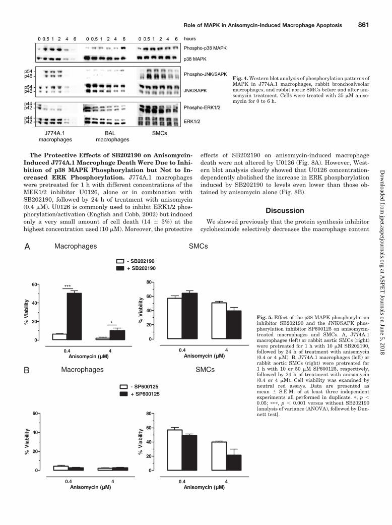
Anisomycin Altered Phosphorylation Patterns of Mi-togen-Activated Protein Kinases. Western blot analysisof MAPK signaling in macrophages (J774A.1 or BAL) andSMCs did not reveal major differences between the cell types(Fig. 4). Similar to J774A.1 macrophages, p38 MAPK andJNK/SAPK were hyperphosphorylated in primary rabbit
macrophages. Although JNK phosphorylation was muchmore difficult to pick up compared with p38 MAPK phosphor-ylation in primary rabbit macrophages. ERK1/2 was rapidlydephosphorylated in J774A.1 and BAL macrophages. InSMCs, p38 MAPK, JNK/SAPK, and ERK1/2 showed a tran-sient phosphorylation status (increased phosphorylation fol-lowed by dephosphorylation).
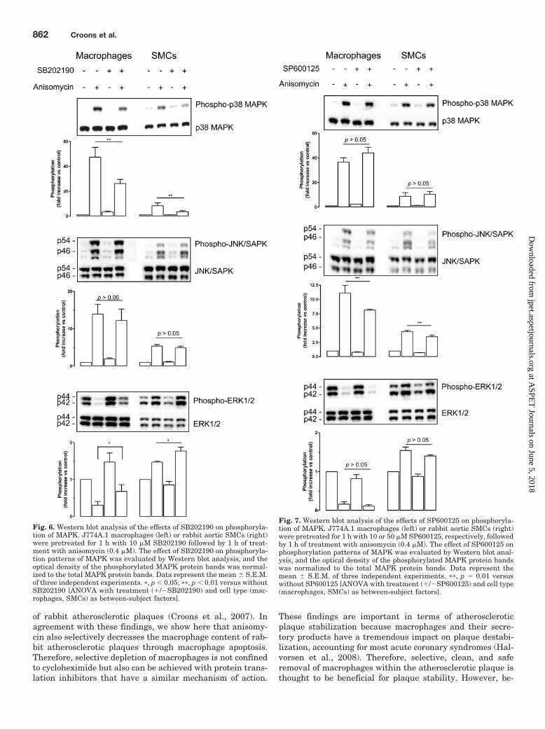
SB202190, but Not SP600125, Selectively PreventedAnisomycin-Induced Macrophage Death. J774A.1 mac-rophages or SMCs were preincubated with the p38 MAPKphosphorylation inhibitor SB202190 or the JNK/SAPK phos-phorylation inhibitor SP600125 for 1 h, followed by 24 h oftreatment with anisomycin (0.4 or 4 �M). SB202190 (10 �M)partially prevented anisomycin-induced macrophage celldeath (Fig. 5A). In contrast to macrophages, SB202190 wasunable to affect anisomycin-induced SMC death (Fig. 5A).SP600125 (10 or 50 �M, respectively) had no effect on aniso-
TABLE 1Effect of in vivo treatment of atherosclerotic rabbit carotid arteries with 4 or 20 �M anisomycin in the osmotic minipump for 3 days on intimaland medial areaData are shown as mean � S.E.M.; n � 5–11; p 0.05 (Kruskal-Wallis test).
Control
Anisomycin
4 �M 20 �M
mm²
Intimal area 0.25 � 0.04 0.26 � 0.03 0.20 � 0.04Medial area 0.60 � 0.04 0.58 � 0.02 0.56 � 0.03
Control AN 4 µM AN 20 µM
0
20
40
60
80
100
%αα αα
-SM
C a
ctin
in m
edia
Control AN 4 µM AN 20 µM
0
10
20
30
*
% R
AM
-11
in in
tim
a
Control AN 4 µM AN 20 µM
0
20
40
60
80
%αα αα
-SM
C a
ctin
in in
tim
a
A
B
Control AN 20 µM
Control AN 20 µM
I
M
I
M
Fig. 2. In vivo effect of anisomy-cin locally administered for 3days via an osmotic minipumpthat was connected to a collararound atherosclerotic rabbit ca-rotid arteries. A, immunoreac-tivity (left) and quantification(right) of RAM11 (brown � mac-rophages). B, immunoreactivity(left) and quantification (right)for �-SMC actin (brown �SMCs), treated for 3 days withsaline (control) or 4 or 20 �Manisomycin (AN) in the osmoticminipump. I, intima; M, media;bar, 50 �m. Individual valuesare shown with the median.�, p � 0.05 versus control(Kruskal-Wallis test followed byDunn’s multiple comparisontest).
Role of MAPK in Anisomycin-Induced Macrophage Apoptosis 859
at ASPE
T Journals on June 5, 2018
jpet.aspetjournals.orgD
ownloaded from

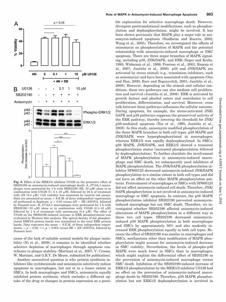
mycin-induced macrophage or SMC death (Fig. 5B). Westernblot analysis showed that expression levels of phosphorylatedp38 MAPK, JNK/SAPK, and ERK1/2 after 1 h of treatmentwith anisomycin (0.4 �M) were different between macro-phages and SMCs (p � 0.05, independent samples t test) (Fig.6). SB202190 (10 �M) decreased anisomycin-induced phos-phorylation of p38 MAPK in macrophages and in SMCs (Fig.6). SB202190 also modestly increased ERK1/2 phosphoryla-
tion but had no effect on JNK phosphorylation in both celltypes. SP600125 (10 or 50 �M, respectively) decreased ani-somycin-induced phosphorylation of JNK/SAPK in macro-phages and in SMCs and had no effect on the phosphoryla-tion patterns of p38 MAPK or ERK1/2 (Fig. 7). Inmacrophages, SP600125 (50 �M) further decreased anisomy-cin-induced JNK/SAPK phosphorylation (data not shown)but caused 80 � 6% cell death after 24 h of treatment.
Control AN 4 µM AN 20 µM
0
10
20**
# N
ucl
ear
frag
men
ts/1
0-2m
m²
inti
ma
Control AN 4 µM AN 20 µM
0
2
4
6
8
10
# N
ucl
ear
frag
men
ts/1
0-2m
m²
med
ia
A
B
C Control AN 20 µM
I
M
Control AN 20 µM
I
M
Fig. 3. Determination and characterization of cell death induced by AN (4 or 20 �M) locally administered for 3 days via an osmotic minipump thatwas connected to a collar around atherosclerotic rabbit carotid arteries. Nuclear fragments were counted in the intima (A) and in the media (B) on H/Estainings and expressed per 10�2 mm2. Individual values are shown with the median. �, p � 0.05 versus control (Kruskal-Wallis test followed byDunn’s multiple comparison test). C, apoptotic cells were visualized using fluorescein isothiocyanate-labeled Val-Ala-DL-Asp(O-methyl)-fluorometh-ylketone labeling (green), whereas smooth muscle cells were detected by immunohistochemical staining for �-SMC actin (red). Bar, 25 �m; I, intima;M, media; arrowheads, fragmented nuclei; triangles, internal elastic lamina.
860 Croons et al.
at ASPE
T Journals on June 5, 2018
jpet.aspetjournals.orgD
ownloaded from
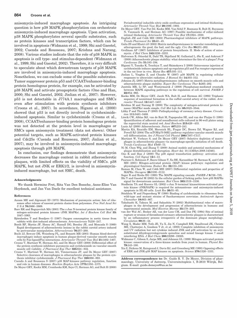
The Protective Effects of SB202190 on Anisomycin-Induced J774A.1 Macrophage Death Were Due to Inhi-bition of p38 MAPK Phosphorylation but Not to In-creased ERK Phosphorylation. J774A.1 macrophageswere pretreated for 1 h with different concentrations of theMEK1/2 inhibitor U0126, alone or in combination withSB202190, followed by 24 h of treatment with anisomycin(0.4 �M). U0126 is commonly used to inhibit ERK1/2 phos-phorylation/activation (English and Cobb, 2002) but inducedonly a very small amount of cell death (14 � 3%) at thehighest concentration used (10 �M). Moreover, the protective
effects of SB202190 on anisomycin-induced macrophagedeath were not altered by U0126 (Fig. 8A). However, West-ern blot analysis clearly showed that U0126 concentration-dependently abolished the increase in ERK phosphorylationinduced by SB202190 to levels even lower than those ob-tained by anisomycin alone (Fig. 8B).
DiscussionWe showed previously that the protein synthesis inhibitor
cycloheximide selectively decreases the macrophage content
Fig. 4. Western blot analysis of phosphorylation patterns ofMAPK in J774A.1 macrophages, rabbit bronchoalveolarmacrophages, and rabbit aortic SMCs before and after ani-somycin treatment. Cells were treated with 35 �M aniso-mycin for 0 to 6 h.
0.4 4
0
20
40
60
80
Anisomycin (µM)
% V
iab
ility
sCMSsegahporcaM
0.4 4
0
20
40
60
- SB202190+ SB202190
***
*
Anisomycin (µM)
% V
iab
ility
A
B sCMSsegahporcaM
0.4 4
0
20
40
60
- SP600125+ SP600125
Anisomycin (µM)
% V
iab
ility
0.4 4
0
20
40
60
80
Anisomycin (µM)
% V
iab
ility
Fig. 5. Effect of the p38 MAPK phosphorylationinhibitor SB202190 and the JNK/SAPK phos-phorylation inhibitor SP600125 on anisomycin-treated macrophages and SMCs. A, J774A.1macrophages (left) or rabbit aortic SMCs (right)were pretreated for 1 h with 10 �M SB202190,followed by 24 h of treatment with anisomycin(0.4 or 4 �M). B, J774A.1 macrophages (left) orrabbit aortic SMCs (right) were pretreated for1 h with 10 or 50 �M SP600125, respectively,followed by 24 h of treatment with anisomycin(0.4 or 4 �M). Cell viability was examined byneutral red assays. Data are presented asmean � S.E.M. of at least three independentexperiments all performed in duplicate. �, p �0.05; ���, p � 0.001 versus without SB202190[analysis of variance (ANOVA), followed by Dun-nett test].
Role of MAPK in Anisomycin-Induced Macrophage Apoptosis 861
at ASPE
T Journals on June 5, 2018
jpet.aspetjournals.orgD
ownloaded from
of rabbit atherosclerotic plaques (Croons et al., 2007). Inagreement with these findings, we show here that anisomy-cin also selectively decreases the macrophage content of rab-bit atherosclerotic plaques through macrophage apoptosis.Therefore, selective depletion of macrophages is not confinedto cycloheximide but also can be achieved with protein trans-lation inhibitors that have a similar mechanism of action.
These findings are important in terms of atheroscleroticplaque stabilization because macrophages and their secre-tory products have a tremendous impact on plaque destabi-lization, accounting for most acute coronary syndromes (Hal-vorsen et al., 2008). Therefore, selective, clean, and saferemoval of macrophages within the atherosclerotic plaque isthought to be beneficial for plaque stability. However, be-
Fig. 6. Western blot analysis of the effects of SB202190 on phosphoryla-tion of MAPK. J774A.1 macrophages (left) or rabbit aortic SMCs (right)were pretreated for 1 h with 10 �M SB202190 followed by 1 h of treat-ment with anisomycin (0.4 �M). The effect of SB202190 on phosphoryla-tion patterns of MAPK was evaluated by Western blot analysis, and theoptical density of the phosphorylated MAPK protein bands was normal-ized to the total MAPK protein bands. Data represent the mean � S.E.M.of three independent experiments. �, p � 0.05; ��, p � 0.01 versus withoutSB202190 [ANOVA with treatment (/�SB202190) and cell type (mac-rophages, SMCs) as between-subject factors].
Fig. 7. Western blot analysis of the effects of SP600125 on phosphoryla-tion of MAPK. J774A.1 macrophages (left) or rabbit aortic SMCs (right)were pretreated for 1 h with 10 or 50 �M SP600125, respectively, followedby 1 h of treatment with anisomycin (0.4 �M). The effect of SP600125 onphosphorylation patterns of MAPK was evaluated by Western blot anal-ysis, and the optical density of the phosphorylated MAPK protein bandswas normalized to the total MAPK protein bands. Data represent themean � S.E.M. of three independent experiments. ��, p � 0.01 versuswithout SP600125 [ANOVA with treatment (/�SP600125) and cell type(macrophages, SMCs) as between-subject factors].
862 Croons et al.
at ASPE
T Journals on June 5, 2018
jpet.aspetjournals.orgD
ownloaded from
cause of the lack of suitable animal models for plaque insta-bility (Ni et al., 2009), it remains to be identified whetherselective depletion of macrophages through apoptosis con-tributes to plaque stability (Martinet et al., 2007; V. Croons,W. Martinet, and G.R.Y. De Meyer, submitted for publication).
Another unresolved question is why protein synthesis in-hibitors like cycloheximide and anisomycin selectively induceapoptosis in macrophages, but not or to a lesser extent inSMCs. In both macrophages and SMCs, anisomycin equallyinhibited protein synthesis, which excludes a different up-take of the drug or changes in protein expression as a possi-
ble explanation for selective macrophage death. However,divergent posttranslational modifications, such as phosphor-ylation and dephosphorylation, might be involved. It hasbeen shown previously that MAPK play a major role in ani-somycin-induced apoptosis (Stadheim and Kucera, 2002;Wang et al., 2005). Therefore, we investigated the effects ofanisomycin on phosphorylation of MAPK and the potentialrelationship with anisomycin-induced macrophage or SMCapoptosis. There are three major branches of MAPK signal-ing, including p38, JNK/SAPK, and ERK (Seger and Krebs,1995; Widmann et al., 1999; Pearson et al., 2001; Raman etal., 2007; Junttila et al., 2008). p38 and JNK/SAPK areactivated by stress stimuli (e.g., translation inhibitors, suchas anisomycin) and have been associated with apoptosis (Onoand Han, 2000; Barr and Bogoyevitch, 2001; Junttila et al.,2008). However, depending on the stimuli and cellular con-ditions, these two pathways can also mediate cell prolifera-tion and survival (Junttila et al., 2008). ERK is activated bygrowth factors and phorbol esters and contributes to cellproliferation, differentiation, and survival. Moreover, crosstalk between these pathways influences the cellular outcome.During apoptosis, for example, the stress-activated JNK/SAPK and p38 pathways suppress the prosurvival activity ofthe ERK pathway, thereby lowering the threshold for JNK/p38-mediated apoptosis (Xia et al., 1995; Junttila et al.,2008). In this study, anisomycin modified phosphorylation ofthe three MAPK branches in both cell types. p38 MAPK andJNK/SAPK were hyperphosphorylated in macrophages,whereas ERK1/2 was rapidly dephosphorylated. In SMCs,p38 MAPK, JNK/SAPK, and ERK1/2 showed a transientphosphorylation status (increased phosphorylation followedby dephosphorylation). To further elucidate the involvementof MAPK phosphorylation in anisomycin-induced macro-phage and SMC death, we subsequently used inhibitors ofMAPK phosphorylation. The JNK/SAPK phosphorylation in-hibitor SP600125 decreased anisomycin-induced JNK/SAPKphosphorylation to a similar extent in both cell types and didnot have an effect on the other MAPK phosphorylation pat-terns. Pretreatment of macrophages or SMCs with SP600125did not affect anisomycin-induced cell death. Therefore, JNK/SAPK phosphorylation is not involved in anisomycin-inducedmacrophage or SMC apoptosis. In contrast, the p38 MAPKphosphorylation inhibitor SB202190 prevented anisomycin-induced macrophage but not SMC death. Therefore, we in-vestigated whether SB202190 affected anisomycin-inducedalterations of MAPK phosphorylation in a different way inthese two cell types. SB202190 decreased anisomycin-induced p38 MAPK phosphorylation in both macrophagesand SMCs by approximately 50% and also modestly in-creased ERK phosphorylation equally in both cell types. Be-cause the effect of SB202190 was similar in macrophages andSMCs, mechanisms other than modification of MAPK phos-phorylation might account for anisomycin-induced decreasein SMC viability. Nevertheless, the levels of phospho-p38MAPK were much lower in SMCs than in macrophages,which might explain the differential effect of SB202190 inthe prevention of anisomycin-induced macrophage versusSMC death. Inhibition of the SB202190-induced increase ofERK1/2 phosphorylation by the MEK1/2 inhibitor U0126 hadno effect on the prevention of anisomycin-induced macro-phage death by SB202190. Therefore, p38 MAPK phosphor-ylation but not ERK1/2 dephosphorylation is involved in
Fig. 8. Effect of the MEK1/2 inhibitor U0126 on the protective effect ofSB202190 on anisomycin-induced macrophage death. A, J774A.1 macro-phages were pretreated for 1 h with SB202190 (SB, 10 �M) alone or incombination with U0126 (U, 0.1–10 �M), followed by 24 h of treatmentwith AN (0.4 �M). Cell viability was examined by neutral red assays.Data are presented as mean � S.E.M. of three independent experimentsall performed in duplicate. p 0.05 versus AN SB (ANOVA, followedby Dunnett test). B, J774A.1 macrophages were pretreated for 1 h withSB202190 (10 �M) alone or in combination with U0126 (0.1–10 �M)followed by 1 h of treatment with anisomycin (0.4 �M). The effect ofU0126 on the SB202190-induced increase in ERK phosphorylation wasevaluated by Western blot analysis. The optical density of the phosphor-ylated ERK1/2 protein bands was normalized to the total ERK proteinbands. Data represent the mean � S.E.M. of three independent experi-ments. �, p � 0.05; ���, p � 0.001 versus SB AN (ANOVA, followed byDunnett test).
Role of MAPK in Anisomycin-Induced Macrophage Apoptosis 863
at ASPE
T Journals on June 5, 2018
jpet.aspetjournals.orgD
ownloaded from
anisomycin-induced macrophage apoptosis. An intriguingquestion is how p38 MAPK phosphorylation can orchestrateanisomycin-induced macrophage apoptosis. Upon activation,p38 MAPK phosphorylates several specific substrates, suchas protein kinases and transcription factors, which can beinvolved in apoptosis (Widmann et al., 1999; Shi and Gaestel,2002; Cuenda and Rousseau, 2007; Krishna and Narang,2008). Various studies indicate that the role of p38 MAPK inapoptosis is cell type- and stimulus-dependent (Widmann etal., 1999; Shi and Gaestel, 2002). Therefore, it is very difficultto speculate about which downstream targets of p38 MAPKare involved in anisomycin-induced macrophage apoptosis.Nonetheless, we can exclude some of the possible substrates.Tumor suppressor protein p53 and CCAAT/enhancer-bindingprotein homologous protein, for example, can be activated byp38 MAPK and activate proapoptotic factors (Ono and Han,2000; Shi and Gaestel, 2002; Jinlian et al., 2007). However,p53 is not detectable in J774A.1 macrophages and SMCseven after stimulation with protein synthesis inhibitors(Croons et al., 2007). In accordance, Higami et al. (2000)showed that p53 is not directly involved in cycloheximide-induced apoptosis. Similar to cycloheximide (Croons et al.,2008), CCAAT/enhancer-binding protein homologous proteinwas not detected at the protein level in macrophages orSMCs upon anisomycin treatment (data not shown). Otherpotential targets, such as MAPK-activated protein kinasesand Cdc25s (Cuenda and Rousseau, 2007; Jinlian et al.,2007), may be involved in anisomycin-induced macrophageapoptosis through p38 MAPK.
In conclusion, our findings demonstrate that anisomycindecreases the macrophage content in rabbit atheroscleroticplaques, with limited effects on the viability of SMCs. p38MAPK, but not JNK or ERK1/2, is involved in anisomycin-induced macrophage, but not SMC, death.
Acknowledgments
We thank Hermine Fret, Rita Van Den Bossche, Anne-Elise VanHoydonck, and Jan Van Daele for excellent technical assistance.
ReferencesAzzam ME and Algranati ID (1973) Mechanism of puromycin action: fate of ribo-
somes after release of nascent protein chains from polysomes. Proc Natl Acad SciU S A 70:3866–3869.
Barr RK and Bogoyevitch MA (2001) The c-Jun N-terminal protein kinase family ofmitogen-activated protein kinases (JNK MAPKs). Int J Biochem Cell Biol 33:1047–1063.
Bjornheden T and Bondjers G (1987) Oxygen consumption in aortic tissue fromrabbits with diet-induced atherosclerosis. Arteriosclerosis 7:238–247.
Booth RF, Martin JF, Honey AC, Hassall DG, Beesley JE, and Moncada S (1989)Rapid development of atherosclerotic lesions in the rabbit carotid artery inducedby perivascular manipulation. Atherosclerosis 76:257–268.
Boyle JJ, Bowyer DE, Weissberg PL, and Bennett MR (2001) Human blood-derivedmacrophages induce apoptosis in human plaque-derived vascular smooth musclecells by Fas-ligand/Fas interactions. Arterioscler Thromb Vasc Biol 21:1402–1407.
Croons V, Martinet W, Herman AG, and De Meyer GRY (2008) Differential effect ofthe protein synthesis inhibitors puromycin and cycloheximide on vascular smoothmuscle cell viability. J Pharmacol Exp Ther 325:824–832.
Croons V, Martinet W, Herman AG, Timmermans JP, and De Meyer GRY (2007)Selective clearance of macrophages in atherosclerotic plaques by the protein syn-thesis inhibitor cycloheximide. J Pharmacol Exp Ther 320:986–993.
Cuenda A and Rousseau S (2007) p38 MAP-kinases pathway regulation, functionand role in human diseases. Biochim Biophys Acta 1773:1358–1375.
De Meyer GRY, Kockx MM, Cromheeke KM, Seye CI, Herman AG, and Bult H (2000)
Periadventitial inducible nitric oxide synthase expression and intimal thickening.Arterioscler Thromb Vasc Biol 20:1896–1902.
De Meyer GRY, Van Put DJ, Kockx MM, Van Schil P, Bosmans R, Bult H, BuyssensN, Vanmaele R, and Herman AG (1997) Possible mechanisms of collar-inducedintimal thickening. Arterioscler Thromb Vasc Biol 17:1924–1930.
English JM and Cobb MH (2002) Pharmacological inhibitors of MAPK pathways.Trends Pharmacol Sci 23:40–45.
Galis ZS and Khatri JJ (2002) Matrix metalloproteinases in vascular remodeling andatherogenesis: the good, the bad, and the ugly. Circ Res 90:251–262.
Grollman AP (1967) Inhibitors of protein biosynthesis. II. Mode of action of aniso-mycin. J Biol Chem 242:3226–3233.
Halvorsen B, Otterdal K, Dahl TB, Skjelland M, Gullestad L, Øie E, and Aukrust P(2008) Atherosclerotic plaque stability: what determines the fate of a plaque? ProgCardiovasc Dis 51:183–194.
Higami Y, Tanaka K, Tsuchiya T, and Shimokawa I (2000) Intravenous injection ofcycloheximide induces apoptosis and up-regulates p53 and Fas receptor expressionin the rat liver in vivo. Mutat Res 457:105–111.
Jinlian L, Yingbin Z, and Chunbo W (2007) p38 MAPK in regulating cellularresponses to ultraviolet radiation. J Biomed Sci 14:303–312.
Johnson JL (2007) Matrix metalloproteinases: influence on smooth muscle cells andatherosclerotic plaque stability. Expert Rev Cardiovasc Ther 5:265–282.
Junttila MR, Li SP, and Westermarck J (2008) Phosphatase-mediated crosstalkbetween MAPK signaling pathways in the regulation of cell survival. FASEB J22:954–965.
Kockx MM, De Meyer GRY, Jacob WA, Bult H, and Herman AG (1992) Triphasicsequence of neointimal formation in the cuffed carotid artery of the rabbit. Arte-rioscler Thromb 12:1447–1457.
Krishna M and Narang H (2008) The complexity of mitogen-activated protein ki-nases (MAPKs) made simple. Cell Mol Life Sci 65:3525–3544.
Libby P and Theroux P (2005) Pathophysiology of coronary artery disease. Circula-tion 111:3481–3488.
Lowik CW, Alblas MJ, van de Ruit M, Papapoulos SE, and van der Pluijm G (1993)Quantification of adherent and nonadherent cells cultured in 96-well plates usingthe supravital stain neutral red. Anal Biochem 213:426–433.
Lusis AJ (2000) Atherosclerosis. Nature 407:233–241.Martin KA, Rzucidlo EM, Merenick BL, Fingar DC, Brown DJ, Wagner RJ, and
Powell RJ (2004) The mTOR/p70 S6K1 pathway regulates vascular smooth musclecell differentiation. Am J Physiol Cell Physiol 286:C507–C517.
Martinet W, Verheye S, and De Meyer GRY (2007) Selective depletion of macro-phages in atherosclerotic plaques via macrophage-specific initiation of cell death.Trends Cardiovasc Med 17:69–75.
Ni M, Chen WQ, and Zhang Y (2009) Animal models and potential mechanisms ofplaque destabilization and disruption. Heart doi: 10.1136/hrt.2008.143461.
Ono K and Han J (2000) The p38 signal transduction pathway: activation andfunction. Cell Signal 12:1–13.
Pearson G, Robinson F, Beers Gibson T, Xu BE, Karandikar M, Berman K, and CobbMH (2001) Mitogen-activated protein (MAP) kinase pathways: regulation andphysiological functions. Endocr Rev 22:153–183.
Raman M, Chen W, and Cobb MH (2007) Differential regulation and properties ofMAPKs. Oncogene 26:3100–3112.
Seger R and Krebs EG (1995) The MAPK signaling cascade. FASEB J 9:726–735.Shi Y and Gaestel M (2002) In the cellular garden of forking paths: how p38 MAPKs
signal for downstream assistance. Biol Chem 383:1519–1536.Stadheim TA and Kucera GL (2002) c-Jun N-terminal kinase/stress-activated pro-
tein kinase (JNK/SAPK) is required for mitoxantrone- and anisomycin-inducedapoptosis in HL-60 cells. Leuk Res 26:55–65.
Stocklein W and Piepersberg W (1980) Binding of cycloheximide to ribosomes fromwild-type and mutant strains of Saccharomyces cerevisiae. Antimicrob AgentsChemother 18:863–867.
Takahashi K, Takeya M, and Sakashita N (2002) Multifunctional roles of macro-phages in the development and progression of atherosclerosis in humans andexperimental animals. Med Electron Microsc 35:179–203.
van der Wal AC, Becker AE, van der Loos CM, and Das PK (1994) Site of intimalrupture or erosion of thrombosed coronary atherosclerotic plaques is characterizedby an inflammatory process irrespective of the dominant plaque morphology.Circulation 89:36–44.
Wang X, Mader MM, Toth JE, Yu X, Jin N, Campbell RM, Smallwood JK, ChristeME, Chatterjee A, Goodson T Jr, et al. (2005) Complete inhibition of anisomycinand UV radiation but not cytokine induced JNK and p38 activation by an aryl-substituted dihydropyrrolopyrazole quinoline and mixed lineage kinase 7 smallinterfering RNA. J Biol Chem 280:19298–19305.
Widmann C, Gibson S, Jarpe MB, and Johnson GL (1999) Mitogen-activated proteinkinase: conservation of a three-kinase module from yeast to human. Physiol Rev79:143–180.
Xia Z, Dickens M, Raingeaud J, Davis RJ, and Greenberg ME (1995) Opposing effectsof ERK and JNK-p38 MAP kinases on apoptosis. Science 270:1326–1331.
Address correspondence to: Dr. Guido R. Y. De Meyer, Division of phar-macology, University of Antwerp, Universiteitsplein 1, B-2610 Wilrijk, Bel-gium. E-mail: [email protected]
864 Croons et al.
at ASPE
T Journals on June 5, 2018
jpet.aspetjournals.orgD
ownloaded from


![Sorafenib, a multikinase inhibitor, induces formation of ...€¦ · Sorafenib (Nexavar®), an Raf1/Mek/Erk kinase inhibitor approved for advanced hepatocarcinoma cells (HCC) [35],](https://img.pdfslide.us/doc/110x75/5f439704553a106cae7db5ff/sorafenib-a-multikinase-inhibitor-induces-formation-of-sorafenib-nexavar.jpg)