Embed Size (px)
Citation preview
Kidney transplant is the method of choice for
treating terminal renal failure. The timely trans
plant of a suitable donor kidney is the only way to
ensure optimal medical management and social
participation of affected patients. A kidney graft
is susceptible to numerous postoperative com
plications and deleterious effects and therefore
requires close postoperative followup. Ultrasound
(US) is the most widely employed noninvasive
modality for the postoperative assessment of kidney
grafts. Contrastenhanced ultrasound (CEUS) is
a promising and straightforward method that is
superior to established sonographic techniques
such as conventional Bmode scanning (volume
measurement, demonstration of hematoma) and
color Doppler (rejection, perfusion defects, vascu
larization) in the diagnostic evaluation of kidney
grafts. Moreover, CEUS potentially allows tumor
characterization in transplant and shrunken
kidneys. A single contrastenhanced ultrasound
examination can answer a variety of questions in
the early postoperative phase (rejection) and long
term followup (chronic damage). Initial studies
indicate that efficient and early diagnosis of rejec
tion or acute tubular necrosis (ATN) is possible as
these conditions show characteristic bolus kinetics
curves. Surgical complications like perfusion
defects secondary to thrombosis of a polar artery
or postoperative hematoma are also identified.
Moreover, perfusion effects of a hematoma can
be assessed. Further technical developments of
ultrasound equipment will trigger new applications
of CEUS. The rapid technical advances seen in
recent years led to the introduction of many new
software tools for the analysis of raw data sets or
improved visualization of microbubbles at very low
energy, for instance by means of techniques that
accumulate and depict the microbubble signals
over time such as MicroFlow Imaging by Toshiba.
This new technique in turn laid the foundation for
the socalled Parametric Imaging (Fig. 5) which
relies on bolus or replenishment kinetics and is
able to analyze individual curve parameters such
as timetopeak on a pixel by pixel basis. The
information is displayed in a colorcoded image
that presents all US data in a standardized manner.
This simple and fast technique offers high diag
nostic yield that may improve the acceptance of
CEUS as a routine diagnostic tool that provides
all diagnostic information on contrast medium
dynamics in a single image of the transplant
kidney.
Thomas Fischer
Ultrasound Research Laboratory, Department of Radiology
Charité – University Medicine Berlin, Central Campus
Berlin, Germany
The Role of CEUS in the Assessment of Renal Graft – Immediate and Long-Term Transplant Follow-Up
Fig. 1: Temporal course of contrast inflow after bolus administration of 1.6 ml of
SonoVue™ via the left cubital vein. Patient with good graft function. Analysis shows
that signal intensity in the cortex increases as early as 1 s (green color) after the
increase in the main renal artery (red color).
Fig. 2: Delayed inflow in the renal cortex in a patient with histologically proven
rejection. Arterial inflow of the contrast medium is shown. Analysis shows that the
signal intensity in the cortex increases as late as 4 s after the increase in the main
renal artery.
ULTRASOUND CT MRI X-RAY SERVICES
www.toshibamedical.eu
© Toshiba Medical Systems Corporation 2011 all rights reserved.Design and specifications subject to change without notice.02/2011 TCSUS0010EC.EU
Printed in Europe
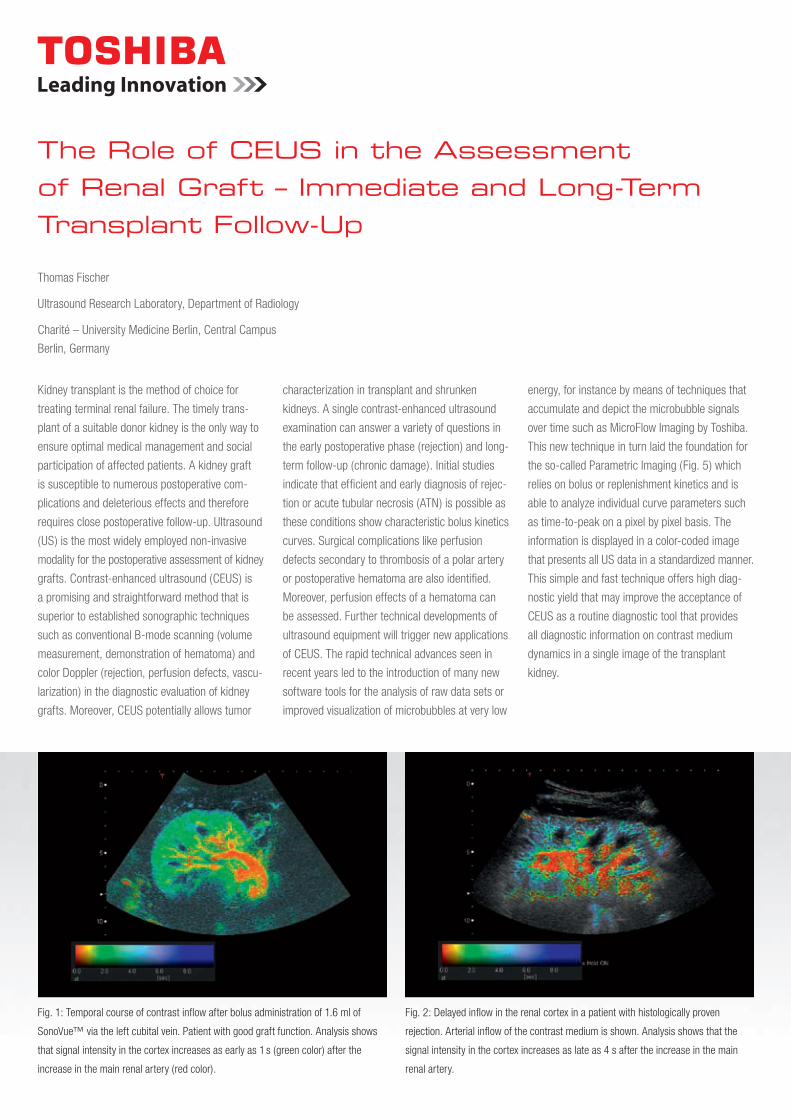
Fig. 3: Following contrast administration, a perfusion defect is well depicted in the
polar area covering about 30 %.
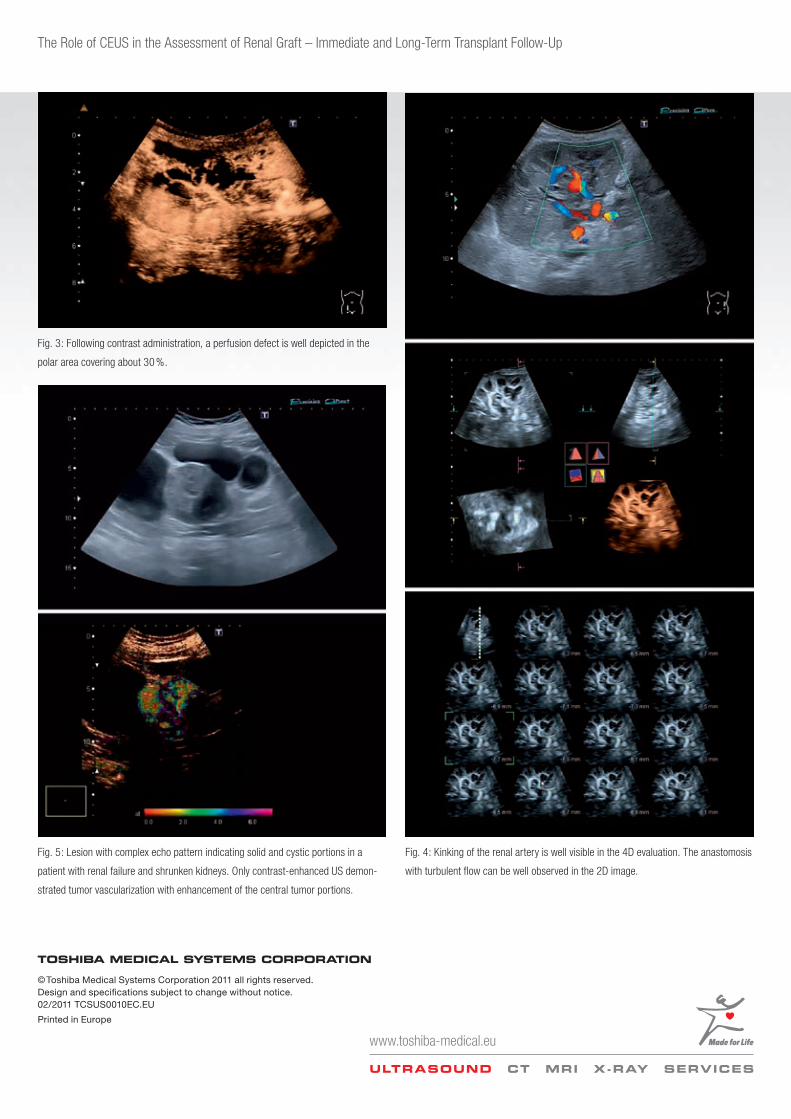
Fig. 5: Lesion with complex echo pattern indicating solid and cystic portions in a
patient with renal failure and shrunken kidneys. Only contrastenhanced US demon
strated tumor vascularization with enhancement of the central tumor portions.
Fig. 4: Kinking of the renal artery is well visible in the 4D evaluation. The anastomosis
with turbulent flow can be well observed in the 2D image.
The Role of CEUS in the Assessment of Renal Graft – Immediate and LongTerm Transplant FollowUp