Embed Size (px)
DESCRIPTION
The Respiratory System. Chapter 13. The term respiration has a broad meaning. Internal respiration refers to the metabolic processes occurring in the mitochondria. Molecular oxygen is used by tissue cells. Cardon dioxide is produced. - PowerPoint PPT Presentation
Citation preview
The Respiratory System
Chapter 13
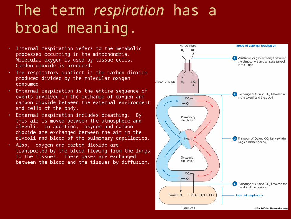
The term respiration has a broad meaning.
• Internal respiration refers to the metabolic processes occurring in the mitochondria. Molecular oxygen is used by tissue cells. Cardon dioxide is produced.
• The respiratory quotient is the carbon dioxide produced divided by the molecular oxygen consumed.
• External respiration is the entire sequence of events involved in the exchange of oxygen and carbon dioxide between the external environment and cells of the body.
• External respiration includes breathing. By this air is moved between the atmosphere and alveoli. In addition, oxygen and carbon dioxide are exchanged between the air in the alveoli and blood of the pulmonary capillaries.
• Also, oxygen and carbon dioxide are transported by the blood flowing from the lungs to the tissues. These gases are exchanged between the blood and the tissues by diffusion.
The respiratory system carries out nonrespiratory functions.
• It provides a route for water loss and heat elimination.• It enhances venous return.• It contributes to the maintenance of normal acid-base
balance.• It enables various kinds of vocalizations.• It defends against inhaled foreign matter.• It modifies, activates, and inactivates materials
passing through the circulatory system.
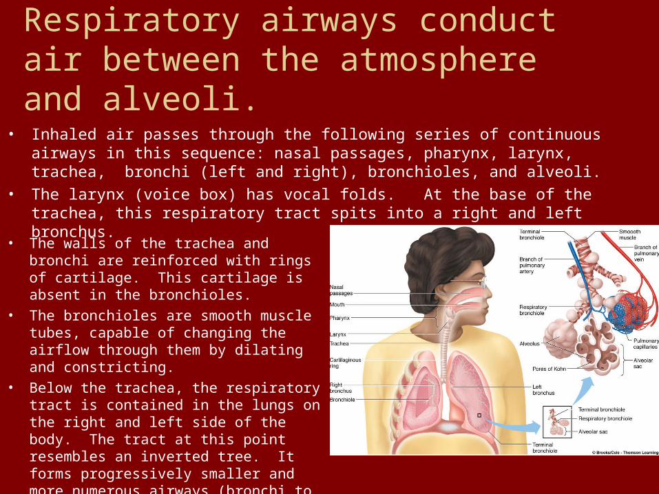
Respiratory airways conduct air between the atmosphere and alveoli.
• The walls of the trachea and bronchi are reinforced with rings of cartilage. This cartilage is absent in the bronchioles.
• The bronchioles are smooth muscle tubes, capable of changing the airflow through them by dilating and constricting.
• Below the trachea, the respiratory tract is contained in the lungs on the right and left side of the body. The tract at this point resembles an inverted tree. It forms progressively smaller and more numerous airways (bronchi to bronchioles to alveoli).
• Inhaled air passes through the following series of continuous airways in this sequence: nasal passages, pharynx, larynx, trachea, bronchi (left and right), bronchioles, and alveoli.
• The larynx (voice box) has vocal folds. At the base of the trachea, this respiratory tract spits into a right and left bronchus.
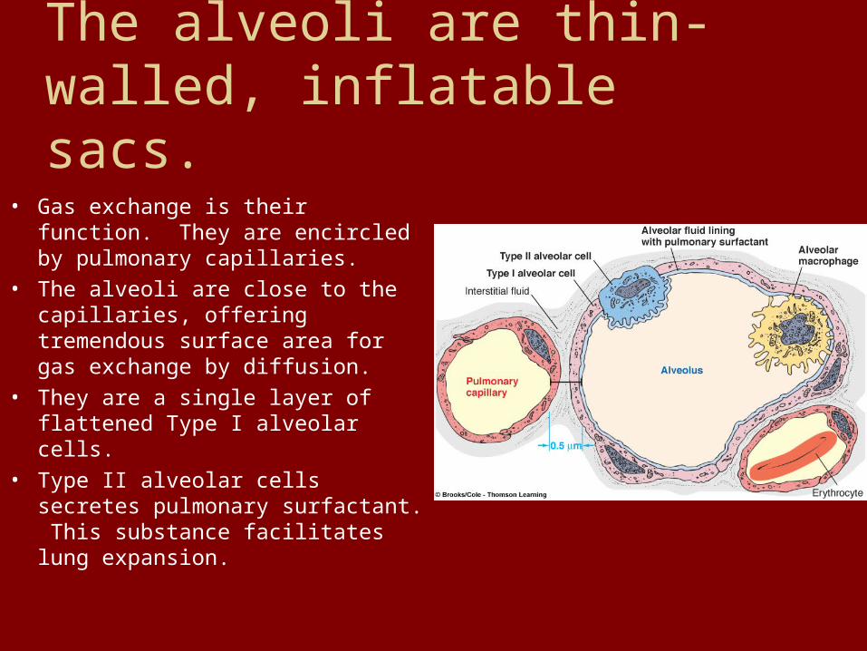
The alveoli are thin-walled, inflatable sacs.
• Gas exchange is their function. They are encircled by pulmonary capillaries.
• The alveoli are close to the capillaries, offering tremendous surface area for gas exchange by diffusion.
• They are a single layer of flattened Type I alveolar cells.
• Type II alveolar cells secretes pulmonary surfactant. This substance facilitates lung expansion.
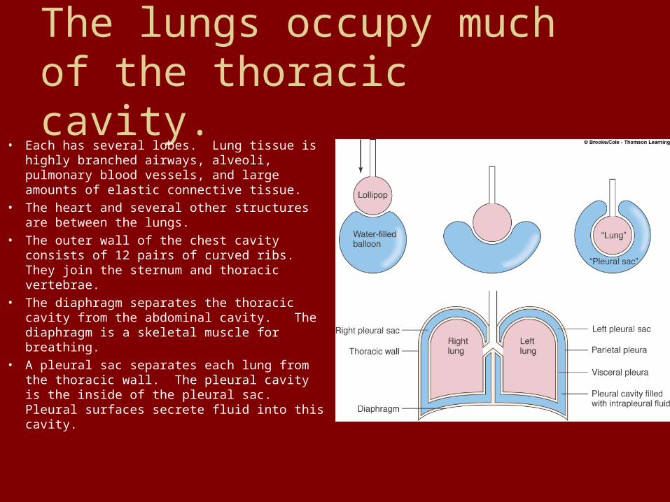
The lungs occupy much of the thoracic cavity.
• Each has several lobes. Lung tissue is highly branched airways, alveoli, pulmonary blood vessels, and large amounts of elastic connective tissue.
• The heart and several other structures are between the lungs.
• The outer wall of the chest cavity consists of 12 pairs of curved ribs. They join the sternum and thoracic vertebrae.
• The diaphragm separates the thoracic cavity from the abdominal cavity. The diaphragm is a skeletal muscle for breathing.
• A pleural sac separates each lung from the thoracic wall. The pleural cavity is the inside of the pleural sac. Pleural surfaces secrete fluid into this cavity.
There are several pressures inside and outside the lungs.
• Atmospheric pressure is produced by the weight of the air on objects on the surface of the Earth. It is 760 mm of Hg at sea level and decreases with increasing altitude above sea level.
• Intra-alveolar (intrapulmonary) pressure is the pressure in the alveoli.
• Intrapleural pressure is in the intrapleural cavity. It averages 756 mm Hg at rest. This is also written as minus 4, as it is four units below 760 in the atmosphere. It has a slight vacuum compared to normal atmospheric pressure.
• This is lost during pneumothorax.
• The lungs are normally stretched, filling the large thorax. This is due, in part, to the intrapleural fluid’s cohesiveness. This stickiness pulls the lungs outward.
• Also, a transmural pressure pushes the lungs outward. This is produced by an intra-alveolar pressure that is greater than the pressure outside the alveoli.
– See Figures 13-6 and 13-8
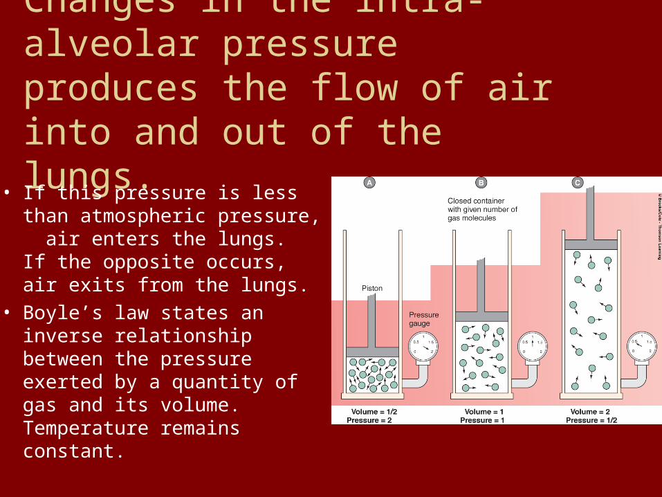
Changes in the intra-alveolar pressure produces the flow of air into and out of the lungs.
• If this pressure is less than atmospheric pressure, air enters the lungs. If the opposite occurs, air exits from the lungs.
• Boyle’s law states an inverse relationship between the pressure exerted by a quantity of gas and its volume. Temperature remains constant.
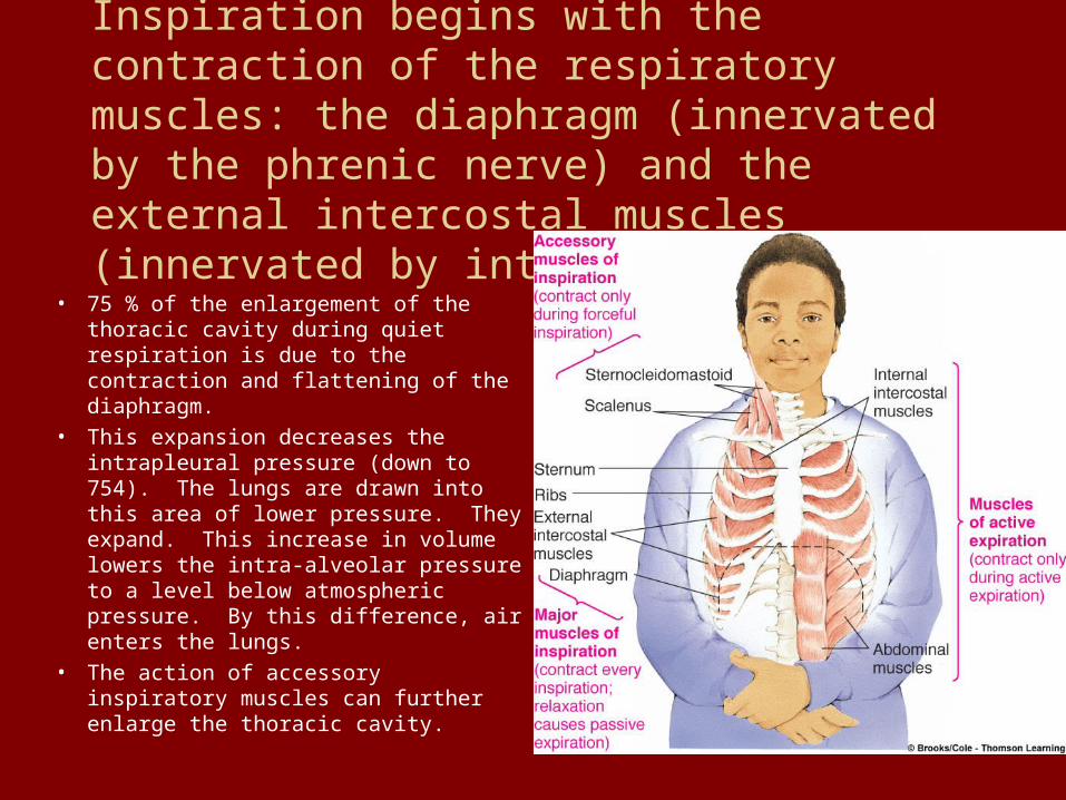
Inspiration begins with the contraction of the respiratory muscles: the diaphragm (innervated by the phrenic nerve) and the external intercostal muscles (innervated by intercostal nerves).
• 75 % of the enlargement of the thoracic cavity during quiet respiration is due to the contraction and flattening of the diaphragm.
• This expansion decreases the intrapleural pressure (down to 754). The lungs are drawn into this area of lower pressure. They expand. This increase in volume lowers the intra-alveolar pressure to a level below atmospheric pressure. By this difference, air enters the lungs.
• The action of accessory inspiratory muscles can further enlarge the thoracic cavity.
The onset of expiration begins with the relaxation of the inspiratory muscles.
• Relaxation of the diaphragm and the muscles of the chest wall, plus the elastic recoil of the alveoli, decrease the size of the chest cavity.
• The intrapleural pressure increases and the lungs are compressed. • The intra-alveolar pressure increases. When it increases to a level
above atmospheric pressure, air is driven out - an expiration. • Forced expiration can occur by the contraction of expiratory muscles. • These skeletal muscles are ones in the abdominal wall and the
internal intercostal muscles. Their contraction further increases the pressure gradient between the alveoli (greater pressure) and the atmosphere.– See Figure 13-12
Airway resistance in the respiratory tract influences the rate of airflow.
• F = delta P
R
• As the difference between the atmospheric and intra-alveolar pressures (delta P) is greater, the air flow is greater. This relationship is a direct proportion.
• However, if the resistance (R) increases, the airflow is decreased (inverse proportion.
• The major determinant of resistance is the radius of the conducting airways. The autonomic nervous system controls the contraction of the smooth muscle in the walls of the bronchioles, changing their radii.
• Sympathetic stimulation and epinephrine cause bronchodilation.
• Airway resistance is increased abnormally with chronic obstructive pulmonary disease. Expiration is more difficult than inspiration. Chronic bronchitis is the long-term inflammatory condition of the respiratory airways.
• Asthma is the obstruction of the airways due to inflammation. Emphysema is the collapse of the alveoli.
The lungs have elastic behavior.• The lungs have elastic recoil, rebounding if they are stretched. • Compliance is the effort required to stretch or distend the lungs. A thin, toy
balloon is more compliant than a thick, rubber balloon.• A highly-compliant lung stretches further for a given increase in pressure than
a lung with less compliance. • Numerous factors decrease lung compliance.• Pulmonary elastic behavior depends on the pulmonary elastic behavior and
alveolar surface tension. This tension is determined by the thin liquid film that lines the outside of each alveolus.
• This film allows the alveolus to resist expansion. This film also squeezes the alveolus, producing recoil.
• A coating of pulmonary surfactant prevents the alveoli from collapsing from this surface tension.
• An insufficient amount of pulmonary surfactant can produce newborn respiratory distress syndrome.
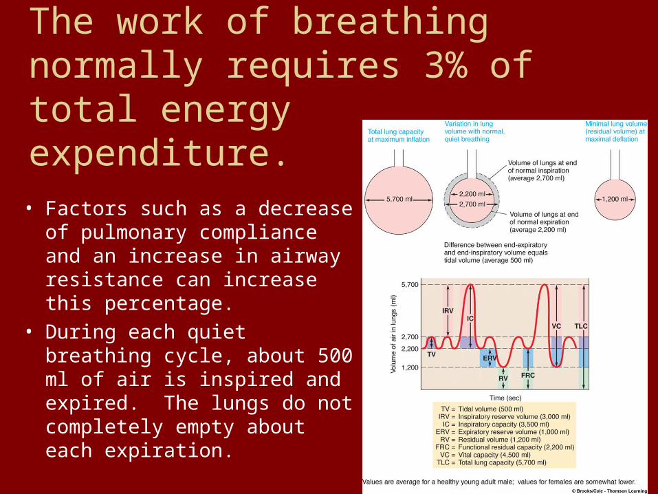
The work of breathing normally requires 3% of total energy expenditure.
• Factors such as a decrease of pulmonary compliance and an increase in airway resistance can increase this percentage.
• During each quiet breathing cycle, about 500 ml of air is inspired and expired. The lungs do not completely empty about each expiration.
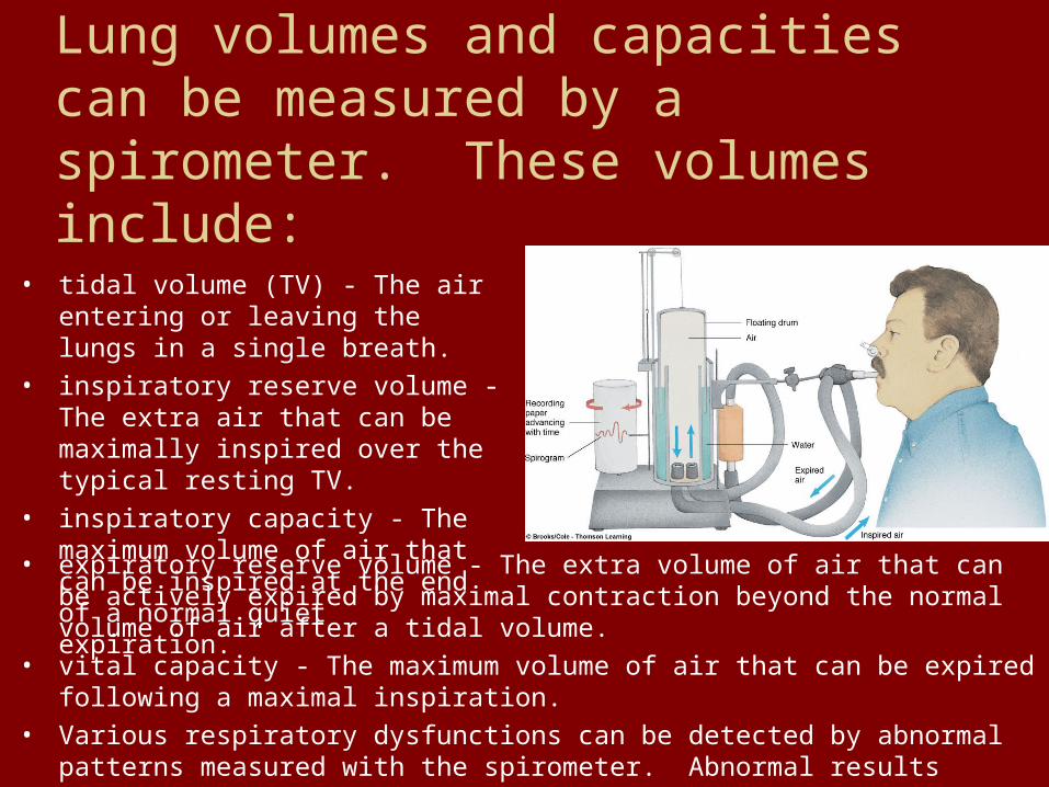
Lung volumes and capacities can be measured by a spirometer. These volumes include:
• tidal volume (TV) - The air entering or leaving the lungs in a single breath.
• inspiratory reserve volume - The extra air that can be maximally inspired over the typical resting TV.
• inspiratory capacity - The maximum volume of air that can be inspired at the end of a normal quiet expiration.
• expiratory reserve volume - The extra volume of air that can be actively expired by maximal contraction beyond the normal volume of air after a tidal volume.
• vital capacity - The maximum volume of air that can be expired following a maximal inspiration.
• Various respiratory dysfunctions can be detected by abnormal patterns measured with the spirometer. Abnormal results include obstructive lung disease and restrictive lung disease.
Pulmonary ventilation is the tidal volume x respiratory rate.• Alveolar ventilation is less because of the anatomic dead space.
These are the airways where air is not available for gas exchange. • Due to this dead space:• alveolar ventilation = • (tidal volume - dead space volume) x respiratory rate• Breathing patterns (e.g., deep and slow) can affect alveolar
ventilation. • An alveolar dead space also exists, but it is usually small. • There are local controls on the smooth muscle of the airways. An
accumulation of carbon dioxide in the alveoli decreases airway resistance.
• An increase of oxygen in the alveoli causes pulmonary vasodilation. It causes vasoconstricion of pulmonary arterioles
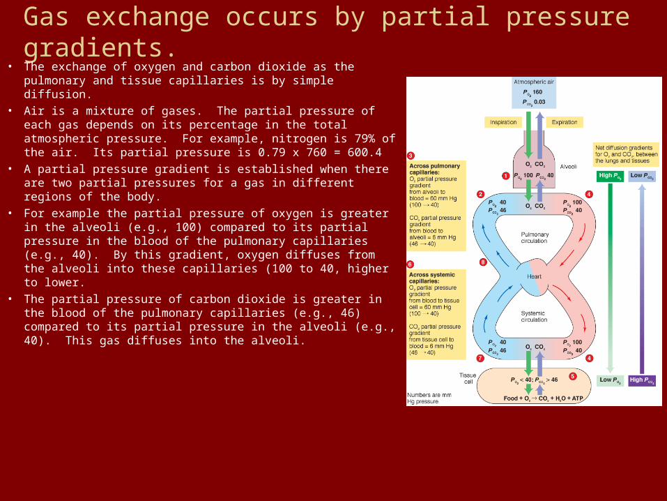
Gas exchange occurs by partial pressure gradients.• The exchange of oxygen and carbon dioxide as the
pulmonary and tissue capillaries is by simple diffusion.• Air is a mixture of gases. The partial pressure of each gas
depends on its percentage in the total atmospheric pressure. For example, nitrogen is 79% of the air. Its partial pressure is 0.79 x 760 = 600.4
• A partial pressure gradient is established when there are two partial pressures for a gas in different regions of the body.
• For example the partial pressure of oxygen is greater in the alveoli (e.g., 100) compared to its partial pressure in the blood of the pulmonary capillaries (e.g., 40). By this gradient, oxygen diffuses from the alveoli into these capillaries (100 to 40, higher to lower.
• The partial pressure of carbon dioxide is greater in the blood of the pulmonary capillaries (e.g., 46) compared to its partial pressure in the alveoli (e.g., 40). This gas diffuses into the alveoli.
Partial pressure gradients change the partial pressures of oxygen(e.g., 100) and carbon dioxide (e.g., 40) in the blood returning to the heart from the lungs.• By diffusion, the partial pressures for oxygen and carbon dioxide
in the pulmonary capillaries equilibrate with the partial pressures for these gases in the alveoli.
• The greater the partial pressure gradients between the alveoli and the blood, the greater the rate of transfer for the gases.
• The blood passing through the lungs gains oxygen and eliminates some of its carbon dioxide.
• This blood passes through the left side of the heart and enters the systemic circulation. It arrives at the tissues with the same gas content (e.g., 100 for oxygen and 40 for carbon dioxide) established at lung equilibration.
Other factors in addition to the partial pressure gradient affect the rate of gas transfer.• As surface area increases the rate increases. The alveoli
collectively offer a tremendous surface area. Increased pulmonary blood pressure, from an increased cardiac output, increases the area.
• The walls of the alveoli and pulmonary capillaries are thin for rapid gas transfer. Pulmonary edema, pulmonary fibrosis, and pneumonia thicken the barriers for gas exchange.
• Gas exchange is also directly proportional to the diffusion coefficient for a gas. This coefficient is twenty times as great for carbon dioxide compared to oxygen, as carbon dioxide is more soluble.
Gas exchange across systemic capillaries also occurs down partial pressure gradients.• By equilibration in the alveoli, the oxygen in the systemic capillaries
has a high partial pressure (e.g., 100) compared to tissue cells (e.g., 40). These cells are using oxygen.
• The partial pressure for carbon dioxide in the systemic capillaries is low (e.g., 40) compared to the tissue cells (e.g., 46), which are making this gas through their metabolism.
• By partial pressure gradients, oxygen diffuses from the systemic capillaries into the tissue cells (100 to 40, higher to lower). Carbon dioxide diffuses in the opposite direction.
• Having equilibrated with the tissue cells, the blood leaving the systemic capillaries is low in oxygen and high in carbon dioxide.
• This blood returns to the right side of the heart and on to the lungs. At the pulmonary capillaries, the blood acquires oxygen and releases some of its carbon dioxide.
Most oxygen in the blood is transported by binding with hemoglobin.
• Hemoglobin combines with oxygen to form oxyhemoglobin. This is a reversible process, favored to form oxyhemoglobin in the lungs.
• Hemoglobin tends to combine with oxygen as oxygen diffuses from the alveoli into the pulmonary capillaries.
• A small percentage of oxygen is dissolved in the plasma.
• The dissociation of oxyhemoglobin into hemoglobin and free molecules of oxygen occurs at the tissue cells. The reaction is favored in this direction as oxygen leaves the systemic capillaries and enters tissue cells.
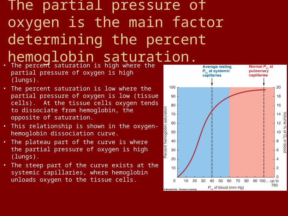
The partial pressure of oxygen is the main factor determining the percent hemoglobin saturation.
• The percent saturation is high where the partial pressure of oxygen is high (lungs).
• The percent saturation is low where the partial pressure of oxygen is low (tissue cells). At the tissue cells oxygen tends to dissociate from hemoglobin, the opposite of saturation.
• This relationship is shown in the oxygen-hemoglobin dissociation curve.
• The plateau part of the curve is where the partial pressure of oxygen is high (lungs).
• The steep part of the curve exists at the systemic capillaries, where hemoglobin unloads oxygen to the tissue cells.
Hemoglobin promotes the net transfer of oxygen at both the alveolar and tissue levels.• There is a net diffusion of oxygen from the alveoli to the blood. This occurs
continuously until hemoglobin is as saturated as possible (97.5% at 100 mm of Hg).
• At the tissue cells hemoglobin rapidly delivers oxygen into the blood plasma and on to the tissue cells. Various factors promote this unloading.
• An increase in carbon dioxide from the tissue cells into the systemic capillaries increased hemoglobin dissociation from oxygen (shifts the dissociation curve to the right).
• Increased acidity has the same effect.• This shift of the curve to the right (more dissociation) is called the Bohr effect.• Higher temperatures also produces this shift, as does the production of BPG.• Hemoglobin has more affinity for carbon monoxide compared to oxygen.
Most carbon dioxide (about 60%) is transported as the bicarbonate ion. • Carbon dioxide combines with water to form carbonic acid. The enzyme
carbonic anhydrase facilitates this in the erythrocyte. Carbonic acid dissociates into hydrogen ions and the bicarbonate ion.
• This two-step, reversible process is favored at the tissue cells. The reverse of this process (bicarbonate ions forming free molecules of carbon dioxide) occurs in the lungs.
• 30% of the carbon dioxide is bound to hemoglobin in the blood. This is another means of transport.
• About 10% of the transported carbon dioxide is dissolved in the plasma.• By the chloride shift, the plasma membrane of the erythrocyte passively
facilitates the diffusion of bicarbonate ions (out of the red cell) and chloride ions.
• By the Haldane effect the removal of oxygen from hemoglobin at the tissue cells increases the ability of hemoglobin to bind with carbon dioxide.
Various respiratory states are characterized by abnormal blood gas levels.
•There are four general categories of hypoxias.
•Examples include hypoxic hypoxia, which is characterized by a low partial pressure in the arterial blood. In anemia hypoxia there is reduced oxygen-carrying capacity in the blood.
•Hyperoxia ia an above-normal arterial partial pressure of oxygen.
•Hypercapnia is an excess of carbon dioxide in the blood caused by hypoventilation.
•Hypocapnia is a below-normal arterial level of carbon dioxide in the blood, due to hyperventilation.
•Hyperpnea is an increased need for oxygen delivery and carbon dioxide elimination during exercise.
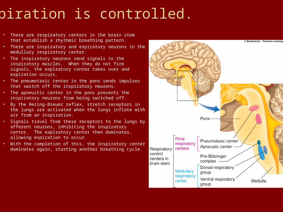
Respiration is controlled.• There are respiratory centers in the brain stem that
establish a rhythmic breathing pattern.• There are inspiratory and expiratory neurons in the
medullary respiratory center. • The inspiratory neurons send signals to the inspiratory
muscles. When they do not fire signals, the expiratory center takes over and expiration occurs.
• The pneumotaxic center in the pons sends impulses that switch off the inspiratory neurons.
• The apneustic center in the pons prevents the inspiratory neurons from being switched off.
• By the Hering-Breuer reflex, stretch receptors in the lungs are activated when the lungs inflate with air from an inspiration.
• Signals travel from these receptors to the lungs by afferent neurons, inhibiting the inspiratory center. The expiratory center then dominates, allowing expiration to occur.
• With the completion of this, the inspiratory center dominates again, starting another breathing cycle.
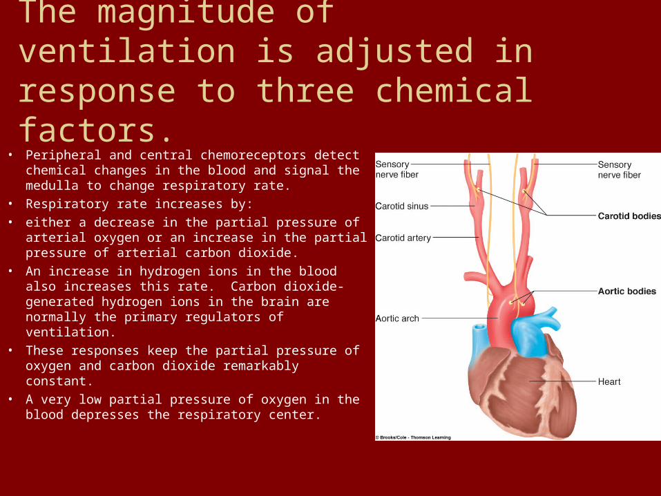
The magnitude of ventilation is adjusted in response to three chemical factors.
• Peripheral and central chemoreceptors detect chemical changes in the blood and signal the medulla to change respiratory rate.
• Respiratory rate increases by:• either a decrease in the partial pressure of arterial
oxygen or an increase in the partial pressure of arterial carbon dioxide.
• An increase in hydrogen ions in the blood also increases this rate. Carbon dioxide-generated hydrogen ions in the brain are normally the primary regulators of ventilation.
• These responses keep the partial pressure of oxygen and carbon dioxide remarkably constant.
• A very low partial pressure of oxygen in the blood depresses the respiratory center.
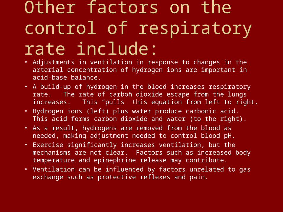
Other factors on the control of respiratory rate include:• Adjustments in ventilation in response to changes in the arterial
concentration of hydrogen ions are important in acid-base balance.
• A build-up of hydrogen in the blood increases respiratory rate. The rate of carbon dioxide escape from the lungs increases. This “pulls” this equation from left to right.
• Hydrogen ions (left) plus water produce carbonic acid. This acid forms carbon dioxide and water (to the right).
• As a result, hydrogens are removed from the blood as needed, making adjustment needed to control blood pH.
• Exercise significantly increases ventilation, but the mechanisms are not clear. Factors such as increased body temperature and epinephrine release may contribute.
• Ventilation can be influenced by factors unrelated to gas exchange such as protective reflexes and pain.
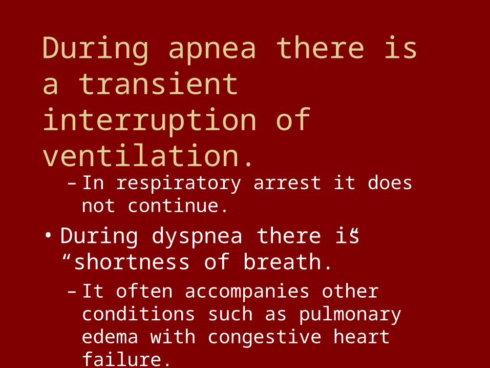
During apnea there is a transient interruption of ventilation.
– In respiratory arrest it does not continue.
• During dyspnea there is “shortness of breath.”– It often accompanies other conditions such
as pulmonary edema with congestive heart failure.


![Respiratory system roadmap.pptx [Repaired] - Loginanatomical-sciences.health.wits.ac.za/roadmaps/Respiratory system... · DIVISION OF THE RESPIRATORY SYSTEM CONDUCTING PORTION Nasal](https://img.pdfslide.us/doc/110x75/5a78c3d87f8b9ae6228c9db0/respiratory-system-repaired-loginanatomical-scienceshealthwitsaczaroadmapsrespiratory.jpg)