Embed Size (px)
Citation preview
The respiratory system
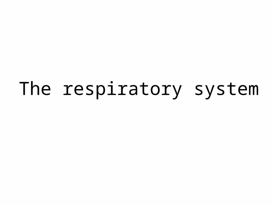
Respiratory System Function
Major Functions
Upper respiratory system:
1. Air conditioning
2. Defense against pathogens
Lower respiratory system:
1. Speech & other respiratory sounds
2. Gas exchange (Supplies body with oxygen and Disposes of carbon dioxide )
3. Maintenance of homeostasis, e.g. pH
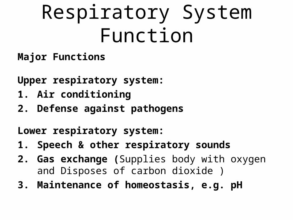
Organs of the Respiratory SystemOrgans of the Respiratory System
• Divided into:
– Conducting zone – respiratory passage which serve as conduits that carry air to the sites of gas exchange
•Serve to filter, humidify, and warm the incoming air
– Respiratory zone – the actual site of gas exchange in the lungs
•Respiratory bronchioles, alveolar ducts, alveolar sacs – contain alveoli
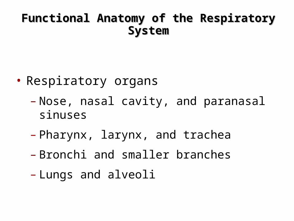
Functional Anatomy of the Respiratory Functional Anatomy of the Respiratory SystemSystem
• Respiratory organs
– Nose, nasal cavity, and paranasal sinuses
– Pharynx, larynx, and trachea
– Bronchi and smaller branches
– Lungs and alveoli
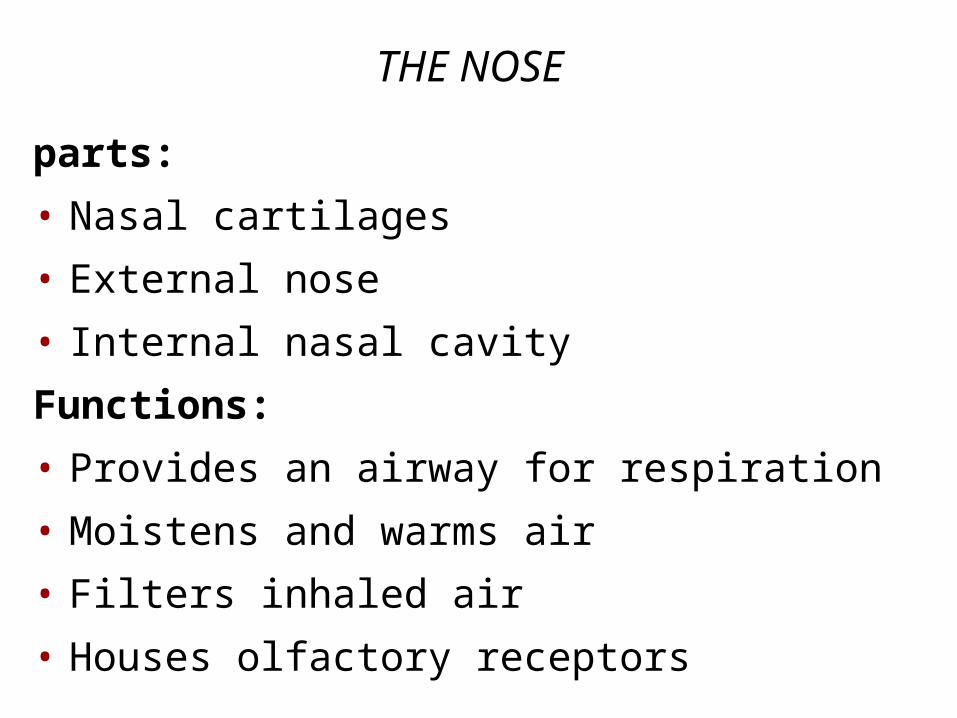
THE NOSE
parts:
• Nasal cartilages
• External nose
• Internal nasal cavity
Functions:
• Provides an airway for respiration
• Moistens and warms air
• Filters inhaled air
• Houses olfactory receptors
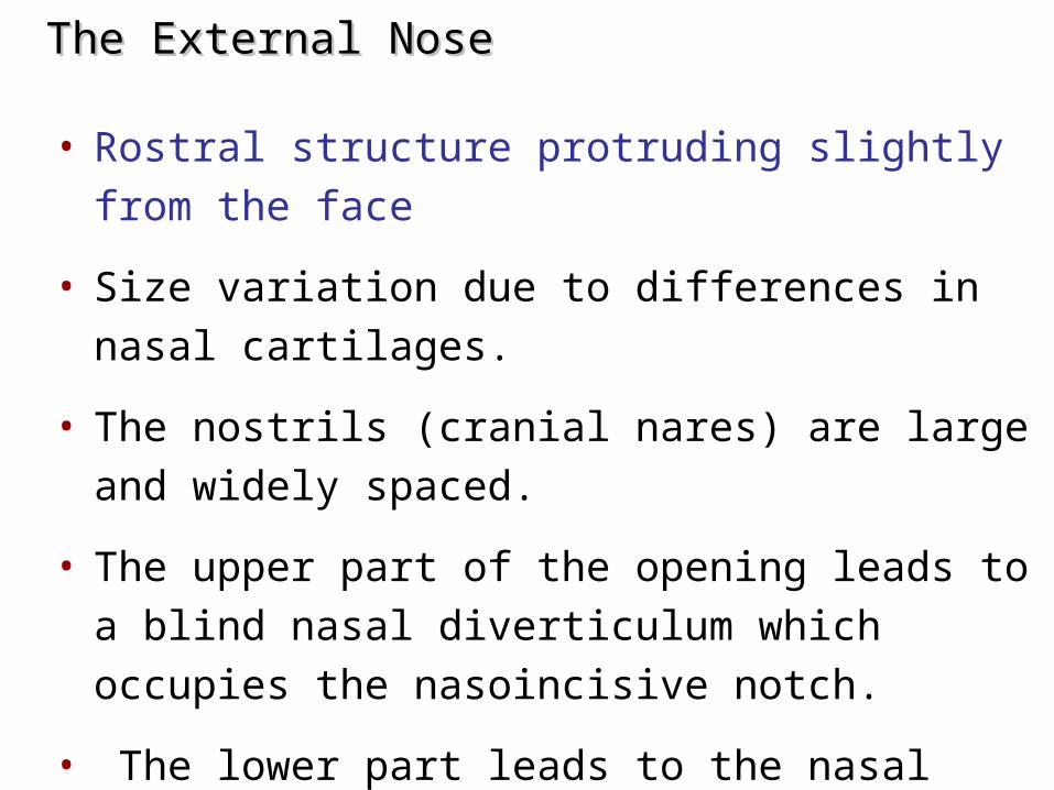
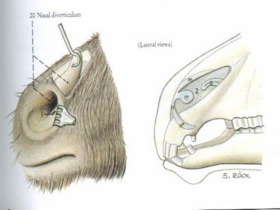
The External NoseThe External Nose
• Rostral structure protruding slightly from the face
• Size variation due to differences in nasal cartilages.
• The nostrils (cranial nares) are large and widely
spaced.
• The upper part of the opening leads to a blind nasal
diverticulum which occupies the nasoincisive notch.
• The lower part leads to the nasal cavity
– it is essential when passing a stomach tube to ensure that
it is guided into the lower par
The Nasal CavityThe Nasal Cavity
• External nares – nostrils
• Lies in and posterior to the external nose
• Divided by – nasal septum.
• Continuous with nasopharynx
• Internal nares – posterior nasal apertures (choana)
• Roof – Ethmoid and sphenoid bones
• Floor – hard palate.
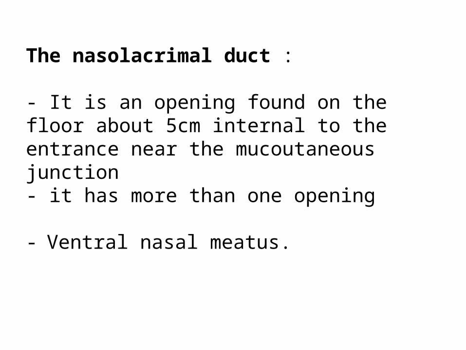
The nasolacrimal duct :
- It is an opening found on the floor about 5cm internal to the entrance near the mucoutaneous junction - it has more than one opening
- Ventral nasal meatus.
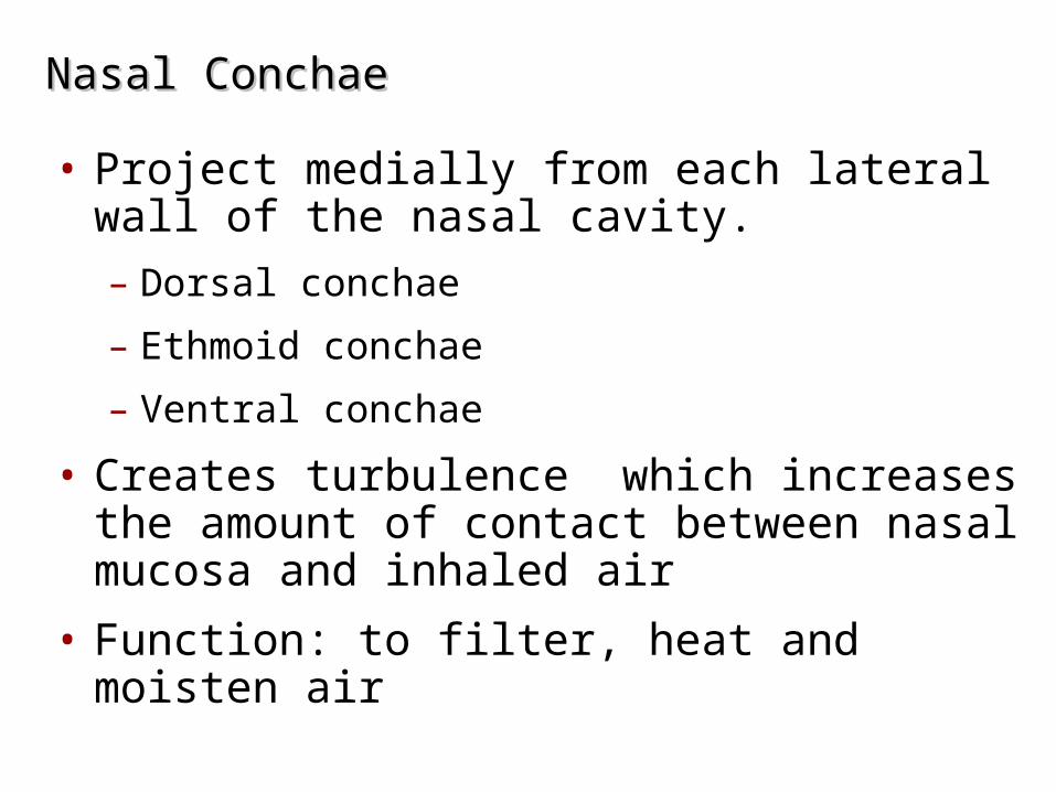
Nasal ConchaeNasal Conchae
• Project medially from each lateral wall of the nasal cavity. – Dorsal conchae
– Ethmoid conchae
– Ventral conchae
• Creates turbulence which increases the amount of contact between nasal mucosa and inhaled air
• Function: to filter, heat and moisten air
The paranasal sinuses are diverticula of the nasal cavity that excavate the skull bones largely after birth.
The separation of the inner and outer tables of the bones alters the conformation of the head
and is specially striking in pigs and cattle, in which certain sinuses eventually extend dorsal and even caudal to the cranial cavity.
Paranasal Sinuses
The sinuses retain their connections with the nasal cavity
these openings are generally narrow a relatively slow exchange of air occurs
The narrowness and locations of the openings make them prone to blockage when the mucosa is thickened by inflammation or congestion.
Not all the sinuses are of equal importance.
Paranasal Sinuses
The function
The function of the sinuses is obscure Offer some thermal Mechanical protection to the orbit and
nasal and cranial cavities Enlarge the skull areas available for
muscular attachment without unduly increasing weight,
Affect the resonance of the voice.
All species have frontal and maxillary systems
neither communicating with its contralateral counterpart.
The frontal system consists of one or more spaces within the bones at the border between the nasal and
cranial cavities.
In most species the various frontal compartments open separately into ethmoidal meatus in the nasal
cavity
in the horse the frontal sinus communicates with the nasal cavity indirectly via the caudal maxillary sinus.
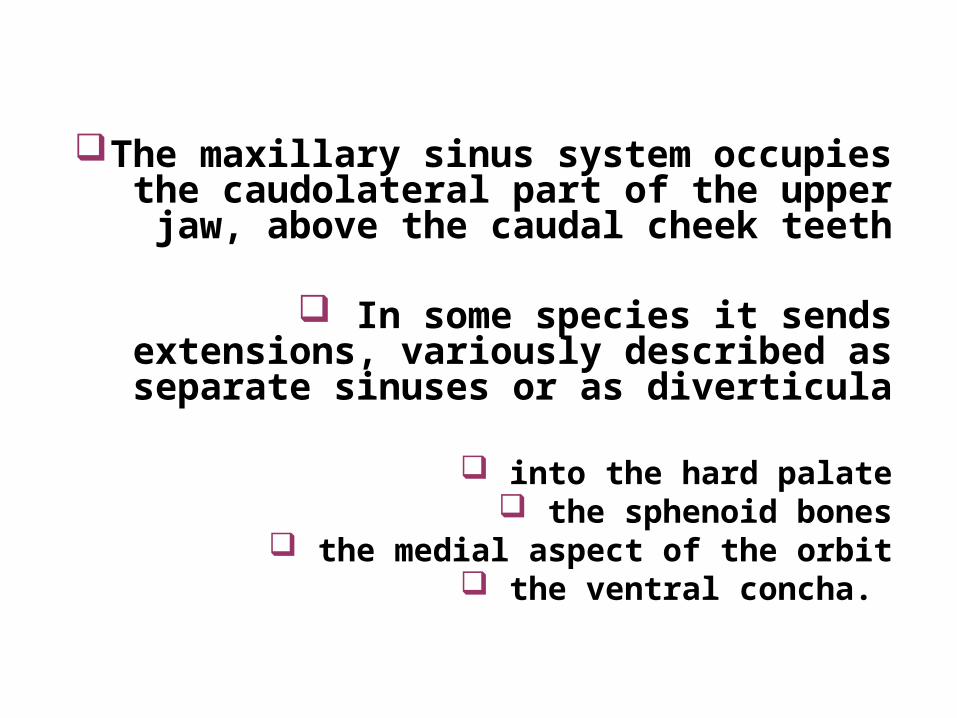
The maxillary sinus system occupies the caudolateral part of the upper jaw, above the
caudal cheek teeth
In some species it sends extensions, variously described as separate sinuses or
as diverticula
into the hard palate the sphenoid bones
the medial aspect of the orbit the ventral concha.
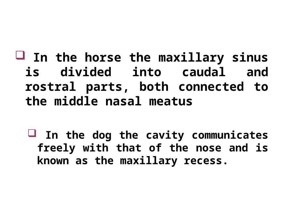
In the horse the maxillary sinus is divided into caudal and rostral parts, both connected to the middle nasal meatus
In the dog the cavity communicates freely with that of the nose and is known as the maxillary recess.
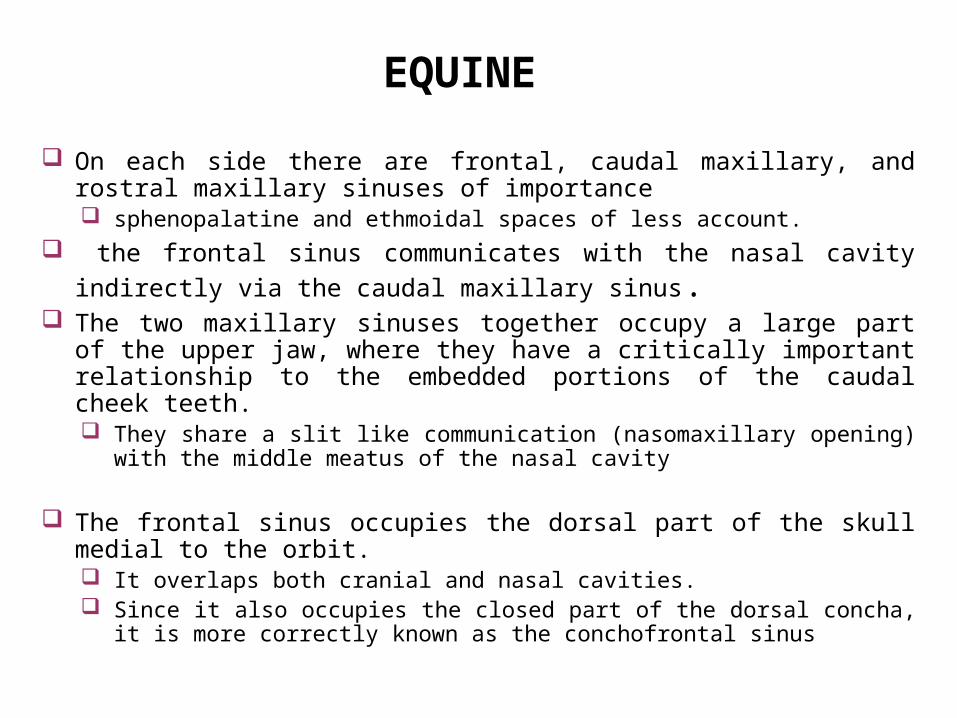
On each side there are frontal, caudal maxillary, and rostral maxillary sinuses of importance sphenopalatine and ethmoidal spaces of less account.
the frontal sinus communicates with the nasal cavity indirectly via the caudal
maxillary sinus. The two maxillary sinuses together occupy a large part of the upper jaw,
where they have a critically important relationship to the embedded portions of the caudal cheek teeth. They share a slit like communication (nasomaxillary opening) with the middle
meatus of the nasal cavity
The frontal sinus occupies the dorsal part of the skull medial to the orbit. It overlaps both cranial and nasal cavities. Since it also occupies the closed part of the dorsal concha, it is more correctly
known as the conchofrontal sinus
EQUINE
BOVINE, OVINE, CAPRINE
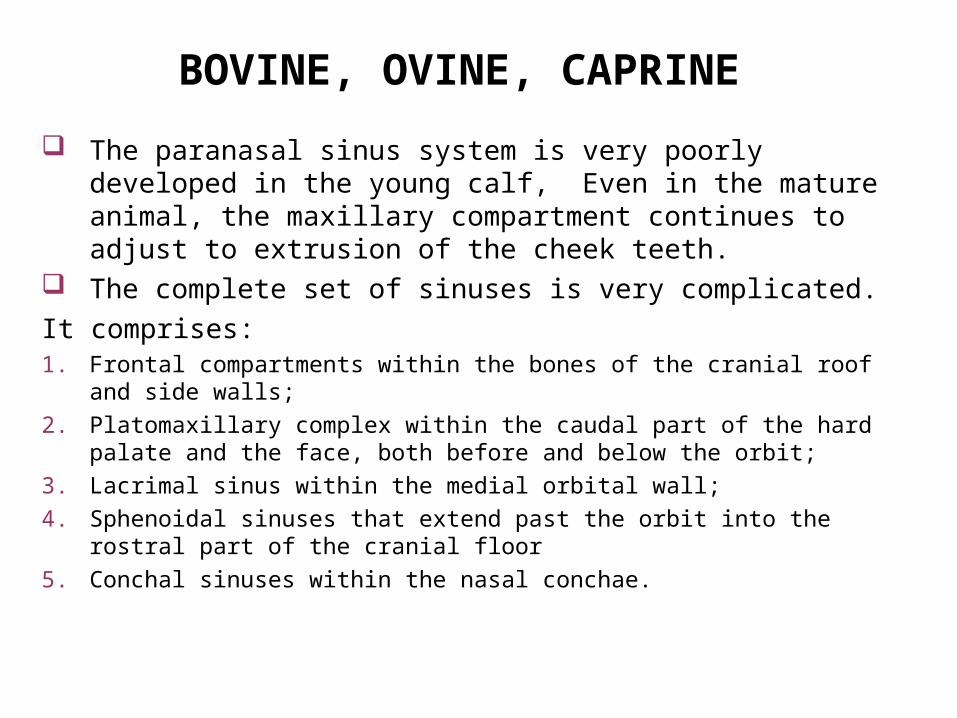
The paranasal sinus system is very poorly developed in the young calf, Even in the mature animal, the maxillary compartment continues to adjust to extrusion of the cheek teeth.
The complete set of sinuses is very complicated.
It comprises:1. Frontal compartments within the bones of the cranial roof and side walls;
2. Platomaxillary complex within the caudal part of the hard palate and the face, both before and below the orbit;
3. Lacrimal sinus within the medial orbital wall;
4. Sphenoidal sinuses that extend past the orbit into the rostral part of the cranial floor
5. Conchal sinuses within the nasal conchae.
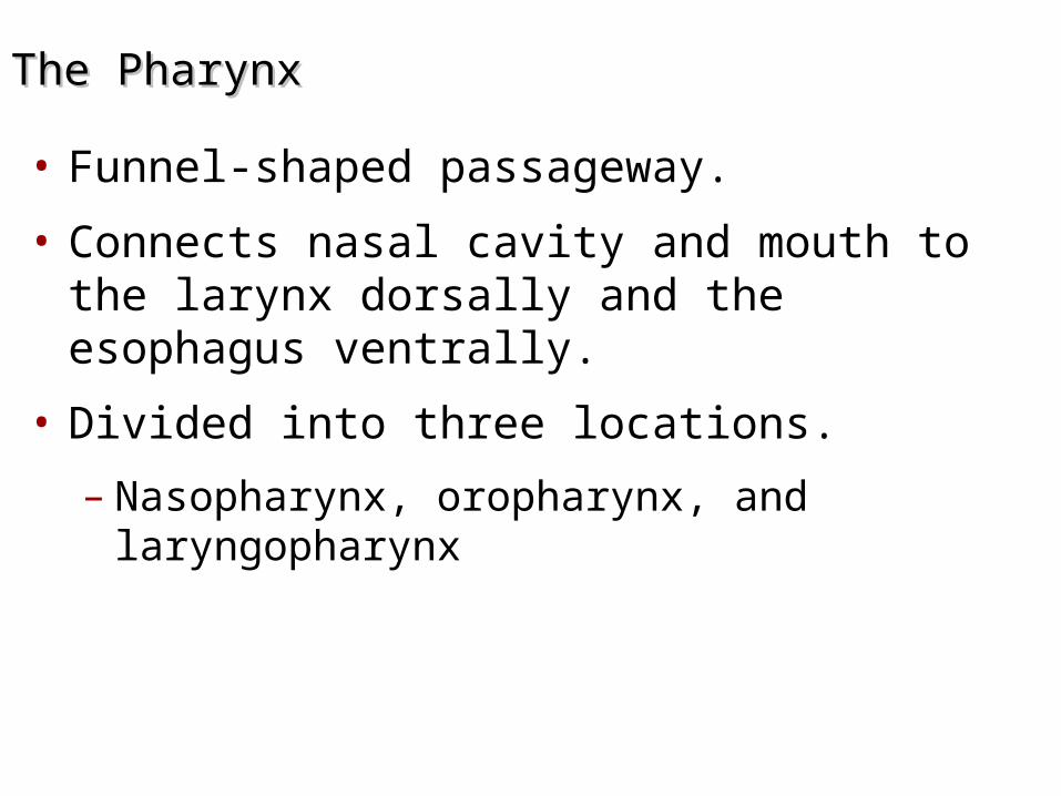
The PharynxThe Pharynx
• Funnel-shaped passageway.
• Connects nasal cavity and mouth to the larynx dorsally and the esophagus ventrally.
• Divided into three locations.
– Nasopharynx, oropharynx, and laryngopharynx


The NasopharynxThe Nasopharynx
• Superior to the point where food enters
• Only an air passageway
• Closed off during swallowing by soft palate
• Contains the opening to the auditory tube
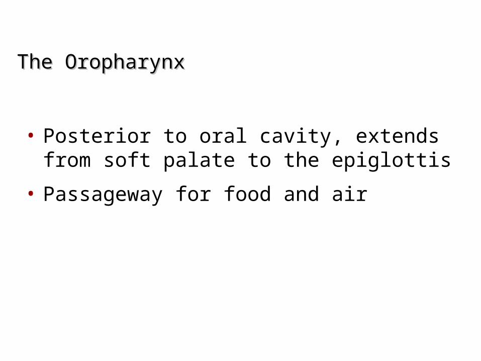
The OropharynxThe Oropharynx
• Posterior to oral cavity, extends from soft palate to the epiglottis
• Passageway for food and air
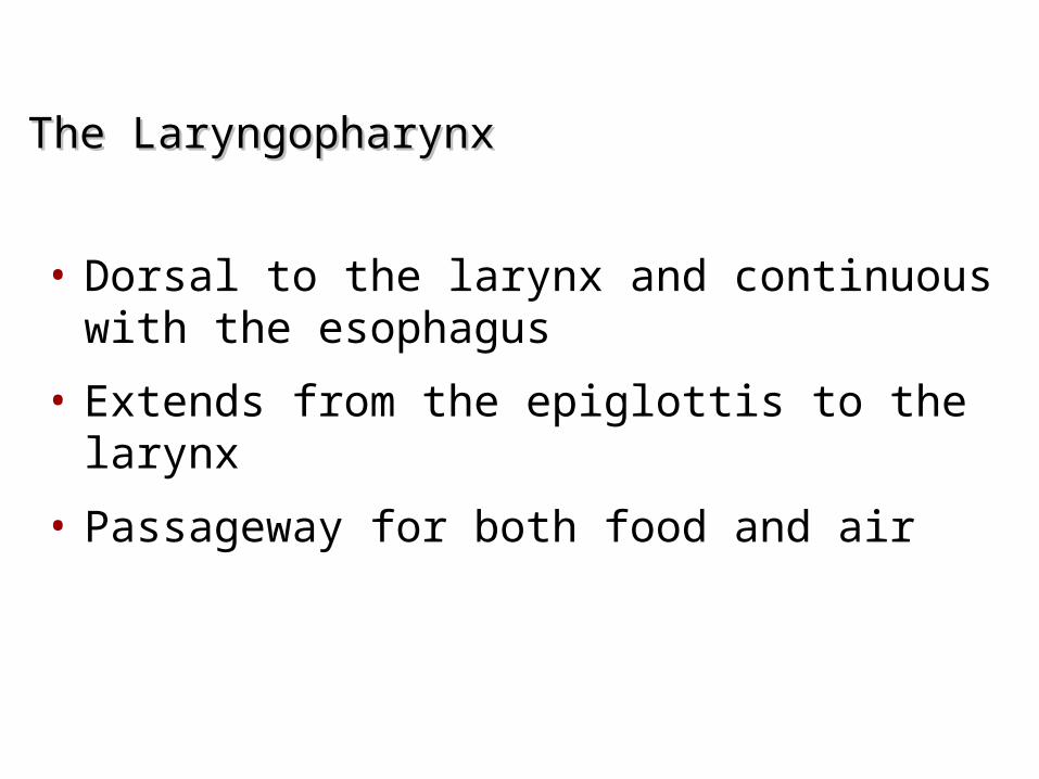
The LaryngopharynxThe Laryngopharynx
• Dorsal to the larynx and continuous with the esophagus
• Extends from the epiglottis to the larynx
• Passageway for both food and air

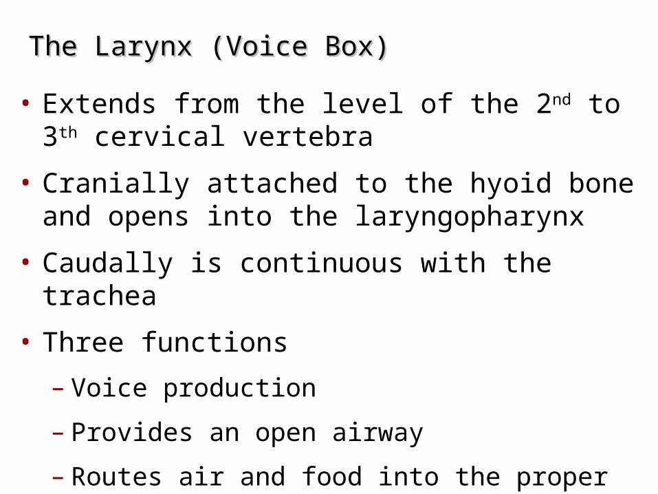
The Larynx (Voice Box)The Larynx (Voice Box)
• Extends from the level of the 2nd to 3th cervical vertebra
• Cranially attached to the hyoid bone and opens into the laryngopharynx
• Caudally is continuous with the trachea
• Three functions
– Voice production
– Provides an open airway
– Routes air and food into the proper channels
• The larynx is the part that forms the connection between the pharynx and the tracheobronchial tree.
• Position: the larynx can be found behind the mouth, below the pharynx.
• Its position is altered when the animal swallows due to its connection with the hyoid apparatus and tongue.
• The larynx's main function is to protect the rest of the respiratory tract and system as well as producing sound (vocalisation)
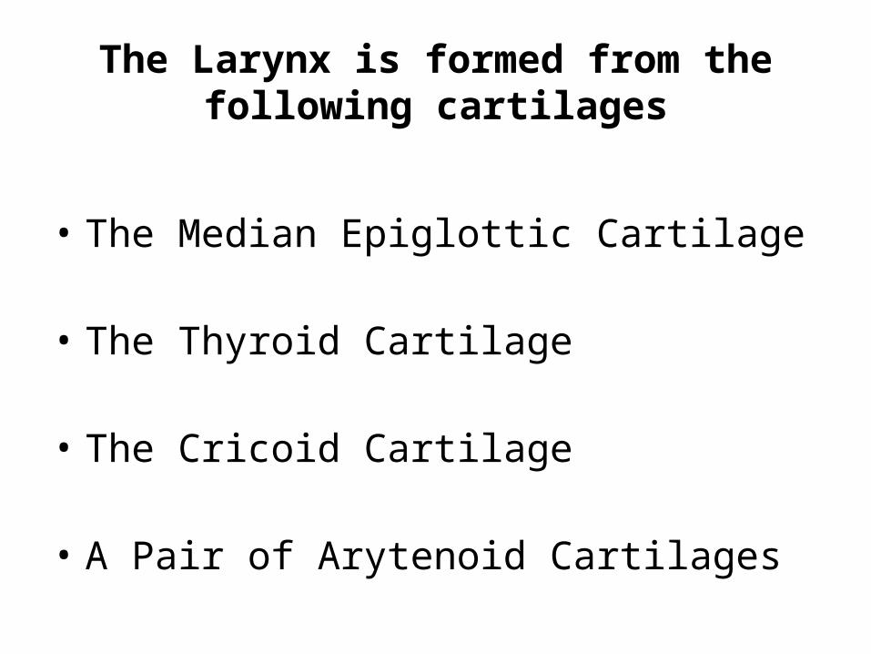
The Larynx is formed from the following cartilages
• The Median Epiglottic Cartilage
• The Thyroid Cartilage
• The Cricoid Cartilage
• A Pair of Arytenoid Cartilages
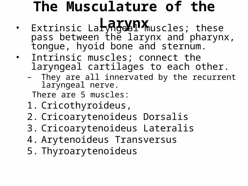
The Musculature of the Larynx• Extrinsic Laryngeal muscles; these pass
between the larynx and pharynx, tongue, hyoid bone and sternum.
• Intrinsic muscles; connect the laryngeal cartilages to each other.
– They are all innervated by the recurrent laryngeal nerve.
There are 5 muscles:1. Cricothyroideus,2. Cricoarytenoideus Dorsalis3. Cricoarytenoideus Lateralis4. Arytenoideus Transversus5. Thyroarytenoideus
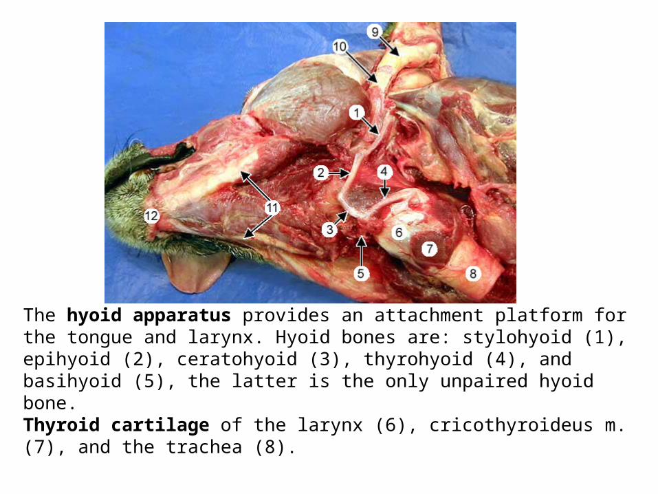
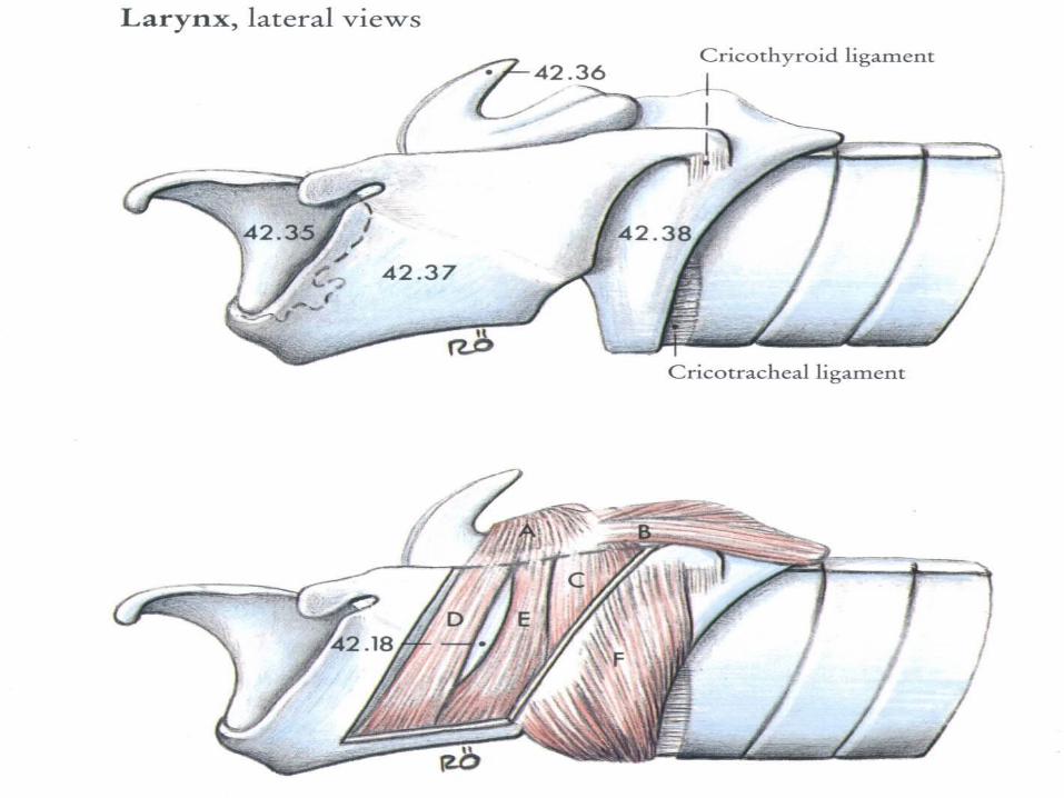
The hyoid apparatus provides an attachment platform for the tongue and larynx. Hyoid bones are: stylohyoid (1), epihyoid (2), ceratohyoid (3), thyrohyoid (4), and basihyoid (5), the latter is the only unpaired hyoid bone. Thyroid cartilage of the larynx (6), cricothyroideus m. (7), and the trachea (8).
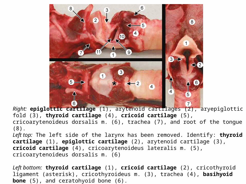
Right: epiglottic cartilage (1), arytenoid cartilages (2), aryepiglottic fold (3), thyroid cartilage (4), cricoid cartilage (5), cricoarytenoideus dorsalis m. (6), trachea (7), and root of the tongue (8). Left top: The left side of the larynx has been removed. Identify: thyroid cartilage (1), epiglottic cartilage (2), arytenoid cartilage (3), cricoid cartilage (4), cricoarytenoideus lateralis m. (5), cricoarytenoideus dorsalis m. (6)
Left bottom: thyroid cartilage (1), cricoid cartilage (2), cricothyroid ligament (asterisk), cricothyroideus m. (3), trachea (4), basihyoid bone (5), and ceratohyoid bone (6).


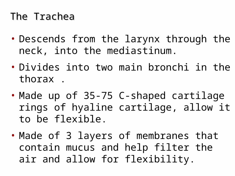
The Trachea The Trachea
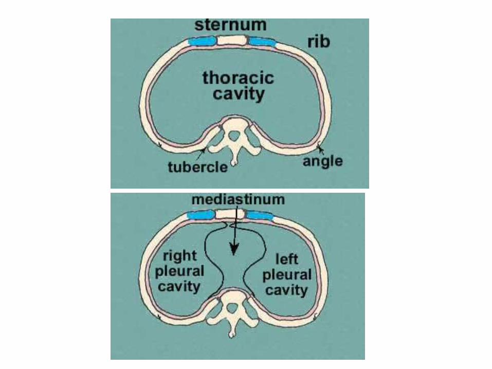
• Descends from the larynx through the neck, into the mediastinum.
• Divides into two main bronchi in the thorax .
• Made up of 35-75 C-shaped cartilage rings of hyaline cartilage, allow it to be flexible.
• Made of 3 layers of membranes that contain mucus and help filter the air and allow for flexibility.
Bronchi in the Conducting ZoneBronchi in the Conducting Zone
• Bronchial tree – extensively branching respiratory passageways within the lungs
– Division of the trachea at T4/T7
• Primary bronchi (main bronchi) – largest bronchi or conduits in the bronchial tree
• Right main bronchi – wider and shorter than the left and more vertical (clinical important)
Bronchi in the Conducting ZoneBronchi in the Conducting Zone
• Secondary (lobar) bronchi – each of which supply one lung lobe
– Three-four on the right, Two on the left
• Tertiary (segmental) bronchi
– Branch into each lung segment
• Overall, 23 orders of air passageways
• Bronchioles – little bronchi, less than 1 mm in diameter
• Terminal bronchioles – less than 0.5 mm in diameter
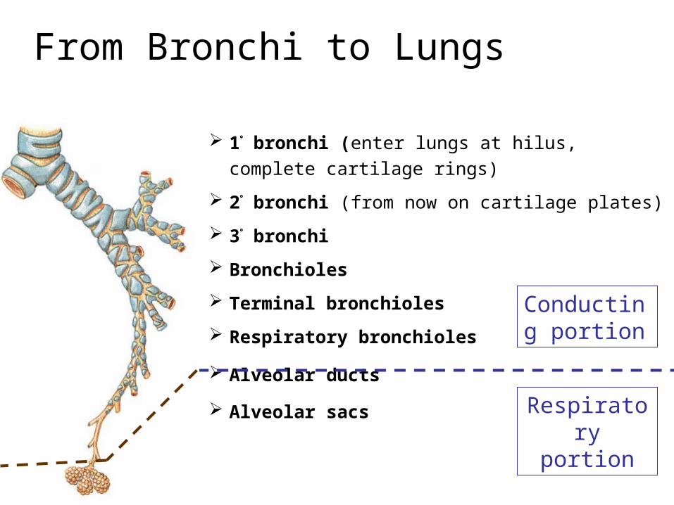
From Bronchi to Lungs
1 bronchi (enter lungs at hilus, complete cartilage
rings)
2 bronchi (from now on cartilage plates)
3 bronchi
Bronchioles
Terminal bronchioles
Respiratory bronchioles
Alveolar ducts
Alveolar sacs
Conducting portion
Respiratory portion
The Respiratory ZoneThe Respiratory Zone
• Consists of air-exchanging chambers – called alveoli
• Begins at the terminal bronchioles
• Respiratory bronchioles – branch from terminal bronchioles, have scattered alveoli protruding from their walls
– Lead to alveolar ducts – walls consist almost entirely of alveoli
•Lead to alveolar sacs – terminal clusters of alveoli
•The opening from an alveolar sac into an alveolar sac is call an atrium “entrance chamber”
The PleuraeThe Pleurae
Membranes that cover the lungs and line the pleural cavity.
• A double-layered sac surrounding each lung
• Parietal pleura
• Visceral pleura
• Pleural cavity - Potential space between the visceral and parietal pleurae, filled with pleural fluid
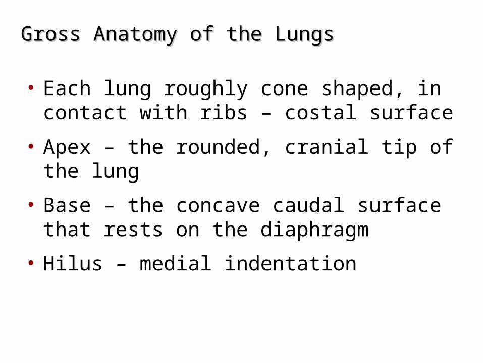
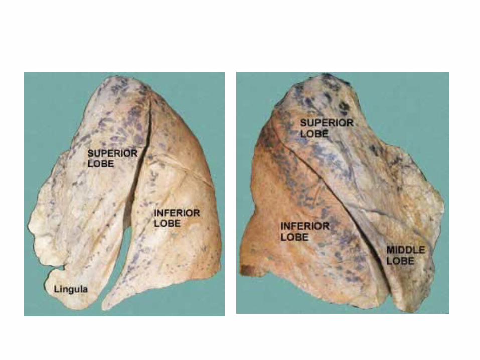
Gross Anatomy of the LungsGross Anatomy of the Lungs
• Each lung roughly cone shaped, in contact with ribs – costal surface
• Apex – the rounded, cranial tip of the lung
• Base – the concave caudal surface that rests on the diaphragm
• Hilus – medial indentation
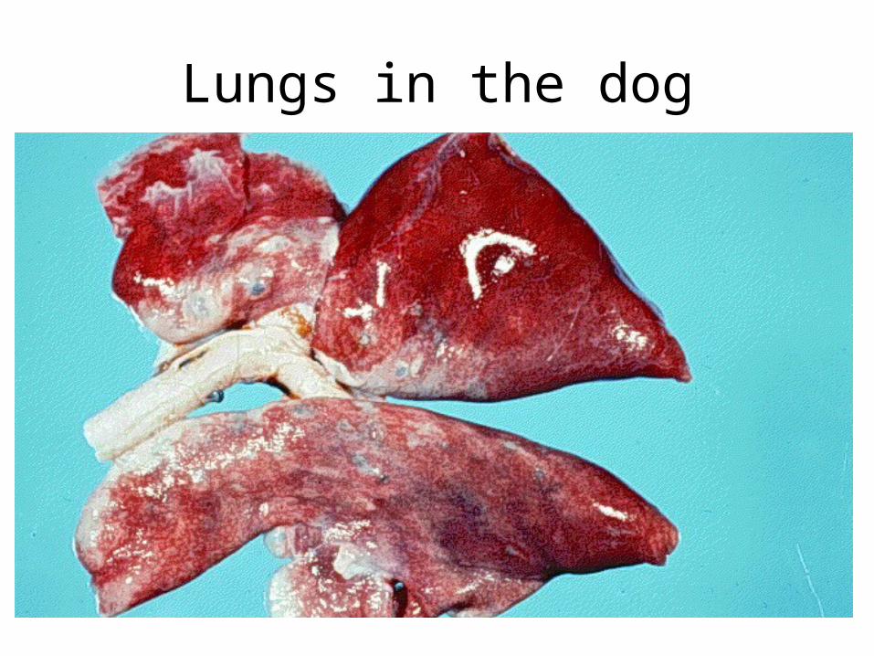
Lungs in the dog
Gross Anatomy of the LungsGross Anatomy of the Lungs
• Lobes – deep fissures separate the lungs into divisions
• Left lung – 2 lobes
• Right lung – 3-4 lobes
• Lobule – smallest subdivision of the lung seen by naked eye
• In horses the lungs are elongated and shallow
• The left and right lungs are more equally in size than the
other species
• The difference between left and right manly in
thickens ,the right lung more thicker than the left lung
and it ha an accessory lob appended to the base
• There is no evidence of lobation
• The right lung consist of cranial,caudal and accessory
lobes
• The left lung consist of cranial and caudal lobes
• The tow lungs are extensively joined by connective
tissue caudal to the bifurcation of the trachea
• At the junction between the trachea and the bronchi
there are lymph nods which called (tracheobronchial
lymph nodes)
-The pattern in sheep often varies between parts
of the lung some show the connective tissue
septa through the visceral pleura while others
unmarked.
-The lung of cattle possess the usual roughly
pyramidal shape .
Blood Supply and Innervation of the LungsBlood Supply and Innervation of the Lungs
• Pulmonary arteries – deliver oxygen-poor blood to the lungs
• Pulmonary veins – carry oxygenated blood to the heart
• Innervation
– Sympathetic, parasympathetic, and visceral sensory fibers
•Parasympathetic – constrict airways
•Sympathetic – dilate airways